HEAVY CHAIN SUBCLASSES OF HUMAN ³G-GLOBULIN: SERUM
17
HEAVY CHAIN SUBCLASSES OF HUMAN 7G-GLOBULIN SERUM DISTRIBUTION AND CELLULAR LOCALIZATION* BY G. M. BERNIER,~ M.D., R. E. BALLIEUX,§ PH.D., K. T. TOMINAGA,H M.D., AND F. W. PUTNAM,¶ PH.D. (From the Department of Biochemistry, College of Medicine, University of Florida, Gainesville) Pi~z 22 (Received for publication 2 September 1966) Investigation of the complex heterogeneity of immunoglobulins in man has led to the finding of subclasses of 7G-globulin, distinguished by partial differences in the heavy or 7-chain. Initially, the subclassific~fion was entirely based on antigenic dif- ferences detectable with primate antisera. Thus, Dray (1) described three subclasses of normal human 7G-globniin termed "r~, 7~b, and 7~o. Variations of these three sub- classes in disease were studied by Peetom and Kramer (2). Terry and Fahey (3) have extensively studied normal and myeloma 7G-globulius and have been able to classify most normal and pathological molecules into one of four such subcategories (~/~, 72b, 72°, 7~a). Grey and Kunkel (4), using both rabbit and monkey anfisera prepared against individual myeloma globulins, have divided ~G-myeloma globulins and normal ~G-globulins into four subgroups based on heavy chain differences (Vi, Ge, Ne, and a major group comprising 60-70% of all 7G myeloma globulins, We). The apparent relationship among these subdassificafionsis: 72a ffi Ne; ~2b ~--" We; ~o -- Vi; ~/2d ffi Ge (5). In a previous report (6), we described a similar, less extensive, heavy chain subclassification of "},G-globulin based on differences detected by rabbit anti- sera prepared against proteins from patients with "heavy chain disease" (7-9). The subpopulatious which we have termed Cr, Zu, and Cr variant, are not strictly coextensive with those of other investigators. However, based on *Supported by Grants CA-02803 and CA-08497 from the National Cancer Institute, Na- tional Institutes of Health, and by a grant from the Netherlands Organization for the Ad- vancement of Pure Research. ~Present address: University Hospitals, Cleveland, Ohio. §Present adress: Division of lmm~mochemistry, Department of Medicine, University Hospital, Utrecht, Holland. [[Present address: Third Department of Internal Medicine, Faculty of Medicine, Kyushu University, Fukuoka, Japan. ¶Present address: Division of Biological Sciences, Indiana University, Bloomington, Indi- ana. Reprint requests should be sent to this address. 303
Transcript of HEAVY CHAIN SUBCLASSES OF HUMAN ³G-GLOBULIN: SERUM

HEAVY C H A I N SUBCLASSES OF HUMAN 7G-GLOBULIN
SERUM DISTRIBUTION AND CELLULAR LOCALIZATION*
BY G. M. BERNIER,~ M.D., R. E. BALLIEUX,§ PH.D., K. T. TOMINAGA, H M.D., AND F. W. PUTNAM,¶ PH.D.
(From the Department of Biochemistry, College of Medicine, University of Florida, Gainesville)
P i ~ z 22
(Received for publication 2 September 1966)
Investigation of the complex heterogeneity of immunoglobulins in man has led to the finding of subclasses of 7G-globulin, distinguished by partial differences in the heavy or 7-chain. Initially, the subclassific~fion was entirely based on antigenic dif- ferences detectable with primate antisera. Thus, Dray (1) described three subclasses of normal human 7G-globniin termed "r~, 7~b, and 7~o. Variations of these three sub- classes in disease were studied by Peetom and Kramer (2). Terry and Fahey (3) have extensively studied normal and myeloma 7G-globulius and have been able to classify most normal and pathological molecules into one of four such subcategories (~/~, 72b, 72°, 7~a). Grey and Kunkel (4), using both rabbit and monkey anfisera prepared against individual myeloma globulins, have divided ~G-myeloma globulins and normal ~G-globulins into four subgroups based on heavy chain differences (Vi, Ge, Ne, and a major group comprising 60-70% of all 7G myeloma globulins, We). The apparent relationship among these subdassificafionsis: 72a ffi Ne; ~2b ~--" We; ~o -- Vi; ~/2d ffi Ge (5).
In a previous report (6), we described a similar, less extensive, heavy chain subclassification of "},G-globulin based on differences detected by rabbit anti- sera prepared against proteins from patients with "heavy chain disease" (7-9). The subpopulatious which we have termed Cr, Zu, and Cr variant, are not strictly coextensive with those of other investigators. However, based on
*Supported by Grants CA-02803 and CA-08497 from the National Cancer Institute, Na- tional Institutes of Health, and by a grant from the Netherlands Organization for the Ad- vancement of Pure Research.
~Present address: University Hospitals, Cleveland, Ohio. §Present adress: Division of lmm~mochemistry, Department of Medicine, University
Hospital, Utrecht, Holland. [[Present address: Third Department of Internal Medicine, Faculty of Medicine, Kyushu
University, Fukuoka, Japan. ¶Present address: Division of Biological Sciences, Indiana University, Bloomington, Indi-
ana. Reprint requests should be sent to this address.
303

304 ~G-GLOBIYLINSIYBCLASSES
cross-typing t and correlative evidence (4, 5) the Zu subclass is most certainly the same as q,~o and Vi. The Cr subclass contains the major i ty of normal and
pathological ~'G-globulins and would appear to be roughly coextensive with ~'2b and We. The Cr var iant subclass is less well defined. The present paper is concerned with the distribution of these antigenic markers in normal and pathological ,yG-globulins, and the biological functions and cellular origins of the ~,G-globulln subpopulations.
Materials and Methods
Pro tdns . - - Franklin proteins: The urinary proteins (Cr and Zu) of two patients with the disorder
termed "heavy chain disease" were purified by ammonium sulfate precipitation, ion exchange chromatography, and gel filtration (G-100 Sephadex).
"yG-globulin: Two preparations of normal ~'G-globulin were used. One was obtained as Cohn fraction II (Merck Sharp and Dohme, lot 91179). Tiffs was purified by chromatography using diethy]amlnoethyl cellulose equilibrated with 0.01 ~, pH 7.5 sodium phosphate buffer. Protein which eluted with the wash buffer front was pooled and pressure-dialyzed to a concentration of 60 mg/ml. As judged by immunoelectrophoresis at this concentration against anti-human serum, no protein other than "tO-globulin was present.
The other "yG-globulin preparation was obtained from a single normal individual (G. B.) by plasmapheresis. Purification was carried out by the Rivanol method (10) followed by am- monium sulfate precipitation, and finally by the chromatographic procedure outlined. This preparation, after pressure dialysis to 60 mg/ml, was also free of other contaminating protein as judged by immunoelectrophoresis.
Myelorna globulins: Some myeloma glob,lin.* were prepared by ammonium sulfate precipita- tion and some by ammonium sulfate precipitation and chromatography. In some instances, samples of whole serum were tested by immunoelectrophoresis and ngar double diffusion.
Bence Jones proteins: These were prepared by previously described methods of ammonium sulfate precipitation, DEAE Sephadex (A-S0) chromatography, and gel filtration (11).
Antisera.--Male New Zealand rabbits were immunized with a total of 0.5-1.0 mg of purified antigen in complete Freund's adjuvant divided over multiple intradermal sites. Mter 4 wk, when the antibody levels were suflidently iffgh, the rabbits were repeatedly bled. Antisera were prepared in this fashion to the Cr protein, the Zu protein, the F, and F,b fragments of ~G-myeloma globnlln% and to Bence Jones proteins of each antigenic type.
Analytical Technique~.--Immunoelectrophoresis and agar double diffusion were done ac- cording to the methods of Scheidegger (12) and Ouchterlony (13), respectively. Ultracentrif- ngation was carried out in the Spineo model E at 59,780 rpm at 20°C and the values corrected for solvent.
Papain Cle, avage.--Enzymatic cleavage using crystalline papaln was performed according to the method of Porter (14) as modified by Putnam et al. (15). Hydrolysis was performed at 37°C for 16 hr except where noted. One preparation of "yG-globulin was split using the water- insoluble papaln cleavage method of Cebra et al. (16). Hydrolysis of 100 mg protein in 10 ml
ZDr. W. D. Terry has typed a number of our myeloma globulins according to his system, and it seems well established that the Zu determinant is antigenically the same as 3'~ in his classification. Terry et al. (5) have stated that the antigenic determinants of the Cr protein itself have not been dearly identified with monkey antisera. Although the Cr protein appears to be of type 'Y2d (5), one of our myeloma globulins (Ag) of the Cr type was classed by Dr. Terry as a type q'~b.

BERNIER~ BALLIEUX~ TO~IINAGA, AND PUTNAM 305
of buffer was performed at 37°C for 30 rain; the sample was then reduced in 0.005 M cysteine at 37°C for 3 hr. Under these conditions approximately one-haft of the sample was reduced to 3.5S fragments. The products were twice passed through a Sephadex G-100 column, and the resultant preparation contained no 7S material in the ultracentrifuge and no immunologi- cally detectable intact 7G-globulin molecules.
Estimagon of Relative Concentrations of Subtypes of 7G-GlobuUns.--The two pools of 7G- globulin (purified Cohn fraction I I and purified G. B. preparation) were trace-labeled according to the method of Talmage et al. (17) with ~ I obtained as carrier-free sodium iodide from Nuclear Science & Engineering Corporation, Pittsburg, Pa. Uncoupled radioactivity was removed by passage of the sample through Sephadex G-50 columns. The radioactivity in the protein fraction was almost entirely precipitable with 10% trichloroacetic acid (99.2 and 99.6% in the two preparations).
Precipitin curves were constructed using the ~Llabeled "yG-globulin antigen and three rabbit antisera: (a) anti-Cr (unabsorbed), (b) anti-Cr (absorbed with the Zu protein), and (¢) anti-Zu (absorbed with the Cr protein). Increasing amounts of antigen were added to 0.5 ml of antiserum and the final volume was adjusted to 1.2 ml per tube. After incubation at 37°C for 1 hr, the tubes were kept at 0°C for 24 hr, then centrifuged at 10,000 rpm. The super- natant was collected, and the sediment was washed three times in chilled 0.15 x sodium chlo- ride.
Aliquots (0.3 ml) of the original supernatants were dissolved in 1 ml Hyamine, and 1 ml Hyamlne was added to the washed precipitates. Scintillation fluid was prepared according to the method of Kinard (18). Each sample was added to 5 ml of scintillation fluid and counted in a Packard Tri-Carb scintillation counter. Values of the supematant aliquots were corrected to full volume. Tubes of control samples of antigen and 0.15 u sodium chloride were treated in the same manner as the precipitates; the radioactivity of these samples was not significantly greater than background.
Fluorescent, Antibody Preparation and S~aining.--The fluorescent antibody technique of Coons et al. (19) was used to stain splenic cells for the presence of specific antigenic deter- minants. The methods of Wood et al. (20) and Cebra and Goldstein (21) were used for prepara- tion and purification of immune globulin conjugates. Fluorescein isothiocyanate or tetramethyl rhodamine isothiocyanate (Baltimore Biological Company, Baltimore, Md.) was coupled to the immune globulin preparation of the appropriate antiserum. Splenic tissue prints were obtained from human subjects at postmortem, stained with the various fluorescent conju- gates, and examined by ultraviolet light microscopy. The optical filter system described by Cebra and Goldstein (21) was used to objectively discriminate the colors of stained cells.
To prepare the specific fluorescent conjugates, the following precedures were performed in sequence:
1. Ammonium sulfate precipitation (1.6 ~) of the appropriate rabbit antisera to obtain the immune globulin fractions.
2. Chromatography using DEAE-cellulose (0.01 M phosphate buffer, pH 7.5). 3. Pressure dialysis to concentrate the unretarded fraction. 4. Absorption with water-insoluble antigen, e.g., an insoluble "l'G-myeloma globulin of the
Cr type was used to absorb the anti-Zu immune globulin. [The preparation of insoluble antigen by coupling with bisdiazotized benxidine has been described by Bemier and Cebra (22)]. Be- cause of the high degree of solubility of the Zu protein, it was necessary to mix this with equal amounts of inert rabbit 7G-globulin before it could be rendered insoluble by the bisdiazotized benzidine.
5. Conjugation of the absorbed immune globulin preparation to fluorescent dye. 6. When rhodamine was used as the fluorescent dye, the preparation was passed through
Sephadex G-50 to remove the excess dye. 7. Fractionation of the conjugated immune globulin by DEAE-cellulose chromatography.
306 VG-GLOBULIN SUBCLASSES
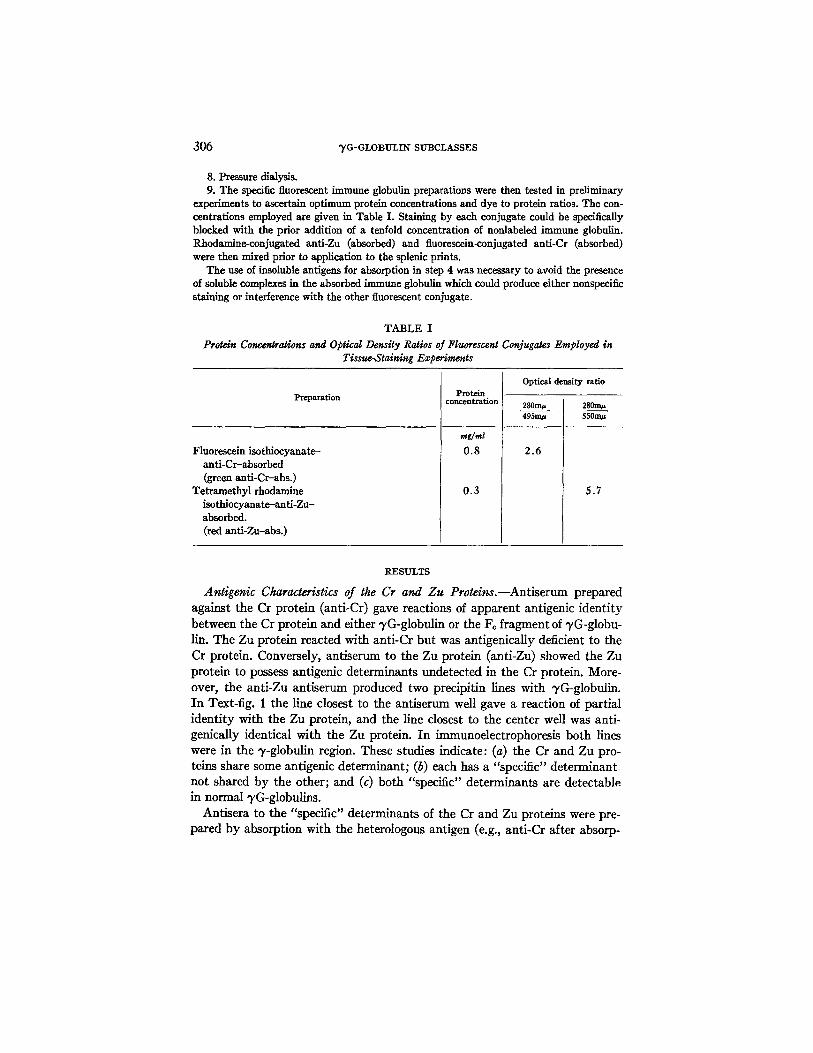
8. Pressure dialysis. 9. The sp~t ic fluorescent immune globulin preparations were then tested in preliminary
experiments to ascertain optimum protein concentrations and dye to protein ratios. The con- centrations employed are given in Table I. Staining by each conjugate could be specifically blocked with the prior addition of a tenfold concentration of nonlabeled immune globulin. Rhodamine~onjugated anti-Zu (absorbed) and fluoreseein-conjugated anti-Cr (absorbed) were then mixed prior to application to the splenic prints.
The use of insoluble antigens for absorption in step 4 was necessary to avoid the presence of soluble complexes in the absorbed immune globulin which could produce either nonspecific staining or interference with the other fluorescent conjugate.
TABLE I
Protein Concerdragons and Optical Density Raldos of Fluorescent Conjugates Employed in Tissue-Stainlng Experinwrgs
Preparation
Fluorescein isothiocyanate- anti-Cr-absorbed (green anti-Crabs.)
Tetramethyl rhodamine isothiocyanate-anti-Zu- absorbed. (red anti-Zu-abs.)
Protein concentration
r#g/mt 0.8
0.3
Optical density ratio
280rn# 495m#
2.6
280m~ 5S0n~
5.7
RESULTS
Antigenic Charac~iaics of the Cr and Zu Protdns.--Antiserum prepared against the Cr proton (anti-Cr) gave reactions of apparent antigenic identity between the Cr protein and either vG-globulin or the Fo fragment of vG-globu- fin. The Zu protein reactcd with anti-Cr but was antigcnicaliy deficient to the Cr protein. Conversely, antiserum to the Zu protein (anti-Zu) showed the Zu protein to possess antigenic determinants undetected in the Cr protein. More- over, the anti-Zu antiserum produced two precipitin lines with vG-globulin. In Text-fig. I the line closest to the antiserum well gave a reaction of partial identity with the Zu protein, and the line closest to the center well was anti- gcnically identical with the Zu protein. In immunoclcctrophoresis both lines were in the V-globulin region. These studies indicate: (a) the Cr and Zu pro- tcins share some antigenic determinant; (b) each has a "specific" determinant not shared by the other; and (c) both "specific" determinants are detectable. in normal vG-globulins.
Antiscra to the "specific" determinants of the Cr and Zu proteins were pre- pared by absorption with the heterologons antigen (e.g., anti-Cr after absorp-

BERNIER~ BALLIEUX~ TOMINAGA~ AND PIITNA~ 307
tion with the Zu protein was "specific" for the Cr determinant) . With anti- Cr-absorbed and ant i -Zu-absorbed antisera it was possible to demonstrate tha t one population of normal ~,G-globulin molecules possesses the Cr deter- minant, and another population possesses the Zu determinant.
Cr and Zu Types of Normal ~G-globulins.--The "specific" Cr and Zu anti- genic determinants were studied with regard to their frequency in the general population, the percentage of normal ~G-globulin molecules bearing the "spe- cific" determinants, and the location of the specific determinants on the "yG- globulin molecule.
TEXT-FIG. 1. Ouchterlony double diffusion showing reactions with anti-Zu (well 3) and anti-Zu (absorbed with the Cr protein) (well 5). Antigen wells contain normal ")'G-globulin (well 1) the Cr protein (well 2), and the Zu protein (well 4). A double line develops between anti-Zu and normal q~G-globulin. The line closest to the anti-Zu fuses with the Cr protein, and the other line fuses with the Zu protein. The anti-Zu absorbed with the Cr protein (well 5) still reacts with normal "yG-globulin and with the Zu protein.
Normal sera: The sera of 250 normal blood donors were tested with anti-Cr and anti-Zu antisera, and each normal serum was found to possess "specific" Zu and Cr determinants. A number of sera of known Gm specificity [Gm ( a + b - - ) , Gin(a-- b + ) , Gm (x)] 2 were tested, and each serum was found to possess both determinants. In each instance a single line was formed with unabsorbed anti-Cr while two lines were formed with unabsorbed anti-Zu.
Relative percentages: The percentage of "yG-globulin molecules possessing the Cr determinant and the percentage possessing the Zu determinant were determined by quanti tat ive precipitin curves using 125I-labeled ~,G-globulin from a single individual (Text-fig. 2 a). Anti-Cr before absorption precipitated approximately 97 % of the radioactivity; after absorption with the Zu protein, it precipitated approximately 75 %. Anti-Zu, on the other hand, after absorp-
2Sera of known Gm specificities were given to us by Dr. Wallace Epstein and Dr. A. G. Steinberg.

7 -
6 -
5 -
i~ 4-
2 . 5 %
.~ 2 -
~1.5-
I
0.5
I 0 -
~8-
A-Or / 9 3
¢ ~,o
i
I 0 2~0 3~0 /zg Antigen added
A-Cr-Absorbed y 5 6
75
7 7
i I 0 2~0
p.g Antigen added 30
I0-
5 ~
"6
2-
¢0
A-Zu-Absorbed 2.1
= 250 5 ; 0 7 ;0 ' ' I000 1250
p.g Antigen added (a)
2.0
1.6
1,0-
~ 0 . 6 6 -
0,33
TEXT Fio. 2
308
A - C r ~ 6
I 96.6
/, 97.0
/rgzo Ioo 200 500
~g Antigen added
A-Cr-Abserbed 5S4
~71.9" 0.1
.3
5 I 15
/zg Antigen added
6
H 4.52
i ~o 2'o 3'o 4'o 5'o
/zg Antigen added
(b)

BERNIER, BALLIEUX, TOMINAGA, AND PUTNAM 309
tion with the Cr protein, precipitated only 5-6 % of the radioactive antigen. Similar, though not identical, results were obtained when a pool of ~,G-globu- lin (Cohn fraction I I ) was used as the radioactive antigen (Text-fig. 2 b). Thus, when corrected for the 3 % of radioactivity not precipitated by the unabsorbed anti-Cr, the individual preparation contained 77% Cr-type molecules, 6% Zu-type molecules, and (by difference) 17% molecules lacking both deter- minants. Similarly, the pooled "yG-globulin preparation contained 74% Cr- type molecules, and 10% Zu-type molecules. By difference, 16% of molecules lack both determinants.
Molecular location: After papain digestion of %'G-globulin under the condi- tions of Porter (14), F~ and F~b fragments are obtained. The Cr determinant was found by immunoelectrophoresis in the Fc fragment of normal "yG-globu- lin, and in the Fc fragments of several myeloma globulins. However, the Zu determinant could not be detected in either the Fc or F~b fragments of ~,G- globulin digested in this fashion. When controlled hydrolysis with water-insol- uble papain was performed [according to the method of Cebra et al. (16)], the Zu determinant was found in the Fc fragment (Text-fig. 3). The lability of the Zu determinant to lengthy papain hydrolysis suggests that its location in the polypeptide chain is extremely close to the point of papain cleavage. By inference, the Cr determinant would be in an analogous position but might be relatively more resistant to papain degradation. I t should be pointed out that Takatsuki and Osserman have described differences in products of papain hydrolysis of Zu and Cr proteins (23).
Classification of "yG-Myeloma Globulins.--Since individual myeloma globu- lins appear to be single homogeneous molecular species of v-globulin, the inci- dence frequency of specific antigenic characteristics in a large series of myeloma globulins would be expected to approximate the frequency of that character- istic in a pool of normal "y-globulin molecules. This has been shown to be the case with the relative incidence of types K and L. Type K molecules are ap- proximately twice as common as type L molecules in normal pools and in a large series of myeloma globulins (24, 25). Accordingly, 108 "~,G-myeloma globulin specimens were tested for the presence of the Cr and Zu determinants with antisera rendered specific for these determinants by absorption. The
TExT-FIG. 2. Precipitin curves using 125I-labeled "}'G-globulin obtained from (a) a single individual (G. B.) and (b) purified Cohn fraction II, normal human pool. The symbol in the upper left corner designates the antiserum used in the construction of the precipitin curves. The numerical values given at points along the curves indicate the percentage of radioactive antigen precipitated by the antiserum. The unabsorbed anti-Cr antiserum (A-Cr) at the maxi- mum precipitated 97% of the 5,G-globulin in each case. The anti-Cr antiserum absorbed with the Zu protein (A-Cr absorbed) precipitated 73 and 77% of the radioactive antigen maxi- mally in the two instances. The anti-Zu antiserum, absorbed with the Cr protein (A-Zu ab- sorbed) precipitated 10 and 6% of the radioactive antigen maximally.

310 ~G-GLOBULIN SUBCLASSES
findings are shown in Table II. The relative incidence of Zu-type 3'G--myeloma globulins (7.4 %) and of the Cr-type (81.5 %) is remarkably similar to the dis- tribution of these molecules in the normal "gG-globulin pool and in the serum of the one individual studied. An additional number of 7G-myeloma globulins either lacked the Cr and Zu determinants entirely (0.9%), or they possessed some of the Cr determinants but were antigenically deficient to the Cr protein and the other Cr-type myeloma globulins (10.2 %). Those with some but not all of the Cr determinants are called Cr variants.
TEXT-FIG. 3. Immunoelectrophoresis of normal 3'G-globulin (1 and 5) and of normal 3~G cleaved with water-insoluble papain (3). The antisera are anti-Zu (2) and anti TG-globulin (4). Anode is at the right. The anti-3'G-globulin reacts with the intact "}'G-globulin (5) and with Fc and Fab fragments (3). The anti-Zu reacts with the intact 3'G-globulin (1) and with the Fc fragment (3). The inner, "specific Zu" line is found in both reactions. (The inner llne has been accentuated in this figure for clarity.)
Most of the myeloma globulins were studied in terms of light chain type as well as heavy chain subgroup. As is shown in Table II, the K :L ratio of the entire group of myeloma globulins and of the Cr-type subgroup was slightly higher than the expected 2:1 ratio. In the relatively small number sampled, the K : L ratio of the Cr variants was near unity and conversely, the K : L ratio of the Zu-type globulins was higher than would be expected. However, these differences probably reflect sampling. For instance, in terms of the Zu group, Grey and Kunkel found eight type K and two type L light chain types in 10 Vi group myeloma proteins (4) and Terry, Fahey, and Steinberg found 13 type K and 14 type L light chain types in 27 3%-myeloma globulins (5). Hence, in the cumulative number of 45 myeloma globulins in the three series typed as Zu, Vi, or 3'2°, 28 are type K and 17 are type L. These values are reasonably close to the 2:1 ratio randomly expected.

BEP.NIER, BALLIEUX~ TOMINAGA~ AND PUTNAM 311
Of the 11 proteins classified as Cr variants, eight were studied as regards electrophoretic mobility in agar gel electrophoresis and all eight were found to have mobilities in the slow fl- or fast v-globulin range. This was in marked contrast to the Cr and Zu groups where the vast majority of myeloma proteins had a 7-globulin mobility. The one myeloma globulin (Ca) which could not be classified also had a E-globulin mobility. These findings are of interest in terms of the Ge subgroup of Grey and Kunkel which is also comprised of pro- teins with rapid electrophoretic mobility (4).
Biological Considerations.--The Cr and Zu types of normal 7G-globulin were studied regarding antibody function and other biological characteristics. The presence of the two antigenieally defined types of 7G-globulin was deter-
TABLE II
Classif~a~ion o] 7G--Mydoma Globulins Based on Heavy Chain A n~genic Determinants, and the Light Chain Class Distribution within These Subgroups
Heavy chain classification
Cr type Cr variant Zu type Not classified
Total
Number
88 11 8 1
108
Per cent
81.5 10.2 7.4 0.9
100
Light chain classification
Number Type K typed*
75 52 11 6 8 7 1 1
95 66
TypcL
23 5 1 0
29
* Light chain typing was not performed on 13 Cr-type 7G-myeloma globulins.
mined in: (a) a specific immune precipitate, (b) fetal cord blood, (c) neonatal sera, and (d) severe hypogammaglobulinemia.
(a) Serum was obtained from a single individual immunized with diphtheria toxoid. I t was reacted with diphtheria toxoid, and the immune precipitate was centrifuged, washed, dissolved in acid, and separated from the antigen by passage through Sephadex G-200. When this specific antibody was tested in immunodiffnsion, both the Cr and Zu populations were present. In this experi- ment, there appeared to be a relative increase in concentration of the Zu mole- cules acting as antibodies, but both types of 7G appeared to be effective.
(b) Placental transfer of the two types of 7G-globulin was determined by examination of six paired samples of maternal serum and fetal cord blood. In each instance, both Zu and Cr populations were present, with a predominance of the Cr type. Since 7G-globulin synthesis in the fetus is negligible (26), these results appear to indicate that placental transfer of both types of 7G-globulin occurs.
(c) Sera from 16 infants varying in age from 7 wk to 24 months were

312 "yG-GLOBULIN SUBCLASSES
studied by immunodiffusion with use of unabsorbed antiserum. In all cases both types of 7G-globulins could be detected. The relative amounts of the two populations seemed to be comparable to the ratio found in the serum of normal adults, as judged from the position of the precipitin lines in the immu- nodiffusion plates. This was the case even in infants of 7 and 10 wk of age. Both classes of molecules appeared antigenically identical with those seen in adult sera.
(d) Sera from three adult patients with severe hypogammaglobulinemia (30, 100, and 100 mg of 7G-globulin per 100 ml of serum, respectively) were examined by immunodiffusion. These sera were chosen because the patients had not received therapeutic injections of -g-globulin. The two sera containing 100 mg/ml could readily be shown to have both populations. When relatively large amounts of the more deficient serum were used as antigen, both the Cr and Zu populations of 3,G-globulin were detectable. Again, the same relative proportion of molecules appeared to be present as in sera from normal patients, indicating that the virtual agenesis of 7G-globulin in these individuals was not selective in terms of either the Cr or Zu populations.
Thus, in the four biological situations--antibody activity, placental transfer, neonatal synthesis, and severe hypogammaglobulinemia--the two antigenically distinguishable populations of 3'G-globulin exhibited the characteristics of classical 3'G-globulin antibodies, and the relative proportion of the two popu- lations was approximately the same as that of normal serum. Since precise quantitation was not performed, moderate variations may, of course, be pres- ent.
Cellular Localization.--The cellular sites of synthesis of Cr and Zu deter- minants were investigated by use of fluorescent antibody techniques. Insoluble antigens were used to absorb the antisera before conjugation with fluorescent dyes in order to leave the anfisera free of any excess antigen as well as of cross- reacting antibodies. Human splenic prints were stained with the two fluorescent conjugated antisera, both sequentially and in mixtures. Fig. 1 and Table I I I show the results of staining splenic tissue prints with mixtures of red anti-Zu- abs. and green anti-Cr-abs. Cells stained either red or green, but not at all with both reagents. The percentage of cells stained with green anti-Cr-abs. was considerably greater than the percentage stained with red anti-Zu-abs. This 9:1 ratio approximates the ratio of the serum concentration of the two ~G-globulin types, and also the relative incidence of the two types of myeloma globulins.
As a control, fluorescent antisera directed against the two types of light chains (~, X) were used in combination with either the red anti-Zu-abs, or the green anti-Cr-abs. Results of one experiment are presented in Table HI. Pre- vious experiments with this particular spleen (specimen 6 of reference 22) had shown that 43 % of the immunoglobulin-containing cells contained ~/G-globulin, and that 44% of the immunoglobulin-containing cells contained type L light

BERNIER, BALLIEUX, TOMINAGA~ AND PUTNAM 313
chains. When this specimen was stained with red anti-Zu abs. and green anti- type L light chain, 6% of cells stained red, 4 % were mixed, and 90 % were green. The green and mixed cells are all those containing immunoglobulins of type L (total, 94%), but only 43% of these contain ~/G-globulins. Therefore, of 100 cells staining in this experiment, 41 may be considered to contain type L ~G-globulin. Four of these (the mixed cells) also contain Zu-type heavy chain (9.8%). Hence, the Zu-type heavy chain-containing cells comprise about one-tenth of the "yG-globulin-containing cells.
The ratio of red cells to mixed cells (3:2) is the ratio expected assuming that the red cells contain a type a-light chain (22).
TABLE III Percentages of Stained Cells in Splenic Tissue Preparations Showing Red, Green,
or Mixed Fluorescence
Reagents Spleen No. Red Green Mixed
Red anti-Zu-abs, and green anti-Cr-abs.
Red anti-Zu-abs, and green anti-k
I I I
III
II
6 14 9
94 86 91
90
DISCUSSION
The two antigenically distinguishable subclasses of ~,G-globulins studied appear to have extensive similarities to one another in terms of biological char- acteristics, chemical structure, and antigenic features. The studies reported here are, however, more concerned with the differences which can be shown to exist.
Numerically, the Cr type of normal %G-globulin molecules are approxi- mately 10 times more common than the Zu. Similarly, myeloma globulins possess the Zu determinant at one-tenth the frequency of the Cr determinant. As judged by the myeloma analogues of these populations in the peptide- mapping studies of Frangione and Franklin (27) and of our laboratory, s definite differences in primary sequence occur and account in part for the observed antigenic differences between the two populations. From the studies cited, it would appear that the area where sequence differences occur is in that portion of the molecule which is susceptible to the prolonged action of papain. It is pertinent in this regard that Grey and Kunkel (4) have found that the anti- genic characteristics of the Vi class of proteins are destroyed, at least partially, by papain, yet are preserved when proteolysis with pepsin is performed.
SBrackenridge, C., F. W. Putnam, and C. W. Easley. Comparative structural study of the heavy chains and Fc fragments of normal and pathologic G immunoglobuUns. In preparation.

314 ~G-GLOBULINSUBCLASSES
It has been demonstrated by the fluorescent antibody techniques described that individual cells do not contain both types of "rG-globulin heavy chains. We may infer that this means a given TG-globulin-producing cell does not synthesize both the Zu or Cr type of heavy chain, at least at one time in the cell's existence. This may indicate a degree of cellular predifferentiation and is in accord with the findings of Bernier and Cebra (22) regarding the cellular localization of the major classes of immunoglobulin polypeptide chains and with the close correlation between numbers of cells containing individual chains and the relative serum concentration of the chains. These findings accord with many of the observations of Chiappino and Pernis (28, 29). How- ever, these authors did find cells staining for more than one kind of light chain or heavy chain in the germinal center of lymph nodes.
Finally, the complex heterogeneity of the immunoglobulins has been in- creasingly recognized in recent, years. In addition to the class and type cate- gories, subcategories have been demonstrated for the ~/G-globulins and, as has been shown by Stein et al. (30), for Bence Jones proteins. Similar subcategories have now been found for the ~,A- and ~,M-globulin classes (31-33). Added to these subcategories are the individual segmental differences in amino acid sequence which presumably confer antibody specificity to an individual mole- cule. Attempts are underway in various laboratories to delineate these latter differences by analogy in the case of Bence Jones proteins (34-36). When the amino acid sequences of a number of related Bence Jones proteins are com- pleted, it is to be expected that a formulation of the biosynthetic mechanisms of antibody formation will be more feasible.
In spite of the apparent, endless chemical complexities revealed by detailed studies of the immunoglobulins, it seems clear that the many facets of immu- noglobulin heterogeneity will have to be appreciated and explained before a unified hypothesis can provide an understanding of these molecules as products of protein synthesis.
S12"~ARY
Two antigenic types of naturally occurring proteins related to the papain produced Fo fragment of ~,G-globulin have been studied. Most normal and myeloma ~,G-globulins are related to one or the other of these proteins, based on antigenic and structural characteristics of their heavy chain. Molecules bearing the determinant "Cr" are approximately 10 times as abundant as those bearing the determinant termed "Zu" in pools of normal ~,G-globulin and among a large group of q,G-myeloma globulins. Both populations have properties of typical ~,G-globulin antibodies. Splenic cells, stained for these two populations with specific fluorescent antibody preparations, are found to contain one or the other of the two populations of ~,G-globulin, but not both. Approximately 10 times as many cells contain the Cr determinant as contain the Zu determinant.

BERNIER, BALLIEUX, TOMINAGA~ AND PUTNAm[ 315
The authors thank Dr. E. C. Franklin of the New York University Medical School and Dr. E. F. Osserman of the College of Physicians and Surgeons, Columbia University, for making available proteins from the patients with "heavy chain disease". Doctors E. F. Osser- man, C. F. Hinz, C. E. Bucldey, and H. M. Rawnsley generously provided sera from myeloma patients. Dr. J. Robbins provided purified antidiphtheria toxoid antibody. Dr. J. Cebra pro- vided the water-insoluble papain and offered valuable suggestions in this work. We thank Mrs. Judith Byerley for technical assistance.
BIBLIOGRAPHY
i. Dray, S. 1960. Three v-globulins in normal human serum revealed by monkey precipitins. Science. 132:313.
2. Peetom, F., and B. Kramer. 1964. Development and anomalies of 7S gamma globulin (3'ss) investigated with monkey anti-human serum. In Proceedings at the 9th Congress of the European Society of Haematology, Lisbon, 1963. Vedag S. Karger, Basel, Switzerland.
3. Terry, W. D., and J. L. Fahey. 1964. Subclasses of human "r~-globulin based on differences in the heavy polypeptide chains. Science. 146:400.
4. Grey, H. M., and H. G. Kunkd. 1964. H chain subgroups of myeloma proteins and normal 7S 3,-globulin. Y. Exptl. Med. 120:253.
5. Terry, W. D., J. L. Fahey, and A. G. Steinberg. 1965. Gm and Inv factors in subdasses of human IgG. Y. Exptl. Meal. 122:1087.
6. Ballieux, R. E., G. M. Bernier, K. Tominaga, and F. W. Putnam. 1964. Gamma globulin antigenic types defined by heavy chain determinants. Science. 145:168.
7. Franklin, E. C., J. Lowenstein, B. Bigelow, and M. Meltzer. 1964. Heavy chain disease: a new disorder of serum gamma globulins. Report of the first case. Am. Y. Med. 37:332.
8. Franklin, E. C. 1964. Structural studies of human 7S T-globulin (G immuno- globulin). Y. Exptt. Med. 120:691.
9. Osserman, E. F., and K. Takatsuki. 1964. Clinical and immunochemical studies Of 4 cases of H'y 2 chain disease. Am. J. Med. 37:374.
10. Boettcher, E. W., P. Kisfler, and H. Nitschmann. 1958. Method of isolating the flrmetal-combining globulin from human blood plasma. Nature. 181:490.
11. Bernier, G. M., and F. W. Putnam. 1964. Polymerism, polymorphism, and im- purities in Bence-Jones proteins. Biochim. Biophys. Acta. 86:295.
12. Scheidegger, J. J. 1955. Une micro-m&hode de l'immuno-~lectrophor6se. Intern. Arch. Allergy Appl. Immunol. "/:103.
13. Ouchteflony, O. 1958. Diffusion-in-gel methods for immunological analysis. Progr. Allergy. 5:1.
14. Porter, R. R. 1959. The hydrolysis of rabbit 3,-globulin and antibodies with crystalline papain. Biochem. J. "/3:119.
15. Putnam, F. W., C. W. Easley, and L. T. Lynn. 1962. Site of cleavage of "/-globulins by papain. Biochim. Biophys. Acta. 58:279.
16. Cebra, J. J., D. Givol, H. I. Silman, and E. Katchalski. 1961. A two-stage deavage of rabbit v-globulin by a water-insoluble papain preparation followed by cysteine. J. Biol. Chem. 9.,$6:1720.
17. Talmage, D. W., H. R. Baker, and W. Akeson. 1954. Separation and analysis of labelled antibodies. ]. Infect. Diseases. 94:199.

316 '~G-GLOBULIN SUBCLASSES
18. Kinard, F. E. 1957. Liquid scintillator for the analysis of proteins in water. Rev. Sci. Instr, 28:293.
19. Coons, A. H., E. H. Leduc, and J. M. Connolly. 1955. Studies on antibody pro- duction. I. A method for the histochemical demonstration of specific antibody and its application to a study of the hyperimmune rabbit, f . E, xptl. Meal. 102: 49.
20. Wood, B. T., S. H. Thompson, and G. Goldstein. 1965. Fluorescent antibody staining. III . Preparation of fluorescein-isothiocyanate-labeled antibodies. J. Immunol. 95:246.
21. Cebra, J. J., and G. Goldstein. 1965. Chromatographic purification of tetramethyl- rhodamine-immune globulin conjugates and their use in the cellular localization of rabbit 7-globulin polypepfide chains. J. Immunol. 95:230.
22. Bernier, G. M., and J. J. Cebra. 1965. Frequency distribution of a, % ~ and polypeptide chains in human lymphoid tissues. J. Immunol. 95:246.
23. Takatsuki, K., and E. F. Osserman. 1964. Structural differences between two types of "heavy chain" disease proteins and myeloma globulins of corresponding types. Science. 145:499.
24. Mannik, M., and H. G. Kunkel. 1963. Two major types of normal 7S 3,-globulins. J. Exptl. Med. 117:213.
25. Korngold, L. 1961. Abnormal plasma components and their significance in disease. Ann. N. Y. Aead. Sci. 94:110.
26. Eichwald, H. F., and H. R. Shinefield. 1963. Antibody production by the human fetus. J. Pediat. 63:870.
27. Frangione, B., and E. C. Franklin. 1965. Structural studies of human immuno- globulins. Differences in the Fd fragments of the heavy chains of G myeloma proteins. J. Exptl. Med. 122:1.
28. Chiappino, G., and B. Pernis. 1964. Demonstration with immunofluorescence of 19S macroglobulins and 7S gamma globulins in different cells of the human spleen. Pathol. Microbiol. 27:8.
29. Pernis, B., and G. Chiappino. 1964. Identification in human lymphoid tissues of cells that produce group 1 or group 2 gamma globulins. Immunology. 7:500.
30. Stein, S., R. L. Nachman, and R. L. Engle. 1963. Individual and sub-group anti- genic specificity of Bence-Jones protein. Nature. 200:1180.
31. Vaerman, J-P., and J. F. Heremans. 1966. Subclasses of human immunoglobulin A, based on differences in the alpha polypeptide chains. Science. 153:647.
32. Harboe, M., J. Deverill, and It. C. Godal. 1965. Antigenic heterogeneity of Waldenstr6m type ~M-globulins. Scand. J. ttaematol. 2:137.
33. Terry, W. D., and M. S. Roberts. 1966. Antigenic heterogeneity of human im- munoglobulin A proteins. Science. 153:1007.
34. Hilschman, N., and L. C. Craig. 1965. Amino acid sequence studies with Bence- Jones proteins. Proc. Nat. Acad. Sci. U.S. 53:1403.
35. Milstein, C. 1966. Variations in amino acid sequence near the disulphide bridges of Bence-Jones proteins. Nature. 209:370.
36. Titani, K., E. Whitley, Jr., and F. W. Putnam. 1965. Immunoglobulin structure: Variation in the sequence of Bence-Jones proteins. Science. 152:1513.

BERNIER~ BALLIEUX~ TOMINAGA~ AND PUTNAM 317
PLATE 22

318 "yG-GLOBULIN SUBCLASSES
FIG. 1. Photomicrographs of human splenic prints stained with a mixture of rho- damine-conjugated anti-Zu (absorbed with Cr) and fluorescein-conjugated anti-Cr (absorbed with Zu). The top row shows two green stained cells (left) and one red stained cell (right). Both fields viewed with a neutral K-2 falter and a 100 X oil- immersion objective. The bottom row shows three pictures of one field as viewed with the neutral K-2 filter (center), a 57A red-excluding filter (right) and a 23A green- excluding falter (left). A green-staining cell and a red-staining cell are evident in the center picture. The green fluorescence passes the 57A filter, and the red fluores- cence passes the 23A filter.

THE JOURNAL OF EXPERIMENTAL MEDICINE VOL. 125 PLATE 22
(Bernier et al.: ",/G-globulin subclasses)