
Heart failure ASSESSMENT OF DIASTOLIC...
15
Heart failure ASSESSMENT OF DIASTOLIC FUNCTION: WHAT THE GENERAL CARDIOLOGIST NEEDS TO KNOW Philip M Mottram, Thomas H Marwick Heart 2005; 91:681–695. doi: 10.1136/hrt.2003.029413 Take the online multiple choice questions associated with this article (see page 695) See end of article for authors’ affiliations _________________________ Correspondence to: Professor Thomas H Marwick, University of Queensland Department of Medicine, Princess Alexandra Hospital, Ipswich Road, Brisbane, Q4102, Australia; tmarwick@ soms.uq.edu.au _________________________ D iastolic dysfunction has a major impact on symptom status, functional capacity, medical treatment, and prognosis in both systolic and diastolic heart failure (HF), irrespective of the cause. w1 w2 When systolic dysfunction is clearly present, the central clinical question concerns the presence or absence of elevated filling pressure; a restrictive filling pattern is highly specific for elevated pulmonary wedge pressure in this setting. 1 w3 The transmitral flow pattern is also predictive of outcome; non-reversibility of restrictive filling with treatment portends a very poor prognosis. 2 Thus, diastolic evaluation is an important component of the evaluation of the patient with systolic left ventricular (LV) impairment. IS ASSESSMENT OF DIASTOLIC FUNCTION NECESSARY ? c Paradoxically, the role of diastolic function assessment is more difficult to define in patients with diastolic HF. Diastolic dysfunction is the predominant cardiac abnormality in this syndrome, which is associated with increased risk of hospitalisation and death. 3 However, the American College of Cardiology/American Heart Association guidelines for the evaluation and management of HF w4 support a diagnosis of exclusion—that is, clinical evidence of HF with preserved systolic function. Indeed, such a definition of diastolic HF has been adopted by the majority of previous reports, w5 and is supported by the results of recent studies which indicate that the presence of diastolic dysfunction may be assumed in patients presenting with HF and normal LV ejection fraction (LVEF). 4 w6 w7 Zile and colleagues demonstrated that at least one abnormal index of diastolic function was present in patients with HF and normal systolic function. These data suggest that a diagnosis of diastolic HF may accurately be made as a diagnosis of exclusion, 4 albeit in a highly selected population of relatively young, predominantly male patients who were scheduled to undergo cardiac catheterisation (contrasting with the large clinical population of elderly, hypertensive, predominantly female patients with HF and preserved systolic function). Nonetheless, a recent review has highlighted the disconnect between Doppler echo measurements and true diastolic properties of the left ventricle, and has questioned the prevailing assumption that HF with preserved systolic function is always caused by diastolic dysfunction. 5 A third scenario is perhaps the most difficult. These patients present with exertional dyspnoea in the context of normal systolic function, and in this situation, symptoms may be ascribed to diastolic HF. The lack of specificity of HF symptoms has led to concern regarding the heterogeneity of patients who are labelled with diastolic HF based only upon the presence of clinical HF and normal systolic function; many such patients may in fact have non-HF causes of their clinical presentations. w8 For these reasons, recent guidelines have called for invasive determination of diastolic dysfunction to make a definite diagnosis of diastolic HF, 6 although the feasibility of this approach is limited. Doppler echocardiography is ideally suited for assessment of diastolic function, being widely available, non-invasive, and less expensive than other techniques. Echocardiographic assessment of diastolic function makes the diagnosis of diastolic HF more specific, allows serial assessment of the response of diastolic dysfunction to treatment, and facilitates inclusion of more homogeneous populations into intervention trials. Echocardiography has a broader role by allowing exclusion of overt LV systolic dysfunction (LVEF , 50%), significant valvar dysfunction (such as mitral regurgitation or aortic stenosis), and pericardial disease. However, a significant limitation is the requirement for expert interpretation of multiple parameters that vary with loading conditions and often provide conflicting information. The clinical importance of the evaluation of diastolic function warrants a simplified approach to diastology that is broadly accessible to general cardiologists. This article will therefore attempt to clarify this complex area, focusing on the practical application of Doppler echocardiography for the clinical assessment of the dyspnoeic patient, both in patients with preserved and impaired systolic function. 681 www.heartjnl.com on 31 August 2018 by guest. Protected by copyright. http://heart.bmj.com/ Heart: first published as 10.1136/hrt.2003.029413 on 14 April 2005. Downloaded from
Transcript of Heart failure ASSESSMENT OF DIASTOLIC...
Heart failure
ASSESSMENT OF DIASTOLICFUNCTION: WHAT THE GENERALCARDIOLOGIST NEEDS TO KNOW
Philip M Mottram, Thomas H Marwick
Heart 2005;91:681–695. doi: 10.1136/hrt.2003.029413
Take the online multiple choicequestions associated with thisarticle (see page 695)
See end of article for authors’affiliations_________________________
Correspondence to:Professor Thomas H Marwick,University of QueenslandDepartment of Medicine,Princess Alexandra Hospital,Ipswich Road, Brisbane,Q4102, Australia; [email protected]_________________________
Diastolic dysfunction has a major impact on symptom status, functional capacity, medical
treatment, and prognosis in both systolic and diastolic heart failure (HF), irrespective of the
cause.w1 w2 When systolic dysfunction is clearly present, the central clinical question
concerns the presence or absence of elevated filling pressure; a restrictive filling pattern is highly
specific for elevated pulmonary wedge pressure in this setting.1 w3 The transmitral flow pattern is
also predictive of outcome; non-reversibility of restrictive filling with treatment portends a very
poor prognosis.2 Thus, diastolic evaluation is an important component of the evaluation of the
patient with systolic left ventricular (LV) impairment.
IS ASSESSMENT OF DIASTOLIC FUNCTION NECESSARY?cParadoxically, the role of diastolic function assessment is more difficult to define in patients with
diastolic HF. Diastolic dysfunction is the predominant cardiac abnormality in this syndrome,
which is associated with increased risk of hospitalisation and death.3 However, the American
College of Cardiology/American Heart Association guidelines for the evaluation and management
of HFw4 support a diagnosis of exclusion—that is, clinical evidence of HF with preserved systolic
function. Indeed, such a definition of diastolic HF has been adopted by the majority of previous
reports,w5 and is supported by the results of recent studies which indicate that the presence of
diastolic dysfunction may be assumed in patients presenting with HF and normal LV ejection
fraction (LVEF).4 w6 w7 Zile and colleagues demonstrated that at least one abnormal index of
diastolic function was present in patients with HF and normal systolic function. These data
suggest that a diagnosis of diastolic HF may accurately be made as a diagnosis of exclusion,4 albeit
in a highly selected population of relatively young, predominantly male patients who were
scheduled to undergo cardiac catheterisation (contrasting with the large clinical population of
elderly, hypertensive, predominantly female patients with HF and preserved systolic function).
Nonetheless, a recent review has highlighted the disconnect between Doppler echo measurements
and true diastolic properties of the left ventricle, and has questioned the prevailing assumption
that HF with preserved systolic function is always caused by diastolic dysfunction.5
A third scenario is perhaps the most difficult. These patients present with exertional dyspnoea
in the context of normal systolic function, and in this situation, symptoms may be ascribed to
diastolic HF. The lack of specificity of HF symptoms has led to concern regarding the
heterogeneity of patients who are labelled with diastolic HF based only upon the presence of
clinical HF and normal systolic function; many such patients may in fact have non-HF causes of
their clinical presentations.w8 For these reasons, recent guidelines have called for invasive
determination of diastolic dysfunction to make a definite diagnosis of diastolic HF,6 although the
feasibility of this approach is limited. Doppler echocardiography is ideally suited for assessment of
diastolic function, being widely available, non-invasive, and less expensive than other techniques.
Echocardiographic assessment of diastolic function makes the diagnosis of diastolic HF more
specific, allows serial assessment of the response of diastolic dysfunction to treatment, and
facilitates inclusion of more homogeneous populations into intervention trials. Echocardiography
has a broader role by allowing exclusion of overt LV systolic dysfunction (LVEF , 50%),
significant valvar dysfunction (such as mitral regurgitation or aortic stenosis), and pericardial
disease. However, a significant limitation is the requirement for expert interpretation of multiple
parameters that vary with loading conditions and often provide conflicting information. The
clinical importance of the evaluation of diastolic function warrants a simplified approach to
diastology that is broadly accessible to general cardiologists. This article will therefore attempt to
clarify this complex area, focusing on the practical application of Doppler echocardiography for
the clinical assessment of the dyspnoeic patient, both in patients with preserved and impaired
systolic function.
681
www.heartjnl.com
on 31 August 2018 by guest. P
rotected by copyright.http://heart.bm
j.com/
Heart: first published as 10.1136/hrt.2003.029413 on 14 A
pril 2005. Dow
nloaded from
DEFINING DIASTOLIC DYSFUNCTION WITHECHOCARDIOGRAPHYNo comprehensive consensus regarding diagnostic echocar-
diographic criteria for diastolic HF has been reached, but
guidelines have been proposed.7 w9 The European Study
Group on Diastolic HF has provided criteria which relate to
abnormal LV relaxation, abnormal LV filling, or reduced LV
diastolic distensibility based on transmitral and pulmonary
vein Doppler data.7 However, even these guidelines do not
necessarily provide for echocardiographic identification of
specific subgroups of patients with pseudonormal or restric-
tive filling patterns, potentially reducing their sensitivity for
the diagnosis of diastolic dysfunction in the absence of
cardiac catheterisation data. They are further limited by
failure to include newer echo techniques such as tissue
Doppler imaging (TDI). The assessment of diastolic function
should be based on a comprehensive echocardiographic study
integrating all available two dimensional and Doppler data.8
Such an approach will be outlined below.
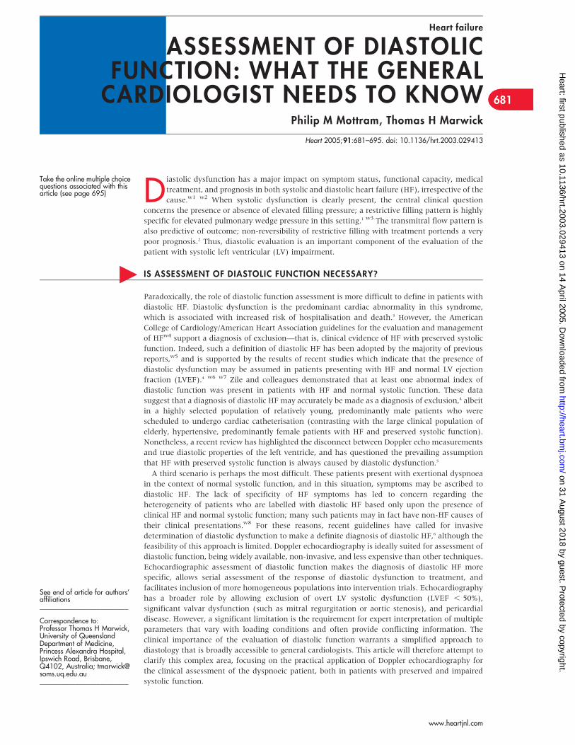
Assessment of transmitral flowEchocardiographic evaluation of diastolic function has been
traditionally performed by measurement of transmitral flow
parameters including the early (E) and late (A) diastolic
filling velocities, the E/A ratio, and the E deceleration time
(DT) from an apical four chamber view with conventional
pulsed wave Doppler (fig 1A).w9 The transmitral E wave is
related to the time course of active LV relaxation which
generates a pressure gradient from the left atrium through
the LV inflow tract to the LV apex.w10 Early diastolic LV
filling is therefore largely influenced by the interaction of left
atrial compliance and the rate of ventricular relaxation. The
peak E velocity may be increased by either elevated left atrial
pressure (the cause of high E/A ratios in cardiac disease), or
alternatively, by low LV minimal diastolic pressure caused by
rapid LV relaxation (which drives the high E/A ratios typical
of normal young adults).9
Based upon age adjusted interpretation of the transmitral
flow profile, diastolic function is initially classified as either
normal, impaired relaxation, pseudonormal, restrictive
(which may be reversible or non-reversible with preload
reduction), or indeterminate (if normal or pseudonormal
cannot be differentiated) (fig 1). These patterns of LV filling
represent progressively worse diastolic dysfunction as the LV
becomes increasingly abnormal. It is important to consider
that increasing diastolic dysfunction is usually accompanied
by a progressive increase in LV filling pressures, which in turn
have a major impact on the transmitral flow profile. Slow or
prolonged LV relaxation therefore causes a decrease in E
velocity but at the same time contributes to elevation of left
atrial pressure, which in turn tends to increase the E
velocity.10 These opposing effects of left atrial pressure and
LV relaxation are also operative on the E deceleration time,
which tends to be prolonged by impaired LV relaxation and
shortened by increased filling pressures. Thus the effects of
diastolic dysfunction on the E/A ratio and E wave decelera-
tion time become progressively compensated and then
over-compensated by the effects of loading, resulting in a
non-linear (in fact ‘‘U’’ shaped) relation between these
indices and severity of diastolic dysfunction.11 The isovolumic
relaxation time bears a similar relation to diastolic dysfunc-
tion and load and does not provide additional information. In
individual patients therefore, the filling pattern can change
from mild (impaired relaxation) to more severe (pseudo-
normal or restrictive) diastolic dysfunction with either a
Figure 1 Pathophysiological characterisation of left ventricular (LV) filling patterns. (A) Normal transmitral flow in a patient in sinus rhythm.(B) Impaired relaxation with normal filling pressures. (C) Pseudonormal filling. (D) Restrictive filling.
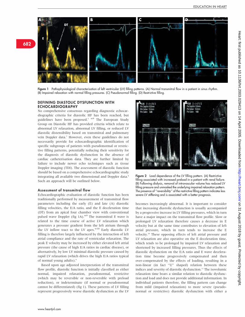
Figure 2 Load dependence of the LV filling pattern. (A) Restrictivefilling associated with increased preload in a patient with renal failure.(B) Following dialysis, removal of intravascular volume has reduced LVfilling pressure and unmasked the underlying impaired relaxation pattern.The presence of ‘‘reversibility’’ of the restrictive filling pattern indicates lesssevere LV stiffening and is associated with a better prognosis.
682
EDUCATION IN HEART
www.heartjnl.com
on 31 August 2018 by guest. P
rotected by copyright.http://heart.bm
j.com/
Heart: first published as 10.1136/hrt.2003.029413 on 14 A
pril 2005. Dow
nloaded from
progression of the underlying pathophysiological process, or
alteration of loading conditions (fig 2). Similarly, improve-
ment in the Doppler filling profile may occur over a longer
period with treatments targeting the underlying cause (fig 3).
Thus transmitral flow parameters must be further interpreted
in the light of LV loading. This requires either incorporation
of alternative load dependent parameters, or use of newer
less load dependent techniques (or preferably both). Our
standard approach is to gather the pulmonary venous flow
profile, the medial and lateral early diastolic mitral annular
velocities by TDI (Ea), the E/Ea values, and the response of
E/A to Valsalva manoeuvre.
Tissue Doppler imaging: long axis relaxation rateLong axis shortening (contraction) and lengthening (relaxa-
tion) of myocardial segments results in longitudinal motion
of the mitral annulus toward or away from the (relatively
fixed) LV apex during systole and diastole, respectively.
Although long axis segmental shortening remains fairly
uniform along the myocardial wall,w11 a gradient of increas-
ing velocity from apex to base has been demonstrated.w12 w13
Mitral annular velocities may therefore be regarded as an
‘‘aggregate’’ of segmental myocardial velocities and in the
absence of regional LV dysfunction accurately reflect global
long axis LV function. The systolic velocity (Sa) corresponds
to ventricular ejection while the early (Ea) and late (Aa)
diastolic velocities correspond to the transmitral Doppler
flow. In normal subjects the Ea occurs coincident with, or
just before, the transmitral E wave, whereas in heart failure
there is a progressive delay in Ea with respect to E.w10 Of
more practical importance, the Ea velocity progressively
decreases as the long axis relaxation rate becomes increas-
ingly reduced in the setting of a wide range of cardiac disease
processes including dilated, restrictive, and hypertrophic
cardiomyopathies. Invasive studies have demonstrated that
the Ea velocity correlates strongly with the time constant of
isovolumic relaxation over a wide range of filling pressures.12–14
Specifically, the Ea velocity is much less susceptible to the
effects of increased preload and remains low in patients
with advanced diastolic dysfunction and pseudonormalisa-
tion of the transmitral E velocity.14 w14 Further, Ea is
typically lowest in patients with severe LV dysfunction and
restrictive filling.
Pulsed wave TDI, whereby a sample volume (2–5 mm) is
placed at the septal or lateral border of the mitral annulus in
an apical four chamber view, is the most commonly used
technique to record longitudinal velocities at the mitral
annulus.11 Colour tissue Doppler is an alternative method,
based on autocorrelation, which permits a colour velocity map
to be gathered over the entire image sector, and therefore
permits off-line measurement of annular or myocardial
velocities at any point in the image. While pulsed wave
Doppler offers superior temporal resolution, current colour
Doppler algorithms can acquire data at high frame rate (. 100
frames per second) and generate clear velocity–time curves
without the problem of gain dependent spectral broadening
which affects the pulsed wave technique. However, because of
its wider availability and large clinical evidence base, pulsed
wave Doppler is the preferred technique for routine assess-
ment of diastolic function in the clinical setting. Published
normal ranges have been produced for both methods, with
colour TDI velocities significantly lower compared with
pulsed wave velocities.w15 w16 In keeping with the normal
age dependent reduction in diastolic function, reference
values for Ea must be adjusted for agew17 w18 (table 1).
A major advantage of TDI is its high feasibility, high
reproducibility, and ease of application in the clinical
setting.12 15 Velocities at both the septal and lateral mitral
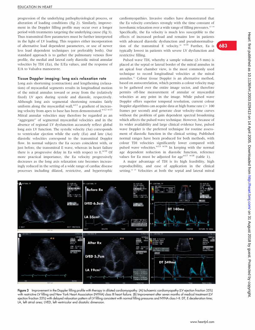
Figure 3 Improvement in the Doppler filling profile with therapy in dilated cardiomyopathy. (A) Ischaemic cardiomyopathy (LV ejection fraction 35%)with restrictive LV filling and New York Heart Association (NYHA) class III heart failure. (B) Improvement after seven months of medical treatment (LVejection fraction 55%) with delayed relaxation pattern of LV filling consistent with normal filling pressures and NYHA class I–II. DT, E deceleration time;LA, left atrial area; LVED, left ventricular end diastolic dimension.
683
EDUCATION IN HEART
www.heartjnl.com
on 31 August 2018 by guest. P
rotected by copyright.http://heart.bm
j.com/
Heart: first published as 10.1136/hrt.2003.029413 on 14 A
pril 2005. Dow
nloaded from
annulus may be obtained with minimal increase in the
duration of the study. Recent evidence suggests that
velocities at the septal and lateral annulus may be affected
by different variables and are not interchangeable.w18 If
obtaining only a single measurement, the lateral Ea may be
preferredw19 as the septal Ea velocity has been demonstrated
to be altered by preload in subjects with normal LV
function,w20 although this effect may decrease as LV
relaxation becomes progressively impaired.16 In addition,
the septal Ea velocity may be influenced by right ventricular
diastolic function. Potential pitfalls to be considered when
acquiring and interpreting pulsed wave TDI signals include
ensuring that the two dimensional image quality is optimised
and that the ultrasound beam is well aligned (, 30 )̊ with
the direction of longitudinal motion (which may be more
challenging at the lateral annulus). Finally, localised seg-
mental hypokinesis in a given LV wall will result in reduced
velocity of annular motion at the corresponding site, possibly
leading to a spuriously low estimate of global LV function. In
this situation it is recommended to obtain an average Ea from
multiple annular sites.w19
Tissue Doppler imaging: estimation of LV fil l ingpressuresProgressive diastolic dysfunction is associated with both
impairment of LV relaxation and an increase in left atrial
pressure. These concurrent events tend to have opposing
effects on the transmitral E velocity, rendering it poorly
predictive of either process. However, the E velocity (which
increases with elevation of left atrial pressure) may be
‘‘corrected’’ for the degree of impairment in LV relaxation
rate by relating it to the Ea velocity (which is a relatively load
independent measure of reduced LV relaxation) to provide an
index, the E/Ea ratio, which has been demonstrated to
correlate with mean left atrial pressure.12 This concept has
been validated in various clinical conditions including normal
and impaired systolic function, tachycardia, atrial fibrillation,
and hypertrophic cardiomyopathy.12 15 w21–23 This application
of TDI for estimation of LV filling pressures has significantly
advanced the ability of the echocardiographer to distinguish
normal from pseudonormal LV filling. It is particularly useful
in difficult cases such as patients with early elevation of left
atrial pressure whose filling profile is in a transition phase
between impaired relaxation and pseudonormal filling (fig 4).
As discussed for assessment of LV relaxation rate, the
lateral Ea may be preferable for E/Ea ratio estimation of
filling pressure as more defined cut offs have been reported.
Nagueh and colleagues demonstrated that E/Ea . 10 using
the lateral mitral annular velocity reliably predicts a
pulmonary capillary wedge pressure of . 12 mm Hg.12 In
comparison, using the septal Ea velocity, Ommen and
colleagues found that while pulmonary capillary wedge
pressure is likely normal if the E/Ea ratio is , 8 and likely
Table 1 Age adjusted normal cut offs for selecteddiastolic parameters. w9 w16 w27 w44 w96
,40 years 40–60 years .60 years
E deceleration time (ms) ,220 140–250 140–275Septal Ea velocity (cm/s) .9 .7 .6Lateral Ea velocity (cm/s) .11 .10 .7
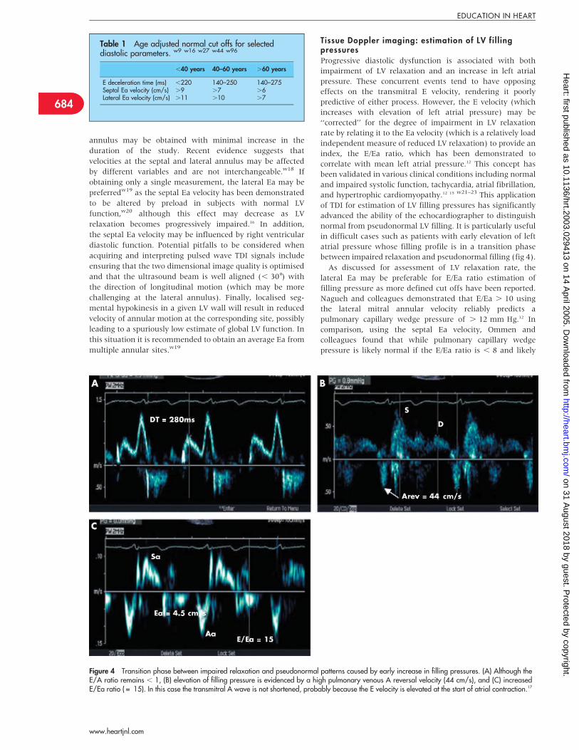
Figure 4 Transition phase between impaired relaxation and pseudonormal patterns caused by early increase in filling pressures. (A) Although theE/A ratio remains , 1, (B) elevation of filling pressure is evidenced by a high pulmonary venous A reversal velocity (44 cm/s), and (C) increasedE/Ea ratio ( = 15). In this case the transmitral A wave is not shortened, probably because the E velocity is elevated at the start of atrial contraction.17
684
EDUCATION IN HEART
www.heartjnl.com
on 31 August 2018 by guest. P
rotected by copyright.http://heart.bm
j.com/
Heart: first published as 10.1136/hrt.2003.029413 on 14 A
pril 2005. Dow
nloaded from

elevated if . 15, intermediate values were less useful.15 A
recent report found lateral E/Ea to be superior to septal E/Ea
for predicting wedge pressure when ejection fraction is
. 50%,w19 although an average of both values is more
accurate in the presence of regional dysfunction.w19 w24
Pulmonary venous flowThe pulmonary venous Doppler signal comprises ‘‘forward’’
systolic (S) and diastolic (D) velocities into the left atrium,
and a ‘‘backwards’’ late diastolic A reversal wave correspond-
ing to atrial contraction. The major factors influencing the
pulmonary venous Doppler profile are illustrated in fig 5. The
systolic flow wave is often biphasic; the ‘‘S1’’ occurs in
response to atrial relaxation while the ‘‘S2’’ (often more
dominant) is thought to be caused by mitral annular descent
towards the ventricular apex resulting from long axis LV
shortening, which increases left atrial capacity and reduces
chamber pressure.w25 The pulmonary venous D flow occurs
just after, and is largely determined by, the transmitral E
wave.w26 In older adults, systolic flow is dominant such that
S/D is . 1.w27 Like the E and A velocities in the transmitral
Doppler profile, the pulmonary venous S/D ratio exhibits a
non-linear relation to progressive diastolic dysfunction.
Importantly, however, this load dependence can be used to
advantage to aid correct interpretation of the transmitral flow
pattern. Therefore in the early stages of diastolic dysfunction
(evidenced by a low E/A ratio), the D velocity is low and S/D
is . 1. As detailed in fig 5, the S velocity is also influenced
by left atrial pressure. As such, increased left atrial pressure
will tend to ‘‘blunt’’ the S flow and reduce the S/D flow ratio
to , 1, particularly when filling pressures are notably
raised.w28 w29 However, while a useful index in the setting
of reduced LV ejection fraction, this criterion is relatively
insensitive for the detection of elevated left atrial pressure
when LV systolic function is preservedw30 as brisk mitral
annular descent tends to maintain the systolic forward
fraction of pulmonary venous flow. Preservation of atrial
contractile function has a similar effect by fostering low left
atrial preload at the onset of LV systole.
The pulmonary venous A wave provides an additional tool
for assessment of LV filling pressure and diastolic function.
The peak A reversal velocity increases as resistance to atrial
forward flow increases as a result of increased ventricular
stiffness and/or end diastolic pressure, such that a peak
velocity . 35 cm/s is suggestive of elevated filling pres-
sures.w9 However, left atrial mechanical dysfunction often
accompanies advanced diastolic dysfunction (particularly in
association with paroxysmal atrial fibrillation) and may lead
to low A reversal velocities. A more robust pulmonary venous
parameter may be derived from the difference in the
transmitral and pulmonary venous A wave durations. As LV
compliance decreases and diastolic pressure rises, increased
afterload on the left atrium tends to shorten the transmitral
A wave, while its duration measured in the pulmonary vein
may be increased. A difference in the respective durations of
. 20–30 ms accurately predicts significant elevation of LV
end diastolic pressurew28 and may be an early marker of
transformation from impaired relaxation to a pseudonormal
filling pattern (fig 6). The major advantage of this para-
meter is its utility in the setting of preserved LV systolic
function,w30 w31 while the obvious limitation is the difficulty
in acquisition of accurate measurements of the pulmonary
venous A duration from a transthoracic window.15
Load altering manoeuvresThe principle behind this step is to remove the effects of
preload compensation and thereby unmask the underlying
relaxation abnormality. Thus the aim is to produce a
transient lowering of left heart filling pressures which in
the clinical setting is most practically achieved with the
Valsalva manoeuvre, although a similar effect may be
obtained with sublingual glyceryl trinitrate.w32 During the
Valsalva manoeuvre, an initial minor increase in systemic
blood pressure (caused by increased pulmonary venous
return) is followed by a decrease in systemic venous return
and a gradual decrease in stroke volume, leading, after a few
cardiac cycles, to a reduction in left atrial and LV filling
pressures and potential ‘‘conversion’’ of pseudonormal filling
to an impaired relaxation pattern (fig 7).w33 This approach
remains largely qualitative, and the required decrease in
either the E velocity or the E/A ratio to reach a diagnostic
threshold varies with different studies and will depend upon
the baseline values for E and A, the quality of Valsalva,
degree of patient effort, and other factors.15 Even in the
research setting, ability to obtain adequate data may be
particularly low,15 w34 thus limiting the sensitivity of the
technique. A reduction of E velocity by 50% or complete
reversal of the E/A ratio to , 1w35 may be useful criteria,
although other investigators have been unable to determine
an accurate cutoff.w34 The Valsalva manoeuvre may also be
less predictive of elevated LV filling pressure when ejection
fraction is preserved.w31
Flow propagation velocityThe high temporal and spatial resolution of colour Doppler M
mode can be used to provide a two dimensional representa-
tion of the velocity of early diastolic filling as the bolus of
blood propagates through the mitral valve towards the LV
apex. This flow propagation velocity (Vp) can be measured
from an apical four chamber view with the M mode beam
aligned parallel to LV inflow.11 With colour Doppler activated
and the aliasing velocity reduced, a sharp colour wavefront
can be demarcated which represents progression of a bolus of
blood towards the LV apex in response to early diastolic LV
relaxation; Vp may be determined from the slope of this
isovelocity line. Analogous to Ea, Vp correlates with invasive
measurements of LV relaxation and has been shown to be
relatively independent of loading conditions.w36 Similarly,
Figure 5 Determinants of the pulmonary venous Doppler profile. AF,atrial fibrillation; LAP, left atrial pressure; LVEDP, left ventricular enddiastolic pressure.
685
EDUCATION IN HEART
www.heartjnl.com
on 31 August 2018 by guest. P
rotected by copyright.http://heart.bm
j.com/
Heart: first published as 10.1136/hrt.2003.029413 on 14 A
pril 2005. Dow
nloaded from
the E/Vp ratio has been demonstrated to correlate with LV
filling pressure.w37 More recently, however, Vp has been
shown to be significantly influenced by LV systolic function,
which may act to normalise Vp values in the presence of
impaired LV relaxation.w16 w38 Other investigators have
found Vp to be relatively more preload dependent in com-
parison with Ea in patients with normal systolic function.w39
Thus although the E/Vp ratio correlates well with pulmonary
wedge pressure in the setting of LV dilatation and reduced
ejection fraction, it may be limited in patients with normal
systolic function and particularly those with small or
hypertrophic ventricles—that is, the same conditions under
which conventional Doppler data becomes less reliable.
We do not use this technique clinically because several
practical technical issues contribute to significant inter-
observer variability.w40 w41 The colour map of the early filling
wave is often partially biphasic, with a near vertical initial
column related to movement of blood already present in the
LV as the mitral valve opens, followed by a second column
which represents true LV inflow. Rather than the ideal straight
line, the colour map of the aliased velocity is frequently
curvilinear which makes accurate measurement problematic.
Accuracy is also limited when the gradient is steep (high Vp)
as minute adjustments in the slope of the line result in large
changes in the value of Vp. Therefore, this technique is most
useful when a satisfactory colour M mode signal is achievable
in the setting of impaired LV ejection fraction.
CATEGORISATION OF DIASTOLIC DYSFUNCTIONImpaired relaxationRecognitionSlowing and prolongation of LV relaxation becomes apparent
at an early stage of LV dysfunction,w42 perhaps because this
part of the cardiac cycle is metabolically very demanding.
Impaired LV relaxation reduces the peak transmitral pressure
gradient, thereby reducing the E velocity and E/A ratio (to
, 1 in young patients, , 0.5 in the elderly).7 Continued slow
or discoordinate LV relaxation maintains a low transmitral
pressure gradient into mid diastole resulting in prolongation
of the E deceleration slope (. 220 ms in young patients,
. 280 ms in the elderly)7 (fig 1B). As left atrial pressure
remains relatively normal at rest in this early stage of
diastolic dysfunction, patients may have symptoms only with
exertion, and transmitral flow may be close to normal at rest.
Nonetheless, even this mild degree of diastolic dysfunction
places patients at increased risk for adverse cardiovascular
events.3 w43 However, the functional significance of an
impaired relaxation pattern of LV filling is less clear, and
estimation of resting LV filling pressures in this group is
independently predictive of exercise capacity.17
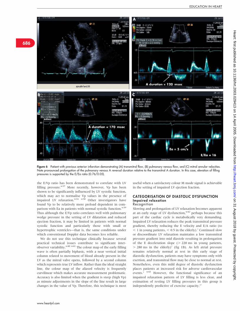
Figure 6 Patient with previous anterior infarction demonstrating (A) transmitral flow, (B) pulmonary venous flow, and (C) mitral annular velocities.Note pronounced prolongation of the pulmonary venous A reversal duration relative to the transmitral A duration. In this case, elevation of fillingpressures is supported by the E/Ea ratio (0.76/0.05).
686
EDUCATION IN HEART
www.heartjnl.com
on 31 August 2018 by guest. P
rotected by copyright.http://heart.bm
j.com/
Heart: first published as 10.1136/hrt.2003.029413 on 14 A
pril 2005. Dow
nloaded from
Effects of age and heart rateAs there is an age dependent decline in diastolic function in
normal subjects,w9 w27 w44 all diastolic indices should be
interpreted in conjunction with age. Sex differences may also
be significant—for example, elderly females tend to have
lower E/A ratios and longer E deceleration times compared
with males.w9 In some ways, the distinction of ‘‘age related
normal changes’’ from pathologic filling patterns seems
artificial. Studies of backscatter and strain characteristics
have shown abnormal tissue characterisation in association
with abnormal filling, suggesting the involvement of an age
related process such as fibrosis.w45
The patient’s heart rate may also influence the LV Doppler
filling pattern. In elderly subjects in the Framingham heart
study both the transmitral E velocity and E/A ratio were
inversely related to heart rate,w46 suggesting that in mild
disease, abnormal filling patterns follow the exhaustion of
‘‘filling reserve’’. In contrast, exercise induced tachycardia
resulted in proportional increases in both E and A (such that
the E/A ratio remained unchanged) as well as an increase in
the mitral annular Ea velocity in healthy younger subjects.w47
Importantly, Nagueh and colleagues have demonstrated that
the lateral E/Ea ratio remains a relatively accurate measure of
pulmonary wedge pressure in a mixed clinical population
during sinus tachycardia.w21
Other causes of delayed relaxationRegional LV dysfunction caused by ischaemia or abnormal
activation may be associated with dyssynchronous relaxa-
tion, which has a major impact on the dynamics of early
diastolic filling.18 Pacing induced asynchrony has been shown
to decrease LV filling indices in an animal model,w48 while
left bundle branch block is associated with prolongation of
isovolumic relaxation and reduced filling time in patients
with dilated cardiomyopathy.w49 Isolated left bundle branch
block with normal global systolic function is also associated
with alterations of LV filling parameters.w50 Therefore,
cardiac pacing and left bundle branch block may preclude
accurate assessment of delayed relaxation from the LV inflow
pattern, as this technique relies on the assumption of regional
uniformity. Asynchronous long axis LV relaxation is also
common in patients with coronary artery disease and may in
fact be the major cause of reduced E velocity and an
abnormal relaxation pattern in these patients.w51 Finally,
right ventricular overload and pulmonary hypertension
can significantly influence LV filling patterns, possibly result-
ing from abnormal septal motion and changes in LV
geometry.w52
Normal or pseudonormal fi l l ingRecognitionThe finding of apparently normal filling in the dyspnoeic
patient may suggest one of two problems—either the filling
pattern is pseudonormal, or the patient does not have heart
failure. A normal transmitral flow pattern is age and sex
dependent but may be generally characterised by an E/A ratio
of 0.75–1.5 and a deceleration time of 160–260 ms. As
discussed above, the entities of normal and pseudonormal
filling cannot be distinguished on the basis of transmitral
flow alone. The pseudonormal pattern occurs in advanced
cardiac disease (often with concomitant systolic dysfunction)
where progressive impairment of LV relaxation and com-
pliance leads to elevation of LV filling pressures. As preload
increases, the rise in left atrial pressure begins to increase
(and therefore ‘‘pseudonormalise’’) the E velocity while
elevation of LV end diastolic pressure tends to favour
earlier equilibration of the transmitral pressure gradient
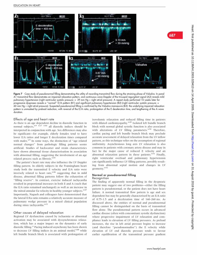
Figure 7 Case study of pseudonormal filling demonstrating the utility of recording transmitral flow during the straining phase of Valsalva. In panelA1 transmitral flow demonstrates an impaired relaxation pattern, and continuous wave Doppler of the tricuspid regurgitant signal (A2) reveals mildpulmonary hypertension (right ventricular systolic pressure = 39 mm Hg + right atrial pressure). A repeat study performed 10 weeks later forprogressive dyspnoea reveals a ‘‘normal’’ E/A pattern (B1) and significant pulmonary hypertension (B2) (right ventricular systolic pressure =60 mm Hg + right atrial pressure). Suspected pseudonormal filling is confirmed by the Valsalva manoeuvre (B3): the underlying impaired relaxationpattern is unmasked by preload reduction, with reversal of the E/A ratio, prolongation of the E deceleration time, and lengthening of the A waveduration.
687
EDUCATION IN HEART
www.heartjnl.com
on 31 August 2018 by guest. P
rotected by copyright.http://heart.bm
j.com/
Heart: first published as 10.1136/hrt.2003.029413 on 14 A
pril 2005. Dow
nloaded from
and therefore shortens the E deceleration time back into the
normal range. In only one setting may the transmitral flow
give the diagnosis—the typical pseudonormal filling pattern
may become modified during slow heart rates, such that the
underlying impaired LV relaxation may be discernible.w53
This may manifest as a distinct decrease in the gradient of the
E wave deceleration slope (fig 8A), or alternatively, as a
period of mid diastolic transmitral flow distinct from the E
and A waves, corresponding to the underlying prolonged or
dyssynchronous LV relaxation which is uncovered by the
bradycardia (fig 8B).
Differentiating normal from pseudonormal LV fil l ingWe take the following initial steps to distinguish normal from
pseudonormal filling:c Integrate the clinical information—An E/A ratio of 2 with
deceleration time of 160 ms is likely normal in a young
adult with a structurally normal heart referred for
palpitations, but is almost certainly pseudonormal in an
elderly hypertensive patient who was referred for inves-
tigation of dyspnoea.c Consider the status of the left ventricle on two dimensional echo—
Even if the E/A ratio and deceleration time are in the
normal range, LV filling is unlikely to be normal if there is
significant LV hypertrophy or LV dysfunction. In this
setting, preserved E velocity is better explained by elevated
left atrial pressure than by brisk relaxation and a rapid
decline in LV diastolic pressure. This effect of LV ‘‘suction’’
on transmitral flow is important to consider when
attempting to differentiate normal from pseudonormal
filling in older patients with small LV cavities. When such
individuals exhibit vigorous LV systolic function (for
example, LVEF . 70%), a high E velocity and E/A ratio
may reflect lower LV minimum diastolic pressure due to
brisk elastic recoil of the LV rather than elevated left atrial
pressure.w23 Such patients will also tend to have a high
tissue velocity (since this is directly proportional to LV
minimal pressure)w23 so that the E/Ea ratio (see below)
will remain low, indicating that filling is in fact normal
rather than pseudonormal.c Evaluate LA size—Left atrial size correlates with mean
pulmonary wedge pressure19 and is therefore a relatively
sensitive marker of chronic diastolic dysfunction.w54
Diastolic HF is not a plausible explanation for chronic
dyspnoea if the left atrial size is normal. The disadvantage
is, however, that specificity may be compromised by
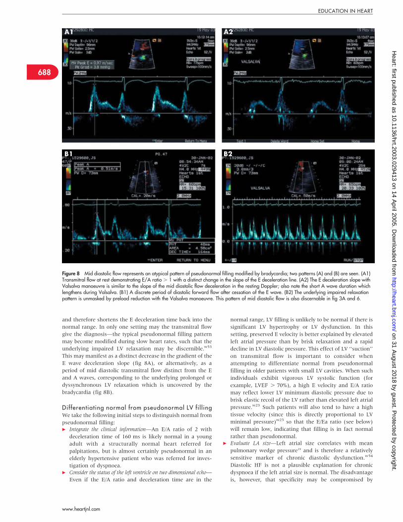
Figure 8 Mid diastolic flow represents an atypical pattern of pseudonormal filling modified by bradycardia; two patterns (A) and (B) are seen. (A1)Transmitral flow at rest demonstrating E/A ratio . 1 with a distinct change in the slope of the E deceleration line. (A2) The E deceleration slope withValsalva manoeuvre is similar to the slope of the mid diastolic flow deceleration in the resting Doppler; also note the short A wave duration whichlengthens during Valsalva. (B1) A discrete period of diastolic forward flow after cessation of the E wave. (B2) The underlying impaired relaxationpattern is unmasked by preload reduction with the Valsalva manoeuvre. This pattern of mid diastolic flow is also discernable in fig 3A and 6.
688
EDUCATION IN HEART
www.heartjnl.com
on 31 August 2018 by guest. P
rotected by copyright.http://heart.bm
j.com/
Heart: first published as 10.1136/hrt.2003.029413 on 14 A
pril 2005. Dow
nloaded from
conditions such as atrial fibrillation and mitral valve
disease which commonly cause left atrial dilatation.
The next steps in the discrimination of normal from
pseudonormal LV filling involve acquisition of tissue Doppler,
pulmonary venous flow, load altering manoeuvres, and
(sometimes) measurement of inflow propagation.
Integration of (conflicting) echo Doppler parametersThe integration of the clinical and two dimensional echo
information with a full complement of Doppler data usually
allows reliable discrimination of pseudonormal from normal
filling, as illustrated in fig 9. The most useful Doppler
parameters for this purpose are listed in table 2. However,
it should be emphasised that in contrast to their value
with overt systolic dysfunction, several parameters of dia-
stolic function have reduced accuracy when LV ejection
fraction is preserved.w55 In this setting the E deceleration
time correlates poorly with filling pressures, and the
Valsalva manoeuvre, pulmonary venous S/D ratio, and flow
propagation velocity are all relatively unreliable indicators of
diastolic dysfunction. The most robust parameters for esti-
mating filling pressure to aid interpretation of the transmitral
Doppler profile when LV ejection fraction is normal are the
E/Ea ratio and the difference in pulmonary venous and
transmitral A wave durations.w31 Even so, diastolic function
may remain inconclusive in occasional patients who present
very contradictory data that cannot be resolved.
Cardiac catheterisationInvasive measurement of diastolic function is often invoked
as the gold standard method for assessment of diastolic
function.6 However the cost, complexity, and expertise
required, as well as patient risk and lack of tolerability
associated with such procedures, mean that cardiac catheteri-
sation is rarely performed specifically to evaluate diastolic
function. In addition, unlike echocardiography, cardiac
catheterisation does not lend itself to serial assessment for
monitoring disease progression and response to treatment.
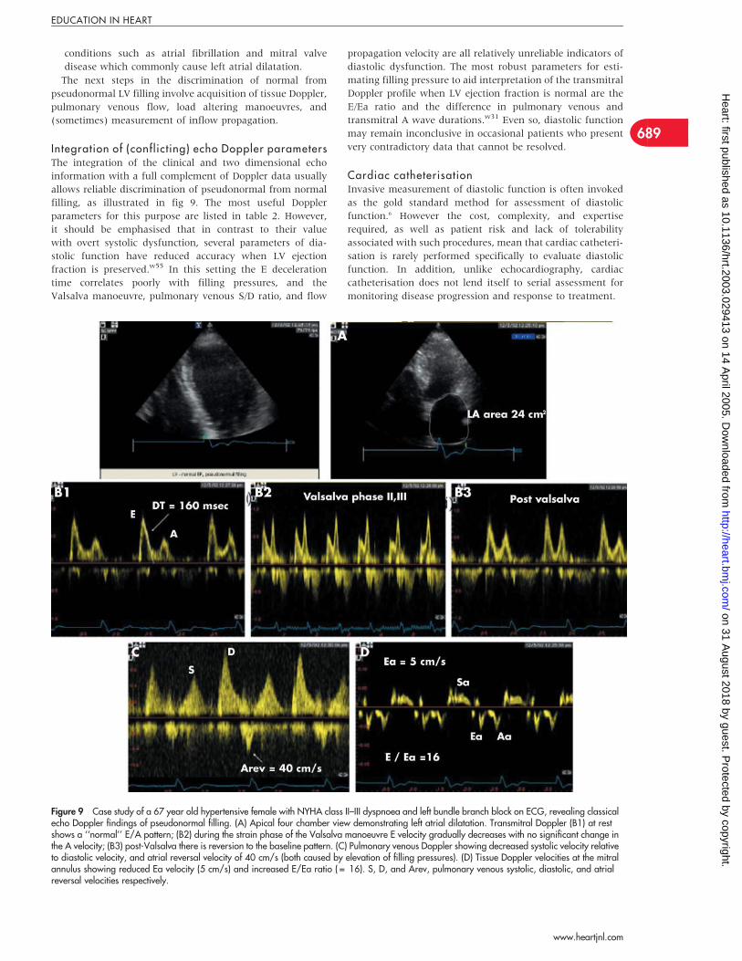
Figure 9 Case study of a 67 year old hypertensive female with NYHA class II–III dyspnoea and left bundle branch block on ECG, revealing classicalecho Doppler findings of pseudonormal filling. (A) Apical four chamber view demonstrating left atrial dilatation. Transmitral Doppler (B1) at restshows a ‘‘normal’’ E/A pattern; (B2) during the strain phase of the Valsalva manoeuvre E velocity gradually decreases with no significant change inthe A velocity; (B3) post-Valsalva there is reversion to the baseline pattern. (C) Pulmonary venous Doppler showing decreased systolic velocity relativeto diastolic velocity, and atrial reversal velocity of 40 cm/s (both caused by elevation of filling pressures). (D) Tissue Doppler velocities at the mitralannulus showing reduced Ea velocity (5 cm/s) and increased E/Ea ratio ( = 16). S, D, and Arev, pulmonary venous systolic, diastolic, and atrialreversal velocities respectively.
689
EDUCATION IN HEART
www.heartjnl.com
on 31 August 2018 by guest. P
rotected by copyright.http://heart.bm
j.com/
Heart: first published as 10.1136/hrt.2003.029413 on 14 A
pril 2005. Dow
nloaded from
Restrictive fil l ingRestrictive filling, characterised by a pronounced increase in
the E/A ratio (. 2) and shortening of the E deceleration time
(, 150 ms) is seen in the ‘‘sickest’’ ventricles and indicates
severely reduced LV compliance and notable elevation of left
atrial pressure. In the setting of preserved ejection fraction,
restrictive filling usually indicates severe infiltrative myocar-
dial disease such as cardiac amyloidosis rather than
hypertensive heart disease. In clinical practice, restrictive
filling is most commonly seen in association with LV
dilatation and severe systolic dysfunction and is strongly
predictive of mortality in this population, particularly if it is
not reversible with treatment.2
Systolic dysfunctionWhen systolic dysfunction is clearly present, the central
clinical question concerns the presence or absence of elevated
filling pressure. Impaired LV relaxation without preload
compensation indicates relatively normal filling pressures. In
this situation, no further evaluation of diastology is necessary
unless one is dealing with borderline abnormal values (for
example LVEF 40%, E/A 0.8, deceleration time 250 ms), in
which case one should proceed with more comprehensive
assessment. However, if the E/A ratio and deceleration time
appear relatively normal (or increased and shortened,
respectively) despite pronounced systolic dysfunction, pre-
served E velocity likely reflects elevated left atrial pressure.
Corroboration may be reliably obtained with TDI, as low
mitral annular amplitude in the presence of systolic
dysfunction results in reduced early diastolic lengthening
rate,w51 and therefore a high E/Ea ratio in the presence of
elevated left atrial pressure.
Effects of atrial fibril lationAs transmitral flow reflects the left atrial to LV pressure
gradient, the pattern of ventricular filling may be notably
influenced by both the compliance and contractile function of
the atrium. This issue attains practical clinical significance in
the setting of paroxysmal atrial fibrillation. The return of
sinus rhythm is often accompanied by atrial stunning, a loss
of mechanical function that is proportional to the duration of
fibrillation as well as anatomical characteristics of the
atrium,w56 and which may lead to inappropriate interpreta-
tion of diastolic function. Recent evidence indicates that Aa
correlates well with quantitative methods of left atrial
function.w57 Figure 10 illustrates abnormalities of diastolic
echo Doppler parameters that are typically observed in the
setting of impaired left atrial contractile function. In contrast,
brisk left atrial function is evident in fig 8, resulting in a high
transmitral A velocity, preserved systolic (S1) dominance,
and high atrial reversal velocity in pulmonary venous flow,
and a high Aa TDI velocity.
Evaluation of diastolic function is also difficult when atrial
fibrillation is sustained, although in patients with severe
systolic dysfunction the E deceleration time correlates well
with LV filling pressure.20 w58 In particular, a very short
deceleration time—for example, 120–140 ms—is strongly
predictive of elevated pulmonary wedge pressure.w58
Combining transmitral and pulmonary vein Doppler data
may also allow estimation of pulmonary wedge pressure in
patients with systolic HF.w59 When LV systolic function is
preserved in atrial fibrillation, estimation of filling pressure
from transmitral flow remains possible, but requires more
complicated Doppler indices.20 Of potentially more practical
use, the TDI appears to retain diagnostic value in patients
with atrial fibrillation. Sohn and colleagues demonstrated
that Ea correlated with prolongation of tau and E/Ea
predicted elevation of LV filling pressure, both at cut offs
which are similar to those reported for patients in sinus
rhythm.w22 Other investigators have confirmed a strong
relation between Ea and tau and between E/Ea and LV end
diastolic pressure in atrial fibrillation.
NEW DEVELOPMENTS IN DIASTOLIC FUNCTIONASSESSMENTThe use of resting data alone is an important limitation of the
Doppler approach, especially early in the disease, when the
heart is compensated at rest and symptoms occur only with
activity. The evolving techniques that may help with this
diagnosis include assessment of LV filling during exercise, B
type natriuretic peptide (BNP), and tissue characterisation.
BNP for diagnosis of diastolic dysfunctionGiven the complexity of the echocardiographic evaluation of
LV diastolic function, non-invasive diagnosis would be
greatly aided by a simpler approach. In particular, a blood
test with acceptable accuracy would be a very valuable
addition to the diagnostic armamentarium, and recent
attention has turned to BNP for this purpose.
BNP is a peptide secreted from the ventricular myocardium
in response to dilatation and increased intra-cavity pres-
sure.w60 w61 Elevation of BNP has been associated with a
range of cardiac abnormalities which can result in increased
filling pressures.w62 In particular, elevation of BNP has been
demonstrated in the setting of acute HF, and correlates with
the degree of LV systolic dysfunction in this setting.w63 w64 A
BNP of 100 pg/ml has been demonstrated to be accurate for
the emergency room diagnosis of acute systolic heart
failure.w64 In addition, there is increasing evidence that the
N-terminal fragment of the pro-BNP molecule that is
released during secretion of BNP from cardiac myocytes (so
called N-terminal BNP) has similar diagnostic utility.
This relation of BNP to ventricular filling pressure in
systolic HF implies that BNP might also have diagnostic
potential in patients with diastolic HF, in whom symptoms
are also related to elevated LV filling pressures. It also
suggests that BNP might be more relevant for the diagnosis
of clinical diastolic HF, which is usually associated with
pseudonormal or restrictive filling patterns,w65 rather than
for the detection of exertional dyspnoea attributed to
diastolic dysfunction, which is often characterised by an
impaired relaxation pattern and may not be associated with
high filling pressures at rest.w34 Thus BNP is usually elevated
in patients presenting to an emergency room with shortness
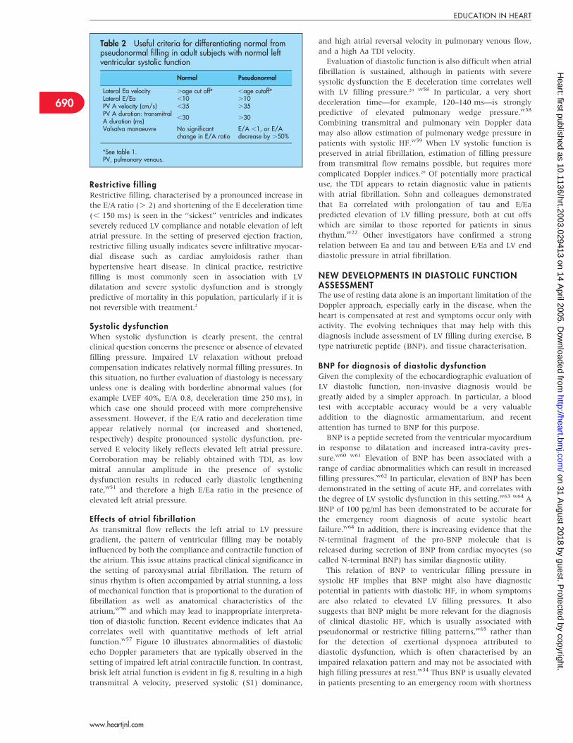
Table 2 Useful criteria for differentiating normal frompseudonormal filling in adult subjects with normal leftventricular systolic function
Normal Pseudonormal
Lateral Ea velocity .age cut off* ,age cutoff*Lateral E/Ea ,10 .10PV A velocity (cm/s) ,35 .35PV A duration: transmitralA duration (ms)
,30 .30
Valsalva manoeuvre No significantchange in E/A ratio
E/A ,1, or E/Adecrease by .50%
*See table 1.PV, pulmonary venous.
690
EDUCATION IN HEART
www.heartjnl.com
on 31 August 2018 by guest. P
rotected by copyright.http://heart.bm
j.com/
Heart: first published as 10.1136/hrt.2003.029413 on 14 A
pril 2005. Dow
nloaded from
of breath due to HF regardless of whether the ejection
fraction is preserved or impaired and a normal BNP value has
high negative predictive value in this setting.w66 Further,
high BNP concentrations have been reported in HF patients
with normal LVEF,w67 and in those with isolated LV diastolic
dysfunction.w65 Even so, BNP is reportedly lower in HF
patients with preserved ejection fraction compared to those
who have systolic dysfunction.w66 This is relevant for the
diagnosis of diastolic HF because population studies have
demonstrated notable increases in BNP with age and female
sex,w68 leading to significant overlap in BNP concentrations
between dyspnoeic elderly patients with HF and preserved
ejection fraction and similar patients without HF.w66
Therefore, a specific BNP cut off may not accurately
discriminate diastolic HF from non-HF presentations in the
elderly, particularly elderly women (in whom the condition is
most prevalent).w69 In addition, since BNP has a short half
life (approximately 20 minutes), the timing of sampling in
relation to the patient’s symptoms may have a profound
influence on the utility of BNP for the detection of diastolic
HF. In particular, clinically stable or treated patients who are
limited by exertional dyspnoea caused by mild diastolic
dysfunction often have relatively normal resting LV filling
pressures and may therefore have normal BNP concentra-
tions at rest.w70 Therefore, given that reported mean BNP
concentrations in diastolic HF have varied from 56 pg/ml in a
community settingw67 to 413 pg/ml in acute hospital pre-
sentations,w66 it is difficult to apply a simple cut off.
However, as a general guide, in symptomatic patients with
preserved systolic function, diastolic HF may be consi-
dered unlikely if BNP is , 50 pg/ml, and likely if BNP is
. 100 pg/ml.
Whether BNP has a wider role in the diagnosis of
hypertensive heart disease remains unclear. While BNP has
been reported to be increased in patients with hyperten-
sionw71 and LV hypertrophy,w72–74 its ability to detect
increased LV mass in a community setting was subopti-
mal.w75 Similarly, a recent study found that BNP was
suboptimal for the identification of diastolic (or indeed
systolic) dysfunction in more than 2000 subjects randomly
selected from the population.21
Responses of LV fil l ing and BNP to stressSince chronic stable diastolic HF is characterised by abnormal
increases in diastolic pressures during exertion, exercise
criteria would seem to be an important part of the diagnostic
criteria. Indeed, if a patient demonstrates a high functional
capacity during exercise testing then a diagnosis of chronic
HF of any cause can be reliably excluded. An ideal test for the
diagnosis of diastolic HF would allow objective demonstra-
tion of reduced exercise capacity caused by limiting dyspnoea
while simultaneously confirming elevation of left atrial
pressure. While cardiac catheterisation during exercise is
not practical, non-invasive estimation of filling pressures
using the E/Ea ratio at rest and immediately after maxi-
mal exercise is a potentially useful approach that has
proved feasiblew47 w76 and warrants further investigation.
Empirically, exercise limitation that is associated with
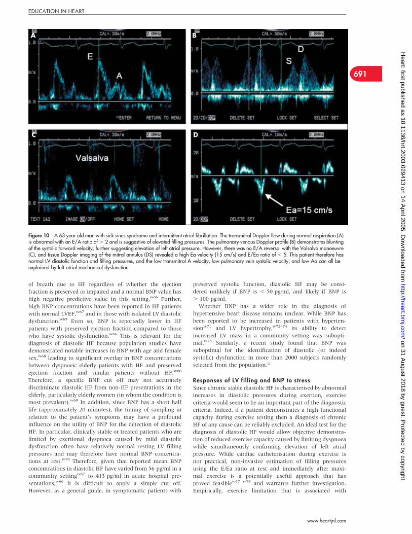
Figure 10 A 63 year old man with sick sinus syndrome and intermittent atrial fibrillation. The transmitral Doppler flow during normal respiration (A)is abnormal with an E/A ratio of . 2 and is suggestive of elevated filling pressures. The pulmonary venous Doppler profile (B) demonstrates bluntingof the systolic forward velocity, further suggesting elevation of left atrial pressure. However, there was no E/A reversal with the Valsalva manoeuvre(C), and tissue Doppler imaging of the mitral annulus (DS) revealed a high Ea velocity (15 cm/s) and E/Ea ratio of , 5. This patient therefore hasnormal LV diastolic function and filling pressures, and the low transmitral A velocity, low pulmonary vein systolic velocity, and low Aa can all beexplained by left atrial mechanical dysfunction.
691
EDUCATION IN HEART
www.heartjnl.com
on 31 August 2018 by guest. P
rotected by copyright.http://heart.bm
j.com/
Heart: first published as 10.1136/hrt.2003.029413 on 14 A
pril 2005. Dow
nloaded from
conversion of an ‘‘impaired relaxation’’ pattern at rest to a
‘‘pseudonormal’’ pattern immediately post-exercise is indi-
cative of elevation of left atrial pressure with exercise, and
suggests that the slow LV relaxation is functionally important
in a particular patient (fig 11). Similarly, augmentation of
BNP with exercise might also have diagnostic potential in
this situation. As a surrogate for LV filling pressures,
increases in BNP during exercise may suggest elevation of
LV filling pressures in a patient with exertional dyspnoea and
suspected diastolic HF.w76
Several other aspects of exercise echocardiography may
provide clinically useful information. A hypertensive blood
pressure response to stress is a preclinical marker of
abnormal ventricular structure and function,w77 and con-
tributes to exercise intolerance by inducing transient diastolic
dysfunction.w78 w79 The presence of exercise induced regional
hypokinesis implicates coronary artery disease as a cause of
the patient’s symptoms, while increased pulmonary artery
systolic pressure at peak exercise may be related to LV
diastolic dysfunction.
Tissue characterisationIt is clear from the previous discussion that currently utilised
techniques for the detection of hypertensive heart disease are
relatively insensitive, demonstrating abnormalities only
when extensive changes have occurred and when patients
are already likely to be at considerably increased risk of
adverse cardiovascular events. More sensitive echocardio-
graphic techniques for characterising myocardial structure
and function include strain imaging and assessment of
integrated backscatter. Echocardiographic measurement of
the extent (strain) and rate (strain rate) of segmental
myocardial deformation utilises colour tissue Doppler to
determine gradients between adjacent myocardial veloci-
ties.w80 While the clinical application of this recently
developed technique is currently limited by a suboptimal
signal to noise ratio and marked angle dependency, it has
recently been applied in the research setting to identify
reduced LV systolic function in patients with hypertension
and isolated diastolic dysfunction.w81 In addition, its high
sensitivity has potential to aid detection of preclinical
myocardial dysfunction.w82 w83 Segmental myocardial prop-
erties may also be assessed by measurement of myocardial
reflectivity with integrated backscatter. The magnitude of
myocardial backscatter normalised to either the blood pool or
pericardium, and the cyclic variation in backscatter with
segmental contraction are related to both myocardial
structure (degree of fibrosis)w84 and function.w85 Changes
in these parameters precede functional changes in hyperten-
sive LV hypertrophyw86 and also correlate with diastolic
changes in the aging heart.w45 These sensitive techniques
therefore provide an alternative means of identifying early
end organ damage in hypertensive patients, and are currently
being used to investigate diastolic function in various disease
states.
In addition to providing specific information about the
diastolic function of the ventricle, early diastolic mitral
annular velocities have a potentially broader role in the
detection of cardiac abnormalities. Conditions such as
hypertension and diabetes contribute to global LV diastolic
dysfunction, usually without significant regional hetero-
geneity. Thus mitral annular velocities, which represent the
‘‘sum’’ of long axis LV velocities, accurately reflect global LV
function in these conditions. The resting Ea correlates with
exercise capacity in hypertensive patients,w87 and has been
demonstrated to be sensitive (more so than Sa) for the
detection of myocardial dysfunction in various cardiac
disease processes including hypertension,w88 diabetes,w89
myocardial infiltration,w90 and hypertrophic cardiomyo-
pathy.w91 Moreover, Shan and colleagues have shown early
diastolic myocardial velocity to be inversely related to
interstitial fibrosis.w92 Finally, the TDI derived Ea velocity is
independently related to cardiac mortality and provides
predictive power which is incremental to data obtained from
clinical and conventional Doppler assessment.22 The applica-
tion of this approach is currently limited by deficiencies in
understanding of the various factors which influence Ea,w18
and a lack of normal ranges with robust cut offs for
identification of abnormal myocardial function.
Global versus segmental diastolic functionAll of the techniques described above provide assessment of
global LV diastolic function. However, it is clear from models
of systolic dysfunction that it is possible to have normal
global function (evidenced by preserved ejection fraction) in
the presence of segmental hypokinesis, provided that enough
normally contracting segments remain. This concept may
also be applied to diastolic function, although at present it is
unclear what extent (or severity) of segmental diastolic
dysfunction is required before parameters of global diastolic
function become abnormal. This regional non-uniformity has
implications for the reporting of echo Doppler studies of LV
diastolic function. If global systolic function (ejection
fraction) is impaired then it can be assumed that global
diastolic function is also abnormal and, for example, a
‘‘normal’’ E/A ratio would likely represent pseudonormal
filling. Alternatively, when regional hypokinesis is present in
the setting of preserved ejection fraction—for example,
localised inferior wall ischaemia—one can assume that the
hypokinetic inferior segments will have abnormal relaxation
and compliance; however, it is possible for global diastolic
function to be either normal or abnormal in this setting,
depending on the extent/severity of segmental diastolic
dysfunction and the diastolic function of the non-infarcted
segments. In addition, as discussed above, the extent of
regional asynchrony in the timing of myocardial relaxation
may significantly affect measures of global performance.
Further work utilising regional information from strain
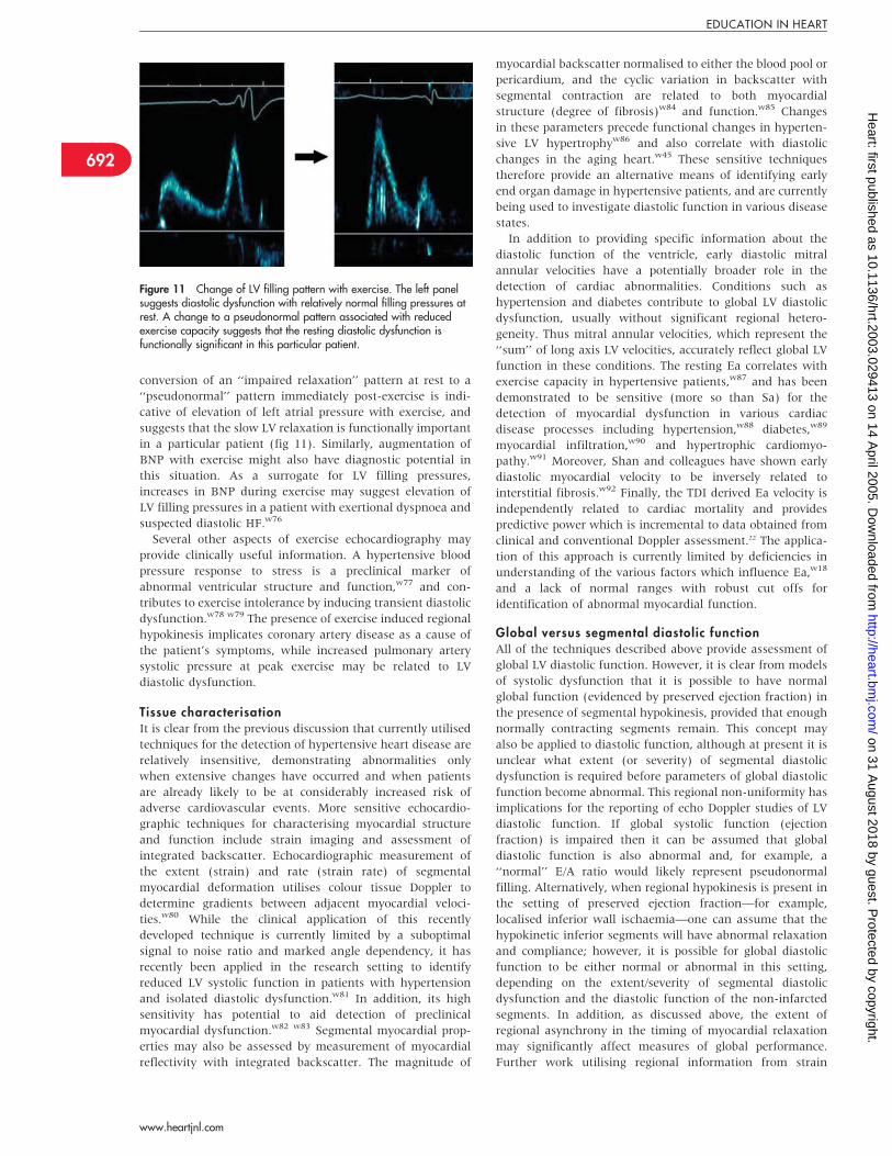
Figure 11 Change of LV filling pattern with exercise. The left panelsuggests diastolic dysfunction with relatively normal filling pressures atrest. A change to a pseudonormal pattern associated with reducedexercise capacity suggests that the resting diastolic dysfunction isfunctionally significant in this particular patient.
692
EDUCATION IN HEART
www.heartjnl.com
on 31 August 2018 by guest. P
rotected by copyright.http://heart.bm
j.com/
Heart: first published as 10.1136/hrt.2003.029413 on 14 A
pril 2005. Dow
nloaded from

imaging with both echocardiographyw93 and magnetic
resonance taggingw94 will likely provide important insight
into these questions.
Alternative imaging techniquesIn symptomatic patients with limited echocardiographic
windows or poor quality data (for example, inadequate
pulmonary vein Doppler), alternative imaging techniques
may provide useful information. Historically, radionuclide
techniques have been used but these are limited by low frame
rates, cycle length variability, and background lung blood
pool attenuation. Magnetic resonance imaging has recently
been demonstrated to be sensitive for the detection of global
diastolic dysfunction in early diabetic heart disease.w95
However, while spatial resolution is excellent, diastolic
function assessment with magnetic resonance may be limited
by lower temporal resolution and further experience is
required.
CONCLUSIONSA comprehensive assessment of diastolic function and filling
pressures should ideally include integration of all available
two dimensional and Doppler data with relevant clinical
information such as age, exercise capacity, and the presence
of hypertension, coronary disease, or diabetes. As multiple
parameters are used to assess diastolic function, each with
imperfect sensitivity and specificity, discordant results in a
given patient are relatively common. A busy echo lab should
therefore favour acquisition of parameters which offer both
accuracy and low inter-observer variability. At the very least,
this requires assessment of transmitral Doppler flow (which
remains central to categorisation of diastolic function) in
combination with newer, less load dependent Doppler echo
techniques. Of available methods, pulsed wave TDI is the
easiest to use and provides robust, relatively unambiguous,
well validated data that are more reliable than the use of
pulmonary vein Doppler, flow propagation velocity or load
Figure 12 Suggested schema for echo Doppler categorisation of diastolic function in patients with normal LV systolic function.
Table 3 Practical classification of diastolic function
Diastology when ejection fraction is (near) normal (.45–50%)1. Normal diastolic function.2. Mild diastolic dysfunction with normal resting filling pressures(impaired relaxation)3. Moderate diastolic dysfunction with elevated filling pressures(pseudonormal filling)4. Severe diastolic dysfunction with notably elevated filling pressures(restrictive filling—reversible or non-reversible)5. Indeterminate diastolic function (paced rhythm, atrial fibrillation, mitralvalve disease)Diastology when ejection fraction is reduced (,45%)1. Diastolic dysfunction with normal filling pressures (impaired relaxationpattern)2. Diastolic dysfunction with elevated filling pressures (pseudonormalfilling pattern)3. Diastolic dysfunction with severely elevated filling pressures (restrictivefilling—reversible or non-reversible)4. Diastolic dysfunction with indeterminate filling pressures
693
EDUCATION IN HEART
www.heartjnl.com
on 31 August 2018 by guest. P
rotected by copyright.http://heart.bm
j.com/
Heart: first published as 10.1136/hrt.2003.029413 on 14 A
pril 2005. Dow
nloaded from
altering techniques, particularly when systolic function is
preserved.15 In difficult cases, a comprehensive echo Doppler
examination should be interpreted by an experienced
echocardiographer and consideration should be given to
further evaluation with BNP and exercise testing (fig 12).
Thus, although echocardiographic evaluation of diastolic
function in HF is complex, a simplified approach can be
widely applied to provide important clinical and prognostic
information for patients with and without overt LV systolic
dysfunction.
The clinical role of echocardiography for evaluating
diastolic function is quite different depending on whether
global LV systolic function (ejection fraction) is preserved or
impaired. When the ejection fraction is greater than 45–50%,
it is generally agreed that symptoms of HF should not be
attributed to systolic dysfunction. In this setting, confirma-
tion of diastolic dysfunction does not define diastolic HF,
which still requires clinical correlation. Nonetheless, evidence
of raised LA pressure supports a contribution from diastolic
HF, and this has important prognostic and therapeutic
implications. When global systolic dysfunction is present,
accompanying diastolic dysfunction is assumed, and the
principal aim of Doppler echocardiography is to estimate
filling pressure, which again has direct implications for
treatment and prognosis. A clinically useful classification for
the reporting of diastolic function should therefore reflect
these aims, and an example is given in table 3.
Deficiencies in understanding of the pathophysiology,
epidemiology, and treatment of HF with preserved ejection
fraction have been largely related to inability to accurately
identify diastolic dysfunction with non-invasive techniques.
Research over the past decade has seen a steady evolution of
our understanding of diastolic function in a wide range of
clinical scenarios. Parallel advances in ultrasound technology
and its application in the non-invasive evaluation of cardiac
function have contributed to significant advances in the
diagnosis of diastolic HF. The development of specific criteria
for the diagnosis of diastolic HF should incorporate modern
non-invasive techniques for assessment of diastolic function
within a clear framework of clinical evaluation, as suggested
in fig 13. Such an approach should form the cornerstone for
entry into urgently needed trials of therapeutic intervention
for this important condition.
Additional references appear on the Heart website—http://
www.heartjnl.com/supplemental
ACKNOWLEDGEMENTSWe wish to thank Dr Roess Pascoe for assistance in obtaining some ofthe echo images.
Authors’ affiliations. . . . . . . . . . . . . . . . . .
P M Mottram, T H Marwick, University of Queensland, Brisbane,Australia
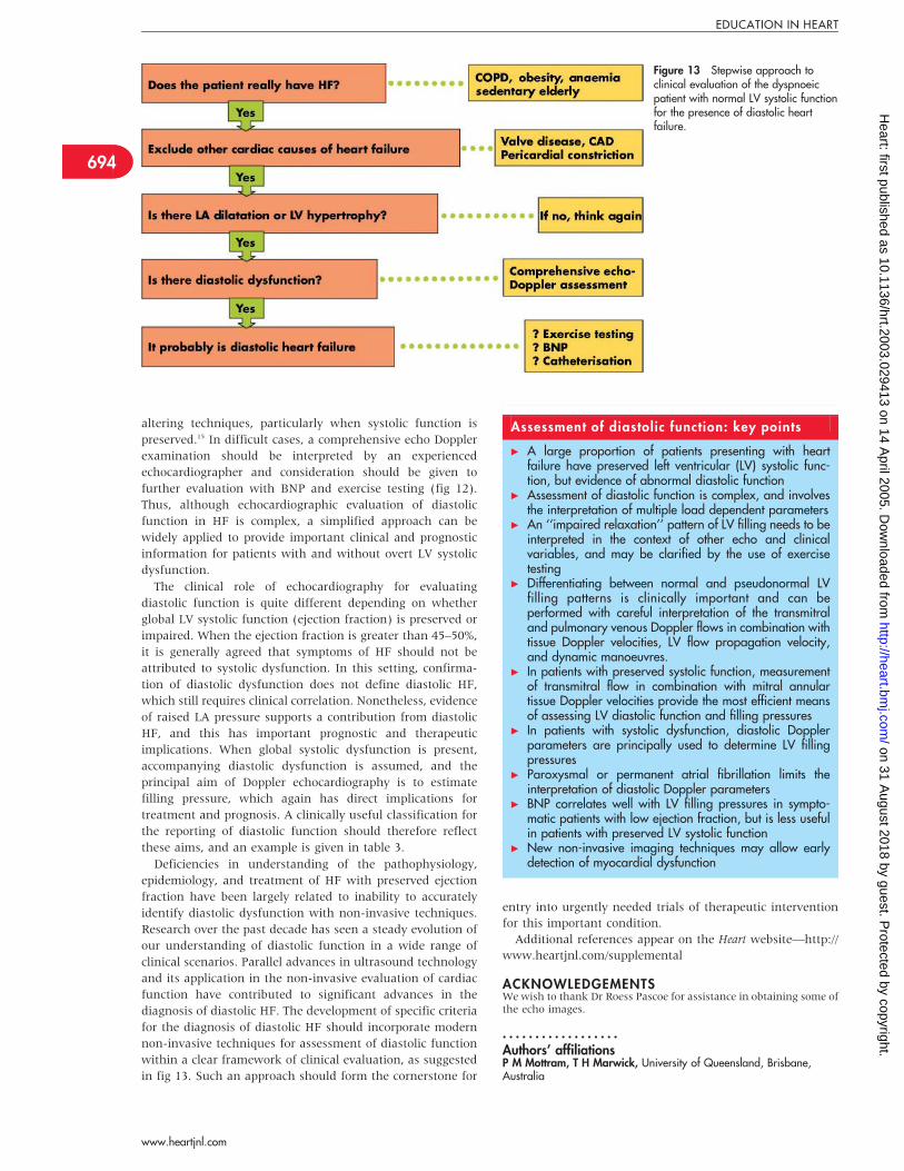
Figure 13 Stepwise approach toclinical evaluation of the dyspnoeicpatient with normal LV systolic functionfor the presence of diastolic heartfailure.
Assessment of diastolic function: key points
c A large proportion of patients presenting with heartfailure have preserved left ventricular (LV) systolic func-tion, but evidence of abnormal diastolic function
c Assessment of diastolic function is complex, and involvesthe interpretation of multiple load dependent parameters
c An ‘‘impaired relaxation’’ pattern of LV filling needs to beinterpreted in the context of other echo and clinicalvariables, and may be clarified by the use of exercisetesting
c Differentiating between normal and pseudonormal LVfilling patterns is clinically important and can beperformed with careful interpretation of the transmitraland pulmonary venous Doppler flows in combination withtissue Doppler velocities, LV flow propagation velocity,and dynamic manoeuvres.
c In patients with preserved systolic function, measurementof transmitral flow in combination with mitral annulartissue Doppler velocities provide the most efficient meansof assessing LV diastolic function and filling pressures
c In patients with systolic dysfunction, diastolic Dopplerparameters are principally used to determine LV fillingpressures
c Paroxysmal or permanent atrial fibrillation limits theinterpretation of diastolic Doppler parameters
c BNP correlates well with LV filling pressures in sympto-matic patients with low ejection fraction, but is less usefulin patients with preserved LV systolic function
c New non-invasive imaging techniques may allow earlydetection of myocardial dysfunction
694
EDUCATION IN HEART
www.heartjnl.com
on 31 August 2018 by guest. P
rotected by copyright.http://heart.bm
j.com/
Heart: first published as 10.1136/hrt.2003.029413 on 14 A
pril 2005. Dow
nloaded from
REFERENCES1 Giannuzzi P, Imparato A, Temporelli PL, et al. Doppler-derived mitral
deceleration time of early filling as a strong predictor of pulmonary capillarywedge pressure in postinfarction patients with left ventricular systolicdysfunction. J Am Coll Cardiol 1994;23:1630–7.
2 Pinamonti B, Zecchin M, Di Lenarda A, et al. Persistence of restrictive leftventricular filling pattern in dilated cardiomyopathy: an ominous prognosticsign. J Am Coll Cardiol 1997;29:604–12.
3 Redfield MM, Jacobsen SJ, Burnett JC Jr, et al. Burden of systolic and diastolicventricular dysfunction in the community: appreciating the scope of the heartfailure epidemic. JAMA 2003;289:194–202.
c Population study utilising comprehensive echo Doppler techniques toassess the prevalence and prognostic significance of abnormal LVfilling patterns.
4 Zile MR, Gaasch WH, Carroll JD, et al. Heart failure with a normal ejectionfraction: is measurement of diastolic function necessary to make the diagnosisof diastolic heart failure? Circulation 2001;104:779–82.
5 Maurer MS, Spevack D, Burkhoff D, et al. Diastolic dysfunction. Can it bediagnosed by Doppler echocardiography? J Am Coll Cardiol2004;44:1543–9.
c Important discussion of the limitations of Doppler echocardiography forthe assessment of diastolic function.
6 Vasan RS, Levy D. Defining diastolic heart failure: a call for standardizeddiagnostic criteria. Circulation 2000;101:2118–21.
7 European Study Group on Diastolic Heart Failure. How to diagnose diastolicheart failure. Eur Heart J 1998;19:990–1003.
8 Appleton CP, Firstenberg MS, Garcia MJ, et al. The echo-Doppler evaluationof left ventricular diastolic function. A current perspective. Cardiol Clin2000;18:513–46.
9 Nishimura RA, Tajik AJ. Evaluation of diastolic filling of left ventricle in healthand disease: Doppler echocardiography is the clinician’s Rosetta Stone. J AmColl Cardiol 1997;30:8–18.
10 Appleton CP, Hatle LK, Popp RL. Relation of transmitral flow velocity patternsto left ventricular diastolic function: new insights from a combinedhemodynamic and Doppler echocardiographic study. J Am Coll Cardiol1988;12:426–40.
c Pioneering study interpreting LV diastolic parameters in the context offilling pressures.
11 Garcia MJ, Thomas JD, Klein AL. New Doppler echocardiographicapplications for the study of diastolic function. J Am Coll Cardiol1998;32:865–75.
c Detailed discussion of the technical and theoretical aspects of tissueDoppler echocardiography and flow propagation velocity.
12 Nagueh SF, Middleton KJ, Kopelen HA, et al. Doppler tissue imaging: anoninvasive technique for evaluation of left ventricular relaxation andestimation of filling pressures. J Am Coll Cardiol 1997;30:1527–33.
13 Nagueh SF, Sun H, Kopelen HA, et al. Hemodynamic determinants of themitral annulus diastolic velocities by tissue Doppler. J Am Coll Cardiol2001;37:278–85.
14 Sohn DW, Chai IH, Lee DJ, et al. Assessment of mitral annulus velocity byDoppler tissue imaging in the evaluation of left ventricular diastolic function.J Am Coll Cardiol 1997;30:474–80.
15 Ommen SR, Nishimura RA, Appleton CP, et al. Clinical utility of Dopplerechocardiography and tissue Doppler imaging in the estimation of leftventricular filling pressures: a comparative simultaneous Doppler-catheterization study. Circulation 2000;102:1788–94.
c Study examining the relative accuracy of various diastolic parametersfor estimation of LV filling pressures.
16 Firstenberg MS, Greenberg NL, Main ML, et al. Determinants of diastolicmyocardial tissue Doppler velocities: influences of relaxation and preload.J Appl Physiol 2001;90:299–307.
17 Skaluba SJ, Litwin SE. Mechanisms of exercise intolerance: insights from tissueDoppler imaging. Circulation 2004;109:972–7.
18 Gibson DG, Francis DP. Clinical assessment of left ventricular diastolicfunction. Heart 2003;89:231–8.
c Insightful review of the pathophysiology of diastolic dysfunction andlimitations of methods used to assess it.
19 Appleton CP, Galloway JM, Gonzalez MS, et al. Estimation of left ventricularfilling pressures using two-dimensional and Doppler echocardiography inadult patients with cardiac disease. Additional value of analyzing left atrialsize, left atrial ejection fraction and the difference in duration of pulmonaryvenous and mitral flow velocity at atrial contraction. J Am Coll Cardiol1993;22:1972–82.
20 Nagueh SF, Kopelen HA, Quinones MA. Assessment of left ventricular fillingpressures by Doppler in the presence of atrial fibrillation. Circulation1996;94:2138–45.
21 Redfield MM, Rodeheffer RJ, Jacobsen SJ, et al. Plasma brain natriureticpeptide to detect preclinical ventricular systolic or diastolic dysfunction: acommunity-based study. Circulation 2004;109:3176–81.
22 Wang M, Yip GW, Wang AY, et al. Peak early diastolic mitral annulusvelocity by tissue Doppler imaging adds independent and incrementalprognostic value. J Am Coll Cardiol 2003;41:820–6.
Additional references appear on the Heart website—http://www.heartjnl.com/supplemental
MULTIPLE CHOICE QUESTIONS
Education in Heart Interactive (www.heartjnl.com/misc/education.shtml)
There are six multiple choice questions associated with each Education in Heart article (thesequestions have been written by the authors of the articles). Each article is submitted to EBAC(European Board for Accreditation in Cardiology; www.ebac-cme.org) for 1 hour of externalCPD credit.
How to find the MCQs: Click on the Online Learning: [Take interactive course] link on thetable of contents for the issue online or on the Education in Heart collection(www.heartjnl.com/cgi/collection/heart_education).
Free access: This link will take you to the BMJ Publishing Group’s online learning website.Your Heart Online user name and password will be recognised by this website. As a Heartsubscriber you have free access to these MCQs but you must register on the site so you cantrack your learning activity and receive credit for completed courses.
How to get access: If you have not yet activated your Heart Online access, please do so byvisiting http://www.bmjjournals.com/cgi/activate/basic and entering your six digit (allnumeric) customer number (found above your address label with your print copy). If you haveany trouble activating or using the site please contact [email protected]
Case based Heart: You might also be interested in the interactive cases published inassociation with Heart (http://cpd.bmjjournals.com/cgi/hierarchy/cpd_node;CBH)
695
EDUCATION IN HEART
www.heartjnl.com
on 31 August 2018 by guest. P
rotected by copyright.http://heart.bm
j.com/
Heart: first published as 10.1136/hrt.2003.029413 on 14 A
pril 2005. Dow
nloaded from


![Review Article Similarities and Differences between the … · 2019. 7. 31. · to diastolic heart failure [ , ]. Compared with systolic heart failure, diastolic heart failure is](https://static.fdocuments.net/doc/165x107/610a99d464112c7f8e47019f/review-article-similarities-and-differences-between-the-2019-7-31-to-diastolic.jpg)


