Translational Informatics: Enabling Knowledge-Driven Healthcare

2 3
Healthcare Imaging InformaticsProduct Portfolio
www.vitalimages.com
© 2016 Vital Images, Inc. | M-06320C
Marks not owned by Vital Images, Inc. are the property of their respective holders.
Vital Images, Inc. | 5850 Opus Parkway, Suite 300 | Minnetonka, MN 55343 | USA | +1 866.433.4624 Vital Images Europe | Zilverstraat 1 | 2718 RP Zoetermeer | Netherlands | +31 79 206 5800
Vital Images, Inc., a Toshiba Medical Company, is a leading provider of healthcare imaging informatics solutions, including advanced visualization, enterprise image viewing solutions and business intelligence technology designed to help healthcare organizations deliver exceptional care while optimizing resources across multi-facility organizations. The company’s solutions are scalable to
meet the unique needs of hospitals and imaging centers and are accessible throughout the enterprise anytime and anywhere.

For more than 25 years, Vital has been a pioneer in imaging solutions. Our suite of software and tools help healthcare organizations deliver exceptional care while optimizing resources across multi-facility organizations. The company’s solutions are scalable to meet the unique needs of hospitals and imaging centers and are accessible throughout
the enterprise anytime and anywhere.

4 5
6 Vitrea®
9 Enterprise Informatics Vitrea Modular Enterprise Imaging
21 Enterprise Analytics Vitrea Image Resource Planning
27 Enterprise Visualization27 Vitrea Enterprise Viewer31 Vitrea Advanced Visualization
35 Vitrea Advanced Visualization 35 Clinical Applications49 Integrated Applications55 Clinical Workflows
59 Support
61 Vital U® Education
Contents
76
Enterprise Informatics Vitrea® Enterprise Informatics solutions offer tailored approaches to data management, with Vitrea Vendor-Neutral Archive (VNA) providing image consolidation and Vitrea Data Stream delivering federated access to images.
Enterprise AnalyticsHealthcare providers frequently struggle for access to meaningful data about their practice’s resources. Vital’s imaging-centric management support tools help you continuously improve efficiency and quality across the practice.
Enterprise Visualization One viewer doesn’t meet all image display needs. While universal viewers give a generic overview, the Vitrea family of viewer options provides a wide array of clinical tools at your disposal, serving every clinical space from the ED, to surgery, radiology and beyond.
Vitrea®
98
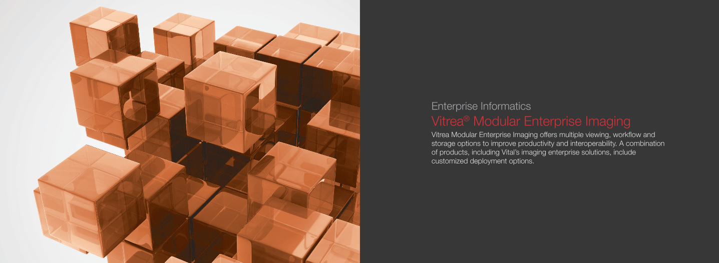
Enterprise Informatics Vitrea® Modular Enterprise Imaging Vitrea Modular Enterprise Imaging offers multiple viewing, workflow and storage options to improve productivity and interoperability. A combination of products, including Vital’s imaging enterprise solutions, include customized deployment options.

10 11
Vitrea® Application-Neutral ArchitectureNon-disruptive, adaptable, interoperability platform
This next-generation approach focuses on customer-centered vendor interoperability, rather than creating additional silos of large data prevalent in the “big bang” VNA realm. The solution was specifically designed to address the semantic interoperability chasm between structured and unstructured data sets, promoting federated, real-time access to information.
n Offers a customer-centered approachn Facilitates advanced interoperability between existing healthcare
informatics applicationsn Communicates with numerous subsystems:
ECM, VNA, PACS, EMR, HIE and other health information systemsn Promotes a federated “real-time access to information” modeln Avoids “big bang” centralization of a typical VNA project
Manage the UnmanagedConsolidate Archives
Vitrea®
Vendor-Neutral ArchiveVitrea®
Data Stream
Non-DICOM External MediaSharing
Connect Archives
Vitrea Application-Neutral Architecture®

12 13
Vitrea® Vendor-Neutral ArchiveNext-generation VNA built on open-source technology
Vitrea Vendor-Neutral Archive is a consolidating image management solution meeting challenges within healthcare systems today, such as easing migration of systems and enabling access to imaging data across modalities, locations and vendors.
n Multimedia storage in native format – DICOM and non-DICOMn Intelligent image management workflow – pre-fetch, IOCM, tag morph, auto routingn Horizontal scalability – quickly expand storage without downtime, avoid “crisis”
hardware purchasesn VNA on demand – avoid costly “big bang” data migrationn Standards-based design – DICOM, DICOMweb, XDS/XDSi, avoid future data migrationsn VNA “on demand” – allows for calculated conversion from existing PACS or VNA
reducing costly migration fees
CT UltrasoundMRDigital X-ray Nuclear Mammography
Vitrea Data Stream
PACSVitrea Enterprise Viewer Vitrea Advanced Visualization
DICOMweb
Vitrea Data Stream
Hospital 1 Hospital 2 Hospital 3 Hospital 4
Vitrea VNA®

14 15
Vitrea® Data StreamDICOMweb™-powered workflow engine that allows for extremely fast query and presentation of clinical images
Vitrea Data Stream provides clinicians access to images that reside in disparate DICOM archives. Using secure Web technologies, it delivers fast access to images without costly consolidation and migration of imaging data.
n Converts DICOM to DICOMwebn Can query multiple, disparate PACS systems simultaneously acrossn Departments/service linesn Different facilitiesn Different vendorsn Uses secure web protocol for maintenance of PHI factorsn Single EMR presentation point when linked to a DICOMweb-capable viewer
Hospital 1
Vitrea VNAStorage
Radiologist readingfrom anywhere.
Vitrea VNA
Hospital 2 Hospital 3 Hospital 4 Hospital 5 Hospital 6
Vitrea Data Stream®

16 17
PersonalizedViewing
Image Management Workflow
An enterprise solution based on anatomy, user preference, and viewing technology with powerful, standards based workflow tools for all types of DICOM and Non-DICOM clinical images.
Storage andBusiness Continuity
Personalized Viewing
Image Management Workflow
Storage and Business Continuity
EasyViz
DiagnosticRadiology
Advanced
AdvancedVisualization
Cardiology Viewer
Cardiology
Enterprise Viewer
Image-enabledEMR
Enterprise Viewer PACS Viewer
Non-DICOM
Data Stream
DICOMwebEnabled
VNA
DICOM Image Workflows
VNA
XDS/XDSi
VNA
eMPI / IOCM
VNA
No downtime�Scalability
VNA
VNA On DemandBackground Migration
Viewing PlatformMultiple viewing options based on the anatomy, disease, viewing device and needs of the end user
n Vitrea® Enterprise Viewer – image enable the EMRn EasyViz* Diagnostic Viewer – diagnostic-quality image presentation across
multiple monitorsn Vitrea Advanced Visualization – premier advanced visualization for all modalitiesn TomTec** – cardiology image displayn Your existing PACS viewer
Workflown Vitrea Data Stream – converting DICOM to DICOMwebTM for an optimized
image viewing experiencen Vitrea VNA – next-generation VNA providing workflow to DICOM and
non-DICOM images
Storagen Expand storage without downtimen Simply create a business continuity solutionn Multiple disaster recovery options
*EasyViz is manufactured by Karos Health. **TomTec is manufactured TomTec Imaging Systems.

18 19
EasyViz Diagnostic Radiology ViewerEasyViz* is a full-fidelity, diagnostic-quality viewer, available in a number of deployment options
Thin Client**n Up to two monitor configurations
and up to 3 MP resolutionn CINE frame rate up to 30 fpsn Supports session collaborationn Advanced hanging protocols,
including mammographyn Full range of diagnostic toolsn Drag and drop image arrangementn Priors managementn Customizable toolbarn Support for 3D, DBTn Simple install through browser
download
Zero Footprint**n Single-monitor configuration
and up to 3 MP resolutionn CINE frame rate up to 30 fpsn Supports session collaborationn Advanced hanging protocols,
including mammographyn Full range of diagnostic toolsn Drag and drop image arrangementn Priors managementn Customizable toolbarn Support for 3D, DBTn No PHI on devicen No downloads
Full client n Up to three monitor configurations
and up to 10MP resolutionn CINE frame rate of 30-90 fpsn Advanced hanging protocols,
including mammographyn Full range of diagnostic toolsn Drag and drop image arrangementn Priors managementn Customizable toolbarn Support for 3D, DBTn Simple install through browser
download
*EasyViz is manufactured by Karos Health. **Not for sale in the US .
2120
Enterprise Analytics
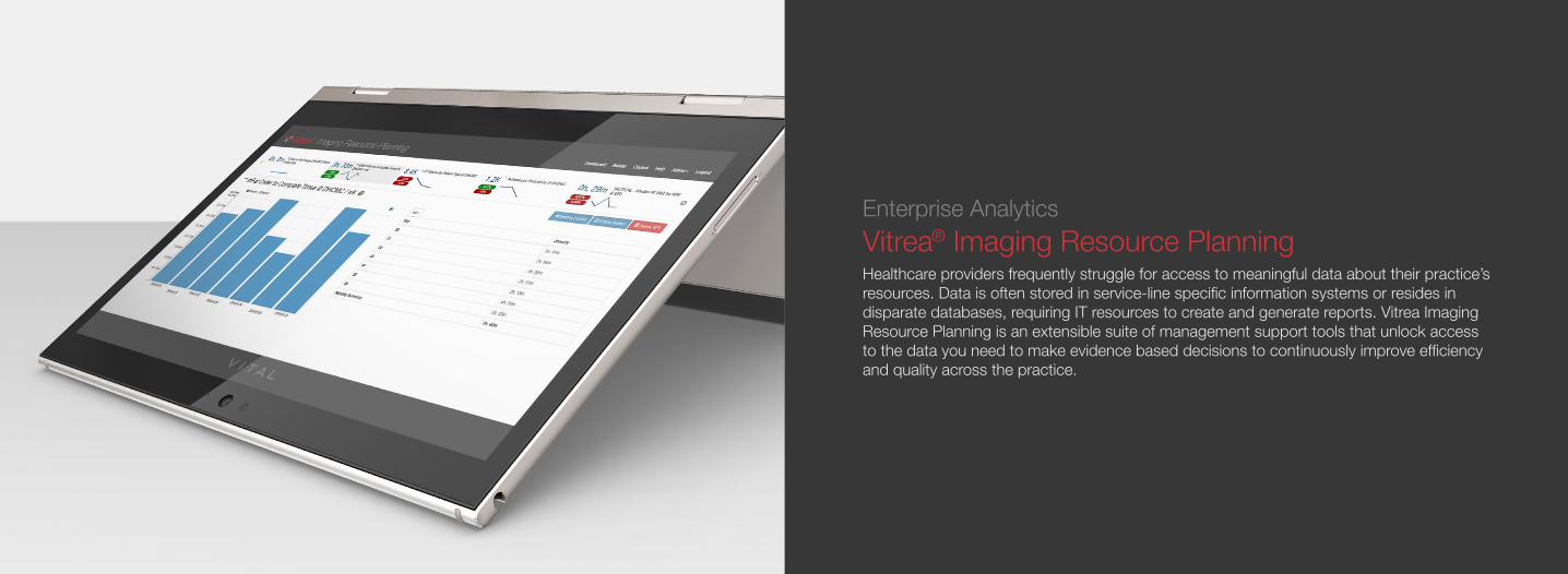
Vitrea® Imaging Resource PlanningHealthcare providers frequently struggle for access to meaningful data about their practice’s resources. Data is often stored in service-line specific information systems or resides in disparate databases, requiring IT resources to create and generate reports. Vitrea Imaging Resource Planning is an extensible suite of management support tools that unlock access to the data you need to make evidence based decisions to continuously improve efficiency and quality across the practice.

2322
Vitrea Imaging Resource PlanningVitrea Imaging Resource Planning provides insight at all organizational levels — from health system-wide trends to the utilization of a specific resource, on demand and in real-time. The application gathers and stores operational data from isolated systems independent of manufacturer or vendor to provide a comprehensive view of your practice. Self-service access to data you need to support resource management decisions are provided through an extensible suite of applications.
These are just some of the questions you can answer with self-service data access:
nHow quickly are we able to perform brain scans on stroke patients?nWhat is the average time to the third available appointment for an MRI at each of my facilities?nHave I lost business from my referring physician base recently? nAm I using my modality resources effectively and efficiently?nWhat kinds of procedures are being performed on each of the CT scanners in my fleet?nWhat is the utilization rate of each of my ultrasound modalities or other portable devices?nWhat is the report turnaround time for emergency patients at each of my facilities?nHow should I staff for the upcoming holiday weekend?nWho are my top referring providers of high RVU exams?nAre we managing radiation dose levels appropriately across procedures, equipment and staff? Do you have self-service access to the data you need to answer these questions today?

2524
Quality KPI ManagerSelect from pre-defined imaging workflow metrics to create powerful dashboards with an easy to use builder interface.
Productivity MonitorAsk questions and get answers when and where you need them in real-time on demand. Investigate data at the highest levels and drill down to specific areas of interest.
Scanner UtilizationEmploy these tactical tools to manage the flow of patients through your imaging departments with real-time visibility into patient wait, exam status, exam time and scheduled exams.
Report SearchUnlock the value of diagnostic reports with full-text report search of your entire report repository.

2726
Enterprise Visualization
Vitrea® Enterprise ViewerVitrea Enterprise Viewer is a diagnostic-quality, zero-footprint viewer, which provides fast and secure Web-based access to patient information from multiple systems and archives. It helps to integrate images effectively into the primary clinical workflow and improve care coordination by providing a single point of access to DICOM images and multimedia files on a browser, tablet or smartphone.
Vitrea Enterprise Viewer is not intended for diagnostic use when accessed from a mobile device.

28 29
Vitrea® Enterprise Viewer helps to improve collaboration among experts across your medical enterprise, helping you consolidate and standardize workflows. Consistent workflows and a unified user experience improve adoption and lower support costs.
CardiologyPACS
FileSystem
Vendor NeutralArchive (Optional)
MiscellaneousImage Repository
DocumentImage Repository
RadiologyPACS
Media
Health Information Exchange Electronic Health Record
Cache (Optional)
Vitrea Data Stream (Optional)
Vitrea Enterprise Viewer®

3130
Enterprise Visualization
Vitrea® Advanced VisualizationFull-powered Solutions for 2D, 3D and 4D
Vitrea software is a multi-modality advanced visualization system providing comprehensive applications in a variety of IT environments.
Multi-modality applications enhance diagnostic confidence across the organization. By providing access to advanced clinical tools, Vitrea software enables physicians to have meaningful interactions wherever they are.
Advanced imaging tools, such as in-suite 3D viewing and automated measurements, provide physicians with patient information anywhere, anytime. Radiologists can share images throughout their enterprise and collaborate in real-time with other physicians to help achieve better outcomes.

32 33
Vitrea® Advanced Visualization DeploymentsWorkstationThe workstation deployment is an intuitive, multi-modality advanced visualization solution that increases scanner productivity by extending workflow beyond the scanning console and optimizing time and resources to produce clinical results. The workstation provides full Vitrea functionality.
ExtendThe extend deployment helps to improve patient care by reducing information delays and by providing quick access to the exams required by your clinical workflows. A centralized database and multiple access points ensure that clinicians can share work using the same tools and interface.
Easy to deploy and maintain, the extend deployment delivers industry-leading clinical applications without adding significantly to the IT footprint. By supporting three concurrent advanced visualization sessions from a variety of access points, the extend deployment eliminates the need to maintain multiple workstations that have low utilization rates and potentially high service costs.
EnterpriseFueled by intelligent automation, the enterprise deployment helps to improve efficiency and offers a straightforward approach to complex information, allowing all members of the care team to share images and clinical functionality anytime, anywhere within the enterprise.
All of our clinical applications leverage the power of Vital’s image management system, giving our customers the ability to centrally manage their images and workflows.

3534
Vitrea® Advanced Visualization Clinical ApplicationsVital’s suite of applications offers full-powered solutions for 2D, 3D and 4D advanced visualization used to process and analyze clinical data from multiple modalities – CT, MR, XA, PET, US and SPECT. Applications for Cardiology, Neurology and Oncology provide comprehensive toolsets that supply medical specialists with information for planning procedures and treating patients.
36 37
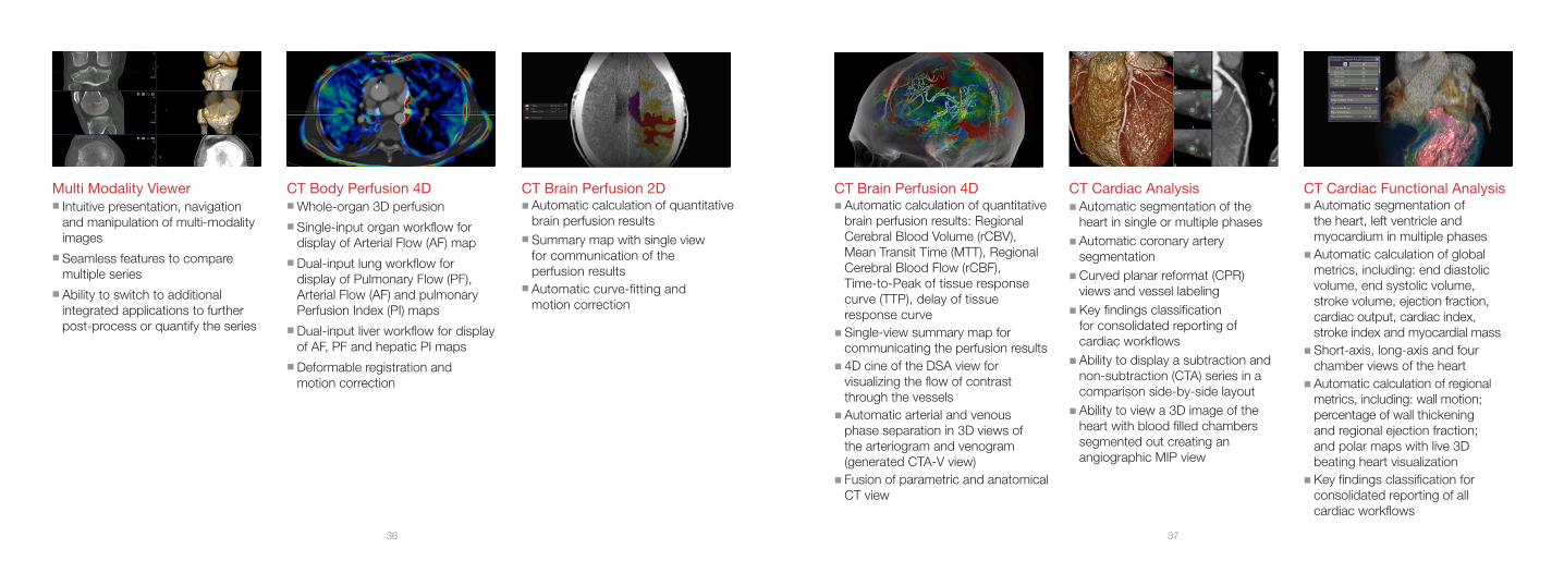
Multi Modality Viewer n Intuitive presentation, navigation
and manipulation of multi-modality images
n Seamless features to compare multiple series
n Ability to switch to additional integrated applications to further post-process or quantify the series
CT Body Perfusion 4Dn Whole-organ 3D perfusionn Single-input organ workflow for
display of Arterial Flow (AF) mapn Dual-input lung workflow for
display of Pulmonary Flow (PF), Arterial Flow (AF) and pulmonary Perfusion Index (PI) maps
n Dual-input liver workflow for display of AF, PF and hepatic PI maps
n Deformable registration and motion correction
CT Brain Perfusion 4D n Automatic calculation of quantitative
brain perfusion results: Regional Cerebral Blood Volume (rCBV), Mean Transit Time (MTT), Regional Cerebral Blood Flow (rCBF), Time-to-Peak of tissue response curve (TTP), delay of tissue response curve
n Single-view summary map for communicating the perfusion results
n 4D cine of the DSA view for visualizing the flow of contrast through the vessels
n Automatic arterial and venous phase separation in 3D views of the arteriogram and venogram (generated CTA-V view)
n Fusion of parametric and anatomical CT view
CT Brain Perfusion 2D n Automatic calculation of quantitative
brain perfusion resultsn Summary map with single view
for communication of the perfusion results
n Automatic curve-fitting and motion correction
CT Cardiac Analysis nAutomatic segmentation of the
heart in single or multiple phasesnAutomatic coronary artery
segmentationnCurved planar reformat (CPR)
views and vessel labelingnKey findings classification
for consolidated reporting of cardiac workflows
nAbility to display a subtraction and non-subtraction (CTA) series in a comparison side-by-side layout
nAbility to view a 3D image of the heart with blood filled chambers segmented out creating an angiographic MIP view
CT Cardiac Functional Analysis nAutomatic segmentation of
the heart, left ventricle and myocardium in multiple phases
nAutomatic calculation of global metrics, including: end diastolic volume, end systolic volume, stroke volume, ejection fraction, cardiac output, cardiac index, stroke index and myocardial mass
nShort-axis, long-axis and four chamber views of the heart
nAutomatic calculation of regional metrics, including: wall motion; percentage of wall thickening and regional ejection fraction; and polar maps with live 3D beating heart visualization
nKey findings classification for consolidated reporting of all cardiac workflows

38 39
CT Colon Analysisn Auto-segmentation of colon with
creation of 2D and 3D centerline for simultaneous multiplanar reformatting (MPR) and 3D review
n Single-click polyp segmentation for morphological characterization and quantification of size, density and distance to rectum
n Integrated filet view and endoluminal fly-through
n Automatic fluid/stool tagging and subtraction
n Polyp assessment and reporting using C-RADS guidelines
CT Endovascular Stent Planning nAutomated segmentation of the
aorta with centerline and contour editing tools
nStent-graft templates for abdominal and thoracic aortic aneurysms
nUser-guided workflow with automated identification of anatomical landmarks and stent-specific endovascular measurements
nKey measurements supporting fenestrated grafts using the clock angle tool and clock overlay functionality
nCreate new, add or modify stent planning templates with the Custom Device Template Editor
CT EP Planning nAutomated segmentation of the left
atrium and pulmonary veinsnAutomatic centerline and lumen
boundaries with 3D fly-through for visualization and measurement of the pulmonary vein ostia
n3D model export to the St. Jude EnSite® system
CT Fat Measurement*nSegment subcutaneous and
visceral fat regions nEvaluate fat segmentation resultsnDedicated application report
with results based on the obesity standard associated with the selected report guideline
CT Liver Analysis n Tumor tracking with RECIST
and WHO measurementsn Single-click liver and vascular
segmentationn Single-click tumor probe with
tumor margin borders viewing in 2D/3D
n Volume fusion, support for up to four timed phases. For example, arterial, venous or delayed
n Virtual resection planning
CT Lung Analysis n Semi-automated segmentation
of lung and airwaysn Restore previously segmented
nodules from prior studies for comparison
n Evaluation of nodules with quantitative measurements
n Dictation Table with Fleischner Criteria for reference within the application
n Structured reporting available for export in the Viewer tab as well as the Reporting tab
n Single click lung nodule segmentation tools to include solid nodules and ground glass opacity (GGO) nodules
*CT Fat Measurement is only available in select countries. It is not available in the US.

40 41
CT Lung Density Analysis n Semi-automatic right lung, left lung
and airway segmentationn Visualization of lung density with
color-defined Hounsfield Unit (HU) ranges
n Lung density result quantification with HU density range, volume measurements, lung density index and the PD15% measurement
n Density graph/histogram of the classified lung voxels’ relative frequencies
n Comparison of upper and lower lung density index ratios
n Adjustable density thresholds for refining and optimizing HU ranges
CT Lung Screening Solution n Vitrea Advanced Visualization’s
flagship CT Lung Analysis application with Image Denoising
n Support of Nuance PowerScribe® 360 Reporting
n PenLungTM by RenRad unified software solution
n Integrated with VisiaTM CT Lung CAD
n Custom report templates with Lung-RADSTM and ACR guideline.
n Comprehensive resource center for education
CT Multi-Chamber CFA nSemi-automatic segmentation of
left atrium (LA), right ventricle (RV), left ventricle (LV) and myocardium, including identification of long axis and mitral valve boundaries across multiple phases
nAutomatic calculation of RV/LV End Diastolic Volume (EDV), End Systolic Volume (ESV), Stroke Volume (SV), Cardiac Output (CO), 3-point LA metrics, LV/RV regurgitation fraction, cardiac index and myocardial mass
nCalculation of regional metrics including wall motion, percentage of wall thickening, regional ejection fraction and polar plots
nKey findings classification for consolidated reporting of cardiac workflows
CT Myocardial PerfusionnSemi-automatic chamber and
myocardium segmentationnQualitative measurements,
including Myocardial Mass, Myocardial Volume and Hounsfield Unit (HU) attenuation
nPolar map plots (contrast, transmural perfusion ratio, perfusion index) highlighting potential myocardium defects
nAutomatic calculation of qualitative perfusion results
nSingle and dual-volume analysis of S1 (early acquisition) and S2
later acquisitionnAbility to view cardiac vessels over
colored attenuation datanSupport for single or dual-volume
exams
CT SUREPlaque™
nSingle-click segmentation, with automatic centerline and lumen boundaries for plaque characterization and quantification based on CT HU values
nAdjustable plaque thresholds for each identified lesion
nAutomatic measurement and display of: lumen area and diameter; plaque area; plaque burden; ratio of wall area and lumen area; plaque volume; and plaque index
CT TAVR PlanningnThree-point aortic valve/annulus
plane definitionnDisplay of C-Arm angle for
device placementnUser guided automation for
annulus sizing with diameters, area and circumference
nUser guided automation for right and left ostium measurements
nUser guided automation for access measurements with histogram and catheter sizing
nComprehensive reporting of measurements, including diameter, area, angle circumference and length
nCreate new, add or modify stent planning templates with the Custom Device Template Editor

42 43
CT VScore™ n2D and 3D visualizationnReport template autofills user
selected scoresnCalculation of calcium score using
Agatston, Volume or Mass
Vessel ProbenThe Vessel Probe tool creates a
centerline through the vessel lumen nMultiple Image viewing formats
including orthogonal MPR (multiplanar reformatted), oblique MPR, curved MPR, 3D, and curved reformat views of the selected vessel.
nAutomated Stenosis Measurement tools that include single and dual reference, NASCET and average
nAutomated internal and external lumen boundary detection, including maximum and minimum lumen diameters
Vitrea® Image DenoisingnSPD denoising algorithmnCustomized filter, with reduced
pixel noise and improved signal contrast to noise ratio (SNR)
nReal time toggle between original and denoised volumes
nPredefined image filter presets may be modified and saved for future use
nInteractive denoising preview capability
nCompatible with multi-phase and multi-volume datasets within select corresponding protocols
3D Printingn3D models can be created
from CT, MR, or XA images and exported from Vitrea in the form of stereolithography (STL). STL files are used in a wide variety of other applications.
n3D print on-demandnBest-in-class 3D printing nAbility to print 3D models in a wide
range of materials and colors– from soft and dissectable to rigid and durable
MR Brain Tumor*Fully automated step-by-step processing for patients suffering from brain tumors, including quantitative and qualitative multiparametric analysis
Includes:n Dynamic Susceptibility Contrast (DSC)
Perfusion Weighted Imaging (PWI)n Dynamic Contrast Enhanced (DCE)
Permeabilityn Diffusion Weighted Imaging (DWI)n Advanced Multiparametric Analysisn Diffusion Tensor Imaging (DTI) /
Fiber Trackingn Intravoxel Incoherent Motion (IVIM)n Longitudinal Analysis
MR Breast*nEfficient tools for breast cancer
detection, characterization and staging.
n Provides instant comprehensive lesion assessment and high quality diffusion assessment
nOffers BI-RADS reporting, which facilitates communication of results between referring physicians
Includes:nDynamic Contrast Enhanced (DCE)
PermeabilitynAdvanced Multiparametric AnalysisnKineticsnBi-RADS® ReportnIntravoxel Incoherent Motion (IVIM)nQuantitative Results
*Powered by Olea Medical

44 45
MR Head and Neck*Advanced post-processing and 3D visualization tools for the diagnosis of head and neck lesions
Includes:n Diffusion Weighted Imaging (DWI)n Dynamic Contrast Enhanced (DCE)
Computationn Advanced Multiparametric Analysisn Kineticsn Intravoxel Incoherent Motion (IVIM)n Quantitative Results
MR Female Pelvis*Analyze morphological changes on the pelvic female organs under pathological conditions
Includes:n Dynamic Contrast Enhanced (DCE)
Computationn Advanced Multiparametric Analysisn Kineticsn Intravoxel Incoherent Motion (IVIM)n Quantitative Results
MR Musculoskeletal*Optimal visualization and assessment of soft tissue and bony structures
Includes:n Analysisn Relaxometryn Automatic Diffusion Computationn Dynamic Contrast Enhanced (DCE)
Computationn Advanced Multiparametric Analysis
MR Neuro ASL*n Neuro Arterial Spin Labeling (ASL)
applications use non-invasive imaging to efficiently measure perfusion.
n Holds an advantage over contrast agent-based methods for patients with contraindications for contrast agent injection
n Uses spatial smoothing and denoising of images to increase signal-to-noise ratio
Includes:n Diffusion Weighted Imaging (DWI)n Analysisn Diffusion Tensor Imaging (DTI) /
Fiber Trackingn Quantitative Results
MR Prostate* Comprehensive lesion assessment and high quality diffusion assessment
Includes:n Diffusion Weighted Imaging (DWI)n Dynamic Contrast Enhanced (DCE)
Computationn Advanced Multiparametric Analysisn Kineticsn PIRADS 2.0n Intravoxel Incoherent Motion (IVIM)n Quantitative Results
MR Rectum* Delineate tumoral margins and assess mesorectal involvement, nodes and distant metastases
Includes:n Dynamic Contrast Enhanced (DCE)
Computationn Advanced Multiparametric Analysisn Kineticsn Intravoxel Incoherent Motion (IVIM)n Quantitative Results
*Powered by Olea Medical
46 47
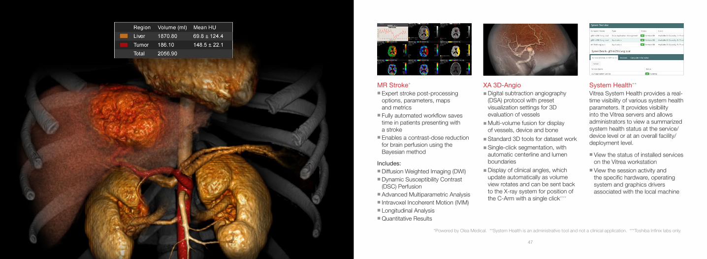
MR Stroke* n Expert stroke post-processing
options, parameters, maps and metrics
n Fully automated workflow saves time in patients presenting with a stroke
n Enables a contrast-dose reduction for brain perfusion using the Bayesian method
Includes:n Diffusion Weighted Imaging (DWI)n Dynamic Susceptibility Contrast
(DSC) Perfusionn Advanced Multiparametric Analysisn Intravoxel Incoherent Motion (IVIM)n Longitudinal Analysisn Quantitative Results
XA 3D-AngionDigital subtraction angiography
(DSA) protocol with preset visualization settings for 3D evaluation of vessels
nMulti-volume fusion for display of vessels, device and bone
nStandard 3D tools for dataset worknSingle-click segmentation, with
automatic centerline and lumen boundaries
nDisplay of clinical angles, which update automatically as volume view rotates and can be sent back to the X-ray system for position of the C-Arm with a single click***
System Health**Vitrea System Health provides a real-time visibility of various system health parameters. It provides visibility into the Vitrea servers and allows administrators to view a summarized system health status at the service/device level or at an overall facility/deployment level.
nView the status of installed services on the Vitrea workstation
nView the session activity and the specific hardware, operating system and graphics drivers associated with the local machine
*Powered by Olea Medical. **System Health is an administrative tool and not a clinical application. ***Toshiba Infinix labs only.

4948
Vitrea® Advanced Visualization Integrated ApplicationsTo provide the best tools to our customers, Vital provides partner applications integrated seamlessly into our software.*
*Check with your Vital Sales Representative or Solutions Architect for your supported applications set.

50 51
Medis® Suite Cardiovascular MRMedis Suite provides an efficient and flexible workflow, including QMass® Viewer with scanline views for cross referencing, caliper measurements and snapshots, and flexible reporting, including predefined textsnCVMR ViewernQMassnQFlow
iCAD VeraLook® CT Colon CADn2D and 3D fly-through visualizationnAutomated identification of regions
of interest (ROI)
nBookmark CAD markingsnCAD summary panel
Cedars-Sinai Medical Center Cardiac SuitenQuantitative Gated SPECT (QGS)nQuantitative Perfusion SPECT (QPS)nQuantitative Blood Pool SPECT
(QBS)nQuantitative PET (QPET)nAutoReconnMoCo (Motion Correction)
Mirada Oncology FusionTM
nIndustry leading comprehensive PET/CT reading platform
nCustom layouts, custom reports and hotkeys for any tool
nSoftware based PET/MRnSupport for Response Evaluation
Criteria in Solid Tumors (RECIST), PET Response Criteria in Solid Tumors (PERCIST) and World Health Organization (WHO)
nSupport for multi-timepoint gated, multi-sequence MR and multi-phase CT data
nAutomatic image alignment of datasets upon load
nCT Segmentation Tool
Mirada RTxnMulti-modal Deformable Image
RegistrationnComprehensive QA tools enable
assessment of Deformable Image Registration
nPET/CT and MR included in planning
nSingle-click multi-atlas contouringnDose warping and summationnAdaptive re-planning
TomTec® UltrasoundnTomTec Radiology Ultrasound
Standard includes 2D, 3D and 4D viewing options, advanced vascular measurement capabilities and the 4D Sono-Scan package for general imaging and Ob/Gyn ultrasound imaging
nTomTec Cardiac Ultrasound Standard provides 2D review and analysis of vascular and cardiac anatomy including stress echo analysis
nTomTec Cardiac Ultrasound Advanced adds 3D/4D visualization options to the “Cardiac Ultrasound Standard” package

52 53
Visia™ Dynamic ReviewnAutomated processing and display
of dynamic MRI studiesnSophisticated 3D rigid or elastic
algorithms allow for advanced motion correction
nInteractive time-intensity curvesnFlexible color overlays through
on-the-fly adjustments of thresholds and time points
nMRI-optimized layoutsnColor overlays available on
interactive 3D Maximum Intensity Projection, subtraction images and any oblique plane
Visia™ CT Lung CADnAutomated segmentation of the
lungs and airwaysnOne-click Nodule Probe tool for
auto-measurement of nodules
nVolume measurementsnLung emphysema visualization
presetnClinically validated CAD
performance - proven to improve reader accuracy and efficiency

5554
Vitrea® Advanced Visualization Clinical WorkflowsVital’s clinical applications contain many workflows to assist users when working on specific clinical exams. These can be used to automatically optimize the screen layout and visualization settings and perform anticipatory segmentation of the volumetric data relative to the anatomy being visualized.

56 57
CT Abdominal AnalysisThe CT Abdominal Analysis workflow visualizes the aorta in the abdomen. CT Aorta AnalysisThe CT Aorta Analysis workflow enables users to visualize and evaluate the aorta vasculature.
CT CarotidThe CT Carotid workflow enables the ability to visualize vessel structures.
CT Circle of WillisThe CT Circle of Willis workflow is intended to view the vascular anatomy in the Circle of Willis.
CT Larynx AirwayThe CT Larynx Airway Analysis workflow is used for the visualization and evaluation of the larynx and airway.
CT MusculoskeletalThe CT Musculoskeletal workflow enables visualization of orthopedic studies.
CT RenalThe CT Renal workflow enables the visualization of renal anatomy using CT angiography studies.
CT RunoffThe CT Runoff workflow provides the ability to visualize and measure bone and vessel structures.
CT UrogramThe CT Urogram workflow enables the ability to evaluate the kidneys, ureters and bladder.
MR Abdominal AnalysisThe MR Abdominal Analysis workflow provides general clinical tools and visualization settings to review and analyze MR abdominal exams.
MR Brain TumorThe MR Brain Tumor workflow is intended for the analysis/quantification of tumor volumes obtained from MR brain series scans using a special 2D view in the Head MR protocol.
MR MusculoskeletalThe MR Musculoskeletal workflow views types of orthopedic studies with presets for optimal visualization of soft tissue and bony structures. Users may export into STL format.
MR VascularThe MR Vascular workflow evaluates vascular anatomy from MR angiography studies.
The following are some of the workflows available in our clinical applications:

58 59
SupportVital understands that the success of your organization depends on reliable technology and minimal downtime. Our dedicated Customer Success Management program and quick response times ensure that you receive the help you need, when you need it. Our consulting services provide the technical and clinical expertise necessary to customize our solution to your environment.
Maintenance and ServicesThe following features are available with our Maintenance and Services contracts:
nSoftware Updates and UpgradesnRapid Response Remote Technical SupportnAccess to Vital U® live WebinarsnAccess to Vital U Online LibrarynStandard Support HoursnAccess to Online Technical Knowledge BasenVital Customer Success ManagernHardware Support Assistancen24/7 Technical Support nProactive System Activity Review with Customer Success Manager nEducation Credits (at additional cost) nOn-site assistance for Critical Issues

60 61
Vital U® EducationWhen you purchase software from Vital, you receive education units per the terms of your purchase. Your Maintenance and Support agreement may include additional, automatically renewing education units. You may purchase more units any time you need them.
Vital U is Vital’s professional educational organization. Vital offers various learning opportunities for you to choose from. Because we value fostering partnerships with our customers, we tailor ongoing education programs for each organization’s size, needs and desired outcomes.
Classroom EducationFor uninterrupted learning in a hands-on classroom environment, sign up for a Vital U classroom course at our headquarters in Minneapolis.
On-Site Classroom EducationOn-site facility education teaches the fundamentals of Vital’s advanced visualization software.
Vital U WebinarsOur live webinars offer lecture-style instruction on the use of clinical applications. New modules are offered every week.
eLearningResources are available online, exactly when and where you need them. Access to our eLearning, educational courses, videos, workflows, user guides and other release-related documentation are available to customers.

62