Health Effects Document for Perfluorooctane Sulfonate (PFOS)
208
Perfluorooctane sulfonate (PFOS) – February 2014 i Draft – Do Not Cite or Quote Health Effects Document for Perfluorooctane Sulfonate (PFOS)
description
Health-Effects-Document-for-Perfluorooctane-Sulfonate-(PFOS).
Transcript of Health Effects Document for Perfluorooctane Sulfonate (PFOS)
Perfluorooctane sulfonate (PFOS) – February 2014 i Draft – Do Not Cite or Quote
Health Effects Document for Perfluorooctane Sulfonate (PFOS)
Health Effects Document
for
Perfluorooctane Sulfonate (PFOS)
U.S. Environmental Protection Agency Office of Water (4304T)
Health and Ecological Criteria Division Washington, DC 20460
EPA Document Number: 822R14002 Date: February 2014
Perfluorooctane sulfonate (PFOS) – February 2014 Draft – Do Not Cite or Quote
ii
Perfluorooctane sulfonate (PFOS) – February 2014 iii Draft – Do Not Cite or Quote
ACKNOWLEDGMENT
This document was prepared under the U.S. EPA Contract No. DW-8992342701, Work Assignment No. 2011-001 with Oak Ridge National Laboratory. The Lead U.S. EPA Scientist is Joyce Morrissey Donohue, Ph.D., Health and Ecological Criteria Division, Office of Science and Technology, Office of Water. The Oak Ridge National Laboratory is managed and operated by UT-Battelle, LLC., for the U.S. Department of Energy under Contract No. DE-AC05-00OR22725.

Perfluorooctane sulfonate (PFOS) – February 2014 iv Draft – Do Not Cite or Quote
Authors, Contributors, and Reviewers
Chemical manager
Joyce Morrissey Donohue, Ph.D. Office of Water, Office of Science and Technology Health and Ecological Criteria Division U.S. Environmental Protection Agency, Washington D.C.
Authors (EPA) Amal Mahfouz, Ph.D. Office of Water, Office of Science and Technology Health and Ecological Criteria Division U.S. Environmental Protection Agency, Washington D.C. Joyce Morrissey Donohue, Ph.D. Office of Water, Office of Science and Technology Health and Ecological Criteria Division U.S. Environmental Protection Agency, Washington D.C. Tina Moore Duke, M.S. Office of Water, Office of Science and Technology Health and Ecological Criteria Division U.S. Environmental Protection Agency, Washington D.C.
Authors (Oak Ridge National Laboratory) Dana Glass-Mattie, D.V.M. Environmental Sciences Division Oak Ridge National Laboratory, Oak Ridge, TN Carol S. Wood, Ph.D., D.A.B.T. Environmental Sciences Division Oak Ridge National Laboratory, Oak Ridge, TN
Peer Reviewers Internal Christopher Lau, Ph.D.

Perfluorooctane sulfonate (PFOS) – February 2014 v Draft – Do Not Cite or Quote
National Health and Environmental Effects Research Laboratory, Office of Research and Development Reproductive Toxicology Division U.S. Environmental Protection Agency, Research Triangle Park, NC Greg Miller, Ph.D. Office of Children’s Health Protection, Office of the Administrator U.S. Environmental Protection Agency, Washington, DC John Wambaugh, Ph.D. National Center for Computational Toxicology, Office of Research and Development Systems Models for Chemical Toxicity and Exposure U.S. Environmental Protection Agency, Research Triangle Park, NC National Center for Environmental Assessment Office of Research and Development U.S. Environmental Protection Agency, Research Triangle Park, NC External

Perfluorooctane sulfonate (PFOS) – February 2014 vi Draft – Do Not Cite or Quote
TABLE OF CONTENTS
ACKNOWLEDGMENT .............................................................................................................................. iii Authors, Contributors, and Reviewers ......................................................................................................... iv TABLE OF CONTENTS ............................................................................................................................. vi LIST OF TABLES ..................................................................................................................................... viii LIST OF FIGURES ...................................................................................................................................... x ABBREVIATIONS AND ACRONYMS .................................................................................................... xi 1.0 EXECUTIVE SUMMARY ............................................................................................................. 1-1 2.0 IDENTITY: CHEMICAL AND PHYSICAL PROPERTIES ......................................................... 2-1 3.0 TOXICOKINETICS ........................................................................................................................ 3-1
3.1 Absorption ................................................................................................................................ 3-1 3.1.1 Oral Exposure ........................................................................................................................... 3-1 3.1.2 Inhalation Exposure .................................................................................................................. 3-2 3.1.3 Dermal Exposure ...................................................................................................................... 3-2
3.2 Distribution ............................................................................................................................... 3-2 3.2.1 Oral Exposure ........................................................................................................................... 3-5 3.2.2 Inhalation and Dermal Exposure ............................................................................................ 3-16 3.2.3 Other Routes of Exposure ....................................................................................................... 3-17
3.3 Metabolism ............................................................................................................................. 3-17 3.4 Excretion ................................................................................................................................. 3-18
3.4.1 Oral Exposure ......................................................................................................................... 3-18 3.4.2 Inhalation Exposure ................................................................................................................ 3-20 3.4.3 Dermal Exposure .................................................................................................................... 3-20 3.4.4 Other Exposure Routes ........................................................................................................... 3-20
3.5 Pharmacokinetic Considerations ............................................................................................ 3-20 3.5.1 Physiologically based models ................................................................................................. 3-20 3.5.2 Half-life data ........................................................................................................................... 3-27 3.5.3 Volume of Distribution Data .................................................................................................. 3-31
4.0 HAZARD IDENTIFICATION........................................................................................................ 4-1 4.1 Human Effects .......................................................................................................................... 4-1
4.1.1 Short-Term Studies and Case Reports ...................................................................................... 4-1 4.1.2 Long-Term and Epidemiological Studies ................................................................................. 4-1
4.1.2.1 Noncancer Systemic Toxicity Studies ............................................................................. 4-1 4.1.2.2 Reproductive Hormones and Reproductive/Developmental Studies ............................... 4-5 4.1.2.3 Thyroid Effect Studies ..................................................................................................... 4-9 4.1.2.4 Immunotoxicity .............................................................................................................. 4-11 4.1.2.5 Carcinogenicity Studies ................................................................................................. 4-13
4.2 Animal Studies ....................................................................................................................... 4-14 4.2.1 Acute Toxicity ........................................................................................................................ 4-14 4.2.2 Short-Term Studies ................................................................................................................. 4-16 4.2.3 Subchronic Studies ................................................................................................................. 4-20 4.2.4 Neurotoxicity .......................................................................................................................... 4-24 4.2.5 Developmental/Reproductive Toxicity ................................................................................... 4-26 4.2.6 Specialized Developmental Studies ........................................................................................ 4-37 4.2.7 Chronic Toxicity ..................................................................................................................... 4-41 4.2.8 Carcinogenicity ....................................................................................................................... 4-42
4.3 Other Key Data ....................................................................................................................... 4-44 4.3.1 Mutagenicity and Genotoxicity .............................................................................................. 4-44 4.3.2 Immunotoxicity ...................................................................................................................... 4-45 4.3.3 Physiological or Mechanistic Studies ..................................................................................... 4-49
Perfluorooctane sulfonate (PFOS) – February 2014 vii Draft – Do Not Cite or Quote
4.3.3.1 Noncancer Effects .......................................................................................................... 4-49 4.3.4 Structure-Activity Relationship .............................................................................................. 4-61
4.4 Hazard Characterization ......................................................................................................... 4-61 4.4.1 Synthesis and Evaluation of Major Noncancer Effects .......................................................... 4-62 4.4.2 Synthesis and Evaluation of Carcinogenic Effects ................................................................. 4-68 4.4.3 Mode of Action and Implications in Cancer Assessment ....................................................... 4-69 4.4.4 Weight of Evidence Evaluation for Carcinogenicity .............................................................. 4-70 4.4.5 Potentially Sensitive Populations ........................................................................................... 4-70
5.0 DOSE-RESPONSE ASSESSMENT ............................................................................................... 5-1 5.1 Dose-Response for Noncancer Effects ..................................................................................... 5-1
5.1.1 RfD Determination ................................................................................................................... 5-1 5.1.1.1 Benchmark Dose Approach ............................................................................................. 5-7 5.1.1.2 Pharmacokinetic Model Approach ................................................................................ 5-11 5.1.1.3 RfD Quantitation ............................................................................................................ 5-21
5.1.2 RfC Determination ................................................................................................................. 5-27 5.2 Dose-Response for Cancer Effects ......................................................................................... 5-27
6.0 REFERENCES ................................................................................................................................ 6-1 APPENDIX A: Summary of Data ................................................................................................................ 1 APPENDIX B ............................................................................................................................................... 1

Perfluorooctane sulfonate (PFOS) – February 2014 viii Draft – Do Not Cite or Quote
LIST OF TABLES
TABLE 2-1. Chemical and Physical Properties of PFOS ......................................................................... 2-2 TABLE 3-1. Percent (%) Binding of PFOS in Rat, Monkey and Human Plasmaa ................................... 3-3 TABLE 3-2. Average PFOS Level (µg/mL or ppm) in Serum of Monkeysa ........................................... 3-6 TABLE 3-3. PFOS Levels in the Serum and Liver of Ratsa ..................................................................... 3-7 TABLE 3-4. Mean (± SD) daily PFOS Consumption and Tissue Residue Levels in Rats Treated for 28
Daysa ................................................................................................................................................. 3-8 TABLE 3-5. Concentrations of PFOS in Male Rats’ Whole Blood (µg/mL) and Various Tissues
(µg/g) After 28 Daysa ........................................................................................................................ 3-8 TABLE 3-6. Levels of PFOS in serum and bile of rats treated for 5 daysa .............................................. 3-9 TABLE 3-7. PFOS Concentrations (Mean ± S.D.) in Samples From Pregnant Dams and Fetuses (GD 21
only) in µg/mL (ppm) for Serum and Urine and µg/g for Liver and Fecesa ................................... 3-10 TABLE 3-8. Mean PFOS (± Standard Error) Concentrations in Serum, Liver and Brain Tissue in Dams
and Offspringa ................................................................................................................................. 3-11 TABLE 3-9. PFOS contents in serum, hippocampus and cortex of offspring (n=6)a ............................. 3-12 TABLE 3-10. Mean PFOS content in serum and lungs of rat offspring (n=6)a ...................................... 3-12 TABLE 3-11. Levels of PFOS (Means ± SE) in Mouse Serum Following Treatment for 10 Daysa ...... 3-13 TABLE 3-12. Mean Concentration of PFOS (±SD) in Various Tissues of Micea .................................. 3-14 TABLE 3-13. Ratios (means ± S.D.) between the concentrations of 35S-labeled PFOS in various organs
and blood of mouse dams, fetuses and pups versus the average concentration in maternal blooda 3-15 TABLE 3-14. Percent Distribution (%) of PFOS in Mice After a 50 mg/kg Subcutaneous Injectiona .. 3-17 TABLE 3-15. Estimation of Toxicokinetic Parameters for PFOSa ......................................................... 3-18 TABLE 3-16. Mean % (± SE) of 14C-K+PFOS in rats after a single dose of 4.2 mg/kga ....................... 3-19 TABLE 3-17. PFOS pharmacokinetic data summary for monkeysa ....................................................... 3-28 TABLE 3-18. PFOS pharmacokinetic data summary for Ratsa .............................................................. 3-29 TABLE 3-19. PFOS pharmacokinetic data summary for micea ............................................................. 3-30 TABLE 3-20. Summary of Half-life Data .............................................................................................. 3-31 TABLE 4-1. Association of Serum PFOS with Serum Lipids and Uric Acid ......................................... 4-4 TABLE 4-2. Association of serum PFOS with reproductive and developmental outcomes .................... 4-9 TABLE 4-3. Association of serum PFOS with the prevalence of thyroid disease and thyroid hormone
levels in studies of general and worker populations ........................................................................ 4-11 TABLE 4-4. Mean (± SD) Values for Select Parameters in Rats Treated for 4 Weeksa ....................... 4-17 TABLE 4-5. Mean (± SD) Values for Select Parameters in Rats Treated for 28 Daysa ......................... 4-18 TABLE 4-6. Mean (± SD) Values for Select Parameters in Monkeys Treated for 182 Daysa ................ 4-22 TABLE 4-7. Mean (± SD) Values for Select Parameters in Rats Treated for 14 Weeksa ....................... 4-24 TABLE 4-8. Fertility and Litter Observations in Dams Administered 0 to 2.0 mg PFOS/kg/Daya ........ 4-31 TABLE 4-9. Effects Observed in the Mice Administered PFOS from GD 0 to GD 17/18a .................... 4-36 TABLE 4-10. Incidence of nonneoplastic liver lesions in rats (number affected/total number) ............ 4-42 TABLE 4-11. Tumor Incidence (%)a ....................................................................................................... 4-43 TABLE 4-12. Genotoxicity of PFOS In Vitro ........................................................................................ 4-44 TABLE 4-13. Genotoxicity of PFOS In Vivo ........................................................................................ 4-44 TABLE 4-14. Thyroid hormone levels in PFOS treated rats .................................................................. 4-51

Perfluorooctane sulfonate (PFOS) – February 2014 ix Draft – Do Not Cite or Quote
TABLE 4-15. Summary of PFAA Transactivation of Mouse and Human PPARα, β/δ and γa ............... 4-53 TABLE 5-1. NOAEL/LOAEL and Effects for Longer-term Duration Studies of PFOS ......................... 5-3 TABLE 5-2. NOAEL/LOAEL Data for Short-term Oral Studies of PFOS .............................................. 5-5 TABLE 5-3. Benchmark Dose Modeling for a 5% Increased Risk of Developmental Toxicity in Rats .. 5-7 TABLE 5-4. Benchmark Dose Modeling for a 10% Increased Incidence of Liver Lesions in Rats ........ 5-8 TABLE 5-5. Benchmark Dose Modeling for a 10% Increase in Liver Weight ...................................... 5-10 TABLE 5-6. Description of prior distributions used. ............................................................................. 5-13 TABLE 5-7. Pharmacokinetic parameters used in the Andersen et al. (2006) model. ........................... 5-15 TABLE 5-8. Predicted final serum concentration and time integrated serum concentration (AUC) for
different treatments of rat. ............................................................................................................... 5-16 TABLE 5-9. Predicted final serum concentration and time integrated serum concentration (AUC) for the
mouse. ............................................................................................................................................. 5-17 TABLE 5-10. Predicted final serum concentration and time integrated serum concentration (AUC) for
the monkey. ..................................................................................................................................... 5-17 TABLE 5-11. Average Serum concentrations Derived from the AUC and the duration of Dosing ....... 5-19 TABLE 5-12. Human Equivalent Doses Derived from the Modeled Animal Average Serum Values .. 5-21 TABLE 5-13. RfD Point of Departure Options from the PFOS Animal Studies ................................... 5-22 TABLE 5-14. The Impact of Quantification Approach on the RfD outcome for the PODs from the
available NOAELs .......................................................................................................................... 5-23 TABLE 5-15. The Impact of Quantification Approach on the RfD Outcome for the BMDLs from liver
and developmental endpoints .......................................................................................................... 5-25 TABLE 5-16. The Impact of Quantification Approach on the RfD Outcomes for the HEDs from the
Pharmacokinetic Model Average Serum Values ............................................................................. 5-26 TABLE A.1. PFOS Toxicokinetic Information ........................................................................................... 2 TABLE A.2. Key Studies Used With Effects Related to Serum Values (Condensed Version) .................. 6 TABLE A.3. Summary of Animal Studies with Exposure to PFOS ......................................................... 14

Perfluorooctane sulfonate (PFOS) – February 2014 x Draft – Do Not Cite or Quote
LIST OF FIGURES
Figure 2- 1. Chemical Structure of PFOS ................................................................................................. 2-1 Figure 3-1. Distribution of radiolabeled PFOS in dams and in fetuses/pups in the liver, lung, kidney and
brain ................................................................................................................................................. 3-16 Figure 3-2. PFOS Contents in Urine, Feces and Overall Excretion in Male Rats Treated for 28 Days . 3-19 Figure 3-3. Schematic for a physiologically-motivated renal resorption pharmacokinetic model. ........ 3-21 Figure 3-4. Structure of model for PFOS in rats and monkeys ............................................................... 3-22 Figure 3-5. Structure of the PFOS PBPK model in monkeys and humans ............................................. 3-23 Figure 3-6. Structure of the PBPK Model for PFOS in the Adult Sprague-Dawley Rat ........................ 3-25 Figure 3-7. Predicted Daily Average Concentration of PFOS in Maternal (black line) and Fetal (gray
line) Plasma at External Doses to the Dam ..................................................................................... 3-26 Figure 4-1. Functional categories of genes modified by PFOS in wild type and null mice. ................... 4-57 Figure 4-2. Function distribution and category enrichment analysis of the differential proteins. .......... 4-59 Figure 5-1. BMDS graphic output from selected model runs; data from Thomford, 2002. ..................... 5-9 Figure 5- 2. BMDS graphic output from liver weight model runs; data from Seacat et al., 2002, 2003 5-11

Perfluorooctane sulfonate (PFOS) – February 2014 xi Draft – Do Not Cite or Quote
ABBREVIATIONS AND ACRONYMS
Ach acetylcholine ACoA Acetyl CoA ACOX1 peroxisomal acyl-coenzyme A oxidase ADAF Age-Dependent Agjustment Factor AIC Akaike's Information Criterion ALP alkaline phosphatase ALT alanine transaminase ANOVA analysis of variance AP-1 activation protein-1 Asp aspartate AST aspartate aninotransferase AUC area under the curve AWWARF American Water Works Association Research Foundation BGS brain growth spurt BMD benchmark dose BMD benchmark dose – Lower 95th percentile confidence bound BMDS benchmark dose software BMI body mass index BQL below quantifiable limit BrdU bromodeoxyuridine BUN blood urea nitrogen bw body weight °C Celsius C Carbon CaMKII calcium/calmodulin-dependent protein kinase II CAR constitutive androstane receptor CAS Chemical Abstracts Service CCL Contaminant Candidate List CCL 3 Contaminant Candidate List 3 CD circular dichroism CFSE 6-carboxyfluorescein succinimidyl ester CI confidence interval CL clearance CoA coenzyme A CREB cAMP response element-binding protein CSF Cancer Slope Factor or cerebrospinal fluid CSM cholestyramine Cte acyl CoA thioesterase CWS community water system CYP4A22 cytochrome P-450 4A22 Cyt c cytochrome c d day DA dansylamide or dopamine DAUDA 11-(5-dimethylaminoapthalenesulphonyl)-undecanoic acid
Perfluorooctane sulfonate (PFOS) – February 2014 xii Draft – Do Not Cite or Quote
DIO1 type 1 deiodinase dL deciliter DMEM Dulbecco’s Minimal Essential Medium DMSO dimethyl sulfoxide DNA Deoxyribonucleic acid DNBC Danish National Birth Cohort DP dansyl-L-proline DPPC dipalmitoylphosphatidylcholine DWI drinking water intake EAA excitatory amino acid EC50 half maximal effective concentration ECF Electro-Chemical Fluorination ED equilibrium dialysis EFSA European Food Safety Authority FOB functional observational battery FT3 free triiodothyronine FT4 free thyroxin g gram GABA gamma-aminobutyric acid GAP-43 growth-associated protein-43 GD gestation day GFAP glial fibrillary acidic protein GGT gamma-glutamyl transpeptidase GJIC gap junction intercellular communication GLP good laboratory practice Glu glutamate Gly glycine GS glutamine synthetase GSH glutathione GSI gonad-somatic index HDL high density lipoprotein HED human equivalent dose HL-60 human promyelocytic leukemia cell line HMG-CoA 3-hydroxy-3-methylglutaryl coenzyme A HOMA homeostatic model assessment HPT hypothalamic-pituitary-thyroid HPLC/ ESMSMS High Performance Liquid Chromatography – electrospray tandem mass spectrometry HRL health reference level HSA Human Serum Albumin HSDB Hazardous Substances Database HSI hepatosomatic index IC50 half-maximal Inhibiting Concentration ICa inward calcium currents ICR imprinting control region IEF induction equivalency factor
Perfluorooctane sulfonate (PFOS) – February 2014 xiii Draft – Do Not Cite or Quote
IL-1α interleukin IL-6 interleukin 6 IRR incidence rate ratio ITC isothermal titration calorimetry IU international unit IV intravenous Ka adsorption rate constant kg kilogram KO knockout Koc organic carbon water partitioning coefficient Kow octanol-water partition coefficient Kt affinity constant L liter LC50 Lethal concentration for 50% (statistical median) of animals LC-ESI- MS/MS liquid chromatography/electrospray ionization with tandem mass spectrometry LC-MS liquid chromatography – mass spectrometry LC-MS/MS liquid chromatography – negative electrospray tandem mass spectrometry LD lactation day LD50 Lethal dose for 50% (statistical median) of animals LDH lactic dehydrogenase LDL low density lipoprotein L-FABP liver fatty acid binding protein LI labeling index LLOQ lower limit of quantification LOAEL lowest observed adverse effect level LOEC lowest observed effect concentration LOQ Limit of Quantitation LPS Lipopolysaccharide m meter MCLG Maximum Contaminant Level Goal MDA malondialdehyde Mdr2 multidrug resistance protein 2 ME malic enzyme µg microgram mg milligram min minute mL milliliter µm micrometer MOA mode of action mol mole MRL minimum reporting level MRP multidrug resistance-associated protein MTBE methyl tertiary-butyl ether NAWQA National Water Quality Assessment NDWAC National Drinking Water Advisory Council ng nanogram
Perfluorooctane sulfonate (PFOS) – February 2014 xiv Draft – Do Not Cite or Quote
NA not applicable ND not detected or not determined NHANES The National Health and Nutrition Examination Survey NIS sodium iodide symporter NK natural killer NMRI Naval Medical Research Institute NOAEL no observed adverse effect level NOEC no observed effect concentration NPDWR National Primary Drinking Water Regulation NRC National Research Council NS no sample NSP newborn screening program NT not tested OA octanoic acid OAT organic anion transporter OATp organic anion transporting peptide OGWDW Office of Ground Water and Drinking Water OR odds ratio p probability PB phenobarbital PBDE polybrominated diphenyl ether PBMC peripheral blood mononuclear cells PBPK physiologically-based pharmacokinetic PBS phosphate buffered saline PCB polychlorinated biphenyl PCNA proliferating cell nuclear antigen PCoAO palmitoyl CoA oxidase PFA perfluoroalkylate PFC perfluorinated carboxylic acids PFAA perfluoroalkyl acid PFBA perfluorobutyric acid PFBS perfluorobutane sulfonate PFDA perfluorododecanoic acid PFHS perfluorohexanesulfonic acid potassium salt PFHxS Perfluorohexanesulfonic acid PFOA Perfluorooctanoic acid PFOC perfluorooctane PFOS perfluoroocatane sulfonate PFOSA perfluorooctane sulfamide PFPA perfluoropropionic acid PFTA perfluorotetradecanoic acid pg picogram PI proliferation index PK pharmacokinetic PND postnatal day POD point of departure POSF perfluorooctanesulfonyl fluoride

Perfluorooctane sulfonate (PFOS) – February 2014 xv Draft – Do Not Cite or Quote
pKa acid dissociation constant PPAR peroxisome proliferator activated receptor ppb parts per billion ppm parts per million ppt parts per trillion mPSC miniature post-synaptic current PTU propylthiouracil PUFA polyunsaturated fatty acid PWS public water system PXR pregnane X receptor Q flow in and out of tissues Qfilc median fraction of blood flow to the filtrate RBC red blood cell Reg Det 2 Regulatory Determinations on the Second CCL RfC reference concentration RfD reference dose RIA radio immunoassay RNA ribonucleic acid RSC relative source contribution RSI renal-somatic index RT-PCR reverse transcription polymerase chain reaction RXRα retinoid X receptor alpha SA serum albumin SPC saponin compound SD standard deviation SDWA Safe Drinking Water Act SIR standardized incidence ratio SMR standardized mortality ratio SOD superoxide dismutase SRBC sheep red blood cells STP sewage treatment plant Syn 1 synapsin 1 SYP synaptophysin T-AOC total antioxidation capability Tmax time of maximum plasma concentration T3 triiodothyronine T4 thyroxine t1/2 chemical half-life T1/2 elimination half-time Tm transporter maximum TAD target administered dose TBG thyroxine-binding globulin TC total cholesterol TG triglycerides TH tyrosine hydroxylase TNFα tumor necrosis factor α TNP trinitrophenol

Perfluorooctane sulfonate (PFOS) – February 2014 xvi Draft – Do Not Cite or Quote
TPO thyroid peroxidase TRH thyrotropin releasing hormone TSH thyroid stimulating hormone TSHR thyroid stimulating hormone receptor TT3 total triiodothyronine TT4 total thyroxin TTP time to pregnancy TTR thyroid hormone transport protein, transthyretin UCB umbilical cord blood UCMR 3 Unregulated Contaminant Monitoring Rule 3 UF uncertainty factor UGT1 uridine diphosphoglucuronosyl transferase URAT urate transporter U.S. EPA U.S. Environmental Protection Agency USGS U.S. Geological Service Vd volume of distribution VLDL very low density lipoprotein VOC volatile organic compound WHAM weighted histogram analysis method WT wild type ww wet weight WWTP waste water treatment plant Wy Wy14,648

Perfluorooctane sulfonate (PFOS) – February 2014 1-1 Draft – Do Not Cite or Quote
1.0 EXECUTIVE SUMMARY PFOS
Perfluorooctane sulfonate (PFOS) is a fluorinated organic compound with an eight-carbon backbone and a sulfonate functional group. PFOS-related chemicals are used in a variety of products including surface treatments for soil/stain resistance, surface treatments of textiles, paper and metals and in specialized applications such as fire fighting foams. Because of strong carbon-fluorine bonds, PFOS is stable to metabolic and environmental degradation and is resistant to biotransformation. Data in humans and animals demonstrate ready absorption of PFOS and distribution of the chemical throughout the body by noncovalent binding to plasma proteins. Both experimental data and pharmacokinetic models show higher level of PFOS in fetal serum and brain compared with the maternal compartments. PFOS is not readily eliminated from humans as evidenced by the half-life of 5.4 years. In contrast, half-life values for the monkey, rat, and mouse are 121 days, 48 days, and 37 days, respectively. The long half-lives appear to be the result of resorption from the kidney. In other words after initial removal from blood by the kidney, a substantial fraction of what would normally be eliminated in urine is returned to the blood.
Peroxisome proliferation is usually associated with hepatic lesions in the rats, but some
uncertainties exist as to whether this is true for PFOS and if this is cause for concern in the human population. Increased hepatic lipid content in the absence of a strong PPARα response is a characteristic of exposure to PFOS. In two studies, mice administered PFOS showed differential expression of proteins mainly involved in lipid metabolism, fatty acid uptake, transport, biosynthetic processes, and response to stimulus. Many of the genes activated are associated with nuclear receptors other than PPARα.
Epidemiology studies have examined occupational and residential populations at or near
large-scale PFOS production plants in the United States in an attempt to determine the relationship between serum PFOS concentration and various health outcomes suggested by standard animal toxicological studies. Exposures were mainly through contaminated drinking water and to multiple PFCs. These studies found a positive association with increased PFOS serum levels and an increase in total cholesterol, triglycerides, and uric acid in the general population. In contrast, occupational studies did not indicate consistent associations between PFOS and cholesterol and/or triglycerides in either cross-sectional surveys or in a longitudinal analysis. Results are inconclusive or inconsistent for associations between increased serum PFOS and affects on thyroid hormones and immunotoxicity.
In general population studies of effects on reproduction and development, the only
finding of note was a slight increase in the risk for low birth weight, however, this was not a consistent finding across the studies.
In most animal studies with PFOS, short-term and chronic exposure resulted in an
increase in liver weight as at least one of the critical effects. Co-occurring effects in these studies included decreased cholesterol, lower body weight, liver histopathology, and developmental toxicity. In rat and monkey repeat-dosing studies (14 or 26 weeks), increased liver weight was accompanied by decreased cholesterol and hepatocellular hypertrophy. As part of a chronic bioassay, rats had low dose liver lesions with liver weight affected at higher doses. The most severe effect observed in the longer-term studies was decreased pup survival in a one-

Perfluorooctane sulfonate (PFOS) – February 2014 1-2 Draft – Do Not Cite or Quote
generation rat study at a LOAEL of 0.8 mg/kg/day. The LOAEL for decreased pup body weight was 0.4 mg/kg/day in one- and two-generation studies. Developmental toxicity studies at slightly higher doses support the concern for low dose-effects on pup survival. In a standard developmental neurotoxicity study, male offspring showed increased motor activity and decreased habituation on PND 17 following a maternal dose of 1 mg/kg/day. Two studies provide evidence for immunological effects in mice.
U.S. EPA has selected 0.00003 mg/kg/day as the RfD for PFOS based on the consistency
of the response and with recognition of the use of developmental toxicity and liver weight as the most sensitive endpoints for protection against co-occurring adverse effects. This value is the outcome for modeled rat serum values for developmental. In the standard developmental neurotoxicity study, male offspring showed increased motor activity and decreased habituation on PND 17 following a maternal dose of 1 mg/kg/day in the absence of effects on pup body weight. The human equivalent dose (HED) used as the basis for the RfD, was calculated from an average serum concentration of 10.87 mg/L derived from the NOAEL of 0.3 mg/kg/day for developmental neurotoxicity. A pharmacokinetic model was used to predict an area under the curve (AUC) for the NOAEL and used to calculate an HEDNOAEL. The total uncertainty factor (UF) applied to the HEDNOAEL from the rat study was 30 which included a UF of 10 for intrahuman variability, and a UF of 3 to account for toxicodynamic differences between animals and humans. Comparable values derived from the HED for liver effects in rats and developmental effects in mice are slightly higher than the RfD indicating that it will be protective.
Under the EPA 2005 cancer guidelines, the evidence for the carcinogenicity of PFOS is
considered “suggestive of carcinogenicity,” but not sufficient to assess human carcinogenicity potential. In a chronic oral toxicity and carcinogenicity study of PFOS in rats, liver, thyroid and mammary fibroadenomas were identified. The biological significance of the mammary fibroadenomas and thyroid tumors was questionable as a true dose-dependent response was not identified. The liver tumors also had a questionable dose-response with slight but statistically significant increases only in high-dose males and females. The liver tumors most found were adenomas (7/60 and 5/60 in high-dose males and females vs. none in the controls of either sex); only one hepatocellular carcinoma was found in a high-dose female. The genotoxicity data are uniformly negative. Human epidemiology studies did not find a direct correlation between PFOS exposure and the incidence of carcinogenicity in worker-based populations. Thus, the weight of evidence for the carcinogenic potential to humans of these tumors was judged to be too limited to support a quantitative cancer assessment.

Perfluorooctane sulfonate (PFOS) – February 2014 2-1 Draft – Do Not Cite or Quote
2.0 IDENTITY: CHEMICAL AND PHYSICAL PROPERTIES
Perfluorooctane sulfonate, commonly known as PFOS, and its salts are fluorinated organic compounds and is part of the group of chemicals called perfluoroalkyl acids (PFAAs). The two most widely known PFAAs have an eight-carbon backbone with either a sulfonate (PFOS) or carboxylate (PFOA- perfluorooctanoic acid) attached (Lau et al., 2007). PFOS-related chemicals are used in a variety of products including surface treatments for soil/stain resistance, coating of paper as a part of a sizing agent formulation and in specialized applications such as fire fighting foams. PFOS is produced commercially from perfluorooctanesulfonyl fluoride (POSF) which is used primarily as an intermediate to synthesize other fluorochemicals. POSF is manufactured through a process called Simons Electro-Chemical Fluorination (ECF) in which an electric current is passed through a solution of anhydrous hydrogen fluoride and an organic feedback of 1-octanesulfonyl fluoride causing the carbon-hydrogen bonds on molecules to be replaced with carbon-fluorine bonds (OECD, 2002). PFOS can also be formed by the degradation of other POSF-derived fluorochemicals.
Due to strong C-F bonds, PFOS is extremely stable and does not biodegrade in the
environment, making it very persistent. Because of this reason, most PFOS manufactured in the United States was discontinued voluntarily by 3M in 2002. PFOS is soluble in fresh water at approximately 519 mg/L. The solubility decreases significantly as the salt content of the water increases. Because of the surface-active properties of PFOS, it forms three layers in octanol/water making an n-octanol/water (Kow) partition co-efficient unable to be determined. The potassium salt of PFOS has a low vapor pressure (OECD, 2002). No direct measurement of the pKa of the acid has been located; however, the chemical is considered to have a low pKa. The chemical structure is provided in Figure 2-1 and the physical properties for PFOS are provided in Table 2-1.
Figure 2- 1. Chemical Structure of PFOS
Perfluorooctane sulfonate (PFOS) – February 2014 2-2 Draft – Do Not Cite or Quote
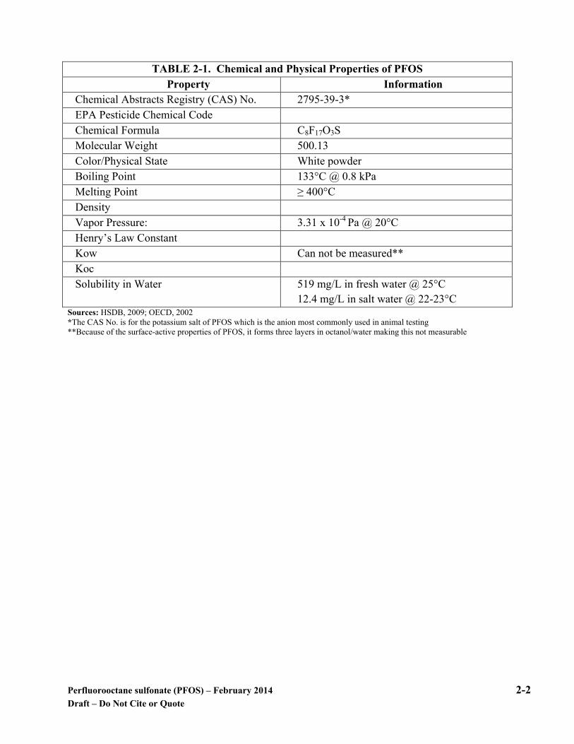
TABLE 2-1. Chemical and Physical Properties of PFOS
Property Information Chemical Abstracts Registry (CAS) No. 2795-39-3* EPA Pesticide Chemical Code Chemical Formula C8F17O3S Molecular Weight 500.13 Color/Physical State White powder Boiling Point 133°C @ 0.8 kPa Melting Point ≥ 400°C Density Vapor Pressure: 3.31 x 10⁻⁴ Pa @ 20°C Henry’s Law Constant Kow Can not be measured** Koc Solubility in Water 519 mg/L in fresh water @ 25°C
12.4 mg/L in salt water @ 22-23°C Sources: HSDB, 2009; OECD, 2002 *The CAS No. is for the potassium salt of PFOS which is the anion most commonly used in animal testing **Because of the surface-active properties of PFOS, it forms three layers in octanol/water making this not measurable
Perfluorooctane sulfonate (PFOS) – February 2014 3-1 Draft – Do Not Cite or Quote
3.0 TOXICOKINETICS
Because of strong carbon-fluorine bonds, PFOS is stable to metabolic and environmental degradation. It is not readily eliminated and can have a long half-life in humans and animals, however, the toxicokinetic profile and the underlying mechanism for the chemical’s long half-life are not completely understood. In the case of another PFC, PFOA, transport families appear to play role in absorption, distribution, and excretion and include organic anion transporters (OATs), organic anion transporting peptides (OATps), multidrug resistance-associated proteins (MRPs) and urate transporters (URAT). The transporters play a critical role in gastrointestinal absorption, uptake by the tissues, and excretion via the kidney. Work is currently in progress to determine if these same transporters are involved in PFOS toxicokinetics and preliminary data appear to indicate that they are. Some preliminary inhibition studies suggest that PFOS has a similar chain length dependent renal excretion and liver accumulation pattern as PFOA, and would involve these same transporters. Animal studies indicate that PFOS is well absorbed orally and distributes primarily in the blood and liver. While PFOS can be a formed as a metabolite from other perfluocompounds, PFOS itself does not undergo further metabolism after absorption takes place. PFAAs are known to activate peroxisome proliferator activated receptor (PPAR) pathways by increasing transcription of mitochondrial and peroxisomal lipid metabolism enzymes, sterol, and bile acid biosynthesis and retinol metabolism genes. However, based on transcriptional activation of many genes in PPARα-null mice, the effects of PFAAs involve more than activation of PPAR (Andersen et al., 2008). A summary of toxicokinetic data are provided in Appendix A, Table A.1 and Table A.2.
3.1 Absorption
Absorption data are available for oral exposure in rats. While there are no absorption studies available for humans that quantify the amounts absorbed relative to dose, extensive data are available demonstrating the presence of PFOS in the serum. These data were reported in Section 5.0 Biomonitoring Data.
The absorption process requires transport across the tissue interface with the external
environment. PFOS displays both hydrophobic and oleophobic properties indicating that movement across the membrane surface is probably achieved with transporters rather than simple diffusion.
3.1.1 Oral Exposure
Absorption in Animals Rats Following ingestion, PFOS is well absorbed. Three male rats were administered a single dose of 4.2 mg/kg of PFOS-14C in solution; 3.45% of the total dose was found in the digestive tract. The mean fecal excretion was 1.55% of the dose at 24 hours and 3.24% at 48 hours. At 24 hours, the mean sum of total carbon-14 in feces and digestive tract plus contents was 5% of the

Perfluorooctane sulfonate (PFOS) – February 2014 3-2 Draft – Do Not Cite or Quote
dose. Some of this 5% likely represented systemically absorbed carbon-14 present either in the digestive tract tissues or in the digestive tract contents as a result of excretion. The data from the 48 hour post dose group of rats were consistent with the 24 hour data. Thus, at least 95% of the PFOS-14C dose was absorbed from solution after administration to non-fasted rats (Chang et al., 2012).
3.1.2 Inhalation Exposure
An acute LC50 study in rats indicated that PFOS absorption occurs by inhalation exposure; however, pharmacokinetic data were not included (Rusch et al., 1979).
3.1.3 Dermal Exposure
No data are available on dermal absorption of PFOS.
3.2 Distribution
It has been suggested that PFOS is distributed within the body by non-covalently binding to a plasma protein, most commonly, albumin. Binding studies are provided to help support this hypothesis. Distribution data are provided only for rats following oral exposure. Indirect distribution data are provided from analysis of PFOS in tissue and blood samples from studies conducted in rats, monkeys and humans. In humans, PFOS has been found to distribute mostly to the liver and blood, but has also been identified in umbilical cord blood and breast milk. In humans, the ratio of PFOS identified in the serum and liver tissue are similar, while in animals the amount found in the liver is higher than that in the serum. In a study by Cui et al. (2009) bioaccumulation of PFOS was liver > heart > kidney > whole blood > lung > testicle, brain and spleen in rats administered 5 or 20 mg/kg/day. The highest level of PFOS was found in the liver of the rats exposed to 20 mg/kg/day and was 648 ± 17 µg/g. Binding Studies The in vitro protein binding of PFOS in rat, monkey and human plasma at concentrations of 1-500 ppm PFOS was investigated (Kerstner-Wood et al., 2003). The PFOS bound to plasma protein in all three species at all concentrations with no sign of saturation (Table 3-1). When incubated with human plasma protein, PFOS was highly bound (99.8 %) to albumin and showed affinity for low density lipoproteins (LDLs, formerly beta-lipoproteins) (95.6%) with some binding to alpha-globulins (59.4%) and gamma-globulins (24.1%).
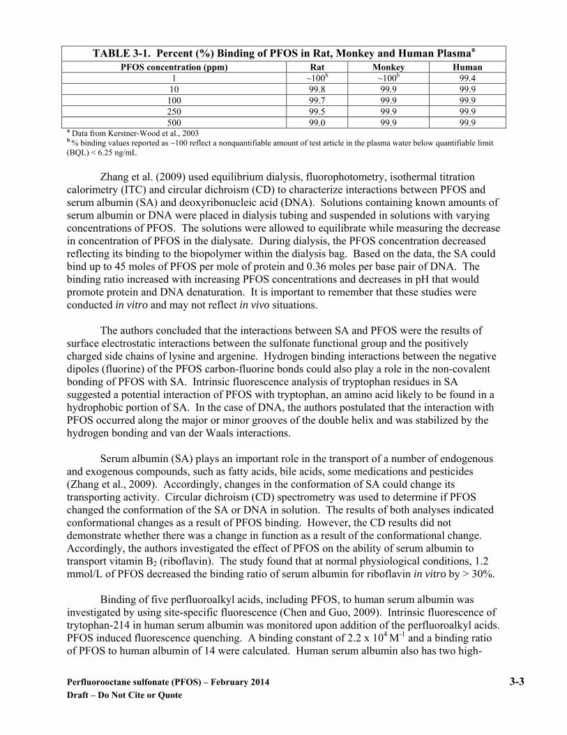
Perfluorooctane sulfonate (PFOS) – February 2014 3-3 Draft – Do Not Cite or Quote
TABLE 3-1. Percent (%) Binding of PFOS in Rat, Monkey and Human Plasmaa PFOS concentration (ppm) Rat Monkey Human
1 ~100b ~100b 99.4 10 99.8 99.9 99.9 100 99.7 99.9 99.9 250 99.5 99.9 99.9 500 99.0 99.9 99.9
a Data from Kerstner-Wood et al., 2003 b % binding values reported as ~100 reflect a nonquantifiable amount of test article in the plasma water below quantifiable limit (BQL) < 6.25 ng/mL
Zhang et al. (2009) used equilibrium dialysis, fluorophotometry, isothermal titration
calorimetry (ITC) and circular dichroism (CD) to characterize interactions between PFOS and serum albumin (SA) and deoxyribonucleic acid (DNA). Solutions containing known amounts of serum albumin or DNA were placed in dialysis tubing and suspended in solutions with varying concentrations of PFOS. The solutions were allowed to equilibrate while measuring the decrease in concentration of PFOS in the dialysate. During dialysis, the PFOS concentration decreased reflecting its binding to the biopolymer within the dialysis bag. Based on the data, the SA could bind up to 45 moles of PFOS per mole of protein and 0.36 moles per base pair of DNA. The binding ratio increased with increasing PFOS concentrations and decreases in pH that would promote protein and DNA denaturation. It is important to remember that these studies were conducted in vitro and may not reflect in vivo situations.
The authors concluded that the interactions between SA and PFOS were the results of surface electrostatic interactions between the sulfonate functional group and the positively charged side chains of lysine and argenine. Hydrogen binding interactions between the negative dipoles (fluorine) of the PFOS carbon-fluorine bonds could also play a role in the non-covalent bonding of PFOS with SA. Intrinsic fluorescence analysis of tryptophan residues in SA suggested a potential interaction of PFOS with tryptophan, an amino acid likely to be found in a hydrophobic portion of SA. In the case of DNA, the authors postulated that the interaction with PFOS occurred along the major or minor grooves of the double helix and was stabilized by the hydrogen bonding and van der Waals interactions. Serum albumin (SA) plays an important role in the transport of a number of endogenous and exogenous compounds, such as fatty acids, bile acids, some medications and pesticides (Zhang et al., 2009). Accordingly, changes in the conformation of SA could change its transporting activity. Circular dichroism (CD) spectrometry was used to determine if PFOS changed the conformation of the SA or DNA in solution. The results of both analyses indicated conformational changes as a result of PFOS binding. However, the CD results did not demonstrate whether there was a change in function as a result of the conformational change. Accordingly, the authors investigated the effect of PFOS on the ability of serum albumin to transport vitamin B2 (riboflavin). The study found that at normal physiological conditions, 1.2 mmol/L of PFOS decreased the binding ratio of serum albumin for riboflavin in vitro by > 30%. Binding of five perfluoroalkyl acids, including PFOS, to human serum albumin was investigated by using site-specific fluorescence (Chen and Guo, 2009). Intrinsic fluorescence of trytophan-214 in human serum albumin was monitored upon addition of the perfluoroalkyl acids. PFOS induced fluorescence quenching. A binding constant of 2.2 x 10⁴ M-1 and a binding ratio of PFOS to human albumin of 14 were calculated. Human serum albumin also has two high-

Perfluorooctane sulfonate (PFOS) – February 2014 3-4 Draft – Do Not Cite or Quote
affinity drug binding sites which are known as Sudlow’s drug Site I and Site II. Past experiments have shown that two fluorescence probes, dansylamide (DA) and dansyl-L-proline (DP) are specific for the two drug binding sites on human serum albumin. These two probes emit negligible fluorescence, but after binding with albumin, fluorescence increases. The titration of PFOS into human serum albumin pretreated with DA (site I), showed that at low concentrations of PFOS (0.07 mM), DA emission increased as the PFOS concentration increased until it was at 140% the original intensity. At the higher PFOS concentrations (0.7 to 4 mM), however, the fluorescence dropped. The author speculated that the rise in fluorescence was induced by the conformational changes of the protein after PFOS binds to it at a site different from Site I and the decrease at higher concentrations was from displacement of DA by PFOS. For the Site II, PFOS caused a fluorescence reduction that was quick at first but then became more gradual making the possibility that PFOS was binding to this site with two different affinities. The binding constant calculated at Site II was 7.6 x 10⁶ M-1. These findings indicate PFOS has binding sites that are similar to those identified for fatty acids. Structure and the energy of binding sites were determined between PFOS and human serum albumin (HSA) using molecular modeling (Salvalaglio et al., 2010). Calculations were based on a compound approach docking, molecular dynamics simulations and estimating free binding energies by adopting weighted histogram analysis method (WHAM)-umbrella sampling and semiempirical methodology. The binding sites impacted were ones identified as human serum albumin fatty acid binding sites. The PFOS binding site with the highest energy (-8.8 kcal/mol) was located near the tip of the Trp 214 binding site and the maximum number of ligands that could bind to HSA for PFOS was 11. The most populated albumin binding site for PFOS was dominated by van der Waals interactions. The author indicated that the number of molecules adsorbed on HSA for PFOA was 9, compared to the 11 for PFOS, which may explain why PFOS has a higher bioaccumulation than PFOA. Weiss et al. (2009) screened the binding of several perfluorinated compounds, including PFOS, to the thyroid hormone transport protein transthyretin (TTR) in a radioligand-binding assay to determine if the compounds can compete with thyroxine (T4), the natural ligand of TTR. Human TTR was incubated with 125I-labeled T4, unlabeled T4, and 10-10,000 nM competitor (PFOS) overnight. The unlabeled T4 was used as a reference compound, and the levels of T4 in the assay were close to the lower range of total T4 measured in healthy adults. PFOS had a high binding potency to TTR. The 50% inhibition concentration was 940 nM. The authors concluded that binding affinity for TTR did occur in perfluorinated compounds with peak binding in compounds having at least an eight carbon length chain, such as PFOS. Luebker et al. (2002) investigated the possibility that PFOS interferes with the binding affinity of liver-fatty acid binding protein (L-FABP) which is an intracellular lipid-carrier protein. This study was performed in vitro with a fluorescent fatty acid analogue 11-(5-dimethy-laminoapthalenesulphonyl)-undecanoic acid (DAUDA). The concentration that can inhibit fifty percent of specific DAUDA-L-FABP binding (IC50) was determined. PFOS demonstrated inhibition of L-FABP in competitive binding assays; with 10 µM PFOS added, 69% of specific DAUDA-L-FABP binding was inhibited with the calculated IC50 being 4.9 µM.

Perfluorooctane sulfonate (PFOS) – February 2014 3-5 Draft – Do Not Cite or Quote
3.2.1 Oral Exposure
Distribution in Humans No studies are available in humans on administration of a controlled dose and PFOS
distribution. Olsen et al. (2003), however, sampled both liver and serum from cadavers for PFOS. Both samples contained PFOS with good correlation between the samples from the same subject. There was no difference in the PFOS concentrations identified in males and females or between age groups. PFOS has been detected in both umbilical cord blood and breast milk indicating that maternal transfer occurs (Apelberg et al., 2007; Von Ehrenstein et al., 2009; Völkel et al., 2008). Kärrman et al. (2010) also identified PFOS in postmortem liver samples (n=12; 6 males and 6 females 27-79 years old) and in breast milk samples from healthy women (n=10; females 30-39 years old) in Catalonia, Spain. The human samples indicate low levels in the milk and good correlation between serum and hepatic levels.
Stein et al. (2012) compared perfluoroalkyl compound levels in maternal serum and
amniotic fluid. Concentrations of eight compounds were measured in paired samples from 28 women in their second trimester. PFOS (3.6-28.7 ng/mL) and three other compounds were detected in all serum samples and PFOS was detected in nine amniotic fluid samples (0.2-1.8 ng/mL). The Spearman correlation coefficient was 0.76 for PFOS (p = 0.01) and the median ratio of maternal serum:amniotic fluid concentration was 25.5:1. Based on simple regression, PFOS was rarely detected in amniotic fluid until the serum concentration reached at least 5.5 ng/mL.
Harada et al. (2007) obtained cerebrospinal fluid (CSF) from seven patients (6 males and 1 female; ages 56-80) to evaluate the partitioning of PFOS between serum and the CSF. The median concentration of PFOS in the serum was 18.4 ng/mL (0.018 ppm), compared to the concentration in the CSF which was 0.10 ng/mL (0.0001 ppm). The CSF to serum ratio was 9.1 x 10⁻³. The levels identified indicate that PFOS does not easily cross into the adult blood-brain barrier. Distribution in Animals Monkey
Seacat et al. (2002; further described under Section 4.2.3) administered 0, 0.03, 0.15 or
0.75 mg/kg/day potassium PFOS orally in a capsule by intragastric intubation to six young-adult to adult cynomolgus monkeys/sex/group, except for the 0.03 mg/kg/day group which was 4/sex, daily for 26 weeks (182 days) in a good laboratory practice (GLP) study, followed by a 52-week recovery period. Levels of PFOS were recorded in the serum and liver. Serum PFOS measurements demonstrate a linear increase with dosing duration in the 0.03 and 0.15 mg/kg/day groups and a non-linear increase in the 0.75 mg/kg/day group. Levels in the high-dose group appeared to plateau after about 100 days (14 weeks). Serum levels of PFOS decreased with recovery in the two highest dosed groups. The average percent of cumulative dose of PFOS in the liver ranged from 4.4 to 8.7% without any correlation to dose group or gender. The concentration of PFOS in the liver decreased during the recovery period. Serum levels are provided in Table 3-2.

Perfluorooctane sulfonate (PFOS) – February 2014 3-6 Draft – Do Not Cite or Quote
TABLE 3-2. Average PFOS Level (µg/mL or ppm) in Serum of Monkeysa
Time (weeks)
Group 1 0.0 mg/kg/day
Group 2 0.03 mg/kg/day
Group 3 0.15 mg/kg/day
Group 4 0.75 mg/kg/day
Males Females Males Females Males Females Males Females 1 < LOQ < LOQ 0.869
± 0.147 0.947 ± 0.110
4.60 ± 0782
3.71 ± 0.455
21.0 ± 1.57
20.4 ± 2.71
4 < LOQ < LOQ 3.20 ± 0.577
3.40 ± 0.291
17.8 ± 1.68
16.5 ± 1.87
95.3 ± 70.4
92.7 ± 39.6
16 0.04 ± 0.01
0.04 ± 0.008
11.2 ± 2.44
10.5 ± 1.90
56.2 ± 5.84
42.1 ± 4.04
189 ± 15.9
162 ± 19.3
27 0.05 ± 0.01
0.04 ± 0.01
15.9 ± 5.54
11.1 ± 1.52
68.1 ± 5.75
58.5 ± 4.67
194 ± 8.93
160 ± 23.9
35 0.05 ± 0.003
0.07 ± 0.004
Not Determined
Not Determined
84.5 ± 12.0
74.7 ± 9.53
181 ± 19.5
171 ± 10.1
57 0.03 ± 0005
0.0445 ± 0.00385
Not Determined
Not Determined
30.2 ± 2.36
32.3 ± 1.34
78.0 ± 16.3
106 ± 3.84
79 0.02 ± 0.003
0.02± 0.003
Not Determined
Not Determined
19.1 ± 0.805
21.4 ± 2.01
41.1 ± 25.9
41.4 ± 1.15
aData from p. 304 in OECD, 2002. LOQ = limit of quantification (value not stated) Rat
In Chang et al. (2012), the three male rats administered the single dose of 4.2 mg/kg of PFOS-14C were found to have approximately 86% of the radioactivity in the carcass at 24 and 48 hours, indicating little had been excreted. Eighty nine days later, male rats had the following mean tissue C-14 concentrations: liver- 20.6 µg/g; plasma- 2.2 µg/g; kidney- 1.1 µg/g; and lung- 1.1 µg/g (Chang et al., 2012). Other tissues such as muscle, bone marrow, skin and spleen had concentrations ranging from 0.2 to 0.6 µg/g. Differences were observed in subcutaneous fat (0.2 µg/g) and abdominal fat (≤ 0.08 µg/g). Little radiolabeled material was found in the eye and none was found in the brain. Liver and plasma contained 25.21% and 2.81% of the dose administered.
A combined chronic toxicity/carcinogenicity GLP study was performed in compliance with Good Laboratory Practice (GLP) in 40-70 male and female Crl:CD (SD)IGS BR rats administered 0, 0.5, 2, 5 or 20 ppm of PFOS in the diet for 104 weeks (Thomford, 2002). Exposure concentrations were equivalent to approximately 0, 0.018-0.023, 0.072-0.099, 0.184-0.247 and 0.765-1.1 mg/kg/day. A recovery group was administered the test substance at 20 ppm for 52 weeks and observed until death. Serum and liver samples were obtained during and at the end of the study to determine the concentration of PFOS. Dose-dependent increases in the PFOS level in the serum and liver were observed, with values slightly higher in females. Further study details are described in Section 4.2.6 Chronic Toxicity. Levels of PFOS identified in the liver and serum are included in Table 3-3.
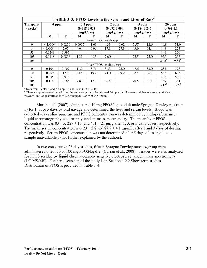
Perfluorooctane sulfonate (PFOS) – February 2014 3-7 Draft – Do Not Cite or Quote
TABLE 3-3. PFOS Levels in the Serum and Liver of Ratsa
Timepoint (weeks)
0 ppm 0.5 ppm (0.018-0.023 mg/k/day)
2 ppm (0.072-0.099 mg/kg/day)
5 ppm (0.184-0.247 mg/kg/day)
20 ppm (0.765-1.1
mg/kg/day) M F M F M F M F M F
Serum PFOS levels (ppm) 0 < LOQ* 0.0259 0.0907 1.61 4.33 6.62 7.57 12.6 41.8 54.0
14 < LOQ** 2.67 4.04 6.96 17.1 27.3 43.9 64.4 148 223 53 0.0249 0.395 146 220
105 0.0118 0.0836 1.31 4.35 7.60 22.5 75.0 69.3 233 106 2.42b 9.51b
Liver PFOS levels (µg/g) 0 0.104 0.107 11.0 8.71 31.3 25.0 47.6 83.0 282 373
10 0.459 12.0 23.8 19.2 74.0 69.2 358 370 568 635 53 0.635 0.932 435 560
105 0.114 0.185 7.83 12.9 26.4 70.5 131 189 381 106 3.12b 12.9b
a Data from Tables 4 and 5 on pp. 38 and 39 in OECD 2002 b These samples were obtained from the recovery group administered 20 ppm for 52 weeks and then observed until death. *LOQ= limit of quantification = 0.00910 pg/mL or ** 0.0457 pg/mL
Martin et al. (2007) administered 10 mg PFOS/kg to adult male Sprague-Dawley rats (n = 5) for 1, 3, or 5 days by oral gavage and determined the liver and serum levels. Blood was collected via cardiac puncture and PFOS concentration was determined by high-performance liquid chromatography-electrospray tandem mass spectrometry. The mean liver PFOS concentration was 83 ± 5, 229 ± 10, and 401 ± 21 μg/g after 1, 3, or 5 daily doses, respectively. The mean serum concentration was 23 ± 2.8 and 87.7 ± 4.1 μg/mL, after 1 and 3 days of dosing, respectively. Serum PFOS concentration was not determined after 5 days of dosing due to sample unavailability (not further explained by the authors). In two consecutive 28-day studies, fifteen Sprague-Dawley rats/sex/group were administered 0, 20, 50 or 100 mg PFOS/kg diet (Curran et al., 2008). Tissues were also analyzed for PFOS residue by liquid chromatography negative electrospray tandem mass spectrometry (LC-MS/MS). Further discussion of the study is in Section 4.2.2 Short-term studies. Distribution of PFOS is provided in Table 3-4.

Perfluorooctane sulfonate (PFOS) – February 2014 3-8 Draft – Do Not Cite or Quote
TABLE 3-4. Mean (± SD) daily PFOS Consumption and Tissue Residue Levels in Rats Treated for 28 Daysa
Parameter 0 mg/kg diet 2 mg/kg diet 20 mg/kg diet 50 mg/kg diet 100 mg/kg diet M F M F M F M F M F
PFOS consumption (mg/kg bw/day)
0 0 0.14 ± 0.02
0.15 ± 0.02
1.33 ± 0.24
1.43 ± 0.24
3.21 ± 0.57
3.73 ± 0.57
6.34 ± 1.35
7.58 ± 0.68
Serum (µg PFOS/g serum)
0.47 ±
0.27
0.95 ± 0.51
0.95 ± 0.13
1.50 ± 0.23
13.45 ± 1.48
15.40 ± 1.56
20.93 ± 2.36
31.93 ± 3.59
29.88 ± 3.53
43.20 ± 3.95
Liver (µg PFOS/g liver)
0.79 ±
0.49
0.89 ± 0.44
48.28 ± 5.81
43.44 ± 6.79
560.23 ±
104.43
716.55 ±
59.15
856.90 ±
353.83
596.75 ±
158.01
1030.40 ± 162.80
1008.59 ± 49.41
Ratio liver:serum PFOS
2.04 ±
1.39
1.30 ± 1.32
51.34 ± 9.20
29.99 ± 8.11
42.10 ± 9.20
46.81 ± 5.26
41.42 ± 16.95
20.23 ± 7.50
35.23 ± 8.50
23.48 ± 1.98
Spleen (µg PFOS/g spleen)
0.27 ±
0.36
2.08 ± 4.17
6.07 ± 1.85
7.94 ± 3.76
45.27 ± 2.16
70.03 ±
36.66
122.51 ± 7.83
139.45 ± 15.44
230.73 ± 11.47
294.96 ± 26.66
Heart (µg PFOS/g heart)
0.10 ±
0.14
1.42 ± 2.91
4.67 ± 1.73
6.54 ± 3.07
33.00 ± 3.44
54.65 ±
30.89
90.28 ± 4.95
107.53 ± 6.24
154.13 ± 11.78
214.45 ± 17.58
a Data from Table 1 on p. 1531 in Curran et al., 2008 SD = standard deviation
Ten three-month old male Sprague-Dawley rats/group were administered 0 (Milli-Q water only), 5 or 20 mg/kg/day of PFOS by oral gavage for 28 days (Cui et al., 2009). Rats were sacrificed after the exposure and blood and tissue samples obtained. Concentrations identified in rat whole blood and various tissues at the end of the exposure are provided in Table 3-5. The study indicated that the highest levels of PFOS were identified in the liver after 28 days of exposure.
TABLE 3-5. Concentrations of PFOS in Male Rats’ Whole Blood (µg/mL) and Various Tissues (µg/g) After 28 Daysa
Tissues Controls 5 mg/kg/day PFOS 20 mg/kg/day PFOS blood ND 72.0 ± 25.7 No sample liver ND 345 ± 40 648 ± 17
kidney ND 93.9 ± 13.6 248 ± 26 lung ND 46.6 ± 17.8 228 ± 122 heart ND 168 ± 17 497 ± 64
spleen ND 38.5 ± 11.8 167 ± 64 testicle ND 39.5 ± 10.0 127 ± 11 brain ND 13.6 ± 1.0 146 ± 34
a Data from Table 1 in Cui et al., 2009. ND = not detected
Yu et al. (2011) administered the following doses to approximately six female Wistar
rats/group are part of a study of PFOS on the thyroid: 1) vehicle (0.5% Tween 20), 2) PFOS at 0.2, 1.0 or 3.0 mg/kg, 3) propylthiouracil (PTU) at 10 mg/kg or

Perfluorooctane sulfonate (PFOS) – February 2014 3-9 Draft – Do Not Cite or Quote
4) PTU at 10 mg/kg and PFOS at 3.0 mg/kg once daily by gavage for 5 consecutive days. Blood, bile and liver tissue were collected 24 hours after the last dose. The serum was used to determine the level of PFOS as well as T4 (TT4) and T3 (TT3). PFOS levels in the serum as well as the bile are provided in Table 3-6. The data demonstrate distribution to serum and bile with a direct relationship to dose.
TABLE 3-6. Levels of PFOS in serum and bile of rats treated for 5 daysa
PFOS (mg/kg bw) Serum PFOS (mg/L) Bile PFOS (mg/L) 0.0 <LOQ <LOQ 0.2 1.09 ± 0.12 1.51 ± 0.42 1.0 8.20 ± 0.13 3.58 ± 0.66 3.0 33.5 ± 1.79 6.51 ± 0.67
a Data from Table 2 in Yu et al. 2011. LOQ = limit of quantification, 0.5 µg/L
Rat- Distribution in Reproductive/Developmental Studies
Two studies administered PFOS up to 3.2 mg/kg/day orally to Sprague-Dawley rats
during cohabitation and either confirmed day of pregnancy or gestation day (GD) 14 or 20 (3M Environmental Laboratory, 2001a and 2001b). Serum, urine, liver and feces were collected. The studies showed that liver samples from adults and fetuses contained higher amounts of PFOS when compared to the serum.
To determine the dose-response curve for neonatal mortality in rat pups born to PFOS exposed dams and to investigate the biochemical and pharmacokinetic parameters, five groups of 16 female Crl:CD(SD)IGS VAF/Plus rats each were administered 0, 0.1, 0.4, 1.6 or 3.2 mg PFOS/kg bw/day by oral gavage beginning 42 days prior to cohabitation and continuing through gestation day (GD) 14 or 20 (Luebker et al., 2005a). Eight rats from each group were randomly chosen and sacrificed on GD 15. Caesarean sections (C-section) were also performed. All remaining animals were sacrificed and C-sectioned on GD 21. Urine and feces were collected overnight from dams on the eve of cohabitation day 1 and during GDs 6-7, 14-15 and 20-21. Serum samples were collected just prior to cohabitation and on GD 7, GD 15 and GD 21. Liver and blood samples were also obtained from the fetuses on GD 21 and pooled by litter. The urine, feces and liver of the control animals all contained PFOS at small concentrations. In treated rats, the highest concentration of PFOS was in the liver. Compared to the dams, the levels of PFOS in the fetuses were similar in the serum, but much lower in the liver. This same study also performed a dose response study that was conducted in a similar manner but obtained liver and serum samples from pups on lactation day 5. In this sampling, serum PFOS levels were similar between the dam and offspring but the liver values were now higher in the neonates. The concentrations found in the pregnant dams and in the fetuses are provided in Table 3-7 below.
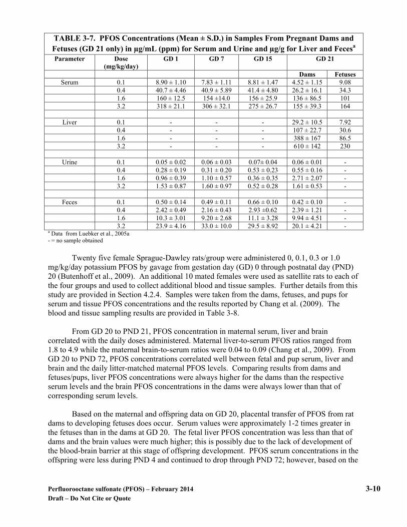
Perfluorooctane sulfonate (PFOS) – February 2014 3-10 Draft – Do Not Cite or Quote
TABLE 3-7. PFOS Concentrations (Mean ± S.D.) in Samples From Pregnant Dams and Fetuses (GD 21 only) in µg/mL (ppm) for Serum and Urine and µg/g for Liver and Fecesa
Parameter Dose (mg/kg/day)
GD 1 GD 7 GD 15 GD 21
Dams Fetuses Serum 0.1 8.90 ± 1.10 7.83 ± 1.11 8.81 ± 1.47 4.52 ± 1.15 9.08
0.4 40.7 ± 4.46 40.9 ± 5.89 41.4 ± 4.80 26.2 ± 16.1 34.3 1.6 160 ± 12.5 154 ±14.0 156 ± 25.9 136 ± 86.5 101 3.2 318 ± 21.1 306 ± 32.1 275 ± 26.7 155 ± 39.3 164
Liver 0.1 - - - 29.2 ± 10.5 7.92 0.4 - - - 107 ± 22.7 30.6 1.6 - - - 388 ± 167 86.5 3.2 - - - 610 ± 142 230
Urine 0.1 0.05 ± 0.02 0.06 ± 0.03 0.07± 0.04 0.06 ± 0.01 - 0.4 0.28 ± 0.19 0.31 ± 0.20 0.53 ± 0.23 0.55 ± 0.16 - 1.6 0.96 ± 0.39 1.10 ± 0.57 0.36 ± 0.35 2.71 ± 2.07 - 3.2 1.53 ± 0.87 1.60 ± 0.97 0.52 ± 0.28 1.61 ± 0.53 -
Feces 0.1 0.50 ± 0.14 0.49 ± 0.11 0.66 ± 0.10 0.42 ± 0.10 - 0.4 2.42 ± 0.49 2.16 ± 0.43 2.93 ±0.62 2.39 ± 1.21 - 1.6 10.3 ± 3.01 9.20 ± 2.68 11.1 ± 3.28 9.94 ± 4.51 - 3.2 23.9 ± 4.16 33.0 ± 10.0 29.5 ± 8.92 20.1 ± 4.21 -
a Data from Luebker et al., 2005a - = no sample obtained
Twenty five female Sprague-Dawley rats/group were administered 0, 0.1, 0.3 or 1.0
mg/kg/day potassium PFOS by gavage from gestation day (GD) 0 through postnatal day (PND) 20 (Butenhoff et al., 2009). An additional 10 mated females were used as satellite rats to each of the four groups and used to collect additional blood and tissue samples. Further details from this study are provided in Section 4.2.4. Samples were taken from the dams, fetuses, and pups for serum and tissue PFOS concentrations and the results reported by Chang et al. (2009). The blood and tissue sampling results are provided in Table 3-8.
From GD 20 to PND 21, PFOS concentration in maternal serum, liver and brain
correlated with the daily doses administered. Maternal liver-to-serum PFOS ratios ranged from 1.8 to 4.9 while the maternal brain-to-serum ratios were 0.04 to 0.09 (Chang et al., 2009). From GD 20 to PND 72, PFOS concentrations correlated well between fetal and pup serum, liver and brain and the daily litter-matched maternal PFOS levels. Comparing results from dams and fetuses/pups, liver PFOS concentrations were always higher for the dams than the respective serum levels and the brain PFOS concentrations in the dams were always lower than that of corresponding serum levels.
Based on the maternal and offspring data on GD 20, placental transfer of PFOS from rat
dams to developing fetuses does occur. Serum values were approximately 1-2 times greater in the fetuses than in the dams at GD 20. The fetal liver PFOS concentration was less than that of dams and the brain values were much higher; this is possibly due to the lack of development of the blood-brain barrier at this stage of offspring development. PFOS serum concentrations in the offspring were less during PND 4 and continued to drop through PND 72; however, based on the
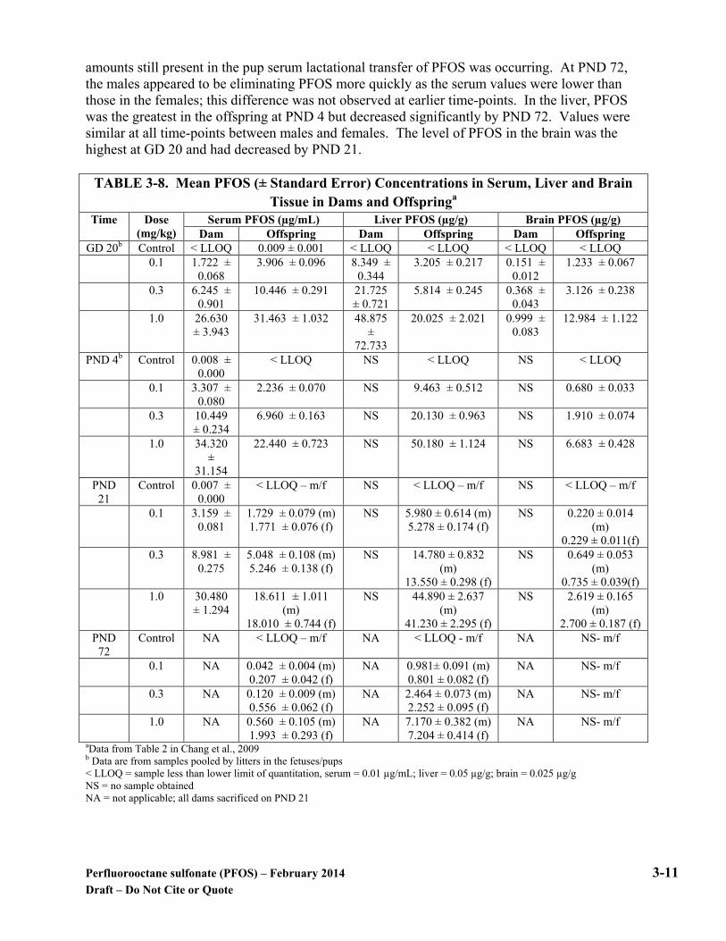
Perfluorooctane sulfonate (PFOS) – February 2014 3-11 Draft – Do Not Cite or Quote
amounts still present in the pup serum lactational transfer of PFOS was occurring. At PND 72, the males appeared to be eliminating PFOS more quickly as the serum values were lower than those in the females; this difference was not observed at earlier time-points. In the liver, PFOS was the greatest in the offspring at PND 4 but decreased significantly by PND 72. Values were similar at all time-points between males and females. The level of PFOS in the brain was the highest at GD 20 and had decreased by PND 21.
TABLE 3-8. Mean PFOS (± Standard Error) Concentrations in Serum, Liver and Brain
Tissue in Dams and Offspringa
Time Dose (mg/kg)
Serum PFOS (µg/mL) Liver PFOS (µg/g) Brain PFOS (µg/g) Dam Offspring Dam Offspring Dam Offspring
GD 20b Control < LLOQ 0.009 ± 0.001 < LLOQ < LLOQ < LLOQ < LLOQ 0.1 1.722 ±
0.068 3.906 ± 0.096 8.349 ±
0.344 3.205 ± 0.217 0.151 ±
0.012 1.233 ± 0.067
0.3 6.245 ± 0.901
10.446 ± 0.291 21.725 ± 0.721
5.814 ± 0.245 0.368 ± 0.043
3.126 ± 0.238
1.0 26.630 ± 3.943
31.463 ± 1.032 48.875 ±
72.733
20.025 ± 2.021 0.999 ± 0.083
12.984 ± 1.122
PND 4b Control 0.008 ± 0.000
< LLOQ NS < LLOQ NS < LLOQ
0.1 3.307 ± 0.080
2.236 ± 0.070 NS 9.463 ± 0.512 NS 0.680 ± 0.033
0.3 10.449 ± 0.234
6.960 ± 0.163 NS 20.130 ± 0.963 NS 1.910 ± 0.074
1.0 34.320 ±
31.154
22.440 ± 0.723 NS 50.180 ± 1.124 NS 6.683 ± 0.428
PND 21
Control 0.007 ± 0.000
< LLOQ – m/f NS < LLOQ – m/f NS < LLOQ – m/f
0.1 3.159 ± 0.081
1.729 ± 0.079 (m) 1.771 ± 0.076 (f)
NS 5.980 ± 0.614 (m) 5.278 ± 0.174 (f)
NS 0.220 ± 0.014 (m)
0.229 ± 0.011(f) 0.3 8.981 ±
0.275 5.048 ± 0.108 (m) 5.246 ± 0.138 (f)
NS 14.780 ± 0.832 (m)
13.550 ± 0.298 (f)
NS 0.649 ± 0.053 (m)
0.735 ± 0.039(f) 1.0 30.480
± 1.294 18.611 ± 1.011
(m) 18.010 ± 0.744 (f)
NS 44.890 ± 2.637 (m)
41.230 ± 2.295 (f)
NS 2.619 ± 0.165 (m)
2.700 ± 0.187 (f) PND
72 Control NA < LLOQ – m/f NA < LLOQ - m/f NA NS- m/f
0.1 NA 0.042 ± 0.004 (m) 0.207 ± 0.042 (f)
NA 0.981± 0.091 (m) 0.801 ± 0.082 (f)
NA NS- m/f
0.3 NA 0.120 ± 0.009 (m) 0.556 ± 0.062 (f)
NA 2.464 ± 0.073 (m) 2.252 ± 0.095 (f)
NA NS- m/f
1.0 NA 0.560 ± 0.105 (m) 1.993 ± 0.293 (f)
NA 7.170 ± 0.382 (m) 7.204 ± 0.414 (f)
NA NS- m/f
aData from Table 2 in Chang et al., 2009 b Data are from samples pooled by litters in the fetuses/pups < LLOQ = sample less than lower limit of quantitation, serum = 0.01 µg/mL; liver = 0.05 µg/g; brain = 0.025 µg/g NS = no sample obtained NA = not applicable; all dams sacrificed on PND 21
Perfluorooctane sulfonate (PFOS) – February 2014 3-12 Draft – Do Not Cite or Quote
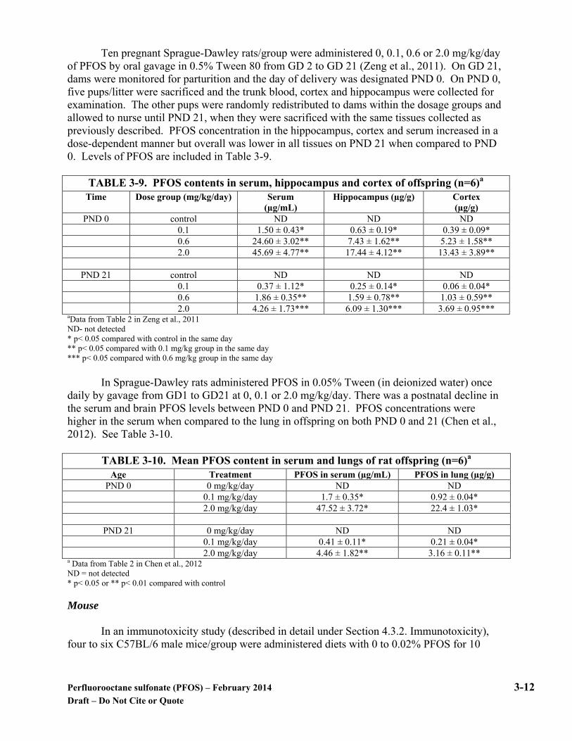
Ten pregnant Sprague-Dawley rats/group were administered 0, 0.1, 0.6 or 2.0 mg/kg/day of PFOS by oral gavage in 0.5% Tween 80 from GD 2 to GD 21 (Zeng et al., 2011). On GD 21, dams were monitored for parturition and the day of delivery was designated PND 0. On PND 0, five pups/litter were sacrificed and the trunk blood, cortex and hippocampus were collected for examination. The other pups were randomly redistributed to dams within the dosage groups and allowed to nurse until PND 21, when they were sacrificed with the same tissues collected as previously described. PFOS concentration in the hippocampus, cortex and serum increased in a dose-dependent manner but overall was lower in all tissues on PND 21 when compared to PND 0. Levels of PFOS are included in Table 3-9.
TABLE 3-9. PFOS contents in serum, hippocampus and cortex of offspring (n=6)a
Time Dose group (mg/kg/day) Serum (µg/mL)
Hippocampus (µg/g) Cortex (µg/g)
PND 0 control ND ND ND 0.1 1.50 ± 0.43* 0.63 ± 0.19* 0.39 ± 0.09* 0.6 24.60 ± 3.02** 7.43 ± 1.62** 5.23 ± 1.58** 2.0 45.69 ± 4.77** 17.44 ± 4.12** 13.43 ± 3.89**
PND 21 control ND ND ND 0.1 0.37 ± 1.12* 0.25 ± 0.14* 0.06 ± 0.04* 0.6 1.86 ± 0.35** 1.59 ± 0.78** 1.03 ± 0.59** 2.0 4.26 ± 1.73*** 6.09 ± 1.30*** 3.69 ± 0.95***
aData from Table 2 in Zeng et al., 2011 ND- not detected * p< 0.05 compared with control in the same day ** p< 0.05 compared with 0.1 mg/kg group in the same day *** p< 0.05 compared with 0.6 mg/kg group in the same day In Sprague-Dawley rats administered PFOS in 0.05% Tween (in deionized water) once daily by gavage from GD1 to GD21 at 0, 0.1 or 2.0 mg/kg/day. There was a postnatal decline in the serum and brain PFOS levels between PND 0 and PND 21. PFOS concentrations were higher in the serum when compared to the lung in offspring on both PND 0 and 21 (Chen et al., 2012). See Table 3-10.
TABLE 3-10. Mean PFOS content in serum and lungs of rat offspring (n=6)a Age Treatment PFOS in serum (µg/mL) PFOS in lung (µg/g)
PND 0 0 mg/kg/day ND ND 0.1 mg/kg/day 1.7 ± 0.35* 0.92 ± 0.04* 2.0 mg/kg/day 47.52 ± 3.72* 22.4 ± 1.03*
PND 21 0 mg/kg/day ND ND 0.1 mg/kg/day 0.41 ± 0.11* 0.21 ± 0.04* 2.0 mg/kg/day 4.46 ± 1.82** 3.16 ± 0.11**
a Data from Table 2 in Chen et al., 2012 ND = not detected * p< 0.05 or ** p< 0.01 compared with control
Mouse In an immunotoxicity study (described in detail under Section 4.3.2. Immunotoxicity), four to six C57BL/6 male mice/group were administered diets with 0 to 0.02% PFOS for 10

Perfluorooctane sulfonate (PFOS) – February 2014 3-13 Draft – Do Not Cite or Quote
days. Levels in the serum increased as the concentration increased (Qazi et al., 2009a). See Table 3-11. TABLE 3-11. Levels of PFOS (Means ± SE) in Mouse Serum Following Treatment
for 10 Daysa
Dietary dose (% w/w) Number of mice ppm PFOS (0) 4 0.0287 ± 0.01
PFOS (0.001%) 4 50.8 ± 2.5 PFOS (0.005%) 4 96.7 ± 5.2 PFOS (0.02%) 4 340 ± 16
a Data from study report by Qazi et al., 2009a Adult male C57/BL6 mice (3 mice/group) were administered 35S-PFOS in the feed at a low and high dose for 1, 3 and 5 days. The dose equivalents were 0.031 mg/kg/day in the low dose group and 23 mg/kg/day in the high dose group. Tissue contents were determined by liquid scintillation (Bogdanska et al., 2011). At 23 mg/kg/day after 5 days, mice had hypertrophy of the liver, atrophy of fat pads and atrophy of epididymal fat when compared to the mice at 0.013 mg/kg/day at 5 days. To determine the amount of radioactivity recovered that was due to blood in the tissues, the hemoglobin content was determined in all of the samples. By correcting for PFOS in the blood, the actual tissue levels were then calculated.
At both doses and at all time-points, the liver contained the highest amount of PFOS. At the low dose, the liver PFOS level relative to blood concentration increased with time, whereas at the high dose, the ratio plateaued after three days. The autoradiography indicated that the distribution within the liver did not appear to favor one area to a greater extent than any other. The liver contained 40 to 50% of the recovered PFOS at the high dose. The authors hypothesized that this could possibly reflect high levels of binding to tissue proteins.
In the high dose mice, the next highest level was found in the lungs. Distribution was
fairly uniform with some favoring of specific surface areas. The tissue: blood ratio for the lung was greater than that for all other tissues except the liver. The lowest PFOS levels were in the brain and fat deposits.
While the levels in Table 3-12 report the PFOS in the whole bone, when the authors did a
whole body autoradiogram of a mouse 48 hours after a single oral dose of 35S-PFOS (12.5 mg/kg), the results indicated that most PFOS was found in the bone marrow and not the calcified bone. Levels for the kidney roughly equal those values observed in the blood at both concentrations and all timepoints. See Table 3-12.

Perfluorooctane sulfonate (PFOS) – February 2014 3-14 Draft – Do Not Cite or Quote
TABLE 3-12. Mean Concentration of PFOS (±SD) in Various Tissues of Micea
Tissues 1 day 3 days 5 days Dose of 0.013 mg/kg/day (PFOS in tissue reported as pmol/g)
Blood 61(6) 129 (41)# 99 (21) Liver 114 (13)** 343 (24)**# 578 (39)**#
Kidney 38 (19) 65 (13) 93 (11)# Lung 39 (29) 88 (6)# 141 (10)*#
Whole bone 113 (15)** 98 (24) 109 (6)
Dose of 23 mg/kg/day (PFOS in tissue reported as nmol/g) Blood 67 (4) 171 (21)# 287 (9)# Liver 246 (31)** 698 (71)**# 1044 (114)**#
Kidney 62 (3) 166 (8)# 233 (12)**# Lung 135 (18)** 336 (69)*# 445 (42)**#
Whole bone 55 (6)* 155 (17)# 207 (8)**# aData from Tables 2 and 3 in Bogdanska et al., 2011 *significantly different (p< 0.05) than blood at the same time-point as evaluated by an independent t-test **significantly different (p<0.01) than blood at the same time-point as evaluated by an independent t-test #significantly different (p< 0.05) from the value for the same tissue at day 1 as determined by one-way analysis of variance (ANOVA) followed by Duncan’s test Mouse- Distribution in Reproductive/Developmental Studies Borg et al. (2010) administered a single dose of 12.5 mg/kg 35S-PFOS by intravenous injection (n=1) or gavage (n=5) on gestation day (GD) 16 to C57Bl/6 mouse dams. Distribution of PFOS was determined in the dams/fetuses (GD 18 and 20) and pups (PND 1) by using whole-body autoradiography and liquid scintillation counting. Distribution in the dams was similar regardless of the route of exposure with the hepatic level being approximately four times greater than the blood. At all timepoints in the dams, PFOS was most concentrated in the liver and lungs. In the fetuses, the highest concentrations of PFOS were found in the kidneys and liver and in pups on PND 1, PFOS was mostly concentrated in the lungs/liver. In dams, the concentration of PFOS in the liver was approximately 4x and in the lung was approximately 2x the blood concentrations, respectively. The distribution of PFOS in the kidneys was similar and the amount in the brain was lower than that of the blood. In the offspring at all timepoints, PFOS was homogeneously distributed in the liver at a level 2.5x higher than maternal blood and 1.7x lower than maternal liver. In the fetuses on GD 18, values in the lungs were similar to the maternal lungs and this value increased by GD 20. Pups on PND 1 had PFOS levels that were 3x higher in the lungs, compared to maternal blood with a heterogeneous distribution. In the kidneys, the highest concentration of PFOS was observed in the fetuses on GD 18 (3x higher than maternal levels) but then was similar to the dams on GD 20 and PND 1. Levels in the brain were similar at all timepoints in the offspring and higher than in the maternal brain, likely due to an immature brain-blood barrier. Select data are provided in Table 3-13 and Figure 3-1.

Perfluorooctane sulfonate (PFOS) – February 2014 3-15 Draft – Do Not Cite or Quote
TABLE 3-13. Ratios (means ± S.D.) between the concentrations of 35S-labeled PFOS in various organs and blood of mouse dams, fetuses and pups versus the average concentration
in maternal blooda [35S-PFOS]organ/[35S-PFOS]maternal blood
Subject Liver (n=6-8) Lungs (n=5-6) Kidneys (n=3-6) Brain (n=6-9) Blood (n=1-6) Dams 4.2** ± 0.7 2.0* ± 0.4 0.9 ± 0.1 0.2** ± 0.05 1.0
Fetus on GD 18 2.6** ± 0.8 2.1* ± 0.6 2.8** ± 0.3 1.2 ± 0.3 2.3 Fetus on GD 20 2.4** ± 0.5 2.5** ± 0.4 1.4 ± 0.2 0.9 ± 0.1 1.1 ± 0.04 Pups on PND 1 2.4* ± 0.4 3.0** ± 0.5 1.0 ± 0.5 0.9 ± 0.2 1.7** ± 0.3
a Data from Table 1 in Borg et al. (2010) *Statistically significant (p≤0.01) in comparison to maternal blood ** Statistically significant (p≤0.001) in comparison to maternal blood

Perfluorooctane sulfonate (PFOS) – February 2014 3-16 Draft – Do Not Cite or Quote
Filled symbols are representative after oral exposure; open after intravenous exposure. A = p≤ 0.001 and a = p≤ 0.01, compared to maternal blood B = p≤ 0.001 and b = p≤ 0.05, compared to maternal tissue
C = p≤ 0.001 and c = p≤ 0.05, comparing between fetuses/pups on GD20/PND1 with corresponding value on GD18;
Figure 3-1. Distribution of radiolabeled PFOS in dams and in fetuses/pups in the liver,
lung, kidney and brain (Figure from Borg et al., 2010)
3.2.2 Inhalation and Dermal Exposure
No data on distribution following inhalation or dermal exposures were identified.
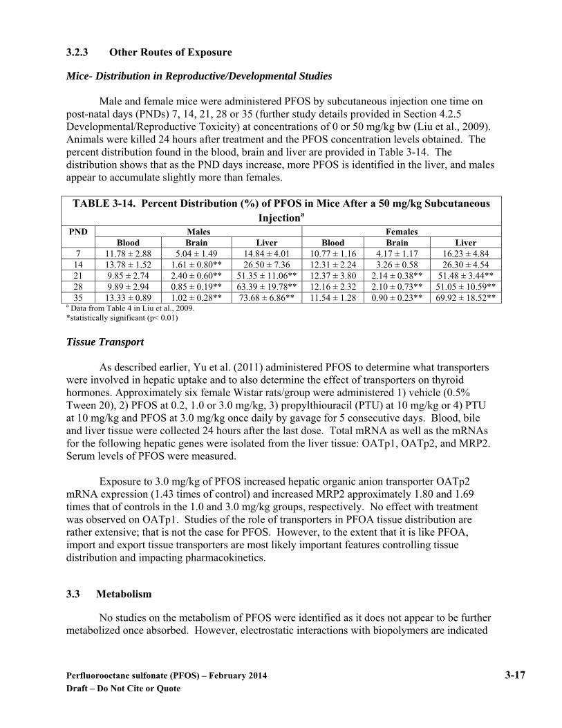
Perfluorooctane sulfonate (PFOS) – February 2014 3-17 Draft – Do Not Cite or Quote
3.2.3 Other Routes of Exposure
Mice- Distribution in Reproductive/Developmental Studies Male and female mice were administered PFOS by subcutaneous injection one time on
post-natal days (PNDs) 7, 14, 21, 28 or 35 (further study details provided in Section 4.2.5 Developmental/Reproductive Toxicity) at concentrations of 0 or 50 mg/kg bw (Liu et al., 2009). Animals were killed 24 hours after treatment and the PFOS concentration levels obtained. The percent distribution found in the blood, brain and liver are provided in Table 3-14. The distribution shows that as the PND days increase, more PFOS is identified in the liver, and males appear to accumulate slightly more than females.
TABLE 3-14. Percent Distribution (%) of PFOS in Mice After a 50 mg/kg Subcutaneous
Injectiona PND Males Females
Blood Brain Liver Blood Brain Liver 7 11.78 ± 2.88 5.04 ± 1.49 14.84 ± 4.01 10.77 ± 1.16 4.17 ± 1.17 16.23 ± 4.84
14 13.78 ± 1.52 1.61 ± 0.80** 26.50 ± 7.36 12.31 ± 2.24 3.26 ± 0.58 26.30 ± 4.54 21 9.85 ± 2.74 2.40 ± 0.60** 51.35 ± 11.06** 12.37 ± 3.80 2.14 ± 0.38** 51.48 ± 3.44** 28 9.89 ± 2.94 0.85 ± 0.19** 63.39 ± 19.78** 12.16 ± 2.32 2.10 ± 0.73** 51.05 ± 10.59** 35 13.33 ± 0.89 1.02 ± 0.28** 73.68 ± 6.86** 11.54 ± 1.28 0.90 ± 0.23** 69.92 ± 18.52**
a Data from Table 4 in Liu et al., 2009. *statistically significant (p< 0.01) Tissue Transport
As described earlier, Yu et al. (2011) administered PFOS to determine what transporters were involved in hepatic uptake and to also determine the effect of transporters on thyroid hormones. Approximately six female Wistar rats/group were administered 1) vehicle (0.5% Tween 20), 2) PFOS at 0.2, 1.0 or 3.0 mg/kg, 3) propylthiouracil (PTU) at 10 mg/kg or 4) PTU at 10 mg/kg and PFOS at 3.0 mg/kg once daily by gavage for 5 consecutive days. Blood, bile and liver tissue were collected 24 hours after the last dose. Total mRNA as well as the mRNAs for the following hepatic genes were isolated from the liver tissue: OATp1, OATp2, and MRP2. Serum levels of PFOS were measured.
Exposure to 3.0 mg/kg of PFOS increased hepatic organic anion transporter OATp2 mRNA expression (1.43 times of control) and increased MRP2 approximately 1.80 and 1.69 times that of controls in the 1.0 and 3.0 mg/kg groups, respectively. No effect with treatment was observed on OATp1. Studies of the role of transporters in PFOA tissue distribution are rather extensive; that is not the case for PFOS. However, to the extent that it is like PFOA, import and export tissue transporters are most likely important features controlling tissue distribution and impacting pharmacokinetics.
3.3 Metabolism
No studies on the metabolism of PFOS were identified as it does not appear to be further metabolized once absorbed. However, electrostatic interactions with biopolymers are indicated

Perfluorooctane sulfonate (PFOS) – February 2014 3-18 Draft – Do Not Cite or Quote
by the Kerstner-Wood et al. (2003) data on binding to plasma proteins plus the Zhang et al. (2009) and Chen and Guo (2009) data from albumin-binding investigations.
3.4 Excretion
Humans Harada et al. (2007) obtained serum and bile samples from patients (2 male and 2 female; ages 63-76) undergoing gallstone surgery to determine the bile to serum ratio and biliary resorption rate. The median concentration for PFOS in the serum was 23.2 ng/mL (0.023 ppm), compared to the bile, 27.9 ng/mL (0.028 ppm). The biliary resorption rate was 0.97 which could contribute to the long-half life in humans. Method of exposure to PFOS was unknown. See Table 3-15.
TABLE 3-15. Estimation of Toxicokinetic Parameters for PFOSa Bile
Participants Serum levels- ng/mL (ppm) Bile levels- ng/mL (ppm) Bile to serum ratio Median (n=4) 23.2 (0.023) 27.9 (0.028) 0.60
a Data from Tables 2 and 3 in Harada et al., 2007
Animals Studies to determine the direct excretion routes are only available for rats. Most excretion for PFOS occurs in the urine.
3.4.1 Oral Exposure
Ten male Sprague-Dawley rats (~ 9 weeks old)/group were administered 0, 5 or 20 mg/kg/day of either PFOA or PFOS by gavage once daily, 7 days a week for 4 weeks (Cui et al., 2010). The dose groups were identified as the following: G0 = ultrapure water; G1= 5 mg/kg/day PFOA; G2 = 20 mg/kg/day PFOA; G3 = 5 mg/kg/day PFOS and G4 = 20 mg/kg/day PFOS. Urine and fecal samples were obtained after the daily gavage by placing the rats in metabolism cages for 24 hour intervals on the following days: prior to treatment (Day 0), Day 1 and Days 3, 5, 7, 10, 14, 18, 21, 24 and 28. Urine was collected three times daily and the volume of the urine sample and weight of the fecal sample were recorded; samples were stored at -40°C prior to analyzing. Target analytes were determined by using a high-performance liquid chromatography-electrospray tandem mass spectrometry system with separation of PFOS and PFOA achieved by the analytical column. An upward trend of increased excretion was observed in the rats administered 5 mg/kg/day PFOS during the study; a similar trend was observed in the rats administered 20 mg/kg/day PFOS, but in the third week, mortalities occurred. By study day 24, there were only 2/10 rats in the 20 mg/kg/day group surviving. The range of PFOS excreted in urine by rats treated with 20 mg/kg/day was 0.080 mg on day 1 to 0.673 mg on Day 14. In the feces, the lowest amount of PFOS was in rats at 5 mg/kg/day on Day 1 (0.0015 mg) and the highest on Day 28 (0.355 mg). A similar trend in feces was observed in the rats treated with 20 mg/kg/day until the deaths occurred; however, the fecal excretion reached a steady state after a maximum on day 18 (0.519 mg). This could have been the result of lower feces volume because the rats had
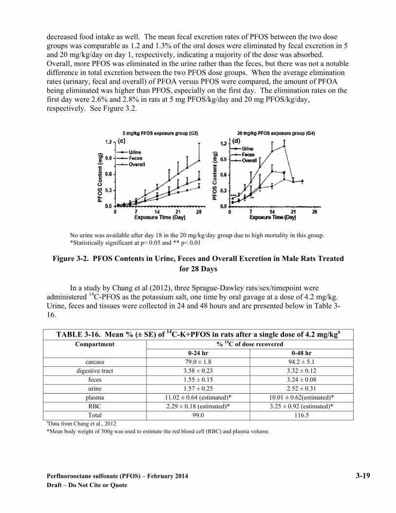
Perfluorooctane sulfonate (PFOS) – February 2014 3-19 Draft – Do Not Cite or Quote
decreased food intake as well. The mean fecal excretion rates of PFOS between the two dose groups was comparable as 1.2 and 1.3% of the oral doses were eliminated by fecal excretion in 5 and 20 mg/kg/day on day 1, respectively, indicating a majority of the dose was absorbed. Overall, more PFOS was eliminated in the urine rather than the feces, but there was not a notable difference in total excretion between the two PFOS dose groups. When the average elimination rates (urinary, fecal and overall) of PFOA versus PFOS were compared, the amount of PFOA being eliminated was higher than PFOS, especially on the first day. The elimination rates on the first day were 2.6% and 2.8% in rats at 5 mg PFOS/kg/day and 20 mg PFOS/kg/day, respectively. See Figure 3.2.
No urine was available after day 18 in the 20 mg/kg/day group due to high mortality in this group. *Statistically significant at p< 0.05 and ** p< 0.01
Figure 3-2. PFOS Contents in Urine, Feces and Overall Excretion in Male Rats Treated for 28 Days
In a study by Chang et al (2012), three Sprague-Dawley rats/sex/timepoint were administered 14C-PFOS as the potassium salt, one time by oral gavage at a dose of 4.2 mg/kg. Urine, feces and tissues were collected in 24 and 48 hours and are presented below in Table 3-16.
TABLE 3-16. Mean % (± SE) of 14C-K+PFOS in rats after a single dose of 4.2 mg/kga Compartment % 14C of dose recovered
0-24 hr 0-48 hr carcass 79.0 ± 1.8 94.2 ± 5.1
digestive tract 3.58 ± 0.23 3.32 ± 0.12 feces 1.55 ± 0.15 3.24 ± 0.08 urine 1.57 ± 0.25 2.52 ± 0.31
plasma 11.02 ± 0.64 (estimated)* 10.01 ± 0.62(estimated)* RBC 2.29 ± 0.18 (estimated)* 3.25 ± 0.92 (estimated)* Total 99.0 116.5
aData from Chang et al., 2012 *Mean body weight of 300g was used to estimate the red blood cell (RBC) and plasma volume.
Perfluorooctane sulfonate (PFOS) – February 2014 3-20 Draft – Do Not Cite or Quote
3.4.2 Inhalation Exposure
In a case report, a 51-year old asymptomatic male researcher lived in a home with carpet flooring that had been treated intermittently with soil/dirt repellants. The carpeting also had an in-floor heating system under the carpets (Genuis et al., 2010). Because of his work the man knew that he had an unusually high amount of PFCs in his serum, primarily perfluorohexanesulfonic acid (PFHxS), PFOS and PFOA. The level of PFOS in his serum was 26 ng/g, the level in his urine was < 0.50 ng/mL and < 0.50 ng/g in sweat and stool samples. The man began treatment with two bile acid sequestrants, cholestyramine (CSM) and saponin compounds (SPCs) to see if they would lower the serum PFC levels. Stool samples were evaluated for PFOS levels after administration of each compound. The concentration of PFOS was increased after CSM treatment, suggesting that it may help with removing PFOS that gains access to the GI tract with bile. The first stool sample after approximately 20 weeks of CSM treatment showed PFOS levels of 9.06 ng/g and the second, 7.94 ng/g. The treatment with SPCs did not increase the PFOS found in the stool. Serum levels of PFOS decreased to 15.6 ng/g after 12 weeks of treatment with CSM and to 14.4 ng/g after 20 weeks of treatment even though the man’s exposure at his home had not changed.
3.4.3 Dermal Exposure
No data on PFOS excretion following dermal exposures were identified.
3.4.4 Other Exposure Routes
At 89 days after a single intravenous (IV) dose of PFOS-14C of 4.2 mg/kg to male rats, urinary excretion was 30.2 ± 1.5% of the total C-14 administered. Mean fecal excretion was 12.6 ± 1.2% (Chang et al., 2012). There was also evidence of enterohepatic circulation of PFOS.
3.5 Pharmacokinetic Considerations
3.5.1 Physiologically based models
Toxicokinetic models have been published as tools to estimate internal doses for humans, monkeys, and rats that can accommodate half-life values that are longer than would be predicted based on standard absorption, distribution, metabolism and excretion concepts. The underlying assumption for all of the models is saturable resorption from the kidney filtrate, which consistently returns a portion of the excreted dose to the systemic circulation and prolongs both clearance from the body (e.g., extends half-life) and the time needed to reach steady state.
One of the earliest physiologically-based pharmacokinetic (PBPK) models (Andersen et
al., 2006) was developed for PFOS using two dosing situations in cynomolgus monkeys. In the first, three male and three female monkeys received a single intravenous dose of potassium PFOS at 2 mg/kg (Noker and Gorman, 2003). For oral dosing, groups of four to six male and female monkeys were administered daily oral doses of 0, 0.03, 0.15 or 0.75 mg/kg PFOS for 26 weeks (Seacat et al., 2002). This model was based on the hypothesis that saturable resorption capacity in the kidney would possibly account for the unique half-life properties of PFOS across species. The model

Perfluorooctane sulfonate (PFOS) – February 2014 3-21 Draft – Do Not Cite or Quote
structure (Figure 3-3; Andersen et al., 2006) was derived from a published model for glucose resorption from the glomerular filtrate via transporters on the apical surface of renal tubule epithelial cells.
Figure 3-3. Schematic for a physiologically-motivated renal resorption pharmacokinetic model.
The model was parameterized using the body weight and urine output for cynomolgus monkeys (Butenhoff et al., 2002 and 2004) and a cardiac output of 15 L/h-kg from the literature (Corley et al., 1990). Other parameters were assumed or optimized to fit the best for monkeys. In the intravenous time course data, some time and/or dose-dependent changes occurred in distribution of PFOS between the blood and tissue compartments, and these changes were less noticeable in the females, therefore, only the female data were used. The simulation captured the overall time course scenario but did not provide good correspondence with the rapid loss from plasma and the apparent rise in plasma concentrations over the first 20 days. For the oral dosing, the 0.15 mg/kg dose simulation was uniformly lower, and the 0.75 mg/kg dose simulation was higher than the data. When compared to PFOA, PFOS had a slower terminal half-life and more rapid approach to steady-state with repeated oral administration. Tan et al. (2008) developed a physiologically-based pharmacokinetic model by modifying the model by Andersen et al. (2006). The new model included time-dependent descriptions and a liver compartment for rats and monkeys to simulate the data on plasma and urine concentrations of PFOS in male and female cynomolgus monkeys after a single intravenous (IV) injection of 2 mg PFOS/kg bw (Noker and Gorman, 2003), and to simulate the time course data on plasma concentrations of PFOS in rats after single oral dosing (See Figure 3.4 below). Only one time-dependent function (protein binding) was needed to fit the plasma data from male monkeys exposed to PFOS while two functions (protein binding and volume of distribution) were needed to fit the male rat data. The PFOS retention in the liver appeared to occur only in male rats and not in male monkeys because of the higher liver: blood partition coefficient and additional binding in the rat liver. The liver: blood partition coefficient was 1 in the monkey and 6.51 in the rat. Comparing the renal resorption parameters, the transport maximum (Tm) was about 1500 times higher in the monkey than the rat. Comparing PFOA and PFOS, the model suggested that PFOS was retained in the tissues longer by the higher
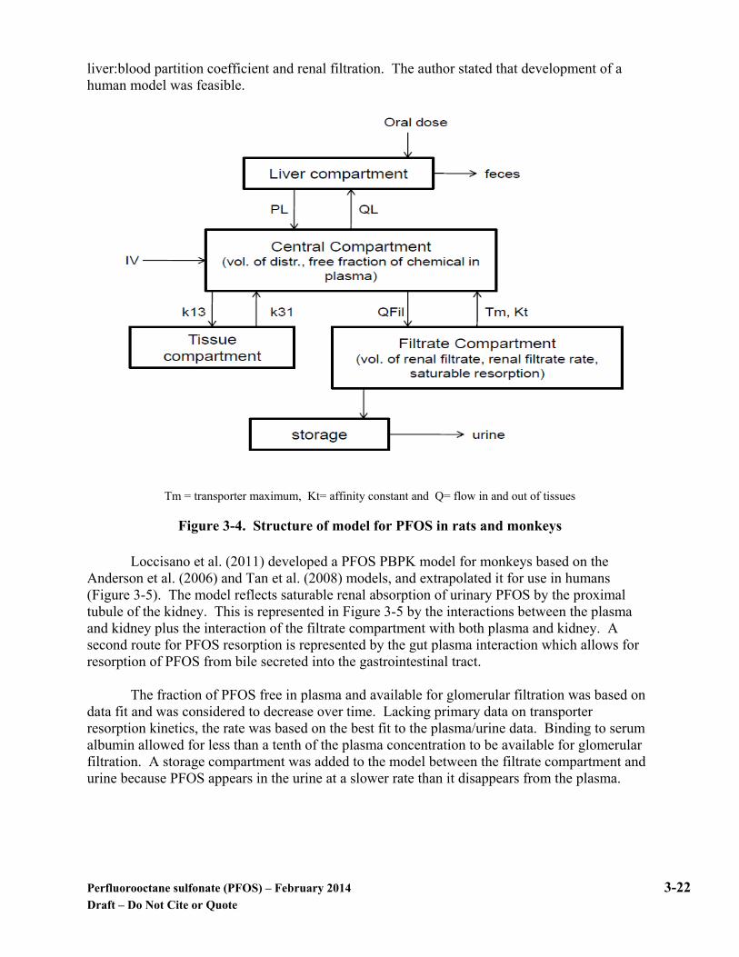
Perfluorooctane sulfonate (PFOS) – February 2014 3-22 Draft – Do Not Cite or Quote
liver:blood partition coefficient and renal filtration. The author stated that development of a human model was feasible.
Tm = transporter maximum, Kt= affinity constant and Q= flow in and out of tissues
Figure 3-4. Structure of model for PFOS in rats and monkeys Loccisano et al. (2011) developed a PFOS PBPK model for monkeys based on the Anderson et al. (2006) and Tan et al. (2008) models, and extrapolated it for use in humans (Figure 3-5). The model reflects saturable renal absorption of urinary PFOS by the proximal tubule of the kidney. This is represented in Figure 3-5 by the interactions between the plasma and kidney plus the interaction of the filtrate compartment with both plasma and kidney. A second route for PFOS resorption is represented by the gut plasma interaction which allows for resorption of PFOS from bile secreted into the gastrointestinal tract. The fraction of PFOS free in plasma and available for glomerular filtration was based on data fit and was considered to decrease over time. Lacking primary data on transporter resorption kinetics, the rate was based on the best fit to the plasma/urine data. Binding to serum albumin allowed for less than a tenth of the plasma concentration to be available for glomerular filtration. A storage compartment was added to the model between the filtrate compartment and urine because PFOS appears in the urine at a slower rate than it disappears from the plasma.

Perfluorooctane sulfonate (PFOS) – February 2014 3-23 Draft – Do Not Cite or Quote
Tm = transporter maximum, Kt= affinity constant and Q= flow in and out of tissues
Figure 3-5. Structure of the PFOS PBPK model in monkeys and humans
Existing data sets for the cynomolgus monkey were used to develop the monkey model.
The IV data came from a single dose of 2 mg/kg (Noker and Gorman, 2003) wherein the concentrations in plasma and urine were monitored for up to 161 days after dosing. The repeat-dose oral data were those from Seacat et al. (2002) with exposures to 0, 0.03, 0.15 or 0.75 mg/kg by capsule for 26 weeks with follow-up monitoring of plasma levels in two monkeys per group at the two highest doses for a year after the cessation of dosing. Both data sets show that the plasma and liver are the primary target tissues for PFOS. The model projections for the repeat dose oral study were in good agreement with the Seacat et al. data for the 0.15 mg/kg dose, but overestimated the plasma values for the 0.75 mg/kg/day dose. The model projected a sharper rise in plasma levels with achievement of steady state more rapidly than indicated by the experimental results.
Human data for PFOS are limited, although serum concentrations were collected from
retired workers (Olsen et al., 2007) and from residents (n=25) in Little Hocking, Ohio. The structure of the human model was similar to that used for the monkeys. The fact that the serum data applied to measurements made following uncertain exposure routes and uncertain exposure durations presented a challenge in the assessment of model fit. The human half-life used for the model (5.4 years) came from an occupational study (Olsen et al., 2007, see Section 3.5.2). No measures of PFOS concentration were available for the drinking water at Little Hocking; thus, the authors estimated the value that could account for the average population serum concentration. The value for the drinking water was estimated to be 0.34 ppb. The model results can be characterized as good when compared to the reported average serum measurements. The
Perfluorooctane sulfonate (PFOS) – February 2014 3-24 Draft – Do Not Cite or Quote
average daily exposure, consistent with the serum value, was estimated as 0.003 µg/kg/day during the period from 1999 to 2000, and about 0.002 µg/kg/day for the 2003 to 2009 time period. The authors concluded that more data are needed on the kinetics of renal transporters, intrahuman variability, and definitive information on exposures in order to refine the human model.
Additional projections of human exposures consistent with measured average serum levels from selected human populations have also been published (Egeghy and Lorber, 2011; Thompson et al., 2010). Both papers used a first-order one-compartment model to assess PFOS exposure from both an intake and body burden perspective using the following equations to determine clearance (CL) with information on volume of distribution (Vd) and chemical half-life (t½).
CL = Vd x (ln2 ÷ t½) Human dose = average serum concentration x CL Egeghy and Lorber (2011) estimated PFOS exposures from both intake and serum
measurements for both typical and contaminated scenarios for adults and children, using available data from peer-reviewed publications. A range of intakes was estimated from the PFOS serum concentrations reported by NHANES as well as published concentrations in various media including dust, air, water, and foods. In the absence of human data, high and low bounding estimates of 3 L/kg and 0.2 L/kg were used for volume of distribution. Total PFOS intakes over all pathways were estimated to be 160 and 2200 ng/day for adults and 50 and 640 ng/day for children in typical and contaminated scenarios, respectively, with food ingestion being the main exposure source in adults and food and dust ingestion being the two main sources in children. Based on the model predictions, the range of intake of PFOS consistent with the serum levels was 1.6 to 24.2 ng/kg-bw/day for adults assuming 70 kg. Thompson et al. (2010) predicted PFOS concentration in blood serum as a function of dose, elimination rate, and volume of distribution. The volume of distribution in this study, 0.23 L/kg bw, was adjusted by 35% from the calibrated data for PFOA in accordance with the differences in PFOA and PFOS volumes of distribution calculated by Anderson et al. (2006). The volume of distribution from PFOA was obtained by calibrating human serum and exposure data collected from two communities in the Little Hocking, OH area (see Section 3.5.3). Applying the volume of distribution and elimination rate values for PFOS calculated from the Little Hocking population to serum data collected from members of the Australian population, the predicted intake by the Australian population was calculated to be 21.7 to 3.6 ng/kg bw/day.
Loccisano et al. (2012a) utilized the saturable resorption hypothesis and pharmacokinetic data from Chang et al. (2012), 3M (2009; unpublished) and Seacat et al. (2003) for adult Sprague-Dawley Rats to develop the model depicted in Figure 3-6. The structure of the model is similar to that for the monkey/human model depicted in Figure 3-5 but lacks the fat and skin compartments and includes a storage compartment to accommodate fecal loss of unabsorbed dietary PFOS as well as that from biliary secretions. Partition coefficients for liver:plasma, kidney:plasma, and rest of the body:plasma were derived from unpublished data on mice by DePierre (2009; personal communication to authors), most of the other kinetic parameters were based on values providing the best fit to the experimental data. The free fraction in plasma was allowed to decrease with time suggesting a strong binding to serum proteins.

Perfluorooctane sulfonate (PFOS) – February 2014 3-25 Draft – Do Not Cite or Quote
Figure 3-6. Structure of the PBPK Model for PFOS in the Adult Sprague-Dawley Rat
The agreement between the experimental data and the model output was generally good
but requires additional data from experimental studies on plasma binding and renal tubular transporters to support further refinement of the parameters derived from model fit. In general, liver and plasma concentrations after daily dosing were overestimated by a factor of about 2. Male and female rats did not differ significantly in their ability to move PFOS from tissues to urine or in resorption capability. PFOS appeared to have a greater capacity to bind to sites in the liver than PFOA. Loccisano et al. (2012b) expanded their adult Sprague-Dawley rat model described above to cover gestational and lactational exposure to the fetus and pups. The data from Thibodeaux et al. (2003) and Chang et al. (2012) for GD 0 to 20 were used in model development. Both studies used multiple dose levels plus data on serum and selected tissue concentrations (liver, brain) from the dams and fetus at one or more time points. The gestational model structure for the dams is similar to Figure 3-6. The model was expanded to include the fetuses linked to the dams by way of the placenta. Uptake from the placenta was described by simple diffusion; the fetal plasma compartment was separate from the dams as was distribution to fetal tissues and amniotic fluid. The model allowed for saturable binding of PFOS within the liver and to serum proteins. Model performance was judged by its ability to predict 24-hour area under the curve (AUC) for plasma, liver, and brain for both the fetus and dam. Brain data were only available from the Cheng et al. (2012) study. According to the model, liver concentrations for the dam are six to seven times greater than those for the fetus and the brain levels for the fetus about eight times greater than those for the dam. Model performance in comparison to the experimental data was judged to be good. The model was used to project the maternal and fetal plasma levels expected at the doses

Perfluorooctane sulfonate (PFOS) – February 2014 3-26 Draft – Do Not Cite or Quote
employed in the Butenhoff et al. (2009), Luebker et al. (2005a,b) and Lau et al. (2003) studies as depicted in Figure 3-7.
Figure 3-7. Predicted Daily Average Concentration of PFOS in Maternal (black line) and Fetal (gray line) Plasma at External Doses to the Dam
The lactational component of the Loccisano et al. (2012b) model allowed for PFOS transport to neonates via mammary-tissues secretion and consequent ingestion by the pups. Pup tissues included in the lactational model included the gut, liver, kidney and the remainder of the body. A renal filtrate compartment linked to plasma and the kidney allowed for neonate PFOS resorption. PFOS transfer to milk via the mammary gland was assumed to be controlled by simple diffusion. Pup urine returned PFOS from the kidney filtrate to the dam. The data of Luebker et al. (2005a,b) and Kuklenyik et al. (2004) were applied in model development. The predicted milk:plasma ratio of 0.1 was in good agreement with the experimental value (0.14) from Kuklenyik et al. (2004). The 24-hour AUC was used to evaluate model predictions. At the beginning of lactation the levels in pup liver were greater than those for the dam; at PND 14 they were equivalent and at PND 20 the levels for the dams were greater than those for the pups. PFOS levels in the pup brain remained higher than those for the dams throughout gestation and lactation but declined gradually across the duration of lactation. After cessation of dosing PFOS levels in the livers of the dams declined gradually. The authors acknowledged the lack of primary experimental data on PFOS transport and potential transporters. Similarity to PFOA was assumed and PFOS was transparently described
Perfluorooctane sulfonate (PFOS) – February 2014 3-27 Draft – Do Not Cite or Quote
as lacking supporting transporter data. Additional research on PFOS binding to serum proteins and liver tissues, its biliary excretion and resorption, plus information on renal resorption transporters in dams and pups, is needed to accomplish further refinements to the published model (Loccisano et al., 2012b).
3.5.2 Half-life data
Differences between species were observed when studies determining the half-life (T1/2) of PFOS in rats, mice, monkeys, and humans were performed. Gender differences in rats do not appear to be as dramatic for PFOS as they are for PFOA (Loccisano et al., 2012a,b).
Humans Occupational Population. Blood sampling was performed on retirees from the 3M plant in Decatur, Alabama where PFOS was produced. These samples were taken approximately every 6 months over a 5-year period to predict the half-life of PFOS. Results ranged from approximately 4 years to 8.67 years (3M Company, 2000; Burris et al., 2002). Both of these studies exhibited some deficiencies in how the samples were taken and the methods used. More recently, Olsen et al. (2007) obtained samples from twenty-six (24 males and 2 females) retired fluorochemical production workers from the 3M Company in Decatur, Alabama to determine the half-life of PFOS. Periodic serum samples (total of 7-8 samples) were collected over a period of 5 years, stored at -80°C and at the end of the study, HPLC-mass spectrometry was used to analyze the samples. The study took place from 1998 to 2004. The mean number of years worked at the plant was 31 years (range: 20 to 36 years), the mean age of the participants at the initial blood sampling was 61 years (range: 55 to 75 years), and the average number of years retired was 2.6 years (range: 0.4 to 11.5 years). The initial arithmetic mean serum concentration of PFOS was 799 ng/mL [0.79 ppm] (range: 145 to 3490 ng/mL), and when samples were taken at the end of the study, the mean serum concentration was 403 ng/mL [0.40 ppm] (range: 37 to 1740 ng/mL). Semi-log graphs of concentration versus time for each of the 26 individuals were created and individual serum elimination half-lives were determined using first-order elimination. The arithmetic and geometric mean serum elimination half-lives of PFOS were 5.4 years (95% CI, 3.9- 6.9) and 4.8 years (95% CI, 4.1-5.4), respectively. General Population. Data were not found for estimation of the half-life of PFOS in the general population. Infants. Newborn Screening Programs (NSPs) collect whole blood as dried spots on filter paper from almost all infants born in the United States. One hundred and ten of these NSPs collected from infants born between 1997 and 2007 in the state of New York were analyzed for PFOS (Spliethoff et al., 2008). The methods used for analysis were validated by using freshly drawn blood from healthy adult volunteers. The mean whole blood concentration for PFOS was 0.81 to 2.41 ng/mL (0.00081 to 0.00241 ppm). The study grouped the blood spots by two different time-points; those collected from 1999-2000 and from 2003-2004 which corresponded to the intervals reported by NHANES. The PFOS concentrations decreased with a mean value of 2.43 ng/mL (0.0024 ppm) reported in 1999-2000 and 1.74 ng/mL (0.0017 ppm) in 2003-2004. The study authors also used regression slopes for natural log concentration versus years since 2000 to calculate the half-life for PFOS. The calculated half-life for PFOS was 4.1 years.

Perfluorooctane sulfonate (PFOS) – February 2014 3-28 Draft – Do Not Cite or Quote
Animal Data
A series of studies was performed to determine the pharmacokinetic parameters of PFOS in rats, mice and monkeys following administration of single doses (Chang et al., 2012). Another study provided half-life information from monkeys (Seacat et al., 2002). Serum half-life estimates were obtained from short-term and long-term studies. Minimal gender-related differences were observed in the species examined. Monkeys
In the study by Chang et al. (2012), three male and three female monkeys were administered a single IV dose of PFOS at 2 mg/kg and followed for 161 days. All monkeys were observed twice daily for clinical signs and body weights were obtained weekly. Urine and serum samples were taken throughout the study. There was no indication that elimination was different from males versus females. Serum elimination half-lives ranged from 122-146 days in male monkeys and 88-138 days in females. Mean values are shown in Table 3-17. The volume of distribution values (Vd) suggest that distribution was predominately extracellular.
TABLE 3-17. PFOS pharmacokinetic data summary for monkeysa
Species Time evaluated
after last dose
Route Sex Amount K+PFOS (mg/kg)
Mean serum T1/2 by sex
(days)
Mean serum T1/2 by species
(days)
Mean serum Vd
by sex (mL/kg)
Cynomolgus monkeys
23 weeks IV M 2 132.0 ± 7 120.8
202
F 2 110.0 ± 15 274 a Data from Chang et al., 2012
Seacat et al. (2002) administered 0, 0.03, 0.15 or 0.75 mg/kg/day potassium PFOS orally in a capsule by intragastric intubation to 6 young-adult to adult cynomolgus monkeys/sex/group, except for the 0.03 mg/kg/day group which had 4/sex, daily for 26 weeks (182 days) in a GLP study. Two monkeys/sex/group in the control, 0.15 and 0.75 mg/kg/day groups were monitored for one year after the end of the treatment period for reversible or delayed toxicity effects. The elimination half-life for potassium PFOS in monkeys was estimated from the elimination curves as approximately 200 days. This value is consistent with that reported by Chang et al. (2012) above. Rats
Chang et al. (2012) conducted a series of pharmacokinetic studies in rats (Table 3-18). First, a single oral dose of 4.2 mg 14C-K+PFOS/kg was administered to male Sprague-Dawley rats (3/timepoint). Urine and fecal samples were collected for 24 and 48 hours. Interim sacrifices to obtain plasma samples were obtained at 1, 2, 6, 12, 24, 48, 96 and 144 hours post-dosing. In the next study, 3 rats/sex were administered 2.2 mg PFOS/kg once by oral gavage or IV administration. The rats had a jugular cannula in place and serum samples from it were obtained at 0.25, 0.5, 1, 2, 4, 8, 18 and 24 hours post-dosing. The T1/2 values must be viewed with caution because the blood samples were limited to a 24-hour post-dose observation period in contrast to the 144-hour (6-day) period from the first study.

Perfluorooctane sulfonate (PFOS) – February 2014 3-29 Draft – Do Not Cite or Quote
In a third study, serum uptake and elimination of PFOS were evaluated at two dose levels: 2 mg/kg and 15 mg/kg. The PFOS was administered as a single oral dose in a 0.5% Tween 20 vehicle to 3 rats/sex or 5/sex at the low and high dose, respectively. Periodic serum, urine and fecal samples were taken for up to 10 weeks. Liver concentrations were evaluated at termination. Half-life estimates (Table 3-18) did not differ significantly with dose, but there was a difference by sex, with values for the males about half those for the females. There were also gender related differences in the volume of distribution values. PFOS concentrations in the liver exceeded those for paired serum concentrations.
The studies by Chang et al. (2012) described above are limited in that they each reflect
pharmacokinetic features associated with a single dose. In an unpublished study by 3M (Butenhoff and Chang, 2007), 5 rats/sex were administered 1 mg/kg/day of PFOS orally for 28 days. Interim blood, urine and feces were obtained for up to 10 weeks. There was no effect on body weight and PFOS elimination was more prominent in the urine than the feces. The elimination of PFOS in this study approximated first order kinetics with a ‘stair-stepping’ pattern. Using nonlinear, noncompartmental software for computation, the half-lives for males ranged from 35 to 53 days and that for females from 33 to 55 days.
TABLE 3-18. PFOS pharmacokinetic data summary for Ratsa
Species Time evaluated after last
dose
Route Sex Amount K+PFOS (mg/kg)
Mean serum T1/2
by dose (days)
Mean serum T1/2 by
sex (days)
Mean serum T1/2 by species (days)
Mean serum Vd by dose (mL/kg)
SD rats 144 hrs Oral M 4.2 8.2 ± 1.5 275
SD rats
24 hrs Oral M 2.2 3.1bc Not determined due
to study design. 765 b
F 1.9c 521 IV M 8.0c 649
F 5.6b 586 b SD rats 10 weeks Oral M 1 x 28
days 35-53 48.2 47.6 -
F 1 x 28 days
33-55 46.9 -
SD rats
10 weeks
Oral
M
2 38.3 ± 2.3 39.8 53.3 1228 15 41.2 ± 2.0 666
F
2 62.3 ± 2.1 66.7 484
15 71.1 ± 11.3
468
a Data from Chang et al., 2012 and Butenhoff and Chang, 2007 (unpublished) bData reflected a single value derived from one rat only cWithin limits of the study design and a follow-up duration of only 24 hrs NA= not available Mice
CD-1 male and female mice were administered PFOS as a single oral dose of 1 or 20 mg/kg (Chang et al., 2012). At designated times (2, 4, 8 hours and 1, 8, 15, 22, 36, 50, 64 and 141 days) post-dosing, four mice/sex were sacrificed and blood, kidneys, and liver samples were obtained. Urine and feces were collected for each 24-hour period up until sacrifice. At the end

Perfluorooctane sulfonate (PFOS) – February 2014 3-30 Draft – Do Not Cite or Quote
of the observation period, the daily urinary and fecal excretion was less than 0.1% of the administered dose. Results are shown in Table 3-19. Serum elimination values were similar for males and females, independent of dose administered: distribution appeared to be mostly extracellular.
TABLE 3-19. PFOS pharmacokinetic data summary for micea Species Time
evaluated after last
dose
Route Sex Amount K+PFOS (mg/kg)
Mean serum T1/2 by dose
(days)
Mean serum T1/2 by
sex (days)
Mean serum T1/2 by species (days)
Mean serum Vd by dose (mL/kg)
CD-1 mice
20 weeks
Oral
M 1 42.8 39.6
36.9
290.0 20 36.4 263.0
F 1 37.8 34.2 258.0 20 30.5 261.0
a Data from Chang et al., 2012
Table 3-20 summarizes the half-life data from the studies discussed above. Despite the limitation that the half-life values from most animal studies were derived from administration of only one dose (Chang et al., 2012), consistency was found in the half-lives for males and females for the monkeys, rats, and mice. In rats, this is in contrast to the results observed for PFOA, where there is a much longer half-life in males than in females. However, similar to PFOA, the half-life of PFOS in humans is much greater than that in laboratory animals. A measure of PFOS half-life in a retired worker population is 5.4 years (Olsen et al., 2007) compared with several months in the laboratory animals.
The animal data summarized in Table 3-20 show fairly consistent half-life values
following single and multiple dosing regimens in both the rat and monkey probably due to the relatively long follow-up in both species after the last dosing was given. In the rat, half-lives for males and females were nearly identical at 48.2 and 46.9 days, respectively, after 28 days of dosing and 10 weeks of follow-up (Butenhoff and Chang, 2007). These results for rats were more consistent between sexes than those half-life values calculated after a single oral dose (Chang et al., 2012). In monkeys similar half-life values were found after either a single intravenous dose (Chang et al., 2012) or repeated oral dosing for 182 days (Seacat et al., 2002). Half-life values for male and female monkeys from Chang et al. (2012) were calculated from the serum concentrations measured over 23 weeks, while the value from Seacat et al. (2002) was estimated from the elimination curves.
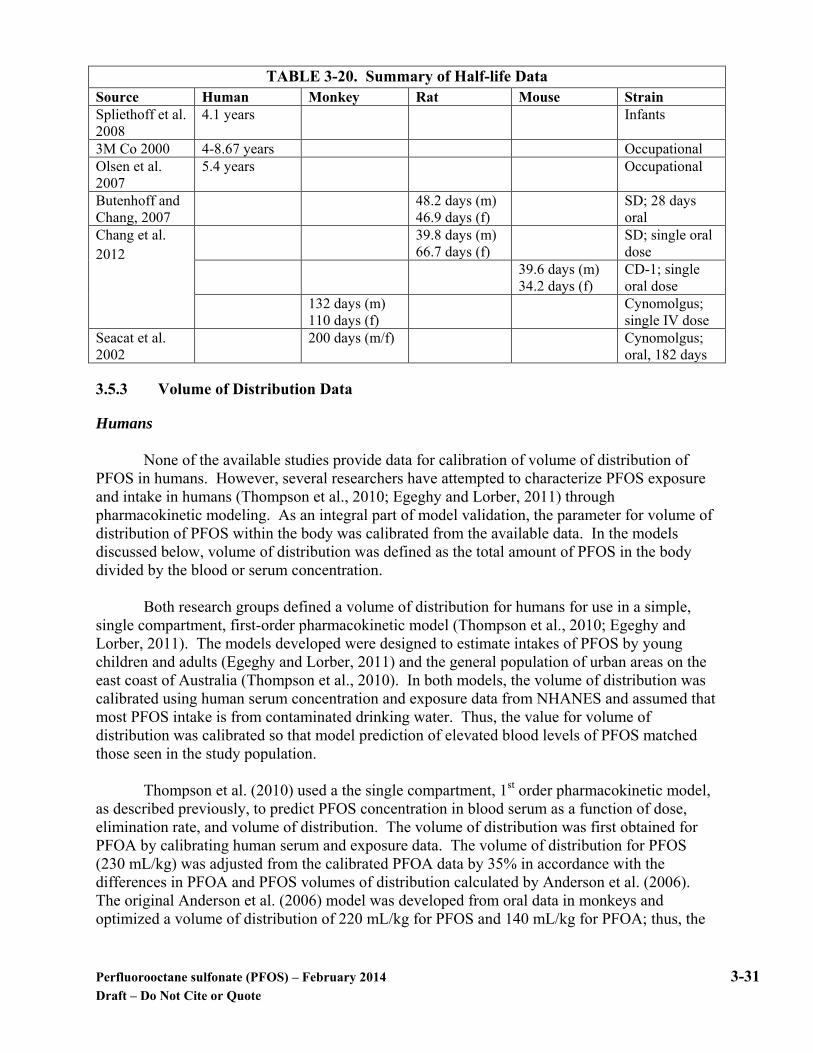
Perfluorooctane sulfonate (PFOS) – February 2014 3-31 Draft – Do Not Cite or Quote
TABLE 3-20. Summary of Half-life Data Source Human Monkey Rat Mouse Strain Spliethoff et al. 2008
4.1 years Infants
3M Co 2000 4-8.67 years Occupational Olsen et al. 2007
5.4 years Occupational
Butenhoff and Chang, 2007
48.2 days (m) 46.9 days (f)
SD; 28 days oral
Chang et al. 2012
39.8 days (m) 66.7 days (f)
SD; single oral dose
39.6 days (m) 34.2 days (f)
CD-1; single oral dose
132 days (m) 110 days (f)
Cynomolgus; single IV dose
Seacat et al. 2002
200 days (m/f) Cynomolgus; oral, 182 days
3.5.3 Volume of Distribution Data
Humans
None of the available studies provide data for calibration of volume of distribution of PFOS in humans. However, several researchers have attempted to characterize PFOS exposure and intake in humans (Thompson et al., 2010; Egeghy and Lorber, 2011) through pharmacokinetic modeling. As an integral part of model validation, the parameter for volume of distribution of PFOS within the body was calibrated from the available data. In the models discussed below, volume of distribution was defined as the total amount of PFOS in the body divided by the blood or serum concentration.
Both research groups defined a volume of distribution for humans for use in a simple,
single compartment, first-order pharmacokinetic model (Thompson et al., 2010; Egeghy and Lorber, 2011). The models developed were designed to estimate intakes of PFOS by young children and adults (Egeghy and Lorber, 2011) and the general population of urban areas on the east coast of Australia (Thompson et al., 2010). In both models, the volume of distribution was calibrated using human serum concentration and exposure data from NHANES and assumed that most PFOS intake is from contaminated drinking water. Thus, the value for volume of distribution was calibrated so that model prediction of elevated blood levels of PFOS matched those seen in the study population.
Thompson et al. (2010) used a the single compartment, 1st order pharmacokinetic model,
as described previously, to predict PFOS concentration in blood serum as a function of dose, elimination rate, and volume of distribution. The volume of distribution was first obtained for PFOA by calibrating human serum and exposure data. The volume of distribution for PFOS (230 mL/kg) was adjusted from the calibrated PFOA data by 35% in accordance with the differences in PFOA and PFOS volumes of distribution calculated by Anderson et al. (2006). The original Anderson et al. (2006) model was developed from oral data in monkeys and optimized a volume of distribution of 220 mL/kg for PFOS and 140 mL/kg for PFOA; thus, the
Perfluorooctane sulfonate (PFOS) – February 2014 3-32 Draft – Do Not Cite or Quote
volume of distribution in monkeys for PFOS was approximately 35% greater than that for PFOA in the optimized models. Therefore, Thompson et al. (2010) used a volume of distribution of 230 mL/kg for humans in their model.
Egeghy and Lorber (2011) used high and low bounding estimates of 3000 mL/kg and 200 mL/kg for volume of distribution since data in humans were not available. The two separate estimates of volume of distribution were used in a first-order, one compartment model to estimate a range of intakes of PFOA. They concluded that the volume of distribution was likely closer to the lower value based on a comparison of predicted modeled intake with estimates of intakes based on exposure pathway analyses. Use of the lower value gave a modeled intake prediction similar to that obtained by a forward-modeled median intake based on exposure assessment. Thus, the authors concluded that the lower value of 200 mL/kg was more appropriate for use in their analysis.
Both of the models described above used a volume of distribution calibrated from actual human data on serum measurements and intake estimates. A calibration parameter obtained from human studies, where constant intake was assumed and blood levels were measured, is considered a more robust estimate for volume of distribution than that optimized within a model developed from animal data.
Animals
The series of pharmacokinetic studies on rats, mice, and monkeys described above, also included volume of distribution calculations (Chang et al., 2012). Values for all species were calculated following a single oral or IV dose of PFOS. As discussed below, the volume of distribution values reported for male and female monkey, female rats, and male and female mice were reasonably similar. The reason for some much higher values found in male rats could not be explained by the study authors.
For male and female cynomolgus monkeys, the volume of distribution was 202 and 274
mL/kg, respectively (Table 3-17), following a single IV dose of 2 mg/kg (Chang et al., 2012). Animals in this pharmacokinetic study were evaluated up to 23 weeks after dosing and the resulting volumes of distribution are similar to that calibrated from human data described above for Thompson et al. (2010).
Volume of distribution findings by Chang et al. (2012) for rats are shown above in Table 3-18. Those values calculated from a follow-up duration of only 24 hours are not considered reliable. In studies with a longer follow-up after dosing, the values for male rats were 275, 666, and 1228 mL/kg and, for female rats, values were 468 and 484 mL/kg. The volume of distribution was notably greater for male rats, with the exception of one value, than that of females or other species including humans. As noted, the authors could not explain the higher value for the male rat but concluded that the volume of distribution for monkeys, rats, and mice is likely in the range of 200-300 mL/kg.
Data for mice (Chang et al., 2012) are shown in Table 3-20. For males and females the
volume of distribution was 263-290 mL/kg and 258-261 mL/kg, respectively, following a single oral dose.
Perfluorooctane sulfonate (PFOS) – February 2014 3-33 Draft – Do Not Cite or Quote
Pharmacokinetic models based on animal data described previously in this chapter
(Anderson et al., 2006; Tan et al., 2008) generally optimized the value for volume of distribution based on model output. The original Anderson et al. (2006) model was developed using data from Seacat et al. (2002) on serum PFOS concentrations in cynomolgus monkeys following oral dosing. The volume of distribution in this model was 220 mL/kg.

Perfluorooctane sulfonate (PFOS) – February 2014 4-1 Draft – Do Not Cite or Quote
4.0 HAZARD IDENTIFICATION
4.1 Human Effects
Studies on PFOS exposures are available from both occupational populations and the general population. Several epidemiologic studies have recently reported associations with PFOS and cholesterol, birth weight changes and various thyroid parameters. These studies are discussed below.
4.1.1 Short-Term Studies and Case Reports
Intentional and Accidental Acute Ingestion No studies of acute accidental or intentional human exposures to PFOS by ingestion were
identified.
Acute and Short-Term Inhalation Exposure No studies of acute and short-term inhalation human exposures to PFOS were identified.
4.1.2 Long-Term and Epidemiological Studies
Several long-term epidemiological studies have been conducted to investigate possible associations between PFOS exposures and various health outcomes. Occupational studies conducted at the 3M Decatur, Alabama plant that produced PFOS are available as well as studies from the C8 Health Project in Ohio and West Virginia. These studies and those on other population groupings are described below. The studies that examined systemic endpoints of toxicity other than cancer are described in Section 4.1.2.1 below where they are subcategorized according to the endpoints examined.
4.1.2.1 Noncancer Systemic Toxicity Studies
Cholesterol, Lipid Homeostasis, Uric Acid, and Biochemical Toxicity Studies
Occupational Populations Cross-sectional as well as a longitudinal analysis of medical surveillance data from the
3M Decatur, Alabama and Antwerp, Belgium plants were conducted to evaluate possible associations between PFOS levels and hematology, clinical chemistry and hormonal parameters (Olsen et. al., 1999; Olsen et. al, 2001b, 2001c). Male volunteers working at the Decatur plant in 1995, 1997 and 2000 and male and female Antwerp volunteers from a 2000 medical surveillance study underwent clinical chemistry tests which evaluated hepatic enzyme activity, renal function tests, thyroid activity and cholesterol levels. There were no consistent associations between worker PFOS levels and any of the clinical chemistry tests in the 1995 and 1997 analyses. The analysis of the data from the 2000 surveillance period indicated a positive association between PFOS and T3, cholesterol, triglycerides and the activity of several hepatic enzymes among male employees at the Decatur plant. However, there were many limitations to combining and comparing the data from the various surveillance periods.

Perfluorooctane sulfonate (PFOS) – February 2014 4-2 Draft – Do Not Cite or Quote
A longitudinal analysis of the above data was performed to determine whether occupational exposure to fluorochemicals over time was related to changes in clinical chemistry and lipid results in employees of the Antwerp and Decatur facilities (Olsen, et al., 2001c). The medical surveillance data from 1995, 1997, and 2000 were analyzed using multivariable regression. The three subcohorts included those who participated in two or more medical exams between 1995 and 2000. When male employees from both plants were combined, no statistically significant (p < 0.05) associations were observed over time between PFOS and serum cholesterol or triglycerides. There were no significant associations between PFOS and changes over time in high density lipoprotein (HDL), alkaline phosphatase, gamma-glutamyl transpeptidase (GGT), aspartate aminotransferase (AST), or alanine transaminase (ALT) activities, total bilirubin, or direct bilirubin. General Population
Several studies on general population exposures to PFOS and other perfluorochemicals
have been generated by the C8 Health Project. The C8 Health Project was conducted in 2005 to 2006 to collect health data from approximately 69,000 residents in Ohio and West Virginia living in the vicinity of a chemical plant producing PFOA. Public drinking water was contaminated in six water districts surrounding the plant (≥0.05 ng/mL of PFOA). The levels of PFOS in this population were similar to those reported in the general U.S. population (median 20.2 ng/mL). Residents were eligible to participate in the study if they had consumed water from any of the 6 water districts for at least one year prior to the study. Blood samples were collected from the participants to determine PFOA and PFOS serum levels and clinical chemistry was performed. Extensive questionnaires were administered as well.
Steenland et al. (2009) examined serum PFOS and PFOA levels and lipids among 46,294
residents, ≥ 18 years old, participating in the C8 Health Project. The mean serum PFOS level among participants was 22.4 ng/mL (0.022 ppm), range (0.25-759.2 ng/mL). Lipid outcomes (total cholesterol, high density lipoprotein [HDL] cholesterol, low density lipoprotein [LDL] cholesterol and triglycerides) were examined in relation to PFOS and PFOA serum levels. All lipid outcomes, except for HDL, showed significant increasing trends with increasing PFOS levels. The predicted increase in cholesterol from lowest to highest PFOS decile (0 to 60 ng/mL) was 11-12 mg/dL. Logistic regression analyses indicate statistically significant increases in cholesterol (≥ 240 mg/dL) with increasing PFOS serum levels. Cholesterol levels ≥ 240 mg/dL are characterized as high, medical intercession is recommended. The odds ratios across quartiles for cholesterol ≥ 240 mg/dL were 1.00, 1.14 [95% CI:1.05, 1.23], 1.28 [95% CI: 1.19, 1.39] and 1.51 [(95% CI: 1.40, 1.64]). The cross-sectional design of this study, as well as the lack of cumulative exposure measurements are limitations in the study design. In addition, the mechanism by which perfluorinated compounds impact serum cholesterol is not yet understood.
Steenland et al. (2010) reported on another analysis of the C8 Health Project participants
≥ 20 years old (n= 54,951) for a possible association between PFOS (and PFOA) serum levels and uric acid. Elevated uric acid is a risk factor for hypertension and may be an independent risk factor for stroke. A statistically significant (p< 0.0001) trend was observed between increasing PFOS levels (untransformed) and uric acid levels. A 0.2-0.3 µg/dL increase in uric acid was associated with an increase from the lowest to highest PFOS decile (10-50 ng/mL). Hyperuricemia (> 6.0 mg/dL for women and > 6.8 mg/dL for men) risk by quintiles increased

Perfluorooctane sulfonate (PFOS) – February 2014 4-3 Draft – Do Not Cite or Quote
slightly with PFOS levels (OR 1.00, 1.02, 1.11, 1.19 and 1.26). The serum of C8 study participants included several PFCs; PFOA appeared to have a greater influence on uric acid trends than PFOS in the models employed by Steenland et al. (2010). Modest associations between PFOS and some lipids existed in children involved in the C8 Health Project (Frisbee et al. 2010). The report stated that 12,476 community children ≤ 18 years old that lived in the C8 Health Project communities were tested for total cholesterol, LDLs, HDLs and fasting triglycerides. The mean level of PFOS was 22.7 ng/mL (0.023 ppm). PFOS was significantly associated with increased total cholesterol, HDL-cholesterol and LDL- cholesterol in a linear regression analysis after adjustment for co-variables. A statistically significant increased risk of abnormal total cholesterol and LDL-cholesterol was also observed between the first and fifth quintiles of PFOS serum concentrations. No trends were observed with triglycerides. As with the other C8 project data, the authors acknowledge that the cross-sectional nature of this study limits causal inference.
Gallo et al. (2012) investigated the correlation between serum PFOS levels and liver enzymes in a total of 47,092 samples collected from members enrolled in the C8 Health Project. The association of ALT, GGT, and direct bilirubin with PFOS was assessed using linear regression models adjusted for various confounders. The ln-transformed values of ALT were significantly associated with ln-transformed PFOS levels and showed a steady increase in fitted levels of ALT per decile of PFOS, leveling off after approximately 30 ng PFOS/mL. Fitted values of GGT showed no overall association with ln-transformed PFOS levels. A positive association was seen with direct bilirubin and PFOS levels in linear regression models but this was not evident with logistic regression models. Limitations of the study include the cross-sectional design and self-reported lifestyle characteristics. Only a small number of ALT values were outside the normal range making the results difficult to interpret in terms of health.
Two studies used data from the U.S. National Health and Nutrition Examination Survey (NHANES) to examine the relationship between perfluorinated chemicals, serum lipid measurements and related conditions. One study analyzed NHANES 2003-2004 data and examined cholesterol levels, obesity, and insulin resistance in relation to PFOS (and other PFCs) (Nelson et al. 2010). The other study analyzed NHANES data from 1999-2000 and 2003-2004 in adolescents and adults and glucose homeostasis and metabolic syndrome (Lin et al. 2009). Nelson et al. (2010) analyzed PFOS and three other perfluorinated chemicals and total cholesterol, HDLs, non-HDL lipoproteins, and LDL. LDL was available only for a subsample of the fasting population and was not measured directly, but was estimated by the Friedewald formula as recommended by CDC. Homeostatic model assessment (HOMA) was used to assess insulin resistance (calculated from fasting insulin and fasting glucose measurements collected in NHANES). Body mass index (BMI) and waist circumference were used to measure body size. Exclusion criteria included current use of cholesterol-lowering medications, participants over the age of 80, pregnant/breastfeeding women or insulin use. After exclusion criteria, ~ 860 participants were included in the cholesterol analyses. The mean PFOS serum concentration for participants 20-80 years old was 25.3 µg/L (range, 1.4 to 392 µg/L). A positive association was identified between total serum cholesterol and serum PFOS concentrations. When analyzed by PFOS serum quartiles, adults in the highest PFOS quartile

Perfluorooctane sulfonate (PFOS) – February 2014 4-4 Draft – Do Not Cite or Quote
had total cholesterol levels of 13.4 mg/dL (95% CI, 3.8, 23.0) higher than those in the lowest quartile. As expected, non-HDL cholesterol accounted for most of the total cholesterol. Consistent trends were not observed for HDL or LDL. Adjusting the cholesterol models for serum albumin produced similar results. Body weight and insulin resistance were not consistently associated with serum PFOS levels. Lin et al. (2009) analyzed the data on perfluorinated chemicals in NHANES 1999-2000 and 2003-2004 and glucose homeostasis and metabolic syndrome. The authors reported an association between PFOS and metabolic syndrome; however, it was not reported how PFOS and other perfluorinated chemicals in the analysis were combined for the NHANES sample years. Effects of PFOS on plasma lipid levels in the Inuit population of Northern Quebec were examined in a cross-sectional epidemiology study (Château-Degat et al., 2010). The relationship between consumption of PFOS-contaminated fish and wild game with blood lipids was assessed in 723 Inuit adults (326 man and 397 women). This traditional diet is also rich in n-3-polyunsaturated fatty acids (n-3 PUFAs) which are known to have hypolipidemic effects; therefore, the n-3 PUFAs were considered as a confounder in the analyses. Multivariate linear regression modeling was used to evaluate the relationship of PFOS levels and blood lipids including TC, HDL cholesterol, LDL cholesterol, and triacylglycerols. Plasma levels of HDL cholesterol were positively associated with PFOS levels, even after adjustment for circulating levels of n-3 PUFAs, but the other blood lipids were not associated with PFOS levels. The geometric mean level of PFOS in plasma for women and men was 18.6 µg/L. Table 4-1 provides a summary of the results from the studies that examined the relationship of serum PFOS with serum lipids and uric acid. The most consistent findings are those for total cholesterol.
TABLE 4-1. Association of Serum PFOS with Serum Lipids and Uric Acid Study TC VLDL LDL HDL Non-
HDL TG UA
Occupational Population Olsen et al., 2001c ↔ NM NM ↔ NM ↔ NM General Population Steenland et al., 2009; 2010
↑ NM ↑ ↔ NM ↑ ↑
Frisbee et al., 2010 (children)
↑ NM ↑ ↑ NM ↔ NM
Château-Degat et al., 2010
↔ NM ↔ ↑ NM ↔ NM
Nelson et al., 2010 ↑ NM ↔ ↔ ↑ NM NM ↑= positive association; ↓= negative association; ↔= no association; TC= total cholesterol; VLDL=very low density lipoprotein; LDL= low density lipoprotein; non-HDL= TC(VLDL,IDL, LDL)-HDL; HDL=high density lipoprotein; TG= triglycerides; UA=uric acid; NM= not monitored

Perfluorooctane sulfonate (PFOS) – February 2014 4-5 Draft – Do Not Cite or Quote
4.1.2.2 Reproductive Hormones and Reproductive/Developmental Studies
Olsen et al. (2009) recently reviewed the epidemiological literature and identified six studies examining potential associations between PFOS and human birth outcomes. Research has focused on birth weight and other measures of fetal growth. Overall, in both the general and occupational populations, inconsistent associations were identified between PFOS exposure/levels and fetal birth weight or gestational age. An occupational cohort study by Grice et al. (2007) examined the potential association between PFOS exposure and adverse pregnancy outcomes in employees at a perfluorinated chemical production facility in Decatur, Alabama. Current and former female employees of the facility completed a questionnaire and provided a brief pregnancy history. The level of exposure was categorized according to a job-specific exposure matrix. A total of 263 women participated and reported 439 births of which there were 421 live births, 14 stillbirths and 4 no data. Birth weight was adjusted for maternal age, smoking status and gravidity. No associations were observed between PFOS exposure and pregnancy outcomes even when birth weight was adjusted for maternal age, smoking status and gravidity.
A series of longitudinal, population-based studies was conducted in a subset of women aged 25 to 35 enrolled in the Danish National Birth Cohort (DNBC) from March 1996 to November 2002 (Fei et al., 2007, 2008a, 2008b, 2009, 2010a). A random sample of 1400 women was selected to investigate the association between blood levels of perfluorinated chemicals and adverse reproductive and developmental outcomes in the women and their children. Study data were collected by telephone interviews at 12 and 30 weeks of gestation and approximately 6 and 18 months after birth. A food frequency questionnaire was filled out at home during approximately week 25 of pregnancy. Maternal blood samples were taken in the first and second trimester and from the cord blood in the infant just after birth. Only blood results from the 1400 women in the first trimester were reported. Mean plasma PFOS levels by age groups were: < 25 years- 38.6 ng/mL (0.039 ppm); 25-29 years- 36.8 ng/mL (0.037 ppm); 30-34 years- 33.9 ng/mL (0.034 ppm) and ≥ 35 years- 33.0 ng/mL (0.033 ppm). Potential confounders included: maternal age, maternal occupation and educational status, parity, pre-pregnancy BMI, smoking/alcohol consumption during pregnancy, gestational weeks at blood draw, child’s sex, child’s age at interview with mother, breast-feeding > 6 months (for 18-month interview), out-of-home child care, hours mother spent with child/day, and home density (the total number of rooms divided by the total number of people in the household). Data from the DNBC were used to investigate the association between plasma levels of PFOS in pregnant women, length of gestation, and the infant’s birth weight (Fei et al., 2007). The average PFOS levels in maternal plasma were 35.3 ng/mL (range, 6.4-106.7 ng/mL). The data were adjusted for confounding factors that might also influence fetal growth or length of gestation and analyzed by analysis of variance and linear regression using both continuous PFOS concentrations, and categorical quartiles of PFOS. Elevated risk estimates were found for PFOS levels and preterm birth, but the odds ratio was significant only for the third quartile of exposure. No significant association was found between PFOS levels and length of gestation, low birth weight, or small for gestational age.

Perfluorooctane sulfonate (PFOS) – February 2014 4-6 Draft – Do Not Cite or Quote
When Fei et al. (2008a) investigated the association between PFOS levels in pregnant women and their newborns and placental weight, birth length, and head and abdominal circumference, he found maternal PFOS levels were not associated with any of the fetal growth indicators when the lowest quartile was compared to the highest. In a stratified analysis of PFOS, inverse associations were found with birth length for postterm and preterm infants and with ponderal index in multiparous women and positive association with ponderal index in nulliparous women. These associations were not statistically significant. In the Fei et al. (2008b) report examining the association between plasma levels of PFOS in pregnant women and the motor and mental development in their children, regression analysis did not indicate any significant association between PFOS and Apgar score after adjustment for potential confounders (odds ratio [OR], 1.20; 95% confidence interval [CI], 0.57-2.25). The developmental measures examined in the infants included Apgar score of child at birth and questionnaire responses about child development milestones at 6 and 18 months. Data from the 6 month interview did not show any association between PFOS levels and motor or mental development. In children at 18 months, mothers with higher PFOS levels were slightly more likely to report that their babies started sitting without support at a later age and “did not use word-like sounds to tell what he/she wants.” Fei et al. (2009) used the same population of pregnant women to determine if there was any association with PFOS levels and fecundity indicated by the time to pregnancy (TTP). In women who had planned pregnancies (n=1240), there was a longer time to pregnancy (TTP) with higher levels of PFOS (p<0.001). PFOS was also associated with irregular menstrual periods (11.6% in the lowest quartile versus 14.2% in the upper three quartiles). The proportion of women with infertility (TTP>12 months) was higher in the upper three quartiles of PFOS versus the lowest quartile. These trends were significant. Women with longer TTP were also older and had a history of spontaneous miscarriages or irregular menstrual cycles. The biological mechanism by which PFOS may reduce fecundity is unknown. Fei et al. (2010a) reported on the effects of PFOS on the length of breastfeeding. Self reported data on the duration of breastfeeding was collected during the telephone interviews at 6 and 18 months after birth of the child. Higher levels of PFOS were significantly associated with a shorter duration of breastfeeding. In multiparous women, the adjusted OR for weaning before 6 months is 1.20 [95% CI, 1.06-1.37]) for each 10 ng/mL increase in PFOS concentration in the maternal blood and the increase was dose-related. A statistically significant positive trend was observed for women having their first child, but no consistent association was found across increasing blood levels of PFOS. The authors speculate that the observed associations may be non-causally related to previous length of breastfeeding or to reduction of PFOS through lactation. Monroy et al. (2008) found no association between the maternal serum levels of PFOS and the birth weight of the neonates in 101 pregnant women enrolled in a large cohort study, Family Study, conducted at McMaster University Medical Center in Canada. PFOS was measured in maternal serum at mid-pregnancy and delivery in 101 healthy pregnant women in Ontario, Canada and in umbilical cord blood (UCB) from 105 babies. PFOS was detected in 100% of samples with mean levels of 18.3, 16.2, and 7.2 ng/L in maternal serum at 24-28 weeks, maternal serum at delivery, and in UCB respectively. The concentration of PFOS in maternal
Perfluorooctane sulfonate (PFOS) – February 2014 4-7 Draft – Do Not Cite or Quote
serum was significantly higher than in UCB. No significant association between levels of PFOS in the maternal serum or UCB and the birth weight of the neonates was found. Maternal PFOS levels were also not associated with maternal body mass index, gestational length, or gender. A prospective cohort study was conducted on birth weight between July 2002 and October 2005 at the Sapporo Toho Hospital in Hokkaido, Japan that included 428 native Japanese women and their infants (Washino et al., 2009). Women enrolled were at 23-35 weeks of gestation with a mean age of 30.5 years. Subjects reported on dietary habits, smoking status, alcohol intake, caffeine intake, household income and educational level. At the time of the questionnaire, a blood sample was obtained. Both PFOS and PFOA were analyzed in the blood using liquid chromatography-tandem mass spectrometry coupled with solid-phase extraction. The mean concentration in the women was 5.6 ng/mL (0.006 ppm) PFOS with every sample having detectable PFOS. The highest PFOS concentration identified was 16.2 ng/mL (0.016 ppm). The results indicated that in utero exposure to PFOS negatively correlated with birth weight in female infants only. A cohort study on 252 pregnant women (≥ 18 years old) at 15-16 weeks gestation in the city of Edmonton, Alberta, Canada was undertaken to examine a possible association between perfluorinated chemicals, fetal growth and gestational age (Hamm et al., 2009). Serum samples collected from December 2005 to June 2006 showed PFOS levels ranging from nondetectable to 35 ng/mL, with the mean and geometric mean being 9.0 ng/mL (0.009 ppm) and 7.4 ng/mL (0.0074 ppm), respectively. Overall, there was no association with the level of PFOS and birth weight or length of gestation. Mean birth weight was 3278 g (n = 83; PFOS < 6.1 ng/mL); 3380 g (n = 83; PFOS 6.1-10.0) and 3387 g (n = 86; PFOS > 10-35). Mean length of gestation for all groups was 38 weeks; the pre-term delivery percentage was similar between groups. Stein et al. (2009) presented on serum levels of PFOS and self-reported pregnancy outcomes of a population of women (5,262 pregnancies; ages 15-55 years) in the Mid-Ohio Valley in 2000-2006. These women were enrollees in the C8 Health Project, a community health study of residents near a chemical plant that used PFOA in the manufacture of fluoropolymers. Pregnancies within the 5 years preceding the exposure measurements were analyzed. The mean level of PFOS in the serum of these women was 14.1 ng/mL (0.014 ppm). There was no association between PFOS levels and miscarriages or pre-term births. PFOS was, however, associated with an increased risk above the median (adjusted odds ratio = 1.5: 95% confidence interval: 1.1, 1.9) for low birth weight, and a dose-response relationship was reported for the 50th-75th, 75th-90th and > 90th percentile serum PFOS exposure concentrations (adjusted OR 1.3, 1.6, 1.8, respectively). PFOS was also weakly associated with pre-eclampsia (adjusted odds ratio = 1.3, 95% confidence interval: 1.1, 1.7). The self-reported nature of pregnancy outcomes in this study is a limitation. Using the C8 Health Project data, blood samples from a population of women aged 18-65 years (n= 25, 957) were analyzed to determine if the onset of menopause, serum estradiol and the amount of PFCs in the blood were inter-related (Knox et al., 2011). These data were cross-sectional with no variable for the length of exposure. The mean PFOS level of all the women was 17.6 ng/mL. Data were eliminated for those reporting undergoing a hysterectomy and adjusted for age within the group, smoking, alcohol consumption, body mass index and exercise. The analysis for menopause was determined upon three groups of women: childbearing (ages 30-

Perfluorooctane sulfonate (PFOS) – February 2014 4-8 Draft – Do Not Cite or Quote
42), perimenopausal (ages >42 but < 51) and menopausal (ages > 51 or < 65). These same groups were used for the estradiol concentrations except the childbearing group was extended to include those > 18 years. For menopause, the odds of having experienced menopause in the menopausal group exposed to PFOS showed a monotonic increase with all quintiles significantly higher relative to the lowest (PFOS odds= 2.1, CI=1.6-2.8). Also, PFOS was negatively associated with estradiol concentrations in all groups but significantly in the perimenapausal group (β = -3.65; p< 0.0001) and menopausal group (β = -0.83; p< 0.007). The level of PFOS was significantly higher in the set of women that had undergone a hysterectomy. While these relationships were associated with PFOS, the authors still recommended caution when interpreting results because the data are cross-sectional. Lopez-Espinosa et al. (2011) also used the C8 Health Project data base to indicate that in 3076 boys and 2931 girls, ages 8-18 years, there was a relationship of reduced odds of reaching puberty (raised testosterone) with increasing PFOS levels, having a delay of 190 days between the highest and lowest quartile, and reduced odds of postmenarche (138 days delay) in girls. This study suggested that PFOS exposure correlated with a delay in puberty. Joensen et al. (2009) investigated the possible associations between perfluoroalkyl acids (PFAAs) and semen quality in 105 Danish men. The median PFOS serum level in men was 24.5 ng/mL. Men with high levels of combined levels PFOA/PFOS had a median level of 6.2 million spermatozoa compared to 15.5 million in men with low PFOA/PFOS levels. There was no significant association between testosterone levels and PFAA levels and no difference in PFAA levels between high and low testosterone groups. The relationships examined and outcomes from the studies that examined reproductive or developmental endpoints as they related to serum PFOS concentrations are summarized in Table 4-2 that follows. For most endpoints no associations were identified. There is some evidence for body weight effects in neonates (2 of 5 studies). Possitive associations were also noted for some developmental endpoints and factors impacting fertility.

Perfluorooctane sulfonate (PFOS) – February 2014 4-9 Draft – Do Not Cite or Quote
TABLE 4-2. Association of serum PFOS with reproductive and developmental outcomes
Study Population Outcome Measures at birth
Growth/ Development
Fecundity/ Fertility
Grice et al., 2007
Occupational ↔ (any adverse)
NM NM NM
Fei et al., 2007; 2008a; 2008b; 2009; 2010a
General ↔ (gestation length) ↔ (length of breastfeeding)
↔ (weight) ↔ (size) ↔ (Apgar score)
↔ (at 6 months) ↑ (at 18 months; sitting up later)
↑ (time to pregnancy) ↑ (infertility)
Monroy et al., 2008
General ↔ (gestation length)
↔ (weight)
NM NM
Washino et al., 2009
General NM ↑ (low weight females only)
NM NM
Hamm et al., 2009
General ↔ (gestation length)
↔ (weight)
NM NM
Stein et al., 2009
General ↔ (miscarriage)
↑ (low weight)
NM NM
Knox et al., 2011 and Lopez-Espinosa et al., 2011
General NM NM ↑ (delayed puberty)
↑ (early menopause)
Joensen et al., 2009 (PFOA/PFOS combined)
General NM NM NM ↑ (lower sperm count) ↔ (testosterone)
↑= positive association; ↓= negative association; ↔= no association; NM = Not Monitored
4.1.2.3 Thyroid Effect Studies
The relationship between exposure to polyhalogenated compounds, including PFOS, and thyroid hormone homeostasis was examined in a cross-sectional study of the adult Inuit population of Nunavik, Quebec, Canada (Dallaire, 2009b). Concentrations of thyroid-stimulating hormone (TSH), free thyroxine (fT4), total triiodothyronine (tT3) and thyroxine-binding globulin (TBG) were measured in 623 individuals. Participants were given a survey to indicate smoking status, frequency of alcohol use, medications taken, and dietary fish consumption. Those using medication for thyroid disease and pregnant women were not included in the results. The study detected PFOS in 100% of individuals with a mean plasma PFOS concentration of 18 ng/ml (95% CI, 17-19 ng/ml). PFOS was negatively associated with circulating levels of TSH, tT3 and TGB and positively associated with fT4. The results suggest that human thyroid hormone levels may be affected by PFOS exposure. However, because the majority of individuals had normal thyroid gland function, it is uncertain whether these relationships are connected to thyroid disease.

Perfluorooctane sulfonate (PFOS) – February 2014 4-10 Draft – Do Not Cite or Quote
National Health and Nutrition Examination Survey (NHANES) data from three independent cross-sectional cycles (1999-2000; 2003-2004 and 2005-2006) were analyzed by Melzer et al. (2010) to estimate associations between serum PFOA and PFOS concentrations and thyroid disease in the general U.S. population. Overall, a total of 3,966 individuals ≥ 20 years of age (1900 men and 2066 women) were included. Of these, 292 women and 69 men reported thyroid disease. The data showed that men with PFOS levels in the highest quartile ≥ 36.8 ng/mL were more likely to report currently treated thyroid disease than men with PFOS levels in the lowest quartile ≤ 25.5 ng/mL (OR=2.68; 95% CI, 1.03-6.98; p=0.043). Women had lower levels of PFOS than men and higher prevalence of thyroid disease, but serum PFOS concentration was not significantly associated with treated thyroid disease. Further studies measuring thyroid hormone levels in a larger sample population could clarify whether pathology, changes in exposure, or altered pharmacokinetics can explain the association.
Bloom et al. (2010) examined the potential association between serum concentrations of
eight polyhalogenated compounds, including PFOS, and human thyroid function. Levels of thyroid stimulating hormone (TSH) and free thyroxine (fT4) were measured in a subsample of participants in the cross-sectional New York State Angler Cohort Study (27 men and 4 women). A survey was conducted to determine smoking status, history of thyroid disease, medications used, and dietary fish consumption. None of the participants reported a thyroid condition or the use of thyroid medication. PFOS occurred at a high concentration compared to the other PFCs measured with a mean concentration of 19.6 ng/mL (95% CI, 16.3-23.5). The results indicated no significant association between PFOS serum concentration and thyroid hormone levels, potentially due to the study’s small sample size.
The potential relationship between PFOS exposure and thyroid disease was investigated
by Pirali et al. (2009) in a sample of 28 patients undergoing thyroid surgery (22 benign and 6 malignant) and a control group of 7 patients with no evidence of thyroid disease. PFOS was detected in thyroid tissue in 100% of the 8 males and 20 females with thyroid disease, with a median PFOS concentration of 5.3 ng/g, with no significant difference in levels between benign and malignant patients. The median PFOS concentration (4.4 ng/g) in the healthy glands of the control group was similar to that found in the diseased thyroid samples indicating that there was no association between PFOS concentration and thyroid disease.
Maternal and umbilical cord blood concentrations of a number of fluorinated organic
compounds, including PFOS, were determined in 15 women (17-37 years of age) and their newborns at Sapporo Toho Hospitals in Hokkaido, Japan from February to July 2003 (Inoue et al., 2004). PFOS was detected in 100% of the maternal and cord blood samples with maternal blood PFOS ranging from 4.9 to 17.6 ng/mL, and cord blood PFOS ranging from 1.6 to 5.3 ng/mL. Thyroid stimulating hormone (TSH) and free thyroxine (fT4) levels in the infants between days 4 and 7 of age were not related to cord blood PFOS concentration in this small study.
Chan et al. (2011) used blood from 974 serum samples collected from women (mean age-
31.3 years) at 15-20 weeks gestation in 2005-2006 in Canada and measured thyroid hormones, fT4 and the level of PFCs to determine if PFC levels were associated with hypothyroxinemia. From the samples, there were 96 cases of hypothyroxinemia (identified as ‘cases’) and 175 controls used. The geometric mean for PFOS was 7.39 ng/mL. The mean free T4 levels were

Perfluorooctane sulfonate (PFOS) – February 2014 4-11 Draft – Do Not Cite or Quote
7.7 pmol/L in the cases and 12.9 in the controls. The mean TSH concentrations were 0.69 mU/L in the cases and 1.13 in the controls. Analysis by conditional logistic regression indicated that the concentration of PFOS was not associated with hypothyroxinemia in this set of pregnant women. For PFOS, the odds ratio for association of hypothyroxinemia with exposure to PFOS was 0.88 with a 95% CI of 0.63-1.24.
The Dallaire et al (2009b) study provides the strongest evidence for effects on thyroid
hormones as illustrated in Table 4-3. The hormone endpoints were not monitored consistently in a number of the other sudies of thyroid effects and where monitored exhibited no significant association.
TABLE 4-3. Association of serum PFOS with the prevalence of thyroid disease and thyroid hormone levels in studies of general and worker populations
Study Population Thyroid Disease TSH T3 T4 Dallaire et al., 2009b General ↔ ↓ ↓ ↑ Bloom et al., 2010 General ↔ ↔ NM ↔ Melzer et al., 2010 General ↔ (women)
↑ (men) NM NM NM
Pirali et al., 2009 General ↔ NM NM NM Inoue et al., 2004 Newborns NM ↔ NM ↔ Chan et al., 2011 Pregnant
women ↔ ↔ NM ↔
↑= positive association; ↓= negative association; ↔= no association; NM = Not Monitored
4.1.2.4 Immunotoxicity
Okada et al. (2012) investigated the relationship between maternal PFOS concentration and infant allergies and infectious diseases during the first 18 months of life as well as cord blood IgE levels. The prospective birth cohort was based on infants delivered at the Sapporo Toho Hospital in Sapporo, Hokkaido, Japan between July 2002 and October 2005. PFOS levels were measured in maternal serum taken after the second trimester (n=343) and total IgE concentration was measured in cord blood (n=231) at the time of delivery. Infant allergies and infectious diseases were assessed in a maternal self-administered questionnaire at 18 months post-delivery. Polynomial regression analyses, adjusted for potential confounders, were performed on log-transformed data. Mean maternal PFOS concentration was 5.6 ng/mL and cord blood IgE level was 0.62 IU/mL. No significant associations were observed between maternal PFOS levels and cord blood IgE levels or incidence of food allergy, eczema, wheezing, or otitis media in infants at 18 months of age. Limitations of the study include the small sample size, potential selection bias of the population, and accurate diagnosis of disease in the infants.
The population from the Danish National Birth Cohort studies by Fei et al. (2010b) was used to determine whether prenatal exposure to PFOS caused an increased risk of infectious diseases leading to hospitalization in early childhood. No clear pattern was identified when results were stratified by child’s age of infection and the level of PFCs in the maternal blood.
Antibody responses to diphtheria and tetanus toxoids following childhood vaccinations
were assessed in context of exposure to perfluorinated compounds (Grandjean et al., 2012). The

Perfluorooctane sulfonate (PFOS) – February 2014 4-12 Draft – Do Not Cite or Quote
prospective study included a birth cohort of 587 singleton births during 1999-2001 from the National Hospital in the Faroe Islands. Serum antibody concentrations were measured in children at age 5 years prebooster, approximately 4 weeks after the booster, and at age 7 years. Prenatal exposures to perfluorinated compounds were assessed by analysis of serum collected from the mother during week 32 of pregnancy; postnatal exposure was assessed from serum collected from the child at 5 years of age. Multiple regression analyses with covariate adjustments were used to estimate the percent difference in specific antibody concentrations per 2-fold increase in PFOS concentration in both maternal and 5-year serum.
Maternal PFOS serum concentration was negatively associated with antidiphtheria
antibody concentration (-39%) at age 5 before booster. An effect was also found in comparison of antibody concentrations at age 7 with serum PFOS concentrations at age 5 where a 2-fold increase in PFOS was associated with a difference in diphtheria antibody of -28% (95% CI, -46% to -3%). Additionally at ages 5 and 7, a small percentage of children had antibody concentrations below the clinically protective level of 0.1 IU/mL. At age 5 the odds ratios of antibody concentrations falling below this level for diphtheria were 2.48 (95% CI, 1.55 to 3.97) compared with maternal and 1.60 (95% CI, 1.10 to 2.34) compared with age 5 serum PFOS concentrations. For age 7 antibody levels correlated with age 5 PFOS serum concentrations, odds ratios for inadequate antibody concentration were 2.38 (95% CI, 0.89 to 6.35) for diphtheria and 2.61 (95% CI, 0.77 to 8.92) for tetanus.
The association between serum levels of perfluorinated compounds and childhood asthma
was investigated by Dong et al. (2013). The cross-sectional study included a total of 231 children aged 10-15 years with physician-diagnosed asthma and 225 age-matched non-asthmatic controls. Between 2009 and 2010, asthmatic children were recruited from two hospitals in Northern Taiwan, while the controls were part of a cohort population in seven public schools in Northern Taiwan. Serum was collected for measurement of ten perfluorinated compounds, absolute eosinophil counts, total IgE, and eosinophilic cationic protein. A questionnaire was administered to asthmatic children to assess asthma control and to calculate an asthma severity score (including frequency of attacks, use of medicine, and hospitalization) during the previous four weeks. Associations of perflourinated compound quartiles with concentrations of immunological markers and asthma outcomes were estimated using multivariable regression models.
Nine of ten perfluorinated compounds were detectable in ≥84.4% of all children with
levels generally higher in asthmatic children compared with non-asthmatics. Serum concentrations of PFOS in asthmatic and non-asthmatic children were 45.5±37.3 and 33.4±26.4 ng/mL, respectively; similar levels were measured for perfluorotetradecanoic acid with concentrations of the remaining compounds much lower. The adjusted odds ratios for asthma association with the highest versus lowest quartile levels were significantly elevated for seven of the compounds. For PFOS the odds ratio was 2.63 (95%CI: 1.48, 4.69). In asthmatic children, absolute eosinophil counts, total IgE, and eosinophilic cationic protein concentration were positively associated with PFOS levels with a significant monotonic trend with increasing serum concentration. None of these biomarkers was significantly associated with PFOS levels in non-asthmatic children. Serum PFOA levels, as well as three other compounds, were significantly associated with higher asthma severity scores.
Perfluorooctane sulfonate (PFOS) – February 2014 4-13 Draft – Do Not Cite or Quote
4.1.2.5 Carcinogenicity Studies
Occupational Populations Several analyses of various health outcomes have occurred on cohorts of workers at the 3M Decatur, Alabama plant (Mandel and Johnson, 1995; Alexander et al., 2003; Alexander and Olsen, 2007). Cancer incidence and mortality have been examined periodically in these workers. A cohort of 2083 workers employed for at least 1 year was examined. Workers were grouped into three PFOS exposure categories: non-exposed, low exposed and high exposed. Cumulative exposures were also estimated using a weighted approach based on biomonitoring data. The geometric mean serum PFOS levels were 0.9 ppm for chemical plant employees and 0.1 ppm for non-exposed workers. A total of 145 deaths were identified with 65 of them in high-exposure jobs. Standardized mortality ratios (SMRs) were calculated using the state of Alabama reference data and when analyzing the entire cohort, SMRs were not elevated for most of the cancer types and for non-malignant causes. SMRs that were above 1 (cancer of the esophagus, liver, breast, urinary organs, bladder, and skin) were also elevated when the cohort was limited to any employee ever employed in a high exposure job (except breast cancer). Only 2 or 3 deaths were reported for each of these cause-specific categories and were not statistically significant, except for bladder cancer. Three male employees in the cohort died of bladder cancer (0.12 expected). All were employed at the Decatur plant for more than 20 years, and had worked in high exposure jobs for at least 5 years. The SMR for bladder cancer for workers who were ever employed in a high exposure job was 12.77 (0.23 expected, CI = 2.63 - 37.35). When the data were analyzed for workers with >5 years of employment in a high exposure job, the SMR was 24.49. This effect remained when the data were analyzed using county death rates. While the 3 deaths from bladder cancer were greater than the expected number observed in the general population, there were limitations observed in this study. The number of deaths was small (especially for females in all categories), all death certificates were not located, exposures to other chemicals were not accounted for, smoking status was not established and animal studies do not indicate any bladder cancer. Based on these results, 3M undertook another study of this cohort to identify bladder cancer incidence (Alexander and Olsen, 2007). Cancer deaths were ascertained from death certificates and via questionnaire for bladder cancer cases, year of diagnosis, and smoking history. Eleven bladder cancer cases were identified; five deaths and 6 incident cases. Only 2 of the 6 self-reported cases were confirmed with medical records. Five of the 6 cases had a history of cigarette smoking. Standardized incidence ratios (SIR) were estimated for 3 exposure categories and compared to US cancer rates. SIRs ranged from less than 1 to 2.72 but none were statistically significant. The highest SIRs were for the lower exposure groups. The SIR was 1.74 (95% CI 0.64-3.79) for those ever employed in a high exposure job. Grice et al. (2007) looked for association with PFOS exposure at the 3M Decatur, Alabama plant to various malignant and benign disorders as well as adverse pregnancy outcomes (results reported in Section 4.1.2.2). Current and past employees at the plant answered questionnaires (n = 1400; 1137 male and 263 female) about any diagnosis of cancers or non-

Perfluorooctane sulfonate (PFOS) – February 2014 4-14 Draft – Do Not Cite or Quote
cancerous conditions (including liver, kidney and gastrointestinal disease). Two exposure models were used in this analysis. The first model grouped workers according to PFOS exposure: unexposed (< 0.29 ppm), low (0.39-0.89 ppm) or high exposure (1.30-1.97 ppm). The other model estimated cumulative exposures by using a weighted approach based on biomonitoring data. Prostate, melanoma and colon cancer were the most frequently reported malignancies. When cumulative exposure measures were analyzed, elevated odds ratios were reported for both colon and prostate cancer, however, they did not reach statistical significance. None of the other health conditions were positively associated with PFOS exposures General Populations A prospective Danish cohort study (1993-2006) compared plasma levels of PFOS and PFOA and the incidence of cancer in 57,053 native Danish individuals (ages 50-65 years) (Eriksen et al., 2009). The participants had no cancer diagnosis at the time of the enrollment. The Danish Cancer Registry and the Danish Pathology Data Bank were used to identify 713, 332, 128 and 67 patients with prostate, bladder, pancreatic and liver cancer, respectively, diagnosed 0-12 years after enrollment in the cohort. A comparison group reflecting the gender ratio of the case group was also chosen from the original cohort (680 men, 92 women). Potential confounders associated with each type of cancer were addressed in questionnaires administered to the participants.
One-time, PFOS and PFOA blood samples were taken at recruitment. Cancer incidence
rate ratios (IRR) were estimated using Cox proportional hazards model, stratified by sex. Median PFOS levels did not differ significantly between male and female cases (35.1 ng/mL, males; 32.1 ng/mL, females) or controls. Adjusted IRRs for the 4 types of cancer based on increasing PFOS quartiles were only above 1 for prostate and pancreatic cancer; none of them were statistically significant, and no increasing trends were noted.
4.2 Animal Studies
A tabular summary of animal studies is provided in Appendix A, Table A.3.
4.2.1 Acute Toxicity
A limited number of acute studies are available for PFOS. The studies indicate an LD50 of 251 mg/kg and an LC50 of 5.2 ppm in rats. PFOS caused no irritation in a dermal irritation study although limited study details were available. An eye irritation study was also conducted but few details were provided on effects observed.
Oral Exposure Rat Dean et al. (1978) exposed 5 CD rats/sex/dose by gavage to a single dose of 0, 100, 215, 464 or 1000 mg/kg of PFOS suspended in a 20% acetone/80% corn oil mixture. Rats were observed for abnormal signs for 4 hours after exposure and then daily for up to 14 days. All rats died in the 464 and 1000 mg/kg group and 3/10 rats died in the 215 mg/kg group. Clinical signs observed included hypoactivity, decreased limb tone, and ataxia; necropsy results indicated stomach distension, lung congestion and irritation of the glandular mucosa. Based on the

Perfluorooctane sulfonate (PFOS) – February 2014 4-15 Draft – Do Not Cite or Quote
findings, the acute oral LD50 was 233 mg/kg in males, 271 mg/kg in females and 251 mg/kg combined. Male Wistar rats (n=2-3/group) were administered a single oral dose of PFOS at 0, 125, 250 or 500 mg/kg and monitored for any neurological signs (Sato et al., 2009). Animals treated with ≥ 250 mg/kg had decreased body weight or delay of body weight gain during the 14 days post-exposure. One of three rats in the 250 mg/kg group and both rats in the 500 mg/kg group died. Mice Sato et al. (2009) also studied male ICR mice (n=2-3/group) administered a single oral dose of PFOS at 0, 125, 250 or 500 mg/kg. Animals treated with ≥ 250 mg/kg had decreased body weight or delay of body weight gain during the 14 days post-exposure. One mouse in each dose group died. Inhalation Exposure Rusch et al. (1979) exposed five Sprague-Dawley rats/sex/dose to PFOS dust (in air) at concentrations of 0, 1.89, 2.86, 4.88, 6.49, 7.05, 13.9, 24.09 or 45.97 mg/L for 1 hour. Rats were observed for abnormal signs prior to exposure, every 15-min during exposure, at removal from the chamber, hourly for 4 hours after exposure and then daily for up to 14 days. The 45.97 mg/L group was not used in determining the LC50 as this portion of the study was terminated on day 2 due to high mortality; the 13.9 mg/L group was also not part of the calculation as this group was terminated early due to mechanical problems. All rats died in the 24.09 mg/L group by day 6. Mortality for the other groups was 0%, 10%, 20%, 80% and 80% in the 1.89, 2.86, 4.88, 6.49 and 7.05 mg/L groups, respectively. Clinical signs observed included emaciation, red material around the nose or other nasal discharges, dry rales, breathing disturbances and general poor condition; necropsy results indicated discoloration of the liver and lung. Based on the findings, the acute inhalation LC50 was 5.2 mg/L (ppm). Dermal/Ocular Exposure Rabbit The only dermal and ocular irritation PFOS studies were performed by Biesemeier and Harris (1974) and were summarized in OECD (2002) with few details. In the dermal study, six albino rabbits were treated by placing 0.5 grams of the test material on their intact or abraded backs, covered and erythema and edema were scored after 24 and 72 hours. The primary irritation score was zero indicating no irritation or edema. No information was provided on the guidelines followed, sex of the animals, and the vehicle used. In the ocular study, six albino New Zealand White rabbits, fitted with Elizabethan collars, were treated with one tenth of a gram of the test substance instilled in one eye; the other eye was used as the untreated control. Reaction to the test material was recorded at 1, 24, 48, and 72 hours after treatment; however, the scale criteria were not presented or referenced. Scores were maximal at 1 hour and 24 hours after treatment then decreased over the rest of the study; however, the raw data were not provided.

Perfluorooctane sulfonate (PFOS) – February 2014 4-16 Draft – Do Not Cite or Quote
4.2.2 Short-Term Studies
Oral Exposure Rat Five Crl:CD (SD) IGS BR rats/sex/dose level/interim necropsy were administered PFOS in the diet at concentrations of 0, 0.5, 2.0, 5.0 or 20 ppm for 4 or 14 weeks (further discussion of the 14-week results is provided in Section 4.2.3) as part of a larger 2-year cancer bioassay design (Seacat et al., 2003). Doses were equivalent to 0, 0.05, 0.18, 0.37 and 1.51 mg/kg in males and 0, 0.05, 0.22, 0.47 and 1.77 mg/kg in females, respectively. Animals were observed twice daily for mortality and morbundity with a clinical exam performed weekly. Body weight and food consumption data were recorded weekly. Food efficiency was determined, and mean daily intake of PFOS, cumulative dose and percentage of dose were identified in the liver and sera. Blood and urine were obtained from 10 animals/sex/treatment during week 4 for clinical chemistry, hematology and urinalysis evaluation. A thorough necropsy was performed at the end of treatment and liver samples were collected for palmitoyl CoA oxidase (PCoAO) activity, cell proliferation index (PI) and PFOS concentration analysis. Microscopic analysis of tissues was performed on the control and high-dose animals. Analysis of PFOS in the liver and sera were determined by HPLC-MS/MS and results were considered quantitative to ± 30%. A summary of findings in the study are provided in Table 4-4. For the animals treated for 4 weeks, terminal body weight in the 20 ppm animals was decreased, although not statistically significant. Absolute liver weight was not affected but relative (to body weight) liver weight was significantly increased in the high dose males. Food consumption and food efficiency were only decreased in the 20 ppm females. No treatment-related effects were observed on hematology or urinalysis; female rats treated with 20 ppm had significant decreases in serum glucose and increases in aspartate aminotransferase (AST). Analysis of PCoAO activity was weakly increased (less than 2-fold) when compared to controls in the 20 ppm dose group males in one laboratory and similar to controls in another laboratory analysis.

Perfluorooctane sulfonate (PFOS) – February 2014 4-17 Draft – Do Not Cite or Quote
TABLE 4-4. Mean (± SD) Values for Select Parameters in Rats Treated for 4 Weeksa
Parameter PFOS (mg/kg/day)
Males 0 0.05 0.18 0.37 1.51 Body wt (g) 323 ± 34 315 ± 16 303 ± 25 309 ± 19 296 ± 21 Liver/body wt (%) 3.6 ± 0.2 4.1 ± 0.4 3.9 ± 0.2 3.5 ± 0.3 4.4* ± 0.3 PCNA LI (%) 0.042± 0.024 0.038 ± 0.014 0.069 ± 0.028 0.043 ± 0.025 0.065 ± 0.029 Glucose (mg/dL) 97 ± 11 97 ± 5 91 ± 11 94 ± 9 84* ± 5 AST (IU/L) 122 ± 26 146 ± 29 104 ± 23 114 ± 17 131 ± 20 PCoAO (IU/g) 9.0 ± 2.2 9.0 ± 2.3 7.0 ± 4.0 8.0 ± 0.8 6.0 ± 1.4
Females 0 0.05 0.22 0.47 1.77 Body wt (g) 213 ± 21 192 ± 11 202 ±15 206 ± 29 193 ± 17 Liver/body wt (%) 3.8 ± 0.2 3.7 ± 0.2 3.8 ± 0.2 3.7 ± 0.4 4.1 ± 0.3 PCNA LI (%) 0.53± 0.032 0.055 ± 0.015 0.059 ± 0.013 0.097 ± 0.036 0.183 ± 0.085 Glucose (mg/dL) 114 ± 7 11 ± 7b 113 ± 18 109 ± 11 107 ± 8 AST (IU/L) 123 ± 28 120 ± 37 101 ± 12 112 ± 24 92 ± 16 PCoAO (IU/g) 5.0 ± 1.5 6.0 ± 1.1 37.0 ± 1.7 2.0* ± 1.1 4.0 ± 1.1 a Data from Seacat et al., 2003 b Reviewer suspects this is a typo and should be 111 mg/dL as it was not marked significant and is not in the text. *Statistically significant from controls, p< 0.05 PCNA LI= proliferating cell nuclear antigen labeling index IU = international unit
Curren et al. (2008) conducted two 28-day studies in groups of 15 Sprague-Dawley rats/sex/dose group. In both studies, the animals were administered 0, 2, 20, 50 or 100 mg PFOS/kg diet which was equivalent to 0, 0.14, 1.33, 3.21 and 6.34 mg/kg/day, respectively, in males and 0, 0.15, 1.43, 3.73 and 7.58 mg/kg/day, respectively, in females. In the first study (Study 1), rats were assessed for changes in clinical chemistry, hematology, histopathology, and gene expression. In Study 2, blood pressure, erythrocyte deformability and liver fatty acid composition were assessed. Tissues were also analyzed for PFOS residues by liquid chromatography negative electrospray tandem mass spectrometry (LC-MS/MS). Tissue residue results showed a dose-dependent increase with most of the PFOS identified in the liver; values for the PFOS residue levels are reported in Section 3.2 Distribution. There were no treatment-related differences observed in hematology and urinalysis parameters. Statistically significant (p ≤ 0.05) decreases in body weight and food consumption were observed in the males and females administered ≥ 50 mg PFOS/kg diet. Food consumption was also statistically decreased in males during one week of treatment in the 20 mg PFOS/kg diet group. No differences in blood pressure measurements were observed across the groups. In red blood cells from both males and females, deformability index values over a range of shear stress levels were significantly lower relative to controls in animals exposed to 100 mg PFOS/kg diet. Absolute and relative (to body weight) liver weight were statistically significantly increased in the male and female rats at ≥ 20 mg PFOS/kg diet. Relative (to body weight) liver weight was also statistically increased in the 2 mg PFOS/kg diet females. Histopathological changes were observed in the liver of the males treated with ≥ 50 mg PFOS/kg diet and included hepatocyte hypertrophy and an apparent increase in cytoplasmic homogeneity. Increased hepatocyte hypertrophy and cytoplasmic homogeneity in the females was seen at dietary concentrations ≥ 50 mg PFOS/kg.
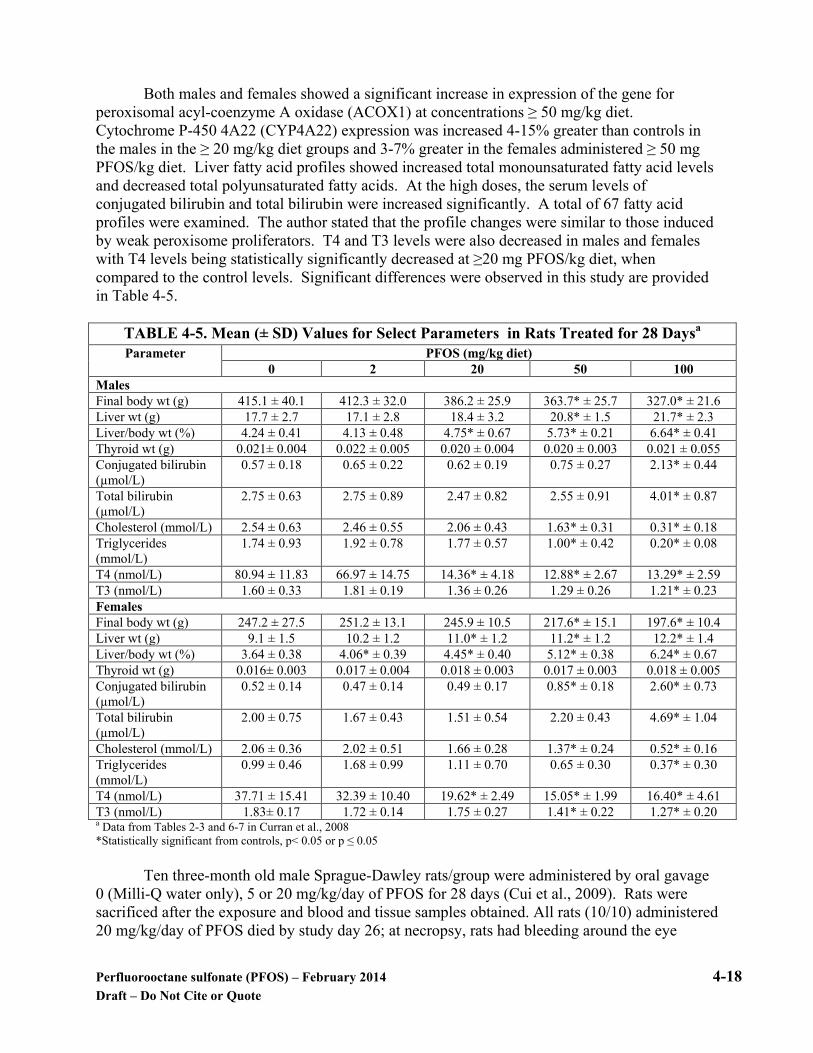
Perfluorooctane sulfonate (PFOS) – February 2014 4-18 Draft – Do Not Cite or Quote
Both males and females showed a significant increase in expression of the gene for
peroxisomal acyl-coenzyme A oxidase (ACOX1) at concentrations ≥ 50 mg/kg diet. Cytochrome P-450 4A22 (CYP4A22) expression was increased 4-15% greater than controls in the males in the ≥ 20 mg/kg diet groups and 3-7% greater in the females administered ≥ 50 mg PFOS/kg diet. Liver fatty acid profiles showed increased total monounsaturated fatty acid levels and decreased total polyunsaturated fatty acids. At the high doses, the serum levels of conjugated bilirubin and total bilirubin were increased significantly. A total of 67 fatty acid profiles were examined. The author stated that the profile changes were similar to those induced by weak peroxisome proliferators. T4 and T3 levels were also decreased in males and females with T4 levels being statistically significantly decreased at ≥20 mg PFOS/kg diet, when compared to the control levels. Significant differences were observed in this study are provided in Table 4-5.
TABLE 4-5. Mean (± SD) Values for Select Parameters in Rats Treated for 28 Daysa
Parameter PFOS (mg/kg diet) 0 2 20 50 100
Males Final body wt (g) 415.1 ± 40.1 412.3 ± 32.0 386.2 ± 25.9 363.7* ± 25.7 327.0* ± 21.6 Liver wt (g) 17.7 ± 2.7 17.1 ± 2.8 18.4 ± 3.2 20.8* ± 1.5 21.7* ± 2.3 Liver/body wt (%) 4.24 ± 0.41 4.13 ± 0.48 4.75* ± 0.67 5.73* ± 0.21 6.64* ± 0.41 Thyroid wt (g) 0.021± 0.004 0.022 ± 0.005 0.020 ± 0.004 0.020 ± 0.003 0.021 ± 0.055 Conjugated bilirubin (µmol/L)
0.57 ± 0.18 0.65 ± 0.22 0.62 ± 0.19 0.75 ± 0.27 2.13* ± 0.44
Total bilirubin (µmol/L)
2.75 ± 0.63 2.75 ± 0.89 2.47 ± 0.82 2.55 ± 0.91 4.01* ± 0.87
Cholesterol (mmol/L) 2.54 ± 0.63 2.46 ± 0.55 2.06 ± 0.43 1.63* ± 0.31 0.31* ± 0.18 Triglycerides (mmol/L)
1.74 ± 0.93 1.92 ± 0.78 1.77 ± 0.57 1.00* ± 0.42 0.20* ± 0.08
T4 (nmol/L) 80.94 ± 11.83 66.97 ± 14.75 14.36* ± 4.18 12.88* ± 2.67 13.29* ± 2.59 T3 (nmol/L) 1.60 ± 0.33 1.81 ± 0.19 1.36 ± 0.26 1.29 ± 0.26 1.21* ± 0.23 Females Final body wt (g) 247.2 ± 27.5 251.2 ± 13.1 245.9 ± 10.5 217.6* ± 15.1 197.6* ± 10.4 Liver wt (g) 9.1 ± 1.5 10.2 ± 1.2 11.0* ± 1.2 11.2* ± 1.2 12.2* ± 1.4 Liver/body wt (%) 3.64 ± 0.38 4.06* ± 0.39 4.45* ± 0.40 5.12* ± 0.38 6.24* ± 0.67 Thyroid wt (g) 0.016± 0.003 0.017 ± 0.004 0.018 ± 0.003 0.017 ± 0.003 0.018 ± 0.005 Conjugated bilirubin (µmol/L)
0.52 ± 0.14 0.47 ± 0.14 0.49 ± 0.17 0.85* ± 0.18 2.60* ± 0.73
Total bilirubin (µmol/L)
2.00 ± 0.75 1.67 ± 0.43 1.51 ± 0.54 2.20 ± 0.43 4.69* ± 1.04
Cholesterol (mmol/L) 2.06 ± 0.36 2.02 ± 0.51 1.66 ± 0.28 1.37* ± 0.24 0.52* ± 0.16 Triglycerides (mmol/L)
0.99 ± 0.46 1.68 ± 0.99 1.11 ± 0.70 0.65 ± 0.30 0.37* ± 0.30
T4 (nmol/L) 37.71 ± 15.41 32.39 ± 10.40 19.62* ± 2.49 15.05* ± 1.99 16.40* ± 4.61 T3 (nmol/L) 1.83± 0.17 1.72 ± 0.14 1.75 ± 0.27 1.41* ± 0.22 1.27* ± 0.20 a Data from Tables 2-3 and 6-7 in Curran et al., 2008 *Statistically significant from controls, p< 0.05 or p ≤ 0.05
Ten three-month old male Sprague-Dawley rats/group were administered by oral gavage 0 (Milli-Q water only), 5 or 20 mg/kg/day of PFOS for 28 days (Cui et al., 2009). Rats were sacrificed after the exposure and blood and tissue samples obtained. All rats (10/10) administered 20 mg/kg/day of PFOS died by study day 26; at necropsy, rats had bleeding around the eye
Perfluorooctane sulfonate (PFOS) – February 2014 4-19 Draft – Do Not Cite or Quote
socket and nose, and tumescence/yellow staining at the urogenital region. Prior to death, rats displayed significant weight loss and a decrease in food consumption when compared to controls. Rats administered 5 mg/kg/day also had a significant decrease in body weight when compared to controls at the study termination. Viscera indices were calculated including the hepatosomatic (HSI), renal-somatic (RSI), and gonad-somatic (GSI) index to evaluate the hyperplasia, swelling and/or atrophy of the organs and all three indices were statistically significantly increased in all of the treated groups. The increases in the HSI and RSI showed a dose dependency. Rats administered 20 mg/kg/day had swelling and discoloration of the liver with hepatocyte hypertrophy and cytoplasmic vacuolation observed on histopathological exam. Rats administered 20 mg/kg/day had congestion and thickened walls in the lungs with the pulmonary congestion also observed in the 5 mg/kg rats. Based on the results, a no observed adverse effect level (NOAEL) could not be identified and the lowest observed adverse effect level (LOAEL) was 5 mg/kg/day in rats. Mouse
Bijand et al. (2011) examined the molecular biology for the hepatic hyperlipidemia in
APOE*3-Leiden.CETP mice, a strain that exhibits human-like lipoprotein metabolism. The experimental animals were fed a western-type diet with or without 3 mg PFOS/kg/day for periods of 4 weeks. Plasma samples were collected via tail vein bleeding and analyzed for a variety of lipid related endpoints including TC, triglycerides (TG), very low density lipoprotein (VLDL) and HDL. Following terminal sacrifice the liver, heart, perigonadal fat, spleen, and skeletal femoralis muscle were collected for analysis. Fecal samples were collected for measurement of bile acids and neutral sterols.
PFOS was found to decrease hepatic VLDL production leading to increased retention of
triglycerides (steatosis) and hepatomegaly. As a consequence there was a decrease in plasma free fatty acids and glycerol, the mass of perigonadal fat pad, and TG uptake by skeletal muscle. Neutral sterols in the feces were not altered, but the presence of bile acids was decreased by 50%. Hepatic clearance of VLDL and HDL cholesterol were decreased primarily because of impaired hepatic production and clearance of these lipoprotein complexes.
Impacted hepatic genes involved with lipid metabolism included those involved with
VLDL metabolism, fatty acid uptake and transport, fatty acid oxidation, and triglyceride synthesis. Overall the genes upregulated were those involved with fatty acid uptake, transport and catabolism; triglyceride synthesis, cholesterol ester synthesis; plus VLDL synthesis and secretion. Genes involved with HDL synthesis, maturation and clearance plus bile acid formation were down regulated. These changes are consistent with increased hepatic hyperlipidemia, decrease in bile acid secretion, and serum hypolipidemia. Many of the genes activated are associated with the nuclear pregnane X receptor (PXR) to a greater extent than PPARα. Lipoprotein lipase activity and mRNA expressions were increased in the liver. This enzyme facilitates removal of TGs from serum LDLs, and uptake into the liver and other organs as free fatty acids and glycerol. Lipoprotein lipase activity in the liver is relatively low compared to that of peripheral tissues.

Perfluorooctane sulfonate (PFOS) – February 2014 4-20 Draft – Do Not Cite or Quote
Inhalation Exposure
No short-term inhalation animal exposure studies of PFOS were identified.
4.2.3 Subchronic Studies
There are three monkey studies of PFOA exposure, two with rhesus- and one with cynomolgus- strains, and two rat subchronic studies. The study with cynomolgus monkeys was a GLP study. There are no subchronic studies by dermal or inhalation routes of exposure with PFOS.
Oral Exposure Monkey Two monkey studies were performed with rhesus monkeys (Goldenthal et al., 1978a and 1979). In the first study, 2 monkeys/sex/group were administered 0, 10, 30, 100 or 300 mg/kg/day of PFOS in distilled water by gavage. The study was terminated on day 20 as all of the 300 mg/kg treated monkeys died beginning on day 4. Clinical signs of toxicity were observed in all groups and included decreased activity, emesis, body stiffening, general body trembling, twitching, weakness and convulsions. At necropsy, several of the 100 and 300 mg/kg/day monkeys had a yellowish-brown discoloration of the liver although there were no microscopic lesions. A NOAEL or LOAEL was not set in the study. In the second study, 2 rhesus monkeys/sex/group were administered 0, 0.5, 1.5 or 4.5 mg/kg/day of PFOS in distilled water by gavage for 90 days. All monkeys in the 4.5 mg/kg/day group died or were euthanized in extremis by week 7 and exhibited decreased body weight, signs of gastrointestinal tract toxicity (anorexia, emesis, black stool), decreased activity and marked to severe rigidity and had a significant decrease in serum cholesterol. Histopathology of the 4.5 mg/kg/day monkeys showed diffuse lipid depletion in the adrenals (4/4), diffuse atrophy of the pancreatic exocrine cells (3/4) and moderate diffuse atrophy of the serous alveolar cells (3/4). All monkeys in the 0.5 and 1.5 mg/kg/day treated groups survived, but exhibited occasional diarrhea, soft stools and anorexia. These clinical signs showed a dose-related increase, and 1/4 of the 1.5 mg/kg/day monkeys had low serum cholesterol. Body weight was decreased in males and females at 1.5 mg/kg/day. There were no treatment-related effects observed in any of the 0.5 or 1.5 mg/kg/day monkeys at necropsy. Based on the findings, the LOAEL was 0.5 mg/kg/day and the NOAEL could not be determined. Seacat et al. (2002) administered 0, 0.03, 0.15 or 0.75 mg/kg/day of potassium PFOS orally in a capsule by intragastric intubation to 6 young-adult to adult cynomolgus monkeys/sex/group, except for the 0.03 mg/kg/day group (4 monkeys/sex), daily for 26 weeks (182 days) in a GLP study. Two monkeys/sex in the control, 0.15, and 0.75 mg/kg/day groups were monitored for one year post-exposure for reversible or delayed toxic effects. Monkeys were observed twice daily for mortality, morbidity, clinical signs and qualitative food consumption. Body weights were recorded pre-dosing and weekly, thereafter, and ophthalmic examinations were performed pre- and post-treatment. PFOS levels were determined in serum and liver tissue and hematology and clinical chemistry were performed. Urine and fecal analyses were done and full histopathology performed at the scheduled sacrifice. Liver samples were also

Perfluorooctane sulfonate (PFOS) – February 2014 4-21 Draft – Do Not Cite or Quote
obtained for hepatic peroxisome proliferation determination and immunohistochemistry was performed by PCNA to look for cell proliferation. Selected results are shown in Table 4-6. Two of the 0.75 mg/kg/day males died; one died on day 155 and one was found moribund and sacrificed on day 179. The monkey that died had pulmonary necrosis and severe acute recurrences of pulmonary inflammation as its cause of death. The specific cause of the moribund condition was not established, however, the clinical chemistry results were suggestive of hyperkalemia. Overall mean body weight gain was significantly (p ≤ 0.05) less in the 0.75 mg/kg/day males and females after the treatment when compared to controls. The 0.75 mg/kg/day males and females lost 8 ± 8% and 4 ± 5%, respectively, while the control males and females gained 14 ± 11% and 5 ± 5%, respectively. Mean absolute and relative (to body weight) liver weight was increased significantly in the 0.75 mg/kg/day males and females. Males and females at 0.75 mg/kg/day had lower total serum cholesterol beginning on day 91 (27-68% and 33-49% lower than controls) and lower high density lipoprotein cholesterol beginning on day 153 (72-79% and 61-68% lower than controls) when compared to the control values. This effect was reversible, however, as the total cholesterol levels were similar to controls by week 5 during recovery and the total high density lipoprotein cholesterol was similar to controls by week 9. Estradiol values were lower at 0.75 mg/kg in males and females on day 182; however, the data were highly variable and the study authors stated that the change was not well understood. Total triiodothyronine (T3) values were significantly decreased and thyroid stimulating hormone (TSH) was increased on day 182 in the high-dose monkeys but a true dose-response was not observed and the monkeys had no sign of hypothyroidism.

Perfluorooctane sulfonate (PFOS) – February 2014 4-22 Draft – Do Not Cite or Quote
TABLE 4-6. Mean (± SD) Values for Select Parameters in Monkeys Treated for 182 Daysa
Parameter PFOS (mg/kg/day)
Males 0 0.03 0.15 0.75 Body wt (g) 3.7 ± 0.7 3.9 ± 0.6 3.3 ± 0.3 3.2 ± 0.8 Body wt change (%) 14 ± 11 16 ± 8 8 ± 7 -8 ± 8* Liver wt (g) 54.9 ± 8.1 62.1 ± 5.3 57.3 ± 5.5 85.3 ± 38.4 Liver/body wt (%) 1.6 ± 0.2 1.7 ± 0.3 1.8 ± 0.1 2.7 ± 0.3* Cholesterol (mg/dL) 152 ± 28 110 ±17** 147 ± 24 48 ± 19** HDL (mg/dL) 63 ± 11 42 ± 4** 48 ± 14 13 ± 5** Total T3 (ng/dL) 146 ± 19.8 145 ± 18.0 129 ± 4.8 76 ± 22** TSH (µU/mL) 0.55 ± 0.44 0.56 ± 0.10 1.38 ± 0.78 1.43 ± 0.25* Estradiol (pg/mL) 23.0 ± 11.5 24.1 ± 14.2 23.2 ± 7.4 0.8 ± 1.0**
Females 0 0.03 0.15 0.75 Body wt (g) 3.0 ± 0.4 3.2 ± 0.7 3.1 ± 0.5 2.8 ± 0.4 Body wt change (%) 5 ± 5 6 ± 7 4 ± 5 -4 ± 5 Liver wt (g) 51.1 ± 9.4 56.8 ± 12.6 57.0 ± 3.1 75.3 ± 13.3* Liver/body wt (%) 1.8 ± 0.2 1.9 ± 0.0 2.1 ± 0.2 2.9 ± 0.3* Cholesterol (mg/dL) 160 ± 47 122 ± 22 129 ± 22 82 ± 15** HDL (mg/dL) 56 ± 16 42 ± 9 36 ± 12** 21 ± 7** Total T3 (ng/dL) 148 ± 21.6 139 ± 11.5 116 ± 16.8 99 ± 16.8* TSH (µU/mL) 1.02 ± 0.69 2.01 ± 2.09 1.33 ± 1.13 1.86 ± 1.29 Estradiol (pg/mL) 148.5 ± 110.1 125.2 ± 101.2 70.6 ± 62.7 39.9 ± 33.6 a Data from Seacat et al., 2002 Statistically significant from controls: *p< 0.05; **p<0.01. Hepatic peroxisome proliferation was measured by palmitoyl CoA oxidase activity and was increased significantly in the 0.75 mg/kg/day females; however, it was less than the two-fold increase typically indicating biological significance. There were no treatment-related effects on cell proliferation in the liver, pancreas or testes when analyzed by proliferating cell nuclear antigen immunohistochemistry cell labeling index. Two high dose males and one high-dose female had ‘mottled’ livers on gross examination at sacrifice; this was also observed in the high-dose male that died during the study. On histopathology, all high-dose females and 3/4 high-dose males had centrilobular or diffuse hepatocellular hypertrophy. Serum and liver samples collected during the study were analyzed for PFOS and animals showed a dose-dependent increase in concentrations. Values decreased with recovery but never returned to control levels. There was not any gender difference in the amount of PFOS identified in the sera or liver. Based on the decreased body weight gain, decreased serum cholesterol, increased absolute and relative liver weight and histopathological lesions in the liver, the LOAEL in male and female monkeys treated with potassium PFOS was 0.75 mg/kg/day and the NOAEL was 0.15 mg/kg/day. Serum concentrations associated with no adverse effect (0.15 mg/kg/day) were 82.6 ppm in males and 66.8 ppm in females. Rat Goldenthal (1978b) administered 0, 30, 100, 300, 1000 or 3000 ppm of PFOS in the diet to five CD rats/sex/group for 90 days. Dietary levels were equivalent to 0, 2, 6, 18, 60 and 200 mg/kg/day, respectively. All rats at ≥ 300 ppm exhibited emaciation, convulsions, hunched
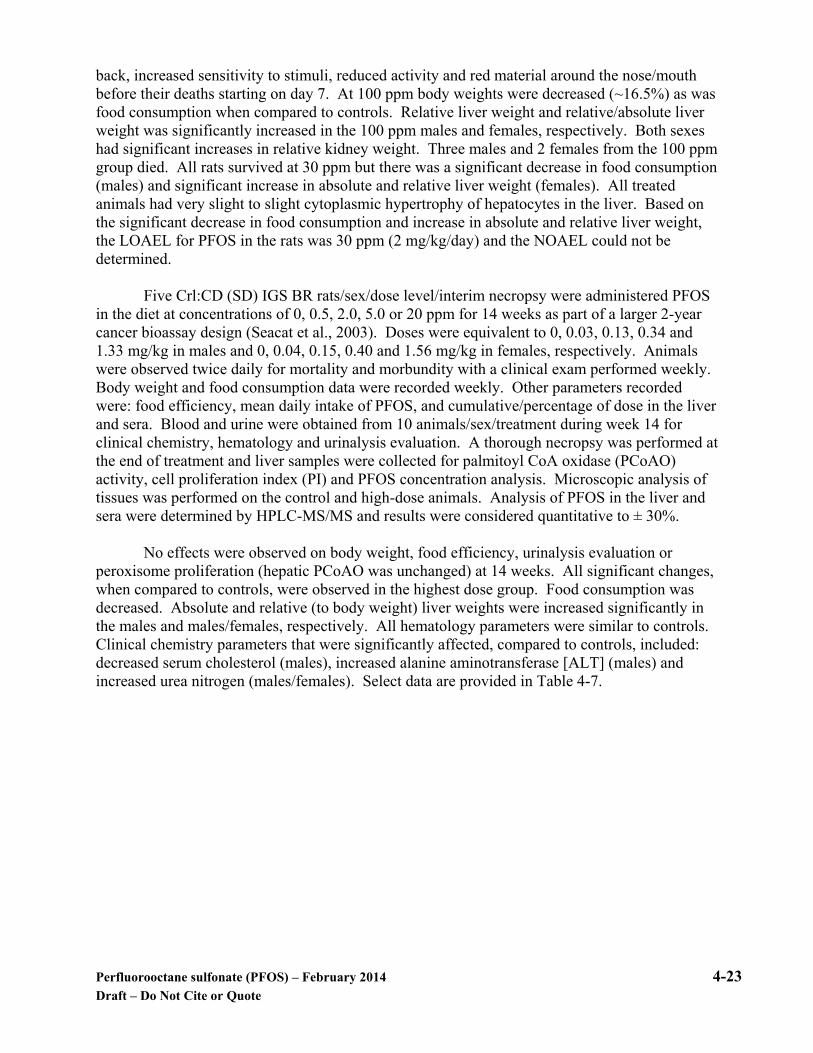
Perfluorooctane sulfonate (PFOS) – February 2014 4-23 Draft – Do Not Cite or Quote
back, increased sensitivity to stimuli, reduced activity and red material around the nose/mouth before their deaths starting on day 7. At 100 ppm body weights were decreased (~16.5%) as was food consumption when compared to controls. Relative liver weight and relative/absolute liver weight was significantly increased in the 100 ppm males and females, respectively. Both sexes had significant increases in relative kidney weight. Three males and 2 females from the 100 ppm group died. All rats survived at 30 ppm but there was a significant decrease in food consumption (males) and significant increase in absolute and relative liver weight (females). All treated animals had very slight to slight cytoplasmic hypertrophy of hepatocytes in the liver. Based on the significant decrease in food consumption and increase in absolute and relative liver weight, the LOAEL for PFOS in the rats was 30 ppm (2 mg/kg/day) and the NOAEL could not be determined. Five Crl:CD (SD) IGS BR rats/sex/dose level/interim necropsy were administered PFOS in the diet at concentrations of 0, 0.5, 2.0, 5.0 or 20 ppm for 14 weeks as part of a larger 2-year cancer bioassay design (Seacat et al., 2003). Doses were equivalent to 0, 0.03, 0.13, 0.34 and 1.33 mg/kg in males and 0, 0.04, 0.15, 0.40 and 1.56 mg/kg in females, respectively. Animals were observed twice daily for mortality and morbundity with a clinical exam performed weekly. Body weight and food consumption data were recorded weekly. Other parameters recorded were: food efficiency, mean daily intake of PFOS, and cumulative/percentage of dose in the liver and sera. Blood and urine were obtained from 10 animals/sex/treatment during week 14 for clinical chemistry, hematology and urinalysis evaluation. A thorough necropsy was performed at the end of treatment and liver samples were collected for palmitoyl CoA oxidase (PCoAO) activity, cell proliferation index (PI) and PFOS concentration analysis. Microscopic analysis of tissues was performed on the control and high-dose animals. Analysis of PFOS in the liver and sera were determined by HPLC-MS/MS and results were considered quantitative to ± 30%. No effects were observed on body weight, food efficiency, urinalysis evaluation or peroxisome proliferation (hepatic PCoAO was unchanged) at 14 weeks. All significant changes, when compared to controls, were observed in the highest dose group. Food consumption was decreased. Absolute and relative (to body weight) liver weights were increased significantly in the males and males/females, respectively. All hematology parameters were similar to controls. Clinical chemistry parameters that were significantly affected, compared to controls, included: decreased serum cholesterol (males), increased alanine aminotransferase [ALT] (males) and increased urea nitrogen (males/females). Select data are provided in Table 4-7.
Perfluorooctane sulfonate (PFOS) – February 2014 4-24 Draft – Do Not Cite or Quote
TABLE 4-7. Mean (± SD) Values for Select Parameters in Rats Treated for 14 Weeksa
Parameter PFOS (mg/kg/day)
Males 0 0.03 0.13 0.34 1.33 Body wt (g) 496 ± 56 481 ± 51 434 ± 31 424 ± 44 470 ± 40 Liver wt (g) 15.5 ± 1.1 15.5 ± 2.7 14.0 ± 1.4 18.8 ± 3.0 20.3*± 2.2 Liver/body wt (%) 3.2 ± 0.3 3.2 ± 0.2 3.2 ± 0.2 3.6 ± 0.3 4.3* ± 0.4 Seg. neutrophils (103/µL) 1.1 ± 0.4 1.3 ± 0.3 1.2 ± 0.3 1.2 ± 0.4 1.6* ± 0.4 Glucose (mg/dL) 102 ± 6.2 106 ± 11 91 ± 14 99 ± 9 95 ± 10 Cholesterol (mg/dL) 63 ± 13 53 ± 17 51 ± 15 57 ± 7 37* ± 13 ALT (IU/L) 36 ± 7 41 ± 6 41 ± 5 44 ± 14 65* ± 53 Urea nitrogen (mg/dL) 13 ± 2 14 ± 2 13 ± 2 14 ± 1 16* ± 2 PCoAO (IU/g) 4.6 ± 1.3 4.8 ± 3.3 5.4 ± 3.0 1.8 ± 1.8 5.4 ± 1.9
Females 0 0.04 0.15 0.40 1.56 Body wt (g) 284 ± 39 298 ± 41 266 ± 16 247 ± 18 249 ± 26 Liver wt (g) 9.3 ± 1.6 9.2 ± 1.3 8.4 ± 0.7 8.7 ± 1.0 10.6 ± 0.7 Liver/body wt (%) 3.3 ± 0.2 3.1 ± 0.1 3.2 ± 0.3 3.5 ± 0.3 4.3* ± 0.4 Seg. neutrophils (103/µL) 1.0 ± 0.5 1.0 ± 0.5 0.7 ± 0.2 0.9 ± 0.6 1.0 ± 0.6 Glucose (mg/dL) 106 ± 12 106 ± 9 108 ± 6 95* ± 8 99 ± 7 Cholesterol (mg/dL) 75 ± 15 88 ± 27 87 ± 24 70 ± 13 66 ± 14 ALT (IU/L) 34 ± 2.4 36 ± 9 37 ± 18 34 ± 5 39 ± 18 Urea nitrogen (mg/dL) 12 ± 2 13 ± 2 13 ± 2 14 ± 3 17* ± 2 PCoAO (IU/g) 1.8 ± 1.6 3.0 ± 2.6 1.0 ± 0.8 1.6 ± 2.6 5.0 ± 2.9 a Data from Table 1 in Seacat et al., 2003 *Statistically significant from controls, p< 0.05
Histopathological changes were not observed in the kidney; however, centrilobular hepatocyte hypertrophy and mid-zonal to centrilobular vacuolization were observed in the livers of the males and females. Based on the findings, the LOAEL for male and female rats administered PFOS in the diet for up to 14 weeks was 20 ppm (1.33 mg/kg in males and 1.56 mg/kg in females) and the NOAEL was 5 ppm (0.34 mg/kg in males and 0.40 mg/kg in females).
4.2.4 Neurotoxicity
In vivo Rat
Yang et al. (2009) determined the effect of PFOS on excitatory amino acids (EAAs) and
glutamine synthetase (GS) in the rat central nervous system. Five adult male Wistar rats/group were administered one dose of 0, 12.5, 25 or 50 mg/kg bw PFOS by oral gavage. The animals were sacrificed 5 days after administration. The EAAs analyzed in brain tissue were glutamate (Glu), aspartate (Asp), glycine (Gly) and gamma-aminobutyric acid (GABA).
All treated rats had a significant (p<0.05) decrease in body weight, 15, 22 and 27% less
than the controls in the 12.5, 25 and 50 mg/kg groups, respectively. Among the EAAs, the Glu content was significantly decreased at the high dose (↓77% compared to controls; p<0.05) in the hippocampus; no other significant differences were recorded. In the cortex, Glu was again the only EAA affected with significant decreases at 25 (↓33% compared to controls) and 50 (↓47

Perfluorooctane sulfonate (PFOS) – February 2014 4-25 Draft – Do Not Cite or Quote
compared to controls) mg/kg. GS activity was significantly increased in the hippocampus at 25 and 50 mg/kg bw. The study had a LOAEL of 12.5 mg/kg/day in rats based on the decreased body weight and indicated an effect on Glu and GS in the central nervous system starting at 25 mg/kg/day.
Male Wistar rats and ICR mice (n=2-3/group) were administered a single oral dose of PFOS at concentrations of 0, 125, 250 or 500 mg/kg bw and monitored for any neurological signs (Sato et al., 2009). Animals were checked daily for startle response, touch response, pain response, righting reflex, visual placing, abdominal tone and limb tone. No neurological signs were observed. However, when stimulated with ultrasound (44 kHz, 10 sec), tonic convulsions occurred and 1/3 rats in the 250 mg/kg group, 2/2 rats in the 500 mg/kg group and 1 mouse in each dose group died within 48 hours of the convulsions. No histopathological changes were observed in the neuronal or glial cells of the cerebrum and cerebellum in rats killed 24 hours after exposure. In these same rats, the highest concentration of PFOS was in the liver and the lowest was in the brain. Rats administered 250 mg/kg bw one time did not show any differences in the levels of catecholamines (norepinephrine, dopamine and serotonin) or amino acids (glutamic acid, glycine and GABA) when compared to the controls at 24 and 48 hours post-exposure. In vitro
Slotkin et al. (2008) evaluated 10-250 µM PFOS, PFOA, perfluorooctane sulfamide
(PFOSA) and perfluorobutane sulfonate (PFBS) in vitro in differentiated and undifferentiated PC12 cells, a neurotypic cell line. In the study, inhibition of DNA synthesis, deficits in cell numbers and growth, oxidative stress, cell viability and shifts in differentiation toward or away from the dopamine (DA) and acetylcholine (ACh) neurotransmitter phenotypes were assessed. No effects on cell size, cell number or neurocyte outgrowth were observed. PFOS decreased cell viability at 250 µM and promoted differentiation into the ACh phenotype at the expense of the DA phenotype. The study suggests that the mechanisms of these neurotypic cell lines are not similar between the tested perfluoroalkyl acids. The rank order of adverse effects was PFOSA > PFOS > PFBS = PFOA.
Liao et al. (2009) assessed the neurotoxic effect of varying chain lengths of the
perfluorinated compounds on cultured Sprague-Dawley rat hippocampal neurons. Spontaneous miniature post-synaptic currents (mPSCs) were recorded in gap-free mode from hippocampal neurons at 8-15 days in vitro. The compounds tested included those in the carboxylic group: perfluoropropionic acid (PFPA; C-3), perfluorobutyric acid (PFBA; C-4), perfluorooctanoic acid (PFOA; C-8), perfluorododecanoic acid (PFDA; C-12) and perfluorotetradecanoic acid (PFTA; C-14); those in the sulfonic group: perfluorobutane sulfonic (PFBS; C-4), perfluorohexanesulfonic acid potassium salt (PFHS; C-6) and perfluorooctane sulfonate (PFOS; C-8) and a nonfluorinated hydrocarbon, octanoic acid (OA; C-8). Also tested was 1 H-perfluorooctane (PFOC; C-8). Testing showed frequency of mPSCs increased in proportion to the increase in carbon length. Eight carbon PFOS had a statistically significant (p< 0.001) increase in the mPSCs when compared to 4 carbon PFBS. Compounds with a carboxylic group also had lower frequencies than those with sulfonic groups. PFOS significantly (p< 0.001) increased the mPSC amplitude. The inward calcium currents (ICa) were recorded in the presence or absence of compounds with a ramp depolarization pulse. Voltage values were recorded and
Perfluorooctane sulfonate (PFOS) – February 2014 4-26 Draft – Do Not Cite or Quote
plotted versus the corresponding ICa every 5 mV and the resulting current-voltage relationship curve established. All three sulfonic compounds increased the ICa with correlation to the chain length with PFOS having the most effect (% increase not provided).
In the same study, the chronic effects of perfluorinated compounds on neuronal
development were evaluated by measuring neurite outgrowth and branching. In the sulfonic compounds, only PFOS statistically suppressed the length of neurites (p < 0.001; 25% below that of controls). The lengths of the longest neurites and sum length of neurites per neuron were not affected with OA or PFOC but were reduced by PFOA and PFOS.
Overall, the adverse effects on cultured neurons increased as the chain length of the test
compound increased. The study also suggested that perfluorinated sulfonates exerted more potent actions on neurons when compared to perfluorinated carboxylates. The study authors hypothesized that this could occur because PFOS was more likely to be incorporated into the lipid bilayer of cell membranes.
4.2.5 Developmental/Reproductive Toxicity
Rats, rabbits, and mice were all found to be affected in developmental/reproductive studies of PFOS. Prenatal exposure to PFOS caused an increase in neonatal mortality when dams were exposed to concentrations ≥ 2 mg/kg/day. Effects were observed on gestation length, birth weight, survival and developmental delays. Structural abnormalities observed include: ocular lens abnormalities (questionable), decreased bone ossification, cleft palate, and enlargement of the right atrium. Many specialized developmental studies (neurotoxicity, immunotoxicity) have been conducted with PFOS.
Reproductive Effects Rat
A two-generation reproductive study was conducted in Crl:CD(SD)IGS VAF rats with
five groups of 35 rats/sex/group administered 0, 0.1, 0.4, 1.6 or 3.2 mg/kg/day of PFOS by gavage for six weeks prior to and during mating (Luebker et al., 2005b). Treatment in males continued until one day prior to sacrifice and females were treated throughout gestation, parturition and lactation.
F0 Generation: Parental animals (F0) were observed twice daily for clinical signs, and body weight and food consumption monitored. Two sets of females in each dose group were treated and had Caesarean-sections (C-section) performed on GD10; others delivered naturally and were killed on lactation day (LD) 21. Typical reproductive parameters were monitored in the females. The F0 male rats were sacrificed and necropsied after the cohabitation interval, with the testes, epididymides, prostate, and seminal vesicles weighed. All livers from adults were removed, weighed and examined. Blood samples were collected from five male rats at sacrifice and five female rats on LD 21 for pharmacokinetic analysis; livers of pups from the litters of these five dams were also collected for analysis. In the F0 generation male rats, no effects were observed on mortality, treatment-related clinical signs and the mating/fertility parameters evaluated. During pre-mating, decreases in

Perfluorooctane sulfonate (PFOS) – February 2014 4-27 Draft – Do Not Cite or Quote
terminal body weight, body weight gain and food consumption occurred at 1.6 and 3.2 mg/kg/day in males. The only effect on weight of the organs evaluated was a significant reduction in the absolute weight of the seminal vesicles (with fluid) and prostate in males administered 3.2 mg/kg/day. In the F0 generation female rats, there were no deaths and no effects on the reproductive parameters measured in both dams sacrificed on GD 10 and those allowed to deliver naturally. The F0 dams administered ≥ 0.4 mg/kg/day had localized alopecia during pre-mating, gestation, and lactation, and a decrease in body weight and food consumption. F1 Generation: The F1 generation pup viability was significantly reduced at 1.6 and 3.2 mg/kg/day, so only the 0.1 and 0.4 mg/kg/day dose groups were carried into the second generation. Twenty-five F1 rats/sex/dose were administered 0, 0.1, or 0.4 mg/kg/day of PFOS by oral gavage beginning at weaning on post-natal day (PND) 22 and continuing through sacrifice. One rat/sex/litter was tested in a passive avoidance paradigm at 24 days of age and one/sex/litter was evaluated in a water-filled M-maze on PND 70. On PND 28, females were evaluated for vaginal patency and on PND 34 males were examined for preputial separation. On PND 90, rats were assigned within each dose group to cohabitation, and once confirmed pregnant, the females were individually housed. The F1 generation male rats were sacrificed after mating, necropsied and evaluated as described in the F0 generation. All F1 generation females delivered naturally and were sacrificed and necropsied on LD 21. Mortality occurred in the F1 offspring of dams administered 1.6 or 3.2 mg/kg/day. At 1.6 mg/kg/day, over 26% of the pups were found dead between LDs 2-4. At 3.2 mg/kg/day, 45% of the pups were found dead on LD 1 with 100% dead by LD 2. The dams dosed with 3.2 mg/kg/day also had a significant increase in stillborns and the viability index was 0% at 3.2 mg/kg/day and 66% at 1.6 mg/kg/day. The lactation index was 94.6% at 1.6 mg/kg/day. At 3.2 mg/kg/day, there were significant decreases in gestation length, number of implantation sites and reductions in litter size. Statistically significant decreases in pup body weight were also observed at the two highest doses. Additional adverse signs in pups at 3.2 mg/kg/day were a high number of pups not nursing, not having milk present in the stomach, being stillborn and a high incidence of maternal cannibalization of the pups. In the F1 generation offspring, pups administered 3.2 mg/kg/day could be evaluated only on LD 1 due to high mortality; all displayed decreased surface righting ability. The acoustic startle reflex and air righting ability were both significantly reduced at 1.6 mg/kg/day. Similar responses were seen for pinna unfolding and eye opening. The physical development delays were transient; all pups were similar to controls by the end of the observation period. No delays were observed in rats administered doses ≤ 0.4 mg/kg/day. F2 Generation: F1 parental animals displayed no clinical signs or mortality. F1 male parents had a transient decrease in food consumption but it was not affected in F1 females. The F1 dams showed no effects in reproductive performance. All F2 generation pups were sacrificed, necropsied, and examined on LD 21 as previously described for the F1 generation pups. In the F2 generation pups, decreases in mean pup body weights at 0.1 mg/kg/day on LDs 4 and 7 were transient; mean body weights were

Perfluorooctane sulfonate (PFOS) – February 2014 4-28 Draft – Do Not Cite or Quote
similar to controls by LD 14. The pups at 0.4 mg/kg/day had significant decreases in body weight on LDs 7-14; after LD 21, body weights remained lower than controls, but were not statistically significant. No other treatment-related effects were observed. Based on the decreases in body weight gain and food consumption, the LOAEL for both the F0 male and female parents was 0.4 mg/kg/day and the NOAEL was 0.1 mg/kg/day. For the F1 parents, the NOAEL was 0.4 mg/kg/day and the LOAEL was not identified. For the F1 offspring, the LOAEL was 1.6 mg/kg/day based on the significant decrease in the pup viability, pup weight and survival; the NOAEL was 0.4 mg/kg/day. In the F2 generation offspring, the LOAEL was 0.4 mg/kg/day, based on the significant decreases in mean pup body weight; the NOAEL was 0.1 mg/kg/day.
Because of the significant reductions in pup viability observed at 1.6 and 3.2 mg/kg/day, a cross-fostering study was conducted as a means of determining whether the effects observed in pups were a result of in utero exposure to PFOS or as a result of exposure during lactation (Luebker et al., 2005b). Twenty five female Sprague-Dawley rats/group were administered 0 or 1.6 mg/kg/day PFOS in 0.5% Tween-80 by gavage, beginning 42 days prior to mating to untreated males, and continuing throughout gestation until LD 21. Parental females were observed twice daily for viability and clinical observations were recorded. Maternal body weight and food consumption were recorded. All maternal rats were sacrificed on LD 22 and gross necropsy was performed; the number and distribution of implantation sites were recorded. After parturition, litters were immediately removed from their respective dams and placed with either a control- or PFOS-treated dam for rearing. This cross-fostering procedure resulted in four groups as follows:
• control dams with litters from control dams (negative control) • control dams with litters from PFOS-treated dams (in utero exposure only); • PFOS-treated dams with litters from control dams (post-natal exposure only) • PFOS-treated dams with litters from PFOS-treated dams (both in utero and post-natal
exposure). There was no mortality or clinical signs associated with treatment in the dams. Mean
maternal body weight gain and food consumption at 1.6 mg/kg/day was reduced compared to controls during premating and continuing throughout gestation, but not lactation. Reductions in gestation length, the average number of implantation sites, delivered sizes, and live litter size were observed in treated animals.
Live litter sizes were comparable between treated and control groups following cross-
fostering. On LDs 2-4, ~ 19% of the pups in the group exposed gestationally and lactationally were either found dead or presumed cannibalized. Pup mortality for the negative control was 1.6%. For pups exposed only prenatally mortality was 9% while it was 1.1% for those exposed during lactation only. Reductions in pup body weights on LD 1were observed in groups exposed both gestationally and lactationally and those with gestational exposure only. On LDs 4-21, pup body weights were reduced in all exposed groups when compared to the negative control, with the greatest decrease in the group with gestational and lactational exposures.
Perfluorooctane sulfonate (PFOS) – February 2014 4-29 Draft – Do Not Cite or Quote
Two litters in the group with lactational exposure and one litter exposed during gestation and lactation did not nurse. No milk was found in the stomachs of the pups found dead and necropsied from the groups with in utero plus lactational exposures (100% of pups), in utero exposures only (57% of pups), and lactational exposures only (87% of pups). Sex ratios and the lactation index were comparable among all groups. Electron microscopic examination of the livers revealed an increase in the number of peroxisomes in pups from dams treated with 1.6 mg/kg/day PFOS. No significant differences were observed between the negative control group and the other groups following examination of pup lungs.
Serum PFOS concentrations in untreated dams ranged from below the limit of detection
(0.05 µg/mL) to 5.34 µg/mL. Serum PFOS concentrations in the pups from the negative controls were below the limit of detection. Serum PFOS concentrations in the pups from treated dams, fostered with untreated dams (in utero exposures) ranged from 47.6 µg/mL to 59.2 µg/mL. Serum PFOS concentrations of treated dams ranged from 59.2 µg/mL to 157 µg/mL. Serum PFOS concentrations in the pups from untreated dams, fostered with treated dams (lactational exposure), ranged from below the limit of detection to 35.7 µg/mL. Serum PFOS concentrations in the pups from treated dams, fostered with treated dams (in utero plus lactational exposures), ranged from 79.5 µg/mL to 96.9 µg/mL. These data indicate that exposure to PFOS can occur both in utero and via milk from treated dams (3M Environmental Laboratory, 1999). The accuracy of quantitation for the analyses was ±30%.
In conclusion, pups from control dams that were cross-fostered with PFOS-treated dams (post-natal exposure only) had the same low mortality rate (1.1%) as pups from control dams cross-fostered with control dams (1.6%; negative control). Mortality rates in the remaining two groups (gestational exposures and gestational plus lactational exposures) were much higher at 9% and 19%, respectively. Although the study is limited, the data appear to indicate that reduced pup survival is mainly a result of in utero exposure to PFOS and that post-natal exposure via milk in conjunction with in utero exposure increases the risk of mortality. In contrast, exposure during lactation alone from exposed dams had no significant affect on pup viability. The dose-response curve for neonatal mortality in rat pups born to PFOS exposed dams and the biochemical and pharmacokinetic parameters were investigated (Luebker et al., 2005a). At 6 weeks prior to mating, female Crl:CD(SD)IGS VAF/Plus rats/dose were administered 0, 0.4, 0.8, 1.0, 1.2, 1.6 or 2.0 mg PFOS/kg bw/day by oral gavage. Dosing continued during the mating interval and through gestation day (GD) 20 for dams assigned to C-section (eight dams in the control, 1.6 and 2.0 mg/kg/day groups). Another group (~20 dams per dose group were allowed to deliver naturally and nurse their pups through LD4. These dams and their pups were killed on LD 5.
The dams in the C-section group were examined for the number of corpora lutea, number of implantation sites, live/dead fetuses, early/late resorptions and placentas. Liver weights were determined for the dams as well and their organs examined by gross necropsy. Fetuses were pooled by litter and mean weight recorded. In the natural delivery dams, typical reproductive and fetal parameters were measured and recorded. Biochemical parameters investigated in the dams and litters included: serum lipids, glucose, mevalonic acid and thyroid hormones (total [TT4] and free thyroxin [FT4], total [TT3] and free triiodothyronine [FT3] and thyroid stimulating hormone [TSH]); milk cholesterol and liver lipids. Mevalonic acid was included as
Perfluorooctane sulfonate (PFOS) – February 2014 4-30 Draft – Do Not Cite or Quote
it is a biomarker of 3-hydroxy-3-methylglutaryl coenzyme A (HMG-CoA) reductase activity. Some chemicals that are inhibitors of this enzyme are known to cause developmental effects in rats. No mortality occurred and no effects were observed in reproductive parameters (corpora lutea, implantations, dead fetuses/litter) in those C-sectioned. Mild decreases in body weight were observed in dams at 1.6 and 2.0 mg/kg during gestation. Body weight gain in the dams was affected statistically during lactation at ≥ 0.8 mg/kg and food consumption showed a general downward trend with increasing dose during pre-mating, gestation and lactation. For those allowed to deliver normally, the fertility index, implantations per delivered litter, gestation index, live births and delivered pups/litter were similar between treated and control dams. Based on the decreased body weight gain, the LOAEL for the F0 dams was 0.8 mg/kg/day and the NOAEL was 0.4 mg/kg/day. In the group sacrificed on LD5, a decrease in gestation length was observed at doses ≥ 0.8 mg/kg and decreases in viability were observed starting at 0.8 mg/kg becoming statistically significant at doses of 1.6 and 2.0 mg/kg. The viability indices were 97.3%, 93.1%, 88.8%, 81.7%, 49.3% and 17.1% at 0, 0.8, 1.0, 1.2, 1.6 and 2.0 mg/kg, respectively (Table 4-8). The decrease in survival did not appear to be caused by a reduction in lipids, glucose utilization or thyroid hormones as these parameters were either only slightly affected or similar between treated and control animals. In all treated groups, pup body weight at birth and on postnatal day 5 was significantly less than that of controls. In one male and one female pup at 2.0 mg/kg/day, the heart and thyroid were collected and examined microscopically. No lesions were found when compared to the controls. The LOAEL for the F1 generation was 0.4 mg/kg/day, respectively, based on decreased body weight and the NOAEL was not identified. Several benchmark dose estimates (BMD5 and BMDL5) were run by the author and provided in the study. They were as follows:
Effect on gestation length BMD5 = 0.45 mg/kg/day BMDL5 = 0.31 mg/kg/day Birth weight effect BMD5 = 0.63 mg/kg/day BMDL5 = 0.39 mg/kg/day Decreased pup wt (day 5) BMD5 = 0.39 mg/kg/day BMDL5 = 0.27 mg/kg/day Pup weight gain (day 5) BMD5 = 0.41 mg/kg/day BMDL5 = 0.28 mg/kg/day Decreased survival of pups through day 5 BMD5 = 1.06 mg/kg/day BMDL5 = 0.89 mg/kg/day

Perfluorooctane sulfonate (PFOS) – February 2014 4-31 Draft – Do Not Cite or Quote
TABLE 4-8. Fertility and Litter Observations in Dams Administered 0 to 2.0 mg PFOS/kg/Daya
0.0 0.4 0.8 1.0 1.2 1.6 2.0 Fertility indexb (%) 96.4 100.0 89.5 95.0 94.7 92.6 96.4 Implantations per delivered litter
14.7 ± 2.3 16.2 ± 1.8
15.1 ± 2.2
15.9 ± 2.0 15.3 ± 2.5 14.3 ± 2.1 14.4 ± 1.9
Gestation length (days) 22.9 ± 0.3 22.6 ± 0.5
22.5 ± 0.5*
22.4 ± 0.6**
22.3 ± 0.5**
22.0 ± 0.0**
22.2 ± 0.4**
Gestation indexc (%) 100 100 100 100 100 100 100 Delivered pups/litter 13.9 ± 2.6 15.0±
2.3 14.5 ±
2.3 15.1 ± 2.3 14.0 ± 2.9 13.6 ± 2.8 13.3 ± 2.5
Live births (%) 98.1 97.0 99.2 99.3 99.6 98.3 99.6 Dams with all pups dying on LD 1-5
0 0 0 1 0 4 14**
Viability indexd (%) 97.3 97.6 93.1 88.8 81.7 49.3** 17.1** a Data from Luebker et al., 2005a b Number of dams pregnant/number of dams mated x 100 c Number of dams with live offspring/number of pregnant dams x 100 d Number of live pups on day 5 postpartum/number of live births x 100 Statistically significant at * p ≤ 0.05 or ** p ≤ 0.01 Developmental Studies Rat
Two older developmental studies were performed on rats. Gortner (1980) administered
0, 1, 5 or 10 mg/kg/day PFOS in corn oil by gavage to four groups of time-mated Sprague-Dawley rats on gestation days (GD) 6-15 and then sacrificed them on GD 20. The maternal LOAEL was 10 mg/kg/day based on significant decreases in body weight during GDs 12-20 and the maternal NOAEL was 5 mg/kg/day. The developmental LOAEL was 1 mg/kg/day based on abnormalities in the lens of the eye, not observed in the control fetuses. The eye abnormalities were localized to the area of the embryonal lens nucleus and appeared to be a lack of complete development of the primary lens fibers. The developmental NOAEL could not be determined. The author added an amendment to the study, however, suggesting that the eye lesion was an artifact created by sectioning and the results were not observed in the Wetzel (1983) study. Therefore, the developmental lesion occurring in the eye is believed to be an artifact and not related to PFOS.
In the second study, groups of 25 pregnant Sprague-Dawley rats were administered 0, 1,
5 or 10 mg/kg/day PFOS in corn oil by gavage on GDs 6-15 (Wetzel, 1983). In this study, the maternal LOAEL was 5 mg/kg/day based on clinical signs observed at 5 and 10 mg/kg/day in dams, including hunched posture, anorexia, uterine stains, bloody vaginal discharge, rough haircoat, and decreased body weight. The maternal NOAEL was 1 mg/kg/day. Two dams in the 10 mg/kg/day group were found dead on GD 17, but information as to the cause of death was not provided. The developmental LOAEL was 5 mg/kg/day based on a decrease in body weight in fetuses; the NOAEL was 1 mg/kg/day. Statistically significant increases in the number of litters containing fetuses with visceral abnormalities, delayed ossifications and skeletal abnormalities were observed at 10 mg/kg/day.
A two-part developmental study with PFOS was performed in rats. Thibodeaux et al.
(2003) reported on the maternal and prenatal evaluation by administering 0, 1, 2, 3, 5 or 10

Perfluorooctane sulfonate (PFOS) – February 2014 4-32 Draft – Do Not Cite or Quote
mg/kg PFOS daily by gavage in 0.5% Tween-20 during gestational days (GDs) 2-20 to groups of 9-16 pregnant Sprague-Dawley rats. Maternal weight gain, food and water consumption and serum clinical chemistries were monitored and recorded. Rats were euthanized on GD 21 and uterine contents examined. At sacrifice, PFOS levels were measured in the serum and maternal and fetal livers.
Maternal body weight, food consumption and water consumption were decreased
significantly (p< 0.0001) in a dose-dependent manner at doses ≥ 2 mg/kg. A dose dependent increase in the serum concentration of PFOS was observed with liver concentrations approximately four times higher. Liver weight was not affected in the treated rats. Serum chemistry showed significant decreases in cholesterol (↓ 14% compared to controls) and triglycerides (↓ 34% compared to controls) at 10 mg/kg. Serum thyroxine (T4) and triiodothyronine (T3) were significantly decreased in all treated rats when compared to controls, however, a feedback response on thyroid stimulating hormone (TSH) was not observed. The number of implantations or live fetuses at term was not affected by treatment. There was a decrease in fetal weight, and birth defects such as cleft palate, ventricular septal defect and enlargement of the right atrium were observed at 10 mg/kg but the litter incidence rates were not given. Benchmark dose estimates were provided for different parameters and were as follows: maternal weight reduction (polynomial model) BMD5 = 0.22 mg/kg and BMDL5 = 0.15 mg/kg; T4 effects on GD7 (Hill model) BMD5 = 0.23 mg/kg and BMDL5 = 0.05 mg/kg; fetal sternal defects (logistic model) BMD5 = 0.31 mg/kg and BMDL5 = 0.12 mg/kg and fetal cleft palate (logistic model) BMD5 = 8.85 mg/kg and BMDL5 = 3.33 mg/kg.
In the second part of the developmental study, the post-natal effects of in utero exposure
to PFOS were evaluated in the rat (Lau et al., 2003). Sprague-Dawley rats were administered 0, 1, 2, 3, 5 or 10 mg/kg PFOS in 0.5% Tween-20 by gavage on GDs 2-21. On GD 22, dams were monitored for signs of parturition. The day after parturition was designated post natal day (PND) 1. The number of pups per litter, number of live pups in the litter daily and body weight were monitored. All pups were weaned on PND 21 and separated by gender. Additional pregnant rats were dosed in the same manner to 0, 1, 2, 3 or 5 mg/kg of PFOS and four pups from each litter were sacrificed within 2-4 hours after birth and used to determine blood and liver PFOS concentrations and thyroid hormone analysis. The other pups were used for additional PFOS concentrations, thyroid hormone analysis and neurobehavioral tests.
In dams administered 10 mg/kg, the neonates became pale, inactive, and moribund within
30-60 minutes and all died. In 5 mg/kg dams, the neonates became moribund after 8-12 hours, with 95% dying within the first 24 hours. A 50% fetal mortality was observed in dams administered 3 mg/kg. Pups from dams treated with 2 mg/kg still had significant increases in mortality but those from dams administered 1 mg/kg were similar to controls. No differences were observed in liver weight in the rats. Pup body weight was affected with PFOS treatment and was significantly decreased starting in dams administered 2 mg/kg. A slight but significant delay in eye opening was observed in the rats (≥ 2 mg/kg), but no difference was observed in onset of puberty. On PND 2, serum levels of both total T4 and free T4 were decreased significantly in all the treated groups but total T4 recovered to levels similar to those of controls by weaning. No changes were observed in serum T3 or TSH. Choline acetyltransferase activity in the prefrontal lobe which is sensitive to thyroid status was slightly reduced in rat pups, but activity in the hippocampus was not. T-maze testing did not demonstrate any learning
Perfluorooctane sulfonate (PFOS) – February 2014 4-33 Draft – Do Not Cite or Quote
deficiencies. Based on the findings, the developmental LOAEL is 2 mg/kg PFOS and the NOAEL is 1 mg/kg. Benchmark dose estimates were provided for survival of the neonates on PND 8 (NCTR model) and were as follows: BMD5 = 1.07 mg/kg and BMDL5 = 0.58 mg/kg.
Because of the high number of fetal deaths, a sub-study was performed with newborns
from the 5 mg/kg PFOS group being cross-fostered with control dams immediately after parturition. Survival was monitored for 3 days. Cross-fostering the PFOS-treated rats (5 mg/kg) with control dams did not increase survival, and all control pups fostered by PFOS treated dams survived.
Grasty et al. (2003) investigated the critical window for prenatal exposure to PFOS by
administering it to pregnant rats by gavage at 25 mg/kg on four consecutive days (GDs 2-5, 6-9, 10-13, 14-17 or 17-20) or at 25 or 50 mg/kg on GDs 19-20. In those administered PFOS in the 4-day intervals, litter size at birth was unaffected but pup weight was decreased. Neonates died after dosing in all time periods but the incidence of death increased as the time of dosing moved closer to the end of gestation period. Mortality was 100% when administered on GD 17-20. Most deaths occurred within 24 hours but all occurred by PND 4. In the two-day treatment, survival of the pups was 98, 66 and 3% in the control, 25 and 50 mg/kg groups on PND 5. Histological examination of the lungs showed differences in maturation between the control and treated pups. Grasty et al. (2005) performed another study to determine if delayed lung maturation was responsible for the deaths. The newborns were found to have thick alveolar walls but had normal pulmonary surfactant profiles leading to doubt that lung maturation was the cause of death.
While the lung maturation did not appear to be the cause of death, on-going studies
support that effects of PFOS on lung surfactant is still likely to be the reason neonatal deaths occurred. PFOS has been shown to interact with dipalmitoylphosphatidylcholine (DPPC) which is a major component of surfactant (Xie et al. 2007, 2010a and 2010b). As discussed in the distribution section, Borg et al. (2010) found radiolabeled PFOS localized in the perinatal lung on GD 18 after it was administered to the dams on GD 16. In these same pups, the PFOS levels in the lungs were three-fold higher than that in the maternal blood on PND 1.
Chen et al. (2012) administered 0, 0.1 or 2.0 mg/kg/day PFOS to 10 pregnant Sprague-
Dawley rats/group in 0.05% Tween 80 in deionized water on GDs 1 to 21 by gavage. After parturition (PND 0), pups were counted, weighed and 2 male and 2 female pups/litter were randomly selected for sacrifice and serum and lung collection. Six offspring/litter were kept until PND 21 when they were sacrificed for serum and lung collection. Lung tissue was used for histopathological examination and assessing oxidative stress and extraction of cytoplasmic protein. The serum and lungs were also analyzed for PFOS concentration. To determine the maternal effect of PFOS on offspring survival, three additional groups of rats were treated as previously described and the number of deaths/litter recorded until PND 4.
Body weight of the dams was decreased and postnatal mortality (by PND 3) increased
significantly (p< 0.05 and 0.01) at 2.0 mg/kg/day, when compared to the control litters. No treatment-related findings were observed at 0.1 mg/kg/day. Postnatal mortality in the control, 0.1 and 2.0 mg/kg/day groups on PND 3 was approximately 4%, 3% and 23%, respectively. On PND 0, PFOS concentrations in the serum (µg/mL) were approximately 2x greater than that
Perfluorooctane sulfonate (PFOS) – February 2014 4-34 Draft – Do Not Cite or Quote
found in the lung (µg/g) at both 0.1 and 0.2 mg/kg/day. PFOS concentrations decreased in both the serum and lungs on PND 21 but were still greater in the serum. PFOS was not detected in controls at either timepoint. Histopathological changes were observed in the lungs of rats at 2.0 mg/kg/day on PND 0 that included marked alveolar hemorrhage, thickened interalveolar septum and focal lung consolidation. At this same dose, the lungs also had alveolar hemorrhage, thickened septum and inflammatory cell infiltration on PND 21. Numerous apoptotic cells were also observed in the lungs of rats at 2.0 mg/kg/day. No abnormalities were observed on examination of the control rats or at 0.1 mg/kg/day. An increase in biomarkers associated with oxidative stress was also observed in the rats at 2.0 mg/kg/day. In the pups, there was an increase in the level of malondialdehyde (MDA; 473% and 305% of controls on PND 0 and 21, respectively) and a decrease in the level of glutathione (GSH) content. Superoxide dismutase (SOD) activity declined also at 2.0 mg/kg/day. Cytochrome c (Cyt c) was increased in rats at 2.0 mg/kg/day which is an apoptogenic factor in the mitochondrial pathway. To determine the intrinsic and extrinsic cell death pathway role in the apoptosis found in the rat offspring, caspase-like activity was measured using specific chromogenic substrates. In the lungs of those administered 2.0 mg/kg/day, caspase -3, -8 and -9 were activated markedly at both PND 0 and 21 when compared to the controls. No changes were observed in the 0.1 mg/kg/day substrates. Overall, the results suggest that both the mitochondrial and the cell death receptor pathways were activated by prenatal PFOS exposure.
To further investigate the effects of PFOS on the fetal lung, Ye et al. (2012) administered
0, 5, or 20 mg PFOS/kg/day by gavage in 0.5% Tween-20 to Sprague-Dawley rats on GDs 12-18. Animals were sacrificed on GD 18.5 and the fetal lungs analyzed for histological lesions and gene expression profiles. Maternal treatment with PFOS did not result in any apparent microscopic changes in the fetal lung. However, gene expression profiling showed a dose-dependent up-regulated expression of 21 genes at 5 mg/kg/day and of 43 genes at 20 mg/kg/day. The genes included five PPARα target genes, four of which are involved in lipid metabolism; the remaining up-regulated genes were involved in significant cytoskeletal, extracellular matrix remodeling, and transporting and secreted proteins in the fetal lung.
Rabbit
Christian et al. (1999) administered 0, 0.1, 1.0, 2.5 or 3.75 mg/kg/day of PFOS in 0.5%
Tween-80 by gavage to 22 pregnant female New Zealand White rabbits/group on GDs 7-20. Does were sacrificed on GD 29 and reproductive parameters measured. A satellite group of 3-5 pregnant rabbits/group were administered the same concentration and euthanized on GD 21 for measurement of PFOS in blood and liver samples. Fetuses from the satellite does were removed, examined grossly and samples pooled by litter. All animals had PFOS in serum and liver although PFOS levels were higher in the liver. Maternal toxicity was observed at 1.0 mg/kg/day and higher. At 2.5 mg/kg/day, one doe and at 3.75 mg/kg/day, nine does aborted between GDs 22-28. Mean maternal body weight was decreased significantly at doses ≥1.0 mg/kg/day. Based on the findings, the maternal rabbit LOAEL was 1.0 mg/kg/day and the NOAEL was 0.1 mg/kg/day. The developmental LOAEL was 2.5 mg/kg/day based on decreased mean fetal body weight and a reduction in the ossification of the sternum of the fetuses. The developmental NOAEL was 1.0 mg/kg/day.

Perfluorooctane sulfonate (PFOS) – February 2014 4-35 Draft – Do Not Cite or Quote
Mouse As described for rats, a two-part developmental study with PFOS was performed also in
mice by Thibodeaux et al. (2003) and Lau et al. (2003). In the first study, groups of 20-29 CD-1 mice were administered 0, 1, 5, 10, 15 or 20 mg/kg PFOS during GDs 1-17 (Thibodeaux et al., 2003). Maternal weight gain, food and water consumption and serum clinical chemistries were monitored and recorded. Mice were euthanized on GD 18. Parameters as described for the rat were recorded for the mice.
Maternal body weight gain was decreased significantly at 20 mg/kg/day. Food and water
consumption were also affected at the high dose. Increases in serum PFOS were comparable to the rat but PFOS treatment increased the liver weight in a dose-dependent manner. T4 was decreased but not as severely as in the rat and the effects of PFOS on the thyroid hormones were not as pronounced as that seen in the rat. A significant increase in post-implantation losses was observed in those administered 20 mg/kg/day and small reductions in fetal weight were observed from dams in the 10 and 15 mg/kg/day groups. Birth defects such as cleft palate, ventricular septal defect and enlargement of the right atrium were observed at ≥ 10 mg/kg. Benchmark dose estimates were provided for different parameters but for most, were much higher than those for the rat. The estimates are as follows: maternal weight reduction (polynomial model) BMD5 = 15.15 mg/kg and BMDL5 = 3.14 mg/kg; maternal T4 effects on GD6 (Hill model) BMD5 = 0.51 mg/kg and BMDL5 = 0.35 mg/kg; fetal sternal defects (logistic model) BMD5 = 0.06 mg/kg and BMDL5 = 0.02 mg/kg and fetal cleft palate (logistic model) BMD5 = 7.03 mg/kg and BMDL5 = 3.53 mg/kg.
In the second part of the developmental study, the post-natal effects of in utero exposure
were evaluated in the mouse (Lau et al., 2003). CD-1 mice were administered 0, 1, 5, 10, 15 or 20 mg/kg of PFOS in 0.5% Tween-20 by gavage on GDs 1-17.
Most mouse pups from dams administered 15 or 20 mg/kg did not survive for 24 hours
after birth. Fifty percent mortality was observed at 10 mg/kg. Survival of pups in the 1 and 5 mg/kg treated dams were similar to controls. A significant (p< 0.0001) increase in absolute liver weight was observed at ≥ 5 mg/kg. A significant delay in eye opening was observed at ≥ 5 mg/kg. Thyroid hormones were not affected as prominently as in the rat. The LOAEL for mouse pups was 5 mg/kg and the NOAEL was 1 mg/kg. Benchmark dose estimates were provided for survival of the neonates on PND 6 (NCTR model) and were as follows: BMD5 = 7.02 mg/kg and BMDL5 = 3.88 mg/kg.
Ten pregnant ICR mice/group were administered 0, 1, 10 or 20 mg/kg of PFOS daily by
gavage from gestational day (GD) 1 to GD 17 or 18 (Yahia et al., 2008). Five dams/group were killed on GD 18 for prenatal evaluation and histological examination of the maternal liver, kidneys, lungs and brain; the other five were left to give birth. Body weight, food consumption and water consumption were monitored in the dams. In the dams killed on GD 18, the gravid uterus was removed and the number of live/dead fetuses as well as resorptions recorded. Fetal weight was obtained and the bone, cartilage and skeletal morphology were examined. Four pups/litter were killed immediately after birth for pulmonary examination.

Perfluorooctane sulfonate (PFOS) – February 2014 4-36 Draft – Do Not Cite or Quote
All dams survived and exhibited no clinical signs. A statistically significant (p< 0.05 or p< 0.01) decrease in body weight was observed in the dams administered 20 mg/kg/day beginning on GD 10. Water consumption was increased. Maternal absolute liver weight increased in a dose-dependent manner, significantly in the 10 (↑ 59%) and 20 (↑ 60%) mg/kg/day groups.
All neonates in the 20 mg/kg/day dose group were born pale, weak, inactive and all died
within a few hours of birth. At 10 mg/kg/day, 45% of those born died within 24 hours. Survival of the 1 mg/kg/day group was similar to that of controls. Neonatal weight was significantly decreased at 10 and 20 mg/kg/day. In the fetuses from dams treated with 20 mg/kg/day, there were large numbers of cleft palates (98.56%), sternal defects (100%), delayed ossification of phalanges (57.23%), wavy ribs (84.09%), spina bifida occulta (100%) and curved fetus (68.47%). Similar defects were observed in the fetuses from dams treated with 10 mg/kg/day except at a lower incidence. See Table 4-9 below. The study did not distinguish or provide any gender differences.
TABLE 4-9. Effects Observed in the Mice Administered PFOS from GD 0 to GD 17/18a
Effects Control 1 mg/kg 10 mg/kg 20 mg/kg Number of dams 5 5 5 5 Total # of fetuses 80 76 79 71 Live fetuses (%) 98.75 ± 1.25 98.88 ± 1.12 96.85 ± 1.97 90.06 ± 3.02*
Body weight of fetuses (g) 1.49 ± 0.01 1.46 ± 0.01 1.41 ± 0.01** 1.10 ± 0.02** # of fetuses examined 60 44 68 60
Cleft palate (%) 0 1.96 ± 1.96 23.36 ± 8.27** 98.56 ± 1.44** Sternal defects (%) 0 15.77 ± 0.99** 52.44 ± 2.79** 100**
Delayed ossification of phalanges (%)
0 1.96 ± 1.96 4.34 ± 1.80 57.23 ± 9.60**
Wavy ribs (%) 0 0 7.31 ± 0.34* 84.09 ± 2.56** Curved fetus (%) 3.55 ± 2.11 4.94 ± 2.47 33.38 ± 8.47** 68.47 ± 6.71**
Spina bifida occulta (%) 0 1.96 ± 1.96 23.13 ± 3.94** 100** Survival rate at PND4 (%) 98.18 ± 1.82 100 55.20 ± 18.98* 0**
a Data from Tables 2-3 in Yahia et al., 2008 *Statistically significant difference between control and treated groups, p< 0.05 or ** p< 0.01
Histopathological exam showed that all fetuses examined on GD 18 from dams treated
with 20 mg/kg were alive, had normal lung structures but mild to severe intracranial dilatation of the blood vessels. Neonates from the 20 mg/kg treated dams had fetal lung atelectasis (partial or complete collapse of the lung or a lobe of the lung) with reduction of alveolar space and intracranial blood vessel dilatation when examined histopathologically. At 10 mg/kg, some pups had normal lungs and some had severe or focal atelectasis. Three neonates from each of the five dams treated with 10 mg/kg were examined, 27% had slight lung atelectasis and 87% had mild to severe dilatation of the brain blood vessel. Based on the significant increase in liver organ weight, the maternal LOAEL was 10 mg/kg/day and the NOAEL was 1 mg/kg/day. Based on the abnormalities observed in the fetuses and decreased survival rate, the developmental LOAEL was 10 mg/kg/day and the NOAEL was 1 mg/kg/day.
Perfluorooctane sulfonate (PFOS) – February 2014 4-37 Draft – Do Not Cite or Quote
4.2.6 Specialized Developmental Studies
Hormonal Disruption Rat
Yu et al. (2009a) fed adult pregnant Wistar rats (n=20/group) a control diet or a diet containing 3.2 mg PFOS/kg feed. Treatment continued for both groups throughout gestation and lactation. Dams were allowed to deliver naturally and on the day of delivery (PND 0), samples were collected from two control litters and two PFOS treated litters. The remaining litters were cross-fostered within 12 hours of birth to make the following groups:
1) litters from control dams fostered by control dams (CC, unexposed control; n=8), 2) litters from treated dams fostered by control dams (TC, prenatal exposure; n=8), 3) litters from control dams fostered by treated dams (CT, post-natal exposure; n=8) and 4) litters from treated dams fostered by treated dams (TT, prenatal + postnatal exposure;
n=10).
The pups were weaned on PND 21 and then fed the diet of their rearing dam. Pups were weighed and killed on PNDs 0, 7, 14, 21 or 35. Serum thyroid hormone analysis was performed and included total thyroxine (T4), total triiodothyronine (T3), reverse T3 (rT3) and hepatic expression of genes involved in thyroid hormone (TH) transport, metabolism and receptors. The genes associated with thyroid metabolism included type 1 deiodinase (DI01) and uridine diphosphoglucuronosyl transferase 1A1 and 1A6 (UGT1A1 and UGT1A6). Those associated with thyroid hormone transport included transthyretin (TTR). The genes for the thyroid hormone receptors α and β (TRα and TRβ) were also studied.
No mortality or clinical signs were observed in the dams. Offspring from PFOS treated groups did not differ significantly from controls in body weight. Liver weights in pups from the TT group were significantly increased on PND 21 and 35. As observed in other studies, levels of PFOS in the dams and offspring were higher in the liver when compared to the serum. The levels of PFOS in both the serum and liver increased with time in the pups exposed postnatally (group CT) but decreased in those exposed only prenatally (group TC). The levels increased in those treated both pre- and postnatally (group TT). These results indicate that PFOS can be transferred by the placenta and by the milk. The total T3 and rT3 were not affected by PFOS treatment of the pups. Pups in all groups, except the controls, had significant (p< 0.05 or 0.01) decreases in total T4 on PNDs 21 and 35 with the response in the CT and TT groups being more severe. Pups in the TT group (exposed pre- and postnatally) were also significantly T4 deficient at PND 14. For gene expression, no statistically significant differences were observed between litters born to control dams or litters born to treated dams on PND 0. The only significant finding in gene expression at the other sacrifice time-points was a significant (p< 0.01) increase (1.5 times greater than the controls) in TTR on PND 21 in the pups that had been treated both in the prenatal and postnatal period (group TT). The author noted, however, that there are metabolic differences between rats and humans that may make the rat’s TH status more susceptible to exogenous exposures than that of humans.
Perfluorooctane sulfonate (PFOS) – February 2014 4-38 Draft – Do Not Cite or Quote
Developmental Immunotoxicity Mouse
Keil et al. (2008) treated pregnant C57BL/6N females bred with male C3H/HeJ mice
with PFOS to evaluate developmental immunity in their inbred B6C3F1 offspring. The females (10-12/group) were administered 0, 0.1, 1 or 5 mg/kg PFOS in 0.5% Tween-20 by gavage daily on gestation day (GDs) 1-17. Pups remained with the dam for approximately 3 weeks with immunotoxicity evaluations performed at 4 and 8 weeks. Body weight was recorded on the dams during the study and pups after delivery. Organ weights (spleen, liver, thymus and uterus) from the pups were recorded at sacrifice.
Natural killer (NK) cell activity was not altered in any pups at 4 weeks old. At 8 weeks,
however, NK cell activity was suppressed in males treated with 1 and 5 mg/kg/day (42.5% and 32.1% decreases compared to controls, respectively) and in females at 5 mg/kg/day (35.1%, compared to controls). The positive control for NK cell activity produced the appropriate response. The plaque-forming cell response for sheep red blood cell (SRBC) IgM production by B cells was only assessed at 8 weeks and was significantly suppressed in the 5 mg/kg/day males (53%); no effect was observed in the females. The only significant differences in lymphocyte immunophenotypes was a 21% decrease in absolute numbers of B220+ cells in 4 week old females in the 5 mg/kg/day group compared to controls; this effect was not observed at 8 weeks. The other significant change was a 25% decrease in CD3+ and 28% decrease in CD4+ thymocytes at 5 mg/kg/day in males at the 8 week evaluation. Functional responses (nitrite production) to LPS and interferon-gamma by peritoneal macrophages were not affected with treatment in the 8 week old mice (not evaluated at 4 weeks). Based on the changes in the immunotoxicity evaluations observed, the LOAEL in mice is 1 mg/kg/day in males and 5 mg/kg/day in females. The NOAEL is 0.1 mg/kg/day in males and 1 mg/kg/day in females.
Developmental Neurotoxicity Rat
Twenty five female Sprague-Dawley rats/group were administered 0, 0.1, 0.3 or 1.0
mg/kg/day of potassium PFOS orally by gavage from gestation day (GD) 0 through postnatal day (PND) 20 (Butenhoff et al., 2009). An additional 10 mated females/group were used to collect additional blood and tissue samples. Offspring were monitored through PND 72 for growth, maturation, motor activity, learning and memory, acoustic startle reflex, and brain weight.
There were no treatment-related effects on the pregnancy rates, gestation length, number
of implantation sites, number of pups born, % gender differences, birth to PND 4 survival, PND 4-21 survival, pup body weights through PND 72, and no gross internal findings were identified at necropsy. A statistically significant, but not toxicologically significant, decrease in mean maternal body weight and food consumption was observed in rats administered 1.0 mg/kg/day. Based on results, the maternal toxicity NOAEL was 1.0 mg/kg/day and the LOAEL could not be determined.
No treatment related effects were observed on functional observational battery (FOB)
assessments performed on PND 4, 11, 21, 35, 45 and 60. Male offspring from dams administered 0.3 and 1.0 mg/kg/day had statistically significant (p< 0.05) increases in motor

Perfluorooctane sulfonate (PFOS) – February 2014 4-39 Draft – Do Not Cite or Quote
activity on PND 17 but this was not observed on PND 13, 21 or 61. No effect on habituation was observed in the 0.3 mg/kg/day males but it was decreased at 1.0 mg/kg/day. This effect was not observed in the females. There were no effects in males or females on acoustic startle reflexes or in the Biel swimming maze trials. Mean absolute and relative (to body weight) brain weight and brain measurements (length, width) were similar between the control and treated animals. Based on the increased motor activity observed with decreased habituation, the LOAEL for developmental neurotoxicity in male rats was 1.0 mg/kg/day and the NOAEL was 0.3 mg/kg/day.
Ten pregnant Sprague-Dawley rats/group were administered 0, 0.1, 0.6 or 2.0 mg/kg/day
of PFOS by oral gavage in 0.5% Tween 80 from GD 2 to GD 21 (Zeng et al., 2011). On GD 21, dams were monitored for parturition and the day of delivery was designated PND 0. On PND 0, five pups/litter were sacrificed and the trunk blood, cortex and hippocampus were collected for examination. The other pups were randomly redistributed to dams within the dosage groups and allowed to nurse until PND 21, when they were sacrificed with the same tissues collected as described for PND 0. PFOS concentration in the hippocampus, cortex and serum increased in a dose-dependent manner but overall was lower in all tissues on PND 21 when compared to PND 0.
Astrocyte activation markers, glial fibrillary acidic protein (GFAP) and S100 calcium
binding protein B, which are associated with morphological changes inside the cell, were evaluated with immunohistochemistry. The number of GFAP positive cells was significantly increased in the hippocampus and cortex of offspring from treated dams on PND 21. The protein levels of GFAP in PND 21 offspring were also increased in the hippocampus and cortex on Western Blot tests. The S100 calcium binding protein B was increased in the offspring’s hippocampus and cortex on PND 21 in those from dams treated with 0.6 and 2.0 mg/kg/day.
In other tests, PFOS increased the mRNA expression of two inflammatory cytokines,
interleukin (IL-1β) and tumor necrosis factor (TNF)-α. The expression of IL-1β and TNF-α was significantly increased compared to controls in all treated offspring in the hippocampus on PND 0 and in those from dams administered ≥ 0.6 mg/kg on PND 21. In the cortex, IL-1β and TNF-α were only significantly increased in the 0.6 mg/kg group and 2.0 mg/kg group, respectively, on PND 0. On PND 21 in the cortex, IL-1β was increased in those administered ≥ 0.6 mg/kg and TNF-α only in the highest dosed group.
To determine the mechanisms leading to the inflammatory effect after PFOS exposure,
mRNA levels of three pro-inflammatory transcription factors in both brain tissues were examined. The most increased effect was observed in the hippocampus on PND 0 with all treated offspring having a significant increase in activation protein-1 (AP-1) and increases in nuclear factor-ĸB (NF- ĸB) and cAMP response element-binding protein (CREB) observed in the ≥ 0.6 mg/kg groups. Two synaptic proteins, synapsin 1 (Syn 1) and synaptophysin (Syp) were also affected; Syn 1 was decreased with PFOS exposure primarily in the hippocampus. Syp was decreased in the hippocampus but increased in the cortex.
Perfluorooctane sulfonate (PFOS) – February 2014 4-40 Draft – Do Not Cite or Quote
Mouse Fuentes et al. (2007) treated 8-10 pregnant Charles River CD-1 mice/group to 0 or 6
mg/kg of PFOS dissolved in 0.5% Tween-20 daily by gavage on gestation days (GDs) 12-18. After treatment, mice were either left alone or restrained (immobilized) three times per day for 30 minutes each time to induce maternal stress. Maternal body weight and food and water consumption were monitored. At birth, the length of gestation, number of live/dead pups, and sex/weight of pups were recorded.
During the post-natal period, the body weight of the pups was recorded, landmarks for
development were monitored and neuromotor maturation tests (i.e. surface righting reflex, forelimb grip strength) were conducted. At 3 months of age, the pups were tested in open-field and rotarod to further assess development. The PFOS treatment had no effect on maternal body weight or food/water consumption. Pups from dams treated with 6 mg/kg of PFOS had reduced body weight on PND 4 and 8. Pups from dams exposed to 6 mg/kg of PFOS and having exposure to restraint exhibited reduced activity in the open-field.
Ten-day old male neonatal NMRI mice (4-7/group) were exposed once to 0, 0.75 or 11.3
mg/kg bw of PFOS by oral gavage (Johansson et al., 2008). Spontaneous behavior (locomotion, rearing and total activity) and habituation were examined in the mice at 2 and 4 months old. Behavior was tested in an automated device equipped with horizontal infrared beams. Motor activity was measured during a 60 minute period divided into three 20 minute sessions. Locomotion, rearing and total activity were recorded.
No effects were observed on body weight. At 2 months old, mice exposed to 0.75 and
11.3 mg/kg bw of PFOS exhibited significant (p≤ 0.01) decreases in locomotion, rearing, and total activity during the first 20 minutes compared to controls. After 60 minutes, activity was significantly increased in the 11.3 mg/kg bw dose group when compared to controls. The expected habituation response is for the highest activity pattern to occur in the first 20-minute period not the last period. The same trend was observed at 4 months in the mice exposed to 11.3 mg/kg bw; at 4 months the responses in the 0.75 mg/kg bw dose group were similar to the controls. Overall, a single PFOS treatment on PND 10 affected habituation even up to 4 months of age for mice in the high dose group (11.3 mg/kg/day).
Johansson et al. (2009), administered a single oral dose of 0 (3 litters) or 11.3 (four
litters) mg of PFOS/kg bw to NMRI male mice (10 days old). The exact number of male mice in each litter was not given. Sacrifice occurred 24 hours after treatment and the brain was dissected. The cerebral cortex and hippocampus were homogenized to determine if PFOS affected the protein levels of calcium/calmodulin-dependent protein kinase II (CaMKII), growth-associated protein-43 (GAP-43), synaptophysin and tau, which are all proteins involved in neuronal survival, growth and synaptogenesis change during the ‘brain growth spurt’ (BGS).
There were no acute toxic signs, and no treatment-related body weight differences. The
CaMKII and GAP-43 protein levels were both increased in the PFOS treated males in the hippocampus; levels were increased 57% (p< 0.001) and 22% (p< 0.01), respectively, when compared to controls. Protein values in the cerebral cortex were similar between the control and treated mice. Synaptophysin protein levels were increased significantly (p< 0.001; ↑ 48%) in the

Perfluorooctane sulfonate (PFOS) – February 2014 4-41 Draft – Do Not Cite or Quote
hippocampus and (p< 0.01; ↑ 59%) in the cerebral cortex in the treated mice. The tau protein levels were increased significantly (p< 0.05; ↑ 80%) in the cerebral cortex only when compared to controls. Overall the study did support that the neuronal proteins were affected with just a one-time treatment with PFOS.
4.2.7 Chronic Toxicity
Oral Exposure Rat
A combined chronic toxicity/carcinogenicity GLP study was performed in compliance with Good Laboratory Practice (GLP) in 40-70 male and female Crl:CD (SD)IGS BR rats administered 0, 0.5, 2, 5 or 20 ppm of PFOS in the diet for 104 weeks (Thomford, 2002). Interim sacrifice results after 14 weeks (Seacat et al., 2002) were described in Section 4.2.3. Doses were equivalent to approximately 0, 0.018, 0.072, 0.184 and 0.765 mg/kg/day, respectively, for males and 0, 0.023, 0.099, 0.247, and 1.10 mg/kg/day, respectively, for females. A recovery group was administered the test substance at 20 ppm for 52 weeks and observed until death. Five animals/sex in the treated groups were sacrificed during weeks 4, 14 and 53 and liver samples were obtained for mitochondrial activity, hepatocellular proliferation rate, and determination of palmitoyl-CoA oxidase activity. Liver weight was recorded only at weeks 14 and 53. The results from the 4-week and 14 week sacrifices are provided in Section 4.2.2 and 7.2.3 respectively. Serum and liver samples were obtained during and at the end of the study for determination of PFOS concentration.
Survival was not affected by PFOS administration. Males and females administered 20 ppm had statistically significantly decreased mean body weight compared to controls during weeks 9-37 and 3-101, respectively, but was similar to controls by week 105. The females at 20 ppm had decreased food consumption during weeks 2-44. At the weeks 14 and 53 sacrifice, absolute and relative (to body weight) liver weights were significantly increased at 20 ppm in males and relative (to body weight) liver weight was increased at 20 ppm in females. At week 53, organ weight data were given only for the control and 20 ppm groups such that a dose-response could not be evaluated. Nonneoplastic lesions in the liver are shown in Table 4-10. At sacrifice, males at 2 ppm had a significant (p < 0.05) increase in hepatocellular centrilobular hypertrophy. In the males and females at 5 and 20 ppm, there were significant (p < 0.05) increases in centrilobular hypertrophy, centrilobular eosinophilic hepatocytic granules and centrilobular hepatocytic vacuolation. The LOAEL for male rats was 2 ppm (0.072 mg/kg) and for female rats was 5 ppm (0.247 mg/kg) based on the liver histopathology. The NOAEL for the males was 0.5 ppm (0.018 mg/kg) and 2 ppm (0.099 mg/kg) for females. No effects with treatment were observed on hepatic palmitoyl-CoA oxidase activity or increases in proliferative cell nuclear antigen (PCNA) at weeks 4 and 14 or bromodeoxyuridine (BrdU) at week 53. PFOS was identified in the liver and serum samples of the treated animals and trace amounts were identified in the control animals. Additional details from the study in regard to carcinogenicity are under Section 4.2.8 Carcinogenicity.

Perfluorooctane sulfonate (PFOS) – February 2014 4-42 Draft – Do Not Cite or Quote
TABLE 4-10. Incidence of nonneoplastic liver lesions in rats (number affected/total number)
Lesion 0 ppm 0.5 ppm 2.0 ppm 5.0 ppm 20 ppm Males
Centrilobular hypertrophy
0/65 2/55 4/55* 22/55** 42/65**
Eosinophilic granules
0/65 0/55 0/55 0/55 14/65*
Vacuolation 3/65 3/55 6/55 13/55** 19/65** Single cell necrosis
5/65 4/55 6/55 5/55 14/65*
Females Centrilobular hypertrophy
2/65 1/55 4/55 16/55** 52/65**
Eosinophilic granules
0/65 0/55 0/55 7/55** 36/65**
Single cell necrosis
7/65 6/55 6/55 6/55 15/65*
Data from Thomford, 2002 Significantly increased over control: *p<0.05; **p<0.01.
4.2.8 Carcinogenicity
Oral Exposure Rat As described above in Section 4.2.7, a combined chronic toxicity/carcinogenicity GLP study was performed in which 40-70 male and female Crl:CD (SD)IGS BR rats were administered 0, 0.5, 2, 5 or 20 ppm PFOS in the diet for 104 weeks (Thomford, 2002). Doses were equivalent to approximately 0, 0.018, 0.072, 0.184 and 0.765 mg/kg/day, respectively, for males and 0, 0.023, 0.099, 0.247, and 1.10 mg/kg/day, respectively, for females. A recovery group was administered the test substance at 20 ppm for 52 weeks and observed until death. Five animals/sex in the treated groups were sacrificed during weeks 4, 14 and 53, and liver samples were obtained for mitochondrial activity, hepatocellular proliferation rate and determination of palmitoyl-CoA oxidase activity. Serum and liver samples were obtained during and at the end of the study to determine the concentration of PFOS in them.
Tumor incidence from the study is included in Table 4-11. A significant positive trend (P=0.0276) was noted in the incidence of hepatocellular adenoma in male rats. This was associated with a significant increase (P<0.05) in the high-dose group (7/60, 11.7%) over the control (0/60, 0%). A significantly increased incidence was observed for thyroid follicular cell adenoma in the high-dose recovery group (9/39, 23.1%) compared to the control group (3/60, 5%). There was also a significant increase in the combined thyroid follicular cell adenoma and carcinoma in the high-dose males (10/39, 25.6%) compared to that of the control group (6/60, 10%).

Perfluorooctane sulfonate (PFOS) – February 2014 4-43 Draft – Do Not Cite or Quote
TABLE 4-11. Tumor Incidence (%)a
Tumors 0 ppm 0.5 ppm 2 ppm 5 ppm 20 ppm 20 ppm recovery
MALES Liver hepatocellular adenoma+
0 (0/60)
6.0 (3/50)
6.0 (3/50)
2.0 (1/50)
11.7** (7/60)
0 (0/40) Thyroid follicular cell adenoma follicular cell carcinoma combined
5.0 (3/60)
5.0 (3/60)
10.0 (6/60)
10.2 (5/49)
2.0 (1/49)
12.2 (6/49)
8.0 (4/50)
2.0 (1/50)
10.0 (5/50)
8.2 (4/49)
4.1 (2/49)
10.2 (5/49)
6.8 (4/59)
1.7 (1/59)
8.5 (5/59)
23.1** (9/39)
2.6 (1/39)
25.6 (10/39) FEMALES
Liver hepatocellular adenoma+
hepatocellular carcinoma combined+
0 (0/60)
0 (0/60)
0 (0/60)
2.0(1/50)
0 (0/50)
2.0 (1/50)
2.0 (1/49)
0 (0/49)
2.0 (1/49)
2.0 (1/50)
0 (0/50)
2.0 (1/50)
8.3** (5/60)
1.7 (1/60)
10.0** (6/60)
5.0 (2/40)
0 (0/40)
5.0 (2/40) Thyroid follicular cell adenoma follicular cell carcinoma combined
0 (0/60)
0(0/60)
0(0/60)
0 (0/50)
0 (0/50)
0 (0/50)
0 (0/49)
0 (0/49)
0 (0/49)
4.0 (2/50)
2.0 (1/50)
6.0* (3/50)
1.7(1/60)
0 (0/60)
1.7 (1/60)
2.5 (1/40)
0 (0/40)
2.5 (1/40) Mammary Fibroma/ adenoma carcinoma combined
38.3 (23/60)
18.3 (11/60)
48.3 (29/60)
60.0** (30/50)
24.0 (12/50)
72.0* (36/50)
45.8 (22/48)
31.2 (15/48)
64.6** (31/48)
52.04 (26/50)
22.0 (11/50)
58.0 (29/50)
25 (15/60)
23.3 (14/60)
40 (24/60)
40 (16/40)
10 (4/40)
42.5 (17/40) aData from Thomford, 2002. +Significant positive trend. Significantly increased over the control: *p < 0.05; **p < 0.01.
In the females, significant positive trends were observed in the incidences of
hepatocellular adenoma (P=0.0153) and combined hepatocellular adenoma and carcinoma (P=0.0057) at the terminal sacrifice. These cases were associated with significant increases in the high-dose group (5/60, 8.3%, and 6/60, 10%) compared to the control (0/60, 0%). A significant increase (P=0.0471) for combined thyroid follicular cell adenoma and carcinoma was observed in the mid-high (5.0 ppm) group (3/50, 6%) compared to the control group (0/60, 0%). Significant increases in mammary fibroadenoma/adenoma (30/50, 60%) and combined mammary fibroadenoma/adenoma and carcinoma (36/50, 72%) were observed in the low–dose (0.5 ppm) group compared to the respective controls (23/60, 38.3%; and 29/60, 48.3%). In addition, the mid-high (5.0 ppm) group (31/48, 64.6%) exhibited a marginally significant

Perfluorooctane sulfonate (PFOS) – February 2014 4-44 Draft – Do Not Cite or Quote
(P=0.066) increase in the incidence of combined mammary fibroadenoma/adenoma/carcinoma over the control group (29/36, 48.3%).
Because the tumor incidence does not indicate a dose response, the evidence of carcinogenicity is suggestive but not definitive.
4.3 Other Key Data
4.3.1 Mutagenicity and Genotoxicity
Results of genotoxicity testing with PFOS are given in Tables 4-12 and 4-13. PFOS was tested for mutation inductions in the Ames Salmonella/Microsome plate test and in the D4 strain of Saccharomyces cerevisiae (Litton Bionetics, Inc., 1979). It was also tested in a Salmonella-Escherichia coli/ Mammalian-microsome reverse mutation assay with and without metabolic activation (Mecchi, 1999), an in vitro assay for chromosomal aberrations in human whole blood lymphocytes with and without metabolic activation (Murli, 1999) and an unscheduled DNA synthesis assay in rat liver primary cell cultures (Cifone, 1999). In all these assays, PFOS was negative. In an in vivo mouse micronucleus assay, PFOS did not induce any micronuclei in the bone marrow of Crl:CD-1 BR mice (Murli, 1996). A 50% w/w solution of the diethanolammonium salt of PFOS in water (T-2247 CoC) was also tested to determine if induction of gene mutation in 5 strains of S. typhimurium and in S. cerevisiae strain D3 would take place with and without metabolic activation (Simmon, 1978). The results were negative. Overall, PFOS does not appear to be mutagenic.
TABLE 4-12. Genotoxicity of PFOS In Vitro
Species (test system)
End-point With activation Without activation Reference
Salmonella strains and D4 strain of Saccharomyces
cerevisiae
Gene mutation negative negative Litton Bionetics, Inc., 1979
Salmonella strains and Escherichia coli
WP2uvr
Gene mutation negative negative Mecchi, 1999
5 strains of S. typhimurium and S. cerevisiae strain D3
Gene mutation negative negative Simmon, 1978
Human lymphocytes Chromosome aberrations
negative negative Murli, 1999
Hepatocytes from Fisher 344 male rat
DNA synthesis negative Cifone, 1999
TABLE 4-13. Genotoxicity of PFOS In Vivo Species
(test system) End-point Results Reference
Crl:CD-1 BR mice Presence of micronuclei in bone marrow
negative Murli, 1996

Perfluorooctane sulfonate (PFOS) – February 2014 4-45 Draft – Do Not Cite or Quote
4.3.2 Immunotoxicity
Human- in vitro
Eleven volunteers donated blood and peripheral blood mononuclear cells (PBMCs) were isolated (Brieger et al., 2011). The PBMCs were incubated for 24, 48, or 72 hours and a human promyelocytic leukemia cell line (HL-60) was incubated for 24 hours in normal culture medium containing PFOS at the following concentrations: 3.9, 7.8, 15.6, 31.3, 62.5 and 125 µg/mL. Viability was determined after incubation by measuring neutral red uptake. No significant reduction of viability was observed up to 125 µg/mL; however, the highest concentration for PFOS could not be evaluated due to limited solubility. Therefore, 100 µg/mL was the highest concentration used thereafter. The impact of PFOS on TNF-α and IL-6 release was also determined in whole blood. PFOS reduced the release of the pro-inflammatory cytokine TNF-α after lipopolysaccharide (LPS) stimulation. Natural killer cell activity was examined by culturing PBMCs for 24 hours in the presence of 0, 1, 10 or 100 µg/mL of PFOS with labeled K562 target cells (labeled with 6-carboxyfluorecein succinimidyl ester [CFSE] diacetate). PFOS decreased NK-cell-mediated killing of K562 cells, reducing NK-cell cytotoxicity by 32%, and was statistically significant at 100 µg/mL. This study suggests some effects on immunity in humans; however the sample size used is small and the dose in which effects were observed are much higher than that typically observed in humans.
Mouse
Peden-Adams et al. (2008) gave PFOS in Milli-Q water containing 0.5% Tween 20, daily
by gavage for 28 days to five adult male and female B6C3F1 mice/group. Equivalent daily doses to the animals were 0, 0.00017, 0.0017, 0.0033, 0.017, 0.033, and 0.166 mg/kg/day, respectively. Animals were euthanized at the end of treatment. Various immune parameters, including lymphocytic proliferation, natural killer cell activity, lysozyme activity, antigen specific IgM production, lymphocyte immunophenotypes, as well as serum PFOS concentrations were determined after exposure.
The following were not affected with treatment: survival; behavior; body weight; spleen,
thymus, kidney, gonad and liver weights; and lymphocytic proliferation. Lysozyme activity was not affected in males but increased significantly in females at 0.0033 and 0.166 mg/kg/day compared to the control group; however, this did not follow a dose response. Natural killer cell activity was increased significantly (p≤ 0.05) 2 to 2.5-fold in males at 0.017, 0.033 and 0.166 mg/kg/day, but was not affected in any of the females. Splenic T-cell immunophenotypes were slightly affected in females but were significantly altered in males treated with ≥ 0.0033 mg/kg/day. In both genders, thymus cell populations were less sensitive to PFOS. Male thymic T-cell subpopulations were not affected with PFOS treatment and in females were increased only at 0.033 and 0.166 mg/kg/day.
The sheep red blood cell (SRBC) plaque-forming cell response (IgM production) was
suppressed and demonstrated a dose response in males beginning at 0.0017 mg/kg/day and in females at 0.017 mg/kg/day. In males it was suppressed by 52-78% and females by 50-74%. Because IgM suppression can result from effects on both T- and B-cells, a T-cell independent test was performed after the SRBC (T-cell dependent) test and it indicated suppression. An additional group of female mice was treated with 0 or 0.334 mg/kg/day of PFOS orally for 21

Perfluorooctane sulfonate (PFOS) – February 2014 4-46 Draft – Do Not Cite or Quote
days and challenged with a TI antigen [a trinitrophenyl (TNP) lipopolysaccharide (LPS) conjugate]. Serum TNP-specific IgM titers were decreased after the TNP-LPS challenge with serum levels of TNP-specific IgM significantly suppressed by 62% compared with controls. Based on the IgM suppression observed in both the T-cell independent and dependent tests, humoral immune effects can be attributed to B-cells, rather than T-cells. Serum levels of PFOS were similar between males and females. Based on the results the LOAEL in mice is 0.0017 mg/kg/day in males and 0.017 mg/kg/day in females. The NOAELs are 0.00017 mg/kg/day in males and 0.0033 mg/kg/day in females.
Qazi et al. (2009a) administered diets containing 0, 0.001%, 0.005% , 0.02% (40 mg/kg
bw/day), 0.05% (100 mg/kg bw/day), 0.1%, 0.25%, 0.5% or 1% PFOS and 0.02% PFOA for 10 days to four to six male (six to eight week old) C57Bl/6 mice/group. The PFOS and PFOA were dissolved in 20 mL of acetone prior to being mixed with the chow and then dried to allow the odor of the acetone to dissipate prior to administration. At the end of ten days, mice were bled for analysis of PFOA and PFOS, and then killed. Weights were obtained for the thymus, spleen, liver and epididymal fat. The number of thymocytes and splenocytes were also measured and checked for viability. Histology was also performed on the thymus and spleen.
Mice treated with dietary concentrations of > 0.02% (~40 mg PFOS/kg bw/day) PFOS
exhibited pronounced weight loss (>20%), a decrease in food consumption (> 40%), and lethargy and were withdrawn from the experiment after 5 days of exposure. The author stated that this was not due to taste aversion as it was observed when PFOS was administered intraperitoneally or subcutaneously. The background levels of PFOS and PFOA were both similar in the control mice; however, after administration of 0.02% in the diet, the serum level of PFOS was approximately twice that of PFOA. Only the animals treated with 0.02% PFOS had a significant decrease in total body weight and in the wet weights of the thymus, spleen and epididymal fat pads compared to the controls. However, all three doses resulted in a significant increase (p<0.05 or 0.01) in liver weight, compared to controls. Similar findings slightly more pronounced were observed in mice administered PFOA. The mice administered 0.02% of PFOS demonstrated a marked decrease in the total number of thymocytes (84% of controls) and splenocytes (43% of controls) as well as having thymocytes and splenocytes reduced in size. Finally, in the mice administered 0.02% PFOS or PFOA, the thymic cortex was small and devoid of cells and the cortical/medullary junction was not distinguishable. No obvious histological differences in the spleen of the mice administered any dose of PFOA or PFOS were observed. In this same study, work was done to determine the role of PPARα which will be discussed in Section 4.3.4. Briefly, results of that part of the study indicated that the immunomodulation was partially dependent on PPARα.
Qazi et al. (2009b) also performed a study to see if exposure to PFOS influenced the cells
of the innate immune system. Four male C57Bl/6 mice per dose were exposed to rat chow supplemented with 0, 0.001 or 0.02% PFOS for 10 consecutive days. A second similar study was performed to determine if the exposure to the chemicals influenced innate immune responses to bacterial lipopolysaccharide (LPS). In this study, mice were exposed as described and then on day 10, some mice were injected intravenously with 0.1 mL sterile saline containing 300 µg LPS (Escherichia coli), while others received the vehicle only. In the first study, mice were bled directly after the 10 day exposure and in the second study mice were bled 2 hours after

Perfluorooctane sulfonate (PFOS) – February 2014 4-47 Draft – Do Not Cite or Quote
administration of LPS. The spleen, thymus, epididymal fat and liver were collected as well as peritoneal and bone marrow cells.
No effects were observed in any of the mice exposed to 0.001% PFOS. Exposure to
0.02% PFOS caused an increase in liver weight and a decrease in the weight of other organs and overall body weight. Food consumption in these mice was also decreased 25% when compared to control mice. The total intake of PFOS over the 10 days was approximately 6 mg and the total concentration of PFOS in the serum was 340 ± 16 µg/mL (ppm). In the first study of the innate immune system, the overall total number of white blood cells and lymphocytes were decreased; however, neutrophil counts were similar to controls. The number of macrophages in the bone marrow was increased but the peritoneal and splenic macrophages were not. The second study also found that cells isolated from the peritoneal cavity and bone marrow, but not spleen, of mice exposed to the high level of PFOS had enhanced levels of the pro-inflammatory cytokines, TNF-α and IL-6 in response to stimulation by LPS. The levels of these cytokines in the serum were not elevated. This study indicates that PFOS can have an effect on the immunotoxicity of mice.
Potassium PFOS suspensions were made with deionized water with 2% Tween 80 and
administered orally by gavage at doses of 0, 5, 20 or 40 mg/kg bw to twelve male (8-10 wks old) C57BL/6 mice/group daily for 7 days (Zheng et al. 2009). Food consumption and body weight were measured daily for 7 days. Mice were bled on the eighth day (24 hours after the last treatment) and subsequently killed. The blood was analyzed for corticosterone and PFOS concentration. Spleen, thymus, liver and kidneys were collected, weighed and the spleen and thymus were processed into suspensions to look at functional immune endpoints and T-cell immunophenotype determinations.
At 20 and 40 mg/kg bw/day starting about day 3, mean body weights were significantly
decreased compared to the controls. Food consumption decreased with treatment; food consumption in the control animals on study day 0 was 5.9 ± 0.2 g and was similar during the study. In the mice treated with 20 and 40 mg/kg bw/day, day one values were 5.8 ± 0.3g and 5.9 ± 0.2 g and dropped to 2.8 ± 0.1 g and 1.6 ± 0.1 g on day 7, respectively.
At the end of treatment, the body weight, splenic and thymic weights were all decreased
at 20 and 40 mg/kg bw/day, compared to the controls and the liver weight increased by 34, 79 and 117% over controls at 5, 20 and 40 mg/kg bw/day, respectively. A dose-dependent increase in PFOS was observed in the serum samples with no PFOS identified in the control mice; serum corticosterone levels increased significantly in the mice treated with doses ≥ 20 mg/kg/day. Splenic and thymic cellularity was significantly decreased (p ≤ 0.05) at 20 and 40 mg/kg bw/day; cellularity in the spleen and thymus in the mice administered 40 mg/kg/day were decreased by 51 and 61%, respectively, compared to the control mice. To determine population changes in functional cell types of spleen and thymic lymphocytes, CD4/CD8 marker analysis was performed. Significant decreases in CD4+ and CD8+ cells were observed in both the spleen and thymus in the mice administered ≥ 20 mg/kg/day PFOS. A lactate-dehydrogenase (LDH) release assay was performed to determine natural killer (NK) cell activity and this was decreased significantly in the two highest dosed groups. The average NK-cell activity in control mice was 50.33 ± 4.08 with the activity at 20 mg/kg/day being 18.04 ± 1.42 and at 40 mg/kg/day, 13.08 ± 1.11. Finally, treatment in all groups of mice resulted in a significant suppression of the plaque-forming cell response after 7 days of treatment; results were 63, 77 and 86% that of controls at 5,

Perfluorooctane sulfonate (PFOS) – February 2014 4-48 Draft – Do Not Cite or Quote
20 and 40 mg/kg bw/day, respectively. Based on the increase in liver weight and the suppression of the plaque-forming cell response, the LOAEL was 5 mg/kg bw/day in mice and the NOAEL could not be determined.
In order to observe chronic effects of immunotoxicity, adult male C57BL/6 mice
(10/group) were administered PFOS delivered in de-ionized water with 2% Tween 80 daily by gavage for 60 days to doses of 0, 0.008, 0.083, 0.417, 0.833 and 2.083 mg/kg/day (Dong et al., 2009). Parameters similar to those described above for Zheng et al. (2009) were measured.
At sacrifice, mice treated with ≥ 0.417 mg/kg/day had significantly lower body weight
compared to the control mice, as well as significant decreases in spleen, thymus and kidney weight. Food consumption in the study was decreased in mice at 0.833 and 2.083 mg/kg/day. Liver weight was increased significantly in all treated mice starting at 0.083 mg/kg/day. The mean liver weight in control mice was 5.17 ± 0.12 g and the liver weights in the treated mice were 5.21 ± 0.17 g (0.0.008 mg/kg/day), 5.78 ± 0.13 g (0.083 mg/kg/day), 6.67 ± 0.11 g (0.417 mg/kg/day), 8.17 ± 0.21 g (0.833 mg/kg/day) and 11.47 ± 0.12 g (2.083 mg/kg/day). Serum corticosterone was similar to that in controls in the mice receiving 0.008, 0.083 or 0.417 mg PFOS/kg/day but was decreased in those at the two higher doses. As in the shorter-term study, thymic and splenic cellularity was decreased in a dose-dependent trend, with significant decreases observed in mice receiving ≥ 0.417 mg/kg/day. The CD4/CD8 marker analysis performed on splenic and thymic lymphocytes demonstrated that the numbers of T cell and B cell CD4/CD8 subpopulations were decreased starting at 0.417 mg PFOS/kg/day. Splenic NK cell activity was increased significantly compared to controls (31.14 ± 1.93%) in the mice at 0.083 mg PFOS/kg/day (45.43 ± 4.74%) with marked decreases at 0.833 mg/kg/day (20.28 ± 2.51%) and 2.083 mg/kg/day (15.67 ± 1.52%). Based on the findings in the 60 day study, the NOAEL was 0.008 mg/kg/day and the LOAEL was 0.083 mg/kg/day. The serum concentration at the LOAEL was 7.132 mg/L.
Guruge et al. (2009) administered 0, 5 or 25 µg/kg PFOS (0, 0.005 or 0.025 mg/kg,
respectively) in 30 female B6C3F1 mice/group for 21 days and then exposed them intranasally to 100 plaque forming units (pfu; in 30 µL of phosphate buffered saline [PBS]) influenza A virus suspension. Mice were observed for 20 days past inoculation. Concentrations of PFOS in the plasma, spleen, thymus and lung all showed a dose dependent increase; however, there was not a significant change in body or organ weights (spleen, thymus, liver, kidney and lung) of the treated mice compared to the controls. Survival rate was significantly decreased in the mice at 25 µg/kg PFOS after viral exposure. Survival rate in the mice on day 20 was 46% in the controls and 17% in the high-dose group.
Male C57BL/6 (H-2b) mice (n=7) were administered PFOS in the diet at 0.005% (w/w)
for 10 days to determine the effect on the histology and immune status of the liver (Qazi et al., 2010). Control mice were also used. There was no effect on body weight, food intake, thymus, spleen or fat pad mass, serum levels of ALT or AST, hematocrit, hemoglobin or the numbers of thymocytes and splenocytes. However, the liver mass was increased 1.6-fold when compared to untreated controls although necrosis was not observed. Total serum cholesterol was also decreased and there was a moderate increase in serum alkaline phosphatase (ALP). At the end of the study, the total mean serum PFOS concentration for four mice was 125.8 µg/mL. The livers of the treated mice also displayed hypertrophic hepatocytes surrounding the central vein. PFOS
Perfluorooctane sulfonate (PFOS) – February 2014 4-49 Draft – Do Not Cite or Quote
only increased one type of intrahepatic immune cells (TER119+) while a corresponding test with 0.002% PFOA increased all types of intrahepatic immune cells. PFOS treated mice, however, had normal responses in the intrahepatic B and T cells by producing enough IgM. The treated mice also had lower levels of the hepatic cytokines, TNF-α, IFN-γ and IL-4, when compared to the control mice and an increase in hepatic erythropoietin.
4.3.3 Physiological or Mechanistic Studies
4.3.3.1 Noncancer Effects
Hormone Disruption
Martin et al. (2007) administered 10 mg PFOS /kg to adult male Sprague-Dawley rats (n = 5) for 1, 3, or 5 days by oral gavage and determined the impact of PFOS on hormone levels. Blood was collected via cardiac puncture and the serum was analyzed for cholesterol, testosterone, free and total T4, and total T3. RNA extracted from the livers was used for gene expression profiling, genomic signatures, and pathway analyses to determine a mechanism of toxicity.
Following a 1-day, 3-day, and 5-day dose, a significant decrease (p<0.05) was observed in total T4 (~↓47- 80%) and free T4 (~↓60-82%). The total T3 was only significantly deceased after day 5 (↓~ 23%). Serum cholesterol was significantly decreased (p<0.05) after dosing for 3 and 5 days. Serum testosterone was similar to controls at all timepoints. PFOS treatment was matched to hepatomegaly and hepatocellular hypertrophy. Genes associated with the thyroid hormone release and synthesis pathway including Dio3, which catalyzes the inactivation of T3 and Dio1, which deiodinates prohormone T4 to bioactivate T3. Treatment with PFOS caused significant (p<0.05) Dio1 repression and Dio3 induction only on day 5.
Chang et al. (2007) tried to determine if the decrease of free thyroxine (fT4) often
observed in animals upon PFOS exposure was due to competition for carrier protein binding interference. The study used equilibrium dialysis radioimmunoassay (ED-RIA) for fT4 measurements in in vitro and in vivo protocols. PFOS did not decrease serum total thyroxine (TT4) or fT4 at concentrations up to 200 µM in vitro. Female rats administered three daily 5 mg/kg oral doses of PFOS also had no changes to serum TSH and fT4 when checked by ED-RIA. The authors suggested that further testing for thyroid hormone parameters should use a reference method such as ED-RIA for determining serum fT4 as analog methods may falsely appear to decrease free thyroid hormones.
Chang et al. (2008) also used a study on thyroid hormone status to determine if exposure
to PFOS in rats caused a competition with thyroxine for serum binding proteins. Three different experimental designs were employed in this three-part study. In the first part, five to fifteen female Sprague-Dawley rats/group were given either a single oral dose of vehicle (0.5% Tween 20 in distilled water; three groups) or 15 mg potassium PFOS/kg bw (three groups) suspended in vehicle. Rats were killed at 2, 6 and 24 hours post-dosing and blood samples obtained. The following thyroid parameters were measured: serum fT4, total thyroxine (TT4), triiodothyronine (TT3), reverse triiodothyronine (rT3), and thyrotropin (TSH; measured at the 6 and 24 hour timepoints only). PFOS concentrations in the blood and liver were also measured and the following hepatic biochemical markers were measured: UDP-glucuronosyltransferase 1A
Perfluorooctane sulfonate (PFOS) – February 2014 4-50 Draft – Do Not Cite or Quote
[UGT1A] family of mRNA transcripts (involved in glucuronidation and T4 turnover), malic enzyme [ME] mRNA transcripts and ME activity (indicators of tissue response to thyroid hormone).
Serum TT4 decreased significantly (p<0.05) compared to controls after 2 hours (↓24%), 6 hours (↓38%) and 24 hours (↓53%). The TT3 and rT3 only decreased significantly at the 24 hours time-point, while fT4 was increased significantly at 2 and 6 hours (68 and 90% over control, respectively) before becoming similar to that of controls at the 24 hour time-point. Serum levels of PFOS were significantly (p<0.05) higher than controls at all time-points but were the highest at 6 hours. A similar trend was observed with the concentration of PFOS in the liver except the values were slightly less and continued to increase through the 24 hour time-point. The ME and UGT1A mRNA transcripts were significantly increased (p< 0.05) only at the 2 hour time-point, compared to controls, and the ME activity was increased significantly only at the 24 hour sampling. In the second part of the study, Sprague-Dawley rats were injected intravenously with either 9.3 µCi (females; n=5/group) or 11 µCi (males; n= 4/group) of 125I-T4 followed by a single oral dose of either vehicle or 15 mg potassium PFOS/kg bw. Urine and feces were collected for 24 hours after administration to determine the 125I elimination. At the end of the 24 hours, the animals were killed and liver and serum samples collected. Serum TT4 concentration was decreased by 55% in the PFOS treated males and females compared to controls. There was also a decrease in serum 125I in the treated males. Liver 125I radioactivity decreased by 40 and 30% in males and females, respectively, but the urine and feces 125I radioactivity increased with the males exhibiting the most activity. This indicates a loss of thyroid hormones and increased turnover. In the last part of the study, adult male Sprague-Dawley rats (4-6/group) were administered either vehicle only by gavage, 3 mg/kg bw of potassium PFOS suspended in vehicle by gavage, 10 µg/mL (10 ppm) propylthiouracil (PTU) in drinking water, or 10 ppm PTU in drinking water + 3 mg PFOS/kg bw for 7 consecutive days. PTU is an inhibitor of thyroid hormone synthesis. On days 1, 3, 7 and 8, TT4, TT3 and TSH were monitored and on day 8, the pituitaries were removed and placed in static culture to assess thyrotropin releasing hormone (TRH)-mediated release of TSH. During the days of dosing with PFOS, TSH levels did not increase although TT4 and TT3 were decreased. Pituitary response to TRH-mediated TSH release was not affected or lessened after the PFOS-only administration. Results suggest that oral PFOS administration results in a transiently increased tissue availability of thyroid hormones, increased turnover of T4 and a reduction in TT4, but does not induce a typical hypothyroid state or alter the hypothalamic-pituitary-thyroid axis.
In the study by Curren et al. (2008; Section 4.2.2) where Sprague-Dawley rats (15/sex/group) were administered 0, 2, 20, 50 or 100 mg PFOS/kg diet for 28 days, T4 and T3 levels were decreased. T4 levels were statistically significantly decreased at ≥20 mg PFOS/kg diet, when compared to the control levels, in both males and females. T3 levels were decreased significantly at ≥50 mg/kg diet in the females and 100 mg/kg diet in the males. There were no treatment-related changes observed with absolute thyroid weight.

Perfluorooctane sulfonate (PFOS) – February 2014 4-51 Draft – Do Not Cite or Quote
Yu et al. (2009a) fed adult pregnant Wistar rats (n=20/group) a control diet or a diet containing 3.2 mg PFOS/kg feed. Treatment continued for both groups throughout gestation and lactation. Dams were allowed to deliver naturally and on the day of delivery (PND 0), samples were collected from two control litters and two PFOS treated litters. Litters were cross-fostered to help determine if PFOS had more effect when administered prenatally, postnatally or both. The total T3 and rT3 were not affected with PFOS treatment in the pups. Pups in all groups, except the controls, had significant (p< 0.05 or 0.01) decreases in total T4 on PNDs 21 and 35. Pups exposed pre- and postnatally were also significantly T4 deficient at PND 14.
Male Sprague-Dawley rats (8-10/group) were administered 0, 1.7, 5.0 or 15.0 mg/L
PFOS in drinking water for 91 days (Yu et al., 2009b). At the end of exposure, serum was collected and analyzed for total thyroxine (T4), free thyroxine (fT4), total triiodothyronine (T3) and thyrotropin (TSH) as well as liver and thyroid organ weights obtained. Also measured were messenger RNA (mRNA) levels for two isoforms of uridine diphosphoglucuronosyl transferase (UGT1A6 and UGT1A1) and type 1 deiodinase (DIO1) in liver, sodium iodide symporter (NIS), TSH receptor (TSHR) and DIO1 in thyroid and activity of thyroid peroxidase (TPO).
No treatment-related effects were observed on body weight or thyroid absolute and relative weight. Absolute and relative (to body wt) liver weights were increased significantly (p< 0.05 or 0.01) in the rats administered 5 and 15 mg/L. Levels of the thyroid hormone activity measured are in Table 4-14 and show that total T4 decreased in a significant dose-dependent manner in the treated rats. Serum fT4 was only decreased at 5 mg/L, total T3 was only increased at 1.7 mg/L, and there was no effect on TSH.
TABLE 4-24. Thyroid hormone levels in PFOS treated rats Dose administered
mg/L Total T3
(µg/L) Total T4
(µg/L) Free T4 (pmol/L)
TSH (IU/L)
PFOS (mg/L)
0 0.29 ± 0.04 40.9 ± 1.8 19.0 ± 1.3 0.72 ± 0.30 < LOQ 1.7 0.48* ± 0.08 23.9** ± 1.3 16.7 ± 1.4 0.67 ± 0.27 5.0 ± 0.3 5.0 0.23 ± 0.05 16.4** ± 5.4 12.6* ± 1.5 1.12 ± 0.34 33.6 ± 2.1
15.0 0.23 ± 0.03 8.5**± 1.6 17.3 ± 1.1 1.62 ± 0.67 88.2 ± 4.2 Data from Table 3 in Yu et al. 2009b *statistically significant at p< 0.05 or **p< 0.01 LOQ = limit of quantification
Hepatic UGT1A6 was not affected with treatment, but hepatic UGT1A1 mRNA expression was up-regulated in the rats treated with 5 and 15 mg/L. Exposure to PFOS at ≥ 5 mg/L also lowered DIO1 mRNA in the liver when compared to controls. The DIO1 levels in the thyroid increased in these same treatment groups by 1.8 and 2.9 fold, respectively, compared to controls. PFOS treatment had no effect on NIS, TSHR or TPO activity. Six female Wistar rats/dose were administered 0, 0.2, 1.0, or 3.0 mg/kg of PFOS by oral gavage daily for 5 consecutive days (Yu et al., 2011). Groups of six were also administered propylthiouracil at 10 mg/kg or PTU (10 mg/kg) + PFOS (3.0 mg/kg) in the same manner. Serum and bile were evaluated for total T4 (TT4), total T3 (TT3), transthyretin and thyroglobulin. Serum TT4 and TT3 both decreased significantly at 1.0 and 3.0 mg/kg for the TT4 (~63.7 and 58.9% of controls) and 3.0 mg/kg for the TT3 (~ 62.9% of the control value). The values in bile were not affected and were similar to controls. Serum transthyretin and thyroglobulin were also similar to controls. As stated earlier, Yu et al. also found that OATp2
Perfluorooctane sulfonate (PFOS) – February 2014 4-52 Draft – Do Not Cite or Quote
was increased significantly (143% compared to controls) in rats at 3.0 mg/kg indicating that this may be involved in hepatic T4 uptake and could be one reason the serum TT4 was decreased. This could also possibly be causing the TT3 decrease. Relative liver weight, absolute and relative thyroid weight were all increased significantly with treatment of PFOS, PTU and PFOS + PTU. In the thyroid, PTU had the most effect followed by the PFOS/PTU mixture and then the PFOS alone. In the liver, PFOS alone had the most effect. PPAR activity
Studies have been conducted with PFOS in order to determine if it activates peroxisome
proliferator-activated receptors (PPARs). The PPARs are members of the nuclear hormone receptor superfamily of ligand-activated transcription factors. These factors can alter gene expression in response to endogenous and exogenous ligands and are associated with lipid metabolism, energy homeostasis and cell differentiation. The three types, PPARα, β/δ or γ, are encoded by different genes, expressed in many tissues and have specific roles during development as well as in the adult (Takacs and Abbott, 2007).
In vitro
Shipley et al. (2004) tested PFOS to determine if it activated human or mouse PPARα in
a COS-1 cell based luciferase reporter trans-activation assay. The COS-1 is a fibroblast-like cell line derived from monkey kidney. Concentrations at 8, 16, 32, 64, 125, 250, 500 and 100 µM were tested. The COS-1 cells were transfected with either mouse or human PPARα expression plasmid along with the reporter plasmid, pHD(x3)luc, which has three PPAR binding sites that are linked to a minimal promoter controlling the gene for Firefly luciferase. Cells were also coinfected with a plasmid encoding Renilla luciferase to serve as a control. A positive control, Wy-14,643, was also used. In the experiments, PFOS activated both human and mouse PPARα. The highest PFOS-activation was 4-6-fold and was similar to that obtained with the positive control. The average EC50 was 13 µM in the mouse and 15 µM in the human PPARα.
Both PFOS and PFOA were tested to determine whether they could activate peroxisome
proliferator-activated receptors (PPARs) with a transient transfection cell assay (Takacs and Abbott, 2007). The Cos-1 cells were cultured in Dulbecco’s Minimal Essential Medium (DMEM) with fetal bovine serum in 96-well plates and transfected with mouse or human PPARα, β/δ or γ reporter plasmids. Transfected cells were then exposed to PFOS (1-250 µM), positive controls (known agonists and antagonists) or negative controls (DMEM, 0.1% water and 0.1% dimethyl sulfoxide). The positive control agonists and antagonists were WY-14643 and MK-886, respectively, for PPARα and troglitazone and GW9662, respectively, for PPARγ; only the agonist L165,041 was used for PPARβ/δ. After treatment for 24 hours, activity was measured using the Luciferase reporter assay. The agonist used for mouse and human PPARα was WY-14,643 and it exhibited 15- and one-fold increase, respectively over the luciferase response of the negative controls. The agonist for mouse and human PPARβ/δ, L165,041, exhibited 28- and 13-fold increases in the luciferase response, respectively, compared to the negative controls. Finally, troglitazone, the agonist for mouse and human PPARγ, increased the luciferase response 3- and 2-fold over the negative controls, respectively. The antagonists showed appropriate inhibitory responses with maximum inhibition of agonist activity of 90 and 60% for mouse and human PPAR α, respectively, and 47 and 45% for mouse and human PPARγ.

Perfluorooctane sulfonate (PFOS) – February 2014 4-53 Draft – Do Not Cite or Quote
In this study, PFOS activated the mouse PPARα with a significant (p< 0.01) 1.5-fold
increase in activity at 120 µM PFOS, compared to the negative control. PFOS did not significantly increase activity in the human PPARα construct. PFOS activated the mouse PPARβ/δ but not the human PPARβ/δ construct. It did not activate the mouse or human PPARγ construct. Table 4-15 shows summary data. The authors concluded that PFOA activated PPARα more than PFOS and the mouse was more responsive than the human. PFOA and PFOS both activated mouse but not human PPARβ/δ and neither chemical activated human or mouse PPARγ.
TABLE 4-35. Summary of PFAA Transactivation of Mouse and Human PPARα, β/δ and γa
PPAR isoform PFAA Mouse LOECb Human LOECb
α PFOA PFOS
10 µM 120 µM
30 µM NA
β/δ PFOA PFOS
40 µM 20 µM
NA NA
γ PFOA PFOS
NA NA
NA NA
a Data from Table 1 in Takacs and Abbott, 2007 b LOEC = lowest concentration (µM) at which there was a significant difference compared to the negative control (p< 0.05)
Wolf et al. (2008) tested PFAAs, including PFOS, to determine if mouse and human
PPARα activity could be induced in transiently transfected COS-1 cell assays. COS-1 cells were transfected with either a mouse or human PPAR-α receptor-luciferase reporter plasmid and after 24-hours were exposed to either negative controls (water or 0.1% dimethyl sulfoxide [DMSO]), a positive control (WY14,643) or PFOS at 1-250 µM. At the end of 24-hours of exposure, the luciferase activity was measured. The positive and negative controls had the expected results. A lowest observed effect concentration (LOEC) and no observed effect concentration (NOEC) was determined. In the study, the mouse PPARα was more responsive than the human. The NOEC for PFOS was 60 µM in the mouse and 20 µM in humans; the LOEC was 90 µM (48.4 µg/mL) in the mouse and 30 µM (16.2 µg/mL) in humans.
In a study similar to that above but including additional PFAAs, Wolf et al. (2012)
incubated transfected cells with PFAAs at concentrations of 0.5 to 100 µM, vehicle (water or 0.1% DMSO as negative control) or with 10 µM WY14,643 (positive control). Assays were performed with 3 identical plates per compound per species with 9 concentrations/plate and 8 wells/concentration. Cell viability was assessed using the Cell Titer Blue cell viability kit and read in a fluorometer. The positive and negative controls had the expected results. All PFAAs significantly induced human and mouse PPARα. The study also provided the C20max which was the concentration at which a PFAA produced 20% of the maximal response elicited by the most active PFAA. For PFOS, this was 94 µM in mouse PPARα and 262 µM in human PPARα. For comparison, PFOA was 6 µM and 7 µM, respectively.
Ishibashi et al. (2011) assessed the transactivation potencies of the Baikal seal
peroxisome proliferator-activated receptor α (BS PPARα) with various PFCs using an in vitro reporter gene assay. They tested eight perfluoroalkyl carboxylates and two perfluoroalkyl sulfonates, including PFOS. As found in the two Wolf studies above, the authors found that the number of perfluorinated carbons was one of the factors determining the transactivation potencies of the BS PPARα, and the carboxylates were more potent than the sulfonates with the
Perfluorooctane sulfonate (PFOS) – February 2014 4-54 Draft – Do Not Cite or Quote
same carbon numbers. The transactivation potencies were measured between the compounds by estimating the PFOA induction equivalency factor (IEF) which was the ratio of the 50% effective concentration of PFOA to the concentration of each compound that can induce the response corresponding to 50% of the maximal effect of PFOA. The IEF would then be 1 and efficacy (% of PFOA) would be 100%. For PFOS, the IEF was 0.26 and the efficacy was 45%.
In vivo Rats
Martin et al. (2007) administered PFOS to male Sprague-Dawley rats by oral gavage at doses of 0 or 10 mg/kg/day for one, three or five consecutive days. Clinical chemistry, hematology, histopathology and gene expression profiling of the livers from three rats/group were performed. Body weight was not affected with treatment but relative liver weight increased after 5 days of treatment. In gene expression, PFOS exhibited peroxisome proliferator-activated receptor alpha agonist-like effects on genes associated with fatty acid homeostasis. Exposure also caused down-regulation of cholesterol biosynthesis genes. PFOS caused significant type 1 iodothyronine deiodinase (Dio1) repression and Dio3 induction on day 5 of exposure which corresponded to decreases of T3 only on day 5 and total and free T4 decreases. Dio1 deiodinates thyroxine (T4) to bioactivate T3 and Dio3 catalyzes the inactivation of T3. PFOS was poorly correlated with peroxisome proliferators in the global gene expression patterns and indicated weak matches with hepatotoxicity related signatures and weak correlation to PPARα agonist treatment. Expression of HMG-CoA reductase was significantly upregulated and cholesterol biosynthesis was downregulated in a manner suggesting a mechanism distinct from the statins. The authors suggested a link between PFOS, PPAR, and thyroid hormone homeostasis based on work by Miller et al. (2001) who observed decreased serum T4 and T3 levels and increased hepatic proliferation following exposure to peroxisome proliferators. They also noted that PFOS exhibited similarities to compounds that induce xenobiotic metabolizing enzymes through PPARγ and CAR.
Wang et al. (2010) dosed albino Wistar female rats in the feed with 3.2 mg/kg of PFOS
from gestation day (GD) 1 to weaning (postnatal day [PND] 21). Pups were allowed access to the treated feed until PND 35. Pups on PND 2 were divided into groups: pups born to treated dams fostered by control dams; pups born to control dams fostered by treated dams; pups born to control dams fostered by other control dams and pups born to treated dams fostered by other treated dams. This was done to determine if prenatal or lactational exposure had more effect on altering gene expression. Gene expression changes were examined on PNDs 1, 7 and 35. Significant effects were observed on genes involved in neuroactive ligand-receptor interaction, calcium signaling pathways, cell communication, the cell cycle and peroxisome proliferator-activated receptor (PPAR) signaling. Transthyretin (TTR) which is a serum and cerebrospinal fluid carrier of thyroxine (T4) was decreased after PND 1. Based on analysis of PFOS in the serum, the half-life of PFOS in the neonates was approximately 14 days, and overall, prenatal exposure altered gene expression more than lactational exposure.
In a 4-week study in rats, the hepatic effects of PFOS, Wy14,648 and phenobarbital (PB)
were compared (Elcombe et al., 2012). Groups of 30 male Sprague-Dawley rats were administered either 20 ppm PFOS, 100 ppm PFOS, 50 ppm Wy14,648 or 500 ppm PB in the diet

Perfluorooctane sulfonate (PFOS) – February 2014 4-55 Draft – Do Not Cite or Quote
ad libitum for either 1, 7, or 28 days. Control animals received only diet ad libitum for the duration of the study. Ten animals from each group were sacrificed on days 2, 8, and 29 for evaluation of liver weights, peroxisome proliferation, enzyme induction, cell proliferation, apoptosis and other clinical and pathological parameters. The study showed that PFOS exhibits the combined effects of Wy14,643 and PB, behaving as a combined peroxisome proliferator and “phenobarbital-like” enzyme inducer. The data suggested that PFOS may activate not only PPARα, but also CAR and PXR.
Mice
To assess PPAR involvement in developmental effects of PFOS, male and female
129S1/Svlm wild type (WT) and PPARα knockout (KO) mice were bred and pregnancy confirmed (Abbott et al., 2009). The females (n= 8-20 dams/group) were administered either vehicle (0.5% Tween-20) or PFOS by gavage on GDs 15-18; the WT mice were administered 4.5, 6.5, 8.5 or 10.5 mg/kg/day PFOS and the KO mice, 8.5 or 10.5 mg/kg/day. Dams and pups were observed daily and pups were weighed on postnatal day (PND) 1 and 15. Eye opening was recorded on PNDs 12-15. Dams and pups were killed on PND 15 and body and liver weights were recorded and serum collected.
Maternal body weight, maternal body weight gain, and reproductive parameters measured
included implantation sites, total number of pups at birth, and the percent litter loss from implantation to birth. Pup body weight and pup body weight gain were not affected with treatment in either the KO or WT mice. PFOS exposure had no effect on absolute or relative (to body weight) liver weight in any of the dams. PFOS exposure at 10.5 mg/kg/day caused a significant increase in relative liver weight (sexes were combined) in both strains of pups. Survival of the pups was affected with treatment. Most post-natal deaths occurred between PNDs 1 and 2. Survival of the WT pups was significantly (p< 0.001) decreased and was 65% ± 10 (n=16), 45% ± 14 (n=8), 55% ± 6 (n=7), 43% ± 9 (n=20) and 26% ± 9 (n=17) in the control, 4.5, 6.5, 8.5 and 10.5 mg/kg/day groups, respectively. Survival of the KO pups was significantly (p< 0.001) decreased and was 84% ± 9 (n=12), 56% ± 12 (n=13) and 62% ± 8 (n=14) in the control, 8.5 and 10.5 mg/kg/day groups, respectively.
Post-natal development was also affected in the WT and KO pups. On PND 13, open
eyes were reported in 44% of the control pups and none in the 8.5 mg/kg/day WT group. In the KO mice, open eyes were reported in 23% of the 10.5 mg/kg pups on PND 14 and 59% of the controls. All serum samples (pups and adults) showed a linear relationship between the amount of PFOS administered and the amount found in the serum, with levels in treated groups being significantly increased compared to the controls. As the results from the WT and KO pups were similar, the author concluded that PFOS-induced neonatal lethality and delayed eye opening were not dependent on the PPARα activation.
In another developmental study a PFOS solution with 0.5% Tween-20 was administered
to timed-pregnant CD-1 mice by oral gavage at 0, 5 or 10 mg/kg/day from GD 1-17 (Rosen et al., 2009). Five dams per group were euthanized at ‘term’ and three fetuses per litter were collected for preparation of total RNA from liver and lung. Additional liver and lungs were collected from 2 more fetuses/litter for histological examination.

Perfluorooctane sulfonate (PFOS) – February 2014 4-56 Draft – Do Not Cite or Quote
Treatment with PFOS had no effect on body weight, general appearance or litter size. Hematoxylin and eosin stained sections from treated and control fetal livers showed eosinophilic granules characteristic of peroxisome proliferation in the PFOS treated dose groups. A transcriptional response occurred but it was not as prolific as it had been in an earlier PFOA study. At 5 mg/kg/day, PFOS had 753 fully annotated genes in the fetal liver. PFOS up-regulated a number of markers for PPARα activity in the fetal liver. In the fetal lungs, regulation only occurred in a limited group of genes including: Cyp4a14, enoyl-Coenzyme A hydratase (Ehhadh) and fatty acid binding protein 1 (Fabp1). The pathways or functional groups significantly enriched by PFOS included: fatty acid metabolism in the fetal liver and lung, xenobiotic metabolism, peroxisome biogenesis, cholesterol biosynthesis, bile acid biosynthesis and metabolism of glucose and glycogen. Overall, there was little difference in the transcriptional changes made by PFOS when compared to the changes activated by PFOA except for the up-regulation of Cyp3a11 and Cyp3a25 occurring in PFOS treated fetal livers.
Taken together, these studies suggest PPARα-independent mechanism for PFOS-induced
neonatal mortality. While transcription changes induced by PFOS in the fetal mouse liver and lung were related to activation of PPARα and were similar to profiles induced by PFOA (Rosen et al., 2009), neonatal mortality occurs in PPARα-null mice treated with PFOS but not with PFOA (Abbott et al., 2009). Thus neonatal toxicity observed with maternal PFOS administration may not be a result of the transcriptional alterations.
Treatment of mice at a dietary level of 0.05% (500 ppm) PFOS for 5 days caused
increases in relative liver weight and activities of palmitoyl-CoA oxidase, catalase and other peroxisomal enzymes (Sohlenius et al., 1993). Induction of peroxisome proliferation and peroxisomal enzyme activities in the liver were also found in rats exposed to 0.02% (200 ppm) PFOS in the diet for 2 weeks (Ikeda et al., 1987).
Qazi et al. (2009a) tested the effects of 0, 0.005% or 0.02% PFOS on wild-type and
PPARα-null 129/Sv mice. Dietary administration of 0.02% PFOS for 10 days resulted in a significant increase in liver weight and a reduction in the weight of the spleen in both the wild-type and null mice; the thymus and epididymal fat pad were both decreased in the wild-type mice only. The wild-type mice administered 0.02% PFOS in the diet had a pronounced decrease in the total number of thymocytes and splenocytes as well as a decrease in the size of the ones present. In the knock-out mice, there was a reduction in the total number of thymocytes and subpopulations that was partially or almost totally attenuated; effects on splenocytes were mostly eliminated. There were no effects in the wild-type or knock-out mice administered 0.005%. The study indicated that the immunomodulation was partially dependent on PPARα.
Changes in gene expression were examined in wild type and PPARα-null mice
administered PFOS by gavage at 0, 3, or 10 mg/kg/day for seven days (Rosen et al., 2010). At sacrifice liver tissues were processed for histopathology and total RNA; microarray analysis was conducted using Affymetrix GeneChip 430_2 mouse genome arrays. Liver weight was increased at 10 mg/kg/day in both wild type and null mice. Overall gene expression showed dose-related changes in wild type mice while the number of transcripts influenced by PFOS in null mice was similar between the dose groups. This finding suggests robust PPARα-independent effects in null mice.

Perfluorooctane sulfonate (PFOS) – February 2014 4-57 Draft – Do Not Cite or Quote
In wild type mice, PFOS altered the expression of PPARα-regulated genes including those involved in lipid metabolism, peroxisome biogenesis, proteasome activation, and inflammatory response. Altered PPARα-independent genes included those associated with xenobiotic metabolism in both wild type and null mice. PFOS caused induction of a constitutive androstane receptor (CAR) inducible gene, Cyp2b10, indicating the likelihood that PFOS also activates CAR. In null mice, changes induced by PFOS included up-regulation of genes in the cholesterol biosynthesis pathway and modest down-regulation of genes associated with oxidative phosphorylation and ribosome biogenesis (Figure 4-1). Unique in null mice, PFOS up-regulated Cyp7a1, an important gene related to bile acid/cholesterol homeostasis.
Figure 4-1. Functional categories of genes modified by PFOS in wild type and null mice.
Tan et al. (2012) conducted a dose-response study of hepatic proteonomic responses
following exposure of male Kunming mice (5/dose group) to PFOS at levels of 0.1, 1.5 or 5 mg/kg/day by i.p. injection for 7 days. Twenty-four hours after the last dose, the animals were

Perfluorooctane sulfonate (PFOS) – February 2014 4-58 Draft – Do Not Cite or Quote
sacrificed and the livers harvested, weighed, and preserved in liquid nitrogen. Body weight was recorded at study initiation and immediately before sacrifice.
Liver tissues were pooled for each dose group and homogenized for proteonomic
analysis. The liver proteins were extracted and grouped using iTRAQ labeling guidelines, digested with trypsin, and labeled with iTRAQ reagent. The iTRAQ proteonomic analysis is a novel, MS-based approach for the relative quantification of proteins. It relies on the derivatization of primary amino groups in intact proteins using isobaric tags for relative and absolute quantitation (Wiese et al., 2007). The tryptic peptides were separated using reverse phase liquid chromatography, identified following LC-MS/MS analysis, and correlated to intact proteins based on peptide structures.
Treatment with PFOS caused a slight deficit in body weight for the high dose group and a
significant dose-related increase in liver weight for the two highest dose groups. The iTRAQ process identified 1502 unique proteins; 71 showed a greater than 1.5-fold change in expression. Sixty-two proteins showed increased expression and 9 showed decreased expression. Figure 4-2 illustrates the impact of the PFOS exposure on identified proteins as associated with subcompartments within the liver cells compared to the proteins in the reference data base. Enrichment was greatest for peroxisomes and endoplasmic reticulum, mitochondrial, and cell membrane proteins. Relative to biochemical processes, Figure 4-2 shows that the majority of enriched proteins were involved with lipid metabolism, transport, biosynthetic processes, catabolic processes, and carbohydrate metabolic processes.

Perfluorooctane sulfonate (PFOS) – February 2014 4-59 Draft – Do Not Cite or Quote
Figure 4-2. Function distribution and category enrichment analysis of the differential
proteins. Top: cellular component; Bottom: biological process
npro: the number of proteins belongs to one category in the proteome database Expect: the number of proteins having an ontology annotation in the reference database.
Sixteen proteins were identified that showed dose-response for the increase in expression.
Four of these were related to peroxisomal beta-oxidation, four were related to CYP-450 aromatase activity, and three had transferase activity including GSTmu3 and GSTmu6. A GTP binding protein (GTP sar-1b) also displayed a dose-related response. One of the remaining four proteins exhibiting dose-response, cysteine sulfinic acid decarboxylase, is the rate limiting enzyme in taurine production and has been proposed as a biomarker for hepatocarcinogenesis.
In the developmental study by Butenhoff et al. (2009), quantitative reverse transcription
polymerase chain reaction (RT-PCR) to obtain mRNA transcript data for the control and 1.0 mg/kg/day dose groups were recorded on GD 20 in the dams and fetuses and PND 21 in the male pups. Results for this part of the study were reported by Chang et al. (2009). Statistically significant changes included: Cyp2b2 levels in dams and male pups were higher (↑ 2.8-fold and 1.8-fold, respectively) than controls on GD 20 and PND 21; mean acyl CoA (ACoA) and Cyp4a1 levels in male pups were greater (↑ 1.5-fold and 2.1-fold, respectively) than those of controls and the mean Cyp7a1 was lower (↓ 3.5-fold) than those of controls. This suggests induction of PPARα as well as hepatic constitutive androstane receptor (CAR). Transcripts possibly related to thyroid status were all similar between the treated dams and pups and the controls.

Perfluorooctane sulfonate (PFOS) – February 2014 4-60 Draft – Do Not Cite or Quote
Oxidative Damage Liu et al. (2009) conducted a study in which 3-6 male and female KD mice/group were
administered one subcutaneous injection of 0 or 50 mg PFOS/kg bw on postnatal days (PNDs) 7, 14, 21, 28 or 35. The study was done in an attempt to determine the effects of treatment on the oxidation-antioxidation system by measuring maleic dialdehyde (MDA) content, superoxide dismutase (SOD) activity and total antioxidation capability (T-AOC). Animals were killed 24 hours post-treatment, and blood was collected as well as liver and brain removed and weighed.
No treatment-related effects were observed on body weight. Relative (to body weight)
liver weight was significantly increased (p< 0.01 or p<0.05) when compared to controls in both males and females at most time-points. The levels of MDA in the brain and liver and SOD activity were similar between treated mice and the controls at most time-points. On PNDs 7 and 21 in the treated males, brain SOD activity was significantly lower when compared to controls by 19 and 13%, respectively. Liver SOD activity was lower (↓19%) in the treated females on PND 14 when compared to controls. Male brain T-AOC was decreased at all stages of post-natal development but only significantly at PND 21 (↓15%). Male liver T-AOC was decreased significantly at PND 7 (↓25%) and 14 (↓27%). Female brain T-AOC had no significant differences from controls and the liver T-AOC was decreased only at PND 21 (↓15%). The study also demonstrated that distribution increased in the liver and lessened in the blood and brain with post natal development in both the males and females. On PND 7, PFOS concentrations were 11.78, 5.04 and 14.84% in the male mouse blood, brain and liver, respectively. On PND 28, the PFOS concentrations were 9.89, 0.85 and 63.39% in the male mouse blood, brain and liver, respectively. A similar trend was observed in the females. The study suggested that oxidative damage from PFOS can occur, is more prominent in the younger neonates and is slightly more pronounced in the males.
Gap Junctional Intercellular Communication (GJIC)
Gap junctions are found in the cell plasma membrane and formed by proteins that
connect and form an intercellular connection that allows a direct exchange of chemicals from the interior of one cell to that of adjacent cells without passage into the extracellular space. The GJIC is considered to be essential in maintaining healthy cells and thus disruptions are thought to cause abnormal cell growth, including tumor formation. They also appear to be linked to some neurological, reproductive or endocrine abnormalities.
Hu et al. (2002) tested PFOS exposure in vitro and in vivo to determine if disruption to
the GJIC could possibly be a mechanism for the effects observed with PFOS exposure. The study exposed a rat epithelial cell line (WB-F344), a dolphin kidney epithelial cell line (CDK) as well as exposing Sprague-Dawley rats orally to PFOS for 3 days and 3 weeks. GJIC effects were measured using the scrape loading dye technique. The in vitro cell lines were exposed to PFOS at concentrations of 0, 3.1, 6.25, 12.5, 50, 100 or 160 µM for 30 minutes. PFOS inhibited GJIC rapidly in a dose-dependent method starting at 12.5 µM, but it was reversible once exposure ended. Four to six rats/sex/group were exposed to 0 or 5 mg/kg PFOS by gavage for either 3 or 21 days. GJIC was significantly reduced in the liver tissue after 3 days of exposure. Inhibition also occurred in rats exposed for 21 days without an increase in magnitude of the inhibition. No differences were observed between the male and female rats.
Perfluorooctane sulfonate (PFOS) – February 2014 4-61 Draft – Do Not Cite or Quote
Mitochondrial Function Starkov and Wallace (2002) isolated mitochondria from the livers of adult male Sprague-
Dawley rats and used them to measure mitochondrial membrane potential and oxygen consumption when exposed to PFOS. PFOS appeared to be a weak mitochondrial toxicant. At higher concentrations, PFOS caused a small increase in resting respiration rate and slightly decreased the membrane potential. The observed effects were attributed to a slight increase in nonselective permeability of the mitochondria membranes caused by the surface-active property of the compound.
4.3.4 Structure-Activity Relationship
In vitro
Bjork and Wallace (2009) performed a study to see if the PPARα agonism was relevant in human cell lines and whether effects differed with various chain lengths. Primary rat and human hepatocytes and HepG2/C3A hepatoma cells were exposed to 25 µM PFAAs for 24 hours to determine the structure-activity relationships across various chain lengths. The concentration was the maximum concentration not causing cell injury in any of the cell lines. The PFAAs tested included perfluorinated carboxylic acids with carbon chain lengths of 2 to 8 and perfluorinated sulfonic acids with chain lengths of 4 to 8.
The PFAAs stimulated mRNA expression of either acyl CoA oxidase (Acox) or acyl CoA thioesterase (Cte-rats or Acot 1-humans) only in rat hepatocytes and within both series and transcripts; the degree of stimulation of gene expression increased with increasing carbon number. Maximum stimulation of Acox gene expression was 3-fold over control for PFOS; maximum stimulation for Cte/Acot 1 gene expression was 4-fold for PFOS. PFOS did not cause any significant stimulation of Acox or Cte/Acot 1 gene expression in human hepatocytes. The Cyp4a11 gene was not expressed or stimulated by any of the PFAAs in HepG2/C3A cells. However, this gene expression was stimulated by PFAA exposure in both rat and human hepatocytes with the perfluorocarboxylates indicating a chain-length dependent structure activity relationship. Maximum gene expression stimulation was in the longer carbon PFAAs but the variability was large with little statistical difference between the 6 and 8 carbon molecules. Study results suggested that the PPARα related gene expression by PFAAs was induced in primary rat hepatocytes, increased with carbon chain length and appeared to be greater in the carboxylic acids (such as PFOA) when compared to the sulfonates (such as PFOS). There also was not any induction of peroxisome-related fatty acid oxidation gene expression (Acox and Cte/Acot 1) in either primary or transformed human liver cells in culture. This suggests that the PPARα mediated peroxisome proliferation observed in rodent liver may not be relevant as an indicator of human risk.
4.4 Hazard Characterization
Human information from biomonitoring as well as more long-term epidemiology studies is available, however, the actual dose of exposure is not known. Controlled dosing studies are available in a variety of species including monkey, rats, rabbits, and mice. The main toxicity endpoints observed are discussed below for both humans and animals with some possible known or speculated modes of action provided.

Perfluorooctane sulfonate (PFOS) – February 2014 4-62 Draft – Do Not Cite or Quote
4.4.1 Synthesis and Evaluation of Major Noncancer Effects
Liver Effects, Cholesterol and Uric Acid Human
Few studies are available where the effects of PFOS in the liver of humans were examined. Olsen et al. (2003) sampled liver tissue and serum from cadavers and found correlation between the samples; Kärrman et al. (2010) also identified PFOS in human hepatic tissue in twelve subjects. Biomonitoring studies performed at the 3M Decatur, Alabama plant (Olsen et al., 1999; Olsen et al., 2001b, 2001c) identified occasional differences in hepatic clinical chemistry values but there were no reported increases in hepatic disease and or hepatic carcinogenicity associated with them. Several studies examining potential associations between PFOS exposures and cholesterol and other lipid measurements are available. Occupational studies did not reveal consistent associations between PFOS and cholesterol and triglycerides in either cross-sectional surveys or in a longitudinal analysis (Olsen et al., 1999; Olsen et al., 2001b, 2001c). However, community studies and an analysis of NHANES data have shown positive relationships of PFOS with total cholesterol (Steenland et al., 2009; Nelson et al., 2010; Lin et al., 2009), LDL, and triglycerides (Steenland et al., 2009). A statistically significant positive association was also reported in a community study between PFOS and uric acid levels (Steenland et al., 2010). Elevated uric acid is a risk factor for hypertension and may be an independent risk factor for stroke, diabetes, and metabolic syndrome. Animal
PFOS, when absorbed, is primarily found in the liver tissue. In monkeys, rats and mice, PFOS levels in the liver showed a dose-dependent increase that was consistently greater than serum levels (Goldenthal et al., 1978a; Seacat et al., 2002; Thomford, 2002; Curran et al., 2008; Liu et al., 2009). Chang et al. (2009) also identified PFOS levels in the liver of offspring as early as GD 20 and Stein et al. (2012) measured PFOS in amniotic fluid supporting placental transfer.
In experimental studies, increased absolute liver weight was observed in monkeys
exposed to 0.75 mg/kg/day for 182 days (Seacat et al., 2002), in rats at ≥1.33 mg/kg/day for 14 weeks (Curran et al., 2008; Seacat et al., 2003), and in rats at ≥0.77 mg/kg/day for 53 weeks (Thomford, 2002). As part of a chronic bioassay, rats were administered PFOS in the diet for up to 104 weeks (Thomford, 2002). Liver weight was increased in males and females at the highest dietary concentration after both 14 and 53 weeks. Liver weight data were not collected at the 104 week sacrifice.
Histopathological lesions of the liver were observed in rats and monkeys. Lesions were found in rats at 1.33-1.56 mg/kg/day after 14 weeks (Seacat et al., 2003), in rats at 0.072-0.247 mg/kg/day after 104 weeks (Thomford, 2002), and in monkeys at 0.75 mg/kg/day after 53 weeks (Seacat et al., 2002). Liver lesions were similar in both species and included centrilobular hypertrophy and vacuolation after the subchronic and chronic exposures with eosinophilic granules also observed after chronic duration. In these studies, no evidence of peroxisome proliferation was found in either species.
Perfluorooctane sulfonate (PFOS) – February 2014 4-63 Draft – Do Not Cite or Quote
Rat and monkey studies demonstrated a decrease in cholesterol levels and high density
lipoprotein cholesterol at 0.75 mg/kg/day when compared to the controls. Serum concentrations increased during recovery. Male rats had decreased serum cholesterol at 14-weeks at a dose of about 1.4 mg/kg/day. Increased hepatic lipid content in the absence of a strong PPAR-α response is a characteristic of exposure to PFOS.
As discussed above in Section 4.3.3.1, mice administered PFOS showed differential
expression of proteins mainly involved in lipid metabolism, transport, biosynthetic processes, and response to stimulus (Tan et al., 2012) and in genes involved in cholesterol biosynthesis and xenobiotic metabolism (Rosen et al., 2010). More specific investigations into the genes involved in lipoprotein metabolism were conducted by Bijand et al. (2011) as described below. In addition, the nuclear hormone receptors CAR and PXR have been shown to be activated in mice (Bijand et al., 2011; Rosen et al., 2010) and rats (Elcombe et al., 2012). Taken together, these studies consistently show an effect on expression of genes involved in lipid metabolism and cholesterol transport and biosynthesis following in vivo PFOS exposure.
To further examine PFOS-specific effects on lipid metabolism, Bijand et al. (2011)
examined the molecular biology of hepatic hyperlipidemia in APOE*3-Leiden.CETP mice, a strain that exhibits human-like lipoprotein metabolism. Details of the experimental procedure were given in Section 4.2.2. Animals fed 3 mg/kg/day for 4 weeks had decreased hepatic VLDL production leading to increased retention of triglycerides and hepatomegaly, with concomitant decreased hepatic clearance of VLDL and HDL cholesterol. Fecal bile acid content was decreased by 50%.
Overall the genes upregulated were those involved with fatty acid uptake, transport and
catabolism; triglyceride synthesis, cholesterol ester synthesis; plus VLDL synthesis and secretion. Genes involved with HDL synthesis, maturation and clearance plus bile acid formation were down regulated. Lipoprotein lipase activity and mRNA expression, both normally low in the liver, were increased.
Many of the genes activated are associated with the nuclear PXR receptor to a greater
extent than PPARα. Lipoprotein lipase activity facilitates removal of TGs from serum LDLs, and uptake into the liver and other organs as free fatty acids and glycerol. Developmental/Reproductive Toxicity Human
Studies evaluating the reproductive and developmental health in humans exposed to PFOS have been performed in both occupational settings (Grice et al., 2007) and in the general population (Inoue et al., 2004; Apelberg et al., 2007; Fei et al., 2007, 2008a, 2008b, 2010a and 2010b; Monroy et al., 2008; Washino et al., 2009). No adverse correlations were found in the occupational workers with birth outcome. In general population studies, the most frequent associations were those for lower birth weight but this was not a consistent finding in all of the studies. PFOS has been detected in human amniotic fluid samples indicating that the chemical crosses the placenta. The median ratio of maternal serum:amniotic fluid concentration was
Perfluorooctane sulfonate (PFOS) – February 2014 4-64 Draft – Do Not Cite or Quote
25.5:1 and PFOS was rarely detected in amniotic fluid until the serum concentration reached at least 5.5 ng/mL (Stein et al., 2012). Animal
Increased pup mortality was observed when rat dams were treated only during gestation as part of developmental toxicity studies (Chen et al., 2012; Thibodeaux, 2003; Lau et al., 2003). Chen et al. (2012) found increased mortality, decreased body weight and histopathological changes in the lungs (alveolar hemorrhage, thickened interalveolar septum) in rat offspring from dams treated with 2.0 mg/kg/day from GD 1 to 21. No effects were observed in those administered 0.1 mg/kg/day. Developmental delays were found in rat offspring at a lower dose than that affecting survival (1 mg/kg/day; Butenhoff et al., 2009) and in mice at a slightly higher dose (5 mg/kg/day; Thibodeaux et al., 2003; Lau et al., 2003).
Rat dams were treated with PFOS for 63 or 84 days in a one- or two-generation
reproductive study, respectively (Luebker et al., 2005a,b). No changes in maternal liver weight were observed on either protocol. The most sensitive endpoint was decreased pup body weight with reduced survival also observed at higher maternal doses. A NOAEL for pup body weight effects was 0.1 mg/kg/day in the two-generation study (Luebker et al., 2005b); this dose was not tested in the one-generation study (Leubker et al., 2005a) where the LOAEL was 0.8 mg/kg/day for decreased pup survival, decreased maternal body weight and decreased gestation length. A 0.4 mg/kg/day dose was a NOAEL in the one generation and LOAEL for decreased body weight gain in the two generation study.
To help characterize the mechanism of PFOS induced neonatal mortality, Grasty et al. (2003) examined critical windows of exposure by treating rats with a high dose of PFOS (25 mg/kg/day) for a four-day period during various stages of pregnancy. Mortality was highest when treatment occurred on gestation days (GD) 17-20 identifying late gestation as the sensitive window for neonatal death. In a subsequent experiment, exposure to 50 mg/kg/day of PFOS on GD 19 and 20 alone was sufficient to produce almost 100% mortality to pups at birth.
Studies by Grasty et al. (2003; 2005) and Chen et al. (2012) describe significant
histological and morphometric differences in the lungs between control and PFOS-treated newborn pups, suggesting that lung maturation and pulmonary surfactant interactions are potential modes of action (MOAs). Changes in lung morphology were noted in rat pups, but prenatal exposure to PFOS did not affect lung phospholipids or alter the expression of marker genes for alveolar differentiation associated with lung maturation (Grasty et al., 2005). Chen et al. (2012) found that PFOS caused oxidative stress and cell apoptosis in the lungs of offspring from mothers treated with 2.0 mg/kg/day during GDs 1-21.
Currently, the leading hypothesis for the MOA of PFOS-induced neonatal mortality is
that PFOS interacts directly with components of natural lung surfactants (Grasty et al., 2005; Xie et al., 2010a, b). PFOS interacts with the major phosphatidylcholine components of pulmonary surfactants and cell membranes and, therefore, has the potential to alter the dynamic properties of lung surfactant (Xie et al., 2010a). PFOS partitions into phospholipid membranes to increase membrane fluidity in several cell types (Xie et al., 2010b). This high tendency of PFOS to partition into phosphatidylcholine lipid bilayers is consistent with its resemblance to medium

Perfluorooctane sulfonate (PFOS) – February 2014 4-65 Draft – Do Not Cite or Quote
chain fatty acids and may be responsible for interfering with the normal physiological function of pulmonary surfactant. Immunotoxicity Human
Limited data, which focus on infants and children, are inconclusive on the potential immunotoxicity from PFOS exposure to humans. No significant associations were observed between maternal PFOS levels and cord blood IgE levels or incidence of food allergy, eczema, wheezing, or otitis media in infants at 18 months of age (Okada et al., 2012). In another study, maternal serum PFOS concentration, measured at week 32 of pregnancy, was negatively associated with anitdiphtheria antibody concentration in their children at 5 years of age. The odds ratio was increased for a 5-year old child to have inadequate antibody concentrations to diphtheria when compared to both maternal PFOS and the child’s age 5 PFOS serum concentrations. At age 7, lower antibody concentrations to diphtheria and tetanus were correlated with higher serum PFOS levels at age 5 (Grandjean et al., 2012). Animal Peden-Adams et al. (2008) and Dong et al. (2009) both identified immunotoxicity in male mice following exposure to 0.0017 mg/kg and 0.083 mg/kg, respectively. In the Peden-Adams study, IgM suppression occurred after 28 days of treatment although there were not any overt signs of toxicity. Further investigation found that the IgM suppression was observed in both the T-cell independent and dependent tests making the humoral immune effects caused by B-cells. In the Dong et al. study, there was an increase in splenic natural killer (NK) cell activity after 60 days, when PFOS serum concentrations were approximately 7.1 mg/L. Gurunge et al. (2009) found a decrease in survival in mice exposed to 0.025 mg/kg of PFOS after exposure to influenza A virus. Inbred B6C3F1 male mice offspring had a decrease in NK cell activity starting at 1 mg/kg/day after 8 weeks of treatment.
Qazi et al. (2009a) reported that 0.02% PFOS in the diet for 10 days in wild-type and PPARα-null 129/Sv knock-out mice caused a pronounced decrease in the total number of thymocytes and splenocytes as well as a decrease in the size of the ones present in wild-type mice. Knock-out mice had a reduction in the total number of thymocytes that less than that seen in the wild-type mice. Effects on splenocytes were mostly eliminated in knock-out mice. The study thus indicated that the immunomodulation was partially dependent on PPARα. Mechanisms that could cause these effects other than PPAR- activation are not known but are a topic of investigation. Neurotoxicity Human No epidemiology studies of neurotoxicity as associated with PFOS exposure were identified.

Perfluorooctane sulfonate (PFOS) – February 2014 4-66 Draft – Do Not Cite or Quote
Animal
In animals effects were observed on excitatory amino acids in the central nervous systems of rats when administered 25 mg/kg/day of PFOS one time (Yang et al., 2009). Butenhoff et al. (2009) and Wang et al. (2010) both looked for developmental neurotoxicity effects in Sprague-Dawley and Wistar rats, respectively. Butenhoff found significant increases in motor activity of male offspring at one time point (PND 17) and decreased habituation in the 1.0 mg/kg/day males. Wang found that pre-natal exposure to 3.2 mg/kg/day of PFOS in the feed had some effect on gene expression involved in neuroactive ligand-receptor interaction, calcium signaling pathways and PPAR signaling. Transthyretin (CSF carrier of T4) was also decreased. Zeng et al. (2011) also found PFOS administered to pregnant rats as low as 0.1 mg/kg from gestation day 2 to 21 caused significant increases of PFOS in the brain (hippocampus and cortex) of the offspring with effects on inflammatory markers and transcription factors. Two-month old mice exposed to 0.75 mg/kg PFOS (Johansson et al., 2008) displayed abnormal habituation responses in motor activity testing. Thyroid Effects Human
Several epidemiological studies included the evaluations of circulating thyroid hormones.
Olsen et al. (2003) reported no significant associations of TSH, TT4 and fT4 with serum PFOS among production workers although a positive association with T3 was apparent. PFOS was detected in maternal and cord blood samples in a susceptible population in Japan with no significant correlations between PFOS concentration and TSH or fT4 (Inoue et al., 2004). Pirali et al. (2009) found no relationship between intrathyroidal concentrations of PFOS and underlying thyroid diseases in patients. Dallaire et al. (2009b) examined a population of Canadian Inuit adults and noted that PFOS at mean plasma levels of 18.3 ng/mL was negatively associated with TSH, T3 or thyroid binding protein (TBG), but positively associated with fT4. The findings of Bloom et al. (2010) in a non-occupational cohort in the U.S. with mean serum PFOS concentrations of 19.6 ng/mL were similar.
Melzer et al. (2010) examined NHANES records for thyroid disease and serum PFOS
levels in the U.S. general population. The only significant association was for men with thyroid disease with PFOS levels ≥ 36.8 ng/mL compared to men with PFOS levels ≤ 25.5 ng/mL Animal
Several animal models have described changes in thyroid hormone levels after
administration of PFOS. In Cynomolgus monkeys treated with 0.03, 0.15 or 0.75 mg/kg/day of PFOS for 26 weeks, Seacat et al. (2002) saw significant reductions of total triiodothyronine (T3) (~50%), and a less consistent effect in total thyroxine (TT4, females only). This was more pronounced at the end of exposure period in the high-dose group but a dose-response was not observed and no evidence of hypothyroidism was seen. Thyroid-stimulating hormone (TSH) levels were variable during the study, but increased two-fold in the high-dose group at the end of exposure. PFOS-induced alterations of thyroid hormones were also seen in the adult rat models (Thibodeaux et al., 2003; Martin et al., 2007; Yu et al., 2009b; Yu et al., 2011); however, in
Perfluorooctane sulfonate (PFOS) – February 2014 4-67 Draft – Do Not Cite or Quote
contrast to the monkey model, most reductions involved circulating TT4, instead of T3. In all animal studies, the changes in T3 and rT4 failed to activate the hypothalamic-pituitary-thyroid (HPT) feedback mechanism to produce significant elevations of serum TSH.
Typically in pregnancy, serum rT4 will decrease by 70% while TSH will increase 3-fold.
Exposure of pregnant rats to PFOS exacerbated both of these hormonal reactions without further elevating the levels of TSH (Thibodeaux et al., 2003). The effective dose of PFOS on TT4 was 1 mg/kg/day, which corresponded to maternal serum concentrations of 14-26 μg/mL. A similar effect of PFOS on serum TT4 was also seen in the pregnant mouse model, although this rodent species appears to be much less sensitive than the rat, with significant changes noted only at the high dose (20 mg/kg/day, corresponding to 114-261 μg/mL) (Thibodeaux et al., 2003).
In utero exposure to PFOS led to postnatal mortality in rat neonates, in a dose-dependent
fashion (Lau et al., 2003). Among the surviving pups, the ontogenetic increases of serum TT4 during the first two weeks of life were delayed or attenuated. No significant changes were noted in the ontogenetic rises of T3 or TSH. Luebker et al. (2005a) observed significant dose-related reductions of TT4 (and to a lesser extent, T3) on postnatal day 5 when ≥0.4 mg/kg/day of PFOS was administered in the diet during gestation. No effect was observed on serum TSH. Histological and morphometric evaluations of the fetal and neonatal thyroid glands indicated normal number and size distribution of follicles, and normal follicular epithelial cell heights and colloid areas, despite the PFOS-induced TT4 deficits (Chang et al., 2009).
In addition to the evaluation of PFOS’s effects on serum TT4, several studies have
examined the levels of circulating free T4 (fT4) (Thibodeaux et al., 2003; Lau et al., 2003, Luebker et al., 2005a; Yu et al., 2011). In these studies, fT4 was reduced after PFOS administration when measured by analog radioimmunoassays (RIA). However, when the fT4 was measured by an equilibrium dialysis step prior to the standard RIA (ED-RIA), fT4 levels in the PFOS-treated rats were comparable to those of controls (Luebker et al., 2005a).
Mechanisms underlying the PFOS-induced hypothyroxinemia are still under active
investigation, but do not likely involve altered de novo biosynthesis of the hormones or compromised integrity of the HPT axis. Yu et al. (2009b) reported no significant effects of PFOS on the sodium iodide symporter gene expression (for iodide uptake) or thyroid peroxidase activity in the thyroid gland. Chang et al. (2008) showed that release of TSH from the pituitary in response to ex vivo TRH stimulation was not altered by PFOS exposure.
Weiss et al. (2009) demonstrated that perfluorinated chemicals (including PFOS) are
capable of competing with T4 and displacing the hormone from binding to the human thyroid hormone transport protein transthyretin (TTR). In fact, PFOS ranks the second highest in binding potency among all perfluorinated compounds examined, although its TTR binding potency is only one-fifteenth of that for T4. Hence, a plausible scenario can be constructed to account for the hypothyroxinemic responses in the PFOS-treated animals. PFOS in circulation competes with T4 and displaces the hormone from binding to TTR (the primary thyroid hormone transport protein in the rat), initially leading to a transient elevation of fT4 (within 6 h), and a brief compensatory decrease of TSH. Concomitantly, hepatic metabolism of the hormone by UGT1A is enhanced (presumably in response to the transient elevation of the free hormone), which results in an increase of hormonal clearance and urinary excretion of iodide. As the fT4
Perfluorooctane sulfonate (PFOS) – February 2014 4-68 Draft – Do Not Cite or Quote
level returns to normal range subsequently (within 24 h), a new equilibrium is reached between normal complements of fT4 and TSH, but a net reduction of total T4 (resulted from protein binding displacement and metabolism).
A lack of significant change in TSH receptor gene expression in the thyroid gland is also
consistent with the transient nature of TSH depression (Yu et al., 2009b). Thus, so long as fT4 levels are maintained, TSH levels will remain within the normal range, despite the repeated displacement of T4 from TTR by PFOS, which results in net loss of TT4. Because the extent of T3 binding to TTR is less than that of T4, the deficits of serum T3 caused by PFOS exposure are smaller than those of T4. Maintenance of fT4 levels despite the PFOS-induced deficits of TT4 is indirectly supported by a general lack of thyroid hormone-specific responses in the rat (Lau et al., 2003; Chang et al. 2008), suggesting that the functional thyroid status has not been compromised significantly by subchronic exposure to the chemical.
Several possibilities may account for the differential findings of thyroid hormone
disruption between animal models and human biomonitoring data. First, hypothyroxinemia was observed in adult monkeys and rodents only when serum PFOS reached the 70-90 μg/mL (ppm) range. Pregnant rats and neonatal rats appeared to be more sensitive, exhibiting TT4 depression when serum PFOS reached about 20 and 40 μg/mL, respectively. However, serum PFOS in the general populations is estimated to be 15-30 ng/mL (ppb), about three orders of magnitude lower than the effective body burden for thyroid hormone disruption in animal models. Secondly, TBG (rather than TTR as in rodents) is the major thyroid hormone transporter in humans. Although PFOS can bind to human TTR and effectively displaces T4 as illustrated in the rat model, its binding affinity to TBG is unknown and may be lower than that to TTR (suggested by the PBDE findings reported by Cao et al., 2010), thus leading to a weaker thyroidal effect of the chemical in humans. Weiss et al. (2009) have shown that most of these compounds are capable of binding to human TTR, and therefore effective in displacing T4 from its transporter proteins. Indeed, these investigators used a T4-EQ approach to estimate the combined thyroidal effects of multiple perfluorinated compounds, and suggested a margin of safety of 503 for European adults and 306 for North American adults. Synthesis and Evaluation of Carcinogenic Effects
4.4.2 Synthesis and Evaluation of Carcinogenic Effects
A positive association between PFOS exposure and the incidence of cancer was not identified in occupational studies (Alexander et al., 2003; Alexander and Olsen, 2007; Grice et al., 2007), and a study of the general population (Eriksen et al., 2009). The only chronic toxicity/carcinogenicity study in animals was a rat study (Thomford, 2002). This study was ‘suggestive’ of carcinogenicity. Increased incidence of hepatocellular adenomas in the male and female rats and combined adenomas/carcinomas in the females were observed, but did not display a clear dose-related response. All genotoxicity studies including an Ames test, mammalian-microsome reverse mutation assay, an in vitro assay for chromosomal aberrations, an unscheduled DNA synthesis assay, and mouse micronucleus assay were negative. Epidemiological studies in occupational and general populations did not support any increases in the incidence of carcinogenicity with exposure to PFOS.

Perfluorooctane sulfonate (PFOS) – February 2014 4-69 Draft – Do Not Cite or Quote
4.4.3 Mode of Action and Implications in Cancer Assessment
Short-term genotoxicity assays suggested that PFOS is not a DNA-reactive compound Therefore, the induction of tumors by PFOS is probably due to non-genotoxic mechanisms. Induction of peroxisome proliferation has been suggested as the mode of action for an increasing number of non-genotoxic carcinogens that induce liver tumors upon chronic administration to rats and/or mice (Rao and Reddy, 1996; Ashby et al., 1994). The liver-expressed peroxisome PPARα regulates the transcription of genes involved in peroxisome proliferation, cell cycle control, apoptosis, and lipid metabolism. Mode of action (MOA) analysis suggested that the liver tumors induced by PFOS in rats are not clearly related to PPARα activation.
The mode of action (MOA) for PPARα-agonist induced liver tumors has been
hypothesized to involve four key causal key events: • PPARα activation • Cell proliferation/decreased apoptosis • Preneoplastic foci • Clonal Expansion of foci • Liver tumors
The data for PFOS are adequate to support the some but not all of the key events in the PPARα-agonist induced tumorigenic MOA.
Peroxisome proliferation can lead to oxidative stress and may contribute to the mode of action by causing indirect DNA damage and leading to mutations, or by stimulating cell proliferation. Information that would help establish that a chemical is inducing liver tumors via a PPARα agonist MOA includes in vitro evidence of PPARα agonism (i.e., evidence from an in vitro receptor assay), in vivo evidence of an increase in number and size of peroxisomes, increases in the activity of acyl CoA oxidase, and hepatic cell proliferation.
Treatment of rats (Ikeda et al., 1987; Thomford, 2002), and mice (Sohlenius et al., 1993)
caused increases in relative liver weight and increased activities of peroxisomal enzymes. Several studies demonstrated that PFOS activates mouse and human PPARα in a luciferase reporter trans-activation assay in COS-1 or 3T3-L1 cells and in a rat liver cell model where the induction of endogenous PPARα target genes was monitored (Shipley et al., 2004; Takacs and Abbott, 2007). Studies by Martin et al (2007) and Rosen et al (2010) And Tan et al (2012) demonstrated that PFOS exposure is associated with gene expression patterns and induction of proteins associated with PPARα but also to CAR and other cellular receptors. Maloney and Waxman (1999) found that PPARαis activated by endogenous cellular fatty acids which suggests that displacement of endogenous ligands from liver fatty acid binding protein may be one mechanism by which PFOS induces peroxisome proliferation.
Although there are some data supporting the ability of PFOS to activate PPARα, data are
generally lacking for increased cell proliferation, another of the key events in a peroxisome proliferator-induced hepatocarcinogenesis. No increase in cell proliferation in the liver was detected in the subchronic study (Seacat et al., 2003) or the cancer bioassay (Thomford, 2002) of PFOS. No studies were identified wherein evidence of preneoplastic foci were observed in the liver. Liu et al. (2009) studied biomarkers for oxidative stress in the liver and brain in KD mice.

Perfluorooctane sulfonate (PFOS) – February 2014 4-70 Draft – Do Not Cite or Quote
Levels of MDA did not differ between controls and exposed animals; SOD activity was lower than that observed in the controls.
Other possible MOAs for carcinogenicity have been explored including mitochondrial
biogenetics and gap junctional intercellular communication (GJIC). While PFOS was shown to be a weak toxicant to isolated mitochondria (Starkov and Wallace, 2002), it inhibited GJIC in a dose-dependent manner in two cell lines and in liver tissue from rats exposed orally (Hu et al., 2002). However, these are not clearly defined MOAs and their importance relative to PFOS exposure is not certain. The results from genotoxicity studies conducted with PFOS were negative.
PFOS was tested for its ability to induce mutation/genotoxicity in a number of in vitro and in vivo assay systems and it was neither mutagenic or genotoxic when tested under these conditions (see Section on Mutagenicity and Genotoxicity).
4.4.4 Weight of Evidence Evaluation for Carcinogenicity
Based on the available evidence, the data are inadequate to support a PPARα-linked MOA for the liver and thyroid adenoma’s observed by Thomford (2002) in the chronic two year bioassay in Crl:CD(SD)IGS BR rats. Although liver adenomas were significantly increased in males and females at the highest dose, a dose-response pattern was not observed although the test for trend was positive (P=0.03). In males the incidence of thyroid tumors was elevated only in the high-dose, recovery group males exposed for 52 weeks, where it was about 3 times greater than the incidence in rats given the same dose for 104 weeks. As was the case for the liver tumors, the thyroid adenoma data did not show a direct response to dose. The classification for of PFOS under the U.S. EPA Guidelines for Carcinogen Risk Assessment (U.S. EPA, 2005a) is currently consistent with the suggestive evidence of carcinogenic potential descriptor.
4.4.5 Potentially Sensitive Populations
In humans, single blood samplings of different populations within the United States do not support major gender differences. Gender differences could not be determined by those exposed by occupational exposure as the majority of those tested were males. Evidence from animal studies does not suggest major differences between genders in the amount of PFOS identified in the serum and liver tissue of animals or in the toxicity. In the monkey studies and most developmental rat studies, there do not appear to be any differences between the males and females after administration of PFOS. However, in the chronic/carcinogenicity study in rats, the male rats do appear to be slightly more sensitive to liver toxicity. Also males had lower NOAEL and LOAEL values in the animal studies involved in determining immunology function.
Animal studies clearly show that developmental exposure of rats or mice to PFOS
administered during gestation results in rapid, dose-dependent effects on neonatal survival (Lau et al., 2003; Luebker et al., 2005b). Additional long term effects on postnatal growth and delays in developmental landmarks (eye opening, pinna unfolding, surface righting, air righting) occur in surviving rat pups. The mechanistic cause of this developmental toxicity is unknown, but investigations of several potential modes of action are summarized here. Generally, there is a lack of consistency among the epidemiological studies regarding potential associations between PFOS levels during pregnancy and developmental birth outcomes. Some studies indicate a potential impact on birth weight, but this finding is not consistent across studies.

Perfluorooctane sulfonate (PFOS) – February 2014 4-71 Draft – Do Not Cite or Quote
To help characterize the mechanism of PFOS induced neonatal mortality, Grasty et al.
(2003) examined critical windows of exposure by treating rats with a high dose of PFOS (25 mg/kg/day) for a four-day period during various stages of pregnancy. Neonatal mortality occurred after all treatment periods, but the incidence of neonatal death increased when exposure occurred later in gestation. Mortality was highest when treatment occurred on gestation days (GD) 17-20 identifying late gestation as the sensitive window for neonatal death. The evidence for reproductive and developmental effects of PFOS in humans is unclear. The strength of many of the studies is reliance on direct biomonitoring data to assess exposure, therefore minimizing misclassification (Olsen et al., 2009). Birth weight was measured by medical records, birth certificates, or maternal recall within several years of birth leading to good accuracy and reliability of the data, but other birth outcome measures may be subject to a greater degree of error. A number of methodological differences across the investigations may affect the comparability of the risk estimates and could account for the inconsistencies between the studies. Differences in the reported data make comparisons difficult because some reported log-transformed data and others did not (Olsen et al., 2009). No two studies reported statistically significant correlations for the same birth outcome and associations were sometimes specific for birth gender or parity. The subjects of the studies differed substantially in nationality, ethnicity, and pre-pregnancy BMI. Several investigators attempted to correct for potential confounders for birth weight. Parity was found to be a significant confounder. Low response rates may also be a factor affecting the representativeness of the population sample.

Perfluorooctane sulfonate (PFOS) – February 2014 5-1 Draft – Do Not Cite or Quote
5.0 DOSE-RESPONSE ASSESSMENT
5.1 Dose-Response for Noncancer Effects
A Reference Dose (RfD) or Reference Concentration (RfC) is used as a benchmark for the prevention of long-term toxic effects other than carcinogenicity. RfD/RfC determination assumes that thresholds exist for toxic effects, such as cellular necrosis, significant body or organ weight changes, blood disorders, etc. The RfD is expressed in terms of milligrams per kilogram per day (mg/kg-day) and the RfC is expressed in milligrams per cubic meter (mg/m3). The RfD and RfC are estimates (with uncertainties spanning perhaps an order of magnitude) of the daily exposure to the human population (including sensitive subgroups) that is likely to be without an appreciable risk of deleterious effects during a lifetime.
5.1.1 RfD Determination
While the database and number of studies is robust for PFOS, quantifying the dose response is challenging. PFOS is a unique chemical as the toxicokinetics are still not fully understood, the half-life differences between species are great (>100X difference between rats and humans), true exposure data in humans are lacking, and a difference in effects has been observed between animal studies and human epidemiology studies.
Human Data. In humans, data have been obtained for occupational and general
population exposure scenarios. Some studies have monitored similar populations over time to determine the trend observed. In general, PFOS levels in the serum of the general population have decreased since production was stopped in the United States. Some limitations associated with the epidemiology studies include the small population of persons evaluated, the lack of control over other factors that may be contributing to the effects observed, and concurrent exposures to other perfluorinated chemicals which were measured in serum. In most cases, the findings are suggestive and not conclusive of an effect. Pathways of exposure in the general population appear to be from drinking water, food (especially fish/seafood), and some environmental exposures (i.e. carpets, house dust).
Some human epidemiology studies found an association with increased PFOS serum
levels and an increase in total cholesterol in adults (Chateau-Degat et al., 2010; Nelson et al., 2010; Steenland et al., 2009) as well as children (Frisbee et al., 2010). Steenland et al. (2009) also found a correlation between increased PFOS and higher triglycerides. Occupational studies did not indicate consistent associations between PFOS and cholesterol and/or triglycerides in either cross-sectional surveys or in a longitudinal analysis (Olsen et al., 1999; Olsen et al., 2001b,2001c). A statistically significant positive association was also reported between PFOS and uric acid levels in a community study (Steenland et al., 2010). A number of the studies in the general population were associated with the C8 project in which the mean serum level of PFOS was approximately 22.4 ng/mL or derived from NHANES data in which the mean serum level was approximately 25.3 µg/L (25.3 ng/mL).
The relationship between PFOS and an increase in thyroid hormones was examined in
human populations (Bloom et al., 2010; Dallaire et al., 2009a; Chateau-Degat et al., 2010; Meltzer et al., 2010) with inconsistent results. While a significant increase in free T4 was observed in subjects with higher PFOS levels, thyroid function appeared to be either normal or

Perfluorooctane sulfonate (PFOS) – February 2014 5-2 Draft – Do Not Cite or Quote
not affected. The mean serum concentration observed in these populations was approximately 20 ng/mL. Limited data, which focus on infants and children, are inconclusive with regard to the potential immunotoxicity from PFOS exposure (Okada et al., 2012; Grandjean et al., 2012; Dong et al., 2013).
Studies of the impact of PFOS on reproductive and developmental health have been
conducted in both occupational settings (Grice et al., 2007) and for the general population (Inoue et al., 2004; Apelberg et al., 2007; Fei et al., 2007, 2008a, 2008b, 2010a and 2010b; Monroy et al., 2008; Washino et al., 2009). The researchers focused on endpoints of birth weight and other measures of fetal growth. No significant effects were found in the occupational workers relative to birth outcome. In general population studies, the only finding of note was a slight increase in the risk for low birth weight, however, this was not a consistent finding across the studies. Mean serum concentrations in pregnant females participating in these studies ranged from 14 to 30 ng/mL.
Animal Data. Adequate studies were available for short-term, subchronic, chronic,
developmental and reproductive parameters in rats, mice, rabbits and primates. Subchronic, chronic, and reproductive toxicity animal studies, all with exposure duration greater than 60 days, have been summarized in Table 5-1. Shorter duration studies which focused on immunotoxicity endpoints and developmental toxicity studies are summarized in Table 5-2.

Perfluorooctane sulfonate (PFOS) – February 2014 5-3 Draft – Do Not Cite or Quote
TABLE 5-1. NOAEL/LOAEL and Effects for Longer-term Duration Studies of PFOS Species Study Duration NOAEL
(mg/kg/day) LOAEL
(mg/kg/day) Critical Effect(s) Reference
monkey 90 days ND 0.5 diarrhea, anorexia Goldenthal et al., 1979
monkey 182 days (6 months) 0.15 0.75 ↓ cholesterol ↓ body wt gain
↑ liver wt; histopath.
Seacat et al., 2002
rat 90 days ND 2.0 ↑ liver wt ↓ food consumption
Goldenthal et al., 1978b
rat 98 days (14 weeks) 0.40 (f) 0.34 (m)
1.56 (f) 1.33 (m)
↑ liver wt ↓ cholesterol (m)
↑ ALT (m) and BUN (m/f)
↑ liver hypertrophy
Seacat et al., 2003
rat 2 generation (84 days; 12 weeks)
0.1 0.4 ↓ food consumption ↓ adult body wt gain
↓ pup body wt
Luebker et al., 2005b
rat 1 generation (females only)
63 days
0.4 0.8 ↓ maternal wt gain ↓ gestation length
↓ pup survival
Luebker et al., 2005a
rat 1 generation (females only)
63 days
ND 0.4 ↓ pup body weight Luebker et al., 2005a
rat 728 days (104 weeks; 2 yrs)
0.099 (f) 0.018 (m)
0.247 (f) 0.072 (m)
liver histopathological
changes
Thomford, 2002
mouse 60 days 0.008 0.083 ↑ liver wt ↑ splenic natural killer cell activity
Dong et al., 2009
ND= not determined BUN = blood urea nitrogen
Seacat et al. (2002) treated monkeys for up to 6 months and found increased liver weight
and centrilobular or diffuse hepatocellular hypertrophy at 0.75 mg/kg/day, but no clear evidence of peroxisomal or cell proliferation. Hepatic peroxisome proliferation, measured by palmitoyl CoA oxidase activity, was increased significantly in the females at 0.75 mg/kg/day; however, the magnitude was less than the two-fold increase typically indicating biological significance. There were no treatment-related effects on cell proliferation in the liver, pancreas or testes when analyzed by proliferating cell nuclear antigen immunohistochemistry cell labeling index. At the highest dose, 0.75 mg/kg/day, monkeys also had decreased cholesterol and 2/6 males died. At the concentration with no effects observed (0.15 mg/kg/day), the serum concentration was 83 µg/mL in males and 67 µg/mL in females. At the dose where effects were present (0.75 mg/kg/day), the serum concentrations were 173 µg/mL in males and 171 µg/mL in females. In the 3-month study with monkeys, Goldenthal et al. (1979) found anorexia and clinical signs, but no changes in liver weight at 0.5 mg/kg/day.
As part of a chronic bioassay, rats were administered PFOS in the diet with an interim
sacrifice after 14 weeks (Seacat et al., 2003) or continued for up to 104 weeks (Thomford, 2002). Liver weight was increased in males and females at the highest dietary concentration after both 14 and 53 weeks, but a dose- and time-response could not be evaluated because data for the lower dose groups were not reported at week 53 and no liver weight data were reported for any

Perfluorooctane sulfonate (PFOS) – February 2014 5-4 Draft – Do Not Cite or Quote
group at study termination after 104 weeks. An increase in liver weight was noted in the subchronic study by Goldenthal et al. (1978b) at a slightly higher dose (2 mg/kg/day) than that seen in Seacat et al. (2003) but a NOAEL was not determined in the older study. In mice, Dong et al. (2009) found a significant increase in liver weight after a 60-day exposure to a dose of 0.083 mg/kg/day.
Histopathological lesions of the liver were observed at doses of 1.33 mg/kg/day in males
and 1.56 mg/kg/day in females after 14 weeks (Seacat et al., 2003) and at 0.072 mg/kg/day in males and 0.247 mg/kg/day in females after 104 weeks (Thomford, 2002). Liver lesions included centrilobular hypertrophy and vacuolation after the subchronic and chronic exposures with eosinophilic granules also observed after chronic duration. No evidence of peroxisome proliferation was found during either phase of the study. No effect levels in males and females were 0.34 mg/kg/day and 0.40 mg/kg/day, respectively, after 14 weeks and 0.018 mg/kg/day and 0.099 mg/kg/day, respectively, after 104 weeks.
Rat dams were treated with PFOS for 63 or 84 days in a one- or two-generation
reproductive study, respectively (Luebker et al., 2005a,b). No changes in maternal liver weight were observed with either protocol. The most sensitive endpoint was decreased pup body weight at 0.4 mg/kg/day in both the one- and two-generation studies with reduced pup survival observed in the one generation study at higher maternal doses of ≥0.8 mg/kg/day. A NOAEL for pup body weight effects was 0.1 mg/kg/day in the two-generation study; the one-generation study (Leubeker et al., 2005a) lacked a NOAEL for decreased pup body weight because it was impacted at the lowest dose tested (0.4 mg/kg/day). Offspring survival was affected in a dose-related manner in the one-generation study with a biologically significant decrease attained at 0.8 mg/kg/day and statistical significance reached at 1.6 mg/kg/day. In the two generation study (Luebeker et al 2005b), F1 offspring viability was markedly impacted at a dose of 1.6 mg/kg/day resulting in discontinuation of that dose for production of the F2 generation. The highest dose used during production of the F2 generation was 0.4 mg/kg/day which was a NOAEL for effects on survival. Thus, the most sensitive endpoint was decreased offspring body weight which occurred at a lower dose than that resulting in reduced pup survival and was similar to the dose causing maternal toxicity.
Some effects on thyroid-related parameters were noted in animals, but there did not
appear to be any increase in hypothyroid or hyperthyroid disorders. In the Seacat et al. monkey study (2002), a significant reduction of total triiodothyronine (T3) and increased TSH were observed that were more pronounced at the end of exposure period in the high-dose group. However, a dose-response was not observed and no evidence of hypothyroidism was seen. PFOS-induced alterations of thyroid hormones were also seen studies on adult rats (Thibodeaux et al., 2003; Martin et al., 2007; Yu et al., 2009b; Yu et al., 2011); however, most reductions involved circulating TT4, instead of T3. In two studies (Martin et al., 2007; Yu et al., 2009b) when PFOS serum levels were at 88 μg/mL, TT4 levels were reduced by 75-79%, suggesting effects of PFOS on serum TT4 are directly related to endogenous concentrations of the chemical. In all animal studies, however, the changes in T3 and TT4 failed to activate the hypothalamic-pituitary-thyroid (HPT) feedback mechanism to produce significant elevations of serum TSH.
Across the range of longer-term studies the lowest LOAEL is 0.072 mg/kg/day for histopathological changes in the liver of male Sprague-Dawley rats following a 104-week (2-

Perfluorooctane sulfonate (PFOS) – February 2014 5-5 Draft – Do Not Cite or Quote
year) exposure (Thomford, 2002). Histological changes observed included centrilobular hypertrophy and centrilobular vacuolization in the hepatocytes, likely associated with PFOS or lipid accumulation. Significant increases in absolute and relative liver weights were not noted. The LOAEL for comparable effects in females was about 3 times higher. Increases in liver weight were observed in male and female rats with shorter durations of exposure. After 14 weeks, Seacat et al. (2003) reported increased absolute and relative liver weights in male and absolute liver weight in female Sprague-Dawley rats, accompanied by centrilobular hypertrophy and decreased cholesterol levels at a dose of 1.33 mg/kg/day for the males and 1.56 mg/kg/day for the females. Goldenthal et al. (1978b) identified 2 mg/kg/day as a LOAEL for increased absolute and relative liver weights in rats treated for 90 days. In monkeys, increased relative liver weight and decreased cholesterol were seen at a LOAEL of 0.75 mg/kg/day administered for six months (Seacat et al., 2002).
In the Dong et al. (2009) study, an increase in splenic natural killer (NK) cell activity and increased liver weight were seen in male mice after 60 days of treatment with 0.083 mg/kg/day; resulting PFOS serum concentrations were approximately 7.1 mg/L. No other studies of an immunological endpoint with a comparable exposure duration were identified.
The most severe of the effect observed in the longer-term studies was the decrease pup survival in the one-generation study by Luebker et al. (2005a) in SD rats at a LOAEL of 0.8 mg/kg/day. The LOAEL for the less serious effect of decreased pup body weight was 0.4 mg/kg/day in one- and two-generation studies. The short-term studies compiled in Table 5-2 below support the concern for low dose-effects on pup survival. The NOAEL for liver effects in male rats (0.072 mg/kg/day) appears to be protective for other effects from PFOS exposures.
TABLE 5-2. NOAEL/LOAEL Data for Short-term Oral Studies of PFOS
Species Study Duration NOAEL (mg/kg/day)
LOAEL (mg/kg/day)
Critical Effect(s) Reference
Rat 28 days ND (f) 0.14 (m)
0.15 (f) 1.33 (m)
↑ relative liver wt Curran et al., 2008
Rat GD 1-21 0.1 2.0 ↑ mortality; histopathological changes to lungs
(pups)
Chen et al., 2012
Rat GD 2-20 1.0 2.0 ↓ dam and pup bwt ↓ pup survival
Thibodeaux et al., 2003; Lau
et al., 2003 Rat GD 1-21 + 20 days
postnatal 0.3 1.0 ↑ motor activity in
male pups PND 17 Butenhoff et
al., 2009 Mouse GD 1-17 1.0 5.0 ↑ liver wt, dams and
pups; delayed eye opening
Thibodeaux et al., 2003 ; Lau
et al., 2003 Mouse 28 days 0.00017 (m)
0.0033 (f) 0.0017 (m) 0.017 (f)
↓ SRBC plaque-forming cell response
Peden-Adams et al., 2008
Similar to the decreased offspring survival described in the reproductive toxicity studies
(Luebker et al., 2005a), increased pup mortality was observed when rat dams were treated only during gestation as part of developmental toxicity studies (Chen et al., 2012; Thibodeaux et al., 2003; Lau et al., 2003). Chen et al. (2012) found increased mortality, decreased body weight and histopathological changes in the lungs (alveolar hemorrhage, thickened interalveolar septum) in

Perfluorooctane sulfonate (PFOS) – February 2014 5-6 Draft – Do Not Cite or Quote
rat offspring from dams treated with 2.0 mg/kg/day from GD 1 to 21. No effects were observed in those administered 0.1 mg/kg/day. Thibodeaux et al. (2003) and Lau et al. (2003) both found decreased maternal and pup weight gain, but no effects on maternal liver weight, when dams were dosed at 2 mg/kg/day from GD 2 to 20.
Developmental neurotoxicity was found in rat offspring at a lower dose than that
affecting survival (1 mg/kg/day; Butenhoff et al., 2009) and developmental delays were observed in mice at a slightly higher dose (5 mg/kg/day; Thibodeaux et al., 2003; Lau et al., 2003). In the standard developmental neurotoxicity study by Butenhoff et al. (2009), male offspring showed increased motor activity and decreased habituation on PND 17 following a maternal dose of 1 mg/kg/day; no effects on body weight were reported. Evaluating postnatal effects of in utero exposure in the mouse, Lau et al. (2003) reported increased liver weight and delayed eye opening in offspring from dams treated with 5 mg/kg/day.
Several studies (Lau et al., 2003; Thibodeaux et al., 2003; Curran et al., 2008) found
increased liver weight generally concurrent with other endpoints. Rats treated for 28 days showed dose-related decreased body weight and increased liver weight with statistical significance beginning at 1.33 mg/kg/day for males and 0.15 mg/kg/day for females (Curran et al., 2008). Treatment of mice during gestation resulted in increased maternal liver weight and developmental delays in the offspring at 5 mg/kg/day (Thibodeaux et al., 2003; Lau et al., 2003).
Peden-Adams et al. (2008) identified immunotoxicity in male mice exposed to 0.0017
mg/kg/day. IgM suppression occurred after 28 days of treatment although no overt signs of toxicity or effects on liver weight were observed at any dose. The only effect at a LOAEL less than that from the Thomford (2002) chronic study was a decrease (52 to 78%) in the SRBC plaque-forming cell response in male mice in the study by Peden-Adams et al. (2008) with a LOAEL of 0.0017 mg/kg/day and an NOAEL of 0.00017 mg/kg/day. The number of animals per dose groups in this study was small (n=5) suggesting the need for additional research to confirm the NOAEL and LOAEL for this endpoint.
In the process of RfD development, all relevant endpoints must be considered within the
context of the database as a whole. As part of that analysis, data from the mouse do not appear to be the best choice on which to base the risk assessment of PFOS. The in vitro measures of immunocompetence on mice may not be relevant to the human experience and limited human data from epidemiology studies are inconclusive regarding the immunotoxicity of PFOS in humans. Values associated with immunotoxicity endpoints in mice were markedly lower, by several orders of magnitude, than those from developmental and liver endpoints in monkeys and rats (Tables 5-1 and 5-2), but are not supported by other immunotoxicity studies in the database (Keil et al., 2008; Zhang et al., 2009; Dong et al., 2009; Qazi et al., 2010). Developmental delays occurred in mice at higher doses than those affecting development and survival in rats. Thus, the mouse does not appear to be the most sensitive species for evaluation of the potential developmental toxicity of PFOS.

Perfluorooctane sulfonate (PFOS) – February 2014 5-7 Draft – Do Not Cite or Quote
5.1.1.1 Benchmark Dose Approach
As a second step in the dose-response analysis, benchmark modeling of dose-response for key endpoints was evaluated. Endpoints considered as critical effects in several studies included decreased offspring body weight, reduced pup survival, liver histopathology, and liver weight changes. For the developmental endpoints, benchmark dose estimates were calculated and published by the authors of the studies. Liver endpoints were modeled as described below based on the available data.
Benchmark dose estimates for developmental endpoints in rats were published in a two-
part developmental toxicity study (Thibodeaux et al., 2003; Lau et al., 2003) and in a one-generation study in which only dams were treated (Luebker et al., 2005a). Protocols varied slightly between studies, but dams were treated throughout gestation and the offspring evaluated either on GD 21 or by postnatal day 8 in each study. As recommended for continuous, normally distributed developmental toxicity endpoints, a shift in the distribution of 0.67 standard deviations was used as the benchmark response, which represents approximately an extra 5% of the individual values being greater than approximately the 99th percentile or about an extra 5% less than approximately the 1st percentile of the distribution in controls (U.S. EPA, 1995). Goodness-of-fit information was used by the authors to choose the best model for each data set. The estimated values from the modeling are reported as the BMD5 and its lower 95% confidence limit, the BMDL5 as shown in Table 5-3.
TABLE 5-3. Benchmark Dose Modeling for a 5% Increased Risk of Developmental
Toxicity in Rats Maternal treatment
Best fit Model
BMD5 mg/kg/day
BMDL5 mg/kg/day
Effects Modeled Reference
GD 2-20 Polynomial 0.22 0.15 Decreased maternal body weight.
Thibodeaux et al., 2003
Hill 0.23 0.05 Decreased maternal T4. Logistic 0.31 0.12 Fetal sternal defects. Logistic 8.85 3.33 Fetal cleft palate
GD 2-21 NCTR 1.07 0.58 Reduced neonatal survival on postnatal day 8
Lau et al., 2003
6 weeks prior to mating until lactation day 4
Not stated 0.45 0.31 Reduced gestation length Luebker et al., 2005a Not stated 0.63 0.39 Decreased pup birth weight
Not stated 0.39 0.27 Decreased pup weight on day 5 Not stated 0.41 0.28 Decreased pup weight gain Not stated 1.06 0.89 Reduced pup survival on day 5
Benchmark dose estimates were not published in the two-generation study (Luebker et
al., 2005b) which had the lowest NOAEL, 0.1 mg/kg/day, for decreased pup body weight and a LOAEL of 0.4 mg/kg/day. Significantly reduced body weight was noted in the F2 pups on lactation days 7 and 14 but not on days 1, 4, or 21. Data presented included mean pup weight/litter but number of litters was not reported. As a consequence benchmark dose analyses could not be run on these data.
Benchmark dose estimates for maternal and developmental endpoints were also published
for mice (Thibodeaux et al., 2003; Lau et al., 2003) treated similarly to the protocol for rats. The resulting estimates for offspring survival and malformations were approximately an order of
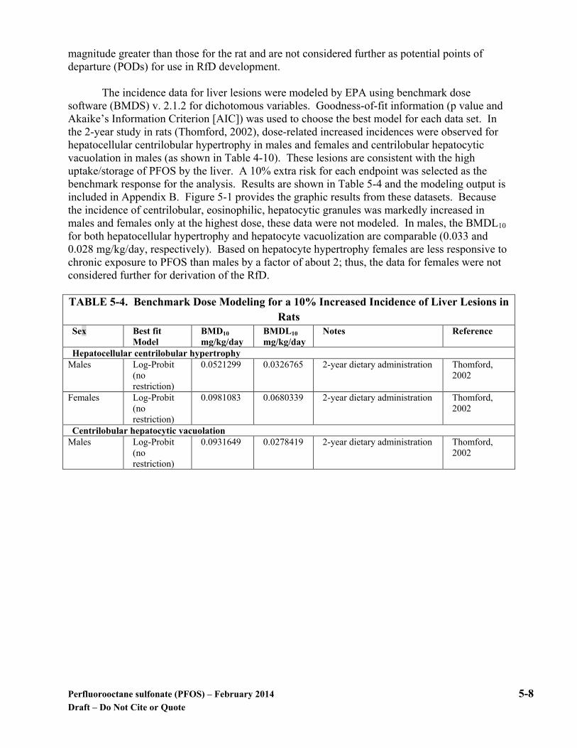
Perfluorooctane sulfonate (PFOS) – February 2014 5-8 Draft – Do Not Cite or Quote
magnitude greater than those for the rat and are not considered further as potential points of departure (PODs) for use in RfD development.
The incidence data for liver lesions were modeled by EPA using benchmark dose
software (BMDS) v. 2.1.2 for dichotomous variables. Goodness-of-fit information (p value and Akaike’s Information Criterion [AIC]) was used to choose the best model for each data set. In the 2-year study in rats (Thomford, 2002), dose-related increased incidences were observed for hepatocellular centrilobular hypertrophy in males and females and centrilobular hepatocytic vacuolation in males (as shown in Table 4-10). These lesions are consistent with the high uptake/storage of PFOS by the liver. A 10% extra risk for each endpoint was selected as the benchmark response for the analysis. Results are shown in Table 5-4 and the modeling output is included in Appendix B. Figure 5-1 provides the graphic results from these datasets. Because the incidence of centrilobular, eosinophilic, hepatocytic granules was markedly increased in males and females only at the highest dose, these data were not modeled. In males, the BMDL10 for both hepatocellular hypertrophy and hepatocyte vacuolization are comparable (0.033 and 0.028 mg/kg/day, respectively). Based on hepatocyte hypertrophy females are less responsive to chronic exposure to PFOS than males by a factor of about 2; thus, the data for females were not considered further for derivation of the RfD.
TABLE 5-4. Benchmark Dose Modeling for a 10% Increased Incidence of Liver Lesions in
Rats Sex Best fit
Model BMD10 mg/kg/day
BMDL10 mg/kg/day
Notes Reference
Hepatocellular centrilobular hypertrophy Males Log-Probit
(no restriction)
0.0521299 0.0326765 2-year dietary administration Thomford, 2002
Females Log-Probit (no restriction)
0.0981083 0.0680339 2-year dietary administration Thomford, 2002
Centrilobular hepatocytic vacuolation Males Log-Probit
(no restriction)
0.0931649 0.0278419 2-year dietary administration Thomford, 2002

Perfluorooctane sulfonate (PFOS) – February 2014 5-9 Draft – Do Not Cite or Quote
Males – hypertrophy
Females – hypertrophy
Males – vacuolation
Figure 5-1. BMDS graphic output from selected model runs; data from Thomford, 2002.
For the studies wherein liver weight was a critical effect, the data were also modeled using BMDS v. 2.1.2 for a continuous dataset with modeled variance where applicable. A 10% increase in absolute liver weight was chosen as the benchmark response for the initial analysis. This endpoint is considered to be a biomarker for systemic exposure in rodents when the chemical is an activator of PPAR-α, rather than a biomarker of adversity. Although the PFOS data support an increase in the expression of proteins involved in peroxisomal fatty acid catabolism (Tan et al., 2012) and lipid transport and metabolism (Bijland et al., 2011), those effects may not totally be a reflection of PPAR-α activation. Compared to PFOA, PFOS appears to be a relatively weak activator of the PPAR-α receptor (Shipley et al., 2004; Wolf et al., 2008,2012; Ishibashi et al., 2011).
0
0.1
0.2
0.3
0.4
0.5
0.6
0.7
0.8
0 0.1 0.2 0.3 0.4 0.5 0.6 0.7 0.8
Frac
tion
Affe
cted
dose
LogProbit Model with 0.95 Confidence Level
14:57 10/03 2012
BMDL BMD
LogProbit
0
0.2
0.4
0.6
0.8
0 0.1 0.2 0.3 0.4 0.5 0.6 0.7 0.8
Frac
tion
Affe
cted
dose
LogProbit Model with 0.95 Confidence Level
13:03 01/29 2013
BMDL BMD
LogProbit
0
0.05
0.1
0.15
0.2
0.25
0.3
0.35
0.4
0.45
0 0.1 0.2 0.3 0.4 0.5 0.6 0.7 0.8
Frac
tion
Affe
cted
dose
LogProbit Model with 0.95 Confidence Level
08:56 10/04 2012
BMDL BMD
LogProbit
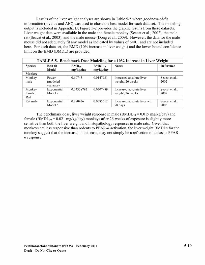
Perfluorooctane sulfonate (PFOS) – February 2014 5-10 Draft – Do Not Cite or Quote
Results of the liver weight analyses are shown in Table 5-5 where goodness-of-fit information (p value and AIC) was used to chose the best model for each data set. The modeling output is included in Appendix B; Figure 5-2 provides the graphic results from these datasets. Liver weight data were available in the male and female monkey (Seacat et al., 2002), the male rat (Seacat et al., 2003), and the male mouse (Dong et al., 2009). However, the data for the male mouse did not adequately fit any model as indicated by values of p<0.1 and are not included here. For each data set, the BMD (10% increase in liver weight) and the lower-bound confidence limit on the BMD (BMDL) are provided.
TABLE 5-5. Benchmark Dose Modeling for a 10% Increase in Liver Weight
Species Best fit Model
BMD10 mg/kg/day
BMDL10 mg/kg/day
Notes Reference
Monkey Monkey male
Power (modeled variance)
0.60743 0.0147931 Increased absolute liver weight; 26 weeks
Seacat et al., 2002
Monkey female
Exponential Model 2
0.03338792 0.0207989 Increased absolute liver weight; 26 weeks
Seacat et al., 2002
Rat Rat male Exponential
Model 5 0.280426 0.0585612 Increased absolute liver wt;
98 days Seacat et al., 2003
The benchmark dose, liver weight response in male (BMDL10 = 0.015 mg/kg/day) and
female (BMDL10 = 0.021 mg/kg/day) monkeys after 26-weeks of exposure is slightly more sensitive than both the liver weight and histopathology responses in male rats. Given that monkeys are less responsive than rodents to PPAR-α activation, the liver weight BMDLs for the monkey suggest that the increase, in this case, may not simply be a reflection of a classic PPAR-α response.

Perfluorooctane sulfonate (PFOS) – February 2014 5-11 Draft – Do Not Cite or Quote
Male monkey
Female monkey
Male rat
Figure 5- 2. BMDS graphic output from liver weight model runs; data from Seacat et al., 2002, 2003
5.1.1.2 Pharmacokinetic Model Approach
The pharmacokinetics of PFOS are not well understood but are considered to be roughly similar to that of the more extensively studied PFOA (Andersen et al., 2006; Loccisano et al., 2011, 2012a,b). Pharmacokinetic studies with PFOS have measured a serum half-life of approximately 48 days in rats (Butenhoff and Chang, 2007) and approximately 121 days in monkeys (Chang et al., 2012). Compared with a half-life of months in animals, the human PFOS half-life of roughly five years (Olsen et al., 2007) is clearly inconsistent with simple allometric scaling across species. The reasons for the contrast between the multi-year PFOS elimination half-life in humans and the shorter half-lives of laboratory animals are not fully understood.
In the process of developing pharmacokinetic models for PFOS in monkeys and rats,
several assumptions had to be made by the authors in order to fit the model outcome to the available pharmacokinetic data (Anderson et al., 2006; Tan et al., 2008; Loccisano et al., 2011, 2012a,b). Parameters for transporter characteristics were assumed to be the same for both sexes
20
40
60
80
100
120
140
0 0.1 0.2 0.3 0.4 0.5 0.6 0.7
BMDBMDL
Power
Mea
n R
espo
nse
dose
Power Model with 0.95 Confidence Level
09:33 01/29 2013
30
40
50
60
70
80
90
100
0 0.1 0.2 0.3 0.4 0.5 0.6 0.7
Mea
n R
espo
nse
dose
Exponential Model 2 with 0.95 Confidence Level
10:17 01/29 2013
BMDBMDL
Exponential
12
14
16
18
20
22
24
0 0.2 0.4 0.6 0.8 1 1.2
Mea
n R
espo
nse
dose
Exponential Model 5 with 0.95 Confidence Level
11:21 01/29 2013
BMDBMDL
Exponential
Perfluorooctane sulfonate (PFOS) – February 2014 5-12 Draft – Do Not Cite or Quote
consistent with no observed differences in elimination; a storage compartment was added because PFOS appears in the urine of male rats at a slower rate than it disappears from plasma. To account for the concentrations of PFOS found in the liver at long times (72–99 days) after dosing, description of saturable binding in the liver was used in the adult rat model (Loccisano et al., 2012a), and a time-dependent description of the free fraction of chemical in the central (plasma) compartment was added to describe possible changes in binding or elimination that may be occurring over time.
It is hypothesized that the pharmacokinetics of perfluorinated compounds, including
PFOS, are driven by transporters, notably in kidney and liver (Kudo, 2006). All published pharmacokinetic models assume that perfluoroalkyl acids are rapidly eliminated into the urine, but then reabsorbed into the body by transporters before they can be excreted. Whenever the ability of transporters to reabsorb the compounds is saturated, however, the compounds can be rapidly eliminated.
Currently, no data characterizing the uptake kinetics of PFOS by renal organic anion
transporters are available. The long half-life of PFOS in both the male and female rat, the monkey, and humans, suggests that it is most likely reabsorbed back into the plasma by a renal transport process, similar to the documented process for PFOA. However, because no significant difference is observed for the elimination half-life between male and female rats (Chang et al., 2012), it can be assumed that the transporters responsible for reabsorption are similar in both sexes, and different in some way from those for PFOA. Differences could be due to either the kinetic interactions of the chemicals with the same transporters or differences in transporters. Studies characterizing the uptake and reuptake of PFOS by renal transporters will help in refinement of these parameters.
Because the exposures of interest typically involve repeated doses, the saturable renal
resorption model (Andersen et al., 2006, Section 3.5.1) was used to describe how PFOS can reach steady state faster than the elimination half-life would indicate. Using a simpler, linear pharmacokinetic model (e.g. the one-compartment model) would result in estimated exposures due to repeated doses that are much greater than actually seem to occur (Seacat et al., 2003). Despite the uncertainties described above, application of this model to PFOS yielded good agreement between the experimental data and the model simulations.
The biological basis for the saturable resorption model parameters is uncertain (Lou et al.
2009), particularly with respect to the idea that glomerular filtration alone is responsible for the movement of PFOS into the renal filtrate given that PFOS is highly protein bound (Kerstner-Wood et al., 2003) and the interactions with the renal transporters are unknown. However, the saturable resorption model functions well in describing the internal doses in studies of PFOS (Andersen et al., 2006; Loccisano et al., 2011, 2012a,b).
The saturable resorption model is an empirical model similar to the commonly used one-
and two-compartment models. Because it is an empirical model, extrapolation between species is difficult. Resorption parameters cannot be directly linked to the properties of the relevant serum transport proteins or membrane transporters. Therefore the saturable resorption model parameters must be estimated using species-specific pharmacokinetic data.

Perfluorooctane sulfonate (PFOS) – February 2014 5-13 Draft – Do Not Cite or Quote
Pharmacokinetic data (serial blood concentrations following treatment with known quantities of PFOS) were collected for three species: cynomolgus monkeys (Chang et al., 2012; Seacat et al., 2002), Sprague-Dawley rat (Chang et al., 2012), and CD1 mouse (Chang et al., 2012). Given that the available pharmacokinetic studies were not necessarily designed with the expectation of non-linear pharmacokinetics, many parameters associated with saturable resorption should be expected to be uncertain. In the absence of ideal data, the question of parameter values becomes one of what range of values are consistent with the data, rather than which single value is most consistent with the data. Considering a range of possible values that might equally well explain the data can be addressed with Bayesian statistical analysis, which determines the distributions of parameter values that are likely to reproduce the observed data (Gelman et al., 2004).
In this case, a non-hierarchical model for parameter values was applied in which there is
a single value shared by all individuals of the same species and gender. Body-weight and treatment (number and magnitude of doses) are the only parameters that may vary between individuals. Body-weight was only varied when animal-specific body-weights were available.
Bayesian analysis allows formal inclusion of prior knowledge in the form of set
distributions on the parameters being estimated (Gelman et al., 2004). Given that empirical pharmacokinetic parameters can have a wide range of values, vague, bounded prior distributions are appropriate (Wambaugh et al., 2008). For all estimated parameters, the prior knowledge was distributed log-normally. This constrained the parameters to positive values. The mean and variances used are given in Table 5-6.
TABLE 5-6. Description of prior distributions used.
Parameter Mean Variance Bounds Lower Upper
ka 1 1000 1/6 105 Vcc 1 1000 10-10 105 k12 1 1000 10-10 105 RV2:V1 1 0.5 10-10 100 Tmaxc 1 1000 10-10 105 kT 1 1000 10-10 105 free 0.01 0.5 10-10 1 Qfilc 1 1000 0.01 105 Vfilc 1 1000 10-10 10
Parameters were log-normally distributed with the mean and variance listed. Bounds were used to reduce the time spent sampling in areas thought to have low probability, and were expanded when large posterior mass at the bounds indicated that the bounds were too narrow.
The deep tissue compartment of the Andersen et al. (2006) model is characterized in
terms of the rates to and from that compartment (k12 and k21, respectively). This corresponds to a volume of distribution V2 = k12*V1/k21, so that the ratio of the volume of the second compartment to the first is Rv2:v1 = k12/k21. In order to enforce the assumption that the primary (serum) compartment contains a significant portion of the PFOS, the volume of the deep tissue was constrained to be no more than 100 times greater than the volume of distribution of the serum. For this reason the ratio of the two volumes is estimated, rather than the rate from the second compartment to the first compartment. The rate of flow from the deep tissue back to the serum was calculated as k21 = k12/Rv2:v1.

Perfluorooctane sulfonate (PFOS) – February 2014 5-14 Draft – Do Not Cite or Quote
Bayesian analysis was performed using Markov Chain Monte Carlo (Gelman et al.,
2004). The distribution of parameter values were considered to “burned in” (i.e., true draws from the posterior combination of the prior distributions and the available data and independent of the starting values) when they passed the Heidelberger and Welch Stationarity test (Heidelberger and Welch, 1983) as implemented in the Coda Package (Best et al., 1995) for R (R Development Core Team, 2010).
The estimated parameters (median and 95% interval), presented in Table 5-7, generally
appear plausible. The volumes of distributions were all less than 1 L/kg BW, and varied between species. The parameters for the male mouse were extremely uncertain, reflecting a combination of the limited amount of data (two single dose treatments) and perhaps inappropriateness of the model used. However, data to support the model assumptions are needed. Parameters such as the flow to and volume of the filtrate compartment were found to be very uncertain, as in previous studies (Andersen et al. 2006). The median fraction of blood flow to the filtrate (Qfilc) was physiological (less than or equal to the fraction of blood flow to the kidney) for the male and female rats, but appears too high for the mouse and monkeys, which might be explained by either a lack of sufficient data or perhaps the role of secreting transporters in the kidney. Although the physical interpretation is moot, the form of the saturable resorption model seems appropriate.

Perfluorooctane sulfonate (PFOS) – February 2014 5-15 Draft – Do Not Cite or Quote
TABLE 5-7. Pharmacokinetic parameters used in the Andersen et al. (2006) model.
Parameter Units CD1 Mouse Femalea
CD1 Mouse Malea
Sprague-Dawley Rat Femalea
Sprague-Dawley Rat Malea
Cynomolgus Monkey Male and Femalea
BWb kg 0.02 0.02 0.203 0.222 3.42 Cardiac Outputc
L/h/kg0.74 8.68 8.68 12.39 12.39 19.8
ka 1/h 1.16 (0.617 - 42400)
433.4 (0.51 – 803.8
4.65 (3.02 - 1980)
0.836 (0.522 - 1.51)
132 (0.225 - 72100)
Vcc L/kg 0.264 (0.24 - 0.286)
0.292 (0.268 – 0.317
0.535 (0.49 - 0.581)
0.637 (0.593 - 0.68)
0.303 (0.289 - 0.314)
k12 1/h 0.0093 (2.63e-10 - 38900)
2976 (2.8e-10 – 4.2e4)
0.0124 (3.1e-10 - 46800)
0.00524 (2.86e-10 - 43200)
0.00292 (2.59e-10 - 34500)
RV2:V1 Unitless 1.01 (0.251 - 4.06)
1.29 (0.24 – 4.09)
0.957 (0.238 - 3.62)
1.04 (0.256 - 4.01)
1.03 (0.256 - 4.05)
Tmaxc M/h 57.9 (0.671 - 32000)
1.1e4 (2.1 – 7.9e4)
1930 (4.11 - 83400)
1.34e-06 (1.65e-10 - 44)
15.5 (0.764 - 4680)
kT M 0.0109 (1.44e-05 - 1.45)
381 (2.6e-5 – 2.9e3)
9.49 (0.00626 - 11100)
2.45 (4.88e-10 - 60300)
0.00594 (2.34e-05 - 0.0941)
Free Unitless 0.00963 (0.00238 - 0.0372)
0.012 (0.0024 – 0.038)
0.00807 (0.00203 - 0.0291)
0.00193 (0.000954 - 0.00249)
0.0101 (0.00265 - 0.04)
Qfilc L/h 0.439 (0.0125 - 307)
27.59 (0.012 – 283)
0.0666 (0.0107 - 8.95)
0.0122 (0.0101 - 0.025)
0.198 (0.012 - 50.5)
Vfilc L/kg 0.00142 (4.4e-10 - 6.2)
0.51 (3.5e-10 – 6.09)
0.0185 (8.2e-07 - 7.34)
0.000194 (1.48e-09 - 5.51)
0.0534 (1.1e-07 - 8.52)
Means and 95% confidence interval from Bayesian analysis are reported. For some parameters the distributions are quite wide, indicating uncertainty in that parameter (i.e., the predictions match the data equally well for a wide range of values). aData sets modeled for the mouse and rat were from Chang et al., 2012 and for the monkey from Seacat et al., 2002 and Chang et al., 2012. a Average bodyweight for species -- individual-specific bodyweights b Cardiac outputs obtained from Davis & Morris, Pharmaceutical Res 10, 1093, 1993
For each study with a toxicological endpoint and NOAEL/LOAEL, the time-integrated
serum concentration (area under the curve or AUC) was determined for the exposure duration investigated in that study. Generally, it was assumed that animals were observed at the end of dosing. The data for studies in the rats are summarized in Table 5-8. For the Butenhoff et al. (2009) study two different AUCs were calculated – gestational only (for the male offspring endpoint) and gestational plus twenty days postnatal (for the maternal endpoint). This separation of the two exposures neglects lactational transfer of compound, which was not modeled.

Perfluorooctane sulfonate (PFOS) – February 2014 5-16 Draft – Do Not Cite or Quote
TABLE 5-8. Predicted final serum concentration and time integrated serum concentration (AUC) for different treatments of rat.
Study Species / Strain
Study Duration And Type
Oral Doses
mg/kg/day
Measured Serum
Concentration mg/L
Species / Strain Used
for Prediction
Predicted Final Serum
Concentration mg/L
Predicted AUC mg/L*h
Curran et al. 2008
Male Rat/Sprague-Dawley
28 days
0.14 No data Male Rat/Sprague-Dawley
4.88 (0.148) 1840 (56.9) 1.33 No data 46.4 (1.4) 17400 (540) 3.21 No data 112 (3.39) 42100 (1300) 6.34 No data 221 (6.7) 83100 (2580)
Curran et al. 2008
Female Rat/Sprague-Dawley
28 days
0.15 No data Female Rat/Sprague-Dawley
6.88 (0.212) 2500 (85.9) 1.43 No data 65.5 (2.03) 23800 (820) 3.73 No data 170 (5.76) 62100 (2170) 7.58 No data 344 (18.3) 126000 (5090
Seacat et al. 2003
Male Rat/ Crl:CD(SD) IGS BR
98 days
0.03 4.04 (0.80) Male Rat/Sprague-Dawley
2.64 (0.103) 3970 (128) 0.13 17.1 (1.22) 10.5 (0.408) 15900 (507) 0.34 43.9 (4.9) 26.3 (1.02) 39600 (1270) 1.33 148 (14) 105 (4.08) 158000
(5070) Seacat et al. 2003
Female Rat/ Crl:CD(SD) IGS BR
98 days
0.04 6.96 (0.99) Female Rat/Sprague-Dawley
4.24 (0.0867) 5780 (128) 0.15 27.3 (2.3) 17 (0.345) 23100 (511) 0.40 64.4 (5.5) 42.3 (0.882) 57700 (1280) 1.56 223 (22) 168 (6.38) 230000
(6390) Butenhoff et al. 2009 and Chang et al. 2009
Rat/Sprague-Dawley
Gestation (22 Days)
0.1 1.722 (0.068) Female Rat/Sprague-Dawley
3.7 (0.121) 1060 (37.8) 0.3 6.245 (0.096) 11.1 (0.364) 3170 (114) 1 26.630 (3.943) 37 (1.21) 10600 (378)
Butenhoff et al. 2009 and Chang et al. 2009
Rat/Sprague-Dawley
Gestation (21 Days) + 20 Days Postnatal
0.1 3.159 (0.081) Female Rat/Sprague-Dawley
6.49 (0.172) 3570 (111) 0.3 8.981 (0.275) 19.5 (0.515) 10700 (333) 1 30.480 (1.294) 64.8 (1.74) 35600 (1110)
Thibodeaux et al. 2003 and Lau et al. 2003
Rat/Sprague-Dawley
GDs 2-20 (19 days)
1 19.69a Female Rat/Sprague-Dawley
32.4 (1.1) 8010 (293) 2 44.33a 64.7 (2.2) 16000 (585) 3 70.62a 97 (3.31) 24000 (879) 5 79.39a 161 (5.69) 40000 (1470) 10 189.4a 321 (15.6) 79800 (3190)
Luebker et al. 2005b
Rat/Crl:CD (SD)IGS VAF/Plus
6 wks prior to mating through gestation and lactation 84 days
0.1 4.52 (1.15) Female Rat/Sprague-Dawley
9.65 (0.202) 9100 (235) 0.4 26.2 (16.1) 38.5 (0.814) 36400 (942) 1.6 136 (86.5) 153 (4.74) 145000
(4100) 3.2 155 (39.3) 303 (17.5) 288000
(11200)
Luebker et al. 2005a
Rat/Crl:CD (SD)IGS VAF/Plus
6 wks prior to mating through gestation; 63 days
0.4 NT Female Rat/Sprague-Dawley
38.5 (0.814) 36400 (942) 0.8 NT 76.9 (1.75) 72600 (1910) 1.0 NT 96.1 (2.32) 90700 (2410) 1.2 NT 115 (2.99) 109000
(2940) 1.6 NT 153 (4.74) 145000
(4100) 2.0 NT 191 (7.07) 181000
(5460) Numbers in parentheses indicate standard deviation GD = gestation day; NT = not tested aThibodeaux et al. (2003) data available only in a graph; values obtained from author.

Perfluorooctane sulfonate (PFOS) – February 2014 5-17 Draft – Do Not Cite or Quote
The data on the results from studies in mice and the monkey are provided in Tables 5-9 and 5-10, respectively. The Lau et al. (2003) data on mice are representative of the impact of PFOS on developmental endpoints as described in Table 5-2 although the duration of this study is relatively short at 17 days. The Seacat et al. (2002) study on monkeys is a long term (6 months) multiple dose study of systemic toxicity in which the LOAEL for effects on liver weight and cholesterol was accompanied by death of 2/6 monkeys.
TABLE 5-9. Predicted final serum concentration and time integrated serum concentration
(AUC) for the mouse. Study Species /
Strain Study Duration And Type
Administered Doses mg/kg/day
Measured Final Serum Concentration mg/L
Species / Strain Used for Prediction
Predicted Final Serum Concentration mg/L
Predicted AUC mg/L*h
Lau et al. 2003
Female Mouse/CD-1
GD 1-17 (17 days)
1 NT Female Mouse / CD1
62.7 (2.28) 14700 (533)
5 NT 287 (19.2) 70900 (2560)
10 NT 421 (107 123000 (14500)
15 NT 484 (186) 156000 (32100)
20 NT 532 (257) 180000 (49900
Numbers in parentheses indicate standard deviation GD = gestation day; NT = not tested
TABLE 5-10. Predicted final serum concentration and time integrated serum concentration (AUC) for the monkey.
Study Species / Strain
Study Durat-ion And Type
Adminis-tered Doses mg/kg/day
Measured Final Serum Concentrat-ion mg/L
Species / Strain Used for Prediction
Predicted Final Serum Concentrat-ion mg/L
Predict-ed AUC mg/L*h
Seacat et al. 2002
Monkey / Cynomol-gus
182 days 0.03 F: 13.2 (1.4) M: 15.8 (1.4)
Monkey / Cynomol-gus
7.58 (0.16) 22100 (382)
0.15 F: 66.8 (10.8) M: 82.6 (25.2)
32.9 (0.557) 102000 (1530)
0.75 F: 171 (22) M: 173 (37)
86.7 (2.14) 332000 (6450)
Numbers in parentheses indicate standard deviation M = male; F = female
The AUC for the LOAEL or NOAEL of each data set can be used to determine an
average serum concentration by dividing it by the duration of the study in days with adjustment for the number of hours in a day. Averaging the terminal serum concentrations for the duration of exposure is important because of the variability in the times of exposure across the studies (17-182 days). The following equation is used for the conversion:
Average Serum Concentration = AUC (mg/L*h) × 1 day/24 hours ÷ exposure duration
(days)
Perfluorooctane sulfonate (PFOS) – February 2014 5-18 Draft – Do Not Cite or Quote
For example, in a case where the AUC was 30,000 mg/L*h and the study duration was 90 days, the Average Serum Concentration would be calculated as follows:
Average Serum Concentration = 30,000 mg/L*h ÷ (90 days × 24 h/day) = 13.89 mg/L This calculation has the advantage of normalizing the serum concentration across the
exposure durations to generate a uniform metric for internal dose in situations where the dosing durations varied and serum measurements were taken immediately prior to sacrifice. The averaged serum concentration is a hybrid of the AUC and the maximum serum concentration. As applied to the database for PFOS, average serum concentration appears to be a stable reflection of internal dosimetry.
Table 5-11 provides the AUC from the model, the dosing duration from each of the
modeled studies, and the resultant average serum concentration. Internal doses associated with developmental toxicity were 18.06 and 24.07 mg/L for reduced pup body weight (Luebker et al., 2005a,b), 36.18 mg/L for changes in motor activity (Butenhoff et al., 2009), and 35.09-48.02 mg/L for pup survival (Lau et al., 2003; Luebker et al., 2005a). In comparison, internal doses associated with increased liver weight were 67.18-76.01 mg/L (Seacat et al., 2002; 2003). Thus, the internal doses associated with the developmental and liver effect levels (LOAELs) differ by much less than an order of magnitude (18.06 mg/L to 76.01 mg/L) while the corresponding AUC values differ by more than an order of magnitude (16000 mg/L*h to 332000 mg/L*h).
The internal doses associated with no adverse effects on developmental and liver
endpoints (NOAELs) were very similar with overlapping ranges; the average serum concentrations ranged from 4.51-24.07 mg/L for developmental endpoints (Butenhoff et al., 2009; Lau et al., 2003; Luebker et al., 2005a,b) and from 16.84-23.35 mg/L for liver weight changes (Seacat et al., 2002; 2003). Despite the similarity in average serum concentrations, the AUC values differ by two orders of magnitude (8010 mg/L*h to 102000 mg/L*h). Given the differences in external doses, the projected serum levels are proportionally quite similar.

Perfluorooctane sulfonate (PFOS) – February 2014 5-19 Draft – Do Not Cite or Quote
TABLE 5-11. Average Serum concentrations Derived from the AUC and the duration of Dosing
Study Dosing duration days
NOAEL mg/kg/day (AUC mg/L*h)
NOAEL (Av serum mg/L)
LOAEL mg/kg/day (AUC mg/L*h)
LOAEL (Av serum mg/L)
Seacat et al., 2002 monkey ↑liver weight
182 0.15 (102000)
23.35 0.75 (332000)
76.01
Luebker et al., 2005b ↓ rat pup body weight
84 0.1 (9100)
4.51 0.4 (36400)
18.06
Luebker et al., 2005a ↓ rat pup body weight
63 None None 0.4 (36400)
24.07
Luebker et al., 2005a ↓maternal weight, pup survival
63 0.4 (36400)
24.07 0.8 (72600)
48.02
Lau et al., 2003 ↓rat pup survival
19 1.0 (8010)
17.56 2.0 (16000)
35.09
Butenhoff et al., 2009 rat ↑DNT
41 0.3 (10700)
10.87 1.0 (35600)
36.18
Seacat et al., 2003 male ↑rat liver weight
98 0.34 (39600)
16.84 1.33 (158000)
67.18
Table 5-11 identifies 4.51 and 10.87 mg/L as the lowest average serum concentrations
which were associated with a NOAEL for offspring effects; the LOAELs were associated with decreased pup body weight (Luebker et al., 2005b) and increased motor activity in male pups (Butenhoff et al., 2009). Average serum values for no increases in liver weight in monkeys (Seacat et al., 2002) and male rats (Seacat et al., 2003) are very similar to the average serum value in Butenhoff et al. (2009). Thus, it appears that the NOAELs are consistent across gender, species, and treatment with respect average serum concentration. Assuming that mode of action and susceptibility to toxicity do not vary and that pharmacokinetics alone explains variation, it is reasonable to expect similar concentrations to cause similar effects in humans.
The EPA model employed here to generate the predicted AUC values that became the
basis for the average serum concentrations shown in Table 5-11, does not include a gestational or lactational component. However the results are in good agreement with those of Loccisano et al. (2012b) from their gestational and lactational model. Comparison of the average maternal serum concentrations calculated for developmental endpoints (Butenhoff et al., 2009; Lau et al., 2003; Luebker et al., 2005a) with those depicted graphically in Figure 3-7 (from Loccisano et al., 2012b), demonstrates good agreement between the two models. For example the LOAEL of 1 mg/kg/day for developmental neurotoxicity (Butenhoff et al., 2009) yields a calculated average maternal serum of 36.18 mg/L as seen in Table 5-11 which is very similar to the approximately 25 mg/L for the dams that can be estimated from the graph (Loccisano et al., 2012b). The slightly higher value calculated from the EPA model may be due to the longer dosing interval, 41 days, used by Butenhoff et al. (2009) versus GD 20 levels presented graphically by Loccisano et al. (2012b). Fetal PFOS serum concentration on GD 20 was published by Chang et al. (2009), but because the EPA model predicts maternal values, not fetal, a direct comparison to the fetal

Perfluorooctane sulfonate (PFOS) – February 2014 5-20 Draft – Do Not Cite or Quote
plasma predicted by Loccisano et al. (2012b; Figure 3-7) cannot be made. However, despite the limitations in the fetal data, values generated by the EPA model can be accepted with reasonable confidence that the predicted AUC values accurately represent maternal levels during gestational and lactational exposures.
The predicted serum concentrations can be converted into an oral equivalent dose at
steady state by recognizing that, at steady state, clearance from the body must equal dose to the body. Clearance can be calculated if the rate of elimination (derived from half-life) and the volume of distribution are both known.
A reliable measure of half-life in humans is from a retired worker population followed for
five years. Olsen et al. (2007) calculated the PFOS half-life in this former worker population as 5.4 years (see Section 3.5.2). Thompson et al. (2010) gives a volume of distribution of 0.23 L/kg bw (see Section 3.5.3). These values combined give a clearance of 8.1×10⁻⁵ L/kg bw/day as determined by the following equation:
CL = Vd x (ln 2 ÷ t1/2) = 0.23 L/kg bw/day x (0.693 ÷ 1971 days) = 0.000081 L/kg bw/day
Where: Vd = 0.23 L/kg ln 2 = 0.693 t1/2 = 1971 days (5.4 years x 365 days/year = 1971 days) These values combined give a clearance of 8.1 x 10⁻⁵ L/kg bw/day. Scaling the derived average concentrations (in mg/L) for the NOAELs and LOAELs in
Table 5-9 gives predicted oral human equivalent doses (HEDs) in mg/kg bw/day for each corresponding serum measurement. The HED values are the predicted human oral exposures necessary to achieve serum concentrations equivalent to the NOAEL or LOAEL in the animal toxicity studies. Note that this scaling uses linear human kinetics in contrast to the non-linear phenomena observed at high doses in animals. Thus, HED = average serum concentration (in mg/L) x CL Where: Average serum is from model output in Table 5-11 CL = 0.000081 L/kg bw/day
The resulting HED values are shown in Table 5-12. Endpoints considered as critical effects in multiple studies include offspring growth and survival, liver weight changes, and liver histopathology.
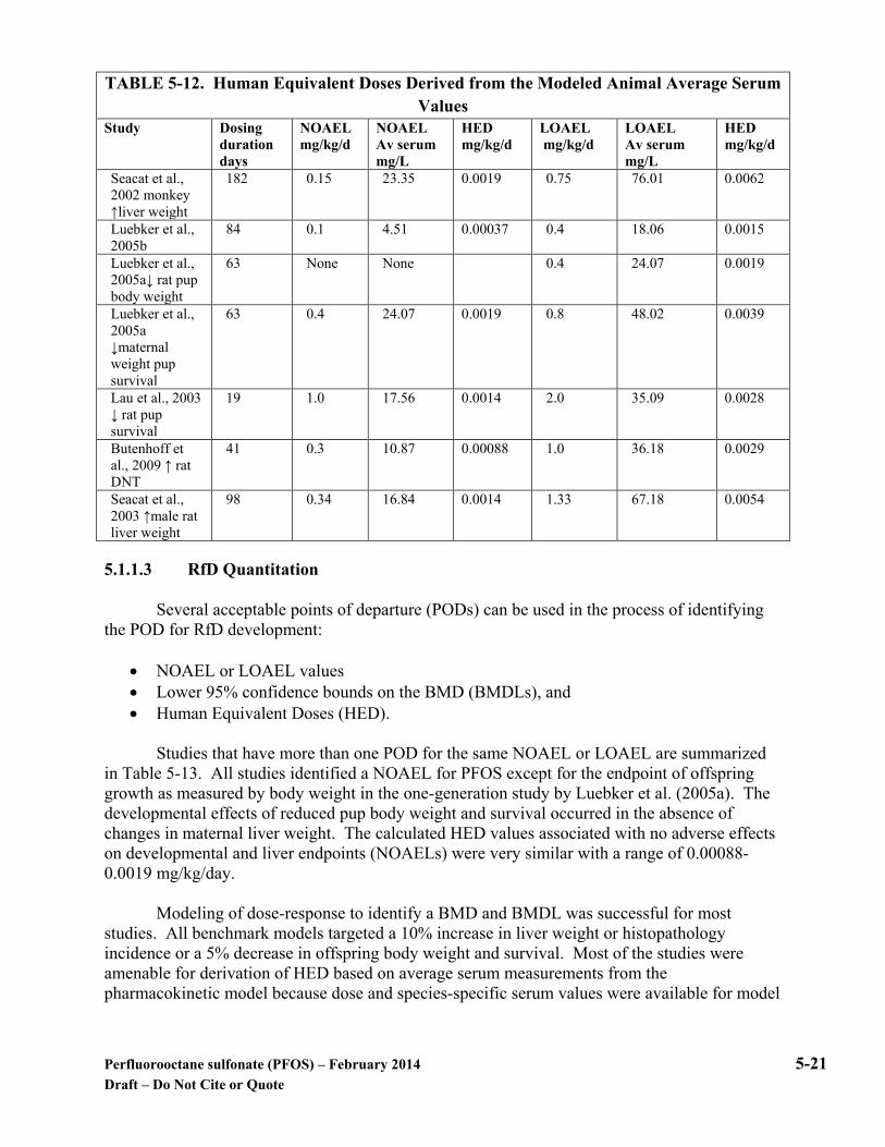
Perfluorooctane sulfonate (PFOS) – February 2014 5-21 Draft – Do Not Cite or Quote
TABLE 5-12. Human Equivalent Doses Derived from the Modeled Animal Average Serum Values
Study Dosing duration days
NOAEL mg/kg/d
NOAEL Av serum mg/L
HED mg/kg/d
LOAEL mg/kg/d
LOAEL Av serum mg/L
HED mg/kg/d
Seacat et al., 2002 monkey ↑liver weight
182 0.15 23.35 0.0019 0.75 76.01 0.0062
Luebker et al., 2005b
84 0.1 4.51 0.00037 0.4 18.06 0.0015
Luebker et al., 2005a↓ rat pup body weight
63 None None 0.4 24.07 0.0019
Luebker et al., 2005a ↓maternal weight pup survival
63 0.4 24.07 0.0019 0.8 48.02 0.0039
Lau et al., 2003 ↓ rat pup survival
19 1.0 17.56 0.0014 2.0 35.09 0.0028
Butenhoff et al., 2009 ↑ rat DNT
41 0.3 10.87 0.00088 1.0 36.18 0.0029
Seacat et al., 2003 ↑male rat liver weight
98 0.34 16.84 0.0014 1.33 67.18 0.0054
5.1.1.3 RfD Quantitation
Several acceptable points of departure (PODs) can be used in the process of identifying the POD for RfD development:
• NOAEL or LOAEL values • Lower 95% confidence bounds on the BMD (BMDLs), and • Human Equivalent Doses (HED).
Studies that have more than one POD for the same NOAEL or LOAEL are summarized
in Table 5-13. All studies identified a NOAEL for PFOS except for the endpoint of offspring growth as measured by body weight in the one-generation study by Luebker et al. (2005a). The developmental effects of reduced pup body weight and survival occurred in the absence of changes in maternal liver weight. The calculated HED values associated with no adverse effects on developmental and liver endpoints (NOAELs) were very similar with a range of 0.00088-0.0019 mg/kg/day.
Modeling of dose-response to identify a BMD and BMDL was successful for most
studies. All benchmark models targeted a 10% increase in liver weight or histopathology incidence or a 5% decrease in offspring body weight and survival. Most of the studies were amenable for derivation of HED based on average serum measurements from the pharmacokinetic model because dose and species-specific serum values were available for model

Perfluorooctane sulfonate (PFOS) – February 2014 5-22 Draft – Do Not Cite or Quote
development. Entries in Table 5-13 provide at least three potential PODs for consideration; four studies provide five values.
TABLE 5-13. RfD Point of Departure Options from the PFOS Animal Studies Studies NOAEL
(mg/kg/ day)
LOAEL (mg/kg/ day)
BMDL10 (mg/kg/ day)
HED NOAEL (mg/kg/ day)
HED LOAEL (mg/kg/ day)
Endpoint
Seacat et al., 2002
0.15 0.75 0.015 0.0019 0.0062 Increased liver weight.
Seacat et al., 2003
0.34 1.33 0.059 0.0014 0.0054 Increased liver weight.
Thomford, 2002
0.018 0.072 0.033 - - Liver hypertrophy.
Thibodeaux et al., 2003
1.0 2.0 0.12* - - Decreased maternal and pup body weight (LOAEL); fetal sternal defects (BMDL5)
Lau et al., 2003
1.0 2.0 0.58* 0.0014 0.0028 Reduced pup survival.
Butenhoff et al., 2009
0.3 1.0 - 0.00088 0.0029 Increased motor activity in male pups on PND 17.
Luebker et al., 2005b
0.1 0.4 - 0.00037 0.0015 Decreased pup body weight.
Luebker et al., 2005a
None 0.4 0.27* - 0.0019 Decreased pup body weight.
Luebker et al., 2005a
0.4 0.8 0.89* 0.0019 0.0039 Reduced pup survival.
* The value provided for developmental endpoints is the BMDL05 rather than the BMDL10. As explained previously, human data have identified significant relationships between
serum levels and specific indicators of adverse health effects but lack the exposure information for dose-response modeling. For this reason none of the human studies provided an appropriate POD for RfD derivation. The pharmacokinetically-modeled average serum values from the animal studies are restricted to the animal species selected for their low dose response to oral PFOS intakes. Extrapolation to humans adds a layer of uncertainty that must be accommodated in deriving the RfD.
Each of the POD values represented in Table 5-13 requires a different quantification approach. Thus, EPA has systematically examined the impact of POD on outcome through three sets of calculations as follows:
• Derivation from the NOAEL values; • Derivation from the BMDL values; • Derivation from HED values derived from modeled average serum values.
NOAEL PODs: Table 5-14 provides the potential RfDs derived from the seven studies which identified a NOAEL. These studies also provided a LOAEL which is critical for dose-response characterization. The NOAEL is used as the POD according to Agency policy instead of the LOAEL because lower uncertainty is applied to the NOAEL when calculating the RfD. The lowest NOAEL is from a chronic bioassay in the rat (Thomford, 2002) and is the dose which did

Perfluorooctane sulfonate (PFOS) – February 2014 5-23 Draft – Do Not Cite or Quote
not cause microscopic lesions in the liver. Because it is a chronic study the uncertainty applied in derivation of the RfD is less than that used in the other rat studies and the monkey study. The NOAEL from Thomford (2002) is also protective of co-critical developmental toxicity effects observed in other studies.
TABLE 5-14. The Impact of Quantification Approach on the RfD outcome for the PODs
from the available NOAELs Study POD dose
mg/kg/day UFH UFA UFL UFS UFD UFtotal Potential
RfD mg/kg/day
Seacat et al., 2002 monkey
0.15 10 48 1 10 1 4800 0.00003
Seacat et al., 2003 rat 0.34 10 123 1 10 1 12300 0.00003 Thomford, 2002 rat 0.018 10 123 1 1 1 1230 0.00001 Thibodeaux et al., 2003 rat
1.0 10 123 1 10 1 12300 0.00008
Lau et al., 2003 rat 1.0 10 123 1 10 1 12300 0.00008 Butenhoff et al., 2009 rat
0.3 10 123 1 10 1 12300 0.00002
Luebker et al., 2005b rat
0.1 10 123 1 10 1 12300 0.000008
Luebker et al., 2005a rat
0.4 10 123 1 10 1 12300 0.00003
Uncertainty Factor (UF) Application UFH A ten-fold adjustment is assigned to account for intrahuman variability and applied for all PODs. UFA. Determination of the interspecies uncertainty factor for the NOAEL requires application of the equation for first order kinetics in order to determine the pharmacokinetic adjustment associated with differences in half-life between humans and rats or monkeys. Pharmacokinetic studies with PFOS have measured a serum half-life of approximately 48 days in rats (Butenhoff and Chang, 2007) and approximately 121 days in monkeys (Chang et al., 2012). The equation also utilizes a volume of distribution component, which for humans has been calibrated as 230 mL/kg (Thompson et al., 2010). This volume of distribution is similar to those reported for monkeys, female rats, and mice in pharmacokinetic studies (Chang et al., 2012) and utilized in pharmacokinetic models on monkeys (Anderson et al., 2006). The equation that describes first order kinetics is as follows (Medinsky and Klaassen, 1996): CL = Vd x (ln 2 ÷ t1/2) Where: Vd = 0.23 L/kg Ln 2 = 0.693 Half-life = 48 days for rats; 121 days for monkeys; and 1971 days for humans CLrat = 0.23 L/kg x (0.693 ÷ 48 days) = 0.0033 L/kg/day

Perfluorooctane sulfonate (PFOS) – February 2014 5-24 Draft – Do Not Cite or Quote
CLmonkey = 0.23 L/kg x (0.693 ÷ 121 days) = 0.0013 L/kg/day CLhuman = 0.23 L/kg x (0.693 ÷ 1971 days) = 0.000081 L/kg/day The ratio of CLrat to CLhuman (0.0033 L/kg/day ÷ 0.000081 L/kg/day = 41) is used as the pharmacokinetic adjustment for differences between these species. The total UFA requires an additional 3-fold factor for species differences in pharmacodynamics (41 x 3 = 123). The ratio of CLmonkey to CLhuman (0.0013 L/kg/day ÷ 0.000081 L/kg/day = 16) is used as the pharmacokinetic adjustment for differences between these species. The total UFA requires an additional 3-fold factor for species differences in pharmacodynamics (16 x 3 = 48). UFL A UF of 1 was used for the NOAEL-derived PODs following Agency policies. UFS A ten-fold factor was applied to the NOAEL PODs to account for studies with less than lifetime exposure. UFD In all cases the uncertainty factor for the strength of the database (UFD) is 1. The data base for oral PFOS exposure studies is essentially complete although mechanistic questions relative to the MOA have not yet been fully elucidated. BMDL PODs. Potential PODs from benchmark dose estimates are listed in Table 5-13. The lowest, species-specific benchmark dose values are those from the Seacat et al. (2002; 2003) studies in the male monkey and male rat and the Thomford (2002) study in the male rat. For the Seacat et al. (2002; 2003) studies the effect modeled was a 10% change in liver weight. For the Thomford (2002) study the effect modeled was a 10% increase in incidence of hepatocellular centrilobular hypertrophy. The BMDL estimates for the liver effects are lower than those calculated for developmental effects, indicating that liver endpoints are protective of other co-critical endpoints. The values for developmental endpoints are included in Table 5-15 for comparison.

Perfluorooctane sulfonate (PFOS) – February 2014 5-25 Draft – Do Not Cite or Quote
TABLE 5-15. The Impact of Quantification Approach on the RfD Outcome for the BMDLs from liver and developmental endpoints
POD Dose mg/kg/d
UFH UFA UFL UFS UFD UFtotal Potential RfD mg/kg/day
BMDL10 (monkey Seacat et al., 2002)
0.015 10 48 1 10 1 4800 0.000003
BMDL10 (rat Seacat et al., 2003)
0.059 10 123 1 10 1 12300 0.000005
BMDL10 (rat Thomford, 2002)
0.033 10 123 1 1 1 1230 0.00003
BMDL05 (rat Thibodeaux et al., 2003)
0.12 10 123 1 10 1 12300 0.00001
BMDL05 (rat Lau et al., 2003)
0.58 10 123 1 10 1 12300 0.00005
BMDL05 (rat Luebker et al., 2005a)
0.27 10 123 1 10 1 12300 0.00002
Uncertainty Value Application The UFH, UFA, UFS and UFD values are assigned as described for the NOAEL data in Table 5-14. UFL A UF of 1 was used for the BMDL-derived PODs following Agency policies. HED PODs. The pharmacokinetic (PK) HEDs derived from Seacat et al. (2002; 2003), Lau et al. (2003), Butenhoff et al. (2009), and Luebker et al. (2005a,b) were each examined as the potential basis for the RfD (Table 5-16). Each of these studies contained a NOAEL from which the HED could be derived. The outcomes for potential RfD values are quite similar demonstrating the ability of the model to normalize the animal data across species, gender, and exposure duration. Co-critical effects of liver lesions and developmental toxicity resulted in similar HED values which yielded similar potential RfD values.

Perfluorooctane sulfonate (PFOS) – February 2014 5-26 Draft – Do Not Cite or Quote
TABLE 5-16. The Impact of Quantification Approach on the RfD Outcomes for the HEDs from the Pharmacokinetic Model Average Serum Values
POD Value mg/kg/day
UFH UFA UFL UFS UFD UFtotal Potential RfD mg/kg/day
PK-HED monkey Seacat et
al., 2002 0.0019 10 3 1 1 1 30 0.00006
PK-HED rat Seacat et al., 2003 0.0014 10 3 1 1 1 30 0.00005 PK-HED rat Lau et al., 2003 0.0014 10 3 1 1 1 30 0.00005 PK-HED rat Butenhoff et al.,
2009 0.00088 10 3 1 1 1 30 0.00003
PK-HED rat Luebker et al.,
2005b 0.00037 10 3 1 1 1 30 0.00001
PK-HED rat Luebker et al.,
2005a 0.0019 10 3 1 1 1 30 0.00006
Uncertainty Value Application The UFH, UFL, and UFD values are assigned as described for the NOAEL data in Table 5-14. The UFA is 3 for each study because the HED was derived using the steady state serum values from the model to account for pharmacokinetic differences between animals and humans. The 3-fold factor is applied to account for toxicodynamic differences between the animals and humans The UFS is 1 bacause the point of departure is based on steady state serum concentrations. RfD Selection
Based on the consistency of the response and with recognition of the use of developmental toxicity as the sensitive endpoint, the 0.00003 mg/kg/day outcome is selected as the RfD for PFOS. This value is the outcome for the modeled rat serum for developmental neurotoxicity (Butenhoff et al., 2009) and supported by the slightly higher 0.00005 and 0.00006 mg/kg/day values for increases in liver weight and other developmental effects. Thus, co-occurring critical endpoints are protected by the chosen RfD.
In the standard developmental neurotoxicity study by Butenhoff et al. (2009), male
offspring showed increased motor activity and decreased habituation on PND 17 following a maternal dose of 1 mg/kg/day; no effects on pup body weight were reported. Animal data consistently show higher PFOS levels in fetal tissues, including brain, as compared to maternal tissues (Chang et al., 2009; Borg et al., 2010) resulting in the higher modeled predictions for fetal versus maternal levels (Loccisano et al., 2012b; see Figure 3-7). Use of the developmental toxicity endpoint is directly relevant to human health because in utero and lactational exposures have been demonstrated. PFOS has been measured in the blood of newborns (Spliethoff et al., 2008), in human breast milk (So et al., 2006; Karrman et al., 2010; Tao et al., 2008), and in serum samples from children aged 2-12 (Olsen et al., 2002b). A human epidemiology found no association with maternal PFOS levels and motor or mental development of their children; the mean maternal serum concentration was approximately 35 mg/L (Fei et al., 2008b). The HED used as the basis for the RfD, was calculated from an average serum concentration of 10.87 mg/L

Perfluorooctane sulfonate (PFOS) – February 2014 5-27 Draft – Do Not Cite or Quote
derived from the NOAEL of 0.3 mg/kg/day for developmental neurotoxicity (Butenhoff et al., 2009; Table 5-11). This further supports the selected RfD as being protective of adverse human health risks.
Use of the developmental neurotoxicity and liver weight endpoints as the basis for the
RfD confers protection against co-occurring adverse effects on offspring body weight and survival. The selected RfD is supported by the results from the modeled serum value for rat offspring body weight (Luebker et al., 2005a) and survival (Lau et al., 2003). These supporting values are slightly greater than the recommended RfD, indicating that the RfD is protective of co-occurring effects on the offspring. For these co-occurring effects, both the modeled values were similar (0.00005-0.00006 mg/kg/day) adding confidence to the selected RfD.
The most conservative potential RfD was derived from modeled serum for decreased pup
body weight from the two-generation study (Luebker et al., 2005b). While this represents the most sensitive endpoint, it was described by the authors as transient because the body weights were significantly lower than those for controls for 2 of 5 time points monitored with the weight differences before and after those two time points not significantly different. Benchmark data for the body weight data to frame the NOAEL POD versus the LOAEL POD were not available. Other modeled results are in good agreement with those from a gestational and lactational model (Loccisano et al., 2012b) but cumulative effects from continuous exposure over multiple generations is unknown and not accounted for by the present model.
5.1.2 RfC Determination
For development of an RfC, an adequate subchronic inhalation study must be available for review. At this time, data are insufficient for PFOS to develop an RfC. The only inhalation study available is an acute lethality inhalation study in rats (Rusch et al., 1979); no inhalation data are available in humans. Therefore, an RfC for PFOS cannot be derived.
5.2 Dose-Response for Cancer Effects
In a chronic oral toxicity and carcinogenicity study of PFOS in rats, liver, thyroid and mammary fibroadenomas were identified (Thomford, 2002). The biological significance of the mammary fibroadenomas and thyroid tumors were questionable as a true dose-dependent response was not identified. Mammary fibroadenomas were increased only in low-dose females and the incidence rate showed a significant decreasing trend with dose. The incidence of thyroid follicular cell adenomas was increased in the high-dose males administered PFOS for 52 weeks followed by basal diet until natural death, but was not increased in high-dose males given PFOS for 104 weeks. The liver tumors also had a questionable dose-response with slight but statistically significant increases only in high-dose males and females. The liver tumors most found were adenomas (7/60 and 5/60 in high-dose males and females vs. none in the controls of either sex); only one hepatocellular carcinoma was found in a high-dose female. No evidence of cell proliferation or peroxisome proliferation was found as measured by hepatic palmitoyl-CoA oxidase activity, PCNA, and BrdU.
Human epidemiology studies did not find a direct correlation between PFOS exposure and the
incidence of carcinogenicity in worker-based populations. Several studies looked at worker-related toxicities of PFOS including some on cancer incidence (Mandel and Johnson, 1995; Alexander et al., 2003; Alexander and Olsen, 2007). A slight increase in bladder cancer was identified in workers;

Perfluorooctane sulfonate (PFOS) – February 2014 5-28 Draft – Do Not Cite or Quote
however, the outcome was not adjusted to account for other possible causes such as cigarette smoking practices (Mandel and Johnson, 1995; Alexander et al., 2003). Based on these results, 3M undertook another study of the same cohort to examine bladder cancer incidence (Alexander and Olsen, 2007) and identified eleven bladder cancer cases with five deaths and 6 incident cases. Only 2 of the 6 self-reported cases were confirmed with medical records and five of the 6 cases had a history of cigarette smoking. Standardized incidence ratios (SIR) were estimated for 3 exposure categories and compared to US cancer rates. SIRs ranged from less than 1 to 2.72 but none were statistically significant.
Grice et al. (2007) also looked for association between PFOS exposure the incidence of cancer
at the 3M Decatur plant and found that prostate, melanoma and colon cancer were the most frequently reported malignancies, but none reached statistical significance. In the general population, Eriksen et al. (2009) compared plasma levels of PFOS to the incidence of cancer in 57,000 Danish individuals and found no statistically significant trends.
As discussed in Section 4.4.3., in animals, the induction of peroxisome proliferation has been
suggested to be the mode of action (MOA) for the tumors observed with PFOS. Evidence of this MOA can be identified by an increase in the number of peroxisomes, increases in the activity of CoA oxidase activity and hepatic cell proliferation as described by Rao and Reddy, (1996) and Ashby et al., (1994). While a number of short-term studies in rats and mice (Sohlenius et al., 1993; Ikeda et al., 1987; 3M Company, 2004) have shown that PFOS is capable of inducing peroxisome proliferation, longer-term studies in monkeys and rats have not (Seacat et al., 2002; 2003; Thomford, 2002).
Positive evidence for peroxisome proliferation was found in mice administered a dietary level
of 500 ppm PFOS for 5 days (Sohlenius et al., 1993) and in rats given 200 ppm in the diet for two weeks (Ikeda et al., 1987). In contrast, no increases in hepatic cell proliferation (a precursor for tumor development) were detected in a rat subchronic study (Seacat et al., 2003), the cancer bioassay in rats (Thomford, 2002), or in monkeys administered PFOS for six months (Seacat et al., 2002). Also, in studies by Wolf et al. (2008 and 2012) and Ishibashi et al. (2011), peroxisome proliferation was activated less by the perfluorinated sulfonates, including PFOS, when compared to the perfluorinated carboxylates and mouse PPARα was more activated than human suggesting possible involvement of other MOAs. In contrast to PFOA, PFOS was poorly correlated with peroxisome proliferators in a study of global PPARα gene expression patterns following exposure (Martin et al., 2007). The weight of evidence for the carcinogenic potential to humans of these tumors was judged to be too limited to support a quantitative cancer assessment.
Perfluorooctane sulfonate (PFOS) – February 2014 6-1 Draft – Do Not Cite or Quote
6.0 REFERENCES
3M Company. 2000. Determination of serum half-lives of several fluorochemicals, June 8, 2000, 3M Company. FYI-0700-1378, 8(e) Supplemental Submission, 8EHQ-0373/0374. (as cited in OECD 2002)
3M Company. 2004. Comparative molecular biology of perfluorooctanesulfonate (PFOS, T-6295); N-Ethyl perfluorooctanesulfonamido ethanol (N-EtFOSE, T-6316); N-ethyl- perfluorooctanesulfonamide (N-EtFOSA, T-6868); perfluorooctanesulfonamido acetate (FOSAA, T-7071); and/or perfluorooctanesulfonamide (FOSA, T-7132) in rats and guinea pigs following oral dosing. Final Report, July 16, 2004, AR-226-1813.
3M Environmental Laboratory. 1999. Analytical laboratory report on the determination of the presence and concentration of potassium perfluorooctanesulfonate (CAS No. 2795-39-3) in the serum of Sprague-Dawley rats exposed to potassium perfluorooctanesulfonate via gavage, Laboratory Report No. U2779, Requestor Project No. 3M Tox 6295.13. Study initiation date: June 10, 1999. Completion at signing (2/11/00). Sample analysis completion June 28, 1999. Study identification number: FACT Tox-108. (as cited in OECD 2002)
3M Environmental Laboratory. 2001a. Oral (gavage) pharmacokinetic study of PFOS in rats, analytical laboratory report, determination of the presence and concentration of perfluorooctanesulfonate (PFOS) in serum, liver, urine and feces samples. 3M Medical Department Study T-6295.12, Argus In-Life Study #418-013, Analytical Report FACT TOX-110, 3M Laboratory Request No. U2849, May 4, 2001. (as cited in OECD 2002)
3M Environmental Laboratory. 2001b. Oral (gavage) pharmacokinetic recovery study of PFOS in rats, analytical laboratory report, determination of the concentration of perfluorooctanesulfonate (PFOS) in the serum, liver, urine and feces of Crl:CDBR VAF/Plus rats exposed to PFOS via gavage, 3M Environmental Laboratory Report No. TOX-111, Laboratory Request No. U2994, 3M Ref.No.T-6295.14, May 4, 2001. (as cited in OECD 2002)
Abbott, B.D., C.J. Wolf, K.P. Das, R.D. Zehr, J.E. Schmid, A.B. Lindstrom, M.J. Strynar, and C. Lau. 2009. Developmental toxicity of perfluorooctane sulfonate (PFOS) is not dependent of expression of peroxisome proliferator activated receptor-alpha (PPARα) in the mouse. Reprod. Toxicol. 27: 258-265.
Alexander, B.H., G.W. Olsen, J.M. Burris, J.H. Mandel, and J.S. Mandel. 2003. Mortality of employees of a perfluorooctanesulfonyl fluoride manufacturing facility. Occup. Environ. Med. 60:722-729.
Alexander, B.H. and G.W. Olsen. 2007. Bladder cancer in perfluorooctanesulfonyl fluoride manufacturing workers. Ann. Epidemiol. 17:471-478. (as cited in Olsen et al. 2009)

Perfluorooctane sulfonate (PFOS) – February 2014 6-2 Draft – Do Not Cite or Quote
Andersen, M., H.J. Clewell, Y.-M. Tan, J.L. Butenhoff, and G.W. Olsen. 2006. Pharmacokinetic modeling of saturable, renal absorption of perfluoroalkylacids in monkeys- probing the determinants of long plasma half-lives. Toxicology 227:156-164.
Andersen, M.E., J.L. Butenhoff, S.-C. Chang, D.G. Farrar, G.L. Kennedy, C. Lau, G.W. Olsen, and K.B. Wallace. 2008. Review: Perfluoroalkyl acids and related chemistries- toxicokinetics and modes of action. Tox. Sci. 102:3-14.
Apelberg, B.J., L.R. Goldman, A.M. Calafat, J.B. Herbstman, Z. Kuklenyik, J. Heidler, L.L. Needham, R.U. Halden, and F.R. Witter. 2007. Determinants of fetal exposure to polyfluoroalkyl compounds in Baltimore, Maryland. Environ. Sci. Technol. 41:3891-3897.
Ashby, J., A. Brady, C.R. Elcombe, B.M. Elliot, J. Ishmael, J. Odum, J.D. Tugwood, S. Kettle, and I.F.H. Purchase. 1994. Mechanistically-based human hazard assessment of peroxisome proliferator-induced hepatocarcinogenesis. Hum Exp Toxicol 13 (Suppl. 2), S1-S117.
Best, N.G., M.K. Cowles, and K. Vines. 1995. CODA: convergence diagnosis and output analysis for Gibbs sampling output.
Biesemeier, J.A. and D.L. Harris. 1974. Eye and skin irritation report on sample T-1117. Report. Project No. 4102871, WARF Institute Inc. (as cited in OECD 2002)
Bijland, S., P.C.N. Rensen, E.J. Pieterman, A.C.E. Maas, J.W. vander Hoorn, M.J. van Erk, L.M. Havekes, K.W. van Dijk, S.-C. Chang, E.J. Ehresman, J.L. Butenhoff, and H.M.G. Princen. 2011. Perfluoroalkyl sulfonates cause alkyl chain length-dependent hepatic steatosis and hypolipidemia mainly by impairing lipoprotein production in APOE*3-Leiden CETP mice. Toxicol. Sci. 123:290-303.
Bjork, J.A. and K.B. Wallace. 2009. Structure-activity relationships and human relevance for perfluoroalkyl acid-induced transcriptional activation of peroxisome proliferation in liver cell cultures. Toxicol. Sci. 111:89-99.
Bloom, M.S., K. Kannan, H.M. Spliethoff, L. Tao, K.M. Aldous, and J.E. Vena. 2010. Exploratory assessment of perfluorinated compounds and human thyroid function. Physiol. and Behav. 99:240-245.
Bogdanska, J., D. Borg, M. Sunström, U. Bergström, K. Halldin, M. Abedi-Valugerdi, Å. Bergman, B. Nelson, J. DePierre, and S. Nobel. 2011. Tissue distribution of 35S-labelled perfluorooctane sulfonate in adult mice after oral exposure to a low environmentally relevant dose or a high experimental dose. Toxicology 284:54-62.
Borg, D., J. Bogdanska, M. Sundström, S. Nobel, H. Håkansson, Å. Bergman, J.W. DePierre, K. Halldin, and U. Bergström. 2010. Tissue distribution of 35S-labelled perfluorooctane

Perfluorooctane sulfonate (PFOS) – February 2014 6-3 Draft – Do Not Cite or Quote
sulfonate (PFOS) in C57Bl/6 mice following late gestational exposure. Reprod. Toxicol. 30:550-557.
Brieger, A., N. Bienefeld, R. Hasan, R. Goerlich, and H. Haase. 2011. Impact of perfluorooctane sulfonate and perfluorooctanoic acid on human peripheral leukocytes. Toxicology in Vitro 25(4): 960-968.
Burris, J.M., J.K. Lundberg, G.W. Olsen, C. Simpson and J. Mandel. 2002. Determination of serum half-lives of several fluorochemicals. Interim Report #2. 3M Medical Dept. (as cited in OECD 2002)
Butenhoff, J. and S. Chang. 2007. ADME Study of perfluorooctane sulfonate in rats. ST-179 Final Report, 3M Strategic Toxicology Laboratory, 3M Center, 270-3S-06, St. Paul, MN. 34 pp.
Butenhoff, J., G. Costa, C. Elcombe, D. Farrar, K. Hansen, H. Iwai, R. Jung, G. Kennedy, Jr., P. Lieder, G. Olsen, and P. Thomford. 2002. Toxicity of ammonium perfluorooctanoate in male Cynomologus monkeys after oral dosing for 6 months. Toxicol. Sci. 69:244-257. (As cited in Andersen et al., 2006)
Butenhoff, J., G.L. Kennedy, Jr., P.M. Hindliter, P.H. Lieder, R. Jung, K.J. Hansen, G.S. Gorman, P.E. Noker, and P.J. Thomford. 2004. Pharmacokinetics of perfluorooctanoate in Cynomologus monkeys. Toxicol. Sci. 82:394-406. (As cited in Andersen et al., 2006)
Butenhoff, J.L., D.J. Ehresman, S.-C. Chang, G.A. Parker, and D.G. Stump. 2009. Gestational and lactational exposure to potassium perfluorooctanesulfonate (K+PFOS) in rats: developmental neurotoxicity. Reprod. Toxicol. 27:319-330.
Calafat, A., L.-Y. Wong, Z. Kuklenyik, J.A. Reidy, and L.L. Needham. 2007. Polyfluoroalkyl chemicals in the U.S. population: data from the National Health and Nutrition Examination Survey (NHANES) 2003-2004 and comparisons with NHANES 1999-2000. Environ. Health Perspect. 115:1596-1602.
Cao, J., Y. Lin, L.-H. Guo, A.-Q. Zhang, Y. Wei, and Y. Yang. 2010. Structure-based investigation on the binding interaction of hydroxylated polybrominated diphenyl ethers with thyroxine transport proteins. Toxicology 277:20-28.
Chan, E., I. Burstyn, N. Cherry, F. Bamforth, and J.W. Martin. 2011. Perfluorinated acids and hypothyroxinemia in pregnant women. Environ. Res. 111:559-564.
Chang, S.-C., J.R. Thibodeaux, M.L. Eastvold, D.J. Ehresman, J.A. Bjork, J.W. Froehlich, C. Lau, R.J. Singh, K.B. Wallace, and J.L. Butenhoff. 2007. Negative bias from analog methods used in the analysis of free thyroxine in rat serum containing perfluorooctanesulfonate (PFOS). Toxicology 234:21-33.
Perfluorooctane sulfonate (PFOS) – February 2014 6-4 Draft – Do Not Cite or Quote
Chang, S.-C., J.R. Thibodeaux, M.L. Eastvold, D.J. Ehresman, J.A. Bjork, J.W. Froehlich, C. Lau, R.J. Singh, K.B. Wallace, and J.L. Butenhoff. 2008. Thyroid hormone status and pituitary function in adult rats given oral doses of perfluorooctanesulfonate (PFOS). Toxicology 243:330-339.
Chang, S.-C., D.J. Ehresman, J.A. Bjork, K.B. Wallace, G.A. Parker, D.G. Stump, and J. Butenhoff. 2009. Gestational and lactational exposure to potassium perfluorooctanesulfonate (K+PFOS) in rats: toxicokinetics, thyroid hormone status and related gene expression. Reprod. Toxicol. 27:387-399.
Chang, S.-C., P.E. Noker, G.S. Gorman, S.J. Gibson, J.A. Hart, D.J. Ehresman, and J.L. Butenhoff. 2012. Comparative pharmacokinetics of perfluorooctanesulfonate (PFOS) in rats, mice and monkeys. Reprod. Toxicol. 33:428-440.
Château-Degat, M.-L., D. Pereg, R. Dallaire, P. Ayotte, S. Dery, and É. Dewailly. 2010. Effects of perfluorooctanesulfonate exposure on plasma lipid levels in the Inuit population of Nunavik (Northern Quebec). Environ. Res. 110:710-717.
Chen, Y.-M. and L.-H. Guo. 2009. Fluorescence study on site-specific binding of perfluoroalkyl acids to human serum albumin. Arch. Toxicol. 83:255-261.
Chen, T., L. Zhang, J.-q. Yue, Z.-q. Lv, W. Xia, Y.-j. Wan, Y.-y. Li, and S.-q. Xu. 2012. Prenatal PFOS exposure induces oxidative stress and apoptosis in the lung of rat off-spring. Reprod. Toxicol. 33:538-545.
Christian, M.S., A.M. Hoberman, and R.G. York. 1999. Oral (stomach tube) developmental toxicity study of PFOS in rabbits. Argus Research Laboratories Inc. Protocol No. 418-012, January 1999. FYI-0500-01378 (as cited in OECD 2002)
Cifone, M.A. 1999. Unscheduled DNA synthesis in rat liver primary cell cultures with PFOS. Final Report. Covance Study No. 207840447. Covance Laboratories Inc., Vienna, VA 22182 (as cited in OECD 2002)
Corley, R.A., A.L. Mendrala, F.A. Smith, D.A. Staats, M.L. Gargas, R.B. Conolly, M.E. Andersen, and R.H. Reitz. 1990. Development of a physiologically based pharmacokinetic model for chloroform. Toxicol. Appl. Pharmacol. 103:512-527. (As cited in Andersen, 2006)
Cui, L., Q.-f. Zhou, C.-y. Liao, J.-j. Fu, and G.-b. Jiang. 2009. Studies on the toxicological effects of PFOA and PFOS on rats using histological observation and chemical analysis. Arch. Environ. Contam. Toxicol. 56:338-349.
Cui, L., C.-y. Liao, Q.-f. Zhou, T.-m. Xia, Z.-j. Yun, and G.-b. Jiang. 2010. Excretion of PFOA and PFOS in male rats during a subchronic exposure. Arch. Environ. Contam. Toxicol. 58:205-213.
Perfluorooctane sulfonate (PFOS) – February 2014 6-5 Draft – Do Not Cite or Quote
Curran, I., S.L. Hierlihy, V. Liston, P. Pantazopoulos, A. Nunnikhoven, S. Tittlemier, M. Barker, K. Trick, and G. Bondy. 2008. Altered fatty acid homeostasis and related toxicologic sequelae in rats exposed to dietary potassium perfluorooctanesulfonate (PFOS). J. Toxicol. Environ. Health, Part A 71:1526-1541.
Dallaire, R., P. Ayotte, D. Pereg, S. Déry, P. Dumas, E. Langlois, and É. Dewailly. 2009a. Determinants of plasma concentrations of perfluorooctanesulfonate and brominated organic compounds in Nunavik Inuit adults (Canada). Environ. Sci. Technol. 43:5130-5136.
Dallaire, R., É. Dewailly, D. Pereg, S. Déry, and P. Ayotte. 2009b. Thyroid function and plasma concentrations of polyhalogenated compounds in Inuit adults. Environ. Health Perspect. 117:1380-1386.
Dean, W.P., D.C. Jessup, G. Thompson, G. Romig and D. Powell. 1978. Fluorad fluorochemical surfactant FC-95 acute oral toxicity (LD50) study in rats. Study No. 137-083, International Research and Development Corporation (as cited in OECD 2002)
Dong, G.-H., Y.-H. Zhang, L. Zheng, W. Liu, Y.-H. Jin, and Q.-C. He. 2009. Chronic effects of perfluorooctane sulfonate exposure on immunotoxicity in adult male C57BL/6 mice. Arch. Toxicol. 83:805-815.
Dong, G.-H., K.-Y. Tung, C.-H. Tsai, M.-M. Liu, D. Wang, W. Liu, Y.-H. Jin, W.S. Hsieh, Y.L. Lee, and P.-C. Chen. 2013. Serum polyfluoroalkyl concentrations, asthma outcomes, and immunological markers in a case-control study of Taiwanese children. Environ. Health Perspect. Online 8 January 2013. http://dx.doi.org/10.1289/ehp.1205351
Egeghy, P. and M. Lorber. 2011. An assessment of the exposure of Americans to perfluorooctane sulfonate: a comparison of estimated intakes with values inferred from NHANES data. J. Expos. Sci. Environ. Epidemiol 21:150-168.
Elcombe, C.R., B.M. Elcombe, J.R. Foster, S.-C. Chang, D.J. Ehresman, and J.L. Butenhoff. 2012. Hepatocellular hypertrophy and cell proliferation in Sprague-Dawley rats from dietary exposure to potassium perfluorooctanesulfonate results from increased expression of xenosensor nuclear receptors PPARα and CAR/PXR. Toxicology 293:16-29.
Eriksen, K., M. Sørensen, J.K. McLaughlin, L. Lipworth, A. Tjønneland, K. Overvad, and O. Raaschou-Nielsen. 2009. Perfluorooctanoate and perfluorooctanesulfonate plasma levels and risk of cancer in the general Danish population. J. Nat. Cancer Instit. 101:605-609.
Fei, C., J.K. McLaughlin, R.E. Tarone, and J. Olsen. 2007. Perfluorinated chemicals and fetal growth: a study within the Danish National Birth Cohort. Environ. Health Perspect 115:1677-1682.

Perfluorooctane sulfonate (PFOS) – February 2014 6-6 Draft – Do Not Cite or Quote
Fei, C., J.K. McLaughlin, R.E. Tarone, and J. Olsen. 2008a. Fetal growth indicators and perfluorinated chemicals: a study in the Danish National Birth Cohort. Am. J. Epidemiol. 168:66-72.
Fei, C., J.K. McLaughlin, L. Lipworth, and J. Olsen. 2008b. Prenatal exposure to perfluorooctanoate (PFOA) and perfluorooctane sulfonate (PFOS) and maternally reported developmental milestones in infancy. Environ. Health Perspect. 116:1391-1395.
Fei, C., J.K. McLaughlin, L. Lipworth, and J. Olsen. 2009. Maternal levels of perfluorinated chemicals and subfecundity. Human Reprod. 1:1-6.
Fei, C., J.K. McLaughlin, L. Lipworth, and J. Olsen. 2010a. Maternal concentrations of perfluorooctanesulfonate (PFOS) and perfluorooctanoate (PFOA) and duration of breastfeeding. Scan. J. Work Environ. Health 36:413-421.
Fei, C., J.K. McLaughlin, L. Lipworth, and J. Olsen. 2010b. Prenatal exposure to PFOA and PFOS and risk of hospitalization for infectious diseases in early childhood. Environ Res 110:773-777.
Frisbee, S.J., A. Shankar, S.S. Knox, K. Steenland, D.A. Savitz, T. Fletcher, and A. Ducatman. 2010. Perfluorooctanoic acid, perfluorooctanesulfonate, and serum lipids in children and adolescents: results from the C8 health project. Arch. Pediat. Adolescent Med. 164:860-869.
Fuentes, S., M.T. Colomina, P. Vicens, N. Franco-Pons, and J. Domingo. 2007. Concurrent exposure to perfluorooctane sulfonate and restraint stress during pregnancy in mice: effects on post-natal development and behavior of the offspring. Toxicol. Sci. 98:589-598.
Gallo, V., G. Leonardi, B. Genser, M.-J. Lopez-Espinosa, S.J. Frisbee, L. Karlsson, A.M. Ducatman, and T. Fletcher. 2012. Serum perfluorooctanoate (PFOA) and perfluorooctane sulfonate (PFOS) concentrations and liver function biomarkers in a population with elevated PFOA exposure. Environ. Health Perspect. 120:655-660.
Gelman, A., J.B. Carlin, H.S. Stern, and D.B. Rubin. 2004. Bayesian data analysis, 2nd edition. Chapman and Hall/CRC.
Genuis, S.J., D. Birkholz, M. Ralitsch and N. Thibault. 2010. Review paper: human detoxification of perfluorinated compounds. Public Health 124: 367-375.
Goldenthal, E.I., D.C. Jessup, R.G. Geil, and J.S. Mehring. 1978a. Ninety-day subacute rhesus monkey toxicity study. Study No. 137-092, International Research and Development Corporation, Mattawan, MI. FYI-0500-1378. (as cited in OECD 2002)

Perfluorooctane sulfonate (PFOS) – February 2014 6-7 Draft – Do Not Cite or Quote
Goldenthal, E.I., D.C. Jessup, R.G. Geil, N.D. Jefferson. and R.J. Arceo. 1978b. Ninety-day subacute rat study. Study No. 137-085, International Research and Development Corporation, Mattawan, MI. FYI-0500-1378. (as cited in OECD 2002)
Goldenthal, E.I., D.C. Jessup, R.G. Geil and. J.S. Mehring. 1979. Ninety-day subacute rhesus monkey toxicity study. Study No. 137-087, International Research and Development Corporation, Mattawan, MI. FYI-0500-1378. (as cited in OECD 2002)
Gortner, E.G. 1980. Oral teratology study of FC-95 in rats. Safety Evaluation Laboratory and Riker Laboratories, Inc. Experiment No.: 0680TR0008, December 1980. FYI-0500-01378. (as cited in OECD 2002)
Grandjean, P., E.W. Andersen, E. Budtz-Jørgensen, F. Nielsen, K. Mølbak, P. Weihe, and C. Heilmann. 2012. Serum vaccine antibody concentrations in children exposed to perfluorinated compounds. JAMA 307:391-397.
Grasty, R.C., B.E. Grey, C.S. Lau, and J.M. Rogers. 2003. Prenatal window of susceptibility to perfluorooctane sulfonate-induced neonatal mortality in the Sprague-Dawley rat. Birth Defects Research (Part B) 68:465-471.
Grasty, R.C., J.A. Bjork, K.B. Wallace, D.C. Wolf, C. Lau, and J.M. Rogers. 2005. Effects of prenatal perfluorooctane sulfonate exposure on lung maturation in the perinatal rat. Birth Defects Research (Part B) 74:405-416.
Grice, M., B. Alexander, R. Hoffbeck, and D. Kampa. 2007. Self-reported medical conditions in perfluorooctanesulfonyl fluoride manufacturing workers. J. Occupat. Environ. Med. 49:722-729.
Guruge, K.S., H. Hikono, N. Shimada, K. Murakami, J. Hasegawa, L.W.Y. Yeung, N. Yamanaka, and N. Yamashita. 2009. Effect of perfluorooctane sulfonate (PFOS) on influenza A virus-induced mortality in female B6C3F1 mice. The J. Toxicol. Sci. 34:687-691.
Hamm, M., N.M. Cherry, E. Chan, J. Martin, and I. Burstyn. 2009. Maternal exposure to perfluorinated acids and fetal growth. J. Expos. Sci. Environ. Epidemiol. 1-9.
Harada, K.H., S. Hashida, T. Kaneko, K. Takenaka, M. Minata, K. Inoue, N. Saito, and A. Koizumi. 2007. Biliary excretion and cerebrospinal fluid partition of perfluoroctanoate and perfluorooctane sulfonate in humans. Environ. Toxicol. Pharmacol. 24:134-139.
Haug, L.S., C. Thomsen, and G. Becher. 2009. Time trends and the influence of age and gender on serum concentrations of perfluorinated compounds in archived human samples. Environ. Sci. Technol. 43:2131-2136.

Perfluorooctane sulfonate (PFOS) – February 2014 6-8 Draft – Do Not Cite or Quote
Heidelberger, P. and P.D. Welch. 1983. Simulation run length control in the presence of an initial transient. Opns. Res. 31:1109-1144.
HSDB (Hazardous Substances Data Bank). 2009. Perfluorooctane sulfate. U.S. National Library of Medicine, Bethedsa, MD. Available from: http://toxnet.nlm.nih.gov/cgi-bin/sis/htmlgen?HSDB
Hu, W., P.D. Jones, B.L. Upham, J.E. Trosko, C. Lau, and J.P. Giesy. 2002 Inhibition of gap junctional intercellular communication by perfluorinated compounds in rat liver and dolphin kidney epithelial cell lines in vitro and Sprague-Dawley rats in vivo. Toxicol. Sci. 68:429-436.
Ikeda, T., K. Fukuda, I. Mori, M. Enmoto, T. Komai, and T. Suga. 1987. Induction of cytochrome P-450 and peroxisome proliferation in rat liver by perfluorinated octanesulfonic acid. In : Peroxisomes in Biology and Medicine. (H.D. Fahimi and H. Sies, Eds.), Springer Verlag, New York, 304-308.
Inoue, K., F. Okada, R. Ito, S. Kato, S. Sasaki, S. Nakajima, A. Uno, Y. Saijo, F. Sata, Y. Yoshimura, R. Kishi, and H. Nakazawa. 2004. Perfluorooctane sulfonate (PFOS) and related perfluorinated compounds in human maternal and cord blood samples: assessment of PFOS exposure in a susceptible population during pregnancy. Environ. Health Perspect. 112:1204-1207.
Ishibashi, H., E.-Y. Kim, and H. Iwata. 2011. Transactivation potencies of the Baikal seal (Pusa sibirica) peroxisome proliferator-activated receptor α by perfluoroalkyl carboxylates and sulfonates: estimation of PFOA induction equivalency factors. Environ. Sci. Technol. 45:3123-3130.
Joensen, U.N., R. Bossi, H. Leffers, A.A. Jensen, N. Skakkebaek, and N. Jørgensen. 2009. Do perfluoroalkyl compounds impair human semen quality? Environ. Health Perspect 117:923-927.
Johansson, N., A. Fredriksson, and P. Eriksson. 2008. Neonatal exposure to perfluorooctane sulfonate (PFOS) and perfluorooctanoic acid (PFOA) causes neurobehavioural defects in adult mice. Neurotoxicol. 29:160-169.
Johansson, N., P. Eriksson, and H. Viberg. 2009. Neonatal exposure to PFOS and PFOA in mice results in changes in proteins which are important for neuronal growth and synaptogenesis in the developing brain. Toxicol. Sci 108:412-418.
Kärrman, A., J.L. Domingo, X. Llebaria, M. Nadal, E. Bigas, B. van Bavel, and G. Lindström. 2010. Biomonitoring perfluorinated compounds in Catalonia, Spain: concentrations and trends for human liver and milk samples. Environ. Sci. Pollut. Res. 17:750-758.

Perfluorooctane sulfonate (PFOS) – February 2014 6-9 Draft – Do Not Cite or Quote
Kato, K., L.-Y. Wong, L.T. Jia, Z. Kuklenyik, and A.M. Calafat. 2011. Trends in exposure to polyfluoroalkyl chemicals in the U.S. population: 1999-2008. Environ. Sci. Tech. 45:8037-8045.
Keil, D., T. Mehlmann, L. Butterworth, and M. Peden-Adams. 2008. Gestational exposure to perfluorooctane sulfonate suppresses immune function in B6C3F1 mice. Toxicol. Sci. 103:77-85.
Kerstner-Wood, C., L. Coward, and G. Gorman. 2003. Protein binding of perfluorohexane sulfonate, perfluorooctane sulfonate and perfluorooctanoate to plasma (human, rat and monkey) and various human-derived plasma protein fractions. Southern Research Institute, Study ID 9921.7.
Kim, S.-K., K.T. Lee, C.S. Kang, L. Tao, K. Kannan, K.-R. Kim, C.-K. Kim, J.S. Lee, P.S. Park, Y.W. Yoo, J.Y. Ha, Y.-S. Shin, and J.-H. Lee. 2011. Distribution of perfluorochemicals between sera and milk from the same mothers and implications for prenatal and postnatal exposure. Environ. Pollut. 159:169-174.
Knox, S.S., T. Jackson, B. Javins, S.J. Frisbee, A. Shankar, and A.M. Ducatman. 2011. Implications of early menopause in women exposed to perfluorocarbons. J. Clin. Endocrin. Metab. 96:1747-1753.
Kudo, N. 2006. Mechanisms for bioaccumulation of perfluorinated detergents and their risk assessment. Advan. Pharmaceut. Sci. 22:21-26.
Kuklenyik, Z., J.A. Reich, J.S. Tully, L.L. Needham, and A.M. Calafat. 2004. Automated solid-phase extraction and measurement of perfluorinated organic acids and amides in human serum and milk. Environ. Sci. Technol. 38:3698-3704.
Lau, C., J.R. Thibodeaux, R.G. Hanson, J.M. Rogers, B.E. Grey, M.E. Stanton, J.L. Butenhoff, and L.A. Stevenson. 2003. Exposure to perfluorooctane sulfonate during pregnancy in rat and mouse. II: postnatal evaluation. Toxicol. Sci. 74:382-392.
Lau, C., K. Anitole, C. Hodes, D. Lai, A. Pfahles-Hutchens, and J. Seed. 2007. Perfluoroalkyl acids: a review of monitoring and toxicological findings. Toxicol. Sci. 99:366-394.
Liao, C., T. Wang, L. Cui, Q. Zhou, S. Duan, and G. Jiang. 2009. Changes in synaptic transmission, calcium current, and neurite growth by perfluorinated compounds are dependent on the chain length and functional group. Environ. Sci. Technol. 43:2099-2104.
Lin, C.-Y., P.-C. Chen, Y.-C. Lin, and L.-Y. Lin. 2009. Association among serum perfluoroalkyl chemicals, glucose homeostasis, and metabolic syndrome in adolescents and adults. Diabetes Care 32:702-707.

Perfluorooctane sulfonate (PFOS) – February 2014 6-10 Draft – Do Not Cite or Quote
Litton Bionetics, Inc. 1979. Mutagenicity evaluation of T-2014 CoC in the Ames Salmonella/Microsome plate test. Final Report. LBI Project No. 20838 (as cited in OECD 2002)
Liu, L., W. Liu, J. Song, H. Yu, Y. Jin, K. Oami, I. Sato, N. Saito, and S. Tsuda. 2009. A comparative study on oxidative damage and distributions of perfluorooctane sulfonate (PFOS) in mice at different postnatal developmental stages. The J. Toxicol. Sci. 34:245-254.
Loccisano, A.E., J.L. Campbell, M.E. Andersen, and H.J. Clewell. 2011. Evaluation and prediction of pharmacokinetics of PFOA and PFOS in the monkey and human using a PBPK model. Reg. Toxicol. Pharmacol. 59:157-175.
Loccisano, A.E., J.L. Campbell, J.L. Butenhoff, M.E. Andersen, and H.J. Clewell. 2012a. Comparison and evaluation of pharmacokinetics of PFOA and PFOS in the adult rat using a physiologically based pharmacokinetic model. Reprod. Toxicol. 33:452-467.
Loccisano, A.E., J.L. Campbell, J.L. Butenhoff, M.E. Andersen, and H.J. Clewell. 2012b. Evaluation of placental and lactational pharmacokinetics of PFOA and PFOS in the pregnant, lactating, fetal and neonatal rat using a physiologically based pharmacokinetic model. Reprod. Toxicol. 33:468-490.
Lopez-Espinosa, M.-J., T. Fletcher, B. Armstrong, B. Genser, K. Dhatariya, D. Mondal, A. Ducatman, and G. Leonard. 2011. Association of perfluorooctanoic acid (PFOA) and perfluorooctane sulfonate (PFOS) with age of puberty among children living near a chemical plant. Environ. Sci. Tech. 45:8160-8166.
Lou, I., J.F. Wambaugh, C. Lau, R.G. Hanson, A.B. Lindstrom, M.J. Strynear, R.D. Zehr, R.W. Setzer, and H.A. Barton 2009. Modeling single and repeated dose pharmacokinetics of PFOA in mice. Toxicol. Sci. 107:331-341.
Luebker, D.J. K.J. Hansen, N.M. Bass, J.L. Butenhoff, and A.M. Seacat. 2002. Interactions of fluorochemicals with rat liver fatty acid-binding protein. Toxicology 176:175-185.
Luebker, D.J., R.G. York, K.J. Hansen, J.A. Moore, and J.L. Butenhoff. 2005a. Neonatal mortality from in utero exposure to perfluorooctanesulfonate (PFOS) in Sprague-Dawley rats: dose-response and biochemical and pharmacokinetic parameters. Toxicology 215:149-169.
Luebker, D.J., M.T. Case, R.G. York, J.A. Moore, K.J. Hansen, and J.L. Butenhoff. 2005b. Two-generation reproduction and cross-foster studies of perfluorooctanesulfonate (PFOS) in rats. Toxicology 215:126-148.
Maloney, E.K. and D.J. Waxman. 1999. trans-Activation of PPARα and PPARγ by structurally diverse environmental chemicals. Toxicol. Appl. Pharmacol. 161:209-218.
Perfluorooctane sulfonate (PFOS) – February 2014 6-11 Draft – Do Not Cite or Quote
Mandel J. and R. Johnson. 1995. Mortality study of employees at 3M plant in Decatur, Alabama. Minneapolis: Division of Environmental and Occupational Health, School of Public Health, University of Minnesota. (as cited in OECD 2002)
Martin, M.T., R.J. Brennan, W. Hu, E. Ayanoglu, C. Lau, H. Ren, C.R. Wood, J.C. Corton, R.J. Kavlock, and D.J. Dix. 2007. Toxicogenomic study of triazole fungicides and perfluoroalkyl acids in rat livers predict toxicity and categorizes chemicals based on mechanisms of toxicity. Toxicol. Sci. 97:595-613.
Mecchi, M.S. 1999. Salmonella- Escherichia coli/- Mammalian-microsome reverse mutation assay with PFOS. Final Report. Covance Study No.: 20784-0-409. Covance Laboratories, Vienna VA 22182 (as cited in OECD 2002)
Medinsky, M.A. and C.D. Klaassen. 1996. Toxicokinetics. In: Cassarett and Doull’s Toxicology The Basic Science of Poisons, 5th Ed., C.D. Klaassen, ed. pp. 187-198.
Melzer, D., N. Rice, M. Depledge, W. Henley, and T. Galloway. 2010. Association between serum perfluorooctanoic acid (PFOA) and thyroid disease in the NHANES study. Environ. Health Perspect. 118:686-692.
Miller, R.T., L.A. Scappino, S.M. Long, and J.C. Corton. 2001. Role of thyroid hormones in hepatic effects of peroxisome proliferators. Toxicol. Pathol. 29, 149–155.
Monroy, R., K. Morrison, K. Teo, S. Atkinson, C. Kubwabo, B. Stewart, and W. Foster. 2008.
Serum levels of perfluoroalkyl compounds in human maternal and umbilical cord blood samples. Environ. Res. 108:56-62.
Murli, H. 1996. Mutagenicity test on T-6295 in an in-vivo mouse micronucleus assay. Final Report. CHV Study No.: 17403-0-455. Corning Hazelton Inc. (CHV) Vienna, VA 22182. (as cited in OECD 2002)
Murli, H. 1999. Chromosomal aberrations in human whole blood lymphocytes with PFOS. Final Report. Covance Study No.: 2784-0-499. Covance Laboratories Inc., Vienna, VA 22182. (as cited in OECD 2002)
Nelson, J.W., E.E. Hatch, and T.F. Webster. 2010. Exposure to polyfluoroalkyl chemicals and cholesterol, body weight, and insulin resistance in the general US population. Environ. Health Perspect. 118:197-202.
Noker, P. and G. Gorman. 2003. A pharmacokinetic study of potassium perfluorooctanesulfonate in the Cynomolgus monkey. In Southern Research Institute Study ID: 9921.6. USEPA public docket, administrative record AR-226-1356. (As cited in Andersen et al., 2006)
OECD (Organization for Economic Co-operation and Development). 2002. ENV/JM/RD(2002)17/FINAL: Hazard assessment of perfluorooctane sulfonate (PFOS)

Perfluorooctane sulfonate (PFOS) – February 2014 6-12 Draft – Do Not Cite or Quote
and its salts. Report of the Environmental Directorate, Joint Meeting of the Chemicals Committee and the Working Party on Chemicals, Pesticides and Biotechnology, Co-operation on Existing Chemicals, Paris, November 21, 2002
Okada, E., S. Sasaki, Y. Saijo, N. Washino, C. Miyashita, S. Kobayashi, K. Konishi, Y.M. Ito, R. Ito, A., Nakata, Y. Iwasaki, K. Saito, H. Nakazawa, and R. Kishi. 2012. Prenatal exposure to perfluorinated chemicals and relationship with allergies and infectious diseases in infants. Environ. Res. 112:118-125.
Olsen, G.W., P.W. Logan, C.A. Simpson, K.J. Hansen, J.M. Burris, M.M. Burlew, J.C. Schumpert, J.H. Mandel. 1999. Fluorochemical exposure assessment of Decatur chemical and film plant employees. Final Report, 3M Medical Department. FYI-0500-01378, August 11, 1999. (As cited in OECD 2002)
Olsen, G.W., P.W. Logan, C.A. Simpson, J.M. Burris, M.M. Burlew, J.K. Lundberg, and J.H. Mandel. 2001a. Descriptive summary of serum fluorochemical levels among employee participants of the year 2000 Decatur fluorochemical medical surveillance program. Final Report. March 19, 2001. (as cited in OECD 2002)
Olsen, G.W., M.M. Burlew, J.M. Burris, and J.H. Mandel. 2001b. A cross-sectional analysis of serum perfluorooctane sulfonate (PFOS) and perfluorooctanoate (PFOA) in relation to clinical chemistry, thyroid hormone, hematology and urinalysis results from male and female employee participants of the 2000 Antwerp and Decatur fluorochemical medical surveillance program. Final Report. 3M Medical Department. (as cited in OECD 2002)
Olsen, G.W. M.M. Burlew, J.M. Burris, and J.H. Mandel. 2001c. A longitudinal analysis of serum perfluorooctane sulfonate (PFOS) and perfluorooctanoate (PFOA) levels in relation to lipid and hepatic clinical chemistry test results from male employee participants of the 1994/95, 1997 and 2000 fluorochemical medical surveillance program. 3M Final Report. (as cited in OECD 2002)
Olsen, G.W., J.M. Burris, J.K. Lundberg, K.J. Hansen, J.H. Mandel, and L.R. Zobel. 2002a. Identification of fluorochemicals in human sera. I. American Red Cross adult blood donors. Final Report; February 25, 2002. (as cited in OECD 2002)
Olsen, G.W., J.M. Burris, J.K. Lundberg, K.J. Hansen, J.H. Mandel, and L.R. Zobel. 2002b. Identification of fluorochemicals in human sera. III. Pediatric participants in a Group A Streptococci clinical trial investigation. Final Report; 3M Company, Medical Dept. (as cited in OECD 2002)
Olsen, G., K. Hansen, L. Stevenson, J. Burris, and J. Mandel. 2003. Human donor liver and serum concentrations of perfluorooctanesulfonate and other perfluorochemicals. Environ. Sci. Technol. 37:888-891.

Perfluorooctane sulfonate (PFOS) – February 2014 6-13 Draft – Do Not Cite or Quote
Olsen, G.W., J.M. Burris, D.J. Ehresman, J.W. Froehlich, A.M. Seacat, J.L. Butenhoff, and L.R. Zobel. 2007. Half-life of serum elimination of perfluorooctanesulfonate, perfluorohexanesulfonate and perfluorooctanoate in retired fluorochemical production workers. Environ. Health Perspect. 115:1298-1305.
Olsen, G.W., D.C. Mair, T.R. Church, M.E. Ellefson, W.K. Reagen, T.M. Boyd, R.M. Herron, Z. Medhdizadehkashi, J.B. Nobiletti, J.A. Rios, J.L. Butenhoff, and L.R. Zobel. 2008. Decline in perfluorooctane sulfonate and other polyfluoroalkyl chemicals in American Red Cross adult blood donors, 2000-2006. Environ. Sci. Technol. 42:4989-4995.
Olsen, G.W., J.L. Butenhoff, and L.R. Zobel. 2009. Perfluoroalkyl chemicals and human fetal development: an epidemiologic review with clinical and toxicological perspectives. Reprod. Toxicol. 27:212-230.
Peden-Adams, M.M., J.M. Keller, J.G. EuDaly, J. Berger, G.S. Gilkeson, and D.E. Keil. 2008. Suppression of humoral immunity in mice following exposure to perfluorooctane sulfonate. Toxicol. Sci. 104:144-154.
Pirali, B., S. Negri, S. Chytiris, A. Perissi, L. Villani, L. La Manna, D. Cottica, M. Ferrari, M. Imbriani, M. Rotondi, and L. Chiovato. 2009. Perfluorooctane sulfonate and perfluorooctanoic acid in surgical thyroid specimens of patients with thyroid disease. Thyroid 19:1407-1412.
Qazi, M.R., Z. Xia, J. Bogdanska, S.-C. Chang, D.J. Ehresman, J.L. Butenhoff, B.D. Nelson, J.W. DePierre, and M. Abedi-Valugerdi. 2009a. The atrophy and changes in the cellular compositions of the thymus and spleen observed in mice subjected to short-term exposure to perfluorooctane sulfonate are high-dose phenomena mediated in part by peroxisome proliferator-activated receptor-alpha (PPARα). Toxicology 260:68-76.
Qazi, M.R., J. Bogdanska, J.L. Butenhoff, B.D. Nelson, J.W. DePierre, and M. Abedi-Valugerdi. 2009b. High-dose, short-term exposure of mice to perfluorooctanesulfonate (PFOS) or perfluorooctanoate (PFOA) affects the number of circulating neutrophils differently, but enhances the inflammatory responses of macrophages to lipopolysaccharide (LPS) in a similar fashion. Toxicology 262:207-214.
Qazi, M.R., M.R. Abedi, B.D. Nelson, J.W. DePierre, and M. Abedi-Valugerdi. 2010. Dietary exposure to perfluorooctanate or perfluorooctane sulfonate induces hypertrophy in centrilobular hepatocytes and alters the hepatic immune status in mice. Int. Immunopharmacol. 10:1420-1427.
R Development Core Team. 2010. R: a language and environment for statistical computing, ISBN 3-900051-07-0. http://www.R-project.org
Rao, M.S. and J.K. Reddy. 1996. Hepatocarcinogenesis of the peroxisome proliferators. Ann. N.Y. Acad. Sci. 804:573.
Perfluorooctane sulfonate (PFOS) – February 2014 6-14 Draft – Do Not Cite or Quote
Rosen, M.B., J.E. Schmid, K.P. Das, C.R. Wood, R.D. Zehr, and C. Lau. 2009. Gene expression profiling in the liver and lung of perfluorooctane sulfonate-exposed mouse fetuses: comparison to changes induced by exposure to perfluorooctanoic acid. Reprod. Toxicol. 27:278-288.
Rosen, M.B., J.R. Schmid, J.C. Corton, R.D. Zehr, K.P. Das, B.D. Abbott, and C. Lau. 2010. Gene expression profiling in wild-type and PPARα-null mice exposed to Perfluorooctane sulfonate reveals PPARα-independent effects. PPAR Res. 2010, Article ID 794739, 23 pp. doi:10.1155/2010/794739.
Rusch, G.M., W.E. Rinehart, and C.A. Bozak. 1979. An acute inhalation toxicity study of T-2306 CoC in the rat. Project No. 78-7185. Bio/dynamics Inc. (as cited in OECD 2002)
Salvalaglio, M., I. Muscionico, and C. Cavallotti. 2010. Determination of energies and sites of binding of PFOA and PFOS to human serum. J. Phys. Chem. 114:14860-14874.
Sato, I., K. Kawamoto, Y. Nishikawa, S. Tsuda, M. Yoshida, K. Yaegashi, N. Saito, W. Liu, and Y. Jin. 2009. Neurotoxicity of perfluorooctane sulfonate (PFOS) in rats and mice after single oral exposure. The J. Toxicol. Sci. 34:569-574.
Seacat, A.M., P.J. Thomford, K.J. Hansen, G.W. Olsen, M.T. Case, and J.L. Butenhoff. 2002. Subchronic toxicity studies on perfluorooctanesulfonate potassium salt in cynomolgus monkeys. Toxicol. Sci. 68:249-264.
Seacat, A.M., P.J. Thomford, K.J. Hansen, L.A. Clemen, S.R. Eldridge, C.R. Elcombe, and J.L. Butenhoff. 2003. Sub-chronic dietary toxicity of potassium perfluorooctanesulfonate in rats. Toxicology 183:117-131.
Shipley, J.M., C.H. Hurst, S.S. Tanaka, F.L. DeRoos, J.L. Butenhoff, A.M. Seacat, and D. Waxman. 2004. Trans-activation of PPARα and induction of PPARα target genes by perfluorooctane-based chemicals. Toxicol. Sci. 80:151-160.
Simmon, V.F. 1978. In-vitro microbiological mutagenicity assays of 3M company compounds T-2247 CoC and T-2248 CoC. Final Report. SRI Project: LSC-4442-016. SRI International, Menlo Park, CA 94025. (as cited in OECD 2002)
Slotkin, T., E. MacKillop, R. Melnick, K. Thayer, and F. Seidler. 2008. Developmental neurotoxicity of perfluorinated chemicals modeled in vitro. Environ. Health Perspect. 116:716-722.
So, M.K., N. Yamashita, S. Taniyasu, Q. Jiang, J.P. Giesy, K. Chen, and P.K.S. Lam. 2006. Health risks in infants associated with exposure to perfluorinated compounds in human breast milk from Zhoushan, China. Environ. Sci. Technol. 40:2924-2929.

Perfluorooctane sulfonate (PFOS) – February 2014 6-15 Draft – Do Not Cite or Quote
Sohlenius, A.-K., A.M. Eriksson, C. Högström, M. Kimland, and J.W. DePierre. 1993. Perfluorooctane sulfonic acid is a potent inducer of peroxisomal fatty acid-β oxidation and other activities known to be affected by peroxisome proliferators in mouse liver. Pharmacol. Toxicol. 72:90-93.
Spliethoff, H.M., L. Tao, S.M. Shaver, K.M. Aldous, K.A. Pass, K. Kannan, and G.A. Eadon. 2008. Use of newborn screening program blood spots for exposure assessment: declining levels of perfluorinated compounds in New York state infants. Environ. Sci. Technol. 42:5361-5367.
Starkov, A.A. and K.B. Wallace. 2002. Structural determinations of fluorochemical-induced mitochondrial dysfunction. Toxicol. Sci. 66:244-252.
Steenland, K., S. Tinker, S. Frisbee, A. Ducatman, and V. Vaccarino. 2009. Association of perfluorooctanoic acid and perfluorooctane sulfonate with serum lipids among adults living near a chemical plant. Am. J. Epidemiol. 170:1268-1278.
Steenland, K., S. Tinker, A. Shankar, and A. Ducatman. 2010. Association of perfluorooctanoic acid (PFOA) and perfluorooctane sulfonate (PFOS) with uric acid among adults with elevated community exposure to PFOA. Environ. Health Perspect. 118:229-233.
Stein, C.R., D.A. Savitz, and M. Dougan. 2009. Serum levels of perfluorooctanoic acid and perfluorooctane sulfonate and pregnancy outcome. Am. J. Epidemiol 170:837-846.
Stein, C.R., M.S. Wolff, A.M. Calafat, K. Kato, and S.M. Engel. 2012. Comparison of polyfluororalkyl compound concentration in maternal serum and amniotic fluid: a pilot study. Reprod. Toxicol. 34:312-316.
Takacs, M.L. and B.D. Abbott. 2007. Activation of mouse and human peroxisome proliferator-activated receptors (α ,β/δ,γ) by perfluorooctanoic acid and perfluorooctane sulfonate. Toxicol. Sci. 95:108-117.
Tan, Y.-M., H.J. Clewell, III, and M.E. Andersen. 2008. Time dependencies in perfluorooctylacids disposition in rat and monkeys: a kinetic analysis. Toxicol. Lett. 177:38-47.
Tan, F., Y. Jin, W. Liu, X. Quan, J. Chen, and Z. Liang. 2012. Global liver proteome analysis using iTRAQ labeling quantitative proteomic technology to reveal biomarkers in mice exposed to perfluorooctane sulfonate (PFOS). Environ. Sci. Technol. 46:12170-12177.
Tao, L., J. Ma, T. Kunisue, E.L. Libelo, S. Tanabe, and K. Kannan. 2008. Perfluorinated compounds in human breast milk from several Asian countries and in infant formula and dairy milk from the United States. Environ. Sci. Technol. 42:8597-8602.
Perfluorooctane sulfonate (PFOS) – February 2014 6-16 Draft – Do Not Cite or Quote
Thibodeaux, J.R., R.G. Hanson, J.M. Rogers, B.E. Grey, B.D. Barbee, J.H. Richards, J.L. Butenhoff, L.A. Stevenson, and C. Lau. 2003. Exposure to perfluorooctane sulfonate during pregnancy in rat and mouse. I: maternal and prenatal evaluations. Toxicol. Sci. 74:369-381.
Thomford, P.J. 2002. 104-week dietary chronic toxicity and carcinogenicity study with perfluorooctane sulfonic acid potassium salt (PFOS; T-6295) in rats. Final Report, 3M T-6295 (Covance Study No. 6329-183), Volumes I-IX, 4068 pages, January 2, 2002. 3M, St. Paul, MN.
Thompson, J., M. Lorber, L.-M.L. Toms, K. Kato, A.M. Calafat, and J.F. Mueller. 2010. Use of pharmacokinetic modeling to characterize exposure of Australians to perfluorooctanoic acid and perfluorooctane sulfonate. Environ. Int. 36:392-397.
U.S. EPA (United States Environmental Protection Agency). 1986a. Guidelines for the health risk assessment of chemical mixtures. Fed. Reg. 51(185):34014-34025.
U.S. EPA (United States Environmental Protection Agency). 1986b. Guidelines for mutagenicity
risk assessment. Fed. Reg. 51(185):34006-34012. U.S. EPA (United States Environmental Protection Agency). 1988. Recommendations for and
documentation of biological values for use in risk assessment. EPA 600/6-87/008. Available from: National Technical Information Service, Springfield, VA; PB88-179874/AS.
U.S. EPA (United States Environmental Protection Agency). 1991. Guidelines for developmental
toxicity risk assessment. Fed. Reg. 56(234):63798-63826. U.S. EPA (United States Environmental Protection Agency). 1994a. Interim policy for particle
size and limit concentration issues in inhalation toxicity studies. Fed. Reg. 59(206):53799.
U.S. EPA (United States Environmental Protection Agency). 1994b. Methods for derivation of
inhalation reference concentrations and application of inhalation dosimetry. EPA/600/8-90/066F. Available from: National Technical Information Service, Springfield, VA; PB2000-500023, and http://www.epa.gov/iris/backgrd.html
U.S. EPA (United States Environmental Protection Agency). 1995. Use of the benchmark dose
approach in health risk assessment. U.S. Environmental Protection Agency. EPA/630/R-94/007. Available from: National Technical Information Service, Springfield, VA; PB95-213765, and http://www.epa.gov/iris/backgrd.html

Perfluorooctane sulfonate (PFOS) – February 2014 6-17 Draft – Do Not Cite or Quote
U.S. EPA (United States Environmental Protection Agency). 1996. Guidelines for reproductive toxicity risk assessment. Fed. Reg. 61(212):56274-56322. Available from: http://www.epa.gov/iris/backgrd.html
U.S. EPA (United States Environmental Protection Agency). 1998. Guidelines for neurotoxicity
risk assessment. Fed Reg 63(93):26926-26954. Available from: http://www.epa.gov/iris/backgrd.html
U.S. EPA (United States Environmental Protection Agency). 2000a. Science Policy Council
Handbook: peer review. 2nd edition. Prepared by the Office of Science Policy, Office of Research and Development, Washington, DC. EPA 100-B-00-001. Available from: http://www.epa.gov/iris/backgrd.html
U.S. EPA (United States Environmental Protection Agency). 2000b. Benchmark dose technical
guidance document [external review draft]. EPA/630/R-00/001. Available from: http://www.epa.gov/iris/backgrd.html
U.S. EPA (United States Environmental Protection Agency). 2000c. Supplemental guidance for
conducting for health risk assessment of chemical mixtures. EPA/630/R-00/002. Available from: http://www.epa.gov/iris/backgrd.html
U.S. EPA (United States Environmental Protection Agency). 2002. A review of the reference
dose and reference concentration processes. Risk Assessment Forum, Washington, DC; EPA/630/P-02/0002F. Available from: http://www.epa.gov/iris/backgrd.html
U.S. EPA (United States Environmental Protection Agency). 2005a. Guidelines for carcinogen
risk assessment. Risk Assessment Forum, Washington, DC; EPA/630/P-03/001B. Available from: http://www.epa.gov/iris/backgrd.html
U.S. EPA (United States Environmental Protection Agency). 2005b.Supplemental guidance for
assessing susceptibility from early-life exposure to carcinogens. Risk Assessment Forum, Washington, DC; EPA/630/R-03/003F. Available from: http://www.epa.gov/iris/backgrd.html
U.S. EPA (United States Environmental Protection Agency). 2006a. Peer review workshop
report on “Draft framework for assessing health risks of environmental exposures to children”. National Center for Environmental Assessment, Washington, DC; EPA/600/R-06/123. Available from: http://www.epa.gov/iris/backgrd.html
U.S. EPA (United States Environmental Protection Agency). 2006b. A framework for assessing
health risks of environmental exposures to children. National Center for Environmental
Perfluorooctane sulfonate (PFOS) – February 2014 6-18 Draft – Do Not Cite or Quote
Assessment, Washington, DC; EPA/600/R-05/093F. Available from: http://www.epa.gov/iris/backgrd.html
Völkel, W., O. Genzel Boroviczény, H. Demmelmair, C. Gebauer, B. Koletzko, D. Twardella, U.
Raab, and H. Fromme. 2008. Perfluorooctane sulphonate (PFOS) and perfluorooctanoic acid (PFOA) in human breast milk: results of a pilot study. Int. J. Hyg. Environ. Health 211:440-446.
Von Ehrenstein, O., S. Fenton, K. Kato, Z. Kuklenyik, A. Calafat, and E. Hines. 2009. Polyfluoroalkyl chemicals in the serum and milk of breast-feeding women. Reproduct. Toxicol. 27:239-245.
Wambaugh, J., H.A. Barton, and R.W. Setzer. 2008. Comparing models for perfluorooctanoic acid pharmacokinetics using Bayesian analysis. J. Pharmacokinet. Pharmacodyn. 35:683-712.
Wang, F., W. Liu, Y. Jin, J. Dai, W. Yu, X. Liu, and L. Liu. 2010. Transcriptional effects of prenatal and neonatal exposure to PFOS in developing rat brain. Environ. Sci. Technol. 44:1847-1853.
Washino, N., Y. Saijo, S. Sasaki, S. Kato, S. Ban, K. Koishi, R. Ito, A. Nakata, Y. Iwasaki, K. Saito, H. Nakazawa, and R. Kishi. 2009. Correlations between prenatal exposure to perfluorinated chemicals and reduced fetal growth. Environ. Health Perspect. 117:660-667.
Weiss, J.M., P.L. Andersson, M.H. Lamoree, P.E.G. Leonards, S.P.J. van Leeuwen, and T. Hamers. 2009. Competitive binding of poly-and perfluorinated compounds to the thyroid hormone transport protein transthyretin. Toxicol. Sci. 109:206-216.
Wetzel, L.T. 1983. Rat teratology study, T-3351, final report. Hazelton Laboratories America, Inc. Project No. 154-160, December 19, 1983 (8EHQ-0399-374) (as cited in OECD 2002)
Wiese, S., K.A. Reidegeld, H.E. Meyer, and B. Wardheild. 2007, Protein labeling by iTRAQ: A new tool for quantitative mass spectrometry in proteome research. Proteomics Feb 7 (3):340-350.
Wilhelm, M., J. Angerer, H. Fromme, and J. Hölzer. 2009. Contribution to the evaluation of reference values for PFOA and PFOS in plasma of children and adults from Germany. Int. J. Hyg. Environ. Health 212:56-60.
Wolf, C.J., M.L. Takacs, J.E. Schmid, C. Lau, and B.D. Abbott. 2008. Activation of mouse and human peroxisome proliferator- activated receptor alpha by perfluoroalkyl acids of different functional groups and chain lengths. Toxicol. Sci. 106:162-171.

Perfluorooctane sulfonate (PFOS) – February 2014 6-19 Draft – Do Not Cite or Quote
Wolf, C., J. Schmid, C. Lau, and B. Abbott. 2012. Activation of mouse and human peroxisome proliferator- activated receptor-alpha (PPARα) by perfluoroalkyl acids (PFAAs); further investigation of C4-C12 compounds. Reprod. Toxicol. 33:546-551.
Xie, W., I. Kania-Korwel, P.M. Bummer, and H.-J. Lehmler. 2007. Effect of potassium perfluorooctanesulfonate, perfluorooctanoate and octanesulfonate on the phase transition of dipalmitoylphosphatidycholine (DPPC) bilayers. Biocim. Biophys. Acta. 1768:1299-1808.
Xie, W., G.D. Bothun, and H.-J. Lehmler. 2010a. Partitioning of perfluorooctanoate into phosphatidylcholine bilayers is chain length-independent. Chem. Phys. Lipids 163:300-308.
Xie, W., G. Ludewig, K. Wang, and H.-J. Lehmler. 2010b. Model and cell membrane partitioning of perfluorooctanesulfonate is independent of the lipid chain length. Colloids and Surfaces B: Biointerfaces 76:128–136.
Yahia, D., C. Tsukuba, M. Yoshida, I. Sato, and S. Tsuda. 2008. Neonatal death of mice treated with perfluorooctane sulfonate. The J. Toxicol. Sci. 33:219-226.
Yang, X., L. Wang, W. Sun, Z. Xue et al. 2009. [Effects of perfluorooctane sulfonate on amino acid neurotransmitters and glutamine synthetase in rats.] J. Hyg. Res. 38:19-21. (Chinese article with translated abstract and ORNL colleague translation)
Ye, L., B. Zhao, K. Yuan, Y. Chu, C. Li, C. Zhao, Q.-Q. Lian, and R.-S. Ge. 2012. Gene expression profiling in fetal rat lung during gestational perfluorooctane sulfonate exposure. Toxicol. Lett. 209:270-276.
Yu, W.-G., W. Liu, Y.-H. Jin, X.-H. Liu, F.-Q. Wang, L. Liu, and S.F. Nakayama. 2009a. Prenatal and postnatal impact of perfluorooctane sulfonate (PFOS) on rat development: a cross-foster study on chemical burden and thyroid hormone system. Environ. Sci. Technol. 43:8416-8422.
Yu, W.-G., W. Liu, and Y.-H. Jin. 2009b. Effects of perfluorooctane sulfonate on rat thyroid hormone biosynthesis and metabolism. Environ. Toxicol. Chem. 28:990-996.
Yu, W.-G., W. Lu, L. Liu, and Y.-H. Jin. 2011. Perfluorooctane sulfonate increased hepatic expression of OAPT2 and MRP2 in rats. Arch. Toxicol. 85:613-621.
Zeng, H.-c., L. Zhang, Y.-y. Li, Y.-j. Wang, W. Xia, Y. Lin, J. Wei, and S.-q. Xu. 2011. Inflammation-like glial response in rat brain induced by prenatal PFOS exposure. Neurotoxicol. 32:130-139.

Perfluorooctane sulfonate (PFOS) – February 2014 6-20 Draft – Do Not Cite or Quote
Zhang, X., L. Chen, X.-C. Fei, Y.-S. Ma, and H.-W. Gao. 2009. Binding of PFOS to serum albumin and DNA: insight into the molecular toxicity of perfluorochemicals. BMC Molecular Biology 10:16.
Zheng, L., G.-H. Dong, Y.-H. Jin, and Q.-C. He. 2009. Immunotoxic changes associated with a 7-day oral exposure to perfluorooctane sulfonate (PFOS) in adult male C57BL/6 mice. Arch. Toxicol. 83:679-689.

Perfluorooctane sulfonate (PFOS) – February 2014 A-1 Draft – Do Not Cite or Quote
APPENDIX A: Summary of Data

Perfluorooctane sulfonate (PFOS) – February 2014 A-2 Draft – Do Not Cite or Quote
TABLE A.1. PFOS Toxicokinetic Information Species Dose Route of
exposure Effects
observed PFOS in liver
(µg/g) PFOS in blood
(ppm) PFOS in brain
(µg/g) M F M F M F
human1 NA Unknown NA 0.19 (serum) 0.015 ppm
NS NS
human2 NA Unknown None observed
NS 0.27 NS NS NS NS
human3 NA Unknown ↑ total cholesterol
NS NS 0.022 NS NS
human4 NA Unknown ↑ total cholesterol
NS NS 0.023 NS NS
human5 NA Unknown ↑ total cholesterol
NS NS 0.023 0.018 NS NS
human6 NA Unknown ↑ incidence of thyroid disease
NS NS 0.037 NS NS NS
monkey7 0.15
mg/kg/day for 26 weeks
with 52 week
recovery
capsule
None observed
NS NS (serum) wk 1: 4.60
wk 35: 84.5 wk 79: 19.1
(serum) wk 1: 3.71
wk 35: 74.7 wk 79: 21.4
NS NS
monkey7 0.75 mg/kg/day
for 26 weeks with 52 week
recovery
capsule
↑ liver wt ↓ cholesterol and body wt
NS NS (serum) wk 1: 21.0 wk 35: 181 wk 79: 41.1
(serum) wk 1: 20.4 wk 35: 171 wk 79: 41.4
NS NS
rat8 0.03
mg/kg/day for 104 weeks
diet None observed
wk 0: 11.0
wk 10: 23.8 wk 105: 7.83
wk 0: 8.71
wk 10: 19.2 wk 105:
12.9
(serum) wk 0: 0.091 wk 14: 4.04
wk 105: 1.31
(serum) wk 0: 1.61
wk 14: 6.96 wk 105: 4.35
NS NS
rat8 0.4 mg/kg/day
diet ↑ liver histopath.
wk 0: 47.6
wk 0: 83.0
(serum) wk 0: 7.57
(serum) wk 0: 12.6
NS NS

Perfluorooctane sulfonate (PFOS) – February 2014 A-3 Draft – Do Not Cite or Quote
TABLE A.1. PFOS Toxicokinetic Information Species Dose Route of
exposure Effects
observed PFOS in liver
(µg/g) PFOS in blood
(ppm) PFOS in brain
(µg/g) M F M F M F
for 104 weeks
lesions wk 10: 358 wk 105: 70.5
wk 10: 370 wk 105:
131
wk 14: 43.9 wk 105: 22.5
wk 14: 64.4 wk 105: 75.0
rat8 1.5 mg/kg/day
for 104 weeks
diet ↑ body and liver wt
↑ hepatocellular
adenoma
wk 0: 282
wk 10: 568 wk 105: 189
wk 0: 373
wk 10: 635 wk 105:
381
(serum) wk 0: 41.8 wk 14: 148
wk 105: 69.3
(serum) wk 0: 54.0 wk 14: 223
wk 105: 233
NS NS
rat9 2 mg/kg diet for 28 days
diet None observed
48.28 43.44 (serum) 0.95 µg/g serum
(serum) 1.50 µg/g serum
NS NS
rat9 20 mg/kg diet for 28
days
diet ↓T4 and ↑ liver wt.
560.23 716.55 (serum) 13.45 µg/g serum
(serum) 15.40 µg/g serum
NS NS
rat9 100 mg/kg diet for 28
days
diet ↓ body wt, T4, T3 and
cholesterol ↑ hepatocyte hypertrophy
1030.40 1008.59 (serum) 29.88 µg/g serum
(serum) 43.20 µg/g serum
NS NS
rat10 (m only)
5 mg/kg for 28 days
drinking water
↓ body wt 345 NS (whole blood) 72.0
NS NS NS
rat10 (m only)
20 mg/kg for 28 days
drinking water
10/10 died (day 26) hepatic
hypertrophy
648 NS (whole blood) NS
NS NS NS
rat11 0.4 mg/kg- 42 days prior to
cohabitation through GD
21
oral gavage
. NS GD 21: dams = 107 fetuses = 30.6
NS (serum) GD 1: 8.90 GD 7: 7.83 GD 21: dams = 26.2 fetuses = 34.3
NS NS
rat11 1.6 mg/kg- 42 days prior to
oral gavage
NS GD 21: dams = 388 fetuses =
NS (serum) GD 1: 160 GD 7: 154
NS NS

Perfluorooctane sulfonate (PFOS) – February 2014 A-4 Draft – Do Not Cite or Quote
TABLE A.1. PFOS Toxicokinetic Information Species Dose Route of
exposure Effects
observed PFOS in liver
(µg/g) PFOS in blood
(ppm) PFOS in brain
(µg/g) M F M F M F
cohabitation through GD
21
86.5 GD 21: dams = 136 fetuses = 101
rat11 3.2 mg/kg- 42 days prior to
cohabitation through GD
21
oral gavage
NS GD 21: dams = 610 fetuses = 230
NS (serum) GD 1: 318 GD 7: 306 GD 21: dams = 155 fetuses = 164
NS NS
rat12 0.1 mg/kg- GD 0 to PND 20
oral gavage None observed in
dams or offspring
PND 21: Offspring = 5.98 PND 72: Offspring = 0.98
GD 20: Dams = 8.35 Offspring = 3.21 PND 21: Dams = NS Offspring = 5.28 PND 72: Dams = NS Offspring = 0.80
(serum) PND 21: Offspring = 1.73 PND 72: Offspring = 0.04
(serum) GD 20: Dams = 1.72 Offspring = 3.91 PND 21: Dams = 3.16 Offspring = 1.77 PND 72: Dams = NS Offspring = 0.21
PND 21: Offspring = 0.22
GD 20: Dams = 0.15 Offspring = 1.23 PND 21: Dams = NS Offspring = 0.23
rat12 1.0 mg/kg- GD 0 to PND 20
oral gavage ↑ motor activity and ↓ habituation in
male offspring
PND 21: Offspring = 44.89 PND 72: Offspring = 7.17
GD 20: Dams = 48.88 Offspring = 20.03 PND 21: Dams = NS Offspring = 41.23 PND 72: Dams = NS Offspring =
(serum) PND 21: Offspring = 18.61 PND 72: Offspring = 0.56
(serum) GD 20: Dams = 26.63 Offspring = 31.46 PND 21: Dams = 30.48 Offspring = 18.01 PND 72: Dams = NS Offspring = 1.99
PND 21: Offspring = 2.62
GD 20: Dams = 0.99 Offspring = 12.98 PND 21: Dams = NS Offspring = 2.70
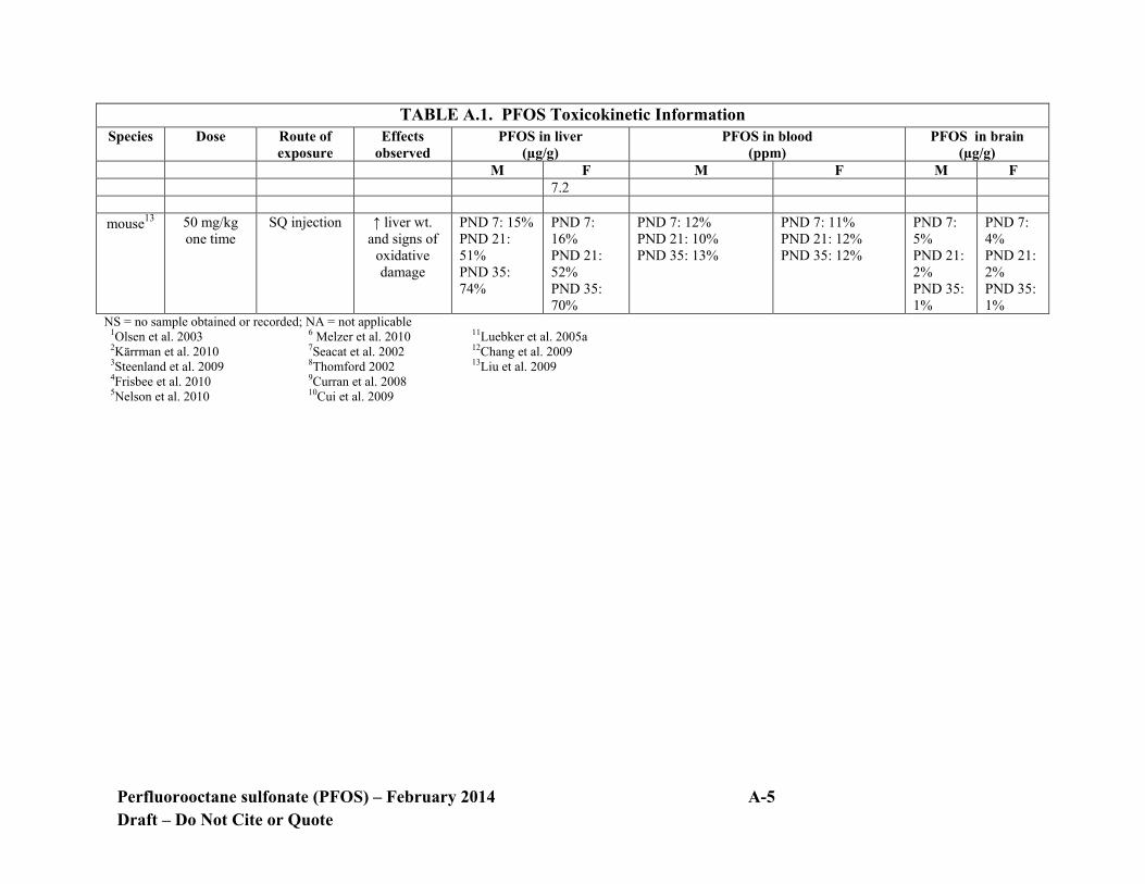
Perfluorooctane sulfonate (PFOS) – February 2014 A-5 Draft – Do Not Cite or Quote
TABLE A.1. PFOS Toxicokinetic Information Species Dose Route of
exposure Effects
observed PFOS in liver
(µg/g) PFOS in blood
(ppm) PFOS in brain
(µg/g) M F M F M F
7.2
mouse13 50 mg/kg one time
SQ injection ↑ liver wt. and signs of
oxidative damage
PND 7: 15% PND 21: 51% PND 35: 74%
PND 7: 16% PND 21: 52% PND 35: 70%
PND 7: 12% PND 21: 10% PND 35: 13%
PND 7: 11% PND 21: 12% PND 35: 12%
PND 7: 5% PND 21: 2% PND 35: 1%
PND 7: 4% PND 21: 2% PND 35: 1%
NS = no sample obtained or recorded; NA = not applicable 1Olsen et al. 2003 2Kärrman et al. 2010 3Steenland et al. 2009 4Frisbee et al. 2010 5Nelson et al. 2010
6 Melzer et al. 2010 7Seacat et al. 2002 8Thomford 2002 9Curran et al. 2008 10Cui et al. 2009
11Luebker et al. 2005a 12Chang et al. 2009 13Liu et al. 2009
Perfluorooctane sulfonate (PFOS) – February 2014 A-6 Draft – Do Not Cite or Quote
TABLE A.2. Key Studies Used With Effects Related to Serum Values (Condensed Version) Study Species/Strain Exposure
duration Dose
(mg/kg/day) PFOS concentration
(µg/mL) NOAEL (mg/kg)
LOAEL (mg/kg)
Critical Effect
M F LIVER EFFECTS Curran et al.
2008 Rat/Sprague-Dawley
15/sex/group
diet
28 days
0
M: 0.14; F: 0.15
M: 1.33; F:
1.43
M: 3.21; F: 3.73
M: 6.34; F:
7.58
serum (values expressed as µg/g serum)
0.47
0.95
13.45
20.93
29.88
serum (values
expressed as µg/g serum)
0.95
1.50
15.40
31.93
43.20
0.14 (M) NA (F)
1.33 (M) 0.15 (F)
F: ↑ final relative (to body weight) liver
weight
M: ↑ final relative (to body weight) liver weight; ↓
serum T4
Seacat et al. 2003
Rat/ Crl:CD(SD) IGS BR
5/sex/group
diet
98 days (14 weeks)
0
0.035
0.14
0.35
1.4
serum
<LOQa
4.04
17.1
43.9
148
serum
2.67
6.96
27.3
64.4
223
0.35 1.4 ↑ absolute liver wt and ALT (m) and ↑ relative liver wt (f)
centrilobular hepatic
hypertrophy (m/f)
Seacat et al. 2002
Monkey/Cynomolgus 6/sex/group
capsule
182 days (26 weeks) followed by
52 week recovery
0
serum
wk 1: NA wk 16: 0.04 wk 27: 0.05
serum
wk 1: NA wk 16: 0.04 wk 27: 0.04
0.15 0.75 M/F: ↑ absolute and relative hepatic wt;
centrilobular or diffuse
hepatocellular

Perfluorooctane sulfonate (PFOS) – February 2014 A-7 Draft – Do Not Cite or Quote
TABLE A.2. Key Studies Used With Effects Related to Serum Values (Condensed Version) Study Species/Strain Exposure
duration Dose
(mg/kg/day) PFOS concentration
(µg/mL) NOAEL (mg/kg)
LOAEL (mg/kg)
Critical Effect
M F
0.03
0.15
0.75
wk 35: 0.05 wk 79: 0.02
wk 1: 0.869 wk 16: 11.2 wk 27: 15.9 wk 35: NS wk 79: ND
wk 1: 4.60
wk 16: 56.2 wk 27: 68.1 wk 35: 84.5 wk 79: 19.1
wk 1: 21.0 wk 16: 189 wk 27: 194 wk 35: 181 wk 79: 41.1
wk 35: 0.07 wk 79: 0.02
wk 1: 0.947 wk 16: 10.5 wk 27: 11.1 wk 35: NS wk 79: ND
wk 1: 3.71
wk 16: 42.1 wk 27: 58.5 wk 35: 74.7 wk 79: 21.4
wk 1: 20.4 wk 16: 162 wk 27: 160 wk 35: 171 wk 79: 41.4
hypertrophy
Thomford, 2002
Rat/Crl:CD (SD)IGS BR
diet
104 weeks
0
0.018-0.023
0.072-0.099
serum
wk 1: <LOQ wk 14: <LOQ wk 53: 0.025
wk 105: 0.012
wk 1: 0.091 wk 14: 4.04 wk 53: NS
wk 105: 1.31
wk 1: 4.33 wk 14: 17.1
serum
wk 1: 0.026 wk 14: 2.67 wk 53: 0.40
wk 105: 0.084
wk 1: 1.61
wk 14: 6.96 wk 53: NS
wk 105: 4.35
wk 1: 6.62 wk 14: 27.3
Males 0.018
Females
0.099
Males 0.072
Females
0.247
Males and Females: liver histopathology

Perfluorooctane sulfonate (PFOS) – February 2014 A-8 Draft – Do Not Cite or Quote
TABLE A.2. Key Studies Used With Effects Related to Serum Values (Condensed Version) Study Species/Strain Exposure
duration Dose
(mg/kg/day) PFOS concentration
(µg/mL) NOAEL (mg/kg)
LOAEL (mg/kg)
Critical Effect
M F
0.184-0.247
0.765-1.1
wk 53: NS wk 105: 7.60
wk 1: 7.57
wk 14: 43.9 wk 53: NS
wk 105: 22.5
wk 1: 41.8 wk 14: 148 wk 53: 146
wk 105: 69.3
wk 53: NS wk 105: NS
wk 1: 12.6
wk 14: 64.4 wk 53: NS
wk 105: 75.0
wk 1: 54.0 wk 14: 223 wk 53: 220 wk 105: 233
THYROID EFFECTS Chang et al.
2008 Rat/Sprague-Dawley 5-15 females/group
oral gavage
Single dose
0
15
No males dosed
serum
< LLOQa at all timepoints
2 hrs: 37.28 6 hrs: 66.90 24 hrs: 61.58
NA 15 Total thyroxine (TT4)- significant ↓
at 2, 6 and 24 hrs Triiodothyronine (TT3) and reverse triiodothyronine
(rT3)- significant ↓ at 24 hrs
Free thyroxine- significant ↑ at 2 and 6 hrs; normal at 24
hrs Curran et al.
2008 Rat/Sprague-Dawley
15/sex/group
diet
28 days
0
M: 0.14; F: 0.15
serum (values expressed as µg/g serum)
0.47
0.95
13.45
serum (values
expressed as µg/g serum)
0.95
1.50
M: 0.14; F: 0.15
M: 1.33; F: 1.43
M/F: ↓ T4

Perfluorooctane sulfonate (PFOS) – February 2014 A-9 Draft – Do Not Cite or Quote
TABLE A.2. Key Studies Used With Effects Related to Serum Values (Condensed Version) Study Species/Strain Exposure
duration Dose
(mg/kg/day) PFOS concentration
(µg/mL) NOAEL (mg/kg)
LOAEL (mg/kg)
Critical Effect
M F M: 1.33; F:
1.43
M: 3.21; F: 3.73
M: 6.34; F:
7.58
20.93
29.88
15.40
31.93
43.20
DEVELOPMENTAL EFFECTS Butenhoff et al. 2009 and Chang et al.
2009
Rat/Sprague-Dawley 25 females/group
oral gavage
GD 0- PND 20
Offspring monitored
through PND 72
0
0.1
serum PND 21: M Offspring = <LLOQ PND 72: M Offspring = <LLOQ PND 21: M Offspring = 1.73 PND 72: M Offspring = 0.04
serum GD 20: Dams = <LLOQ Fetus = 0.009 PND 21: Dams = 0.007 F Offspring = <LLOQ PND 72: Dams = NA F Offspring = <LLOQ GD 20: Dams = 1.72 Fetus = 3.91 PND 21: Dams = 3.16 F Offspring = 1.77 PND 72:
Maternal: 0.3
Male
offspring: 0.3
Female Offspring: >1 mg/kg
Maternal: 1.0
Male
offspring: 1.0
Female Offspring: ND
Maternal: ↓ body wt
Male offspring: based on ↓ habituation response

Perfluorooctane sulfonate (PFOS) – February 2014 A-10 Draft – Do Not Cite or Quote
TABLE A.2. Key Studies Used With Effects Related to Serum Values (Condensed Version) Study Species/Strain Exposure
duration Dose
(mg/kg/day) PFOS concentration
(µg/mL) NOAEL (mg/kg)
LOAEL (mg/kg)
Critical Effect
M F
0.3
1.0
PND 21: M Offspring = 5.05 PND 72: M Offspring = 0.12 PND 21: M Offspring = 18.61 PND 72: M Offspring = 0.56
Dams = NA F Offspring = 0.21 GD 20: Dams = 6.25 Fetus = 10.45 PND 21: Dams = 8.98 F Offspring = 5.25 PND 72: Dams = NA F Offspring = 0.56 GD 20: Dams = 26.63 Fetus = 31.46 PND 21: Dams = 30.48 F Offspring = 18.01 PND 72: Dams = NA F Offspring = 1.99
Thibodeaux et al. 2003 and
Rat/Sprague-Dawley 16-25 females/group
GDs 2-20
serum on GD 21
Maternal = 1
Maternal= 2
Maternal: ↓ body wt

Perfluorooctane sulfonate (PFOS) – February 2014 A-11 Draft – Do Not Cite or Quote
TABLE A.2. Key Studies Used With Effects Related to Serum Values (Condensed Version) Study Species/Strain Exposure
duration Dose
(mg/kg/day) PFOS concentration
(µg/mL) NOAEL (mg/kg)
LOAEL (mg/kg)
Critical Effect
M F Lau et al.
2003
oral gavage
0
1
2
3
5
10
Dam: 0.24 Newborn: 0.188
Dam: 19.6
Newborn: 35.9
Dam: 45.0 Newborn: 71.9
Dam: 71.9
Newborn: 86.5
Dam: 80.6 Newborn: 108.2
Dam: 189.9
Newborn: NS
Develop mental
= 1
Developmental = 2
Developmental: based on ↓ survival and developmental
delays
BMDL5 corresponding to maternal dose for
survival of rat pups at PND 8 was 0.58
mg/kg BMDL5
corresponding to fetal sternal defects was 0.12 and cleft palates was 3.33
mg/kg
REPRODUCTIVE Luebker et al.
2005a and 2005b
Rat/Crl:CD (SD)IGS VAF/Plus
oral gavage
6 wks prior to mating through
gestation and lactation across 2
generations
Due to high number of
pups dying, only 0, 0.1
and 0.4 administered
to 2nd
0
0.1
0.4
NS
serum
NS
Dams GD 1: 8.90 GD 7: 7.83
GD 15: 8.81 GD 21: 4.52 Fetus GD 21:
9.08
Dams GD 1: 40.7 GD 7: 40.9
F0 dams 0.4
F1 pups:
0.4
F0 dams 0.8
F1 pups: 0.8
F0 dams: Decreased body wt
F1 pups:
Decreased gestation length
BMDL5 for decreased survival of
pups through LD5 was 0.89 mg/kg/day
BMDL5 for pup
weight gain was 0.28

Perfluorooctane sulfonate (PFOS) – February 2014 A-12 Draft – Do Not Cite or Quote
TABLE A.2. Key Studies Used With Effects Related to Serum Values (Condensed Version) Study Species/Strain Exposure
duration Dose
(mg/kg/day) PFOS concentration
(µg/mL) NOAEL (mg/kg)
LOAEL (mg/kg)
Critical Effect
M F generation
1.6
3.2
GD 15: 41.4 GD 21: 26.2 Fetus GD 21:
34.3
Dams GD 1: 160 GD 7: 154
GD 15: 156 GD 21: 136
Fetus GD 21: 101
Dams
GD 1: 318 GD 7: 306
GD 15: 275 GD 21: 155
Fetus GD 21: 164
mg/kg/day
IMMUNOTOXICITY Dong et al.
2009 Mouse/B6C3F1
10 m/dose
oral gavage
60 days 0
0.0083
0.083
0.42
0.83
2.08
serum 0.048
0.674
7.132
21.638
65.426
120.67
NS 0.0083 0.083 Based on ↑ splenic natural killer cell
activity

Perfluorooctane sulfonate (PFOS) – February 2014 A-13 Draft – Do Not Cite or Quote
TABLE A.2. Key Studies Used With Effects Related to Serum Values (Condensed Version) Study Species/Strain Exposure
duration Dose
(mg/kg/day) PFOS concentration
(µg/mL) NOAEL (mg/kg)
LOAEL (mg/kg)
Critical Effect
M F Peden-Adams
et al. 2008 Mouse/B6C3F1
5 m/5 f
oral gavage
28 days
0
0.0017
0.0017
0.0033
0.017
0.033
0.166
serum
0.012
0.018
0.092
0.131
NS
NS
NS
serum
0.017
NS
0.088
0.123
0.666
NS
NS
Males 0.00017
Females 0.0033
Males 0.0017
Females 0.017
Males: ↓ plaque forming cell
response
Females: ↓ plaque forming cell
response
a LOQ or LLOQ are below limits that can be quantified NS = no sample; NA= not applicable

Perfluorooctane sulfonate (PFOS) – February 2014 A- 14 Draft – Do Not Cite or Quote
TABLE A.3. Summary of Animal Studies with Exposure to PFOS Method of exposure
Length of study
Species Concentration Results Reference
oral gavage 20 days monkey 0, 10, 30, 100 or 300 mg/kg/day
2 monkeys/sex/dose
NOAEL= NA LOAEL= 10 mg/kg/day from
deaths at all doses
Goldenthal et al. 1978a
oral gavage 90 days monkey 0, 0.5, 1.5 or 4.5 mg/kg/day
2 monkeys/sex/dose
NOAEL= NA LOAEL= 0.5 mg/kg/day
based on diarrhea and anorexia
Goldenthal et al. 1979
oral (capsule)
182 days monkey 0, 0.03, 0.15 or 0.75 mg/kg/day
4-6 monkeys/sex/dose
NOAEL= 0.15 mg/kg/day LOAEL= 0.75 mg/kg/day from ↓ body weight, ↑liver
wt and ↓cholesterol
Seacat et al. 2002
oral gavage single dose rat 0, 100, 215, 464 or 1000 mg/kg
5 rats/sex/dose
LD50 = 251 mg/kg (combined)
Dean et al. 1978
oral gavage single dose rat 0, 12.5, 25 or 50 mg/kg 5 male rats/dose
NOAEL= NA LOAEL= 12.5 mg/kg based
on ↓ body weight
Yang et al. 2009
oral gavage
single dose
thyroid hormone activity
rat 0 or 15 mg/kg 5/15 female rats/group
Total thyroxine (TT4)- significant ↓ at 2, 6 and 24
hrs Triiodothyronine (TT3) and
reverse triiodothyronine (rT3)- significant ↓ at 24 hrs Free thyroxine- significant ↑ at 2 and 6 hrs; normal at 24
hrs
Chang et al. 2008
inhalation 1 hour rat 0, 1.89, 2.86, 4.88, 6.49, 7.05, 13.9, 24.09 or
45.97 ppm 5 rats/sex/dose
LC50 = 5.2 ppm
Rusch et al. 1979
oral (in diet)
28 days rat 0, 0.05, 0.18, 0.37 or 1.51 mg/kg/day- males 0, 0.05, 0.22, 0.47 or
1.77 mg/kg/day- females
(0, 0.5, 2, 5 or 20 ppm) 5 rats/sex/dose
NOAEL = 0.37 mg/kg/day in males and 0.47 mg/kg/day in
females LOAEL = 1.51 mg/kg/day in males and 1.77 mg/kg/day in females, based on ↓ body wt
(m/f) and ↓ food consumption (f)
Seacat et al. 2003
oral (in diet)
28 days rat 0, 2, 20, 50 or 100 mg PFOS/kg diet
NOAEL = 2 mg PFOS/kg in males and NA in females
LOAEL = 20 mg PFOS/kg in males and 2 mg/kg in
females based on ↑ relative liver wt
Curran et al. 2008
oral gavage 28 days rat 0, 5 or 20 mg/kg/day 10 males/dose
NOAEL= NA LOAEL= 5 mg/kg/day based
on ↓ body wt and lung congestion
Cui et al. 2009
oral (in diet)
90 days rat 0, 2, 6, 18, 60 or 200 mg/kg/day
5 rats/sex/dose
NOAEL= NA LOAEL= 2 mg/kg/day, from
↑ liver wt, ↓ food consumption
Goldenthal et al. 1978b
Perfluorooctane sulfonate (PFOS) – February 2014 A- 15 Draft – Do Not Cite or Quote
TABLE A.3. Summary of Animal Studies with Exposure to PFOS Method of exposure
Length of study
Species Concentration Results Reference
oral (in diet)
98 days rat 0, 0.5, 2.0, 5.0 or 20 ppm
0, 0.03, 0.13, 0.34 or
1.33 mg/kg/day- males 0, 0.04, 0.15, 0.40 or
1.56 mg/kg/day- females
5 rats/sex/dose
NOAEL = 0.34 mg/kg/day in males and 0.40 mg/kg/day in
females LOAEL = 1.33 mg/kg/day in males and 1.56 mg/kg/day in females, based on ↑ liver wt (m) and ↑ relative liver wt
(m/f)
Seacat et al. 2003
oral gavage
GD 0 to PND 20a
developmental neurotoxicity
study
rat 0, 0.1, 0.3 or 1.0 mg/kg/day
25 females/dose
Maternal NOAEL= 1 mg/kg/day
LOAEL= NA
Developmental NOAEL= 0.3 mg/kg/day
LOAEL= 1 mg/kg/day based on ↑ motor activity
Butenhoff et al. 2009
oral gavage GD 6-15
developmental study
rat 0, 1, 5 or 10 mg/kg/day 25 pregnant rats
Maternal NOAEL= 1 mg/kg/day
LOAEL= 5 mg/kg/day based on clinical signs
Developmental
NOAEL= 1 mg/kg/day LOAEL= 5 mg/kg/day based
on ↓ body wt
Wetzel 1983
oral gavage GD 2-21 rat 0, 1, 2, 3, 5 or 10 mg/kg
Maternal NOAEL= 1 mg/kg
LOAEL= 2 mg/kg based on ↓ body wt
Developmental
NOAEL= 1 mg/kg LOAEL= 2 mg/kg based on
↓ survival
BMDL5 corresponding to maternal dose for survival of rat pups at PND 8 was 0.58
mg/kg
Thibodeaux et al. 2003 and Lau et
al. 2003
oral gavage GD 2-21 rat 0, 0.1, 0.6 or 2.0 mg/kg Offspring NOAEL = can’t be
determined LOAEL= 0.1 mg/kg based
on changes in the cortex and hippocampus (astrocyte activation markers, pro-
inflammatory transcription factors)
Zeng et al., 2011
oral gavage GD 1-21 rat 0, 0.1, or 2.0 mg/kg/day Offspring NOAEL= 0.1 mg/kg/day LOAEL = 2.0 mg/kg/day
based on histopathological
Chen et al., 2012

Perfluorooctane sulfonate (PFOS) – February 2014 A- 16 Draft – Do Not Cite or Quote
TABLE A.3. Summary of Animal Studies with Exposure to PFOS Method of exposure
Length of study
Species Concentration Results Reference
changes in lungs, ↓ body wt and ↑ mortality
oral gavage 6 wks prior to mating and
Males- 22 days Females- through
gestation, parturition and
lactation
reproductive study
rat 0, 0.1, 0.4, 1.6 or 3.2 mg/kg/day
35 rats/sex/dose
F0 (m/f) parents NOAEL= 0.1 mg/kg/day LOAEL= 0.4 mg/kg/day based on ↓ bwt gain/food
consumption
F1 (m/f) parents NOAEL = 0.4 mg/kg
LOAEL = 1.6 mg/kg/day
F1 offspring NOAEL= 0.4 mg/kg/day LOAEL= 1.6 mg/kg/day
based on ↓ viability, body wt
F2 offspring NOAEL= 0.1 mg/kg/day LOAEL= 0.4 mg/kg/day
based on ↓ body wt
Luebker et al., 2005b
oral gavage 6 wks prior to mating and continued
through mating, gestation and
lactation day 4
reproductive study
rat 0, 0.4, 0.8. 1.0, 1.2, 1.6 and 2.0 mg/kg/day 20-28 dams/dose
F0 dams NOAEL= 0.4 mg/kg/day LOAEL= 0.8 mg/kg/day
based on ↓ bwt gain
F1 offspring NOAEL= not identified LOAEL= 0.4 mg/kg/day
based on ↓ pup body weight
BMDL5 estimates for decreased gestation length was 0.31 and viability was
0.89 mg/kg/day
Luebker et al., 2005a
oral (diet) 104 weeks rat 0, 0.03, 0.1, 0.4 or 1.5 mg/kg/day
0, 0.5, 2, 5 or 20 ppm
40-70 males and females
Males NOAEL= 0.5 ppm (0.03 mg/kg)
Males LOAEL= 2 ppm (0.1 mg/kg) based on liver
histopathology
Females NOAEL= 2 ppm (0.1 mg/kg)
Females LOAEL= 5 ppm (0.4 mg/kg) based on liver
histopathology
Suggestive of carcinogenicity
Thomford, 2002
oral gavage 1 time
developmental
mouse 0, 0.75 or 11.3 mg/kg 4-7 males/group
Mice at both concentrations showed ↓ activity and ↑
neuroprotein levels in the
Johansson et al. 2008 and 2009
Perfluorooctane sulfonate (PFOS) – February 2014 A- 17 Draft – Do Not Cite or Quote
TABLE A.3. Summary of Animal Studies with Exposure to PFOS Method of exposure
Length of study
Species Concentration Results Reference
neurotoxicity hippocampus oral gavage 7 days
immunotoxicity
study
mouse 0, 5, 20 or 40 mg/kg 12 male mice/dose
NOAEL= NA LOAEL= 5 mg/kg based on ↑ liver wt and suppression of
the plaque forming cell response
Zheng et al. 2009
oral gavage GD 1-17
developmental immunotoxicity
mouse 0, 0.1, 1 or 5 mg/kg 10-12 female mice/dose
Males NOAEL= 0.1 mg/kg Males LOAEL= 1 mg/kg
based on ↓NK cell activity
Females NOAEL= 1 mg/kg Females LOAEL= 5 mg/kg based on ↓NK cell activity
Keil et al. 2008
oral gavage GD 1-17 mouse 0, 1, 5, 10, 15 or 20 mg/kg
Maternal NOAEL= 1 mg/kg
LOAEL= 5 mg/kg based on ↑ liver wt
Developmental
NOAEL= 1 mg/kg LOAEL= 5 mg/kg based on
↑ liver wt, delayed eye opening
BMDL5 corresponding to
maternal dose for survival of mouse pups at PND 6 was
3.88 mg/kg
Thibodeaux et al. 2003 and Lau et
al. 2003
oral gavage GD 12-18
developmental
mouse 0 or 6 mg/kg/day 8-10 mice/dose
Maternal NOAEL= 6 mg/kg/day
LOAEL= NA
Developmental NOAEL= NA
LOAEL= 6 mg/kg/day based on ↓ body wt
Fuentes et al. 2007
oral gavage GD 0-17/18
developmental
mouse 0, 1, 10 or 20 mg/kg/day
10 mice/dose
Maternal NOAEL= 1 mg/kg/day
LOAEL= 10 mg/kg/day, based on ↑ liver organ wt.
Developmental
NOAEL= 1 mg/kg/day LOAEL= 10 mg/kg/day,
based on fetal abnormalities and ↓survival
Yahia et al. 2008
oral gavage 28 days
immunotoxicity
mouse 0, 0.005, 0.05, 0.1, 0.5, 1, or 5 mg/kg 5 mice/dose
Males NOAEL= 0.005 mg/kg
Males LOAEL= 0.05 mg/kg based on ↓ plaque forming
cell response
Females NOAEL= 0.1
Peden-Adams et al., 2008
Perfluorooctane sulfonate (PFOS) – February 2014 A- 18 Draft – Do Not Cite or Quote
TABLE A.3. Summary of Animal Studies with Exposure to PFOS Method of exposure
Length of study
Species Concentration Results Reference
mg/kg Females LOAEL= 0.5 mg/kg
based on ↓ plaque forming cell response
oral gavage 60 days
immunotoxicity
mouse 0, 0.008, 0.083, 0.417, 0.833 or 2.083 mg/kg 10 male mice/group
NOAEL= 0.008 mg/kg/day LOAEL= 0.083 mg/kg based on ↑ splenic NK cell activity
and ↑ liver weight
Dong et al. 2009
dermal single dose rabbit 0.5 g*
(no data on gender) No irritation Biesemeier
and Harris 1974
ocular single dose rabbit 0.5 g* (no data on gender)
Exact score not provided except maximal score at 1
and 24 hrs
Biesemeier and Harris
1974 oral gavage GD 7-20
developmental
rabbit 0, 0.1, 1.0, 2.5 or 3.75 mg/kg/day
22 females/dose
Maternal NOAEL= 0.1 mg/kg/day
LOAEL= 1 mg/kg/day based on ↓ body wt.
Developmental
NOAEL= 1 mg/kg/day LOAEL= 2.5 mg/kg/day
based on ↓ fetal body wt and ↓ in sternum ossification
Christian et al. 1999
*Exact dose not provided; NA= not applicable; could not be determined a GD = gestation day and PND = post natal day

Perfluorooctane sulfonate (PFOS) – February 2014 B- 1 Draft – Do Not Cite or Quote
APPENDIX B
Contents: Benchmark Dose Modeling Output Files 10% increased incidence of liver lesions and 10% increase in liver weight for:
1) Thomford, 2002: male rat – hepatocellular centrilobular hypertrophy; 2) Thomford, 2002: female rat – hepatocellular centrilobular hypertrophy; 3) Thomford, 2002: male rat – centrilobular hepatocytic vacuolation; 4) Seacat et al., 2002: male monkey – increased liver weight; 5) Seacat et al., 2002: female monkey – increased liver weight; 6) Seacat et al., 2003: male rat – increased liver weight.
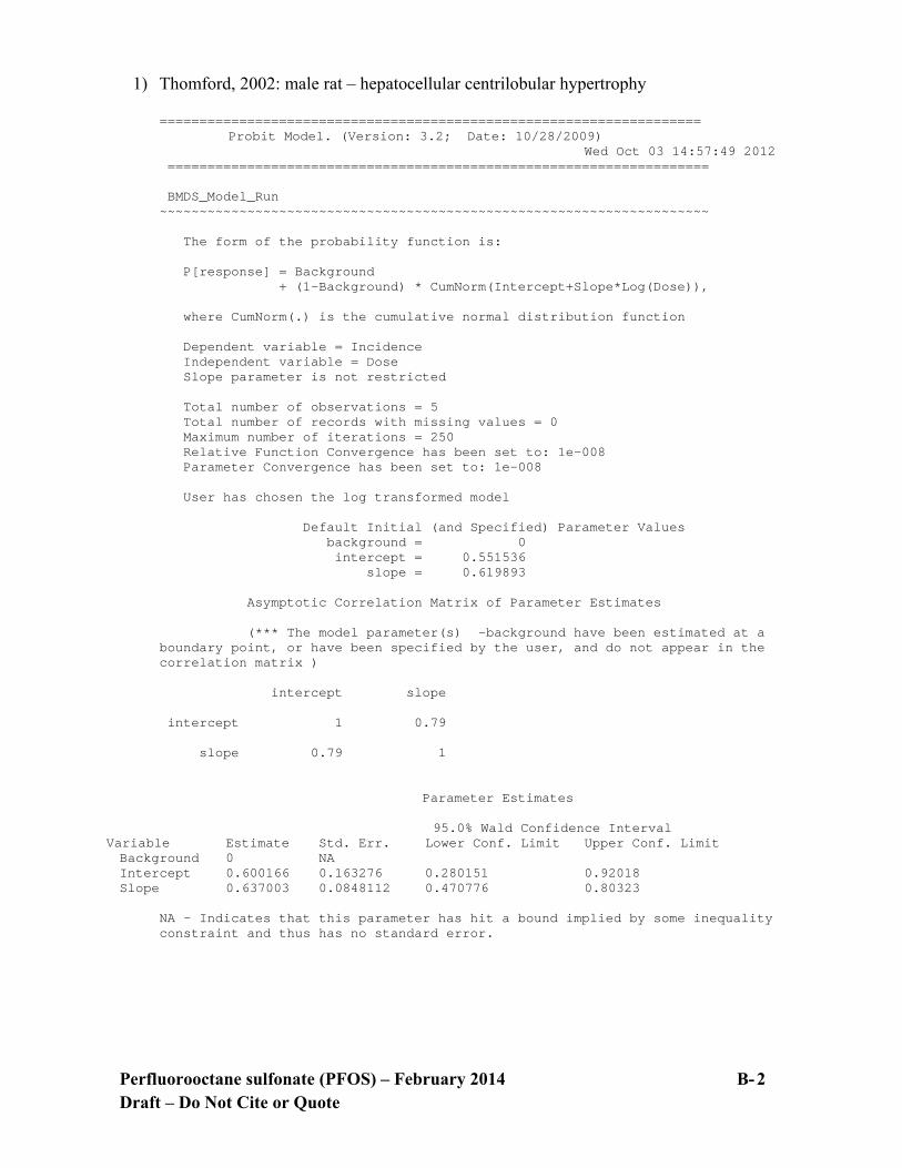
Perfluorooctane sulfonate (PFOS) – February 2014 B- 2 Draft – Do Not Cite or Quote
1) Thomford, 2002: male rat – hepatocellular centrilobular hypertrophy
==================================================================== Probit Model. (Version: 3.2; Date: 10/28/2009) Wed Oct 03 14:57:49 2012 ==================================================================== BMDS_Model_Run ~~~~~~~~~~~~~~~~~~~~~~~~~~~~~~~~~~~~~~~~~~~~~~~~~~~~~~~~~~~~~~~~~~~~~ The form of the probability function is: P[response] = Background + (1-Background) * CumNorm(Intercept+Slope*Log(Dose)), where CumNorm(.) is the cumulative normal distribution function Dependent variable = Incidence Independent variable = Dose Slope parameter is not restricted Total number of observations = 5 Total number of records with missing values = 0 Maximum number of iterations = 250 Relative Function Convergence has been set to: 1e-008 Parameter Convergence has been set to: 1e-008 User has chosen the log transformed model Default Initial (and Specified) Parameter Values background = 0 intercept = 0.551536 slope = 0.619893 Asymptotic Correlation Matrix of Parameter Estimates (*** The model parameter(s) -background have been estimated at a boundary point, or have been specified by the user, and do not appear in the correlation matrix ) intercept slope intercept 1 0.79 slope 0.79 1 Parameter Estimates
95.0% Wald Confidence Interval Variable Estimate Std. Err. Lower Conf. Limit Upper Conf. Limit Background 0 NA Intercept 0.600166 0.163276 0.280151 0.92018 Slope 0.637003 0.0848112 0.470776 0.80323
NA - Indicates that this parameter has hit a bound implied by some inequality constraint and thus has no standard error.

Perfluorooctane sulfonate (PFOS) – February 2014 B- 3 Draft – Do Not Cite or Quote
Analysis of Deviance Table Model Log(likelihood) # Param's Deviance Test d.f. P-value Full model -102.179 5 Fitted model -104.472 2 4.58658 3 0.2047 Reduced model -161.64 1 118.923 4 <.0001 AIC: 212.944 Goodness of Fit Scaled Dose Est._Prob. Expected Observed Size Residual ------------------------------------------------------------------------ 0.0000 0.0000 0.000 0.000 65 0.000 0.0180 0.0251 1.378 2.000 55 0.536 0.0720 0.1410 7.755 4.000 55 -1.455 0.1840 0.3163 17.395 22.000 55 1.335 0.7650 0.6662 43.305 42.000 65 -0.343 Chi^2 = 4.31 d.f. = 3 P-value = 0.2303 Benchmark Dose Computation Specified effect = 0.1 Risk Type = Extra risk Confidence level = 0.95 BMD = 0.0521299 BMDL = 0.0326765

Perfluorooctane sulfonate (PFOS) – February 2014 B- 4 Draft – Do Not Cite or Quote
2) Thomford, 2002: female rat – hepatocellular centrilobular hypertrophy;
==================================================================== Probit Model. (Version: 3.2; Date: 10/28/2009) Tue Jan 29 13:03:16 2013 ==================================================================== BMDS_Model_Run ~~~~~~~~~~~~~~~~~~~~~~~~~~~~~~~~~~~~~~~~~~~~~~~~~~~~~~~~~~~~~~~~~~~~~ The form of the probability function is: P[response] = Background + (1-Background) * CumNorm(Intercept+Slope*Log(Dose)), where CumNorm(.) is the cumulative normal distribution function Dependent variable = Incidence Independent variable = Dose Slope parameter is not restricted Total number of observations = 5 Total number of records with missing values = 0 Maximum number of iterations = 250 Relative Function Convergence has been set to: 1e-008 Parameter Convergence has been set to: 1e-008 User has chosen the log transformed model Default Initial (and Specified) Parameter Values background = 0.0307692 intercept = 0.921158 slope = 0.875963 Asymptotic Correlation Matrix of Parameter Estimates background intercept slope background 1 0.037 0.25 intercept 0.037 1 0.78 slope 0.25 0.78 1 Parameter Estimates
95.0% Wald Confidence Interval Variable Estimate Std. Err. Lower Conf. Limit Upper Conf. Limit Background 0.0242361 0.0139844 -0.0031728 0.0516449 Intercept 1.11217 0.200749 0.718705 1.50563 Slope 1.03103 0.140636 0.755385 1.30667
Analysis of Deviance Table Model Log(likelihood) # Param's Deviance Test d.f. P-value Full model -93.9538 5 Fitted model -94.0971 3 0.286555 2 0.8665 Reduced model -167.248 1 146.589 4 <.0001

Perfluorooctane sulfonate (PFOS) – February 2014 B- 5 Draft – Do Not Cite or Quote
AIC: 194.194 Goodness of Fit Scaled Dose Est._Prob. Expected Observed Size Residual ------------------------------------------------------------------------ 0.0000 0.0242 1.575 2.000 65 0.343 0.0180 0.0254 1.399 1.000 55 -0.341 0.0720 0.0776 4.271 4.000 55 -0.136 0.1840 0.2812 15.464 16.000 55 0.161 0.7650 0.8033 52.215 52.000 65 -0.067 Chi^2 = 0.28 d.f. = 2 P-value = 0.8681 Benchmark Dose Computation Specified effect = 0.1 Risk Type = Extra risk Confidence level = 0.95 BMD = 0.0981083
BMDL = 0.0680339
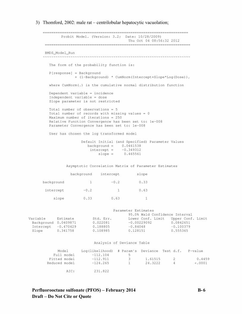
Perfluorooctane sulfonate (PFOS) – February 2014 B- 6 Draft – Do Not Cite or Quote
3) Thomford, 2002: male rat – centrilobular hepatocytic vacuolation;
==================================================================== Probit Model. (Version: 3.2; Date: 10/28/2009) Thu Oct 04 08:56:32 2012 ==================================================================== BMDS_Model_Run ~~~~~~~~~~~~~~~~~~~~~~~~~~~~~~~~~~~~~~~~~~~~~~~~~~~~~~~~~~~~~~~~~~~~~ The form of the probability function is: P[response] = Background + (1-Background) * CumNorm(Intercept+Slope*Log(Dose)), where CumNorm(.) is the cumulative normal distribution function Dependent variable = incidence Independent variable = dose Slope parameter is not restricted Total number of observations = 5 Total number of records with missing values = 0 Maximum number of iterations = 250 Relative Function Convergence has been set to: 1e-008 Parameter Convergence has been set to: 1e-008 User has chosen the log transformed model Default Initial (and Specified) Parameter Values background = 0.0461538 intercept = -0.349312 slope = 0.465561 Asymptotic Correlation Matrix of Parameter Estimates background intercept slope background 1 -0.2 0.33 intercept -0.2 1 0.63 slope 0.33 0.63 1 Parameter Estimates
95.0% Wald Confidence Interval Variable Estimate Std. Err. Lower Conf. Limit Upper Conf. Limit Background 0.0409871 0.022081 -0.00229092 0.0842651 Intercept -0.470429 0.188805 -0.84048 -0.100379 Slope 0.341758 0.108985 0.128151 0.555365
Analysis of Deviance Table Model Log(likelihood) # Param's Deviance Test d.f. P-value Full model -112.104 5 Fitted model -112.911 3 1.61515 2 0.4459 Reduced model -124.265 1 24.3222 4 <.0001 AIC: 231.822

Perfluorooctane sulfonate (PFOS) – February 2014 B- 7 Draft – Do Not Cite or Quote
Goodness of Fit Scaled Dose Est._Prob. Expected Observed Size Residual ------------------------------------------------------------------------ 0.0000 0.0410 2.664 3.000 65 0.210 0.0180 0.0723 3.976 3.000 55 -0.508 0.0720 0.1229 6.759 6.000 55 -0.312 0.1840 0.1821 10.013 13.000 55 1.044 0.7650 0.3163 20.559 19.000 65 -0.416 Chi^2 = 1.66 d.f. = 2 P-value = 0.4357 Benchmark Dose Computation Specified effect = 0.1 Risk Type = Extra risk Confidence level = 0.95 BMD = 0.0931649 BMDL = 0.0278419
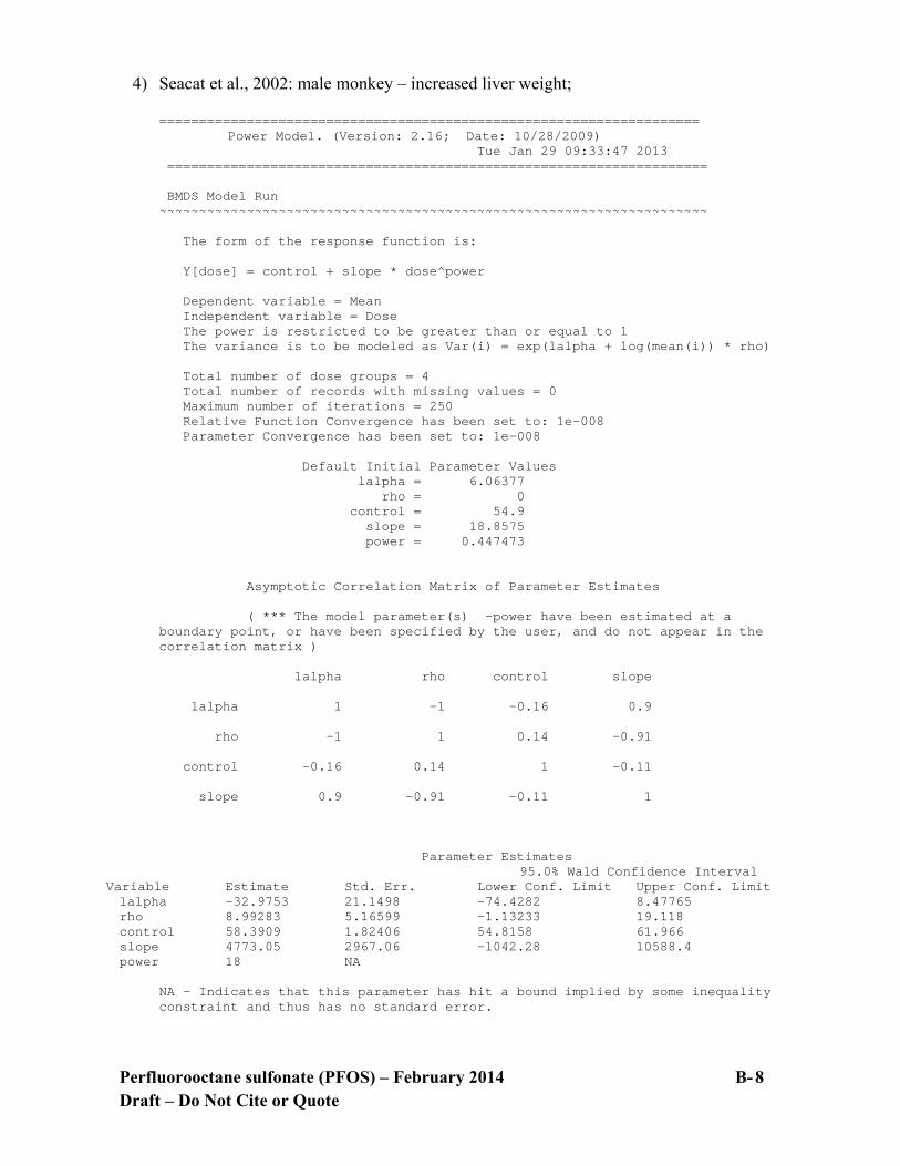
Perfluorooctane sulfonate (PFOS) – February 2014 B- 8 Draft – Do Not Cite or Quote
4) Seacat et al., 2002: male monkey – increased liver weight;
==================================================================== Power Model. (Version: 2.16; Date: 10/28/2009) Tue Jan 29 09:33:47 2013 ==================================================================== BMDS Model Run ~~~~~~~~~~~~~~~~~~~~~~~~~~~~~~~~~~~~~~~~~~~~~~~~~~~~~~~~~~~~~~~~~~~~~ The form of the response function is: Y[dose] = control + slope * dose^power Dependent variable = Mean Independent variable = Dose The power is restricted to be greater than or equal to 1 The variance is to be modeled as Var(i) = exp(lalpha + log(mean(i)) * rho) Total number of dose groups = 4 Total number of records with missing values = 0 Maximum number of iterations = 250 Relative Function Convergence has been set to: 1e-008 Parameter Convergence has been set to: 1e-008 Default Initial Parameter Values lalpha = 6.06377 rho = 0 control = 54.9 slope = 18.8575 power = 0.447473 Asymptotic Correlation Matrix of Parameter Estimates ( *** The model parameter(s) -power have been estimated at a boundary point, or have been specified by the user, and do not appear in the correlation matrix ) lalpha rho control slope lalpha 1 -1 -0.16 0.9 rho -1 1 0.14 -0.91 control -0.16 0.14 1 -0.11 slope 0.9 -0.91 -0.11 1 Parameter Estimates
95.0% Wald Confidence Interval Variable Estimate Std. Err. Lower Conf. Limit Upper Conf. Limit lalpha -32.9753 21.1498 -74.4282 8.47765 rho 8.99283 5.16599 -1.13233 19.118 control 58.3909 1.82406 54.8158 61.966 slope 4773.05 2967.06 -1042.28 10588.4 power 18 NA
NA - Indicates that this parameter has hit a bound implied by some inequality constraint and thus has no standard error.
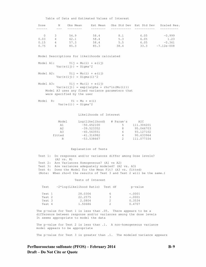
Perfluorooctane sulfonate (PFOS) – February 2014 B- 9 Draft – Do Not Cite or Quote
Table of Data and Estimated Values of Interest Dose N Obs Mean Est Mean Obs Std Dev Est Std Dev Scaled Res. ------ --- -------- -------- ----------- ----------- ---------- 0 3 54.9 58.4 8.1 6.05 -0.999 0.03 4 62.1 58.4 5.3 6.05 1.23 0.15 4 57.3 58.4 5.5 6.05 -0.361 0.75 4 85.3 85.3 38.4 33.3 -7.12e-008 Model Descriptions for likelihoods calculated Model A1: Yij = Mu(i) + e(ij) Var{e(ij)} = Sigma^2 Model A2: Yij = Mu(i) + e(ij) Var{e(ij)} = Sigma(i)^2 Model A3: Yij = Mu(i) + e(ij) Var{e(ij)} = exp(lalpha + rho*ln(Mu(i))) Model A3 uses any fixed variance parameters that were specified by the user Model R: Yi = Mu + e(i) Var{e(i)} = Sigma^2 Likelihoods of Interest Model Log(likelihood) # Param's AIC A1 -50.652100 5 111.304201 A2 -39.523352 8 95.046703 A3 -40.563551 6 93.127102 fitted -41.316982 4 90.633964 R -53.538667 2 111.077334 Explanation of Tests Test 1: Do responses and/or variances differ among Dose levels? (A2 vs. R) Test 2: Are Variances Homogeneous? (A1 vs A2) Test 3: Are variances adequately modeled? (A2 vs. A3) Test 4: Does the Model for the Mean Fit? (A3 vs. fitted) (Note: When rho=0 the results of Test 3 and Test 2 will be the same.) Tests of Interest Test -2*log(Likelihood Ratio) Test df p-value Test 1 28.0306 6 <.0001 Test 2 22.2575 3 <.0001 Test 3 2.0804 2 0.3534 Test 4 1.50686 2 0.4707 The p-value for Test 1 is less than .05. There appears to be a difference between response and/or variances among the dose levels It seems appropriate to model the data The p-value for Test 2 is less than .1. A non-homogeneous variance model appears to be appropriate The p-value for Test 3 is greater than .1. The modeled variance appears

Perfluorooctane sulfonate (PFOS) – February 2014 B- 10 Draft – Do Not Cite or Quote
to be appropriate here The p-value for Test 4 is greater than .1. The model chosen seems to adequately describe the data Benchmark Dose Computation Specified effect = 0.1 Risk Type = Estimated standard deviations from the control mean Confidence level = 0.95 BMD = 0.60743
BMDL = 0.0147931

Perfluorooctane sulfonate (PFOS) – February 2014 B- 11 Draft – Do Not Cite or Quote
5) Seacat et al., 2002: female monkey – increased liver weight;
==================================================================== Exponential Model. (Version: 1.7; Date: 12/10/2009) Tue Jan 29 10:17:21 2013 ==================================================================== BMDS Model Run ~~~~~~~~~~~~~~~~~~~~~~~~~~~~~~~~~~~~~~~~~~~~~~~~~~~~~~~~~~~~~~~~~~~~~ The form of the response function by Model: Model 2: Y[dose] = a * exp{sign * b * dose} Model 3: Y[dose] = a * exp{sign * (b * dose)^d} Model 4: Y[dose] = a * [c-(c-1) * exp{-b * dose}] Model 5: Y[dose] = a * [c-(c-1) * exp{-(b * dose)^d}] Note: Y[dose] is the median response for exposure = dose; sign = +1 for increasing trend in data; sign = -1 for decreasing trend. Model 2 is nested within Models 3 and 4. Model 3 is nested within Model 5. Model 4 is nested within Model 5. Dependent variable = Mean Independent variable = Dose Data are assumed to be distributed: normally Variance Model: exp(lnalpha +rho *ln(Y[dose])) The variance is to be modeled as Var(i) = exp(lalpha + log(mean(i)) * rho) Total number of dose groups = 4 Total number of records with missing values = 0 Maximum number of iterations = 250 Relative Function Convergence has been set to: 1e-008 Parameter Convergence has been set to: 1e-008 MLE solution provided: Exact Initial Parameter Values
Variable Model 2 Model 3 Model 4 Model 5 -------- ------- ------- ------- ------- lnalpha -7.12771 -7.12771 -7.12771 -7.12771 rho 2.78482 2.78482 2.78482 2.78482 a 53.6357 53.6357 48.545 48.545 b 0.452798 0.452798 2.77794 2.77794 c -- -- 1.6287 1.6287 d -- 1 -- 1
Parameter Estimates by Model
Variable Model 2 Model 3 Model 4 Model 5 -------- ------- ------- ------- ------- lnalpha -1.92335 -2.1108 -1.59973 -2.44766 rho 1.54391 1.58965 1.46555 1.6722 a 53.6802 53.8771 53.4554 54.2396 b 0.451454 0.479691 0.00050114 0.0042198 c -- -- 1083.43 721.127 d -- 1.06864 -- 1.30686
Table of Stats From Input Data

Perfluorooctane sulfonate (PFOS) – February 2014 B- 12 Draft – Do Not Cite or Quote
Dose N Obs Mean Obs Std Dev ----- --- ---------- ------------- 0 3 51.1 9.4 0.03 4 56.8 12.6 0.15 4 57 3.1 0.75 4 75.3 13.3 Estimated Values of Interest Model Dose Est Mean Est Std Scaled Residual ------- ------ ---------- --------- ---------------- 2 0 53.68 8.273 -0.5402 0.03 54.41 8.36 0.5712 0.15 57.44 8.717 -0.1012 0.75 75.31 10.74 -0.002181 3 0 53.88 8.276 -0.5812 0.03 54.46 8.347 0.5608 0.15 57.21 8.681 -0.04889 0.75 75.35 10.8 -0.008551 4 0 53.46 8.296 -0.4918 0.03 54.33 8.394 0.5896 0.15 57.8 8.785 -0.1832 0.75 75.2 10.65 0.01899 5 0 54.24 8.29 -0.656 0.03 54.55 8.33 0.5391 0.15 56.82 8.618 0.04191 0.75 75.37 10.92 -0.01305 Other models for which likelihoods are calculated: Model A1: Yij = Mu(i) + e(ij) Var{e(ij)} = Sigma^2 Model A2: Yij = Mu(i) + e(ij) Var{e(ij)} = Sigma(i)^2 Model A3: Yij = Mu(i) + e(ij) Var{e(ij)} = exp(lalpha + log(mean(i)) * rho) Model R: Yij = Mu + e(i) Var{e(ij)} = Sigma^2 Likelihoods of Interest Model Log(likelihood) DF AIC ------- ----------------- ---- ------------ A1 -40.44292 5 90.88584 A2 -36.89929 8 89.79858 A3 -39.91492 6 91.82985 R -45.74039 2 95.48078 2 -40.49219 4 88.98438 3 -40.49157 5 90.98314 4 -40.51305 5 91.0261 5 -40.50101 6 93.00202 Additive constant for all log-likelihoods = -13.78. This constant added to the above values gives the log-likelihood including the term that does not depend on the model parameters.

Perfluorooctane sulfonate (PFOS) – February 2014 B- 13 Draft – Do Not Cite or Quote
Explanation of Tests Test 1: Does response and/or variances differ among Dose levels? (A2 vs. R) Test 2: Are Variances Homogeneous? (A2 vs. A1) Test 3: Are variances adequately modeled? (A2 vs. A3) Test 4: Does Model 2 fit the data? (A3 vs. 2) Test 5a: Does Model 3 fit the data? (A3 vs 3) Test 5b: Is Model 3 better than Model 2? (3 vs. 2) Test 6a: Does Model 4 fit the data? (A3 vs 4) Test 6b: Is Model 4 better than Model 2? (4 vs. 2) Test 7a: Does Model 5 fit the data? (A3 vs 5) Test 7b: Is Model 5 better than Model 3? (5 vs. 3) Test 7c: Is Model 5 better than Model 4? (5 vs. 4) Tests of Interest Test -2*log(Likelihood Ratio) D. F. p-value -------- ------------------------ ------ -------------- Test 1 17.68 6 0.007077 Test 2 7.087 3 0.06917 Test 3 6.031 2 0.04901 Test 4 1.155 2 0.5614 Test 5a 1.153 1 0.2829 Test 5b 0.001241 1 0.9719 Test 6a 1.196 1 0.2741 Test 6b -0.04172 1 N/A Test 7a 1.172 0 N/A Test 7b -0.01888 1 N/A Test 7c 0.02408 1 0.8767 The p-value for Test 1 is less than .05. There appears to be a difference between response and/or variances among the dose levels, it seems appropriate to model the data. The p-value for Test 2 is less than .1. A non-homogeneous variance model appears to be appropriate. The p-value for Test 3 is less than .1. You may want to consider a different variance model. The p-value for Test 4 is greater than .1. Model 2 seems to adequately describe the data. The p-value for Test 5a is greater than .1. Model 3 seems to adequately describe the data. The p-value for Test 5b is greater than .05. Model 3 does not seem to fit the data better than Model 2. The p-value for Test 6a is greater than .1. Model 4 seems to adequately describe the data. The p-value for Test 6b is less than .05. Model 4 appears to fit the data better than Model 2. Degrees of freedom for Test 7a are less than or equal to 0. The Chi-Square test for fit is not valid.

Perfluorooctane sulfonate (PFOS) – February 2014 B- 14 Draft – Do Not Cite or Quote
The p-value for Test 7b is less than .05. Model 5 appears to fit the data better than Model 3. The p-value for Test 7c is greater than .05. Model 5 does not seem to fit the data better than Model 4. Benchmark Dose Computations: Specified Effect = 0.100000 Risk Type = Estimated standard deviations from control Confidence Level = 0.950000 BMD and BMDL by Model Model BMD BMDL ------- ------------ ---------- 2 0.0338792 0.0207989 3 0.0415751 0.0208011 4 0.0286094 0.0165794
5 0.0629252 0.00584753

Perfluorooctane sulfonate (PFOS) – February 2014 B- 15 Draft – Do Not Cite or Quote
6) Seacat et al., 2003: male rat – increased liver weight. ==================================================================== Exponential Model. (Version: 1.7; Date: 12/10/2009) Tue Jan 29 11:21:33 2013 ==================================================================== BMDS Model Run ~~~~~~~~~~~~~~~~~~~~~~~~~~~~~~~~~~~~~~~~~~~~~~~~~~~~~~~~~~~~~~~~~~~~~ The form of the response function by Model: Model 2: Y[dose] = a * exp{sign * b * dose} Model 3: Y[dose] = a * exp{sign * (b * dose)^d} Model 4: Y[dose] = a * [c-(c-1) * exp{-b * dose}] Model 5: Y[dose] = a * [c-(c-1) * exp{-(b * dose)^d}] Note: Y[dose] is the median response for exposure = dose; sign = +1 for increasing trend in data; sign = -1 for decreasing trend. Model 2 is nested within Models 3 and 4. Model 3 is nested within Model 5. Model 4 is nested within Model 5. Dependent variable = Mean Independent variable = Dose Data are assumed to be distributed: normally Variance Model: exp(lnalpha +rho *ln(Y[dose])) rho is set to 0. A constant variance model is fit. Total number of dose groups = 5 Total number of records with missing values = 0 Maximum number of iterations = 250 Relative Function Convergence has been set to: 1e-008 Parameter Convergence has been set to: 1e-008 MLE solution provided: Exact Initial Parameter Values
Variable Model 2 Model 3 Model 4 Model 5 -------- ------- ------- ------- ------- lnalpha 1.35789 1.35789 1.35789 1.35789 rho(S) 0 0 0 0 a 15.3355 15.3355 13.3 13.3 b 0.226423 0.226423 1.66321 1.66321 c -- -- 1.60263 1.60263 d -- 1 -- 1
(S) = Specified Parameter Estimates by Model
Variable Model 2 Model 3 Model 4 Model 5 -------- ------- ------- ------- ------- lnalpha 1.7164 1.7164 1.64166 1.43222 rho 0 0 0 0 a 15.4453 15.4453 14.8246 15 b 0.216725 0.216725 1.73553 2.97948 c -- -- 1.42089 1.35333

Perfluorooctane sulfonate (PFOS) – February 2014 B- 16 Draft – Do Not Cite or Quote
d -- 1 -- 18 Table of Stats From Input Data Dose N Obs Mean Obs Std Dev ----- --- ---------- ------------- 0 5 15.5 1.1 0.03 5 15.5 2.7 0.13 5 14 1.4 0.34 5 18.8 3 1.33 5 20.3 2.2 Estimated Values of Interest Model Dose Est Mean Est Std Scaled Residual ------- ------ ---------- --------- ---------------- 2 0 15.45 2.359 0.0518 0.03 15.55 2.359 -0.0437 0.13 15.89 2.359 -1.788 0.34 16.63 2.359 2.06 1.33 20.61 2.359 -0.2895 3 0 15.45 2.359 0.0518 0.03 15.55 2.359 -0.0437 0.13 15.89 2.359 -1.788 0.34 16.63 2.359 2.06 1.33 20.61 2.359 -0.2895 4 0 14.82 2.272 0.6646 0.03 15.14 2.272 0.3531 0.13 16.08 2.272 -2.052 0.34 17.61 2.272 1.175 1.33 20.44 2.272 -0.1414 5 0 15 2.046 0.5463 0.03 15 2.046 0.5463 0.13 15 2.046 -1.093 0.34 18.8 2.046 1.225e-007 1.33 20.3 2.046 -6.774e-008 Other models for which likelihoods are calculated: Model A1: Yij = Mu(i) + e(ij) Var{e(ij)} = Sigma^2 Model A2: Yij = Mu(i) + e(ij) Var{e(ij)} = Sigma(i)^2 Model A3: Yij = Mu(i) + e(ij) Var{e(ij)} = exp(lalpha + log(mean(i)) * rho) Model R: Yij = Mu + e(i) Var{e(ij)} = Sigma^2 Likelihoods of Interest Model Log(likelihood) DF AIC ------- ----------------- ---- ------------ A1 -29.47369 6 70.94737 A2 -26.27122 10 72.54245 A3 -29.47369 6 70.94737 R -40.48438 2 84.96876

Perfluorooctane sulfonate (PFOS) – February 2014 B- 17 Draft – Do Not Cite or Quote
2 -33.95502 3 73.91004 3 -33.95502 3 73.91004 4 -33.02072 4 74.04144 5 -30.40279 5 70.80558 Additive constant for all log-likelihoods = -22.97. This constant added to the above values gives the log-likelihood including the term that does not depend on the model parameters. Explanation of Tests Test 1: Does response and/or variances differ among Dose levels? (A2 vs. R) Test 2: Are Variances Homogeneous? (A2 vs. A1) Test 3: Are variances adequately modeled? (A2 vs. A3) Test 4: Does Model 2 fit the data? (A3 vs. 2) Test 5a: Does Model 3 fit the data? (A3 vs 3) Test 5b: Is Model 3 better than Model 2? (3 vs. 2) Test 6a: Does Model 4 fit the data? (A3 vs 4) Test 6b: Is Model 4 better than Model 2? (4 vs. 2) Test 7a: Does Model 5 fit the data? (A3 vs 5) Test 7b: Is Model 5 better than Model 3? (5 vs. 3) Test 7c: Is Model 5 better than Model 4? (5 vs. 4) Tests of Interest Test -2*log(Likelihood Ratio) D. F. p-value -------- ------------------------ ------ -------------- Test 1 28.43 8 0.0003996 Test 2 6.405 4 0.1709 Test 3 6.405 4 0.1709 Test 4 8.963 3 0.02979 Test 5a 8.963 3 0.02979 Test 5b -1.421e-014 0 N/A Test 6a 7.094 2 0.02881 Test 6b 1.869 1 0.1716 Test 7a 1.858 1 0.1728 Test 7b 7.104 2 0.02866 Test 7c 5.236 1 0.02213 The p-value for Test 1 is less than .05. There appears to be a difference between response and/or variances among the dose levels, it seems appropriate to model the data. The p-value for Test 2 is greater than .1. A homogeneous variance model appears to be appropriate here. The p-value for Test 3 is greater than .1. The modeled variance appears to be appropriate here. The p-value for Test 4 is less than .1. Model 2 may not adequately describe the data; you may want to consider another model. The p-value for Test 5a is less than .1. Model 3 may not adequately describe the data; you may want to consider another model. Degrees of freedom for Test 5b are less than or equal to 0.
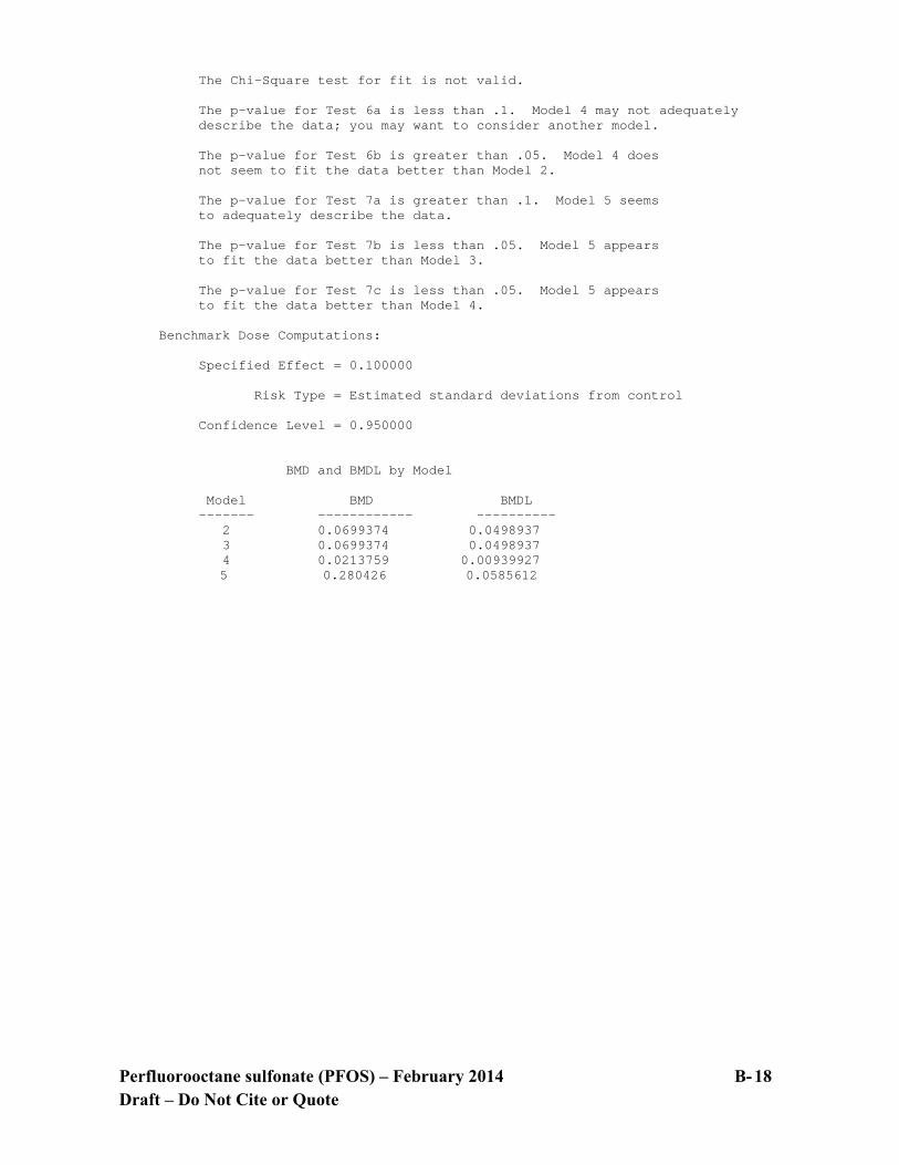
Perfluorooctane sulfonate (PFOS) – February 2014 B- 18 Draft – Do Not Cite or Quote
The Chi-Square test for fit is not valid. The p-value for Test 6a is less than .1. Model 4 may not adequately describe the data; you may want to consider another model. The p-value for Test 6b is greater than .05. Model 4 does not seem to fit the data better than Model 2. The p-value for Test 7a is greater than .1. Model 5 seems to adequately describe the data. The p-value for Test 7b is less than .05. Model 5 appears to fit the data better than Model 3. The p-value for Test 7c is less than .05. Model 5 appears to fit the data better than Model 4. Benchmark Dose Computations: Specified Effect = 0.100000 Risk Type = Estimated standard deviations from control Confidence Level = 0.950000 BMD and BMDL by Model Model BMD BMDL ------- ------------ ---------- 2 0.0699374 0.0498937 3 0.0699374 0.0498937 4 0.0213759 0.00939927
5 0.280426 0.0585612