
Health biotechnology
75
HEALTH BIOTECHNOLOGY Rizwan Abbas baho
-
Upload
bahauddin-zakariya-university-lahore -
Category
Health & Medicine
-
view
284 -
download
9
Transcript of Health biotechnology

HEALTH
BIOTECHNOLOGY
Rizwan Abbas baho
Course Contents:Introduction to Health biotechnology
Social acceptance of medical biotechnology
The molecular basis of disease,
Molecular and genetic markers
Detection of infectious agents
Active and passive immunization
vaccines ,Organ transplantation,
Applications of transgenic animals
Drug delivery systems, Blood transfusion,
Grafting techniques, Pharmacogenetics,
Strategies of gene therapy, gene delivery vehicles, Biopharmaceuticals from plants
Uses of stem cell technology

Reference Books
“Medical Biotechnology” by Judit Pongracz, Mary
Keen “(2009). Elsevier Health Sciences.
“Biotechnology and Your Health: Pharmaceutical
Applications” by Bernice Zeldin Schacter, Bernice
Schacter (2005) Chelsea House Publishers,
“Health and Pharmaceutical Biotechnology” by D.M.
Chetan, K.P. Dinesh, D.M. Chetan (2006) Firewall
Media.

Introduction to health biotechnology
Applications
Drug production
Pharmacogenomics
Gene therapy
Genetic testing
OUTLINE (Lecture-I)
What Is Biotechnology?
Scientific processes to get new organisms
or new products from organisms.
It is the use of living organisms or processes to
develop products useful for mankind.

Has been existing since centuries
Begin with the first action of human on life for his welfare
Term coined by a Hungarian engineer Karl Ereky
Modern biotechnology started in California in 1970’s
History

Origins of Biotechnology
Although it seems like a new thing, biotechnology has actually been around for a while:
Domesticated plants and animals are the result of selective breeding
Using yeast to make bread rise
Using bacteria or yeast to ferment grapes into wine

Any technique that uses living organisms
or substances from those organisms to
make or modify a product, to improve
plants or animals or to develop
microorganisms for specific uses
Definition

Green biotechnology (agricultural)
Red biotechnology (medical)
Blue biotechnology (aquatic)
White biotechnology (industrial)
Applications

The use of biological methods to optimize industrial
processes
Applied by manufacturers of laundry detergents
Includes research for new enzymes (proteins that
remove oily and protein-based stains)
Enzymes that work under extreme conditions (wash
temperatures of 20°C or 90°C)
This often entails modifying the enzymes of
microorganisms for these processes
White biotechnology

Use of biotechnological techniques in agriculture
Vitamin A deficiency is a serious problem and can cause blindness at a young age if left untreated
Golden rice was genetically modified to produce beta-carotene (a precursor of vitamin A that the body converts to vitamin A). A diet including golden rice can thus help to raise vitamin A levels
Green biotechnology:
Also called red biotechnology
It includes:
o Production of medicines and pharmaceutical products for
treating or diagnosing disorders
o Designing of organisms to manufacture antibiotics and
vaccines
o Engineering of genetic defects through genomic
manipulation
o Use in forensics through DNA profiling
Biotechnology and medicine:
Production of human insulin from non- human sources.
Production of hormones like Interferons, Cytokinins,
Steroids and human growth hormones.
Gene therapy for prevention and control of diseases like
hemophilia cystic fibrosis
Development of vaccines and antibodies for rabies, HIV,
etc.
Examples…

Drug production
Pharmacogenomics
Gene therapy
Genetic testing
Health Biotechnology

It is the process in which pharmaceutical products are produced through application of biotechnological techniques
Medicines are produced for:
• Diagnosis
• Cure treatments
• Prevention of diseases
Drug production

Producing medicines through:
Isolating enzymes
Genetically engineering enzymes
Drug production

Recently, plants are being genetically modified to
produce pharmaceutical products instead of their
natural compounds
For Example:
A drug Elelyso for treating Gaucher is being
produced by genetically engineering carrots
Drug production
INSULIN:
Human insulin is being produced using
genetic engineering technique known as
humulin and it is used for the treatment of
diabetes that is low sugar level in the
blood…..
Drug production
INTERFERON:
o Interferon interfere in transmission of viral genome from one cell to another and it also inhibits the cell division of abnormal cells.
o Interferon produced using the recombinant DNA technology is used to treat cancer patients.
o Interferon improved the quality of life of cancer patients…..
Drug production

HUMAN GROWTH HORMONE:
Since dwarfism is caused by growth hormone
deficiency so it can be diagnose by HGH testing.
So HGH is used for the treatment of dwarfism due to
hypo pituitary activity.
Drug production

Pharma = Drug or Medicine
Genomics = The study of genes
Studying response of genetic make up of
an individual to a drug or pharmaceutical
products
Pharmacogenomics

“One-size-fits-all drugs” only work for about 60
percent of the population at best. And the other 40
percent of the population increase their risks
of adverse drug reaction because their genes do
not do what is intended of them.
Use of Pharmacogenomics:

Helps in the development of tailor made medicines
Ensures more appropriate methods of
determining drug dosages
Improve process of drug discovery and approval
Obtaining of better and safer vaccination
Decrease in the overall cost of Health Care
Advanced Screening for Disease
Impotance Of Pharmacogenomics


Opinion: This sort of card would initially (~2025) include
mostly information related to drug metabolizing
enzymes.
Around ~2050 it might include an entire individual
genome
Pharmacogenomics
SMART CARD
(Confidential)
Some barriers faced are:
Complexity of finding gene variation that affect drug response
Limited drug alternatives
Disincentives for drug companies to make multiple pharmacogenomic products
Educating healthcare providers
Pharmacogenomics
The process in which a faulty gene is
removed or replaced with its healthy copy to
restore the normal function of that gene
Gene therapy
Replacing a mutated gene that causes
disease with a healthy copy of the gene
Inactivating or “knocking out” a mutated gene
that is functioning improperly
Introducing the new gene that help fight a
disease
Gene therapy

Some common ways are:
Using fat droplets in nose sprays
Using cold viruses that are modified to carry alleles ,go into the cell and affect them
The direct injection of DNA(might include electroporation or biolistic method)
Gene therapy

The process of gene therapy is of two types:
Stem cell gene therapy:
In this gene therapy is applied on a fully developed
organism and the effects of gene therapy lasts only to
the operated organism
Germ line gene therapy:
In this process gene therapy is done on a fertilized egg
or an early embryo and the altered genome is followed in
next generations.
Gene therapy
Gene therapy
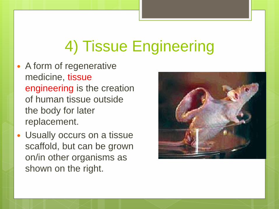
4) Tissue Engineering A form of regenerative
medicine, tissue
engineering is the creation
of human tissue outside
the body for later
replacement.
Usually occurs on a tissue
scaffold, but can be grown
on/in other organisms as
shown on the right.

4) Tissue Engineering Tissue engineers have
created artificial skin, cartilage and bone marrow.
Current projects being undertaken include creating an artificial liver, pancreas and bladder.
Again, we are far from replacing a whole organ, but just looking for “refurbishing” our slightly used ones at the moment.

The examination of a patient’s DNA molecule
to determine his/her DNA sequence for
mutated genes
The genome of an individual is scaned for this
purpose by a scientist
Genetic testing

Forensic/identity testing
Determining sex
Conformational diagnosis of symptomatic
individuals
Newborn screening
Prenatal diagnostic screening
Genetic testing
Better drugs can be obtained by the knowledge of genetics
Genetic testing can be used to detect the mutations regarding genetic disorders like cystic fibrosis, sickle cell anaemia, hutington diseases, etc.
Tests are also being developed to detect various cancers
Genetic testing

Mutation
Detection
Dr. Sajjad Ahmad

Mutations DetectionDetection of mutations has central role in various areas of genetic diagnosis
Preimplantation genetic diagnosis (PGD),
Prenatal diagnosis (PND)
Presymptomatic testing
Confirmational diagnosis
Forensic/identity testing.
Two groups of tests, molecular and cytogenetic, are used in genetic syndromes.

Single Base Pair Mutations
Direct sequencing, DNA hybridization and/or
restriction enzyme digestion methods are used for
detection of single pair mutations. However, there
are two approaches for genetic diagnosis;
Indirect approach depends on the results from a
genetic linkage analysis using DNA markers such as
STR(short tandem repeat) or VNTR (variable number
tandem repeat) markers flanking or within the gene
Direct approach for diagnosis essentially depends on
the detection of the genetic variations responsible for
the disease

Cytogenetics and
Molecular Diagnostics
Karyotyping
Fluorescence in situ hybridization (FISH)
Comparative genomic hybridization (CGH)
Molecular Diagnostics
(Known & Unknown Mutations)
Next Generation Sequencing (NGS)

Karyotyping Karyotyping is the process of pairing and ordering all
the chromosomes of an organism, thus providing a genome-wide image of an individuals chromosomes
Karyotypes are prepared from mitotic cells which are frozen in metaphase.
Characteristic structural features for each chromosome are revealed.
Can reveal changes in chromosome numbers linked to conditions such as Down’s syndrome.
Careful analysis can show more subtle changes as chromosomal deletions, duplications, translocations or inversions.
There is an increasing use of karyotyping for diagnosis of specific birth defects and genetic disorders.

Karyotyping Applications
Chromosome studies are advised in the following situations:
Suspected chromosome abnormality
Sexual disorders
Multiple congenital anomalies/ developmental retardation
undiagnosed learning disabilities
Infertility or multiple miscarriage
Stillbirth and malignancies

Preparation of visual karyotype Traditionally, the microscopic study
of chromosomes is performed oncompacted chromosomes at amagnification of 1000 at metaphase.
Cells are arrested at metaphasestage with a microtubulepolymerization inhibitor such ascolchicine
These cells are spread on a glassslide and stained with Giemsa stain(G banding).
Chromosomes are studied bymaking a photograph or digitalimaging and subsequent assemblingof chromosomes

Process of Karyotyping

Human KaryotypeHuman chromosomes are categorized
based on position of centromere;
Metacentric; the centromere at center (chromosomes
1, 3, 16, 19 and 20),
Acrocentric; the centromere near one end
(chromosomes 13, 14, 15, 21, 22 and Y are)
other chromosomes are submetacentric
The convenient methods of chromosome banding are
G-(Giemsa), R-(reverse),C-(centromere) and Q-
(quinacrine) banding
Fluorescence in situ
hybridization (FISH)

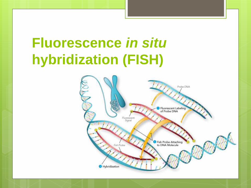
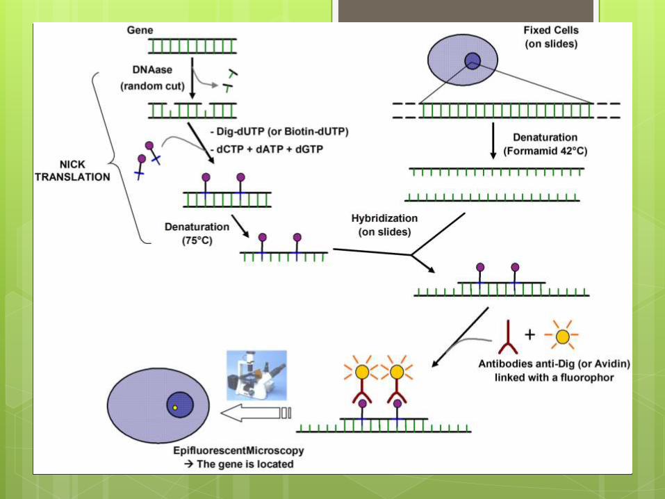
Fluorescence in situ
hybridization (FISH):
FISH is applied to provide specific localization of genes on
chromosomes.
Rapid diagnosis of trisomies and microdeletions is
acquired using specific probes.
Usually a denatured probe is added to a metaphase
chromosome spread and incubated overnight to allow
sequence-specific hybridization.
After washing off the unbound probe, the bound probe is
visualized by its fluorescence under UV light; thus, the site
of the gene of interest is observed as in situ
Comparative genomic
hybridization (CGH)
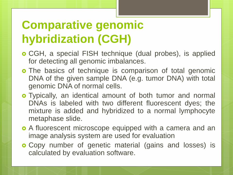
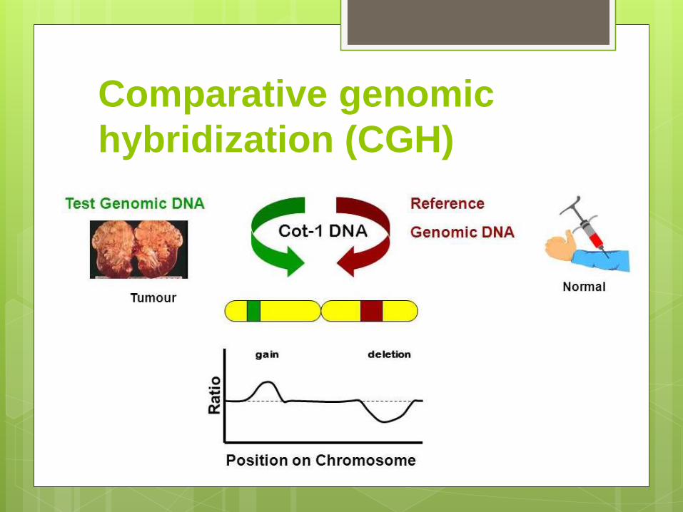
Comparative genomic
hybridization (CGH) CGH, a special FISH technique (dual probes), is applied
for detecting all genomic imbalances.
The basics of technique is comparison of total genomicDNA of the given sample DNA (e.g. tumor DNA) with totalgenomic DNA of normal cells.
Typically, an identical amount of both tumor and normalDNAs is labeled with two different fluorescent dyes; themixture is added and hybridized to a normal lymphocytemetaphase slide.
A fluorescent microscope equipped with a camera and animage analysis system are used for evaluation
Copy number of genetic material (gains and losses) iscalculated by evaluation software.

CGH is used to determine copy
number alterations of genome in
cancer and those cells whose
karyotype is hard or impossible
to prepare or analyze.
In array-CGH, metaphase slide is
replaced by specific DNA
sequences, spotted in arrays on
glass slides, so its resolution is
increased.
Comparative genomic
hybridization (CGH)

Molecular Diagnostics
Molecular Diagnostics
Molecular methods for identification of the disease-
causing mutations could be classified as methods for
known and methods for unknown mutations.
Several criteria, have to be met for choosing a suitable
method; for example
type of nucleic acid (DNA or RNA)
kind of specimen (blood, tissues, etc.)
the number of mutations
reliability of the method
Detection of Known Mutations
Many different approaches have been used for
identifying known mutations
Polymerase chain reaction (PCR) and its versions
DNA microarray
DNA Sequencing
Multiplex ligation-dependent probe amplification
(MLPA)

Detection of Unknown Mutations
Single Strand Conformational
Polymorphism (SSCP)
Denaturing Gradient Gel Electrophoresis
(DGGE)
Restriction fragment length polymorphism
(RFLP)

1. Polymerase chain reaction
In 1980s, Dr Mullis introduced a method for
amplifying DNA fragment to a large number of
fragments by polymerase chain reaction (PCR)
Essential components of PCR are template DNA,
primers , thermostable DNA polymerase enzyme
(e.g. Taq), divalent cations (usually Mg2+),
deoxynucleoside triphosphates (dNTPs) and
buffer solution
PCR, consisting of 25-40 repeated cycles, has
three discrete steps of temperature changes

Steps of PCR Initial denaturation step includes heating the reaction to a temperature
of 92–96°C for 1–9 minutes.
1) Denaturation step includes heating the reaction to 92–98°C for 20–30 seconds. The hydrogen bonds between complementary bases aredisrupted and DNA molecules are denatured, yielding single-strandedDNA molecules (DNA melting).
2) Annealing step is performed by decreasing temperature to 50–65°C for 25–40 seconds; so the primers are annealed to their targetson single stranded DNAs by hydrogen bonds and a polymerase canbind to the primer-template hybrid and begin DNA polymerization innext step.
3) Extension step includes polymerization of the bases to the primers;a thermostable such as Taq polymerase extends a new strandcomplementary to the DNA template strand by adding matched dNTPsin 5' to 3' direction at a temperature of 72°C.
A series of 25-40 repeated cycles of denaturation, annealing ofprimers and extension is performed to amplify the template fragment.
Subsequently, a final elongation is sometimes done at 70–74°C for 5–15 minutes after the last PCR cycle to ensure full extension of anyremaining single-stranded DNA

Types and Applications of PCR
1) Reverse transcriptase PCR (RT-PCR)
2) Multiplex PCR
3) Nested PCR
4) Amplification refractory mutation system
(ARMS) PCR:
5) Real time PCR

1. Reverse transcriptase PCR
(RT-PCR)
In this version, a strand of RNA molecule is
transcribed reversely into its complementary
DNA (cDNA) using the reverse transcriptase
enzyme.
This cDNA is then amplified by PCR.
RT-PCR is applied to study the mutations at
RNA level.

2) Multiplex PCR:
In this technique, multiple selected target regions in
a sample are amplified simultaneously using
different pairs of primers.
3) Nested PCR:
It includes two successive PCRs;
the product of the first PCR reaction is used as a
template for the second PCR.
This type of PCR is employed to amplify templates
in low copy numbers in specimens.
It has the benefits of increased sensitivity and
specificity.

4) Amplification refractory mutation system (ARMS) PCR: Allele-specific amplification (AS-PCR) or ARMS-PCR is a generaltechnique for the detection of any point mutation or smalldeletion
The genotype (normal, heterozygous and homozygous states) of asample could be determined using two complementary reactions:
one containing a specific primer for the amplification of normal DNAsequence at a given
locus and the other one containing a mutants pecific primer foramplification of mutant DNA.
ARMS-PCR has been used to check the most common mutationin GJB2 gene, 35delG mutation
among deaf children.
5) Real time PCR:In this technique, the amplified DNA is detected as the PCRprogresses.
It is commonly used in gene expression studies and quantificationof initial copy number of the target
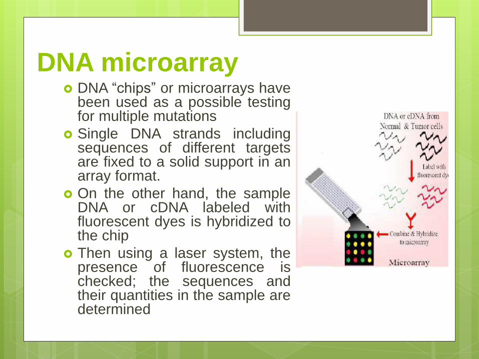
DNA microarray DNA “chips” or microarrays have
been used as a possible testingfor multiple mutations
Single DNA strands includingsequences of different targetsare fixed to a solid support in anarray format.
On the other hand, the sampleDNA or cDNA labeled withfluorescent dyes is hybridized tothe chip
Then using a laser system, thepresence of fluorescence ischecked; the sequences andtheir quantities in the sample aredetermined

DNA Sequencing The main aim of DNA sequencing is to
determine the sequence of small regions ofinterest (~1 kilobase) using a PCR product asa template.
Dideoxynucleotide sequencing or Sangersequencing represents the most widely usedtechnique for sequencing DNA
In this method, double stranded DNA isdenatured into single stranded DNA withNaOH
A Sanger reaction consists of a single strandDNA, primer, a mixture of a particular ddNTPwith normal dNTPs (e.g. ddATP with dATP,dCTP, dGTP, and dTTP).
A fluorescent dye molecule is covalentlyattached to the dideoxynucleotide. ddNTPscannot form a phosphodiester bond with thenext deoxynucleotide so that they terminateDNA chain elongation.
This step is done in four separate reactionsusing a different ddNTP for each reaction
DNA sequencing could be used to check allsmall known and unknown DNA variations.

Multiplex ligation-dependent
probe amplification (MLPA)
MLPA is commonly applied to screen deletions andduplications of up to 50 different genomic DNA orRNA sequences.
Altogether gene deletions and duplications account up to10%, and in many disorders up to 30% of disease-causingmutations
The probe set is hybridized to genomic DNA in solution
Each probe consists of two halves; one half is composedof a target specific sequence and a universal primersequence, and other half has other more sequences, avariable length random fragment to provide the sizedifferences for electrophoretic resolution.

Multiplex ligation-dependent
probe amplification (MLPA)
A pair of probes is hybridized on the target region
adjacently so that they can then be joined by use of a
ligase; the contiguous probe can be amplified by
PCR
After PCR amplification, the copy number of target
sequence i.e. deletion or duplication of target
sequence can be determined and quantified using
the relative peak heights

Multiplex ligation-dependent
probe amplification (MLPA)

Detection of Unknown Mutations
Single Strand Conformational Polymorphism (SSCP)
Denaturing Gradient Gel Electrophoresis (DGGE)
Heteroduplex analysis
Restriction fragment length polymorphism (RFLP)
Single Strand Conformational
Polymorphism (SSCP)
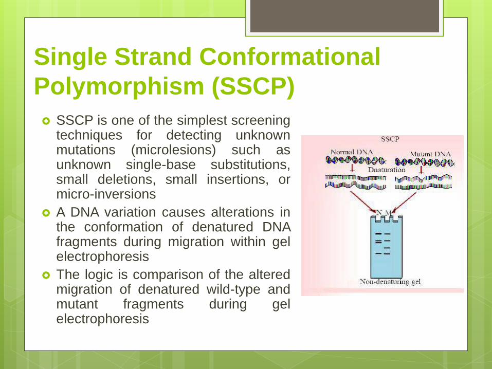
SSCP is one of the simplest screeningtechniques for detecting unknownmutations (microlesions) such asunknown single-base substitutions,small deletions, small insertions, ormicro-inversions
A DNA variation causes alterations inthe conformation of denatured DNAfragments during migration within gelelectrophoresis
The logic is comparison of the alteredmigration of denatured wild-type andmutant fragments during gelelectrophoresis

Single Strand Conformational
Polymorphism (SSCP) DNA fragments are denatured, and renatured under special
conditions preventing the formation of double-strandedDNA and allowing conformational structures to form insingle-stranded fragment
The conformation is unique and resulted from the primarynucleotide sequence
Mobility of these fragments is differed through non-denaturing polyacrylamide gels; detection of variations isbased on these conformational structures.
PCR is used to amplify the fragments, called PCR-SSCP,because the optimal fragment size can be 150 to 200 bp.
About 80–90% of potential point mutations aredetected by SSCP

Denaturing Gradient Gel
Electrophoresis (DGGE): DGGE has been used for screening of
unknown point mutations. It is based ondifferences in the melting behavior ofsmall DNA fragments (200-700 bp);even a single base substitution cancause such a difference.
In this technique, DNA is first extractedand subjected to denaturing gradient gelelectrophoresis.
As the denaturing condition increases,the fragment completely melts to singlestrands.
The rate of mobility in acrylamide gelsdepends on the physical shape of thefragment

Denaturing Gradient Gel
Electrophoresis (DGGE):
Detection of mutated fragments would be possible by
comparing the melting behavior of DNA fragments on
denaturing gradient gels.
Approximately less than 100% of point mutations can
be detected using DGGE.
Maximum of a nearly 1000 bp fragment can be
investigated by this technique

Heteroduplex analysis A mixture of wild-type and mutant DNA molecules is
denatured and renatured to produce heteroduplices
Homoduplices and heteroduplices show different
electrophoretic mobilities through nondenaturing
polyacrylamide gels
In this technique, fragment size ranges between 200
and 600 bp, Nearly 80% of point mutations have
been estimated to be detected by heteroduplex
analysis

Restriction fragment length
polymorphism (RFLP)
Point mutations can change
restriction sites in DNA causing
alteration in cleavage by
restriction endonucleases which
produce fragments with various
sizes
RFLP is used to detect
mutations occurring in restriction
sites

Next Generation Sequencing

Next Generation Sequencing
High speed and throughput, both qualitative and quantitativesequence data are allowed by means of NGS technologies sothat genome sequencing projects can be completed in a few days
NGS systems provide several sequencing approaches includingwhole-genome sequencing (WGS), whole exome sequencing(WES), transcriptome sequencing, methylome, etc.
The coding sequences compromises about 1% (30Mb) of thegenome.
More than 95% of the exons are covered by WES; on the otherhand, 85% of disease-causing mutations in Mendelian disordersare located in coding regions. Sequencing of the complete codingregions (exome), therefore, could potentially uncover themutations causing rare, mostly monogenic, genetic disorders aswell as predisposing variants in common diseases and cancer.


