Headaches in ED - BHS Education...
46
Headaches in ED Ballarat Health Services Emergency Medicine Training Hub Last modified October 2012
Transcript of Headaches in ED - BHS Education...

Headaches in EDBallarat Health Services
Emergency Medicine Training Hub
Last modified October 2012
Learning objectivesDo not miss subarachnoid haemorrhage
Learn the red flags We will examine contrasting clinical cases of
headache that may be due to potentially lethal causes such as subarachnoid haemorrhage or more benign causes such as migraine. lmportant features in the history include establishing the timing of onset of symptoms and using eye witnesses.

Red flags in the diagnosis of headache(1) Sudden onset headache Subarachnoid hemorrhage
Worsening pattern headache Mass lesion, subdural hematoma Medication overuse
Headache with systemic illness Meningitis, encephalitis Systemic infection Collagen vascular disease, arteritis

Red flags CNS vascular:
Bleeds (SAH, EDH, SDH, IPH) Carotid dissection, temporal arteritis Cavernous sinus thrombosis
CNS infection: meningo/encephalitis, abscess
Tumour Others:
Eyes (glaucoma, iritis) Toxic & metabolic (CO poisoning) Preeclampsia & malignant HTN
Sudden onset headache = subarachnoid haemorrhage until proven otherwise
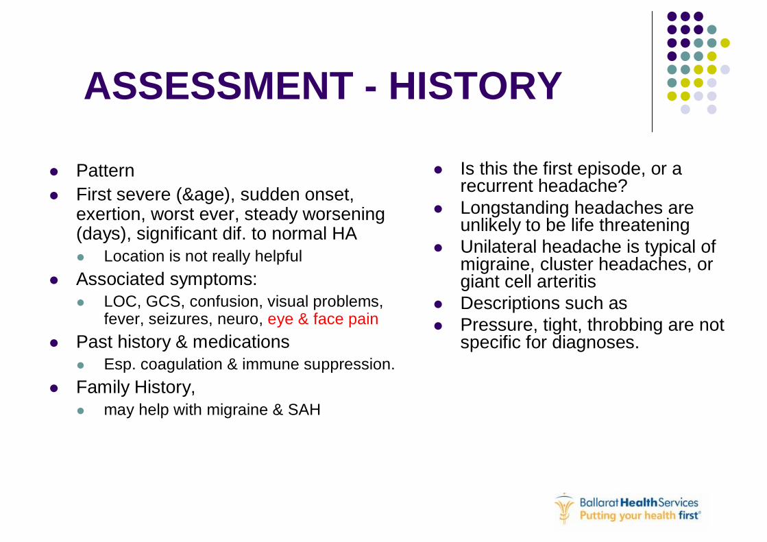
ASSESSMENT - HISTORY
Pattern First severe (&age), sudden onset,
exertion, worst ever, steady worsening (days), significant dif. to normal HA Location is not really helpful
Associated symptoms: LOC, GCS, confusion, visual problems,
fever, seizures, neuro, eye & face pain Past history & medications
Esp. coagulation & immune suppression. Family History,
may help with migraine & SAH
Is this the first episode, or a recurrent headache?
Longstanding headaches are unlikely to be life threatening
Unilateral headache is typical of migraine, cluster headaches, or giant cell arteritis
Descriptions such as Pressure, tight, throbbing are not
specific for diagnoses.

Associated symptoms Focal neurological symptom
(migraines, space occupying lesion)
Nausea & vomiting ( migraine, infections, raised intracranial pressure
Alcoholism, anticoagulants (subdural haematoma)
Recent trauma (concussion, subdural haematoma)
Worse on waking, straining, bending over, coughing (raised ICP)
Fever, photophobia, neck stiffness (meningitis)
Visual disturbance (migraine, glaucoma, giant cell arteritis)
Pain tenderness on side of head, jaw claudication – pain on chewing – visual disturbance (giant cell arteritis)
Coma, seizures, focal neuro symptoms suggest significant problem

ASSESSMENT - EXAM Vitals – BP, PR, fever –
look for focus of infection General – face and eyes, ears, TMJ Meningism
Movement of neck or straight leg raise may cause pain
Neuro – mental state, CN’s, motor, sensory, cerebellar
What’s an adequate screening exam? Plantar response, symmetry, pupils
represent value for time signs.
In patients over 50 years Check temporal artery
tenderness and intraoccular pressure… and look for signs of glaucoma Red eye, cloudy cornea, dilated
pupil, less responsive

Case 1 - Headache Transferred from regional hospital 1-2 hours away Sudden onset of headache during intercourse,
associated nausea, vomiting, neck stiffness Headache occipital No recent illness/fevers No history of similar GCS 15 throughout Gets occasional migraines – not this bad This headache different Pre hospital treatment tramadol and ondansetron,
morphine and stemetil in AV

Case 1 - CT thought to be normal

Case 1 - CT reported normal (ED)
Lesson – radiologist to report suspected SAH CT images

Case 1 - Diagnosis on LP suggested Subarachnoid haemorrhage
Lumbar Puncture Multiple attempts no
success Then successful tap
1st pass Blood stained CSF Opening pressure
28cm H20 Transferred

Investigation in suspected SAH
Lumbar puncture in Lumbar puncture in suspected SAH with suspected SAH with normal CT or MRI brainnormal CT or MRI brain
Differentiate traumatic Differentiate traumatic tap from SAH by tap from SAH by xanthochromiaxanthochromia (colored (colored supernatant)supernatant)
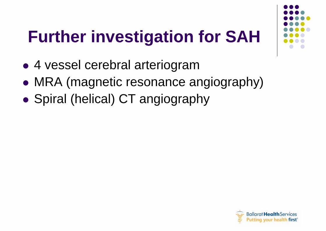
Further investigation for SAH 4 vessel cerebral arteriogram MRA (magnetic resonance angiography) Spiral (helical) CT angiography

SAH management
Early discussion with neurosurgical team Early consideration of transport/retrieval
issues Classification of severity and pre existing
health of patient will influence outcome

Haemorrhagic stroke Subarachnoid haemorrhage can be
contrasted with other haemorrhagic strokes Haemorrhage accounts for approx 20% of all
strokes much more severe than other strokes fatal > 50 percent of the time overall

Haemorrhagic stroke - causes Primary HTN: often basal G, brainstem, cerebellum Some others: amyloidangiopathy
Secondary: Trauma, coagulopathy, tumour, AVM aneurysm

Indications for Transfer
The Prognosis
The roles of neurosurgery

Poor Outcome Predictors Elderly Large or increasing volume of hematoma Interventricular clot extension and/or
hydrocephalus Communication disorder Low GCS on admission Midline shift and herniation syndromes on
imaging Anticoagulation agents

If GCS < 9 and haematoma volume > 60 ml, mortality at one month 90%
GCS > 9 and haematoma volume < 30 ml, mortality at one month > 17%

The ICH Score: A Simple, Reliable Grading Scale for Intracerebral Hemorrhage
Hemphill, J. Claude III, MD; Bonovich, David C. MDStroke. 32(4):891-896, April 2001.
Points 0 1 2
GCS 13-15 5-12 3-4
ICH Volume (cm3) <30 >30
IVH No Yes
Infratentorial No Yes
Age <80 >80

Role of surgery Reversal of mass effect & herniation
syndrome Cerebellar haematoma, large haematoma
Reversal of hydrocephalus Treatment of surgical aetiology Aneurysm, AVM, tumours
Other benefits (????) Reduce the toxic effect of the presence of
intracerebral blood Promote recovery of the ischemic penumbra

Assessment features History: usually associated
with more Nausea &Vomiting, headache, altered conscious state, seizures, sudden onset
Medications important esp. warfarin or other anticoagulants
Underlying functional status!!
ROSIER toolRule Out Stroke In the Emergency Room
Rosier scale to differentiate stroke and "stroke mimics“
Has there been loss of consciousness or syncope Y (-1) N (0) Has there been seizure activity Y (-1) N (0) Is there a new onset (or waking from sleep)?
i Asymmetric facial weakness Y (+1) N (0) ii Asymmetric arm weakness Y (+1) N (0) iii Asymmetric leg weakness Y (+1) N (0) iv Speech disturbance Y (+1) N (0) v Visual field defect Y (+1) N (0)
Stroke is likely if total score > 0Scores of < / = 0 have low probability of stroke but not
excludedAdapted from http://handbook.muh.ie/Neurological/Stroke/rosier.html

Management
ABC issues: airway protect, avoid dextrose in fluids
REVERSE WARFARIN Correct hypoglycaemia, keep <10 IDC, nursing Consider analgesia HTN reduce only if >180/>?90-105 20% reduction, above measures first
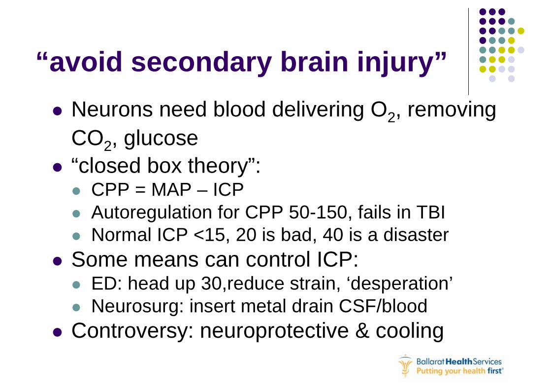
“avoid secondary brain injury” Neurons need blood delivering O2, removing
CO2, glucose “closed box theory”: CPP = MAP – ICP Autoregulation for CPP 50-150, fails in TBI Normal ICP <15, 20 is bad, 40 is a disaster
Some means can control ICP: ED: head up 30,reduce strain, ‘desperation’ Neurosurg: insert metal drain CSF/blood
Controversy: neuroprotective & cooling

Neurosurgical opinion - who for?
Definitely useful in SAH Posterior fossa/cerebellar: Clearly improves outcomes
Brainstem ICH has poor outcome Supratentorial ICH: Possible to evacuate blood, shunt and ICP
monitor Doesn’t improve outcome STICH study Lancet 2005

Aneurysmal haemorrhagic stroke
Aneurysms are common Prevalence in lifetime is 4-6% Most will not rupture Risk of 1st SAH averages 0.1% per year Surgery has significant M&M Not for surg if <10mm unless post SAH 10-25 risk is still < 1% per year >25mm risk approx 6% or more p.a. Position also important
Case 2 - Headache Presents with
headache for many days & Vomiting
Pain on back of head
O/E occipital shingles.

Case 2 - Headache
Examination CNS normal Old neuro signs of weakness Paracetamol, ketorolac 30mg IM,
chlorpromazine 12.5mg IV Plan… exclude…

Case 2 - Headache Further clinical
Headache spreads down left side of face down to neck
Throbbing, 8/10, constant
Nausea, x1 vomit After the LP &CT… Neck stiffness and
photophobia…

Case 3 2-3 days of frontal
headache Also dizzy, nausea,
vomiting Pulled over by police, not
driving well Past history of Aortic
Valve Replacement Medications – Warfarin.
INR 3.1 recent
Temp 37 GCS 14 or 15 Normal neurological
exam
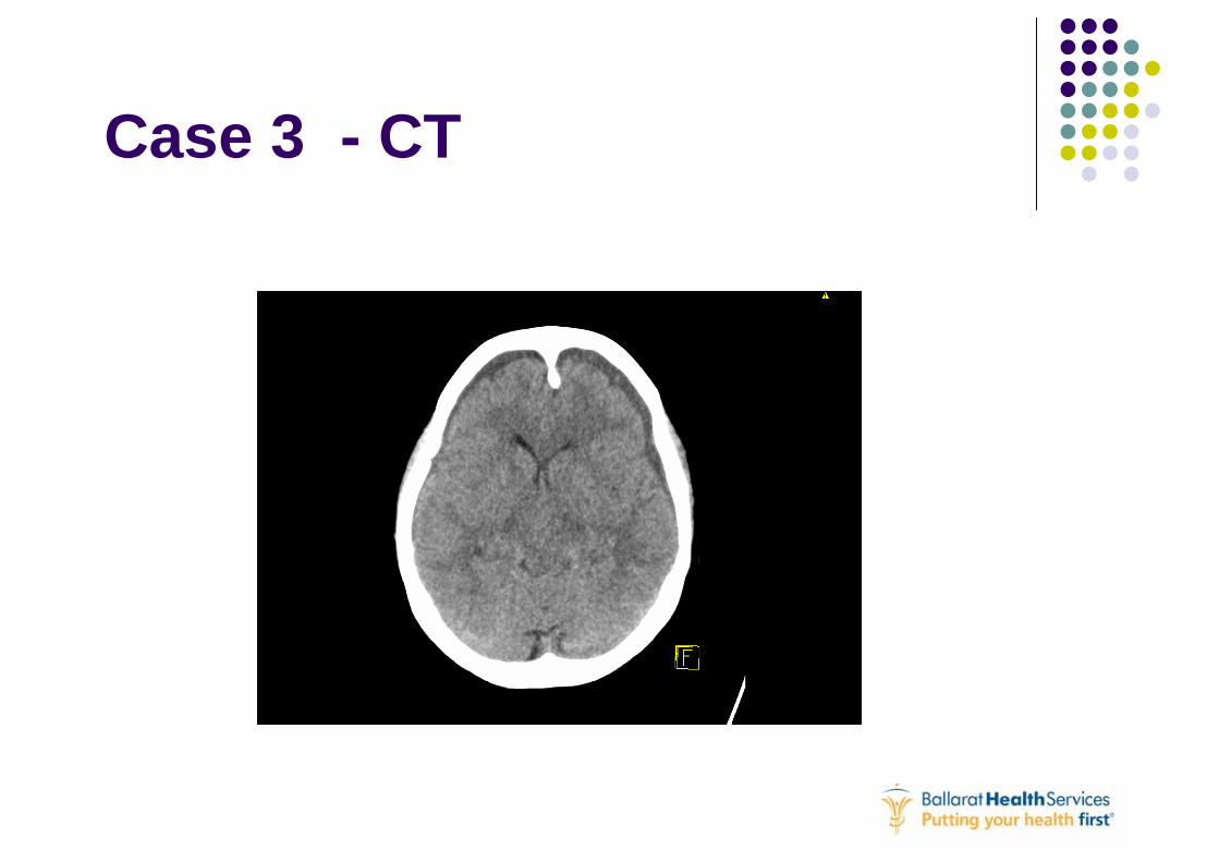
Case 3 - CT

Case 3 – CT report

Case 4 - 3 days ago was
pedestrian, struck by car and hit head
Now confused Diabetic, hypertension MVA was low speed, was
getting better, now more headaches and confused
Unusual behaviour
Note previously seen in ED and discussed with senior ED doc.

Case 4 - CT

Case 4 – CT Report

Primary headache syndromes Purpose is to confidently diagnose: Investigation unnecessary Focus shifts to treatment
There is some good evidence regarding Rx

Migraine Classical history: Aura = spreading depolarisation Strong family history
Treatment (NICS 2006) Narcotics not effective (pethidine<56%) Ketorolac possibly worse Chlorpromazine and tryptans best

Migraine treatment First line - if nothing taken: aspirin 900mg &maxalon 10mg Paracetamol 30/kg / ibuprofen 600-800mg
First line – failed pt meds / vomiting: Metoclopramide 10mg / CPZ 12.5mg im
Moderate-severe: CPZ 25mg in N/S 1litre 30-60/60 OR prochlorperazine 12.5iv OR sumatriptan 6mg s/c

Sumatriptan Selective 5HT Ag. (1D) blood vessels Intranasal 20mg/oral 50mg/ sc 6mg Adverse = HTN, drowsy, dizzy, flushed, rash Contraindications are IHD, HTN Not if concurrent ergotamines 18% of people are non-responders

Cluster Headache
Recurrent brief sudden and severe unilateral periorbital pain
Brief changes Rx options Idiopathic (experimental imaging
hypothalamic dysfunction) Pain from precarotid/cavernous sinus 0.4% males (male:female = 5) Duration 5/60 to 3H

Cluster Headache - clinical
Very distressed patients prominent autonomic phenomena including ipsilateral congestion and rhinorrhea,
lacrimation, conjunctivitis, facial diaphoresis, palpebral edema, and complete or partial Horner syndrome
Tachycardia is a frequent finding.

Cluster Headache - typesEpisodic versus Chronic: Episodic: in clusters, from a week to a year;
pain-free intervals > 2 weeks. Typically last 2 weeks to 3 months.
Chronic CH: more than 1 year without remission or remissions <2 weeks
Chronic CH difficult to treat & resists standard prophylactic agents.

Cluster Headache - treatment
Abortive: O2: High-flow, concentrated 02 extremely effective in
aborting attacks mechanism of action poorly understood
Ergot alkaloids Ergotamine (o/pr) or dihydroergot. (im/iv)
Preventative options inc: anticonvulsants, mood stabilisers, CCB

Other causes of headacheBenign Intracranial HTN
(also ‘pseudotumourcerebri’) Idiopathic raised ICP Mostly obese young women (20-40) May see CN VI lesions, HA with N&V CSF and CT may be normal ICP is raised Rx: LOW, diuretics, drain CSF

HIV and headache It is important to remember that some patient
groups, e.g. those with immunosuppresion, may have different & unique presentations: Toxoplasmosis CNS abscesses more common CNS lymphoma
Useful references freely available
Migraine review http://emedicine.medscape.com/article/1142556-overview
Tension headache review http://emedicine.medscape.com/article/792384-overview
Headache in children http://emedicine.medscape.com/article/802158-overview
Tintanelli www.sjem.org/files/39586667.ppt