

Head injuries - uomustansiriyah.edu.iq10_14_12_AM.pdf · •The Secondary Survey •Recheck A, B, C...
76
Head injuries Dr.Mohammed altamimi
-
Upload
nguyenkhanh -
Category
Documents
-
view
215 -
download
0
Transcript of Head injuries - uomustansiriyah.edu.iq10_14_12_AM.pdf · •The Secondary Survey •Recheck A, B, C...

Head injuries
Dr.Mohammed altamimi

• Pathophysiology
• Local effects on brain tissue
• Systemic effects of TBI
• Management protocols
• Patient triage and classification
• mechanism
• Types of head injuries

• The Primary Survey and Resuscitation
• proceed in parallel
• A –– Clear the Airway. Use chin-lift or jaw-thrust. Immobilize the cervical spine with collars, bags and tape until cleared.
• B –– Check Ventilation. Administer oxygen at 15 litres per minute with tight-fitting mask with reservoir or use bag, valve and mask.
• C –– Check for pulses, skin perfusion and consciousness. Identify obvious sources of blood loss;
• D –– Assess the level of consciousness with A.V.P.U.:
• A (alert); V (responds to verbal communication);
• P (responds only to pain); U (unconscious);
• E –– Expose and examine the patient thoroughly.


• The Secondary Survey
• Recheck A, B, C and D. Proceed to next stage if the patient is stable and analgesia has been effective. The secondary survey is a head-to-toe examination of the patient’s body.
• A.M.P.L.E. - a simple mnemonic for key
• information
• A: allergies (e.g. penicillin or aspirin)
• M: medication (e.g. a beta-blocker or warfarin)
• P: previous medical history (e.g. previous surgery or anaesthetic mishap)
• L: last mealtime (i.e. drink versus major meal)
• E: events surrounding the incident (e.g. fell 5 metres with immediate loss of consciousness)


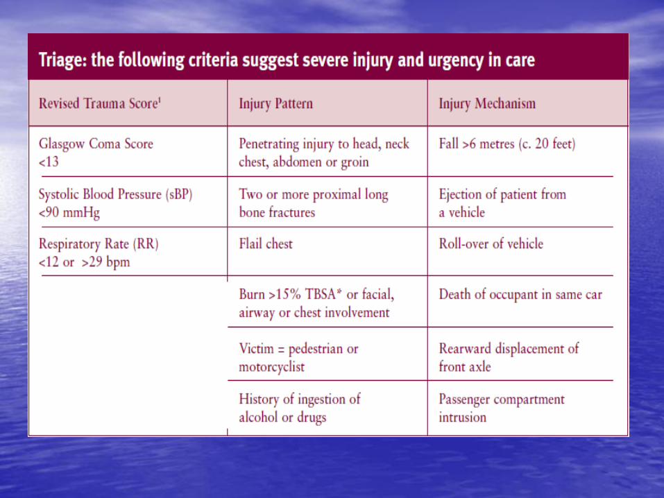
• INITIAL MANAGEMENT OF SEVERE TRAUMA
• Most widely accepted system is that recommended in the American College of Surgeons’ Advanced Trauma Life Support (ATLS) course. The key elements of this approach are the primary survey/resuscitation phase, the secondary survey and the rapid implementation of definitive treatment, which may involve early surgery. The objective is to detect life-threatening conditions as quickly as possible, to stabilise the patient and to start definitive treatment as early as possible, in a prioritised fashion.

Intensive management of traumatic brain
injury (roles and objectives)
• The primary focus of intensive management is directed towards patients suffering severe head injury (GCS of 8)following initial resuscitation and to prevent those suffering moderate injuries from deteriorating into unresponsive coma
• It is universally agreed that a population of neurons is irreversibly damaged at initial impact but neuronal demise continues unchecked until brain resuscitation is initiated
• It is also agreed that primary brain injury initiates a cascade of metabolic derangement affecting the cerebral vascular system, glial supporting cells, and neurons, which leaves brain vulnerable to secondary injury

Head injury Primary impact
Hypoxia .Brain stem dysf. .ventilation prob. . O2 delivery prob.
Traumatic brain (Secondary injury)
. Ion pumps dysfunction
.deranged autoregulation .oedema(vsogenic,c.toxic)
ischemia .Raised ICP .Vasoconstriction .SOL
acidosis acidosis

Herniation syndromes


Systemic effect of T.B.I
Hypothalamic dysfunction
Cholinergic dysfunction
Adrenal hyperactivity
Brain stem dysfunction
.apnoea,
.hypoventilation
.Bradycardia
.hypotention
.hypertention,
.decrease COP
.arrhythmias
.DI
.SIADH
.CSWS
.hypersecretion
of ACTH ,GH
prolactin
.MI
.neurogenic
pulmonary
edema
.bradyrrhythmia.
hypotension
increased secretion of catecholamines and enkephaline, cytokines leading to
hypermetabolism, hyperglycemia hypercatabolism and organ dysfunction (lung, liver,
heart, gut) through lactic acidosis

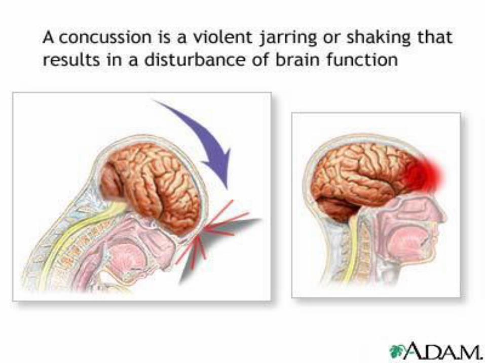
MECHANISMS OF INJURY
• inertial or contact mechanisms
• Inertial injuries are commonly called
"acceleration" or "deceleration" injuries
• Contact injuries are commonly called coup & contercoup injuries.


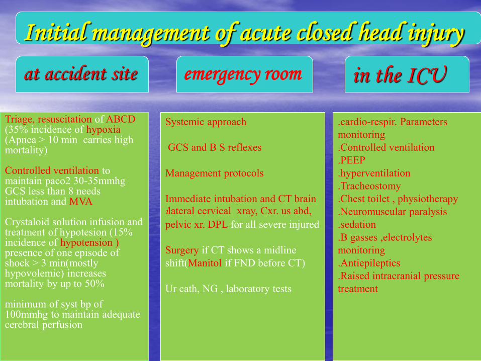
Initial management of acute closed head injury
at accident site emergency room in the ICU
Triage, resuscitation of ABCD (35% incidence of hypoxia (Apnea > 10 min carries high mortality) Controlled ventilation to maintain paco2 30-35mmhg GCS less than 8 needs intubation and MVA Crystaloid solution infusion and treatment of hypotesion (15% incidence of hypotension ) presence of one episode of shock > 3 min(mostly hypovolemic) increases mortality by up to 50% minimum of syst bp of 100mmhg to maintain adequate cerebral perfusion
Systemic approach
GCS and B S reflexes
Management protocols
Immediate intubation and CT brain ilateral cervical xray, Cxr. us abd,
pelvic xr. DPL for all severe injured
Surgery if CT shows a midline
shift(Manitol if FND before CT)
Ur cath, NG , laboratory tests
.cardio-respir. Parameters
monitoring
.Controlled ventilation
.PEEP
.hyperventilation
.Tracheostomy
.Chest toilet , physiotherapy
.Neuromuscular paralysis
.sedation
.B gasses ,electrolytes
monitoring
.Antiepileptics
.Raised intracranial pressure
treatment

1 2 3 4 5 6 GCS
none Extention
to pain Flexion to pain
Withdrawal
to pain Localize the pain
Obey command
Best motor response
none Incomprehensible sound
Inappropriate
word confused oriented
Verbal response
none To pain To verbal Spontaneo
us Eye opening
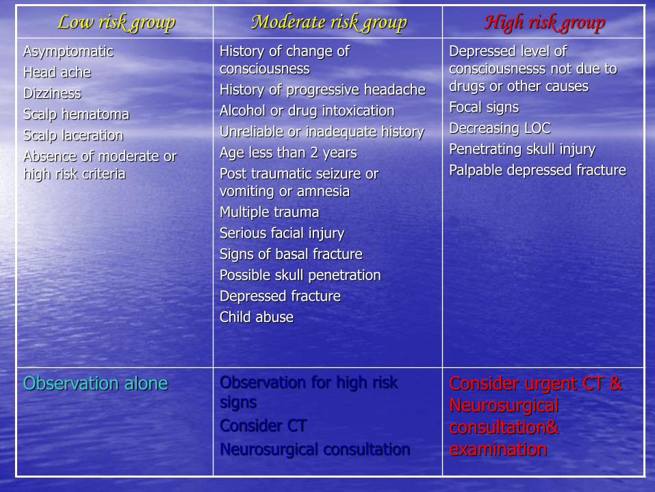
High risk group Moderate risk group Low risk group
Depressed level of consciousnesss not due to drugs or other causes
Focal signs
Decreasing LOC
Penetrating skull injury
Palpable depressed fracture
History of change of consciousness
History of progressive headache
Alcohol or drug intoxication
Unreliable or inadequate history
Age less than 2 years
Post traumatic seizure or vomiting or amnesia
Multiple trauma
Serious facial injury
Signs of basal fracture
Possible skull penetration
Depressed fracture
Child abuse
Asymptomatic
Head ache
Dizziness
Scalp hematoma
Scalp laceration
Absence of moderate or high risk criteria
Consider urgent CT & Neurosurgical consultation& examination
Observation for high risk signs
Consider CT
Neurosurgical consultation
Observation alone
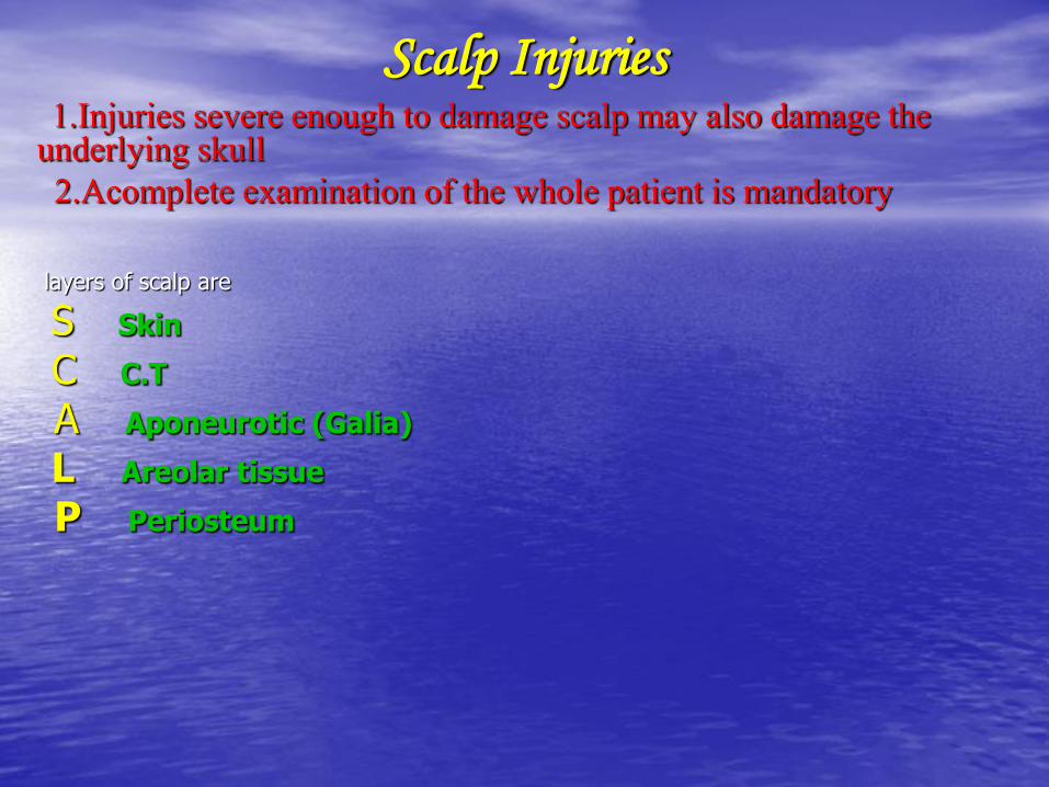
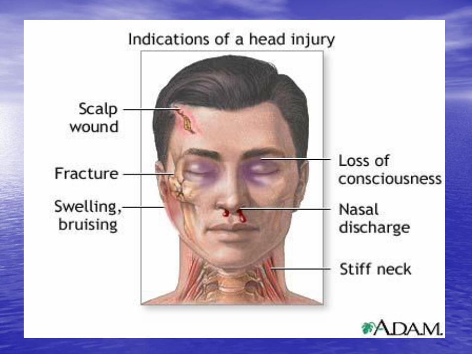
Scalp Injuries
1.Injuries severe enough to damage scalp may also damage the underlying skull
2.Acomplete examination of the whole patient is mandatory
layers of scalp are
S Skin
C C.T
A Aponeurotic (Galia)
L Areolar tissue
P Periosteum


Types
•Lacerations
•Avulsions
•Physical injuries
•Chemical injuries
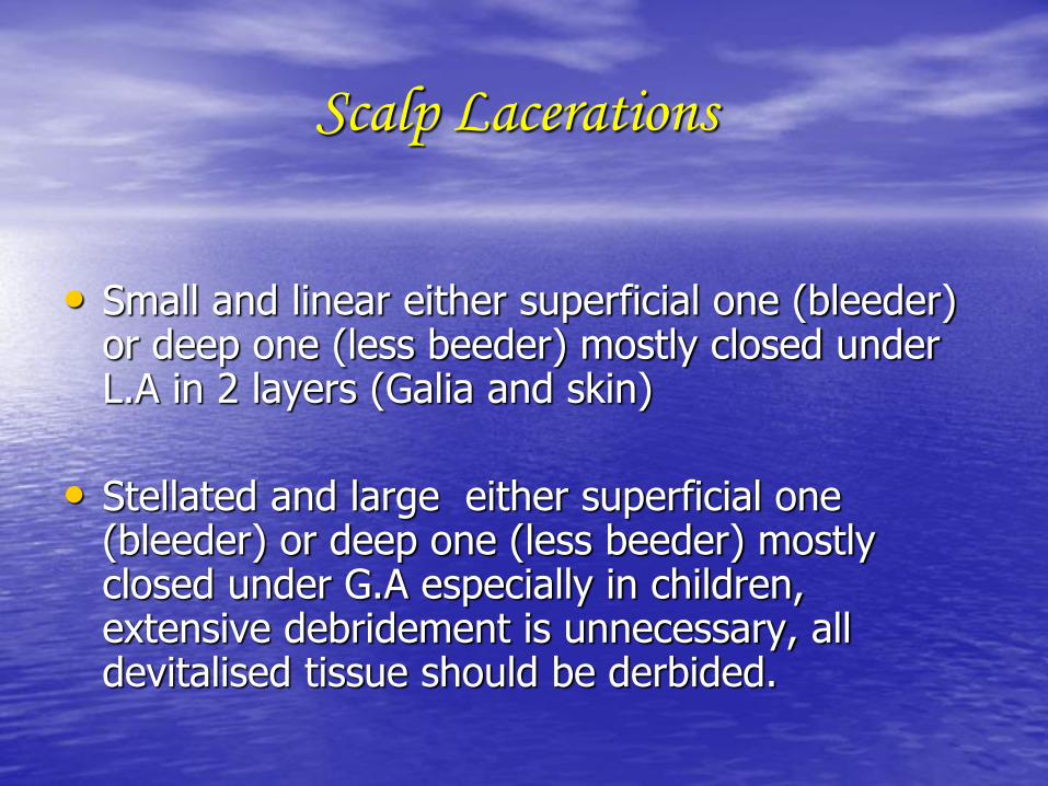
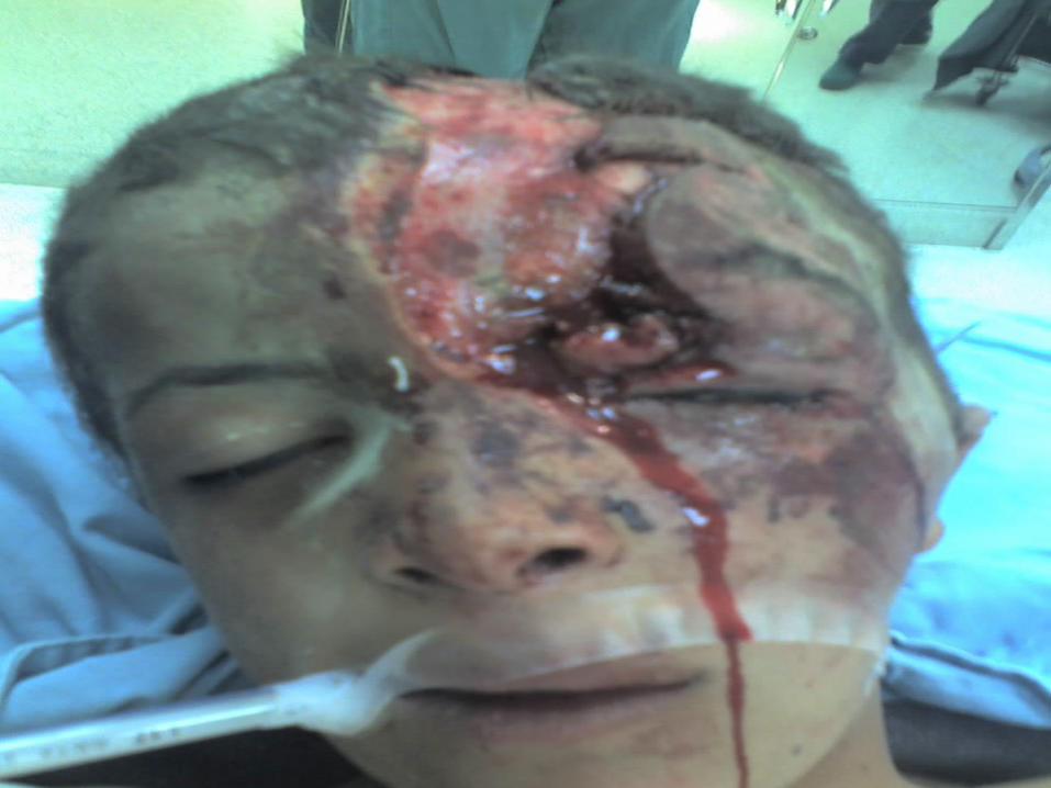
Scalp Lacerations
• Small and linear either superficial one (bleeder) or deep one (less beeder) mostly closed under L.A in 2 layers (Galia and skin)
• Stellated and large either superficial one (bleeder) or deep one (less beeder) mostly closed under G.A especially in children, extensive debridement is unnecessary, all devitalised tissue should be derbided.

Reconstruction ladder
1. primary suturing in simple lacerations ,sharp, without tissue loss and gross contamination
2. secondary intention in wounds with minimum tissue loss
3. debridement and a split thickness graft in wounds with extensive tissue loss and intact pericranium
4. local flaps in wounds with extensive tissue loss and stripped pericranium and a split thickness graft for donor area (if a defect is left)
5. Pedicular flaps can be used if local flaps are not sufficient especially for occipital areas and small defects
6. Free tissue transplant is used for anterior and large areas


Scalp swelling and hematomas
1.Caput succedaneum
2.Cephal hematoma
3.Subglial hematomas

Skull fractures classification
• 1.by pattern (according to amount of energy,ratio of impact force to surface area)
• Comminuted • Depressed • Linear • Diastatic • Basal fractures • Growing
• 2.by type • Opened • Closed
• 3.by anatomic location • Basal • convexity


Treatment roles • Comminuted (replacement as a cranioplasty if not
infected with treatment of other associated injuries)
• Linear fractures (no treatment but observation if closed, debridement if infected and compound)
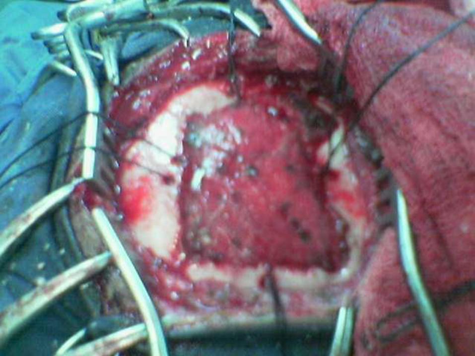
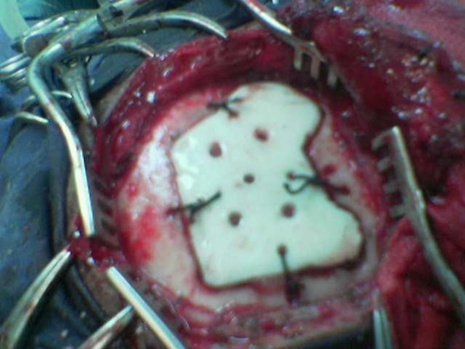
• Depressed(replacement as a cranioplasty if not infected, otherwise good debridement and craniectomy)
• Basilar skull fracture (observation for two days, avoid irrigation of the nose or ear, avoid probing, detailed auditory and vestibular examination is performed at6 weeks interval)
• Frontal sinus fracture (if in the posterior wall cranialization is necessary)

Types of traumatic intracranial leasions
• Focal(epidural hematoma-subdural hematoma-brain contussion-intracerebral hematomas-focal subarachnoid hematoma)
• Diffused(subarachnoid hemorhage-diffused axonal injury-concussion)


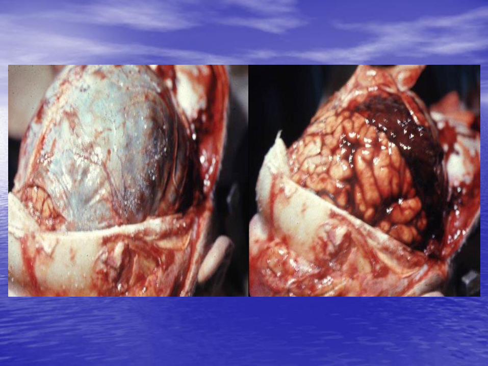
Traumatic intracranial hematomas
•Acute subdural • accelerate high speed impact
• tearing of bridging veins
• bleeding from cortical vessels, venous sinuses
• acute brain trauma may coexist
• altered level of consciousness and focal neurological deficit are common
• CT and rapid evaluation are necessary
• If no signs of rapid deterioration or progressive neurological deficit ,no mass effect so observation and control of intracranial p. is necessary, otherwise surgery is the role)

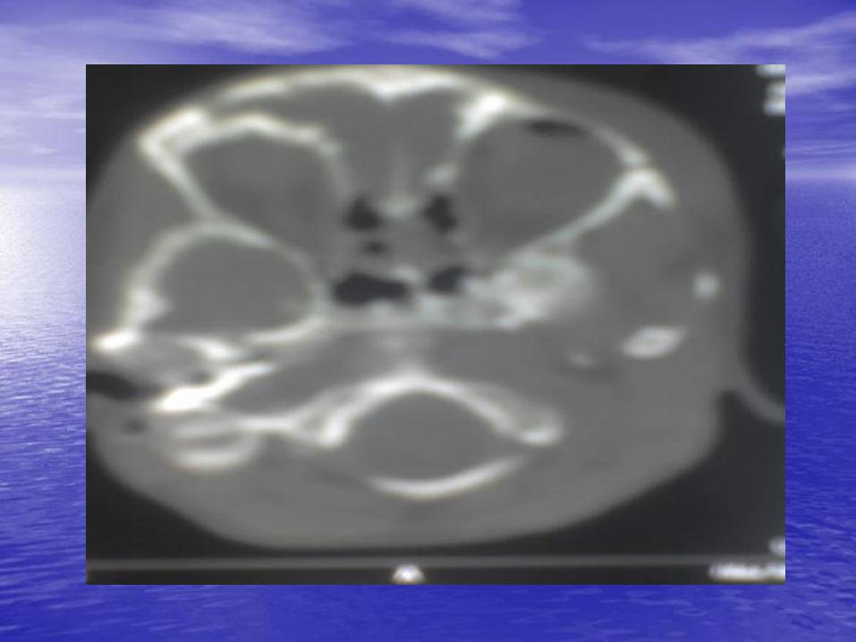
Acute Subdural Hematoma
Another example
of acute subdural
hematoma with a
midline shift
(noncontrast CT)

•Chronic subdural
• Mostly in those over 50 years old • ½ of patients have got no history of trauma • If there is any history of trauma , it is trivial • Alcoholism, epilepsy, coagulopathy are common • Dementia is common presentation • In minimal neuological deficit ,medical
management is the role • If not successful, deterioration of neurological
picture so surgery usually by burr hole evacuation.
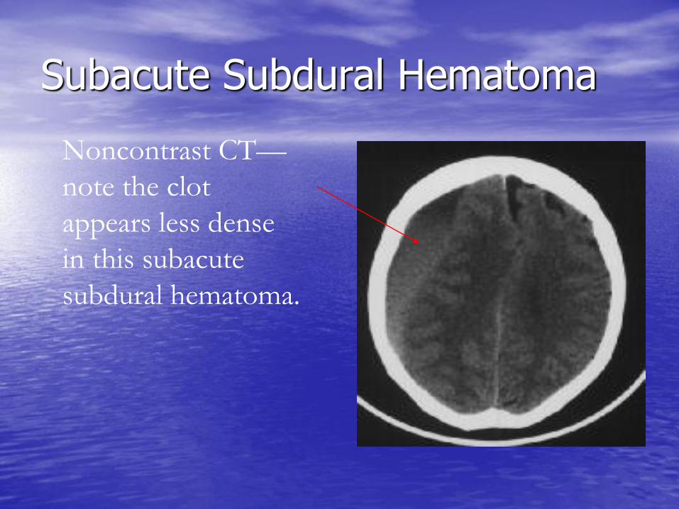
Subacute Subdural Hematoma
Noncontrast CT—
note the clot
appears less dense
in this subacute
subdural hematoma.


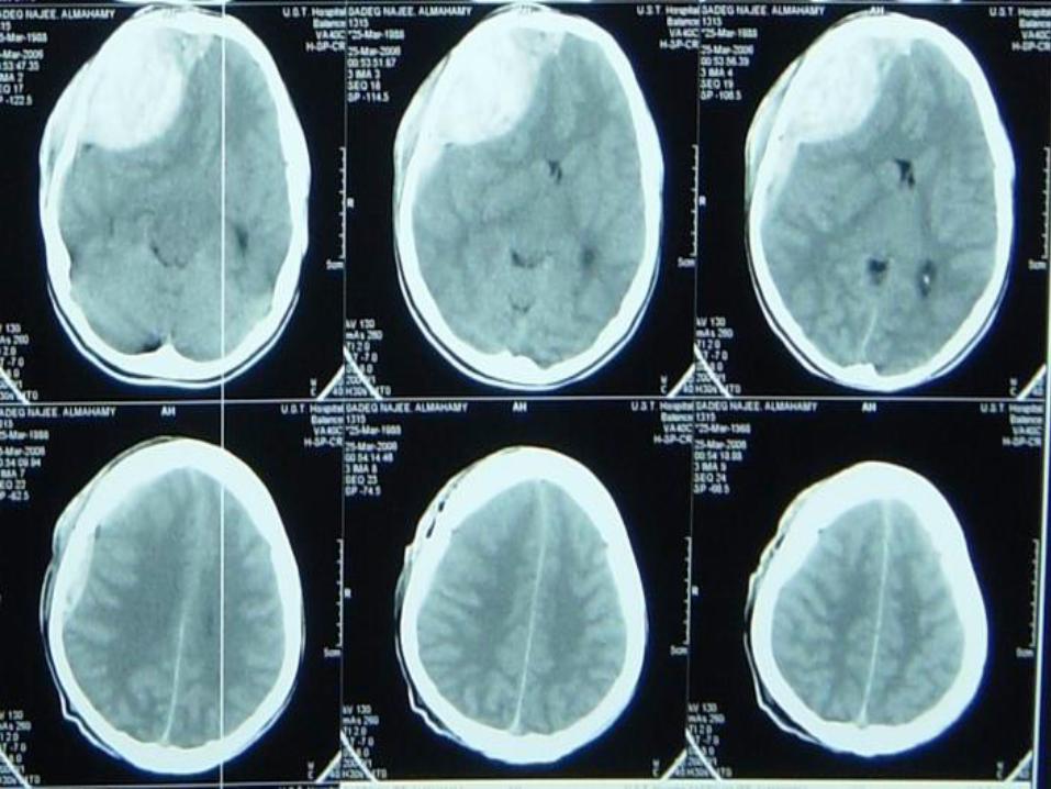
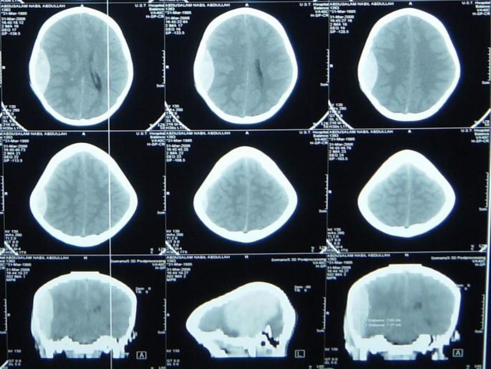
Extradural hematomas
• Mostly resulting from meningial vessel tear (arteries > Veins > sinuses)
• Fractures are common associated injury
• Severe associated brain injury is rare
• Level of consciousness is variable
• If with mass effect must be evacuated within ½ hour
• if small follow up is recommended
• Prognosis depends on level of consciousness at time of presentation


Cerebral contusions
• Small and deep one needs follow up
• Large and with mass effect needs lobectomy
• Large one may herniate as late as 9 days post trauma
• Level of consciousness depends on size of contusion and location


SAH—more examples
• Subarachnoid
hemorrhage in the right
sylvian fissure

SAH—more examples
Blood in the sulci
Edema causing a
midline shift
Posterior fossa hematomas
• Usually arise from venous sinuses hemorhage
• Deterioration in level of consciousness is rappid and signs of brain stem and long tract compression are common innitial finding
• Urgent evacuation is the role
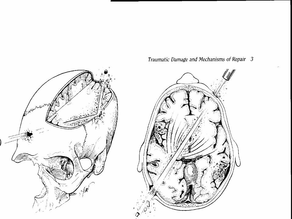
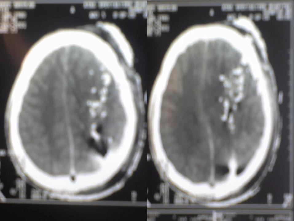
Penetrating head injuries
• Sonic waves and cavitations and decavitation and secondary insults are the injurious mechanisms
• Infection rates are high
• Injury far away from site of entrance and exit is common
• Control of homodynamic state is the initial management
• Surgical interventions are limited to debridement and removal of mass hematomas and for selected cases
• Prognosis is usually bad

Questions • What are the types of skull fractures
• What are the parameters of GCS
• Enumerate the types of traumatic scalp swellings
• Enumerate the signs and symptoms of raised intracranisl pressure
• What are the types of traumatic intracranial hemorrhages
• Right about types of mechanisms of head injury
• What is the pathophysiology of head injury

THANK YOU