Hawaii State Chiropractic Association - c.ymcdn.com State Chiropractic Association ... Interim or...
42
Hawaii State Chiropractic Association STANDARDS OF DOCUMENTATION, CHIROPRACTIC GUIDELINES FOR SPECIFIC CONDITIONS, AND UTILIZATION OF DIAGNOSTIC IMAGING January 25, 1997 © The enclosed material is a copyrighted and exclusive presentation of the Hawaii State Chiropractic Association (HSCA) and it is unlawful to copy, reproduce or distribute any of the following pages of this material, in part or in its entirety, without the expressed written permission of the HSCA.
Transcript of Hawaii State Chiropractic Association - c.ymcdn.com State Chiropractic Association ... Interim or...

Hawaii State Chiropractic Association
STANDARDS OF DOCUMENTATION,CHIROPRACTIC GUIDELINES FOR SPECIFIC CONDITIONS,
AND UTILIZATION OF DIAGNOSTIC IMAGING
January 25, 1997
© The enclosed material is a copyrighted and exclusive presentation of the Hawaii StateChiropractic Association (HSCA) and it is unlawful to copy, reproduce or distribute any ofthe following pages of this material, in part or in its entirety, without the expressed writtenpermission of the HSCA.

TABLE OF CONTENTS
Standards for Chiropractic Documentation
General Information ……………………………..…………………………………………. 1Format ……………………………………………………..……………………………….. 1Legibility …………………………………………………………..……………………….. 1General Considerations for Recordkeeping ……………………………….….……………. 2Documentation of Patient Consent ……………………..………………………………….. 2Initial New Patient:
History …………………………………………….…………………………………. 3Physical Examinations …………………….…………………………………………. 4Assessment …………………………………….…………………………………….. 5Plan ………………………………………………….……………………………….. 5
Interim or Progress Notes ………………………………….………………………………. 6Reevaluation/Interim Examination Frequency ……………….……………………………. 7Guidelines for Frequency of Examinations ………………….…………………………….. 8
Standards for Chiropractic Treatment / Guidelines for Specific Conditions
Disclaimer …………………………………………….……………………………………. 10The Vertebral Subluxation Complex:
Definition of ……………………………………….…………………………………. 11Components of ………………………………….……………………………………. 11Diagnostic Examples of ……………………….……………………………………... 12Importance of ………………………………………………………………………... 14Causes of …………………………………………………………………………….. 14Radiographic Classification of ………………………………………………………. 16
Levels of Chiropractic Care:Active ………………………………………………………………………………... 17Preventative/Maintenance …………………………………………………………… 17
Categories of Conditions Treated:Acute Conditions …………………………………….……………………………… 18Chronic Conditions………………………………………………………………….. 18Acute Exacerbation …………………………………………………………………. 19Maximum Therapeutic Benefit ……………………………………………………… 19PRN Treatment ……………………………………………………………………… 19
General Guidelines for Chiropractic Management ……………………………………….. 20Guidelines for Specific Clinical Conditions ……………………….……………………… 21Complicating Factors ……………………………………………….……………………... 27
Standards for Utilization of Diagnostic Imaging ………………………………………………… 28
Appendices
HSCA Documentation Guidelines ………………………………………………………… ARecords Quick Test ………………………………………………………………………... BDiagnostic Related Groups. ……………………………………………………………….. C

STANDARDS FOR
CHIROPRACTIC DOCUMENTATION
General information: The following elements of chiropractic documentation are presented tothe participating member doctors of the HSCA. The purpose of these guidelines are:
1. To serve as an informational source to doctors. These guidelines represent well-accepted, contemporary views on documentation issues.
2. To serve as a voluntary standard for chiropractors to utilize when developingdocumentation systems for their offices.
3. To serve as an internal benchmark for gauging the quality assurance and qualityimprovement of participating chiropractor of the HSCA.
The HSCA recognizes that the management of each and every and every individual patient isunique. Documentation performed within the parameters of the HSCA guidelines need notbe rigid and inflexible, but may be adapted to the needs of a particular case and used tocommunicate the uniqueness of the individual patient. These guidelines are intended toprovide a common framework for patient records which allows for a more consistent reportingand improvement in communication within the health care delivery system.
Format: A SOAP format must be used, in some form. The advantages of a SOAP-styleformat is that information is recorded in a predictable, repetitive manner. Proprietary systemsof documentation are, by definition, difficult or impossible for a reviewer to understand andshould be avoided. The doctor may complete the documentation process through the use ofa narrative-style writing or may choose to utilize pre-prepared forms which include the crucialelements of case documentation.
Legibility: Documentation must be legible. It is preferred that the chiropractor’s notes betyped and in narrative report form, especially for key portions of the record such as the initialevaluation, interim reexamination / progress report, exacerbation reports and final reports. Ifthe patient records are hand written the records must be legible.

General Considerations for Recordkeeping:
1. The medical record should be complete and legible.
2. The documentation of each patient encounter should include:
the reason for the encounter and relevant history, physical exam findings and priordiagnostic test results.
the physician’s assessment of the patient’s condition, clinical impressions ordiagnoses
a plan for care i.e. treatment plan
the date and legible identity of observer i.e. physician, therapist,
3. If not documented, the rationale for ordering diagnostic and other ancillary servicesshould be easily inferred.
4. Past and present diagnoses should be accessible to the treating and /or consultingphysician.
5. Appropriate health risk factors should be identified.
6. The patient’s progress, response to treatment and changes in the treatment ordiagnosis should be recorded.
7. The CPT and ICD-9-CM codes reported and billed should be supported by thedocumentation in the medical record.
Documentation of Patient Consent: Doctors should review their responsibilities to obtainwritten proof of the patient’s consent in the following critical areas:
general consent to examine and treat: many doctors will have thepatient complete this written consent as part of their initial patient questionnaire.
informed consent: if any proposed treatment procedure poses a meaningful riskto the patient, the doctor is required to disclose that risk to the patient and to obtainwritten consent prior to proceeding with the proposed treatment.
parental consent to examine and treat minor children: required in order to institutetreatment for any child under age 18.
Initial New Patient History: This section forms the subjective area of the documentation.Recommended elements of the subjective portion of the patient records may include thefollowing:
History of the Present Illness: (HPI)
history of trauma
description of the chief complaint(s)
onset of symptomatology
palliative and provocative factors
quality of pain i.e. burning, numbness, tingling, stabbing, dull ache
radiation of pain
severity of pain
frequency or timing of complaint
previous episodes of chief complaint
Past History:
prior major illness and injuries
prior operations
prior hospitalizations
current medications
allergies i.e. food, drugs
age appropriate feeding / dietary / nutritional status
Social History:
current employment
occupational history
use of drugs (recreational and prescription), alcohol and / or tobacco
other relevant social factors

Review of Systems: (ROS)
constitutional symptoms i.e. fever, weight gain or loss, fatigue, etc.
eyes
ears, nose and throat
cardiovascular
respiratory
gastrointestinal
genitourinary
musculoskeletal
integumentary i.e. skin, breast
neurologic
psychiatric
endocrine
hematologic / lymphatic
allergic / immunologic
Initial New Patient Physical Examination:
Purpose: To evaluate a new patient or new condition with the following objectives;
Determine diagnosis
Set treatment plan
Determine prognosis
Refer to other practitioners, as necessary
This information provides the objective area of the documentation. Recommended elementsof the objective portion of the patient records may include:
Vital signs
- Height
- Weight

- Blood pressure (required at initial exam, follow-up, as needed, depending onpatient’s condition)
- Respiration (if symptoms indicate)
- Temperature (if indicated by febrile symptoms)
observation / visualization
palpation
range of motion (AROM, PROM)
Reflexes
Deep tendon
Superficial (if indicated by symptoms)
Pathologic (if indicated by symptoms)
Vascular examination (if indicated by symptoms)
Orthopedics
Neurologic testing
Cranial nerves
Station, gait and balance
Sensory testing
Muscle strength testing
Auscultation
Percussion
Initial New Patient Assessment:
Diagnostic impression in narrative report format
Assessment of risk factors
Initial New Patient Plan:
Diagnostic treatment plan: describe need for further tests, etc.
Therapeutic treatment plan: describe in-office therapies and modalities, to includefrequency and duration
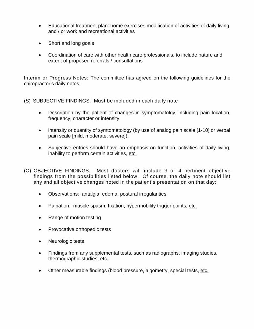
Educational treatment plan: home exercises modification of activities of daily livingand / or work and recreational activities
Short and long goals
Coordination of care with other health care professionals, to include nature andextent of proposed referrals / consultations
Interim or Progress Notes: The committee has agreed on the following guidelines for thechiropractor’s daily notes;
(S) SUBJECTIVE FINDINGS: Must be included in each daily note
Description by the patient of changes in symptomatolgy, including pain location,frequency, character or intensity
intensity or quantity of symtomatology (by use of analog pain scale [1-10] or verbalpain scale [mild, moderate, severe]).
Subjective entries should have an emphasis on function, activities of daily living,inability to perform certain activities, etc.
(O) OBJECTIVE FINDINGS: Most doctors will include 3 or 4 pertinent objectivefindings from the possibilities listed below. Of course, the daily note should listany and all objective changes noted in the patient’s presentation on that day:
Observations: antalgia, edema, postural irregularities
Palpation: muscle spasm, fixation, hypermobility trigger points, etc.
Range of motion testing
Provocative orthopedic tests
Neurologic tests
Findings from any supplemental tests, such as radiographs, imaging studies,thermographic studies, etc.
Other measurable findings (blood pressure, algometry, special tests, etc.

(A) ASSESSMENT: The assessment should note any changes in the doctor’simpression of the case. Possible entries might include:
Change in the diagnostic impression
Subluxation levels
Opinion regarding the patient’s progress
Opinion regarding how well the patient tolerated various treatments
Opinion regarding the patient’s level of compliance with the treatment plan
A referral to a prior, more lengthy assessment (usually at the most recentexamination) if there is not a change in the assessment for that day.
(P) PLAN: The plan section of the daily notes should contain any comments orchanges in the doctor’s treatment plan of the case. Possible entries mightinclude:
Date of return for treatment
Changes in initial treatment plan
Changes in adjusting techniques
Spinal levels of adjustment levels
Therapies performed, including type of therapy, location applied, intensity andduration.
Recommended exercises
Ergonomic changes needed for the patient to maximize the recovery
Imaging or special diagnostic studies which are needed
Referral to a prior, more lengthy plan (usually at the most recent examination orreexamination) if there is not change in the assessment for that day.
Reevaluation/Interim Examination Frequency: It is acknowledge that the chiropractormay reassess and Established Patient at any time during the course of treatment(CPT 99211-99213), especially if the condition changes. The Committee hasformulated the following chart which describes the recommended minimum frequencyof a reevaluation / interim examination for Established patients (CPT 99214-99215):

Purpose: To evaluate an established patient or condition with the following objectives;
Confirm or alter diagnosis
Confirm or alter treatment plan
Evaluate progress
Determine prognosis
GUIDELINES FOR FREQUENCY OF EXAMINATIONS
PATIENT TYPE DEFINITION TYPICAL RECOMMENDEDTREATMENT MINIMUM
PATTERN FREQUENCY
Acute Symptomatology is Variable, depends 30 days or 12 visitspresent less than on severitysix weeks, or acuteexacerbation of achronic or recurringcondition
Chronic Symptomatology Once to twice 3 months orpresent more per week 18 visitsthan 18 weeks
Supportive Symptomatology Twice per month Once or twicepresent due to a per yearknown, permanentdeficit; a full recoveryis not expected.
Maintenance No active Once per month No formalsymptomatology re-exam
required

In addition to the periodically performed reevaluation/interim examinations for an EstablishedPatient, the need for more frequent examinations should be determined by the clinician, aspatient progress warrants, the medical history changes or when one or more of the followingconditions exists or occurs:
a patient presents with a new injury / incident
a patient presents with a new chief complaint (s)
a patient presents with a new, distinct episode / exacerbation of a recurringcondition
a patient has an unexpected response to treatment
an anticipated change in activity level (i.e. return to work)
a patient is being dismissed from care
a patient has not received a treatment for ninety (90) days or more
other significant changes in medical history
Roetgenographic documentation should be included with the OBJECTIVE FINDINGS on theday the film was read/billed. These findings may be in the form of a formal report, or may becondensed to the major findings.

STANDARDS FOR CHIROPRACTIC TREATMENT
AND TREATMENT GUIDELINES
FOR SPECIFIC CONDITIONS

Disclaimer
Specific parameters of care appropriate for each individual case are impossible to define.Each case must be evaluated with complicating factors, concomitant and all extenuatingcircumstances taken into consideration in order to assure the patient maximum benefitthrough conservative chiropractic care.
The following guidelines should serve only as a general guide for parameters of care.Furthermore, these guidelines are not meant to provide cut-off points for treatment, but ratherto assist the analyst in determining when a request for additional information may beappropriate.
Patients may progress at a rate not anticipated in these guidelines. Exacerbation(s)secondary to physical activity may prolong normal rate of recovery. Complicating conditionswherein the patients recovery, because of one or more identifiable factors, may be slow,regressive or retarded in comparison with expectation. On the other hand, patientssometimes respond more quickly than expected. The need for documentation, as outlinedabove, is paramount to good communication between the physician and the insurancecompany and / or employer. Treatment is an essential part of diagnosis. An admittingdiagnosis may be given initially until patient response to treatment confirms or alters thediagnosis, category of condition and its degree of severity. The responsibility of upgrading ordowngrading a patient’s diagnosis and prognosis rests with the treating physician.

THE VERTEBRAL SUBLUXATION COMPLEX
SUBLUXATION COMPLEX DEFINED
The chiropractic subluxation is defined as any alteration of the biomechanical andphysiological dynamics of contiguous spinal and paraspinal structures. These biomedicalalterations are important because they can causeneurological disturbances.
In its simplest form, a subluxation is an alteration in position and/or motion of spinal segmentsand joints of the body, which affects the nervous system, thereby creating dysfunction in thetissues innervated by those nervesinvolved.
The dysfunction may take place at the site of the subluxation or anywhere along the course ofthe involved nerve(s). In its most severe form, many surrounding tissues are involved andmay be permanently damaged ordestroyed.
The chiropractic subluxation, as a complex spinal and paraspinal entity, may be divided intofive following components.
COMPONENTS OF THE VERTEBRAL SUBLUXATION COMPLEX
1. Kinesiopathology (aberrant mobility, etc.)
a. Hypomobilityb. Hypermobilityc. Compensationd. Loss of joint playe. Change in axis of motionf. Positional dyskinesia
2. Neuropathophysiology
a. IrritationFriction, tugging, torsional tension, traction, etc., producing the “facilitatedsegment”.
b. Compression
Direct or indirect – vertebral discogenic, degenerative jointdisease, positional dyskinesia, hypertrophic arthritis, etc.
3. Myopathology
a. From facilitation – (hyperactivity) spasm, contracture, myofibrositis, etc.
b. From compression – (hypoactivity) flaccidity, atrophy, myofibrositis.
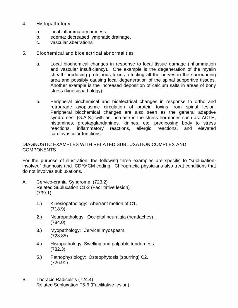
4. Histopathology
a. local inflammatory process.b. edema: decreased lymphatic drainage.c. vascular aberrations.
5. Biochemical and bioelectrical abnormalities
a. Local biochemical changes in response to local tissue damage (inflammationand vascular insufficiency). One example is the degeneration of the myelinsheath producing proteinous toxins affecting all the nerves in the surroundingarea and possibly causing local degeneration of the spinal supportive tissues.Another example is the increased deposition of calcium salts in areas of bonystress (kinesiopathology).
b. Peripheral biochemical and bioelectrical changes in response to ortho andretrograde axoplasmic circulation of protein toxins from spinal lesion.Peripheral biochemical changes are also seen as the general adaptivesyndromes (G.A.S.) with an increase in the stress hormones such as: ACTH,histamines, prostagglandanines, kinines, etc. prediposing body to stressreactions, inflammatory reactions, allergic reactions, and elevatedcardiovascular functions.
DIAGNOSTIC EXAMPLES WITH RELATED SUBLUXATION COMPLEX ANDCOMPONENTS
For the purpose of illustration, the following three examples are specific to “subluxation-involved” diagnosis and ICD*9*CM coding. Chiropractic physicians also treat conditions thatdo not involves subluxations.
A. Cervico-cranial Syndrome (723.2)Related Subluxation C1-2 (Facilitative lesion)(739.1)
1.) Kinesiopathology: Aberrant motion of C1.(718.9)
2.) Neuropathology: Occipital neuralgia (headaches) .(784.0)
3.) Myopathology: Cervical myospasm.(728.85)
4.) Histopathology: Swelling and palpable tenderness.(782.3)
5.) Pathophysiology: Osteophytosis (spurring) C2.(726.91)
B. Thoracic Radiculitis (724.4)Related Subluxation T5-6 (Facilitative lesion)

(739.2)
1.) Kinesiopathology: Mid-thoracic fixation/hypomobility(739.2)
2.) Neuropathology: Neuogenic gastritis, biliary dyskinesia.(535.5)
3.) Myopathology: Fibrositis.(729.0)
4.) Histopathology: Palpable tenderness and tissueinflammation.(782.3)
5.) Pathophysiology: Thoracic scoliosis.(727.39)
C. Lumbar Disc Syndrome (722.73)Related Subluxation L5-S1 (Compressions lesion)(739.2)
1.) Kinesiopathology: Altered range of motion.(780.9)
2.) Neuropathology: Sciatic neuralgia.(724.3)
3.) Myopathology: Atrophy of gastrocnemius muscle.(728.2)
4.) Histopathology: Tissue swelling and palpable tenderness.(782.3)
5.) Pathophysiology: Discogenic spondylosis (L5).(756.11)
The vertebral subluxation complex may be composed of one or more of the abovecomponents, depending upon the severity of the condition. The examples given abovedescribe moderate to severe subluxation complexes in order to give examples containing allfive components. Improvement or reduction of these components of the vertebral subluxationcomplex is an indication of effective treatment.
THE IMPORTANCE OF THE VERTEBRAL SUBLUXATION COMPLEX

The importance of a vertebral subluxation complex is dependent upon its clinical features andwhether this lesion and its neurogenic responses are affecting the total health of theindividual to a significant degree. Minor mechanical errors in position and motion occur in allof us. Consequently, neurological irritations occur. We are all subject to environmentalirritations and respond to these in a manner that creates errors in musculoskeletal symmetryin our body; therefore errors in position and mechanics of our anatomical structure occur.These in turn cause or perpetuate neurological aberrations of motor reflexes. These motorresponses, however, may be of a temporary nature, for the body is capable of correctingmechanical faults within its structure provided they are of minimal nature.
On the other hand, certain forms of lesions cannot be corrected without proper professionalattention. The subluxation’s significance is determined by how it affects the total economy ofthe body and how involved it is in the production of aberrant neurological responses and thecreation of a state of disease. These effects may be determined by a proper chiropracticanalysis and diagnosis.
THE CAUSE OF SUBLUXATION / SEGMENTAL DYSFUNCTION
The immediate causes of subluxation may be divided into two major categories: the unequalor asymmetrical musculature efforts upon the joint structures, and the inequality in thesupporting tissues or a particular joint, such as the cartilage, intervertebral disc, ligaments,etc.
Inequality in Muscular Balance
Inequality in muscular balance may be initiated by many things such as:
1.) Trauma
A. Acute macrotrauma may cause inflammation, degeneration, etc., andparticularly the muscular splinting or spasm reaction that the musculaturemakes when its surrounding tissues are injured and therefore alter theposition and motion of the structural tissues that are related.
B. Microtrauma, though of a less acute nature, may cause a slow continualirritation and may eventually create degenerative pathological changeswhich similarly alter muscular reaction.
2.) Postural Distortion Phenomena

Postural compensations for either mechanical activity or for structural changesin the skeleton itself. These changes, as well as other causes of subluxation,often result in a series or combinations of minor mechanical errors whichtogether may be termed scoliosis, kyphosis, lordosis, distortion, or similarterms.
3.) Biochemical Reactions
The acute or chronic hypo or hypertonicities of musculature may be due tovarious biochemical changes within these tissues.
4.) Psychomotor Reactions
The reaction of the musculature to emotional effects on the nervous activity ofthe body as it depicts its psychological stresses.
5.) Paralytic Affects
Primary disease of the neuromuscular system itself such as polio or otherparalytic diseases which affect the balance of the musculoskeletal tone orstrength and therefore positional quality of motion.
6.) Somatic and Visceral Responses
The secondary reaction of the muscular system to somatic or visceral sensoryirritation which may develop elsewhere in a given neurological segment.
Abnormal Structural Support
Mechanical errors in position or motion may also be brought about by structural alterations inthe supporting tissues of the joint itself. These in turn may be brought about by:
1.) Developmental abnormalities causing asymmetry of the vertebra, cartilage,muscular structure, etc.
2.) Various acquired disease processes within the joint such as arthriticdegeneration, avascular necrosis or a neuropathic process that causes thecartilage, bone, ligaments, or musculature to be structurally altered.
3.) The resolution of macro or microtraumas or of other primary pathology maycause fibrosis, degeneration, or other retrograde changes of a structural naturewithin the joints themselves.

These same various processes, as mentioned, not only develop within the vertebral columnand its paravertebral tissues, but also in the musculoskeletal tissues of the appendicularskeleton, and similar lesions may therein exist which perpetuate neuropathic responses bytheir presence. Within the structures of the vertebral column, the effects of thesesubluxations are more evident because of the close anatomical proximities and the functionalimportance of normal motor motion or mechanical integrity to the various components of thenervous system.
SUBLUXATION CLASSIFICATIONS BY RADIOGRAPHIC FINDINGS WHENRADIOGRAPHIC EVEIDENCE EXISTS
Static1.) Flexion malposition2.) Extension malposition3.) Lateral flexion malposition (left or right)4.) Rotation malposition (left or right) (anterior or posterior)5.) Anteriolisthesis-spondylolisthesis6.) Retrolisthesis7.) Lateral listhesis (left or right)8.) Interosseous spacing
a. decreasedb. increased
9.) Osseous foraminal encroachment
Kinetic
1.) Hypomobility-fixation2.) Hypermobility3.) Aberrant motion
Sectional Subluxations
1.) Scoliosis and/or alteration or curves secondary to musculature imbalance.2.) Scoliosis and/or alteration of curves secondary to structural asymmetries.3.) Decomposition of adaptational curvatures.4.) Abnormalities of motion.
Paravertebral Subluxations
1.) Costovertebral and costotransverse disrelationships.2.) Sacroiliac subluxations.

LEVEL OF CHIROPRACTIC CARE
There are two basic levels of chiropractic care, classified as Active Care andPreventative/Maintenance Care. The type of care the patient is to receive is directlydependent upon the classification of his/her condition.
Active Care: Patient management directed toward returning the patient to pre-clinicalstatus following injury or illness.
1. Relief Care: Patient management directed toward reducing symptoms andimproving function to a tolerable level.
2. Therapeutic Care: Patient management directed to further reducesymtomatology and improve function. This level of care should enable a patientto perform most normal daily activities without frequent exacerbation symptoms.
3. Rehabilitative Care: Patient management directed toward improved functionby restoring optimal strength and flexibility to the musculoskeletal system. Thegoal of this phase is to return the patient to pre-clinical status through educationand therapy.
4. Supportive care: Patient management directed toward support of an unstablecondition which includes periodic monitoring and treatment to reduce thefrequency and severity of anticipated exacerbations. The need for supportivecare is seen in cases where patients having reached maximum therapeuticbenefit, in whom periodic trails of therapeutic withdrawal has failed to sustainprevious therapeutic gains, would otherwise progressively deteriorate. Orwhere the injury or illness results in permanent, unstable conditions ordisabilities and the patient is therefore unable to return to pre-clinical status.
PREVENTATIVE/MAINTENANCE CARE: Patient management directed toward maintainingoptimal body function and health. This is management of the asymptomatic patient who hasreached pre-clinical status or maximum therapeutic benefit where the condition is resolved orstable and is provided to reduce the incidence or prevalence of illness, impairment and riskfactors.
Categories of Conditions Treated, Nomenclature and Operational Definitions
Acute – Sharp, poignant; having a short and relatively severe course. Acute in this instanceis meant to designate the new condition of less than 6 weeks duration.
1. Mild: By nature this condition is uncomplicated and usually does not requirerehabilitative or supportive care. This category of condition does requirerelief care and may require therapeutic care. The patient with a mildcondition would normally require further care only in the event ofsignificant re-injury or acute exacerbation.
An example of mild, acute condition: Strain of the lower back or neck, nomajor joint involvement, recent onset, no past history of similarcomplaints and no complicating factors readily apparent.
2. Moderate: This condition commonly requires rehabilitation care but may notrequire supportive care in the mild-to-moderate case.
An example of moderate, acute condition: Mild-to –moderatelumbosacral sprain/strain injury or cervicalhyperflexion/hyperextension injury, recent onset with significanttrauma, complicated by concomitant or related conditions such asheadaches, nausea, cervicobrachial syndrome.
3. Severe: This condition is complicated and usually requires all four levels ofcare to regain maximum function,which may be maximumtherapeutic (chiropractic) benefit without obtaining pre-existing orpre-clinical status.
An example of severe, acute condition: Joint, muscle, ligament,and/or nerve damage, such as lumbar and cervical disc syndromesor moderate complicated sprain/strain injuries.
Subacute – May be used to describe a condition between acute and chronic with some acutefeatures.
Chronic – Persisting over a long period of time. Chronic in this instance is meant todesignate the long-standing, recurring condition of more than 18 weeksduration.
1. Mild: Without acute exacerbation, this condition may require little or notintensive relief care, but may requires therapeutic, rehabilitative and supportivecare.
Example: Chronic muscle strain from aberrant biomechanics,myofascitis/fibromyositis, without major complicating factors.

2. Moderate: With acute exacerbation, this condition may require all four levelsof care.
Example: Degenerative joint disease, muscle weakness, ligamentousinstability, disc degeneration, with numerous complications andconcomitant conditions.
3. Severe: With serious, acute exacerbation, this condition requires extendedperiods of all four levels of treatment.
Example: Disc, joint and neurologic involvement, post laminectomysyndrome, history of serious and/or numerous traumatic events,numerous complicating factors notes as well as related conditions andconcomitant complaints.
Acute exacerbation – May be used to describe a sharp or severe exacerbation of symptomsor an increase in the severity of a condition.
Maximum Therapeutic Benefit – Maximum therapeutic benefit (MTB) is considered to havebeen reached when the patient’s symptomatology and/or disability has remained unchangedfor two (2) months or longer while under active care. Validation of MTB is to be considereddocumented when there is a lack of significant measurable improvement or reduction indisability on two-(2) consecutive monthly evaluations. Significant residual symptoms,diminished functional capacity and/or disability may still be present, but no furthermeasurable improvement or reduction in disability is anticipated with continued active care.
Significant Residual Symptoms – Residual symptoms are considered significant when theyare severe enough to prevent normal work activities, activities of daily living or animprovement in functional capacity.
PRN Treatment (as needed/supportive care) – PRN treatment or supportive care is definedas treatment for patients who have reached MTB, but continue to experience significantresidual symptoms, that are not adequately controlled with home based self-care and/orlifestyle modifications. PRN treatment should be diagnostically based and administered at alow enough frequency to reduce the chance of physician dependence, somatization, illnessbehavior or secondary gain.
A treating physician may contend that PRN treatment is, by definition, as needed treatmentand in moderate to severe cases ongoing weekly treatment is beneficial even after MTB hasbeen achieved. In an attempt to mitigate the appearance of overutilization and avoid thechance of physician dependence, somatization, illness behavior or secondary gain, weeklychiropractic treatment, for purposes of these treatment utilization guidelines, shall beconsidered “active” treatment. Once MTB has been reached “active” treatment is appropriatein only very unique circumstances, which are justified by and objective clinically documentedcondition or state that elevates the patient to an appropriate Diagnostic Related Group.
GUIDELINES FOR CHIROPRACTIC MANAGEMENT
The following charts shall serve as a general guideline for the treatment of acute and chronicconditions. It is important to note that with proper documentation additional treatment oralteration of treatment schedules may be warranted for the categories of conditions andlevels of care listed herein.
ACUTE CONDITIONS
Definition: New condition, most frequently related to an injury of less than 18 weeks duration.
TYPES OF LEVELS FREQUENCY DURATIONCONDITIONS OF CARE OF TREATMENT OF TREATMENTMild Relief Daily to 3x's/wk 1-15 days
Therapeutic 3x's/wk to 1x/wk 0-30 daysRehabilitative None NoneSupportive None None
Moderate Relief Daily to 3x's/wk 7-21 daysTherapeutic 3x's/wk to 1x/wk 30-60 daysRehabilitative 1x/wk to 2x's/wk 30-60 daysSupportive 2x's/wk to 1x/wk 0-3 months
Severe Relief Daily to 3x's/wk 21-45 daysTherapeutic 3x's/wk to 1x/wk 30-90 daysRehabilitative 1x/wk to 2x's/mo 45-90 daysSupportive 2x's/mo to 1x/mo 3-5 days
TYPES OF LEVELS FREQUENCY DURATIONCONDITIONS OF CARE OF TREATMENT OF TREATMENTMild Relief None None
Therapeutic 3x's/wk to 2x's/wk 14-45 daysRehabilitative 2x's/wk to 2x's/mo 45-90 daysSupportive 2x's/mo to 1x/mo 3-6 months
Moderate Relief Daily to 3x's/wk 14-45 daysTherapeutic 3x's/wk to 2x's/wk 45-90 daysRehabilitative 2x's/wk to 2x's/mo 90-180 daysSupportive 2x's/mo to 1x/mo 6-12 months
Severe Relief Daily to 3x's/wk 30-60 daysTherapeutic 3x's/wk to 2x's/mo 60-120 daysRehabilitative 2x's/wk to 2x's/mo 90-180 daysSupportive 2x's/mo to 1x/3mo 6-18 months
CHRONIC CONDITIONS
Definition: Old condition; arbitrarily set at greater than 18 weeks duration with long-standing or recurring symptomatology. May be history trauma or microtrauma.

DIAGNOSIS ICD*9*CM TREATMENT RANGESTIME/VISITS
847.0847.1847.2846.0-.9839.0-.9839.21839.20839.42739.0739.1739.2-.5
mild 2-4 wks / 6-12 visitsmoderate 5-8 wks / 18-24 visitssevere MAY REQUIRE SURGERY
847.0 6-12 wks / 12-20 visits847.1847.2846.0-.9
Cervical / Thoracic disc syndromedisplacementprotrusion 722.0herniation 722.0
mild 6-8 wks / 8-12 visitsmoderate 12-14 wks / 24-30 visitssevere 18-24 wks / 32-40 visits
Lumbar disc syndrome/displacementprotrusion 722.1herniation 722.1
mild 8-10 wks /18-24 visitsmoderate 14-16 wks / 30-36 visitssevere 18-24 wks / 36-42 visits
Torticollis 723.5mild 33.83 2-3 wks/ 2-6 visitsmoderate 3-5 wks / 6-10 visitssevere 6-12 wks / 10-20 visits
Cervical-occipital neuralgia 729.2 5-8 wks / 12-20 visits
Cervicocranial syndrome 723.2 2-8 wks/ 6-20 visits
Chronic, spinal sprain/strain(cervico-thoracic, lumbosacral,sacroiliac)
Acute, spinal sprain/strain(cervical, thoracic, lumbar,lumbo-sacral, sacroiliac): withor without vertebral
CHIROPRACTIC TREATMENT GUIDELINES FOR SPECIFIC CONDITIONS

DIAGNOSIS ICD*9*CM TREATMENT RANGESTIME/VISITS
Cervicalgia 723.1 3-5 wks / 8-10 visits
Cervicobrachial syndrome 723.1cervical neuralgia/brachial 729.2 6-12 wks / 15-20 visitsneuralgia
Brachial neuritis or radiculitis 723.4 6-12 wks / 24-28 visits
Intercostal neuralgia 353.8729.2 4-7 wks / 6-14 visits
Intercostal neuritis 353.8724.4729.2 7-9 wks / 18-24 visits
Neurovascular headache 784.0
acute 307.81 2-4 wks / 6-8 visitschronic 346.0 8-10 wks / 18-24 visitssevere 12-14 wks / 24-28 visits
Spinal myofascitisacute 729.1 2-4 wks / 3-6 visitschronic 729.8 6-12 wks / 12-20 visits
Lumbar plexus lesion 353.1 6-8 wks / 15-20 visits
Lumbar facet syndrome 724.8 6-8 wks / 15-20 visits
Lumbar trophism 724.8 4-6 wks / 12-14 visits
Lumbar canal stenosis 724.02 5-7 wks / 16-18 visits
Spondylolisthesis 756.12Grade I 738.4 6-8 wks / 14-16 visitsGrade II, III 8-10 wks / 20-24 visitsGrade IV USUALLY REQUIRES
SURGICAL STABILIZATION
Spinal enthesopathy 720.1(cervical, thoracic, lumbosacral) 4-6 wks / 8-12 visits
Scoliosis (treatment of patients 737.0-737.9under 20 years of age) 754.2
mild 4-6 wks / 12-14 visitsmoderate 8-10 wks / 18-24 visitssevere 12-14 wks / 24-30 visits
Lumbago / lumbalgia 724.2 6-8 wks / 14-16 visits
Diagnosis ICD*9*CM TREATMENT RANGESTIME / VISITS
Lumbosacral or 724.4 12- 14 wks / 24-30 visitsSciatic neuritis 724.3
722.10
Sciatic neuralgia 724.3 8-12 wks / 6-24 visits
TMJ syndrome 524.6 6-8 wks / 16-18 visits
Cervical / lumbar 722.4, 722.52Hypo discopathy 722.52, 721.90Degenerative joint 715.09, 715.9Disease / spondylosis 721.0, 721.2, 721.3
Mild 4-8 wks / 4-8 visitsModerate 8-12 wks / 8-12 visitsSevere 12-20 wks / 12-30 visits
Post-laminectomySyndrome / failed back 722.8Surgical syndrome 966.4 12-24 wks / 12-24 visits
Ligamentous laxity / 728.4Hypermobiltiy syndrome 728.5 4-8 wks / 4-8 visits
*Osteoarthrosis / 715.9osteoarthritis 721.90 8-16 wks / 8-16 visits
*Osteoporosis 733.1 8-16 wks / 8-16 visits
*Compression 733.1fractures 805
806 6-12 wks / 6-12 visits
*Congenital 756.19abnormalities 756.10(i.e. hemivertebra, blockvertebra, lumbarization,sacralization) 6-12 wks / 6-12 visits
*Spondylolisthesis 756.12(usually Grade I is listed 738.4 6-8 wks / 14-16 visitsas a complicating factor)
* These factors complicate degenerative structural spinal instability upon which otherconditions are superimposed and increase the probability of non-resolution. Even aftermaximum therapeutic benefit, exacerbations may occur that require periods of scheduledtherapeutic care and periodic non-scheduled care which, if stopped, could result indeteriorations. Treatment of these chronic complicated cases requires proper casedocumentation for justification of treatment beyond the recommended guidelines.
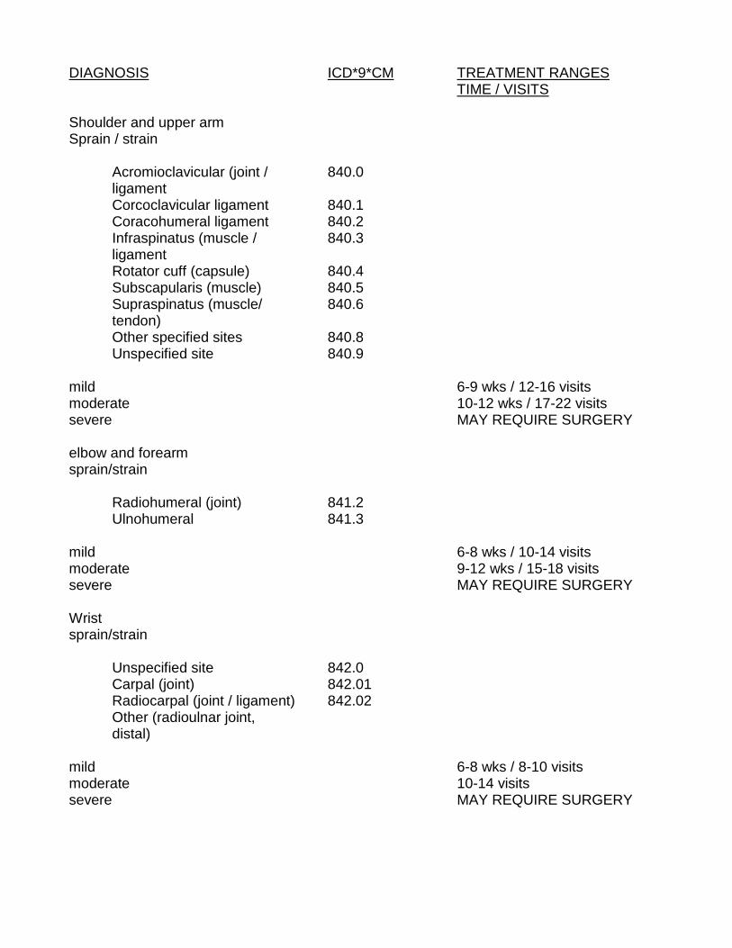
DIAGNOSIS ICD*9*CM TREATMENT RANGESTIME / VISITS
Shoulder and upper armSprain / strain
Acromioclavicular (joint / 840.0ligamentCorcoclavicular ligament 840.1Coracohumeral ligament 840.2Infraspinatus (muscle / 840.3ligamentRotator cuff (capsule) 840.4Subscapularis (muscle) 840.5Supraspinatus (muscle/ 840.6tendon)Other specified sites 840.8Unspecified site 840.9
mild 6-9 wks / 12-16 visitsmoderate 10-12 wks / 17-22 visitssevere MAY REQUIRE SURGERY
elbow and forearmsprain/strain
Radiohumeral (joint) 841.2Ulnohumeral 841.3
mild 6-8 wks / 10-14 visitsmoderate 9-12 wks / 15-18 visitssevere MAY REQUIRE SURGERY
Wristsprain/strain
Unspecified site 842.0Carpal (joint) 842.01Radiocarpal (joint / ligament) 842.02Other (radioulnar joint,distal)
mild 6-8 wks / 8-10 visitsmoderate 10-14 visitssevere MAY REQUIRE SURGERY

DIAGNOSIS ICD*9*CM TREATMENT RANGESTIME / VISITS
Handsprain/strain
Unspecified site 842.10Carpometacarpal (joint) 842.11Metacarpophalangeal (joint) 842.12Interphalangeal (joint) 842.13Other (midcarpal joint) 842.19
mild 6-8 wks / 6-9 visitsmoderate 9-12 wks / 10-12 visitssevere MAY REQUIRE SURGERY
Hip and thigh 843.9sprain/strain
mild 6-9 wks / 12-16 visitsmoderate 10-12 wks / 17-22 visitssevere MAY REQUIRE SURGERY
Knee and legsprain/strain
Lateral collateral ligament 844.0Medial collateral ligament 844.1Cruciate ligament 844.2Tibiofibular (joint / ligament) 844.3Other specified sites 844.8Unspecified site 844.9
mild 6-9 wks / 12-16 visitsmoderate 10-12 wks / 17-22 visitssevere MAY REQUIRE SURGERY
Anklesprain/strain
Unspecified site 845.00
mild 6-8 wks / 8-10 visitsmoderate 9-12 wks / 10-14 visitssevere MAY REQUIRE SURGERY

DIAGNOSIS ICD*9*CM TREATMENT RANGESTIME / VISITS
Foot 845.10sprain/strain 845.19
Unspecified site 845.10Interphalangeal (joint) 845.13
mild 6-8 wks / 8-10 visitsmoderate 9-12 wks / 10-14 visitssevere MAY REQUIRE SURGERY
Bursitis
Anklecalcaneal 726.79
Shoulderscapulohumeral 726.19subacromial 726.19subcoracoid 726.19subdeltoid 726.19
Elbow 726.33Hand 726.4Hip
ischiogluteal 726.5trochanteric area 726.5
Kneeinfrapatellar 726.69prepatellar 726.65subpatellar 726.69pes anserinus 726.61
mild 4-8 wks / 8-10 visitsmoderate 9-12 wks / 11-15- visitssevere 13-15 wks / 16-22 visits
MAY REQUIRE SURGERY
Carpal Tunnel Syndrome 354.0 13-16 wks / 12-25 visits
Shoulder Impingement 13-16 wks / 23-35 visitsSyndrome MAY REQUIRE SURGICAL
DECOMPRESSION

COMPLICATING CONDITIONS/FACTORS
Definition: Where the patient, because of one or more identifiable factors,exhibits regression of retarded recovery in comparison with expectations from thenatural history. 1
Complicating conditions, which may retard the patient’s return to pre-clinicalstatus or maximum therapeutic benefit include, but are not limited to, the following;
History:
TraumaMultiple traumaSeverity of trauma/injuriesPost-traumatic loss of consciousnessMajor illness or injuriesDuration of symptomsAgeEmploymentLifestyle
Radiographic:
Vertebral subluxation complexLoss of normal spinal biomechanical alignmentDegenerative joint diseaseCongenital abnormalitiesLoss of motion segment integrity as documented by radiographs or videoflouroscopy
Physical Examination:
DeconditioningInstability
Neurological:
Loss of relevant reflexesMeasured muscular weakness compared to the contralateral sideMeasured unilateral atrophyAltered dermatomal sensitivityNeurological impairment verified by electrodiagnostic studies
1 Haldeman, Scott, D.C., M.D., PhD., et.al., Guidelines for Chiropractic Quality Assurance and PracticeParameters, Proceedings of the Mercy Center Consesus Conference, Aspen Publication, Gaithersburg,Maryland, 1993

STANDARDS FOR
UTILIZATION OF DIAGNOSTIC IMAGING

STANDARDS FOR UTILIZATION OF DIAGNOSTIC IMAGING IN CHIROPRACTIC
The fundamental purpose of diagnostic imaging is to gain diagnostic informationregarding the patient in terms of diagnosis, prognosis and therapy planning. Studiesare performed at the request of a practitioner with the informed consent of the patient.The basic directive of the practitioner is to use radiology to confirm or contribute to theclinical picture. Each study is performed in a reasonable manner and a formal reportis generated. Only the written radiology report effectively communicates theinformation gained from each study.
Diagnostic imaging, especially plain film radiographs, continue to be a mainstay in theassessment of chiropractic patients. There are four (4) required standards that mustbe met with each imaging study:
1.) The study must be obtained based on clinical need.
2.) The study must be of sufficient diagnostic quality.
3.) There must be interpretation of the study to reach diagnostic conclusionsabout the study.
4.) The information from the study must be correlated with patientmanagement.
Decision Making for Patient Selection
The clinician must select the study which will give the required information withsufficient reliability at a minimal risk to the patient. The value of the information gainedfrom the procedure must be worth the possible detrimental effects. Likewise thedangers inherent in not using the study may be outweighed by the risks. The risk andbenefits analysis must always be considered before selection of a patient forradiographic examination. This selection process must be based on the followingguidelines.
1.) The need for radiographic examination is based on history and
2.) The potential diagnostic benefits of the radiographic examination arejudged to outweigh the risks.
3.) Radiography is used to assist the practitioner in diagnosis of pathology,identify contraindications to chiropractic care, identify bone and jointmorphology, and acquire postural, kinematical and biomechanicalinformation.
4.) Routine radiography of patients as a screening procedure is notappropriate practice except under public health guidelines.

Patient Safety
The HSCA recognizes the role of radiography as a major diagnostic tool in the healingsciences, however harmful effects of ionizing radiation are also well documented andpractitioner discretion and patient safety are essential considerations in determiningwhen and if to use radiography for diagnostic purposes. The HSCA position is asfollows:
1.) Routine radiography of any patient should not be performed without dueregard for clinical need.
2.) Any offer or advertising of x-rays to actual or potential patients shall beaccompanied by a statement that, to avoid needless health hazardsassociated with ionizing radiation, no x-rays will be taken unless there is aprior observable clinical need for it.
3.) Avoidance which compensate for tissue thickness by altering the screensor the light emission from the screens, such as the occluding of one of thescreens of the cassette, is recommended.
4.) Repeat radiographic evaluation of the patient should not be undertakenwithout significant observable clinical indication, as determined by thetreating chiropractor.
5.) Pregnant females should not be radiographed unless the patient’ssymptoms are of such significance that the proper treatment of the patientmight be jeopardized without the use of such radiographs.
6.) Radiographic procedures should not be undertaken without the use ofappropriate compensating filters and gonad shielding, except where suchgonad shielding would exclude an area from examination which isclinically necessary to examine.
7.) Females with reproductive potential, or where the possibility of pregnancyexists, should be radiographed only where clinically necessary andpreferably, during the first ten days following onset of menses.
These guidelines are consistent with the rules of conduct when employingradiographic procedures common throughout the health care community andare consistent with those recommended by the Center for Devices andRadiological Health, Food, & Drug Administration of the U.S. Department ofHealth & Human Services and the Agency for Health Care Policy andResearch, Public Health Service, U.S. Department of Health & HumanServices.

APPENDICES

APPENDIX A
HSCA Documentation Guidelines
The following is an example of a case record which would receive a perfect scoreunder the HSCA Documentation Guidelines. It is provided as a model forinformational purposes. The diction from an initial examination is shown, along withdaily note from the next office visit. Please note the following features of the record:
* The record allows a reviewer to have a clear summary of the patient’scondition and response to treatment, as well as the doctor’s diagnosticimpression and treatment plan.
* The complete initial SOAP workup allows for subsequent daily notes tobe abbreviated.
* The essential subcomponents of the subjective and objective portions ofthe SOAP notes are shown in bold type.
* Each patient is unique. Each patient record should relate the mostpertinent information to that case, but should ideally follow the SOAPformat for organization of the information.
01/30/96 SUBJECTIVE: Joe Frobazz, a 45 year old white male, presents to ourclinic for evaluation and treatment. His chief complaint is of lower back pain over thelower lumbar area, especially at the midline. He rates his pain at 2 out of 10 possiblewhen at rest, and at 7/10 upon movement. The onset of his pain occurred three daysprior when he was twisting and lifting a 50# bag of rice. He states that the pain ismade worse with most movements, and with prolonged sitting or standing. The painis somewhat relieved with the use of ice and aspirin. The pain is characterized asusually dull but is sharp with movement. There is no leg pain or numbness, andbowel/bladder symptomatology is denied. The pain is worse in the morning and atnight with some mild relief in the middle of the day. The patient denies previousepisodes of lower back pain. Past medical history includes five years of mildhypertension with successful management with Lopressor, and running 20 minutesthree times per week. Past family, social history includes an occupation as apineapple worker for the past 15 years. The patient denies the use of alcohol ortobacco. There is no history of familial diseases which would be pertinent to thecurrent chief complaint. Past injuries include a 1992 motor vehicle accident whichresulted in mild symptomatology with no treatment or residuals. Past surgical historyis negative. Review of systems is otherwise negative for the following systems;constitutional, EENT, CV, respiratory, GI, GU, NMS, skin, psychiatric, endocrine,hematologic, allergic.

OBJECTIVE: Vitals include BP=132/86. Pulse=74bpm. Upon observation,patient demonstrated a right lumbar antalgia of approximately 20 degrees to the right,with pronounced spasm of the paraspinal musculature bilaterally. Upon palpation,the spinous process of L4-5 were extremely tender to the touch; sacroiliac, hips andsciatic notch were nontender. Sensory examination indicated a mild loss of lighttouch and sharp sensation of the left lateral thigh to the knee. Vibratory sensation iswithin normal limits. Motor strength of the lower extremity is 5/5; trunk strength testsproduced too much pain to accurately test. DTR’s are =2/5 at patella and Achillesbilaterally. Lumbar range of motion is limited in flexion, extension, lateral bending.Positive orthopedic tests: Kemp’s, DSLR at 80 degrees, minor’s antalgia. Thepatient completed a pain drawing, which was in agreement with the patient’s verbaldescription and our objective findings. An Oswestry questionnaire was completed,with a rating of 28%. AP, lateral and Ferguson radiographs displayed an elevated lefthemipelvis, spinal bifida occulta at L5-S1, anterior weightbearing due to diminishedlumbar lordosis with disc spaces relatively intact.
ASSESSMENT: 1. Acute, moderate lumbar facet joint syndrome. 2. Rule outdisc pathology.
PLAN: With the patient’s consent, interferential current (IFC) was applied at 80-150 Hz, 22mA for 15 minutes to the lower back region and spinal manipulative therapy(SMT) using Gonstead technique to L5-S1. Treatment was well tolerated. Treatmentplan will consist of spinal manipulative therapy and interferential therapy at three (3)times per week for a period of two (2) weeks, with a reevaluation at that time. Thepatient is provided with an excuse from work activities for one (1) week and also withlumbar extension exercises to be performed two (2) times per day to patient tolerance.Short term goals include reduction of pain at rest, and an increased ability to standerect and ambulate without significant pain. Long term goals include restoration offull lumbar range of motion, increased lower back strength and flexibility. Patient hasbeen provided with instructions for the home use of ice. An MRI and/or orthopedicreferral will be considered if symptoms do not significantly improve within the next four(4) weeks.

Appendix B
The following checklist is to be used as a quick test to see how well your recordscompare to the standards of the HSCA.
* Is the record legible?
* Is there a completed problem list?
* Is there an appropriated past medical history in the record?
* Is there a pertinent history and physical exam?
* Are lab and other studies ordered as appropriate?
* Are working diagnoses consistent with findings?
* Are plans of action/treatment consistent with diagnoses?
* Is there a date of return visit or other follow-up plan for each encounter?
* Is there a continuity and coordination of care between primary andspecialty physicians?

02/02/96
S: The patient is having diminished lower back pain, now 1/10 at rest and 3-4/10upon movement. Walking is slightly less painful and he is able to stand more erect.Patient denies leg pain and/or numbness. Sleeping fairly comfortably and using ice asrecommended.
O: Diminished severity of antalgia, now approximately 10 degrees. Fixation L5-S1.+Kemp’s, +Dejerines triad.
A: See 01/30/95.
P: See 01/30/95; SMT to L5 area; IFC to lower back 15 min, 80-150 Hz, 22 Ma;increase frequency of stretching exercises to 4x/day, continue home ice therapy.

APPENDIX C
DIAGNOSTIC RELATED GROUPS
Category I
* The patient has not significant clinical findings.
* No muscle guarding or history of guarding.
* No documentable neurologic impairment.
* No significant loss of structural integrity on lateral flexion and extensionroentgenograms.
* No indication of impairment related to injury or illness.
Category II
* The clinical history and examination findings are compatible with aspecific injury or illness. Findings may include;
* Significant intermittent or continuous muscle quarding which isobservable by the physician.
* Nonuniform loss of range of motion, as measured by Goniometer.
* Nonverifable radicular complaints.
* No objective sign of radiculopathy.
* No loss of structural integrity.
Category III
* Patient presents with significant signs of radiculopathy such as;
* Loss of relevant reflex (es) or
* Measured unilateral atrophy compared to the contralateral side at thesame location.
* Neurological impairment may be verified by electrodiagnostic findings.

Diagnostic Related groupspage 2
Category IV
* Loss of motion segment integrity or structural integrity of at least 5mm oftranslations of one vertebra on another, or angular motion greater than 11degrees of and adjacent motion segment.
* Documented history of muscle guarding and pain is present.
* Neurologic abnormalities need not be present.
Category V
* The patient meets the criteria of Category III and IV i.e.
* Loss of motion segment integrity and
* Radiculopathy is present.
* Significant lower-extremity impairment is indicated by:
- atrophy or
- loss of reflex(es)
- numbness with an anatomic basis and/or
- electromyographic findings
Category VI
Cauda Equina-syndrome with
* Objectively demonstrated permanent, partial loss of extremity function
* May or may not have loss of motion segment integrity
* Do not have objectively documented bowel or bladder impairment.


![INTERNATIONAL CHIROPRACTORS ASSOCIATION REPORT …INTERNATIONAL CHIROPRACTORS ASSOCIATION REPORT TO THE MARYLAND CHIROPRACTIC ASSOCIATION Submitted By ... Public Law 15,[1] ... help](https://static.fdocuments.net/doc/165x107/5f0ed6397e708231d4412d47/international-chiropractors-association-report-international-chiropractors-association.jpg)