Hand Detection and Tracking Using the Skeleton of the Blob ...
8
Hand Detection and Tracking Using the Skeleton of the Blob for Medical Rehabilitation Applications Pedro Gil-Jim´ enez 1 , Beatriz Losilla-L´opez 1 , Rafael Torres-Cueco 2 , Aurelio Campilho 3 , and Roberto L´opez-Sastre 1 1 Universidad de Alcal´a, Alcal´a de Henares, Madrid, Spain, [email protected], http://agamenon.tsc.uah.es/Investigacion/gram/ 2 Universidad de Valencia, Valencia, Spain, 3 INEB - Instituto de Engenharia Biom´ edica, Portugal Universidade do Porto, Faculdade de Engenharia, Portugal Abstract. This article presents an image processing application for hand detection and tracking using the 4-connected skeleton of the seg- mentation mask. The system has been designed to be used with tech- niques of virtual reality to develop an interactive application for phantom limb pain reduction in therapeutic treatments. One of the major contributions is the design of a fast and accurate skele- ton extractor, that has proven to be faster than those available in the literature. The skeleton allows the system to precisely detect the position of all the interest points of the hand (namely the fingers and the hand center). The system, composed of both the hand detector and tracker, and the virtual reality application, can work in real-time, allowing the patient to watch the virtual image of his own hand on a screen. Keywords: hand detection and tracking, blob skeleton, virtual reality, phantom limb pain reduction 1 Introduction In the management of patients with Complex Regional Pain Syndrome (CRPS) or Phantom Limb Pain (PLP), there is compelling evidence that Mirror Visual Feedback (MVF) is effective in reducing pain and disability [13] [14]. The use of virtual reality in medical applications has been given a great interest in recent years. One of these approaches is the Virtual Reality Mirror Visual Feedback (VRMVF), with the same principles of the MVF, but, instead of using a mirror, it uses a different equipment, sometimes quite complex, like sensors inserted in a glove [3] [15]. This equipment can not be used in many cases, since it implies that the patient has to wear a glove (or a similar type of sensor) which some patients can not tolerate. Furthermore, in this kind of rehabilitation techniques,
Transcript of Hand Detection and Tracking Using the Skeleton of the Blob ...
Hand Detection and Tracking Using theSkeleton of the Blob for Medical Rehabilitation
Applications
Pedro Gil-Jimenez1, Beatriz Losilla-Lopez1, Rafael Torres-Cueco2, AurelioCampilho3, and Roberto Lopez-Sastre1
1 Universidad de Alcala, Alcala de Henares, Madrid, Spain,[email protected],
http://agamenon.tsc.uah.es/Investigacion/gram/2 Universidad de Valencia, Valencia, Spain,
3 INEB - Instituto de Engenharia Biomedica, PortugalUniversidade do Porto, Faculdade de Engenharia, Portugal
Abstract. This article presents an image processing application forhand detection and tracking using the 4-connected skeleton of the seg-mentation mask. The system has been designed to be used with tech-niques of virtual reality to develop an interactive application for phantomlimb pain reduction in therapeutic treatments.
One of the major contributions is the design of a fast and accurate skele-ton extractor, that has proven to be faster than those available in theliterature. The skeleton allows the system to precisely detect the positionof all the interest points of the hand (namely the fingers and the handcenter).
The system, composed of both the hand detector and tracker, and thevirtual reality application, can work in real-time, allowing the patient towatch the virtual image of his own hand on a screen.
Keywords: hand detection and tracking, blob skeleton, virtual reality,phantom limb pain reduction
1 Introduction
In the management of patients with Complex Regional Pain Syndrome (CRPS)or Phantom Limb Pain (PLP), there is compelling evidence that Mirror VisualFeedback (MVF) is effective in reducing pain and disability [13] [14]. The use ofvirtual reality in medical applications has been given a great interest in recentyears. One of these approaches is the Virtual Reality Mirror Visual Feedback(VRMVF), with the same principles of the MVF, but, instead of using a mirror,it uses a different equipment, sometimes quite complex, like sensors inserted ina glove [3] [15]. This equipment can not be used in many cases, since it impliesthat the patient has to wear a glove (or a similar type of sensor) which somepatients can not tolerate. Furthermore, in this kind of rehabilitation techniques,
for an application to be useful, it needs to offer some kind of entertainment oramusement for the patient to get his compliance.
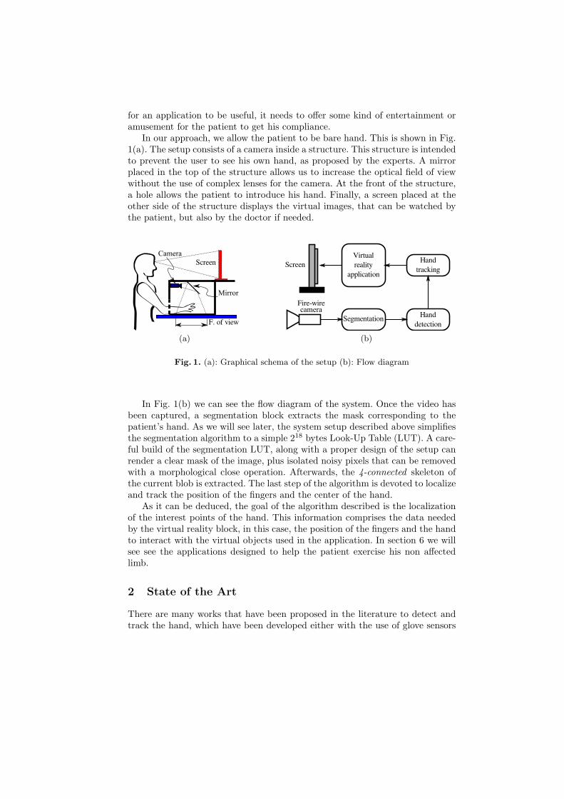
In our approach, we allow the patient to be bare hand. This is shown in Fig.1(a). The setup consists of a camera inside a structure. This structure is intendedto prevent the user to see his own hand, as proposed by the experts. A mirrorplaced in the top of the structure allows us to increase the optical field of viewwithout the use of complex lenses for the camera. At the front of the structure,a hole allows the patient to introduce his hand. Finally, a screen placed at theother side of the structure displays the virtual images, that can be watched bythe patient, but also by the doctor if needed.
(a) (b)
Fig. 1. (a): Graphical schema of the setup (b): Flow diagram
In Fig. 1(b) we can see the flow diagram of the system. Once the video hasbeen captured, a segmentation block extracts the mask corresponding to thepatient’s hand. As we will see later, the system setup described above simplifiesthe segmentation algorithm to a simple 218 bytes Look-Up Table (LUT). A care-ful build of the segmentation LUT, along with a proper design of the setup canrender a clear mask of the image, plus isolated noisy pixels that can be removedwith a morphological close operation. Afterwards, the 4-connected skeleton ofthe current blob is extracted. The last step of the algorithm is devoted to localizeand track the position of the fingers and the center of the hand.
As it can be deduced, the goal of the algorithm described is the localizationof the interest points of the hand. This information comprises the data neededby the virtual reality block, in this case, the position of the fingers and the handto interact with the virtual objects used in the application. In section 6 we willsee see the applications designed to help the patient exercise his non affectedlimb.
2 State of the Art
There are many works that have been proposed in the literature to detect andtrack the hand, which have been developed either with the use of glove sensors
or artificial vision techniques. These techniques include color segmentation [7][16] [8], contour detection [9] [20] and infrared segmentation [1], among others.The hand gesture is normally extracted by recognition algorithms, for example,searching the fingertips [7], estimating the optical flow [16] or detecting thevalleys between the fingers and the fingertips [20].
In the studies mentioned above, the input images have been obtained by acamera (web, fire-wire, etc.) and the recognition will be on real-time [12] [7][19] or not [9][17]. Some authors have improved the system with the use of theMicrosoft Kinect sensor, which has a depth sensor and a RGB camera integratedin one device [17] [19][18]. Although Kinect has its own library (OpenNI) torecognize the individual’s skeleton position, it does not focus on the skeleton ofthe hand.
3 Setup and Segmentation
As we saw in Fig. 1(a), the camera, and its field of view, are inside a closed box.Furthermore, the bottom of the box can be chosen to have a color or textureas different as possible as the color of the hand. Typically, some kind of coloredsheets that are frequently used by physiotherapists can be used. Nevertheless,the color or texture of the sheet can be interchanged to increase the contrastbetween the hand and the background. For instance, a patient wearing a glovewould require a different sheet than one bare hand.
A careful design of the setup will simplify enormously the segmentation pro-cess. In many image processing systems, segmentation is the first and morecritical step. A poor segmentation will make the whole system fail. On the otherhand, a proper segmentation mask would make the design of the rest of thesystem easier. Many algorithms have been designed for hand, or more generally,for skin segmentation [4]. However, those algorithms have one or two drawbacks.On the one hand, many algorithms look for some predefined palette of colors,making it quite difficult for the user to fix it when the skin color of the pa-tient, or just the proper parameters of the camera, make the segmentation fail.On the other hand, some works implement a complex algorithm which makes itimpossible for the system to cope with the real-time requirements.
In this work, we perform the segmentation in a pixel wise fashion, with theuse of a Look-up Table, [5], built with a set of Support Vector Machines (SVMs)[2]. The SVMs are trained with a set of pixels values obtained from a given imageof the patient’s hand. The system works as follows.
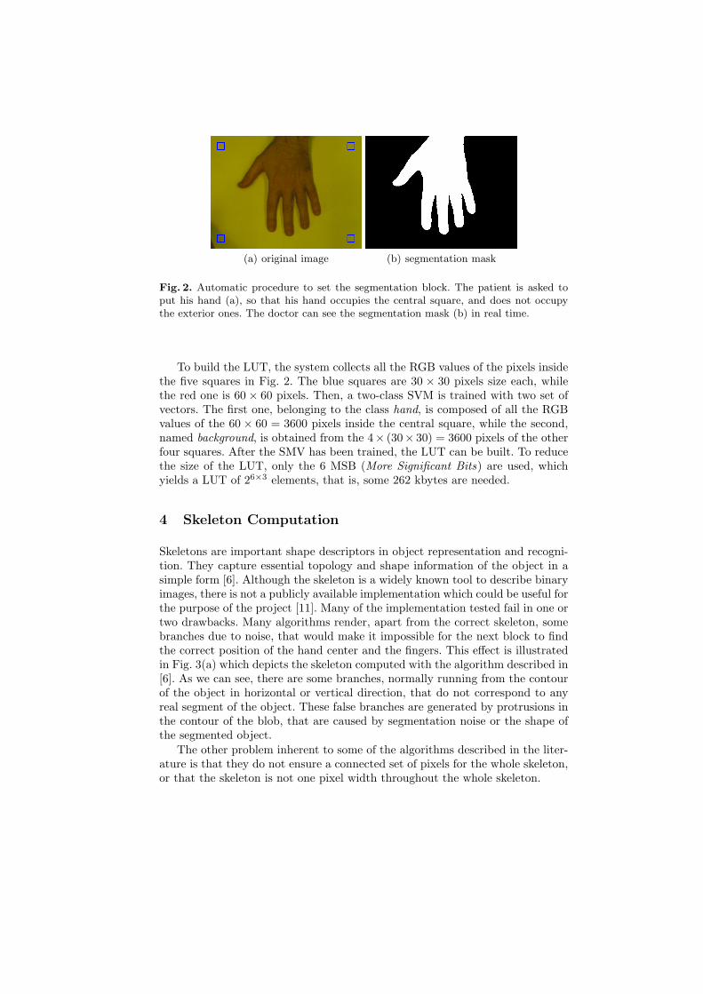
The patient is asked to put his hand inside the setup described in Fig. 1(a),in such a way that the image of his hand occupies a central square, and doesnot occlude a set of four squares at the corners of the image. Fig. 2 shows theprocess. During this step, the patient and the doctor can see the image of thehand, including the five squares superimposed in the image. The red one mustbe occupied completely by the hand, while the other four blue ones must onlybe on the background. In this situation, the doctor only needs to press a keyand the process to build the LUT is started.
(a) original image (b) segmentation mask
Fig. 2. Automatic procedure to set the segmentation block. The patient is asked toput his hand (a), so that his hand occupies the central square, and does not occupythe exterior ones. The doctor can see the segmentation mask (b) in real time.
To build the LUT, the system collects all the RGB values of the pixels insidethe five squares in Fig. 2. The blue squares are 30 × 30 pixels size each, whilethe red one is 60 × 60 pixels. Then, a two-class SVM is trained with two set ofvectors. The first one, belonging to the class hand, is composed of all the RGBvalues of the 60 × 60 = 3600 pixels inside the central square, while the second,named background, is obtained from the 4× (30× 30) = 3600 pixels of the otherfour squares. After the SMV has been trained, the LUT can be built. To reducethe size of the LUT, only the 6 MSB (More Significant Bits) are used, whichyields a LUT of 26×3 elements, that is, some 262 kbytes are needed.
4 Skeleton Computation
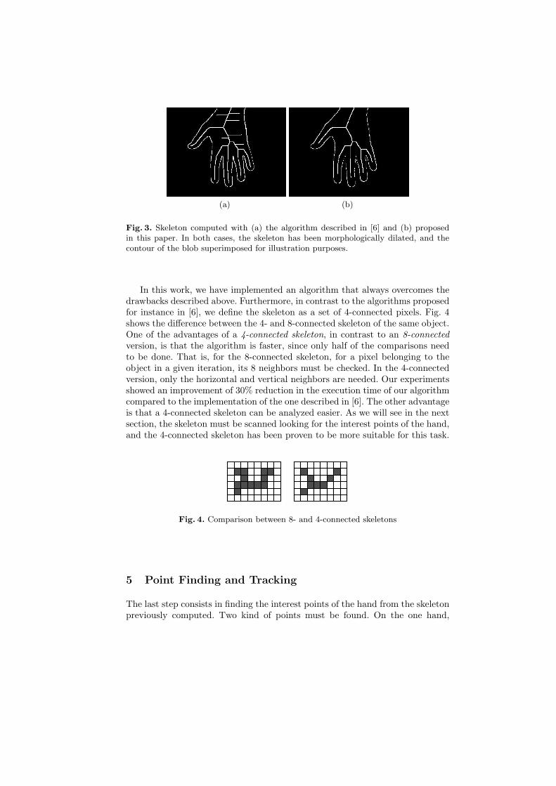
Skeletons are important shape descriptors in object representation and recogni-tion. They capture essential topology and shape information of the object in asimple form [6]. Although the skeleton is a widely known tool to describe binaryimages, there is not a publicly available implementation which could be useful forthe purpose of the project [11]. Many of the implementation tested fail in one ortwo drawbacks. Many algorithms render, apart from the correct skeleton, somebranches due to noise, that would make it impossible for the next block to findthe correct position of the hand center and the fingers. This effect is illustratedin Fig. 3(a) which depicts the skeleton computed with the algorithm described in[6]. As we can see, there are some branches, normally running from the contourof the object in horizontal or vertical direction, that do not correspond to anyreal segment of the object. These false branches are generated by protrusions inthe contour of the blob, that are caused by segmentation noise or the shape ofthe segmented object.
The other problem inherent to some of the algorithms described in the liter-ature is that they do not ensure a connected set of pixels for the whole skeleton,or that the skeleton is not one pixel width throughout the whole skeleton.
(a) (b)
Fig. 3. Skeleton computed with (a) the algorithm described in [6] and (b) proposedin this paper. In both cases, the skeleton has been morphologically dilated, and thecontour of the blob superimposed for illustration purposes.
In this work, we have implemented an algorithm that always overcomes thedrawbacks described above. Furthermore, in contrast to the algorithms proposedfor instance in [6], we define the skeleton as a set of 4-connected pixels. Fig. 4shows the difference between the 4- and 8-connected skeleton of the same object.One of the advantages of a 4-connected skeleton, in contrast to an 8-connectedversion, is that the algorithm is faster, since only half of the comparisons needto be done. That is, for the 8-connected skeleton, for a pixel belonging to theobject in a given iteration, its 8 neighbors must be checked. In the 4-connectedversion, only the horizontal and vertical neighbors are needed. Our experimentsshowed an improvement of 30% reduction in the execution time of our algorithmcompared to the implementation of the one described in [6]. The other advantageis that a 4-connected skeleton can be analyzed easier. As we will see in the nextsection, the skeleton must be scanned looking for the interest points of the hand,and the 4-connected skeleton has been proven to be more suitable for this task.
Fig. 4. Comparison between 8- and 4-connected skeletons
5 Point Finding and Tracking
The last step consists in finding the interest points of the hand from the skeletonpreviously computed. Two kind of points must be found. On the one hand,
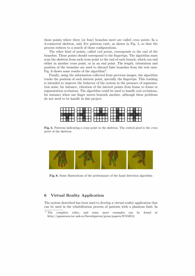
those points where three (or four) branches meet are called cross points. In a4-connected skeleton, only five patterns exist, as shown in Fig. 5, so that theprocess reduces to a search of these configurations.
The other kind of points, called end points, corresponds to the end of thebranches. These points should correspond to the fingertips. The algorithm mustscan the skeleton from each cross point to the end of each branch, which can endeither in another cross point, or in an end point. The length, orientation andposition of the branches are used to discard false branches from the real ones.Fig. 6 shows some results of the algorithm4.
Finally, using the information collected from previous images, the algorithmtracks the position of each interest point, specially the fingertips. This trackingis intended to improve the behavior of the system in the presence of segmenta-tion noise, for instance, vibration of the interest points from frame to frame orsegmentation occlusions. The algorithm could be used to handle real occlusions,for instance when one finger moves beneath another, although these problemsdo not need to be handle in this project.
Fig. 5. Patterns indicating a cross point in the skeleton. The central pixel is the crosspoint of the skeleton.
Fig. 6. Some illustrations of the performance of the hand detection algorithm
6 Virtual Reality Application
The system described has been used to develop a virtual reality application thatcan be used in the rehabilitation process of patients with a phantom limb. In
4 The complete video, and some more examples can be found athttp://agamenon.tsc.uah.es/Investigacion/gram/papers/ICIAR12.
this case, the system includes several games that can be played by the patient.Some videos showing the performance of the system can be downloaded fromthe web page.
The system has been implemented using the OpenCV library, version 2.3. Forthe segmentation block, we used the SVMs implementation included in OpenCV.The skeleton and the interest point finding and tracking were implemented inC++. The input of the system is a fire-wire camera, with 640× 480 pixels colorimages. The system works at 15 frames per second over a Pentium 4 2.4 GHzon a Linux kernel 2.6.32-25.
7 Conclusion and Future Work
In this paper, we have presented an algorithm for hand and finger detectionand tracking, that allows the development of a virtual reality application tobe used in rehabilitation processes, specifically, to reduce the pain in patientswith a phantom limb. The main contributions are the development of a skeletonextractor algorithm, that overcomes the achievement of the ones described inthe literature, and an algorithm which locates the interest points of the handfrom the skeleton. This algorithm has been used to develop a virtual realityapplication, which includes a set of games, that can be played by the patientwithin its rehabilitation process.
Future work includes several directions. The closest one is the developmentof several more applications or games. This must be done working together withdoctors and patients, since each patient could require a different set of activitiesfor his personal rehabilitation process.
The system is also open to depth images fusion. In our case, we intend touse a Microsoft Kinect sensor mainly to get the information about the height ofeach finger with respect to the table. This will allow the system to detect whenthe patient is pressing the background. This would widen the set of applicationsthat can be developed.
Although the main motivation of our work was the development of a virtualreality application, the applications of the system can be extended to other kindof projects, such as Sign Language Recognition [10, 18].
Acknowledgements
This work was supported by the Spanish Ministry for Science and Innovationproject number TIN2010-20845-C03-03, project UAH2011/EXP-030 from theUniversity of Alcala and the INNPACTO Program N. exp. IPT-2011-1366-390000.
References
1. Breuer, P., Eckes, C., Muller, S.: Hand gesture recognition with a novel IR time-of-flight range camera? a pilot study. In: Gagalowicz, A., Philips, W. (eds.) Computer
Vision - Computer Graphics Collaboration Techniques, Lecture Notes in ComputerScience, vol. 4418, pp. 247–260. Springer Berlin - Heidelberg (2007)
2. Chang, C., Lin, C.: LIBSVM: a library for support vector machines (2001), softwareavailable at http://www.csie.ntu.edu.tw/~cjlin/libsvm
3. Cole, J., Crowle, S., Austwick, G., Slater, D.H.: Exploratory findings with vir-tual reality for phantom limb pain; from stump motion to agency and analgesia.Disability and Rehabilitation 31(10), 846–854 (September 2009)
4. Elgammal, A., Muang, C., Hu, D.: Skin detection - a short tutorial. In: Encyclo-pedia of Biometrics (2009)
5. Gomez-Moreno, H., Maldonado-Bascon, S., Gil-Jimenez, P., Lafuente-Arroyo, S.:Goal evaluation of segmentation algorithms for traffic sign recognition. vol. PP,pp. 1–14 (July 2010)
6. Gonzalez, R., Woods, R.: Digital Image Processing. Addison-Wesley (1993)7. von Hardenberg, C., Berard, F.: Bare-hand human-computer interaction. In: Pro-
ceedings of the 2001 workshop on Perceptive user interfaces. pp. 1–8. PUI ’01(2001)
8. Hsieh, C.C., Liou, D.H., Lee, D.: A real time hand gesture recognition systemusing motion history image. In: Signal Processing Systems (ICSPS), 2010 2ndInternational Conference on. vol. 2, pp. V2–394 –V2–398 (july 2010)
9. Imai, A., Shimada, N., Shirai, Y.: 3-d hand posture recognition by training contourvariation. In: Automatic Face and Gesture Recognition, 2004. Proceedings. SixthIEEE International Conference on. pp. 895 – 900 (may 2004)
10. Isaacs, J., Foo, J.: Hand pose estimation for american sign language recognition.In: Southern Symposium on System Theory. pp. 132–136 (2004)
11. Lakshmi, J.K., Punithavalli, M.: A survey on skeletons in digital image processing.Digital Image Processing pp. 260 – 269 (2009)
12. Letessier, J., Berard, F.: Visual tracking of bare fingers for interactive surfaces. In:Proceedings of the 17th annual ACM symposium on User interface software andtechnology. pp. 119–122. UIST ’04, ACM (2004)
13. Lewis, J.S., Kersten, P., McCabe, C.S., McPherson, K.M., Blake, D.R.: Body per-ception disturbance: A contribution to pain in complex regional pain syndrome.In: CRPS (2007)
14. McCabe, C.S., Haigh, R.C., Ring, E.F., Halligan, P.W., Wall, P.D., Blake, D.R.:A controlled pilot study of the utility of mirror visual feedback in the treatmentof complex regional pain syndrome (type 1). Rheumatology 42(1), 97–101 (2003)
15. Murray, C.D., Patchick, E., Pettifer, S., Howard, T., Caillette, F., Kulkarni, J.,Bamford, C.: Investigating the efficacy of a virtual mirror box in treating phantomlimb pain in a sample of chronic sufferers. Disabil Human Dev 5(3), 227–234 (2006)
16. Nope R., S., Loaiza C., H., Caicedo B., E.: Estudio comparativo de tecnicas parael reconocimiento de gestos por vision artificial. Avances en Sistemas e Informtica5(3) (2009)
17. Oikonomidis, I., Nikolaos, K., Argyros, A.A.: Efficient model-based 3d trackingof hand articulations using kinect. In: Tracking Hand Articulations using Kinect(2011)
18. Pugeault, N., Bowden, R.: Spelling it out: Real-time ASL fingerspelling recognition.In: IEEE Workshop on Consumer Depth Cameras for Computer Vision (2011)
19. Ren, Z., Meng, J., Yuan, J., Zhang, Z.: Robust hand gesture recognition with kinectsensor. In: Proceedings of the 19th ACM international conference on Multimedia.pp. 759–760. MM ’11, ACM (2011)
20. Yoruk, E., Konukoglu, E., Sankur, B., Darbon, J.: Shape-based hand recognition.Image Processing, IEEE Transactions on 15(7), 1803 –1815 (july 2006)