Haider Ali, Christophe Hano, and Bilal Haider Abbasi ...
16
Research Article Mehreen Zaka*, Syed Salman Hashmi, Moiz A. Siddiqui, Lubna Rahman, Sadaf Mushtaq, Haider Ali, Christophe Hano, and Bilal Haider Abbasi* Callus-mediated biosynthesis of Ag and ZnO nanoparticles using aqueous callus extract of Cannabis sativa: Their cytotoxic potential and clinical potential against human pathogenic bacteria and fungi https://doi.org/10.1515/gps-2021-0057 received June 07, 2021; accepted August 16, 2021 Abstract: In this paper, we have presented the method of green synthesis of ZnO and Ag-NPs using the callus extract (CE) of medicinally important Cannabis sativa. The synthesis of nanoparticles (NPs) was confirmed by UV-Vis spectroscopy, while as far as the size and shape of the NPs were concerned, they were validated using the techniques of X-ray diffraction and scanning electron micro- scopy, respectively. The energy dispersive X-ray analysis graph confirmed the constitution of elements along with the surface chemical state of NPs. Fourier transform-infrared spectroscopy was utilized for the confirmation of biomole- cules capping the NPs. In order to test the application of these biosynthesized NPs on biological entities, four bac- terial strains, including Bacillus subtilis, Klebsiella pneu- monia, Staphylococcus aureus, and Pseudomonas aerugi - nosa, were used. On the other hand, five fungal strains, namely Mucor, Aspergillus flavus, Aspergillus fumigatus, Aspergillus niger, and Fusarium solani, were utilized for antifungal assay. Cytotoxicity assay was also performed using the HepG2 cell line. The results showed considerable antibacterial and antifungal activities. It also showed better cytotoxicity values as compared to the control. Keywords: Cannabis sativa, callus extract, green synth- esis, Ag-NPs, ZnO-NPs 1 Introduction Apart from the selection of edible plants, including wheat, rice, or maize, by earlier civilizations in order to obtain improved yields and quality, Cannabis sativa has also been selected and grown artificially in large quantities, making it the most renowned plant species on the earth. According to Richard E. Schultes, who was responsible for the origin of ethnobotany, although humans have extensively used the products of this plant for more than 10,000 years, it has not yet been recognized in the positive way it deserves [1]. C. sativa belongs to a group of herbaceous shrubs normally having a height of 1–2 m. It was estimated that its cultivation first started in Russia in about 4000 B.C. and is now widely used as a drug around the world by millions of people in the form of marijuana, hashish, or bhang. It has also been exploited as a source of fiber and oil. As far as its chemistry is concerned, it contains approx. 480 compounds, belonging to various chemical classes, including steroids, fatty acids, amino acids, alkaloids, terpenoids, and flavonoids [2]. The con- centrations of these compounds vary according to the type, age, variety, growth, and storage conditions of the tissue. Phytochemicals derived from C. sativa have been * Corresponding author: Mehreen Zaka, Department of Biotechnology, Quaid-i-Azam University, Islamabad-45320, Pakistan, e-mail: [email protected] Syed Salman Hashmi, Lubna Rahman, Haider Ali: Department of Biotechnology, Quaid-i-Azam University, Islamabad-45320, Pakistan Moiz A. Siddiqui: Department of Chemistry, Faculty of Sciences, Synergy Education, Faisalabad, Pakistan Sadaf Mushtaq: Department of Biotechnology, Quaid-i-Azam University, Islamabad-45320, Pakistan; Functional Genomics Group, Institute of Biomedical & Genetic Engineering (IBGE), Sector G-9/1, Islamabad, Pakistan Christophe Hano: LBLGC (Laboratoire de Biologie des Ligneux et des Grandes Cultures)- INRAE USC1328, University of Orleans, Department of Plant Sciences 45067 Orleans CEDEX 2, France * Corresponding author: Bilal Haider Abbasi, Department of Biotechnology, Quaid-i-Azam University, Islamabad-45320, Pakistan, e-mail: [email protected] Green Processing and Synthesis 2021; 10: 569–584 Open Access. © 2021 Mehreen Zaka et al., published by De Gruyter. This work is licensed under the Creative Commons Attribution 4.0 International License.
Transcript of Haider Ali, Christophe Hano, and Bilal Haider Abbasi ...
Research Article
Mehreen Zaka*, Syed Salman Hashmi, Moiz A. Siddiqui, Lubna Rahman, Sadaf Mushtaq,Haider Ali, Christophe Hano, and Bilal Haider Abbasi*
Callus-mediated biosynthesis of Ag and ZnOnanoparticles using aqueous callus extract ofCannabis sativa: Their cytotoxic potential andclinical potential against human pathogenicbacteria and fungi
https://doi.org/10.1515/gps-2021-0057received June 07, 2021; accepted August 16, 2021
Abstract: In this paper, we have presented the methodof green synthesis of ZnO and Ag-NPs using the callusextract (CE) of medicinally important Cannabis sativa.The synthesis of nanoparticles (NPs) was confirmed byUV-Vis spectroscopy, while as far as the size and shape ofthe NPs were concerned, they were validated using thetechniques of X-ray diffraction and scanning electron micro-scopy, respectively. The energy dispersive X-ray analysisgraph confirmed the constitution of elements along withthe surface chemical state of NPs. Fourier transform-infraredspectroscopy was utilized for the confirmation of biomole-cules capping the NPs. In order to test the application ofthese biosynthesized NPs on biological entities, four bac-terial strains, including Bacillus subtilis, Klebsiella pneu-monia, Staphylococcus aureus, and Pseudomonas aerugi-nosa, were used. On the other hand, five fungal strains,
namely Mucor, Aspergillus flavus, Aspergillus fumigatus,Aspergillus niger, and Fusarium solani, were utilized forantifungal assay. Cytotoxicity assay was also performedusing the HepG2 cell line. The results showed considerableantibacterial and antifungal activities. It also showed bettercytotoxicity values as compared to the control.
Keywords: Cannabis sativa, callus extract, green synth-esis, Ag-NPs, ZnO-NPs
1 Introduction
Apart from the selection of edible plants, including wheat,rice, or maize, by earlier civilizations in order to obtainimproved yields and quality, Cannabis sativa has alsobeen selected and grown artificially in large quantities,making it the most renowned plant species on the earth.According to Richard E. Schultes, who was responsiblefor the origin of ethnobotany, although humans haveextensively used the products of this plant for morethan 10,000 years, it has not yet been recognized in thepositive way it deserves [1]. C. sativa belongs to a group ofherbaceous shrubs normally having a height of 1–2 m. Itwas estimated that its cultivation first started in Russia inabout 4000 B.C. and is now widely used as a drug aroundthe world by millions of people in the form of marijuana,hashish, or bhang. It has also been exploited as a sourceof fiber and oil. As far as its chemistry is concerned, itcontains approx. 480 compounds, belonging to variouschemical classes, including steroids, fatty acids, aminoacids, alkaloids, terpenoids, and flavonoids [2]. The con-centrations of these compounds vary according to thetype, age, variety, growth, and storage conditions of thetissue. Phytochemicals derived from C. sativa have been
* Corresponding author: Mehreen Zaka, Department ofBiotechnology, Quaid-i-Azam University, Islamabad-45320,Pakistan, e-mail: [email protected]
Syed Salman Hashmi, Lubna Rahman, Haider Ali: Department ofBiotechnology, Quaid-i-Azam University, Islamabad-45320, PakistanMoiz A. Siddiqui: Department of Chemistry, Faculty of Sciences,Synergy Education, Faisalabad, PakistanSadaf Mushtaq: Department of Biotechnology, Quaid-i-AzamUniversity, Islamabad-45320, Pakistan; Functional GenomicsGroup, Institute of Biomedical & Genetic Engineering (IBGE), SectorG-9/1, Islamabad, PakistanChristophe Hano: LBLGC (Laboratoire de Biologie des Ligneux et desGrandes Cultures) - INRAE USC1328, University of Orleans,Department of Plant Sciences 45067 Orleans CEDEX 2, France
* Corresponding author: Bilal Haider Abbasi, Department ofBiotechnology, Quaid-i-Azam University, Islamabad-45320,Pakistan, e-mail: [email protected]
Green Processing and Synthesis 2021; 10: 569–584
Open Access. © 2021 Mehreen Zaka et al., published by De Gruyter. This work is licensed under the Creative Commons Attribution 4.0International License.
used in medicines as anti-vomiting, anti-spasm, anti-epi-lepsy, anti-asthma, and as an appetite stimulant [3].
Due to the remarkable properties of nanoparticles(NPs) such as electrical conductivity, mechanical strength,as well as magnetic and thermal properties, NPs havefound their way into research in almost every field. Butthe problem associated with their synthesis is that itrequires processes such as laser ablation, inert gas con-densation, or chemical reduction, which adds to the toxi-city of the process [4]. In order to synthesize NPs, morehuman and environmentally friendly methods are pre-ferred. Green synthesis involving plant extracts, micro-organisms, and enzymes is a very promising alternative tothe chemical and physical methods of NP synthesis [5]. Notonly can it minimize the toxicity of the particles, but it canalso increase the quality of the product. Phyto-nanotech-nology is also emerging as a simple, less expensive, time-friendly, and produces stable NPs [6]. Several types of NPs,especially metallic NPs like silver (Ag) [7], gold (Au) [8],zinc oxide (ZnO) [9], copper oxide (CuO) [10], iron (Fe),and lead oxide (PbO) [11] have been successfully preparedusing different plants. Out of these, silver nanoparticles (Ag-NPs) are among the most widely synthesized NPs owing totheir tremendous antimicrobial potential [8]. Similarly, zincoxide nanoparticles (ZnO-NPs) are also popular in biome-dical research due to their biocompatibility [12].
ZnO-NPs are regarded as metal oxide NPs havinga diverse range of significant characteristics like bio-compatible nature, cheap synthesis methods, and easyaccessibility [13]. The worldwide annual production ofZnO-NPs is approximately 31,000–34,000 metric tons,which made these metal oxide NPs a popular choice[14]. The semiconductor properties of ZnO-NPs due tothe presence of zinc and oxygen are promising. Theexciton binding energy ranges of ZnO-NPs are up to60meV, and the bandgap is 3.37 eV. The tolerance ofelectric fields is increased and falls in detectable ranges.These NPs are effective to work alone as well as in theform of complexes and bimetallic forms with other metals[12]. The ZnO-NP synthesis protocols are eco-friendly andbiocompatible, which make them a popular candidate tobe chosen for a variety of applications, including drugdelivery, diagnosis, biological research, agricultural research,biosensors, cosmetics, solar cells, semiconductors, and bio-logical imaging probes [15].
Ag-NPs are the most common and widely used metalNPs due to their multifarious properties [16]. Ag-NPshave been successfully synthesized using both physicaland chemical approaches; however, the time-consuming,expensive and toxic nature of these approaches make themless attractive, and hence, increasing focus is projected
toward a greener and a much safer synthesis approach[17,18]. The green synthesis approach for the Ag-NP synth-esis involves the use of living organisms like microbes,fungi, and plants or their products [19]. Ag-NPs havebeen effectively employed in research for multiple applica-tions, including optical devices, electronics, biosensors,and biolabeling. Ag-NPs have also been proved to be apotent antimicrobial and anti-cancer, wound-healing, watertreatment, and drug delivery agents [20].
Previously, the wild plant of C. sativa has beenexploited for silver (Ag) and gold (Au) nano-synthesis,but no reports are available on the exploitation of thecallus extract (CE) for the NP synthesis [8]. The inductionof callus in the controlled in vitro conditions offers anincreased quantity of secondary metabolites. The plantgrowth regulators (PGRs) used as stress inducers in thecallus induction are responsible for the increased sec-ondary metabolites production under specific lab condi-tions [12]. In this study, we have utilized Cannabis sativa(CS) CE to synthesize Ag and ZnO NPs. As the extract fromcallus is supposed to have an increased quantity of sec-ondary metabolites, it is a useful source for capping,reduction, and NP stabilization. The characterization ofthe biosynthesized NPs was done using UV-Vis spectro-scopy, Fourier transform-infrared spectroscopy (FT-IR),X-ray diffraction analysis (XRD), scanning electron micro-scopy (SEM), and energy dispersive X-ray analysis (EDX).
2 Materials and methods
2.1 Plant collection
Wild plants of Cannabis sativa (L.) were collected fromthe surroundings of Quaid-i-Azam University, Islamabad,and their fresh leaves were taken. In the next step, theleaves were washed two times, with high care so that allthe debris gets removed before taking it to the laminarflow hood. Then, 1% mercuric chloride solution wasapplied on the leaves for about 15 s, and these werethen washed five times with autoclaved distilled water.The leaves were then dried on autoclaved filter paper.
2.2 Establishment of callus cultures fromthe wild leaf explant
Inoculation of the leaf explants having a size of about1–2 cm was done on the MS (Murishage and Skoog,
570 Mehreen Zaka et al.

1962) medium, consisting of 8 g of agar and 30 g ofsucrose. Different concentrations of PGRs for cannabiscallus induction have been reported in the literatureand were exploited [21–23]. Different combinations ofPGRs like IBA (indole-3-butyric acid), BA (6-benzylami-nopurine), Kn (kinetin), 2,4-D(2,4-dichlorophenoxyaceticacid), NAA (1-naphthalene acetic acid), and TDZ (thidia-zuron) were added into the medium. The combination ofNAA and TDZ gave the best response in callus inductionat different concentrations. Optimization under differentPGR concentrations was done according to the protocolsavailable in the previous literature [22]. Briefly, the con-centrations used for optimization are given in Table 1.
The best response was recorded at concentrations of0.50mg·L−1 NAA and 1.00mg·L−1 TDZ. Under laminarflow, pH was balanced between 5.6 and 5.7, utilizing 1 NHCl and 1 N NaOH. The experiment was conducted usingthree replicates such that the inoculation of the threeexplants was done in each conical flask as one replicatefor one PGR concentration. These flasks were then placedat 25°C for a 16/8 h photoperiod in a growth room.
2.3 Calli extract preparation for thebiosynthesis of NPs
For the CE preparation, 10 g of callus was added to 100mLof distilled water in an Erlenmeyer flask (500mL). The
resulting mixture was boiled for 8min. After boiling, thetemperature of Cannabis CE was cooled to room tempera-ture, and it was filtered through Whatman no. 1 filterpaper. The filtrate was then stored at a temperature of4°C after adjusting its volume to 100mL.
2.4 Biosynthesis of ZnO-NPs
The plant extract and metallic salt concentrations, alongwith pH, temperature, and response time, play a vital rolein the biosynthesis of metal NPs [24]. The precursor saltused for the synthesis of ZnO NPs was zinc acetate dihy-drate (C4H6O4Zn). A 0.02 M zinc acetate dihydrate solu-tion was prepared for the synthesis of ZnO NPs. In thefirst step, 1 mL of the sample extract was mixed sepa-rately with 0.02 M zinc acetate (50mL) solution undercontinuous stirring. The pH of the resulting mixturewas adjusted at 12 using sodium hydroxide (NaOH). Alka-loids, terpenoids, and flavonoids available in the extractcaused the reduction. The resulting precipitates were whiteand pale, which were subjected to washing with ethanolbefore being washed with distilled water twice (Figure 1a).The sample was then centrifuged for 15min at 6,000 rpmprior to incubation at 60°C overnight to dry. Drying of thesamples yielded powdered ZnO-NPs, which were then uti-lized for characterization and biological assays.
2.5 Biosynthesis of Ag-NPs
For the synthesis of Ag-NPs, the protocols of Abbasi et al.[8] were used with slight modifications. In brief, the calliextract was separately mixed with 1 mM AgNO3 (pre-cursor salt) in different ratios (1:2, 1:5, and 1:10). Theincubation of the blend was done at room temperaturefor 24 h. A color change from yellow to brown wasobserved (Figure 1b), then the absorbance of each test
Table 1: C. sativa callus induction using different concentrations ofNAA and TDZ
Sr No NAA (mg·L−1) TDZ (mg·L−1)
1 0.50 1.002 1.00 1.003 1.50 1.004 2.00 1.00
Figure 1: (a) Dried pale white precipitates of ZnO-NPs; (b) color change after the Ag-NP synthesis at different concentrations.
Callus-mediated biosynthesis of Ag and ZnO nanoparticles 571
sample was recorded through a UV-Vis spectrophoto-meter in order to confirm the biosynthesis of Ag-NPsfrom the calli extract. Once the Ag-NP biosynthesis wascomplete, the NPs were separated through centrifugationat 12,000 rpm for 10min. The isolated Ag-NPs were washedthrice using distilled water.
2.6 Characterization techniques
2.6.1 UV vis spectroscopy
A Shimadzu UV-1650 PC Spectrophotometer was used toconfirm the biosynthesis of Ag-NPs and ZnO-NPs. Thelight absorption spectra were provided using distilledwater as a reference. The surface plasmon resonancepeak of Ag-NPs and ZnO-NPs was noted. The spectrawere recorded at different times to check the absorbanceand wavelength of the NPs.
2.6.2 XRD analysis
The crystalline nature of the structures of Ag-NPs andZnO-NPs was determined using an XRD (Model-D8 Advance,Germany) instrument. In this instrument, the cathode rayreleases the X-rays in the direction of the sample. Forthe examination and study, 1 mg of each Ag-NPs andZnO-NPs were taken. For determining the size of theNPs, the Debye–Scherrer equation was used as follows:
= /D kλ β θcos (1)
where λ is the X-ray wavelength (1.5418 Å), k is the shapefactor (0.94), θ is the Bragg’s angle, and β is the full widthat half-maximum in radians.
2.6.3 Fourier transform infrared spectroscopy (FTIR)
FTIR spectroscopy (Spectrum One, Perkin Elmer, Waltham,MA, USA) was used for determining the functional groupsattached to ZnO-NPs and Ag-NPs. In the FTIR spectroscope,KBr pellets were utilized, and the data was recorded in thespectral range of 500 to 3,500 cm−1.
2.6.4 SEM and EDX analysis
The morphology and the sizes of NPs were determinedusing an SEM (MIRA3 TESCAN model), operated at 10 kV.
In the initial step, a small quantity of the sample was placedon a grid made of copper before coating it with carbon. Thiscoat was then subjected to drying under a mercury lamp forabout 5min; the samples were examined, and their photo-graphs were captured. EDX analysis was carried out to ana-lyze the elemental composition. The samples were coatedwith carbon, dried, and further analysis was performedusing SEM equipped with an EDX detector.
2.7 Antibacterial activity
For antibacterial activity assay, a well diffusion methodwas used according to Bereksi et al. [25]. The bacteriaagainst which the activity of these NPs was evaluatedincluded both Gram-positive and Gram-negative bacteria.Gram-positive bacteria included Bacillus subtilis (ATCC:6633) and Staphylococcus epidermidis (ATCC: 14990), andGram-negative bacterial strains included Klebsiella pneu-monia (ATCC: 4617) and Pseudomonas aeruginosa (ATCC:9721). In the first step, the inoculation of bacterial strainswas done in 10mL tryptic soy broth (TSB), which wasthen incubated at 120 rpm for 24 h at 37°C temperature ina shaking incubator. The inoculum was prepared andstandardized at 1 × 10⁸ CFU·mL−1 for each bacterial strain.Further, on a Trypticase soy agar media plate, each of thebacterial broth cultures (100 µL)was poured and spread inorder to get a uniform bacterial lawn using a sterilizedglass rod. In the case of NPs, in each of the 5 wells,30µL of each NP with serial dilutions of 5, 4, and 3mg·mL−1
for Ag-NPs and 10, 5, and 4mg·mL−1 for ZnO-NPs was intro-duced. The positive control used in this experiment wasCefixime. The broth dilution method of Tai et al. [26] wasutilized to calculate the corresponding MICs.
2.8 Antifungal activity
For antifungal activity assay, the agar well diffusionmethodof Nath and Joshi [27] was used with some slight modifica-tions. Five fungal strains (pathogenic), includingAspergillusfumigatus (FCBP-1264), Aspergillus niger (FCBP-0198),Fusarium solani (FCBP-1114), Aspergillus flavus (FCBP-0064), andMucor (FCBP-0041), were taken and the activityof NPs was investigated. Themedia used for the preparationof broth cultures of pathogenic fungi was the Sabourauddextrose liquid medium (Oxoid: CM0147). After inoculatingthe fungal strains in the broth, it was incubated in a shakingincubator at a temperature of 37°C for 24 h. The opticaldensity (OD) of the broth cultures was adjusted to 0.5 beforecarrying out the assay. About 50 µL of the broth culture
572 Mehreen Zaka et al.
from standardized cultures was taken and was spread on aPetri plate containing the Sabouraud dextrose agar mediumusing a sterilized glass rod in order to obtain a steady lawnof pathogenic fungal strains. The positive control used inthis experiment was Amphotericin B. The plates were incu-bated at 37°C for 24–48 h, and the inhibition zones wererecorded after a certain period of time.
2.9 Cytotoxicity screening
2.9.1 Cell culture
Dulbecco’s modified Eagle’s medium consisting of 10%fetal calf serum along with 100 U·mL−1 penicillin, L-glu-tamine (2 mM), and 100 μg·mL−1 streptomycin at a
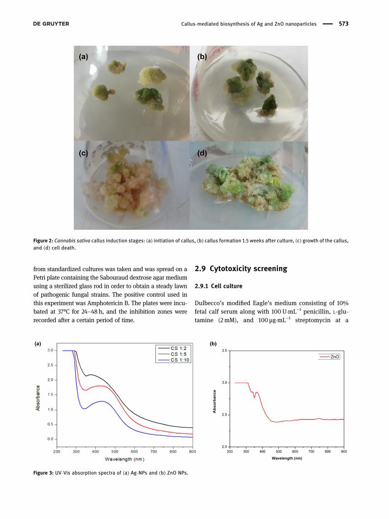
Figure 2: Cannabis sativa callus induction stages: (a) initiation of callus, (b) callus formation 1.5 weeks after culture, (c) growth of the callus,and (d) cell death.
Figure 3: UV-Vis absorption spectra of (a) Ag-NPs and (b) ZnO NPs.
Callus-mediated biosynthesis of Ag and ZnO nanoparticles 573
temperature of 37°C and a humid atmosphere that con-tained 5% CO2 and 1mM Na-pyruvate as a supplementwas used for culturing human hepatocellular carcinomacells (ATCC HB-8065). Apart from this, 0.5mM trypsin/EDTAwas used at room temperature for 1min in order to harvestthe cells.
2.9.2 Cell viability assay
In a sulforhodamine B (SRB) assay, ZnO-NPs and Ag-NPswere tested to check their cytotoxic effect on the HepG2cell line. For cytotoxic screening assay, NP samples wereprepared by suspending them in deionized water. Then, a
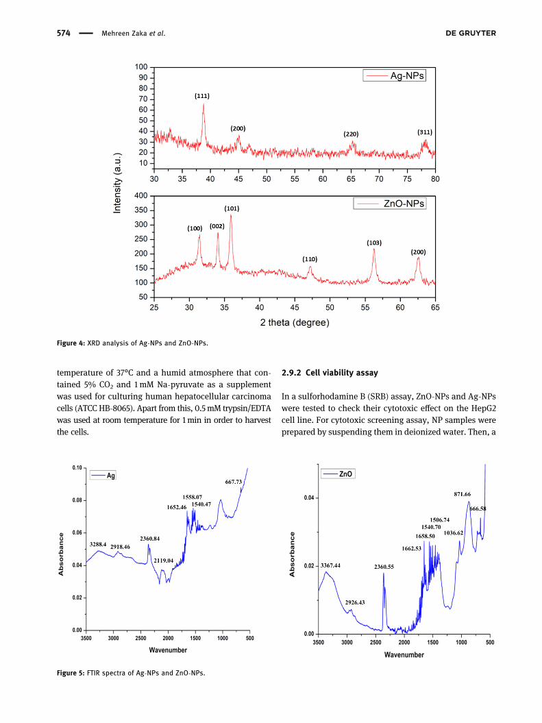
Figure 4: XRD analysis of Ag-NPs and ZnO-NPs.
Figure 5: FTIR spectra of Ag-NPs and ZnO-NPs.
574 Mehreen Zaka et al.
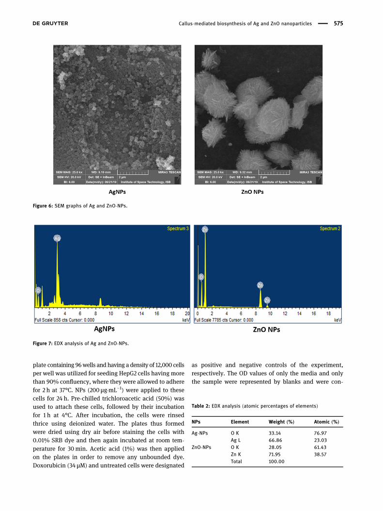
plate containing 96wells and having a density of 12,000 cellsper well was utilized for seeding HepG2 cells having morethan 90% confluency, where they were allowed to adherefor 2 h at 37°C. NPs (200 µg·mL−1) were applied to thesecells for 24 h. Pre-chilled trichloroacetic acid (50%) wasused to attach these cells, followed by their incubationfor 1 h at 4°C. After incubation, the cells were rinsedthrice using deionized water. The plates thus formedwere dried using dry air before staining the cells with0.01% SRB dye and then again incubated at room tem-perature for 30min. Acetic acid (1%) was then appliedon the plates in order to remove any unbounded dye.Doxorubicin (34 µM) and untreated cells were designated
as positive and negative controls of the experiment,respectively. The OD values of only the media and onlythe sample were represented by blanks and were con-
Figure 6: SEM graphs of Ag and ZnO-NPs.
Figure 7: EDX analysis of Ag and ZnO-NPs.
Table 2: EDX analysis (atomic percentages of elements)
NPs Element Weight (%) Atomic (%)
Ag-NPs O K 33.14 76.97Ag L 66.86 23.03
ZnO-NPs O K 28.05 61.43Zn K 71.95 38.57Total 100.00
Callus-mediated biosynthesis of Ag and ZnO nanoparticles 575
sidered as controls. An Olympus CK2 light microscopeequipped with a digital camera was utilized to take thesnapshots. Using triplicates of each of the samples, thisexperiment was repeated two times. Then, 100 µL of10 mM Tris having a pH of 8 was introduced into eachof the wells at room temperature for 5 min in orderto make SRB dye more soluble. A microplate reader(Platos R 496, AMP) was then utilized to analyze theabsorbance values at a wavelength of 565 nm. The viabi-lity percentage was calculated relative to the untreatedsample by the following formula:
( ) =
−
−
×
Cell viability percentage %Absorbance of sample Absorbance of control
Absorbance of untreated cells Absorbance of media100
(2)
While the cell inhibition percentage was determinedby the following formula:
( ) = −
( )
Cell inhibition percentage % 100 Cell viabilitypercentage %
(3)
2.10 Statistical analysis
All the statistical calculations were done in triplicates. Thevalues present in the text as well as in the figures werestatistically examined as mean ± SE (standard error).Origin Pro (version 8.5) software was used to analyze allthe graphs. The probability value was viewed to be notablycontrasting at the point of P < 0.05 (95%).
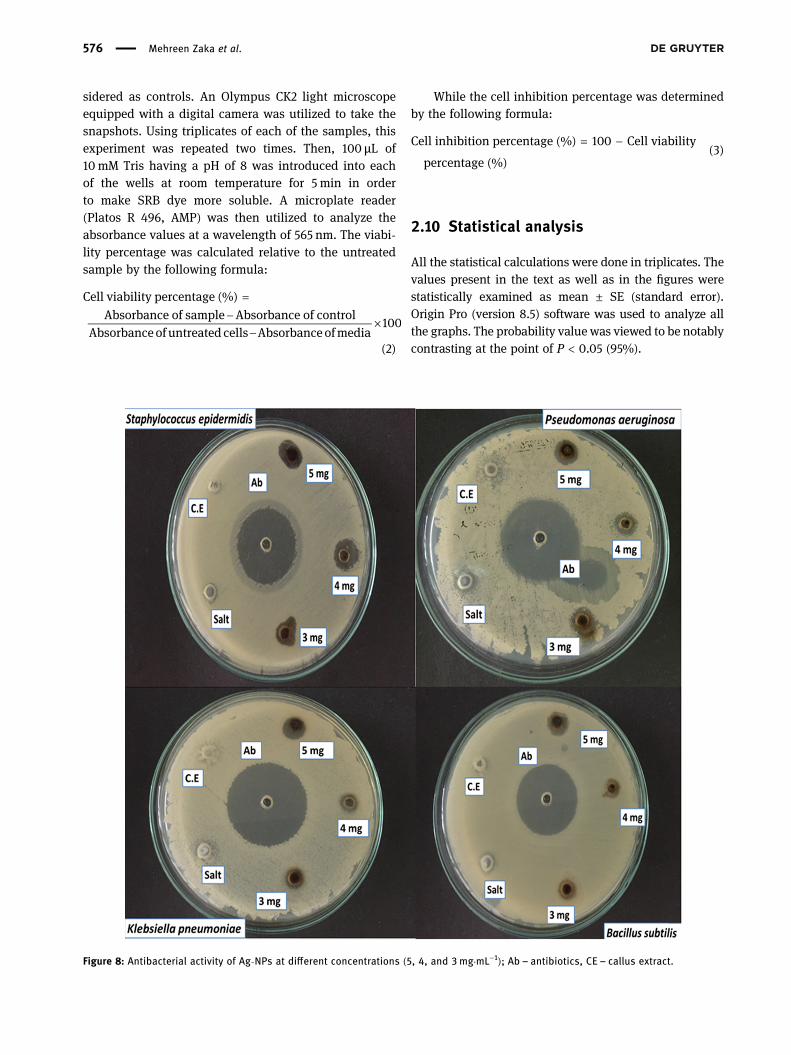
Figure 8: Antibacterial activity of Ag-NPs at different concentrations (5, 4, and 3mg·mL−1); Ab – antibiotics, CE – callus extract.
576 Mehreen Zaka et al.
3 Results and discussion
3.1 Callus induction
Different studies have shown the optimization of Cannabiscallus induction at different concentrations of PGRs [21–23].The protocol of callus induction was optimized by using thePGR concentrations reported in the previous literature. Thedevelopment of callus cultures was done using leaf explantsfrom the wild C. sativa plant. Leaf portions of about 3–4mmwere cut out and were then cultured on the Murashige andSkoog medium. The optimized callus was off-white and fri-able obtained at concentrations of 1.00mg·L−1 TDZ and0.50mg·L−1 NAA of PGRs (Figure 2).
3.2 UV-Vis analysis
A preliminary confirmation of biosynthesis of Ag-NPs andoptimization of suitable CE to precursor salt ratio for thebiosynthesis using CE was carried out. The mixture ofAgNO3 and the extracts mixed in different ratios werecharacterized using a UV-Vis spectrophotometer. The mix-ture for CE and AgNO3 in 1:2 displayedmaximumabsorbance(2.188) at a wavelength of 382 nm. At 1:5, the maximumabsorbance (1.811) was recorded at 422 nm, while at 1:10the maximum absorbance (1.284) was recorded at 430nm.Due to the optimized absorbance and a sharp peak near422 nm, 1:5 was selected for further experiments and charac-terization. Figure 3a shows the spectrophotometric analysisof CE-mediated Ag-NPs at the aforementioned ratios. Thebiosynthesis of ZnO NPs was also confirmed using spectro-photometric analysis. Soon after the formation of precipi-tates, the mixture was subjected to a spectrophotometer.The maximum absorbance was recorded at 362 nm, whichis a characteristic of ZnO NPs. Figure 3b represents the spec-trophotometric analysis of CE-mediated ZnO NPs.
3.3 XRD results
For determining the properties like film thickness, purityof phase (crystalline nature), and the ordering of atoms inamorphous materials, the XRD technique is applied. Inthis analysis, the overall structure of the material isobserved based on the X-ray penetration. The distinctpeaks for silver were obtained at 38.10°, 46.42°, 69.27°,and 76.87°, which correspond to the lattice patterns (111),(200), (220), and (311) as discussed by Amin [28]. Thepeak at the (111) plane is the predominant orientationbecause it is more intense as compared to other peaks.Ta
ble3:
Antibacterial
activity
ofCEan
dcallu
s-med
iatedAg-NPs
andZn
O-NPs
agains
ttw
oGram-neg
ative(Klebs
iella
pneu
mon
iae,
Pseu
domon
asae
rugino
sa),an
dtw
oGram-pos
itiveba
cteria
(Staph
ylococcu
sep
idermidis,Bacillus
subtilis)
Bacterial
strains
Diameter
oftheinhibition
zone
(mm)
Ag-NPs
ZnO-NPs
5mg·mL−
14mg·mL−
13mg·mL−
1CE
Cefixime
(10μg
·mL−
1 )Precurso
rsa
lt10
mg·mL−
15mg·mL−
14mg·mL−
1Ce
fixime
(10μg
·mL−
1 )CE
Precurso
rsa
lt
Gram
positive
Stap
hylococcus
epidermidis
13±0.65
14±0.70
10±0.50
5±0.25
35±1.75
5±0.25
13±0.65
6±0.30
6±0.30
30±1.50
7±0.35
8±0.40
Bacillus
subtilis
11±0.55
8±0.40
7±0.35
4±0.20
28±1.40
6±0.30
14±0.70
15±0.75
15±0.75
27±1.35
10±0.50
8±0.40
Gram
nega
tive
Kleb
siella
pneu
mon
iae
11±0.55
8±0.40
7±0.35
5±0.25
35±1.75
6±0.30
14±0.70
12±0.60
20±1.00
30±1.50
14±0.70
7±0.35
Pseu
domon
asae
rugino
sa12
±0.60
10±0.50
12±0.60
5±0.25
30±1.50
9±0.45
18±0.90
12±0.60
6±0.30
30±1.50
13±0.65
8±0.40
Value
sareaverag
eof
triplic
ates
±stan
dard
error.
Abb
reviation:
CE–callu
sextract.
Callus-mediated biosynthesis of Ag and ZnO nanoparticles 577
The results show that the synthesized Ag-NPs had aface-centered cubic (fcc) structure. Figure 4 representsthe XRD pattern for CE-mediated Ag-NPs and ZnO-NPs.The 2theta values of the peaks obtained were 33.81°, 34.06°,35.91°, 47.19°, 56.29°, and 62.65° which corresponds to(100), (002), (101), (110), (103), and (200) planes of lattices,respectively, as discussed by Siddiquah et al. [12]. A crystal-line structure for callus-mediated ZnO-NPs was revealedthrough XRD data. The average size of Ag-NP and ZnO-NPnanoparticles was calculated by using Debye–Scherer’sequation. First, individual peaks sizes were calculated,and then the average of all the individual peaks in the graphwas taken, and the average size was found to be 14.85 nmfor Ag-NPs and 16.43 nm for ZnO-NPs.
3.4 FT-IR analysis
The confirmation of capping of phytochemicals upon NPswas done by subjecting the powdered NPs to FT-IR ana-lysis. FT-IR analysis of C. sativa CE-mediated Ag-NPsshowed peaks at 3288.44, 2918.46, 2360.84, 2119.04, 1652.46,1558.07, 1540.47, 1521.11, 1506.06, 1456.70, 1036.62, and667.73 cm−1 (Figure 5a), while C. sativa CE-mediated ZnO-NPsshowed peaks at 3367.44, 2926.43, 2360.55, 1662.53, 1658.50,1540.70, 1506.74, 1456.51, 1032.80, 871.86, and 666.85 cm−1
(Figure 5b). The peaks at 3288.44 and 3367.44 cm−1 showO–H stretching in alcohols, the peaks at 2918.46 and2926.43 cm−1 show C–H stretching of alkanes [7], the peaksat 2360.84 and 2360.55 cm−1 show asymmetric C]O
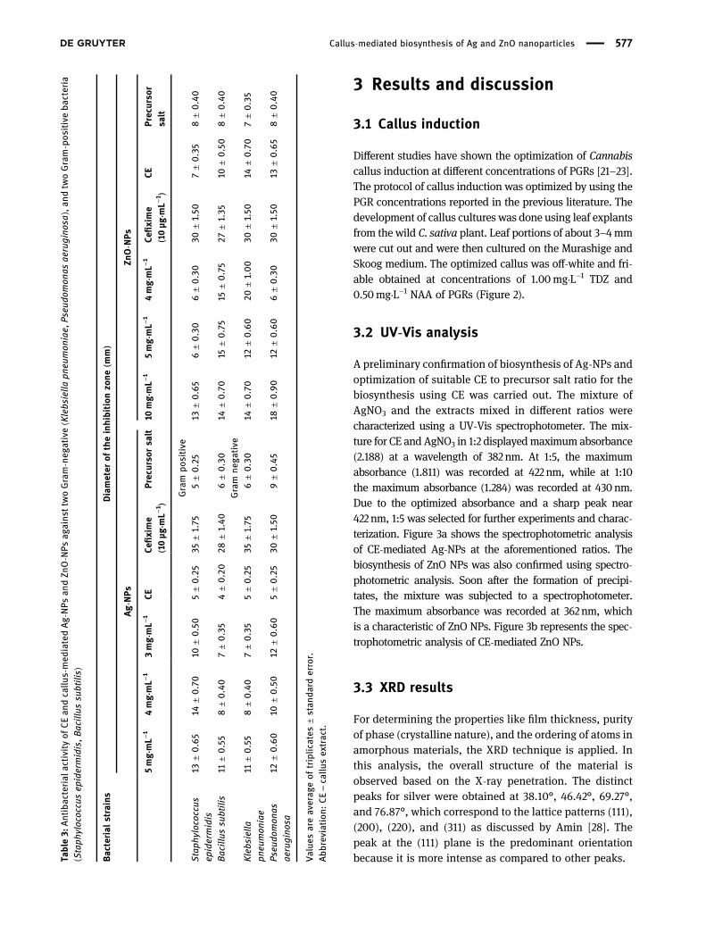
Figure 9: Antibacterial activity of ZnO-NPs at different concentrations (10, 5, and 4mg·mL−1); Ab – antibiotics, CE – callus extract.
578 Mehreen Zaka et al.
stretching [12], the peaks at 1652.46 and 1658.50 cm−1 showC]C stretching in alkenes, the multiple peaks between1,400 and 1,600 cm−1 show C]C stretching in aromaticcompounds, the peaks at 1036.62 and 1032.80 cm−1 showC–O stretching, the peak at 871.86 cm−1 shows C–N stretchingin amines [8], while the peaks at 667.73 and 666.85 cm−1 showC–Cl stretching in alkyl halides. The relationship betweenthe primary and secondary metabolites and the Ag-NPs andZnO-NPs can be clearly observed from the findings. Thisattachment of functional groups is due to the presence ofelectrostatic forces between the positively charged zinc ionsandnegatively charged groups present in organicmolecules. Itis the result of this binding force that makes these NPs per-fectly suitable for different applications including the study ofbiological interactions [29] and drug delivery [30].
3.5 SEM and EDX analysis
Figure 6 shows the SEM images of Ag-NPs and ZnO-NPs,respectively, while Figure 7 shows the EDX analyses ofAg-NPs and ZnO-NPs, respectively. In the case of Ag-NPs,highly aggregated NPs having roughly spherical shapewas observed. Similar results were reported by Hashmiet al. [7]. The ZnO-NPs synthesized were shown to behighly aggregated and needle-shaped stacked togetherin flower-like morphology. The EDX spectrum for Ag-NPs
confirms the presence of elemental silver and a hint of thepresence of oxygen in the sample. A strong signal of ele-mental silver can be seen around 3 keV. Noohpisheh et al.[31] also reported the synthesis of Ag-ZnO compositesusing Trigonella foenum-graecum leaf extract. In thecase of ZnO-NPs, the presence of both elemental Zn andO is evident. Strong Zn signals were observed around1 keV while two other signals can be seen at 8.6 and9.6 keV. The signal for elemental O was obtained around0.5 keV. No signals were found for any other metal in thesample, which proves that the NPs were highly pure.The results are in agreement with Siddiquah et al. [12].The relative concentration of each elemental componentis given in Table 2 for Ag-NPs and ZnO-NPs. The data inthe table also show that the synthesized NPs were free ofany impurities.
3.6 Antibacterial activity
In antibacterial activity, inhibition against all the bacterialstrains was observed after treatment with CE-mediatedAg-NPs and ZnO-NPs. The experiment was performed atvarious concentrations, i.e., 5, 4, and 3mg·mL−1 for Ag-NPsand 10, 5, and 4mg·mL−1 for ZnO-NPs. It was found thatCE-mediated Ag-NPs showed the highest activity againstS. epidermidis with 14mm zone followed by P. aeruginosa
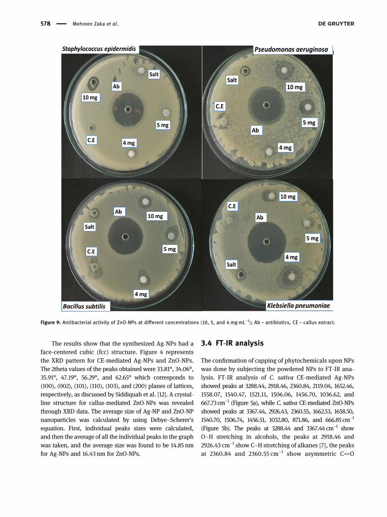
Figure 10: Antifungal activity of Ag-NPs against five different fungal strains at different concentrations (5, 4, and 3 mg·mL−1);Ab – antibiotics, CE – callus extract.
Callus-mediated biosynthesis of Ag and ZnO nanoparticles 579
with 12mm zone (Figure 8). ZnO-NPs showed promisingactivity against K. pneumoniae with an inhibition zone of20mm, P. aeruginosa with a maximum zone of inhibitionof 18mm, followed by B. subtilis with 15mm zone withinMIC ranges (300–120 μg·mL−1) (Table 3 and Figure 9). Ourresults are in agreement with Lara et al. [32]. According tothem, the bactericidal effect of Ag-NPs was potent againstthe P. aeruginosa strain. Ag-NPs have significant antibac-terial potential against various strains of bacteria, such asGram-negative and Gram-positive bacterial strains. Sev-eral antimicrobial properties of Ag-NPs have been investi-gated due to their chemical stability, wound-healing cap-ability, catalytic activity, or high conductivity [33]. Ag-NPsexhibit antimicrobial properties that mainly rely on super-ficial contact; thus, they prevent respiratory chains byinhibiting the enzymatic system and change the synthesispattern of DNA. Since the small size of NPs gives a largesurface area as compared to other salts (including silverparticulate), it thus provides a platform to bind withmicro-organisms via the cell membrane and enter inside a cell[34]. The mode of antibacterial action of ZnO-NPs involvesentering into a bacterium and getting attached to its cellmembrane. These NPs alter the process of productionof energy by affecting the cell membrane, thus releasingthe cell contents [35]. This result is in agreement with theprevious literature. Siddiquah et al. [12] reported thatZnO-NPs have a broad-spectrum bactericidal effect. Accord-ing to Gunalan et al. [45], the bactericidal activity of thebiosynthesized ZnO-NPs is increased as compared to che-mically synthesized ZnO-NPs. Owing to the high surfacearea and chemical stability of ZnO-NPs, it is available tothe cytoplasm of microbes and, as a result, the destructionof cells leads to apoptosis [46,47]. Likewise, Nazir et al. [9]examined that the biosynthesized ZnO-NPs by using CE ofSilybum marianum has greater potential against B. subtilisand K. pneumonia, as compared to chemically synthesizedZnO-NPs.
3.7 Antifungal assay
The antifungal potential of callus-mediated Ag-NP andZnO-NP samples was evaluated by a modified agar well-diffusion assay described previously by Ginovyan et al.[36]with some modifications. All the NPs showed differentactivities. Antifungal activity was investigated for the givenstock concentrations of 5, 4, and 3mg·mL−1 for CE-mediatedAg-NPs and 10, 5, and 4mg·mL−1 for CE-mediated ZnO-NPs.The NPs were evaluated against pathogenic fungi includ-ing Mucor (FCBP-0041), Aspergillus flavus (FCBP-0064),Ta
ble4:
Antifun
galactivities
ofAg-NPs
,Zn
O-N
Ps,an
dCEof
Cann
abis
sativ
aan
alyzed
agains
tpa
thog
enic
fung
iMuc
or,As
pergillus
flav
us,As
pergillus
fumigatus
,As
pergillus
nige
r,an
dFusa
rium
solani
Fung
alstrains
Diameter
oftheinhibition
zone
(mm)
Ag-NPs
ZnO-NPs
CE5mg·mL−
14mg·mL−
13mg·mL−
1Precurso
rsa
ltAmph
otericin
B(10μg
·mL−
1 )CE
10mg·mL−
15mg·mL−
14mg·mL−
1Precurso
rSalt
Amph
otericin
B(10μg
·mL−
1 )
Aspe
rgillus
nige
r6±0.30
7±0.35
5±0.25
10±0.50
12±0.60
30±1.50
7±0.35
12±0.60
10±0.50
15±0.75
10±0.50
40±2.00
Aspe
rgillus
fumigatus
5±0.25
6±0.30
7±0.35
5±0.25
5±0.25
45±2.25
15±0.75
20±1.00
15±0.75
5±0.25
6±0.30
45±2.25
Aspe
rgillus
flav
us7±0.35
6±0.30
5±0.25
6±0.30
7±0.35
30±1.50
6±0.30
11±0.55
12±0.60
7±0.35
10±0.50
40±2.00
Muc
or6±0.30
7±0.35
4±0.20
4±0.20
5±0.25
17±0.85
7±0.35
30±1.50
30±1.50
20±1.00
15±1.00
20±1.00
Fusa
rium
solani
6±0.30
6±0.30
6±0.30
6±0.30
5±0.25
35±1.75
6±0.30
10±0.50
6±0.30
7±0.35
5±0.25
25±1.25
Value
sareaverag
eof
triplic
ates
±stan
dard
error.
Abb
reviation:
CE–callu
sextract.
580 Mehreen Zaka et al.
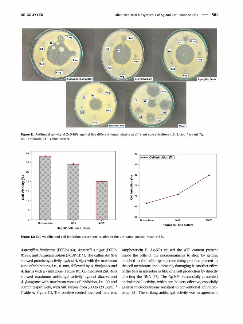
Aspergillus fumigatus (FCBP-1264) Aspergillus niger (FCBP-0198), and Fusarium solani (FCBP-1114). The callus Ag-NPsshowed promising activity againstA. nigerwith themaximumzone of inhibitions, i.e., 10mm, followed by A. fumigatus andA. flavuswith a 7mm zone (Figure 10). CE-mediated ZnO-NPsshowed maximum antifungal activity against Mucor, andA. fumigatus with maximum zones of inhibition, i.e., 30 and20mm respectively, with MIC ranges from 300 to 120 μg·mL−1
(Table 4, Figure 11). The positive control involved here was
Amphotericin B. Ag-NPs caused the ATP content presentinside the cells of the microorganisms to drop by gettingattached to the sulfur group containing proteins present inthe cell membrane and ultimately damaging it. Another effectof the NPs in microbes is blocking cell production by directlyaffecting the DNA [37]. The Ag-NPs successfully presentedantimicrobial activity, which can be very effective, especiallyagainst microorganisms resistant to conventional antimicro-bials [38]. The striking antifungal activity was in agreement
Figure 11: Antifungal activity of ZnO-NPs against five different fungal strains at different concentrations (10, 5, and 4mg·mL−1);Ab – antibiotic, CE – callus extract.
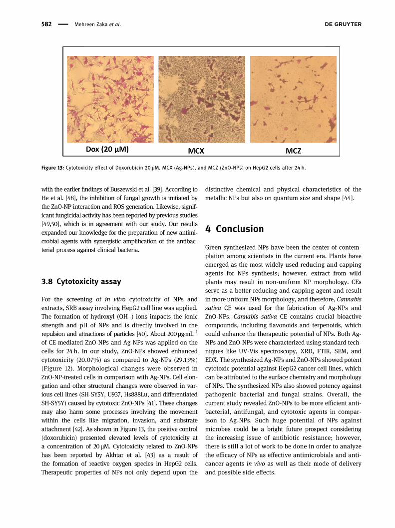
Figure 12: Cell viability and cell inhibition percentage relative to the untreated control (mean ± SE).
Callus-mediated biosynthesis of Ag and ZnO nanoparticles 581
with the earlier findings of Buszewski et al. [39]. According toHe et al. [48], the inhibition of fungal growth is initiated bythe ZnO-NP interaction and ROS generation. Likewise, signif-icant fungicidal activity has been reported by previous studies[49,50], which is in agreement with our study. Our resultsexpanded our knowledge for the preparation of new antimi-crobial agents with synergistic amplification of the antibac-terial process against clinical bacteria.
3.8 Cytotoxicity assay
For the screening of in vitro cytotoxicity of NPs andextracts, SRB assay involving HepG2 cell line was applied.The formation of hydroxyl (OH−) ions impacts the ionicstrength and pH of NPs and is directly involved in therepulsion and attractions of particles [40]. About 200µg·mL−1
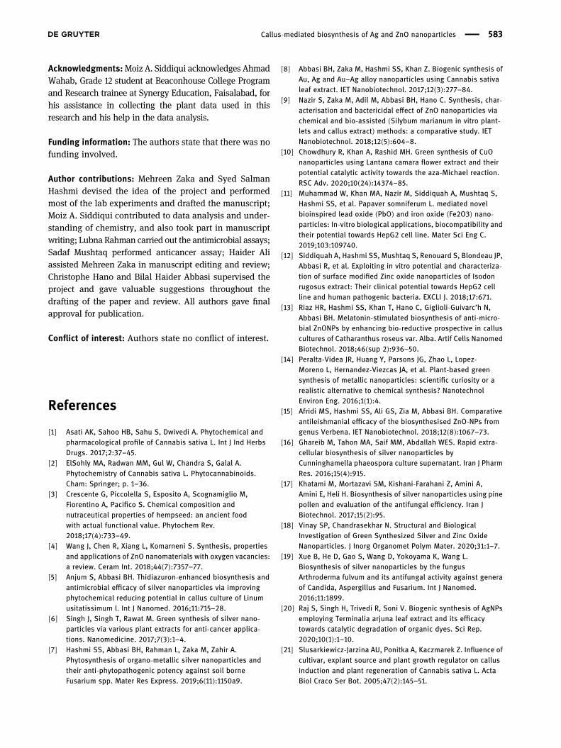
of CE-mediated ZnO-NPs and Ag-NPs was applied on thecells for 24 h. In our study, ZnO-NPs showed enhancedcytotoxicity (20.07%) as compared to Ag-NPs (29.13%)(Figure 12). Morphological changes were observed inZnO-NP-treated cells in comparison with Ag-NPs. Cell elon-gation and other structural changes were observed in var-ious cell lines (SH-SY5Y, U937, Hs888Lu, and differentiatedSH-SY5Y) caused by cytotoxic ZnO-NPs [41]. These changesmay also harm some processes involving the movementwithin the cells like migration, invasion, and substrateattachment [42]. As shown in Figure 13, the positive control(doxorubicin) presented elevated levels of cytotoxicity ata concentration of 20 µM. Cytotoxicity related to ZnO-NPshas been reported by Akhtar et al. [43] as a result ofthe formation of reactive oxygen species in HepG2 cells.Therapeutic properties of NPs not only depend upon the
distinctive chemical and physical characteristics of themetallic NPs but also on quantum size and shape [44].
4 Conclusion
Green synthesized NPs have been the center of contem-plation among scientists in the current era. Plants haveemerged as the most widely used reducing and cappingagents for NPs synthesis; however, extract from wildplants may result in non-uniform NP morphology. CEsserve as a better reducing and capping agent and resultin more uniform NPs morphology, and therefore, Cannabissativa CE was used for the fabrication of Ag-NPs andZnO-NPs. Cannabis sativa CE contains crucial bioactivecompounds, including flavonoids and terpenoids, whichcould enhance the therapeutic potential of NPs. Both Ag-NPs and ZnO-NPs were characterized using standard tech-niques like UV-Vis spectroscopy, XRD, FTIR, SEM, andEDX. The synthesized Ag-NPs and ZnO-NPs showed potentcytotoxic potential against HepG2 cancer cell lines, whichcan be attributed to the surface chemistry and morphologyof NPs. The synthesized NPs also showed potency againstpathogenic bacterial and fungal strains. Overall, thecurrent study revealed ZnO-NPs to be more efficient anti-bacterial, antifungal, and cytotoxic agents in compar-ison to Ag-NPs. Such huge potential of NPs againstmicrobes could be a bright future prospect consideringthe increasing issue of antibiotic resistance; however,there is still a lot of work to be done in order to analyzethe efficacy of NPs as effective antimicrobials and anti-cancer agents in vivo as well as their mode of deliveryand possible side effects.
Figure 13: Cytotoxicity effect of Doxorubicin 20 µM, MCX (Ag-NPs), and MCZ (ZnO-NPs) on HepG2 cells after 24 h.
582 Mehreen Zaka et al.
Acknowledgments:Moiz A. Siddiqui acknowledges AhmadWahab, Grade 12 student at Beaconhouse College Programand Research trainee at Synergy Education, Faisalabad, forhis assistance in collecting the plant data used in thisresearch and his help in the data analysis.
Funding information: The authors state that there was nofunding involved.
Author contributions: Mehreen Zaka and Syed SalmanHashmi devised the idea of the project and performedmost of the lab experiments and drafted the manuscript;Moiz A. Siddiqui contributed to data analysis and under-standing of chemistry, and also took part in manuscriptwriting; Lubna Rahman carried out the antimicrobial assays;Sadaf Mushtaq performed anticancer assay; Haider Aliassisted Mehreen Zaka in manuscript editing and review;Christophe Hano and Bilal Haider Abbasi supervised theproject and gave valuable suggestions throughout thedrafting of the paper and review. All authors gave finalapproval for publication.
Conflict of interest: Authors state no conflict of interest.
References
[1] Asati AK, Sahoo HB, Sahu S, Dwivedi A. Phytochemical andpharmacological profile of Cannabis sativa L. Int J Ind HerbsDrugs. 2017;2:37–45.
[2] ElSohly MA, Radwan MM, Gul W, Chandra S, Galal A.Phytochemistry of Cannabis sativa L. Phytocannabinoids.Cham: Springer; p. 1–36.
[3] Crescente G, Piccolella S, Esposito A, Scognamiglio M,Fiorentino A, Pacifico S. Chemical composition andnutraceutical properties of hempseed: an ancient foodwith actual functional value. Phytochem Rev.2018;17(4):733–49.
[4] Wang J, Chen R, Xiang L, Komarneni S. Synthesis, propertiesand applications of ZnO nanomaterials with oxygen vacancies:a review. Ceram Int. 2018;44(7):7357–77.
[5] Anjum S, Abbasi BH. Thidiazuron-enhanced biosynthesis andantimicrobial efficacy of silver nanoparticles via improvingphytochemical reducing potential in callus culture of Linumusitatissimum l. Int J Nanomed. 2016;11:715–28.
[6] Singh J, Singh T, Rawat M. Green synthesis of silver nano-particles via various plant extracts for anti-cancer applica-tions. Nanomedicine. 2017;7(3):1–4.
[7] Hashmi SS, Abbasi BH, Rahman L, Zaka M, Zahir A.Phytosynthesis of organo-metallic silver nanoparticles andtheir anti-phytopathogenic potency against soil borneFusarium spp. Mater Res Express. 2019;6(11):1150a9.
[8] Abbasi BH, Zaka M, Hashmi SS, Khan Z. Biogenic synthesis ofAu, Ag and Au–Ag alloy nanoparticles using Cannabis sativaleaf extract. IET Nanobiotechnol. 2017;12(3):277–84.
[9] Nazir S, Zaka M, Adil M, Abbasi BH, Hano C. Synthesis, char-acterisation and bactericidal effect of ZnO nanoparticles viachemical and bio-assisted (Silybum marianum in vitro plant-lets and callus extract) methods: a comparative study. IETNanobiotechnol. 2018;12(5):604–8.
[10] Chowdhury R, Khan A, Rashid MH. Green synthesis of CuOnanoparticles using Lantana camara flower extract and theirpotential catalytic activity towards the aza-Michael reaction.RSC Adv. 2020;10(24):14374–85.
[11] Muhammad W, Khan MA, Nazir M, Siddiquah A, Mushtaq S,Hashmi SS, et al. Papaver somniferum L. mediated novelbioinspired lead oxide (PbO) and iron oxide (Fe2O3) nano-particles: In-vitro biological applications, biocompatibility andtheir potential towards HepG2 cell line. Mater Sci Eng C.2019;103:109740.
[12] Siddiquah A, Hashmi SS, Mushtaq S, Renouard S, Blondeau JP,Abbasi R, et al. Exploiting in vitro potential and characteriza-tion of surface modified Zinc oxide nanoparticles of Isodonrugosus extract: Their clinical potential towards HepG2 cellline and human pathogenic bacteria. EXCLI J. 2018;17:671.
[13] Riaz HR, Hashmi SS, Khan T, Hano C, Giglioli-Guivarc’h N,Abbasi BH. Melatonin-stimulated biosynthesis of anti-micro-bial ZnONPs by enhancing bio-reductive prospective in calluscultures of Catharanthus roseus var. Alba. Artif Cells NanomedBiotechnol. 2018;46(sup 2):936–50.
[14] Peralta-Videa JR, Huang Y, Parsons JG, Zhao L, Lopez-Moreno L, Hernandez-Viezcas JA, et al. Plant-based greensynthesis of metallic nanoparticles: scientific curiosity or arealistic alternative to chemical synthesis? NanotechnolEnviron Eng. 2016;1(1):4.
[15] Afridi MS, Hashmi SS, Ali GS, Zia M, Abbasi BH. Comparativeantileishmanial efficacy of the biosynthesised ZnO-NPs fromgenus Verbena. IET Nanobiotechnol. 2018;12(8):1067–73.
[16] Ghareib M, Tahon MA, Saif MM, Abdallah WES. Rapid extra-cellular biosynthesis of silver nanoparticles byCunninghamella phaeospora culture supernatant. Iran J PharmRes. 2016;15(4):915.
[17] Khatami M, Mortazavi SM, Kishani-Farahani Z, Amini A,Amini E, Heli H. Biosynthesis of silver nanoparticles using pinepollen and evaluation of the antifungal efficiency. Iran JBiotechnol. 2017;15(2):95.
[18] Vinay SP, Chandrasekhar N. Structural and BiologicalInvestigation of Green Synthesized Silver and Zinc OxideNanoparticles. J Inorg Organomet Polym Mater. 2020;31:1–7.
[19] Xue B, He D, Gao S, Wang D, Yokoyama K, Wang L.Biosynthesis of silver nanoparticles by the fungusArthroderma fulvum and its antifungal activity against generaof Candida, Aspergillus and Fusarium. Int J Nanomed.2016;11:1899.
[20] Raj S, Singh H, Trivedi R, Soni V. Biogenic synthesis of AgNPsemploying Terminalia arjuna leaf extract and its efficacytowards catalytic degradation of organic dyes. Sci Rep.2020;10(1):1–10.
[21] Slusarkiewicz-Jarzina AU, Ponitka A, Kaczmarek Z. Influence ofcultivar, explant source and plant growth regulator on callusinduction and plant regeneration of Cannabis sativa L. ActaBiol Craco Ser Bot. 2005;47(2):145–51.
Callus-mediated biosynthesis of Ag and ZnO nanoparticles 583
[22] Lata H, Chandra S, Khan IA, ElSohly MA. High frequency plantregeneration from leaf derived callus of high Δ9-tetrahydro-cannabinol yielding Cannabis sativa L. Planta Medica.2010;76(14):1629–33.
[23] Movahedi M, Ghasemi-Omran V, Torabi S. The effect of dif-ferent concentrations of TDZ and BA on in vitro regeneration ofIranian cannabis (Cannabis sativa) using cotyledon and epi-cotyl explants. J Plant Mol Breed. 2015;3(2):20–7.
[24] Thi TUD, Nguyen TT, Thi YD, Thi KHT, Phan BT, Pham KN. Greensynthesis of ZnO nanoparticles using orange fruit peelextract for antibacterial activities. RSC Adv.2020;10(40):23899–907.
[25] Bereksi MS, Hassaïne H, Bekhechi C, Abdelouahid DE.Evaluation of antibacterial activity of some medicinal plantsextracts commonly used in Algerian traditional medicineagainst some pathogenic bacteria. Pharmacogn J.2018;10:3507–512.
[26] Tai Z, Cai L, Dai L, Dong L, Wang M, Yang Y, et al. Antioxidantactivity and chemical constituents of edible flower of Sophoraviciifolia. Food Chem. 2011;126(4):1648–54.
[27] Nath A, Joshi S. Anti-candidal effect of endophytic fungi iso-lated from Calotropis gigantean. Rev de Biol Trop.2017;65(4):1437–47.
[28] Amin M, Anwar F, Janjua MRSA, Iqbal MA, Rashid U. Greensynthesis of silver nanoparticles through reduction withSolanum xanthocarpum L. berry extract: characterization,antimicrobial and urease inhibitory activities againstHelicobacter pylori. Int J Mol Sci. 2012;13(8):9923–41.
[29] Song JY, Jang HK, Kim BS. Biological synthesis of gold nano-particles using Magnolia kobus and Diopyros kaki leafextracts. Process Biochem. 2009;44(10):1133–8.
[30] Wang R, Billone PS, Mullett WM. Nanomedicine in action: anoverview of cancer nanomedicine on the market and in clinicaltrials. J Nanomater. 2013;2013:629681.
[31] Noohpisheh Z, Amiri H, Farhadi S, Mohammadi-gholami A.Green synthesis of Ag-ZnO nanocomposites using Trigonellafoenum-graecum leaf extract and their antibacterial, anti-fungal, antioxidant and photocatalytic properties.Spectrochim Acta Part A Mol Biomol Spectrosc.2020;245:118595.
[32] Lara HH, Ayala-Núñez NV, Turrent LDCI, Padilla CR. Bactericidaleffect of silver nanoparticles against multidrug-resistant bac-teria. World J Microbiol Biotech. 2010;26(4):615–21.
[33] Sweet MJ, Singleton I. Silver nanoparticles: a microbial per-spective. Adv Appl Microbiol. Academic Press. 2011;77:115–33.
[34] Hsiao IL, Hsieh YK, Wang CF, Chen IC, Huang YJ. Trojan-horsemechanism in the cellular uptake of silver nanoparticles ver-ified by direct intra-and extracellular silver speciation ana-lysis. Environ Sci Technol. 2015;49(6):3813–21.
[35] Manivasagan P, Venkatesan J, Senthilkumar K, Sivakumar K,Kim SK. Biosynthesis, antimicrobial and cytotoxic effect ofsilver nanoparticles using a novel Nocardiopsis sp. MBRC-1.BioMed Res Int. 2013;2013:287638.
[36] Ginovyan M, Petrosyan M, Trchounian A. Antimicrobial activityof some plant materials used in Armenian traditional medi-cine. BMC Compl Alternat Med. 2017;17(1):50.
[37] Bhaduri GA, Little R, Khomane RB, Lokhande SU, Kulkarni BD,Mendis BG, et al. Green synthesis of silver nanoparticles usingsunlight. J Photochem Photobiol A Chem. 2013;258:1–9.
[38] Sharma VK, Yngard RA, Lin Y. Silver nanoparticles: greensynthesis and their antimicrobial activities. Adv ColloidInterface Sci. 2009;145(1–2):83–96.
[39] Buszewski B, Railean-Plugaru V, Pomastowski P, Rafińska K,Szultka-MLynska M, Golinska P, et al. Antimicrobial activity ofbiosilver nanoparticles produced by a novel Streptacidiphilusdurhamensis strain. J Microbiol Immunol Infect.2018;51(1):45–54.
[40] Peng C, Zhang W, Gao H, Li Y, Tong X, Li K, et al. Behavior andpotential impacts of metal-based engineered nanoparticles inaquatic environments. Nanomaterials. 2017;7(1):21.
[41] Najim N, Rusdi R, Hamzah AS, Shaameri Z, Mat Zain M,Kamarulzaman N. Effects of the absorption behaviour of ZnOnanoparticles on cytotoxicity measurements. J Nanomater.2014;2014:1–10.
[42] Brandhagen BN, Tieszen CR, Ulmer TM, Tracy MS,Goyeneche AA, Telleria CM. Cytostasis and morphologicalchanges induced by mifepristone in human metastatic cancercells involve cytoskeletal filamentous actin reorganization andimpairment of cell adhesion dynamics. BMC Cancer.2013;13(1):1–15.
[43] Akhtar MJ, Ahamed M, Kumar S, Khan MM, Ahmad J,Alrokayan SA. Zinc oxide nanoparticles selectively induceapoptosis in human cancer cells through reactive oxygenspecies. Int J Nanomed. 2012;7:845.
[44] Khan ZUH, Khan A, Chen Y, Shah NS, Muhammad N, Khan AU,et al. Biomedical applications of green synthesized Nobelmetal nanoparticles. J Photochem Photobiol B Biol.2017;173:150–64.
[45] Gunalan S, Sivaraj R, Rajendran V. Green synthesized ZnOnanoparticles against bacterial and fungal pathogens. ProgNat Sci Mater Int. 2012;22(6):693–700.
[46] Yousef JM, Danial EN. In vitro antibacterial activity andminimum inhibitory concentration of zinc oxide and nano-particle zinc oxide against pathogenic strains. J Health Sci.2012;2(4):38–42.
[47] Santos-Filho SD. Erythrocyte membrane and hemolysis:effects of natural products. Int J Life Sci Technol. 2016;9(3):28.
[48] He L, Liu Y, Mustapha A, Lin M. Antifungal activity of zinc oxidenanoparticles against Botrytis cinerea and Penicilliumexpansum. Microbiol Res. 2011;166(3):207–15.
[49] Sharma D, Rajput J, Kaith BS, Kaur M, Sharma S. Synthesis ofZnO nanoparticles and study of their antibacterial and anti-fungal properties. Thin Solid Films. 2010;519(3):1224–9.
[50] Lipovsky A, Nitzan Y, Gedanken A, Lubart R. Antifungal activityof ZnO nanoparticles – the role of ROS mediated cell injury.Nanotechnology. 2011;22(10):105101.
584 Mehreen Zaka et al.