Hafez et al. 2011
8
This article appeared in a journal published by Elsevier. The attached copy is furnished to the author for internal non-commercial research and education use, including for instruction at the authors institution and sharing with colleagues. Other uses, including reproduction and distribution, or selling or licensing copies, or posting to personal, institutional or third party websites are prohibited. In most cases authors are permitted to post their version of the article (e.g. in Word or Tex form) to their personal website or institutional repository. Authors requiring further information regarding Elsevier’s archiving and manuscript policies are encouraged to visit: http://www.elsevier.com/copyright
Transcript of Hafez et al. 2011
7/22/2019 Hafez et al. 2011
http://slidepdf.com/reader/full/hafez-et-al-2011 1/8
This article appeared in a journal published by Elsevier. The attached
copy is furnished to the author for internal non-commercial research
and education use, including for instruction at the authors institution
and sharing with colleagues.
Other uses, including reproduction and distribution, or selling or
licensing copies, or posting to personal, institutional or third partywebsites are prohibited.
In most cases authors are permitted to post their version of the
article (e.g. in Word or Tex form) to their personal website or
institutional repository. Authors requiring further information
regarding Elsevier’s archiving and manuscript policies are
encouraged to visit:
http://www.elsevier.com/copyright
7/22/2019 Hafez et al. 2011
http://slidepdf.com/reader/full/hafez-et-al-2011 2/8
Author's personal copy
Neurobiology
Neprilysin-2 Is an Important -Amyloid Degrading
Enzyme
Daniel Hafez,* Jeffrey Y. Huang,*
Alexis M. Huynh,* Stephanie Valtierra,*
Edward Rockenstein,†‡ Angela M. Bruno,*Bao Lu,§ Luc DesGroseillers,¶ Eliezer Masliah,†‡
and Robert A. Marr*
From the Department of Neuroscience,* Rosalind Franklin
University of Medicine and Science, North Chicago, Illinois; the
Departments of Neurosciences † and Pathology,‡ University of
California, San Diego, La Jolla, California; Children’s Hospital,§
Harvard Medical School, Boston Massachusetts; and the
Department of Biochemistry,¶ University of Montreal, Montreal,
Quebec, Canada
Proteases that degrade the amyloid- peptide (A ) are
important in protecting against Alzheimer’s disease
(AD), and understanding these proteases is critical to
understanding AD pathology. Endopeptidases sensitive
to inhibition by thiorphan and phosphoramidon are
especially important, because these inhibitors induce
dramatic A accumulation (30- to 50-fold) and patho-
logical deposition in rodents. The A -degrading enzyme
neprilysin (NEP) is the best known target of these in-
hibitors. However, genetic ablation of NEP results in
only modest increases (1.5- to 2-fold) in A , indicating
that other thiorphan/phosphoramidon-sensitive en-
dopeptidases are at work. Of particular interest is the
NEP homolog neprilysin 2 (NEP2), which is thiorphan/
phosphoramidon-sensitive and degrades A . We inves-
tigated the role of NEP2 in A degradation in vivo
through the use of gene knockout and transgenic mice.
Mice deficient for the NEP2 gene showed significant
elevations in total A species in the hippocampus and
brainstem/diencephalon (1.5-fold). Increases in A
accumulation were more dramatic in NEP2 knockout
mice crossbred with APP transgenic mice. In NEP/NEP2
double-knockout mice, A levels were marginally in-
creased (1.5- to 2-fold), compared with NEP //
NEP2/ controls. Treatment of these double-knockout
mice with phosphoramidon resulted in elevations of A , suggesting that yet other NEP-like A -degrading en-
dopeptidases are contributing to A catabolism. (Am J
Pathol 2011, 178:306–312; DOI: 10.1016/j.ajpath.2010.11.012)
Alzheimer’s disease (AD) is a neurodegenerative disor-
der currently affecting more than 26 million people world-
wide and, as advances in modern medicine prolong life-span, this number is expected to quadruple by 2050.1 A
major factor believed to be involved in the progression of
AD pathology is the accumulation of amyloid- peptide
(A). Studying the mechanisms of A clearance is, there-
fore, very important to understanding AD.
Currently, enzymatic degradation is thought to play an
integral role in the removal of A. Of the A-degrading
enzymes, neprilysin (NEP) has been shown to be highly
critical for cerebral A control.2 NEP expression has also
been inversely correlated with amyloid pathology in hu-
mans and mice, and NEP gene transfer has been re-
ported to reduce amyloid pathology in transgenic mice
(reviewed by Marr and Spencer3
). Despite the impor-tance of NEP-mediated A degradation, NEP knockout
(KO) mice show only moderately elevated A levels (1.5-
to 2-fold), insufficient to cause plaque deposition.4 How-
ever, when treated with thiorphan, an NEP endopeptidase
inhibitor, mice and rats demonstrate pathological accumu-
lations of A after only 1 month.2,5 This was also found in
mice treated with phosphoramidon, another NEP inhibitor.6
These results indicate that there may exist additional NEP-
like endopeptidases in the metalloprotease 13 (M13) family
that are central to the A clearance pathway.
The NEP homolog neprilysin 2 (NEP2) is one such endo-
peptidase. NEP2 (also known as MMEL1/2, SEP, NL1, NE-
PLP) possesses a 55% sequence identity to NEP and hasbeen shown to degrade vasoactive peptides.7,8 In addition,
the membrane-bound -splice form of murine NEP2 has
demonstrated A-degrading properties in membrane frac-
tions.9 In transduced HEK293T cells, our research group
previously showed that cell surface human NEP2 (-splice
form) was able to degrade both A42 and A40 peptides.10
Subcellular localization studies using transfection into CHO
cells have found that murine NEP2 is present primarily in the
Accepted for publication September 23, 2010.
Supplemental material for this article can be found on http://ajp.
amjpathol.org or at doi:10.1016/j.ajpath.2010.11.012.
Address reprint requests to Dr. Robert A. Marr, Ph.D., Department of
Neuroscience, Rosalind Franklin University of Medicine and Science,
3333 Green Bay Rd., North Chicago, IL 60064. E-mail: robert.marr@
rosalindfranklin.edu.
The American Journal of Pathology, Vol. 178, No. 1, January 2011
Copyright © 2011 American Society for Investigative Pathology.
Published by Elsevier Inc. All rights reserved.
DOI: 10.1016/j.ajpath.2010.11.012
306

7/22/2019 Hafez et al. 2011
http://slidepdf.com/reader/full/hafez-et-al-2011 3/8
Author's personal copy
endoplasmic reticulum.7,11 However, Oh-Hashi and col-
leagues12 did find murine NEP2 activity at the cell surface.
Studies of NEP2 in mice and rats have demonstrated that it
is expressed primarily neurons and shows mild expression
in the cerebral cortex, mild to moderate expression in thehippocampus, and strong expression in numerous tha-
lamic, hypothalamic, and brainstem nuclei.13–15 In mice,
however, expression in the hippocampus could not be con-
firmed, because of high background levels.15 NEP2 is in-
volved in sperm function in mice and modulates fertilization
and early embryonic development.16 However, NEP2 also
functions as a neuropeptidase, one that is possibly involved
in several physiological pathways controlling nociception,
energy, and endocrine functions.15,17,18
The purpose of this study was to investigate the in vivo
role of NEP2 in A degradation in mice. Using NEP2 KO16
and NEP/NEP2 double-knockout (DKO) mice, the impor-
tance of NEP2 was investigated by measuring A levels invarious regions in the brain of KO and control mice. In
addition, APP transgenic mice crossed with NEP2 KO mice
were used to test the effect of NEP2 on A removal when
challenged with high A levels. Defining the role of NEP2 in
A clearance will provide further insight into AD pathogen-
esis and possible therapies for this disease.
Materials and Methods
Animals
All mice were used according to institutional Animal Care
and Use Committee-approved protocols and in accor-
dance with the Guide for Care and Use of Laboratory Ani-
mals as published in 1996 by the U.S. National Academy of
Sciences. NEP2 (Mme/1) KO mice16 were obtained from the
laboratory of Dr. Luc DesGroseillers. NEP/NEP2 DKO mice
were created by cross-breeding NEP2 KO mice (129 back-
ground) with NEP (Mme) KO mice (BALB/c background) (re-
ceived from the laboratory of Dr. Bao Lu).19 These mice were
backcrossed once more with the NEP KO mice (BALB/c back-
ground) before being maintained by inbreeding NEP / /
NEP2 / or NEP / /NEP2 / strains. APPtg/NEP2 KO mice
were created by cross-breeding NEP2 KO mice (129 back-
ground) with APPtg mice (TASD41 line, received from the
laboratory of Dr. Eliezer Masliah) (Blk/Sw background).20
These mice are propagated by breeding heterozygous APP
transgenic mice having either wild-type or NEP2 / back-
grounds with wild-type (129) or NEP2 / (129) mice, re-
spectively. The analysis was done with mice that had been
backcrossed three times with the 129 lines.
Mouse genotypes were confirmed by PCR analysis.
Briefly, NEP2 gene knockout mice were identified using a
specific primer set (wt-F: 5=-TGGAACTGGAGACGCAT-
CTGG-3=, Neo-F: 5=-TCCTGTCATCTCACCTGGCTCC-3=,
NL1-R: 5=-TAGCTCCATCAGGTCCATTCG-3=): APPtg mice
were identified by using a specific primer pair (APPtg-F:
5=-GGCTACGAAAATCCAACCTACAAG-3=, APPtg-R: 5=-
GATGATGGCATGCAGCACTGG-3=); and NEP KO mice
were identified using a specific primer set (Exon12-F: 5=-
GAAATCATGTCAACTGTG-3=, Neo-R: 5=-ATCAGAAGCT-
TATCGAT-3=, Exon13-R: 5=-CTTGCGGAAAGCATTTC-3=). Ex-
amples of PCR genotyping results are presented in
Supplemental Figure S1 (at http://ajp.amjpathol.org).
A Enzyme-Linked Immunosorbent Assay
Mice were anesthetized with 2-bromo-2-chloro-1,1,1-tri-
fluoroethane (Sigma-Aldrich, St. Louis, MO) and were
sacrificed by cervical dislocation for A analysis. Brains
were extracted and dissected into four regions: cerebellum,
brainstem/diencephalon, hippocampus, and cerebral cor-
tex. Samples were stored at 80°C until analysis. Frozen
brain sections were homogenized in lysis buffer (5 mol/L
guanidine HCl; Fisher Scientific, Pittsburgh, PA) using a
Polytron homogenizer (Kinematica, Bohemia, NY). Quanti-
fication of A1-40 and A1-42 was performed using isoform-
specific A enzyme-linked immunosorbent assay (ELISA)
kits (Wako Chemicals, Richmond, VA).4 Statistics were de-
termined using two-tailed Student’s t -test compared withcontrol.
Immunohistochemistry
Mouse hemibrains were placed in 4% paraformaldehyde
(Fisher Scientific) fixative for 24 hours before storage in a
cryoprotectant solution (30% sucrose, 0.1 mol/L PO4). Fixed
hemibrains were sagittally sectioned on a freezing mic-
rotome (40 m) and stored in a cryoprotectant solution at
20°C. Every sixth brain slice was selected for pretreat-
ment with 70% formic acid for 10 minutes. After application
of the primary antibody (4G8, 1:1500, Sigma-Aldrich), sam-
ples were incubated for 48 hours at 4°C before applying
secondary antibody (Alexa Fluor 488 goat anti-mouse
1:250, or Alexa Fluor-594 goat anti-mouse 1:250; Invitrogen,
Carlsbad, CA) and were imaged by fluorescent microscopy
(Microscopy and Imaging Facility, Rosalind Franklin Univer-
sity of Medicine and Science, North Chicago, IL). Aplaque
burden was quantified by averaging the percent area stain-
ing positive (above threshold) within the entire specified
brain region from several sections per animal, using Meta-
Morph software (Molecular Devices, Sunnyvale, CA). Intra-
cellular A was also measured by averaging the percent
area staining positive (excluding plaques) in the cortex or in
the CA fields of the hippocampus.21 Statistics were deter-
mined using two-tailed Student’s t -test compared with con-
trol.
In Vitro A Degradation Assay
The murine NEP2 cDNA construct was generated by
PCR mutagenesis (as previously described)22 of the mu-
rine NEP2 cDNA, to delete the alternative exon missing
in murine NEP2.10 Sequencing analysis confirmed the
correct deletion (Northwestern Genomics Core Facility,
Northwestern University, Chicago, IL). For the assay,
conditioned medium containing A was first collected
from supernatants of HEK293T cells transfected with a
mutant APP expression plasmid, as previously de-
scribed.23
HEK293T cells were then transfected (calci-um-phosphate method)24 with expression plasmids for
murine NEP2, human NEP, or green fluorescent protein
(GFP). Two days after transfection, cell medium was re-
NEP2 Degrades A in Vivo 307AJP January 2011, Vol. 178, No. 1

7/22/2019 Hafez et al. 2011
http://slidepdf.com/reader/full/hafez-et-al-2011 4/8
Author's personal copy
placed with conditioned medium containing A42 (50
pmol/L) with or without 100 mol/L thiorphan (T). After 5
hours of incubation at 37°C (10% CO2), supernatants
were collected, centrifuged to remove cell debris (1000
g, 5 minutes), and quantified using A ELISA (as de-scribed above). Statistics were determined using two-
tailed Student’s t -test compared with control (GFP).
Intracerebroventricular Infusion of Phosphoramidon
Mice (NEP/NEP2 DKO) were anesthetized using isoflu-
rane and placed on a stereotaxic frame with an isoflurane
nose cone (Kopf Instruments, Tujunga, CA). Miniosmotic
perfusion pumps (Alzet model 1007; Durect, Cupertino,
CA) were implanted subcutaneously on the dorsal aspect
of each mouse. Pump cannulae were placed into the
lateral ventricle for delivery of drug or vehicle. Pumps
delivered 10 mmol/L phosphoramidon (0.11 L/hr) in sa-line containing 1 mmol/L ascorbic acid. Mice were sac-
rificed for analysis 2 weeks after implantation and A was
extracted and quantified as described above.
Real-Time PCR Assay of NEP2 Expression
Total RNA was extracted using TRIzol (Invitrogen),
treated with DNase (RNase free; Ambion, Austin, TX),
and then reverse-transcribed into cDNA using a mix of
oligo-dT and random primers (high-capacity cDNA re-
verse transcription kit; Applied Biosystems, Foster City,
CA). qRT-PCR was performed with SYBR Green PCR mas-
ter mix (Applied Biosystems) and analyzed using the com-parative Ct method (normalized to the amplification cy-
cle counts of a house-keeping gene, cyclophilin A). Primer
pairs for NEP2 were, 5=-CCCAGGAAAAGGCCATGAAT-3=
and 5=-CCAGGTGTTTATTGTTATCTTCCAAA-3=. Primers
pairs for cyclophilin A were, 5=-GGCCGATGACGAGCCC-
3=, and 5=-TGTCTTTGGAACTTTGTCTGCAAAT-3=.
Results
Increased A Levels in NEP2 KO Mice
To determine whether NEP2 is required for cerebral control
of A, we analyzed A levels in NEP2 KO mice.16 Total A
extracts from different brain regions (hippocampus, cere-
bral cortex, brainstem/diencephalon, and cerebellum) were
assayed for A42 and A40 levels by highly sensitive and
specific ELISA.4 At 10 months of age, mice deficient in
NEP2 demonstrated significantly increased A levels,
compared with control NEP2 / littermates (Figure 1). In
the hippocampus, only A42 levels were significantly el-evated (measures of A42 and A40 levels in the hip-
pocampus are presented in Figure 1, A and B, respec-
tively). In addition to the hippocampus, A levels in the
brainstem, cerebral cortex, and cerebellum were ana-
lyzed, with the data presented as the percentage of the
average levels found in control (NEP2 / ) mice (Figure 1,
C and D). In the brainstem/diencephalon, both A42 and
A40 levels were elevated (Figure 1, C and D).
Increased A Levels in APPtg/NEP2 KO Mice
To determine the importance of NEP2-aided A degra-
dation when exposed to high A load, NEP2 / mice
were crossbred with an APP transgenic (APPtg) mouse
line (TASD41)20 to generate APPtg/NEP2 / mice. The
APP transgenic line produced approximately an 80-fold
increase in A40 levels, as can be observed by compar-
ing the levels of A in the hippocampus from normal
APPtg/NEP2 / mice (Figure 2B) to wild-type NEP2 /
mice (Figure 1B). A40 levels in the hippocampus dem-
onstrated more than a fourfold increase in the absence of
NEP2, compared with control mice (Figure 2B). It should
also be noted that, although the difference was not sta-
tistically significant, levels of A42 and A40 in the cere-
bral cortex were measured at an average 19 9 and 7
5 times the control mice, respectively (Supplemental Fig-
ure S2, at http://ajp.amjpathol.org).To visualize the pathological effect of a NEP2 knockout
in APPtg mice, immunohistochemistry was performed to
detect deposited A. The data indicate A plaque dep-
osition elevated in the cortex of APPtg/NEP2 / mice,
compared with controls (Figure 3). Plaques were also ob-
served in the hippocampus. Quantification of the percent
area staining positive for A showed increased plaque dep-
osition in the cerebral cortex (Figure 3D). Immunohisto-
chemical analysis of the accumulation of intracellular A
showed no significant differences between NEP2 / and
NEP2 / groups in the cerebral cortex and hippocampus
(see Supplemental Figure S3, at http://ajp.amjpathol.org).
However, the antibody used (4G8) recognizes cell-associ-ated APP and other fragments, which may obscure the
detection of elevated intracellular A. Altogether, these data
complement the findings of the NEP2 KO experiments (Fig-
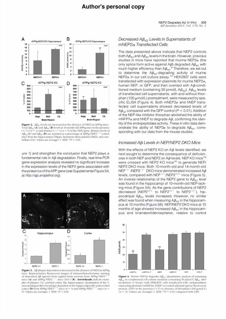
Figure 1. Amyloid (A) levels are increased in NEP2 knockout (KO) mice.
Total A42 ( A ) and A40 (B) levels in the hippocampus of 10-month-old NEP2 wild-type (/) (n 9) and NEP2 KO (/) (n 7) mice. Relative levels of A42 (C) and A40 (D) are reported as a percentage of wild-type mice fromhippocampus (Hipp), cerebral cortex (Ccx), brainstem/diencephalon(BsD), andcerebellum (Cb). Values are averages SEM. *P 0.05, **P 0.01.
308 Hafez et alAJP January 2011, Vol. 178, No. 1
7/22/2019 Hafez et al. 2011
http://slidepdf.com/reader/full/hafez-et-al-2011 5/8
Author's personal copy
ure 1) and strengthen the conclusion that NEP2 plays a
fundamental role in A degradation. Finally, real-time PCR
gene expression analysis revealed no significant increase
in the expression levels of the NEP2 gene associated withthe presence of the APP gene (see Supplemental Figure S4,
at http://ajp.amjpathol.org).
Decreased A 42
Levels in Supernatants of
mNEP2 Transfected Cells
The data presented above indicate that NEP2 controls
both A42 and A40 levels in the brain. However, previousstudies in mice have reported that murine NEP2 (the
only splice form active against A) degraded A40 with
much higher efficiency than A42.9 Therefore, we set out
to determine the A42-degrading activity of murine
NEP2 in our cell culture assay.10 HEK293T cells were
transfected with expression plasmids for murine NEP2,
human NEP, or GFP, and then overlaid with A-condi-
tioned medium (containing 50 pmol/L A42). A42 levels
of transfected cell supernatants, with and without thior-
phan (100 mol/L) pretreatment, were measured by spe-
cific ELISA (Figure 4). Both mNEP2 and hNEP trans-
fected cell supernatants showed decreased levels of
A42 compared with the GFP control (P 0.01). Additionof the NEP-like inhibitor thiorphan abolished the ability of
mNEP2 and hNEP to degrade A, confirming the iden-
tity of the endopeptidase activity. These in vitro data dem-
onstrate the ability of NEP2 to degrade A42, corre-
sponding with our data from the mouse studies.
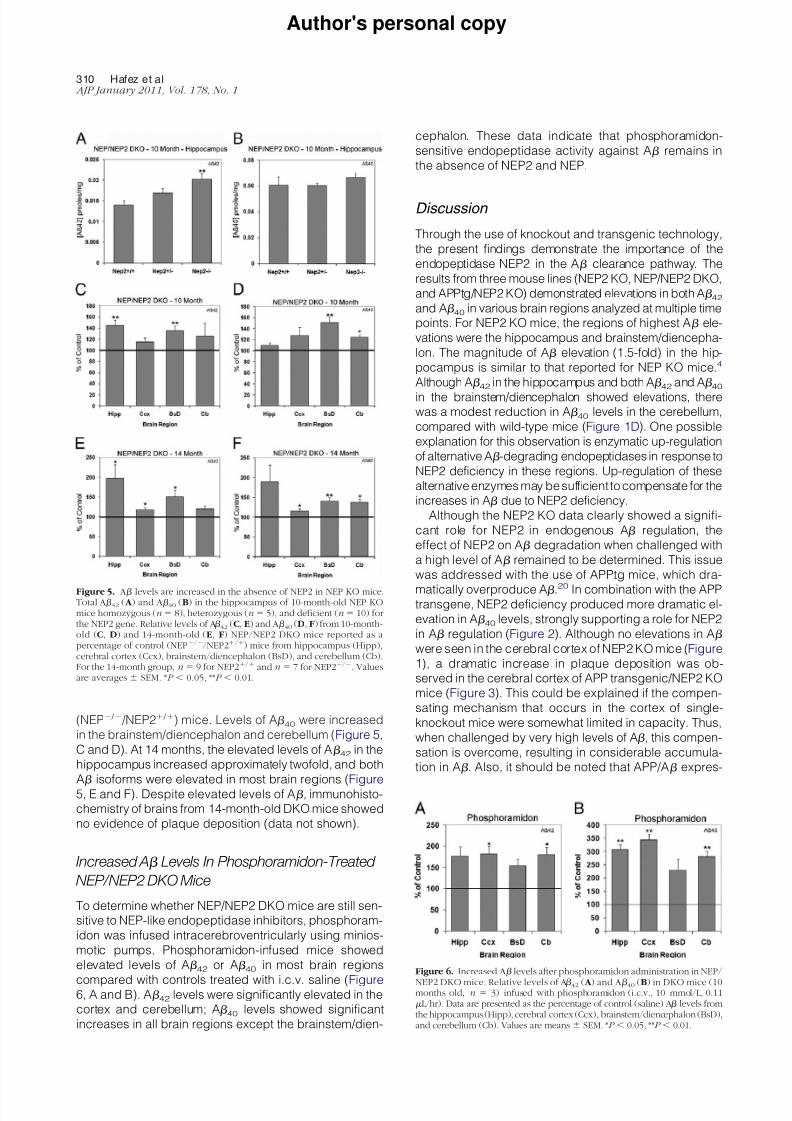
Increased A Levels in NEP/NEP2 DKO Mice
With the effects of NEP2 KO on A levels identified, we
next sought to determine the consequence of deficien-
cies in both NEP and NEP2 on A levels. NEP KO mice19
were crossed with NEP2 KO mice16 to generate NEP/
NEP2 DKO mice. Both 10-month-old and 14-month-oldNEP / /NEP2 / DKO mice demonstrated increased A
levels, compared with NEP / /NEP2 / mice (Figure 5).
An inverse relationship of the NEP2 gene to A42 level
was found in the hippocampi of 10-month-old NEP-lack-
ing mice (Figure 5A). As the gene contributions of NEP2
decreased (NEP2 / to NEP2 / to NEP2 / ), hip-
pocampal A42 levels increased. However, no similar
effect was found when measuring A40 in the hippocam-
pus at 10 months (Figure 5B). NEP/NEP2 DKO mice at 10
months of age showed increased A42 in the hippocam-
pus and brainstem/diencephalon, relative to control
Figure 2. A40 levels are increased in the absence of NEP2 in APPtg mice.Total A42 ( A ) and A40 (B) levels in 10-month-old APPtg mice in the presence(/) (n 4) and absence (/) (n 5) of the NEP2 gene. Relative levels of A42 (C) and A40 (D) are reported as a percentage of APPtg/NEP2/ controlmice from the hippocampus (Hipp), brainstem/diencephalon (BsD), and cere-bellum (Cb). Values are averages SEM. **P 0.01.
Figure 3. A plaque deposition is increased in the absence of NEP2 in APPtgmice. Representative fluorescent images of immunohistochemistry stainingof deposited A (green) from sagittal brain sections from APPtg/NEP2/
mice ( A ) and APPtg/NEP2
/
mice (40) (B). Arrowheads indicate exam-ples of plaques. Cx, cerebral cortex; Hp, hippocampus. Quantitation of the %area staining positive for plaque deposition in the hippocampus (C) andcerebralcortex (D) from APPtg/NEP2/ mice (n 4) and APPtg/NEP2/ mice (n 5). Values are averages SEM. *P 0.05.
Figure 4. Murine NEP2 degrades A42. Quantitative analysis of remaining
A42 in conditioned cell culture medium containing 50 pmol/L A42 afterincubation (5 hours) with HEK293T cells transfected with endopeptidaseexpressing plasmids (mNEP2, hNEP) or control plasmid (green fluorescentprotein, GFP) in the presence (T) or absence of thiorphan (100 mol/L)(n 4). Values are averages SEM. **P 0.01 compared with GFP.
NEP2 Degrades A in Vivo 309AJP January 2011, Vol. 178, No. 1
7/22/2019 Hafez et al. 2011
http://slidepdf.com/reader/full/hafez-et-al-2011 6/8
Author's personal copy
(NEP / /NEP2 / ) mice. Levels of A40 were increased
in the brainstem/diencephalon and cerebellum (Figure 5,
C and D). At 14 months, the elevated levels of A42 in the
hippocampus increased approximately twofold, and both
A isoforms were elevated in most brain regions (Figure5, E and F). Despite elevated levels of A, immunohisto-
chemistry of brains from 14-month-old DKO mice showed
no evidence of plaque deposition (data not shown).
Increased A Levels In Phosphoramidon-Treated
NEP/NEP2 DKO Mice
To determine whether NEP/NEP2 DKO mice are still sen-
sitive to NEP-like endopeptidase inhibitors, phosphoram-
idon was infused intracerebroventricularly using minios-
motic pumps. Phosphoramidon-infused mice showed
elevated levels of A42 or A40 in most brain regions
compared with controls treated with i.c.v. saline (Figure6, A and B). A42 levels were significantly elevated in the
cortex and cerebellum; A40 levels showed significant
increases in all brain regions except the brainstem/dien-
cephalon. These data indicate that phosphoramidon-
sensitive endopeptidase activity against A remains in
the absence of NEP2 and NEP.
Discussion
Through the use of knockout and transgenic technology,
the present findings demonstrate the importance of the
endopeptidase NEP2 in the A clearance pathway. The
results from three mouse lines (NEP2 KO, NEP/NEP2 DKO,
and APPtg/NEP2 KO) demonstrated elevations in both A42and A40 in various brain regions analyzed at multiple time
points. For NEP2 KO mice, the regions of highest A ele-
vations were the hippocampus and brainstem/diencepha-
lon. The magnitude of A elevation (1.5-fold) in the hip-
pocampus is similar to that reported for NEP KO mice.4
Although A42 in the hippocampus and both A42 and A40in the brainstem/diencephalon showed elevations, therewas a modest reduction in A40 levels in the cerebellum,
compared with wild-type mice (Figure 1D). One possible
explanation for this observation is enzymatic up-regulation
of alternative A-degrading endopeptidases in response to
NEP2 deficiency in these regions. Up-regulation of these
alternative enzymes may be sufficient to compensate for the
increases in A due to NEP2 deficiency.
Although the NEP2 KO data clearly showed a signifi-
cant role for NEP2 in endogenous A regulation, the
effect of NEP2 on A degradation when challenged with
a high level of A remained to be determined. This issue
was addressed with the use of APPtg mice, which dra-
matically overproduce A.20 In combination with the APPtransgene, NEP2 deficiency produced more dramatic el-
evation in A40 levels, strongly supporting a role for NEP2
in A regulation (Figure 2). Although no elevations in A
were seen in the cerebral cortex of NEP2 KO mice (Figure
1), a dramatic increase in plaque deposition was ob-
served in the cerebral cortex of APP transgenic/NEP2 KO
mice (Figure 3). This could be explained if the compen-
sating mechanism that occurs in the cortex of single-
knockout mice were somewhat limited in capacity. Thus,
when challenged by very high levels of A, this compen-
sation is overcome, resulting in considerable accumula-
tion in A. Also, it should be noted that APP/A expres-
Figure 6. Increased A levels after phosphoramidon administration in NEP/
NEP2 DKO mice. Relative levels of A42 ( A ) and A40 (B) in DKO mice (10months old, n 3) infused with phosphoramidon (i.c.v., 10 mmol/L, 0.11L/hr). Data are presented as the percentage of control (saline) A levels fromthe hippocampus (Hipp), cerebral cortex (Ccx), brainstem/diencephalon (BsD),and cerebellum (Cb). Values are means SEM. *P 0.05, **P 0.01.
Figure 5. A levels are increased in the absence of NEP2 in NEP KO mice.Total A42 ( A ) and A40 (B) in the hippocampus of 10-month-old NEP KOmice homozygous (n 8), heterozygous (n 5), and deficient (n 10) forthe NEP2 gene. Relative levels of A42 (C, E) and A40 (D, F) from 10-month-old (C, D) and 14-month-old (E, F) NEP/NEP2 DKO mice reported as apercentage of control (NEP//NEP2/) mice from hippocampus (Hipp),cerebral cortex (Ccx), brainstem/diencephalon (BsD), and cerebellum (Cb).For the 14-month group, n 9 for NEP2/ and n 7 for NEP2/. Valuesare averages SEM. *P 0.05, **P 0.01.
310 Hafez et alAJP January 2011, Vol. 178, No. 1
7/22/2019 Hafez et al. 2011
http://slidepdf.com/reader/full/hafez-et-al-2011 7/8
Author's personal copy
sion levels in APP transgenics are also dependent on the
transgene promoter and genomic integration site. The
APP transgenic line we have used is reported to express
APP/A more strongly in the cerebral cortex.20
The accumulation of intracellular A has been implicatedas a major mediator of A toxicity.25 Our analysis of intra-
cellular A did not show significant increases in intracellular
accumulation (see Supplemental Figure S3, at http://ajp.
amjpathol.org). The lack of a clear effect of NEP2 deficiency
on intracellular A, compared with plaque deposition, may
be due to saturation of the intracellular A accumulation
process. This hypothesis suggests that, even in the pres-
ence of the NEP2 gene, A levels are dramatically elevated
in this transgenic model, resulting in high levels of intracel-
lular A that are difficult to elevate further. Also, the contri-
bution of cellular APP and its other fragments complicates
this analysis. Finally, one limitation of the present study is
that our analysis quantified total A and did not specificallymeasure aggregated forms of A, including toxic soluble
oligomers.26 This analysis has important implications for the
ultimate relevance of NEP2 to AD, and we plan to address
it in future studies.
Analyzing A levels in NEP2 KO mice also deficient for
NEP provides further insight into the role of these key
endopeptidases in A catabolism. The results from NEP/
NEP2 DKO mice indicate that ablation of the NEP2 gene
leads to further elevated A42 and A40 levels in the
brain, compared with single knockout of NEP (Figure 5).
Our analysis also confirms that ablation of NEP alone
produces elevated A levels. For example, NEP KO mice
(NEP / .NEP2 / ) displayed approximately 1.5 to 2
times the A levels of wild-type mice (compare Figure 1,
A and B, with Figure 5, A and B). In addition, the absence
of NEP and NEP2 produces an additive elevation in A,
verifying that both endopeptidases cooperate to control
A levels in the brain. Our analysis did not reveal a
significant increase in A levels between 10-month-old
and 14-month-old mice (data not shown). Previous stud-
ies have demonstrated that treating rodents with inhibi-
tors of NEP/NEP2 (ie, phosphoramidon and thiorphan)
produces more dramatic elevations in A (30- to 50-
fold).2,5,6 However, despite the lack of both NEP and
NEP2 genes, only modest elevations in A peptides were
observed in our double-knockout studies—well below
known plaque-inducing levels. As expected, the immu-nohistochemical analysis of 14-month-old DKO mice did
not show evidence of plaque deposition (data not
shown). Therefore, other A clearing mechanisms must
be compensating for the loss of both NEP and NEP2.
Supporting this theory, DKO mice treated with phosphor-
amidon produced increases in both A42 andA40 levels in
certain brain regions (Figure 6). It should be noted that,
considering the smaller pump size and shorter duration of
this study, the values of A elevation due to phosphorami-
don infusion were comparable to the values seen with lower
doses previously used by Nisemblat and colleagues.6
These elevations in A resulting from the application of an
NEP/NEP2 inhibitor suggest the existence of yet other NEP-like endopeptidases that are involved in A catabolism.
Given that both NEP and NEP2 are M13 proteases, the
exploration of other enzymes in this family that are sensitive
to phosphoramidon may lead to the discovery of additional
enzymes involved in A metabolism. Possible enzymes co-
operating with NEP and NEP2 include endothelin-convert-
ing enzymes (ECE), phosphate-regulating gene with homol-
ogies to endopeptidases on the X chromosome (PHEX),and damage-induced neuronal endopeptidase (DINE). The
ECE are clear candidates because they are sensitive to
phosphoramidon. However, early studies using thiorphan
infusion in rodents suggest that ECEs are not involved in the
drug’s effect on A.2,5 PHEX and DINE share 39% and 36%
identity to NEP, respectively.8 It should be mentioned that
aminopeptidases or other A-degrading enzymes may also
be affected by the infusion of relatively large concentrations
of NEP-like inhibitors in these experiments. It is worth noting
that phosphoramidon produced stronger effects on A40 in
the absence of both NEP and NEP2, suggesting that the
remaining NEP-like activity may more efficiently degrade
A40 compared with A42. It is possible, therefore, that NEPand NEP2 could be the major catabolic NEP-like enzymes
(ie, phosphoramidon-sensitive enzymes) responsible for
clearing A42.
Our analyses clearly demonstrate that murine NEP2
degrades both isoforms of A. However, a previous study
reported that murine NEP2 degrades A40 much more
efficiently than does A42.9 To address this issue, we
constructed a murine NEP2 expression vector. Our re-
sults, using A-conditioned medium overlaid on cells
transfected with our engineered murine NEP2 construct,
demonstrate the ability of mNEP2 to degrade A42 in the
extracellular compartment (Figure 4). A possible explana-
tion for the observed differences between our findings and
the previous study may be the different experimental de-
signs used. The previous study measured the cleavage of
A from enzymes extracted in membrane-bound fractions,9
as opposed to the present study’s measurements using the
enzyme expressed from cells in culture.
Besides identifying a novel NEP-like enzyme that con-
trols A in vivo, our results have implications for potential
therapeutic applications. Experiments using NEP gene
therapy have been conducted in APPtg mice using a
variety of systems to overexpress NEP [reviewed by Marr
and Spencer3]. The results of these therapeutic studies
showed reduced plaque pathology and memory im-provements in treated animals.21,27–35 However, be-
cause current approaches could ultimately be ineffective
or produce unwanted side effects,36–38 more tools are
always welcome in the search for therapies for AD, and
exploration of the potential of NEP2 as a therapeutic
agent is warranted.
In conclusion, we have demonstrated in three different
mouse models that NEP2 is required for the endogenous
regulation of A42 and A40 levels in the rodent brain.
However, unlike the results of thiorphan and phosphor-
amidon infusion studies, only modest elevations in these
peptides were observed in nontransgenic mice lacking
NEP and NEP2, suggesting the existence of redundantproteolytic systems for controlling A levels. These sys-
tems may include the class of phosphoramidon-sensitive
(NEP-like) enzymes.
NEP2 Degrades A in Vivo 311AJP January 2011, Vol. 178, No. 1

7/22/2019 Hafez et al. 2011
http://slidepdf.com/reader/full/hafez-et-al-2011 8/8
Author's personal copy
Acknowledgment
We thank Andrew Mensing for assistance with this work.
References 1. Brookmeyer R, Johnson E, Ziegler-Graham K, Arrighi HM: Forecast-
ing the global burden of Alzheimer’s disease. Alzheimers Dement
2007, 3:186–191
2. Iwata N, Tsubuki S, Takaki Y, Watanabe K, Sekiguchi M, Hosoki E,
Kawashima-Morishima M, Lee HJ, Hama E, Sekine-Aizawa Y, Saido
TC: Identification of the major Abeta1-42-degrading catabolic path-
way in brain parenchyma: suppression leads to biochemical and
pathological deposition. Nat Med 2000, 6:143–150
3. Marr RA, Spencer BJ: NEP-like Endopeptidases and Alzheimer’s
Disease. Curr Alzheimer Res 2010, 7:223–229
4. Iwata N, Tsubuki S, Takaki Y, Shirotani K, Lu B, Gerard NP, Gerard C,
Hama E, Lee HJ, Saido TC: Metabolic regulation of brain Abeta by
neprilysin. Science 2001, 292:1550–1552
5. Dolev I, Michaelson DM: A nontransgenic mouse model shows induc-
ible amyloid-beta (Abeta) peptide deposition and elucidates the roleof apolipoprotein E in the amyloid cascade, Proc Natl Acad Sci USA
2004, 101:13909–13914
6. Nisemblat Y, Belinson H, Dolev I, Michaelson DM: Activation of the
amyloid cascade by intracerebroventricular injection of the protease
inhibitor phosphoramidon. Neurodegener Dis 2008;5:166–169
7. Ikeda K, Emoto N, Raharjo SB, Nurhantari Y, Saiki K, Yokoyama M,
Matsuo M: Molecular identification and characterization of novel
membrane-bound metalloprotease, the soluble secreted form of
which hydrolyzes a variety of vasoactive peptides. J Biol Chem 1999,
274:32469–32477
8. Ghaddar G, Ruchon AF, Carpentier M, Marcinkiewicz M, Seidah NG,
Crine P, DesGroseillers L, Boileau G: Molecular cloning and biochem-
ical characterization of a new mouse testis soluble-zinc-metallopep-
tidase of the neprilysin family, Biochem J 2000, 347:419–429
9. Shirotani K, Tsubuki S, Iwata N, Takaki Y, Harigaya W, Maruyama K,
Kiryu-Seo S, Kiyama H, Iwata H, Tomita T, Iwatsubo T, Saido TC:
Neprilysin degrades both amyloid beta peptides 1-40 and 1-42 most
rapidly and efficiently among thiorphan- and phosphoramidon-sensi-
tive endopeptidases. J Biol Chem 2001, 276:21895–21901
10. Huang JY, Bruno AM, Patel CA, Huynh AM, Philibert KD, Glucksman
MJ, Marr RA: Human membrane metallo-endopeptidase-like protein
degrades both beta-amyloid 42 and beta-amyloid 40. Neuroscience
2008, 155:258–262
11. Raharjo SB, Emoto N, Ikeda K, Sato R, Yokoyama M, Matsuo M:
Alternative splicing regulates the endoplasmic reticulum localization
or secretion of soluble secreted endopeptidase. J Biol Chem 2001,
276:25612–25620
12. Oh-Hashi K, Ohkubo K, Shizu K, Fukuda H, Hirata Y, Kiuchi K:
Biosynthesis, processing, trafficking, and enzymatic activity of mouse
neprilysin 2. Mol Cell Biochem 2008, 313:103–111
13. Facchinetti P, Rose C, Schwartz JC, Ouimet T: Ontogeny, regional
and cellular distribution of the novel metalloprotease neprilysin 2 in
the rat: a comparison with neprilysin and endothelin-converting en-
zyme-1. Neuroscience 2003, 118:627–639
14. Ouimet T, Facchinetti P, Rose C, Bonhomme MC, Gros C, Schwartz
JC: Neprilysin II: a putative novel metalloprotease and its isoforms in
CNS and testis. Biochem Biophys Res Commun 2000, 271:565–570
15. Carpentier M, Marcinkiewicz M, Boileau G, DesGroseillers L: The
neuropeptide-degrading enzyme NL1 is expressed in specific neu-
rons of mouse brain. Peptides 2003, 24:1083–1091
16. Carpentier M, Guillemette C, Bailey JL, Boileau G, Jeannotte L, Des-
Groseillers L, Charron J: Reduced fertility in male mice deficient in the
zinc metallopeptidase NL1. Mol Cell Biol 2004, 24:4428–4437
17. Rose C, Voisin S, Gros C, Schwartz JC, Ouimet T: Cell-specific
activity of neprilysin 2 isoforms and enzymic specificity compared
with neprilysin, Biochem J 2002, 363:697–705
18. Turner AJ, Nalivaeva NN: New insights into the roles of metallopro-
teinases in neurodegeneration and neuroprotection. Int Rev Neuro-
biol 2007, 82:113–13519. Lu B, Gerard NP, Kolakowski LF Jr, Bozza M, Zurakowski D, Finco O,
Carroll MC, Gerard C: Neutral endopeptidase modulation of septic
shock. J Exp Med 1995, 181:2271–2275
20. Rockenstein E, Mallory M, Mante M, Sisk A, Masliaha E: Early forma-
tion of mature amyloid-beta protein deposits in a mutant APP trans-
genic model depends on levels of Abeta(1-42). J Neurosci Res 2001,
66:573–582
21. Spencer B, Marr RA, Rockenstein E, Crews L, Adame A, Potkar R, Patrick
C, Gage FH, Verma IM, Masliah E: Long-term neprilysin gene transfer isassociated with reduced levels of intracellular Abeta and behavioral im-
provement in APP transgenic mice. BMC Neurosci 2008, 9:109
22. Marr RA, Addison CL, Snider D, Muller WJ, Gauldie J, Graham FL: Tumour
immunotherapy usingan adenoviral vector expressing a membrane-bound
mutant of murine TNF alpha. Gene Ther 1997, 4:1181–1188
23. Singer O, Marr RA, Rockenstein E, Crews L, Coufal NG, Gage FH,
Verma IM, Masliah E: Targeting BACE1 with siRNAs ameliorates
Alzheimer disease neuropathology in a transgenic model. Nat Neu-
rosci 2005, 8:1343–1349
24. Graham FL, van der Eb AJ: A new technique for the assay of infec-
tivity of human adenovirus 5 DNA. Virology 1973, 52:456–467
25. Oddo S, Caccamo A, Shepherd JD, Murphy MP, Golde TE, Kayed R,
Metherate R, Mattson MP, Akbari Y, LaFerla FM: Triple-transgenic
model of Alzheimer’s disease with plaques and tangles: intracellular
Abeta and synaptic dysfunction. Neuron 2003, 39:409–421
26. Ashe KH, Zahs KR: Probing the biology of Alzheimer’s disease inmice. Neuron 2010, 66:631–645
27. El-Amouri SS, Zhu H, Yu J, Marr R, Verma IM, Kindy MS: Neprilysin:
an enzyme candidate to slow the progression of Alzheimer’s disease.
Am J Pathol 2008, 172:1342–1354
28. Huang SM, Mouri A, Kokubo H, Nakajima R, Suemoto T, Higuchi M,
Staufenbiel M, Noda Y, Yamaguchi H, Nabeshima T, Saido TC, Iwata
N: Neprilysin-sensitive synapse-associated amyloid-beta peptide oli-
gomers impair neuronal plasticity and cognitive function. J Biol Chem
2006, 281:17941–17951
29. Leissring MA, Farris W, Chang AY, Walsh DM, Wu X, Sun X, Frosch
MP, Selkoe DJ: Enhanced proteolysis of beta-amyloid in APP trans-
genic mice prevents plaque formation, secondary pathology, and
premature death. Neuron 2003, 40:1087–1093
30. Marr RA, Rockenstein E, Mukherjee A, Kindy MS, Hersh LB, Gage FH,
Verma IM, Masliah E: Neprilysin gene transfer reduces human amy-
loid pathology in transgenic mice. J Neurosci 2003, 23:1992–1996
31. Poirier R, Wolfer DP, Welzl H, Tracy J, Galsworthy MJ, Nitsch RM,
Mohajeri MH: Neuronal neprilysin overexpression is associated with
attenuation of Abeta-related spatial memory deficit. Neurobiol Dis
2006, 24:475–483
32. Mohajeri MH, Wolfer DP: Neprilysin deficiency-dependent impair-
ment of cognitive functions in a mouse model of amyloidosis. Neuro-
chem Res 2009, 34:717–726
33. Madani R, Poirier R, Wolfer DP, Welzl H, Groscurth P, Lipp HP, Lu B, El
Mouedden M, Mercken M, Nitsch RM, Mohajeri MH: Lack of neprilysin
suffices to generate murine amyloid-like deposits in the brain and be-
havioral deficit in vivo. J Neurosci Res 2006, 84:1871–1878
34. Liu Y, Studzinski C, Beckett T, Guan H, Hersh MA, Murphy MP, Klein
R, Hersh LB: Expression of neprilysin in skeletal muscle reduces
amyloid burden in a transgenic mouse model of Alzheimer disease.
Mol Ther 2009, 17:1381–1386
35. Guan H, Liu Y, Daily A, Police S, Kim MH, Oddo S, LaFerla FM, Pauly
JR, Murphy MP, Hersh LB: Peripherally expressed neprilysin reduces
brain amyloid burden: a novel approach for treating Alzheimer’s
disease. J Neurosci Res 2009, 87:1462–1473
36. Iijima-Ando K, Hearn SA, Granger L, Shenton C, Gatt A, Chiang HC,
Hakker I, Zhong Y, Iijima K: Overexpression of neprilysin reduces Alz-
heimer amyloid-beta42 (Abeta42)-induced neuron loss and intraneuro-
nal Abeta42 deposits but causes a reduction in cAMP-responsive ele-
ment-binding protein-mediated transcription, age-dependent axon
pathology, and premature death in Drosophila. J Biol Chem 2008, 283:
19066–19076
37. Meilandt WJ, Cisse M, Ho K, Wu T, Esposito LA, Scearce-Levie K,
Cheng IH, Yu GQ, Mucke L: Neprilysin overexpression inhibits plaque
formation but fails to reduce pathogenic Abeta oligomers and asso-
ciated cognitive deficits in human amyloid precursor protein trans-
genic mice. J Neurosci 2009, 29:1977–1986
38. Walther T, Albrecht D, Becker M, Schubert M, Kouznetsova E,
Wiesner B, Maul B, Schliebs R, Grecksch G, Furkert J, Sterner-KockA, Schultheiss HP, Becker A, Siems WE: Improved learning and
memory in aged mice deficient in amyloid beta-degrading neutral
endopeptidase. PLoS One 2009, 4:e4590
312 Hafez et alAJP January 2011, Vol. 178, No. 1