(HA)
29
Quantitative Micro Hemagglutination Test (HA)
Transcript of (HA)

Quantitative Micro Hemagglutination Test (HA)
Definitions • Agglutination: The clumping together of biologic
material, such as red blood cells or bacteria, that is suspended in liquid.
• Hemagglutination: The agglutination of red blood cells caused by an antibody either for red blood cell antigens or for antigens that coat red blood cells or by the presence of viruses or other microbes.
• Hemagglutination test: sensitive test to measure certain antigens, antibodies, or viruses, using their ability to agglutinate certain erythrocytes.
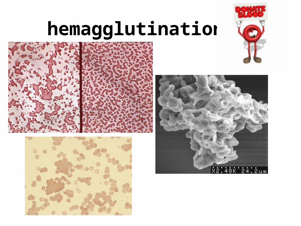
hemagglutination

Hemagglutination
RBC
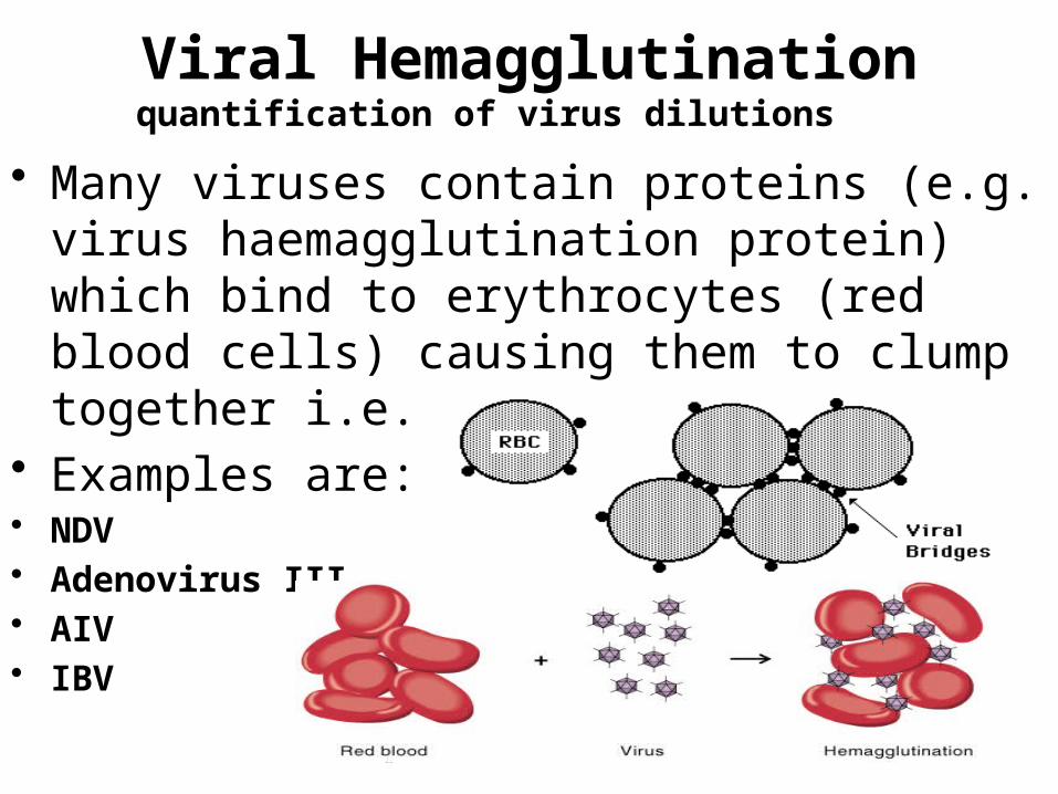
Viral Hemagglutinationquantification of virus dilutions
• Many viruses contain proteins (e.g. virus haemagglutination protein) which bind to erythrocytes (red blood cells) causing them to clump together i.e. agglutinate.
• Examples are:• NDV• Adenovirus III• AIV• IBV
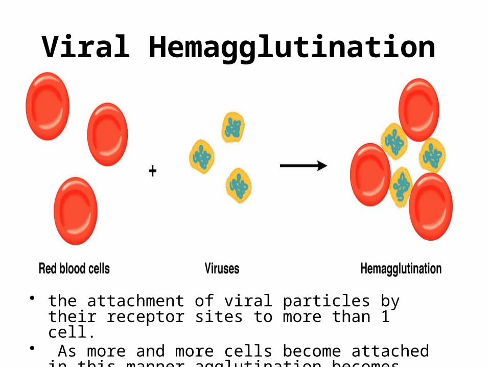
Viral Hemagglutination
• the attachment of viral particles by their receptor sites to more than 1 cell.
• As more and more cells become attached in this manner agglutination becomes visible

Equivalence point: (suitable proportion between the virus particles and
RBCs)

Negative control well (only RBCs+buffer) (no hemagglutinin)
Positive control well (contains haemagglutinin)

Readings The results • Titer: The maximum dilution that gives
visible agglutination.• The end point: is the well with the lowest
concentration of the virus where there is hemagglutination (no button),(HA1)
2 4 8 16 32 64 128 256 512 1024 2048 4096
The HA titer of this virus in this row is 256 or 28
(1:256 dilution contains (1 HA unit) (one hemagglutinating unit)

Example of readings

Titer = 32 HA units/ml
Hemagglutination test: method
1:8
1:2 1:21:21:21:2
8 16 32 64 128 256
virus
serial dilution
mix with red blood cells
side view
top view
One HA unit :minimum amount of virus that will cause complete agglutination of the red blood cells .
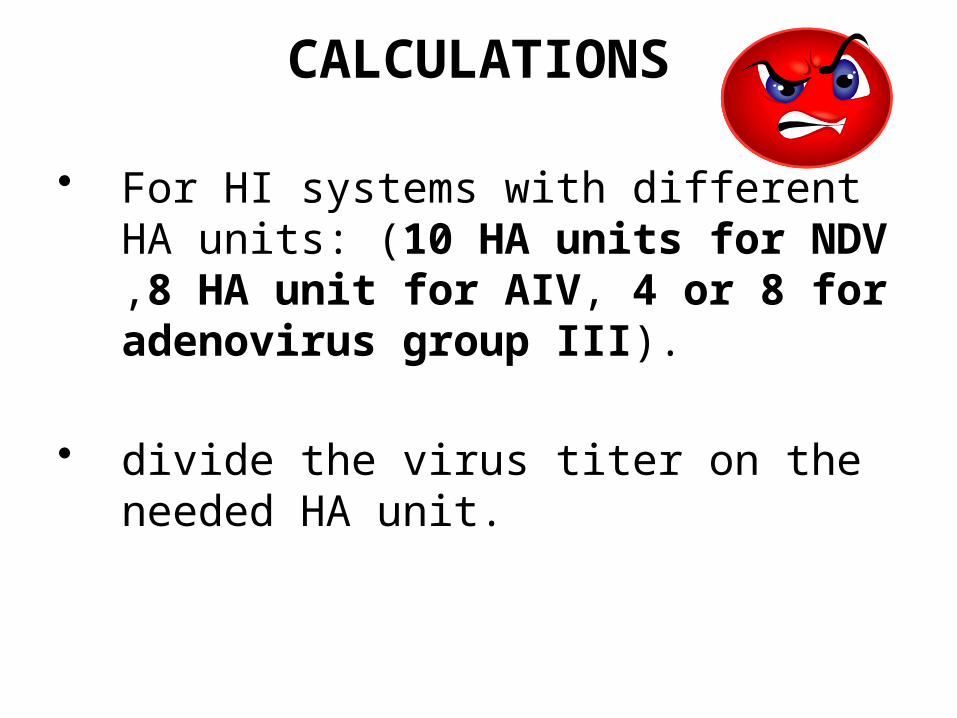
CALCULATIONS
• For HI systems with different HA units: (10 HA units for NDV ,8 HA unit for AIV, 4 or 8 for adenovirus group III).
• divide the virus titer on the needed HA unit.

Hemagglutination assay. Seven different samples of influenza virus, numbered 1 through 7 at the left, were serially diluted as indicated at the top, mixed with chicken red blood cells (RBC), and incubated on ice for 1 to 2 hours. Wells in the bottom row contain no virus. Agglutinated RBCs coat wells evenly, in contrast to nonagglutinated cells, which form a distinct button at the bottom of the well. The HA titer, shown at the right, is the last dilution that shows complete hemagglutination activity. (From Fields Vriology (2007) 5th edition, Knipe, DM & Howley, PM, eds, Wolters Kluwer/Lippincott Williams & Wilkins, Philadelphia Fig. 2.9)
Hemagglutination assay: influenza virus

WHAT WE NEED?

HEMAGGLUTINATION INHIBITION TEST (HI)
VIRUSE SERUM

hemagglutination inhibition (HI) test
• an assay for the presence of specific antiviral antibodies in a test serum.
• convenient and commonly used assay that requires cheap reagents and is read by eye.
• the antibodies bind to the haemagglutinin protein in the envelope of the virus. This blocks the haemagglutinin protein from binding with the receptor site on chicken RBCs
• The highest dilution of serum that inhibits hemagglutination is the HI titer of the serum.

In the absence of anti-virus antibodies
Erythrocytes
Virus
Virus agglutination of erythrocytes
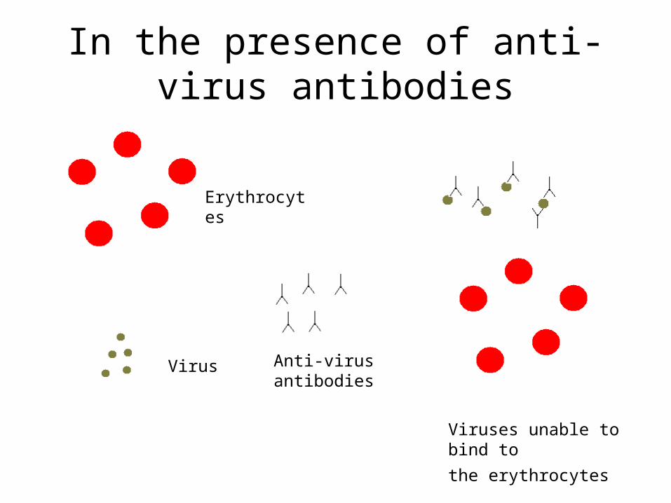
In the presence of anti-virus antibodies
Erythrocytes
Virus Anti-virus antibodies
Viruses unable to bind to
the erythrocytes


Antibody Titer• Is the lowest
concentration of antibodies against a particular antigen.

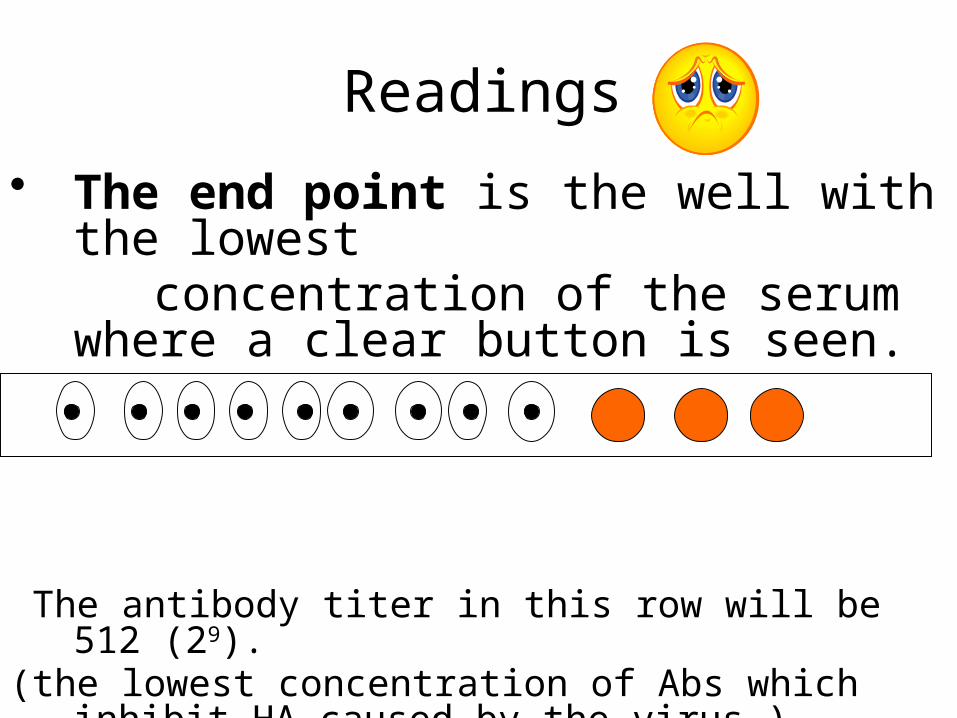
Readings
• The end point is the well with the lowest concentration of the serum where a clear
button is seen. 2 4 8 16 32 64 128 256 512 1024 2048 4096
The antibody titer in this row will be 512 (29).(the lowest concentration of Abs which inhibit HA caused
by the virus )

PROCEDURE(CONTROL)
• Always run four control rows:
_ Positive antiserum row (Contains antibodies against the specific virus)
_ Negative antiserum row (Contains no antibodies against the specific virus)
_ Antigen row.
_ RBCs row.

The commonly-used HI tests in chickens
• for Newcastle disease (Paramyxovirus-1), Infectious bronchitis (Coronavirus), and EDS-76 (adeno-virus),HI tests may also be carried out for Avian Influenza.

WASHING THE RBCs

Why we have to wash them?
• To obtain the RBCs and get rid from any other blood components such as WBCs, immune complexes, and Abs
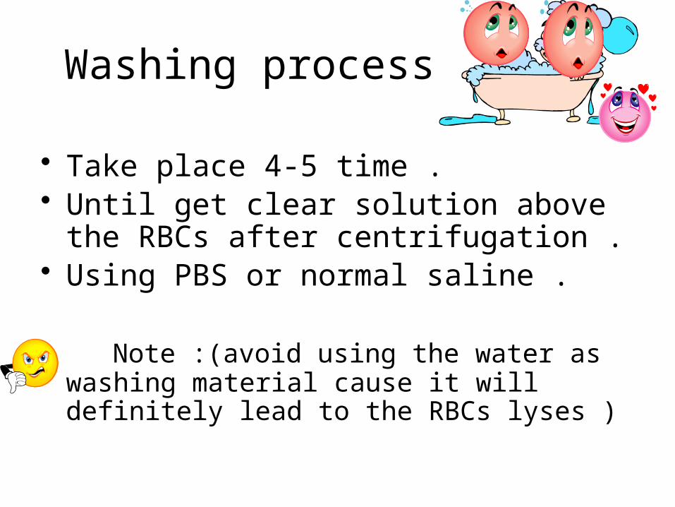
Washing process
• Take place 4-5 time .• Until get clear solution above the RBCs
after centrifugation .• Using PBS or normal saline .
Note :(avoid using the water as washing material cause it will definitely lead to the RBCs lyses )

Procedure
• Fill the blood samples in tubes ,centrifugation at 1500 round/min for 10 min .
• Draw off the plasma and WBCs coat using pipette filter spelling it into the sink .
• Add the PBS to each tube and centrifuge again, each time draw off the washing solution and add new amount until we get a clear soultion above the RBCs layer ,almost 3 times .

THANK YOU


![]s - Началоagup.varna.bg/.../article/492/a_2014_1_PROTOKOL_ESUT.pdfP O T O K O r. Ha Ha No Ha ' I sa Ha - Ha Ha crc ...](https://static.fdocuments.net/doc/165x107/5ad73c2b7f8b9af9068bf3a5/s-agupvarnabgarticle492a20141protokolesutpdfp-o-t-o.jpg)