
Gynecologic Abscess: CT-guided Percutaneous … › files › public › 3 › 34934 › ...catheter...
4
Recently, with the development of intervention- al procedures, indications for computed tomogra- phy (CT) or ultrasound (US)-guided percutaneous abscess drainage (PAD) continue to expand and almost all abscesses in various regions are consid- ered amenable 8) . Previous therapeutic options for gynecologic abscess included antibiotics alone or in combination with US-guided transvaginal drainage or surgical resection 1-3) . Despite the established success of CT-guided PAD for almost all abscesses, the transabdominal approach using this technique 2,7) has not been performed very often for the treatment of gynecologic abscess. This report presents two patients with gynecologic abscesses who had good clinical outcomes by CT- guided PADs. CASE REPORT Case 1 A 42-year-old woman with recurrent bilateral endometrial ovarian cystoma, after having a bilat- eral oophorocystectomy six years before, presented with fever and pelvic pain. US and magnetic reso- nance imaging (MRI) showed the rapid enlarge- ment of a right unilocular ovarian cystoma of 8 cm in diameter with a left ovarian cystoma, and right tubal dilatation (Fig. 1A). The bilateral ovarian cys- tomas also showed hemorrhagic change on MRI. The right rapid-growing cystoma was suspected of having developed into a TOA due to a complicating infection with recent hemorrhage. Since no improvement was obtained using several varieties of intravenous and oral antibiotics for 2 weeks, transabdominal diagnostic aspiration with an 18- gauge needle under CT (SOMATOM Plus4 Volume Zoom; Siemens, Erlangen, Germany) guidance was performed (Fig. 1B). Drab, bloody and purulent fluid was aspirated grossly. Thereafter, a 7F (French) pigtail catheter (Dawson-Mueller drainage catheter, Cook incorporated, Bloomington, IN) was placed immediately using the Seldinger technique under fluoroscopy (Multistar; Siemens, Erlangen, Germany) equipped with CT guidance (Fig. 1C). Approximately 120 ml of purulent fluid was initial- ly aspirated from the catheter. Although the cul- ture was negative, erythrocytosis, leucocytosis, heterophilic leucocytosis, in particular, and histio- 97 Gynecologic Abscess: CT-guided Percutaneous Drainage Hideaki KAKIZAWA 1, * ) , Naoyuki TOYOTA 1) , Masashi HIEDA 1) , Nobuhiko HIRAI 1) , Toshihiro TACHIKAKE 1) , Noriaki MATSUURA 1) , Yoshio FUJIMURA 1) , Ichiro KODAMA 2) , Eiji HIRATA 2) , Tetsuaki HARA 2) and Katsuhide ITO 1) 1) Department of Radiology, Hiroshima University Hospital, 1–2–3, Kasumi, Minami-ku, Hiroshima 734–8551, Japan 2) Department of Obstetrics and Gynecology, Hiroshima University Hospital, 1–2–3, Kasumi, Minami-ku, Hiroshima 734–8551, Japan ABSTRACT A 42-year-old woman with recurrent bilateral endometrial ovarian cystoma presented with fever and pelvic pain caused by a tubo-ovarian abscess (TOA), which was resistant to several varieties of intravenous and oral antibiotics for 2 weeks (Case 1). Computed tomography (CT)- guided diagnostic aspiration for a rapid enlarged right ovarian cystoma through a transabdomi- nal route confirmed that it had developed into a TOA. Subsequent percutaneous abscess drainage (PAD) and irrigation for 3 days were successful. One-year follow-up revealed no recur- rence of TOA. A 58-year-old woman with recurrent cervical cancer after external radiation ther- apy (RT) presented with fever, confusion and tremor caused by pyometra (Case 2). Since transvaginal drainage was impossible due to cervical os obstruction, the patient had undergone CT-guided transabdominal PAD and irrigation for a month. Thereafter, the clinical findings improved and a tracheloplasty was performed to prevent recurrence. CT-guided PAD may be a useful treatment option for gynecologic abscess as a diagnostic aspiration, a temporizing proce- dure until surgery, or an alternative surgery. Key words: Abscess, CT, Percutaneous drainage, Gynecologic organs Hiroshima J. Med. Sci. Vol. 55, No. 3, 97~100, September, 2006 HIJM 55–15 *Correspondence to: Hideaki Kakizawa, M.D. Department of Radiology, Hiroshima University Hospital, 1–2–3, Kasumi, Minami-ku, Hiroshima 734–8551, Japan Fax: 81 82 257 5259, Phone: 81 82 257 5257, E-mail address: [email protected]
Transcript of Gynecologic Abscess: CT-guided Percutaneous … › files › public › 3 › 34934 › ...catheter...

Recently, with the development of intervention-al procedures, indications for computed tomogra-phy (CT) or ultrasound (US)-guided percutaneousabscess drainage (PAD) continue to expand andalmost all abscesses in various regions are consid-ered amenable8). Previous therapeutic options forgynecologic abscess included antibiotics alone or incombination with US-guided transvaginaldrainage or surgical resection1-3). Despite theestablished success of CT-guided PAD for almostall abscesses, the transabdominal approach usingthis technique2,7) has not been performed veryoften for the treatment of gynecologic abscess.This report presents two patients with gynecologicabscesses who had good clinical outcomes by CT-guided PADs.
CASE REPORT
Case 1A 42-year-old woman with recurrent bilateral
endometrial ovarian cystoma, after having a bilat-eral oophorocystectomy six years before, presentedwith fever and pelvic pain. US and magnetic reso-
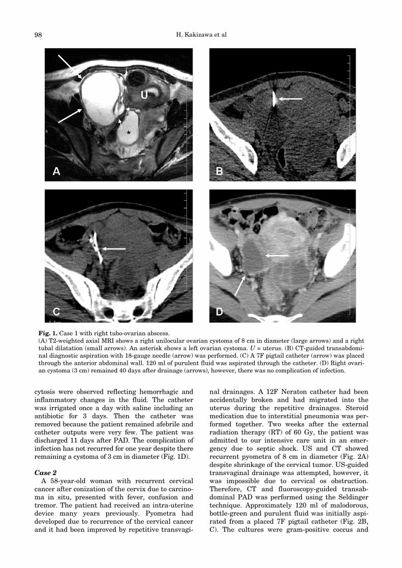
nance imaging (MRI) showed the rapid enlarge-ment of a right unilocular ovarian cystoma of 8 cmin diameter with a left ovarian cystoma, and righttubal dilatation (Fig. 1A). The bilateral ovarian cys-tomas also showed hemorrhagic change on MRI.The right rapid-growing cystoma was suspected ofhaving developed into a TOA due to a complicatinginfection with recent hemorrhage. Since noimprovement was obtained using several varietiesof intravenous and oral antibiotics for 2 weeks,transabdominal diagnostic aspiration with an 18-gauge needle under CT (SOMATOM Plus4 VolumeZoom; Siemens, Erlangen, Germany) guidance wasperformed (Fig. 1B). Drab, bloody and purulentfluid was aspirated grossly. Thereafter, a 7F(French) pigtail catheter (Dawson-Mueller drainagecatheter, Cook incorporated, Bloomington, IN) wasplaced immediately using the Seldinger techniqueunder fluoroscopy (Multistar; Siemens, Erlangen,Germany) equipped with CT guidance (Fig. 1C).Approximately 120 ml of purulent fluid was initial-ly aspirated from the catheter. Although the cul-ture was negative, erythrocytosis, leucocytosis,heterophilic leucocytosis, in particular, and histio-
97
Gynecologic Abscess: CT-guided Percutaneous Drainage
Hideaki KAKIZAWA1,*), Naoyuki TOYOTA1), Masashi HIEDA1), Nobuhiko HIRAI1), Toshihiro TACHIKAKE1), Noriaki MATSUURA1), Yoshio FUJIMURA1), Ichiro KODAMA2),
Eiji HIRATA2), Tetsuaki HARA2) and Katsuhide ITO1)
1) Department of Radiology, Hiroshima University Hospital, 1–2–3, Kasumi, Minami-ku, Hiroshima734–8551, Japan
2) Department of Obstetrics and Gynecology, Hiroshima University Hospital, 1–2–3, Kasumi, Minami-ku,Hiroshima 734–8551, Japan
ABSTRACTA 42-year-old woman with recurrent bilateral endometrial ovarian cystoma presented with
fever and pelvic pain caused by a tubo-ovarian abscess (TOA), which was resistant to severalvarieties of intravenous and oral antibiotics for 2 weeks (Case 1). Computed tomography (CT)-guided diagnostic aspiration for a rapid enlarged right ovarian cystoma through a transabdomi-nal route confirmed that it had developed into a TOA. Subsequent percutaneous abscessdrainage (PAD) and irrigation for 3 days were successful. One-year follow-up revealed no recur-rence of TOA. A 58-year-old woman with recurrent cervical cancer after external radiation ther-apy (RT) presented with fever, confusion and tremor caused by pyometra (Case 2). Sincetransvaginal drainage was impossible due to cervical os obstruction, the patient had undergoneCT-guided transabdominal PAD and irrigation for a month. Thereafter, the clinical findingsimproved and a tracheloplasty was performed to prevent recurrence. CT-guided PAD may be auseful treatment option for gynecologic abscess as a diagnostic aspiration, a temporizing proce-dure until surgery, or an alternative surgery.
Key words: Abscess, CT, Percutaneous drainage, Gynecologic organs
Hiroshima J. Med. Sci.Vol. 55, No. 3, 97~100, September, 2006HIJM 55–15
*Correspondence to: Hideaki Kakizawa, M.D.Department of Radiology, Hiroshima University Hospital, 1–2–3, Kasumi, Minami-ku, Hiroshima 734–8551, JapanFax: 81 82 257 5259, Phone: 81 82 257 5257, E-mail address: [email protected]
H. Kakizawa et al
cytosis were observed reflecting hemorrhagic andinflammatory changes in the fluid. The catheterwas irrigated once a day with saline including anantibiotic for 3 days. Then the catheter wasremoved because the patient remained afebrile andcatheter outputs were very few. The patient wasdischarged 11 days after PAD. The complication ofinfection has not recurred for one year despite thereremaining a cystoma of 3 cm in diameter (Fig. 1D).
Case 2A 58-year-old woman with recurrent cervical
cancer after conization of the cervix due to carcino-ma in situ, presented with fever, confusion andtremor. The patient had received an intra-uterinedevice many years previously. Pyometra haddeveloped due to recurrence of the cervical cancerand it had been improved by repetitive transvagi-
nal drainages. A 12F Neraton catheter had beenaccidentally broken and had migrated into theuterus during the repetitive drainages. Steroidmedication due to interstitial pneumonia was per-formed together. Two weeks after the externalradiation therapy (RT) of 60 Gy, the patient wasadmitted to our intensive care unit in an emer-gency due to septic shock. US and CT showedrecurrent pyometra of 8 cm in diameter (Fig. 2A)despite shrinkage of the cervical tumor. US-guidedtransvaginal drainage was attempted, however, itwas impossible due to cervical os obstruction.Therefore, CT and fluoroscopy-guided transab-dominal PAD was performed using the Seldingertechnique. Approximately 120 ml of malodorous,bottle-green and purulent fluid was initially aspi-rated from a placed 7F pigtail catheter (Fig. 2B,C). The cultures were gram-positive coccus and
98
Fig. 1. Case 1 with right tubo-ovarian abscess.(A) T2-weighted axial MRI shows a right unilocular ovarian cystoma of 8 cm in diameter (large arrows) and a righttubal dilatation (small arrows). An asterisk shows a left ovarian cystoma. U = uterus. (B) CT-guided transabdomi-nal diagnostic aspiration with 18-gauge needle (arrow) was performed. (C) A 7F pigtail catheter (arrow) was placedthrough the anterior abdominal wall. 120 ml of purulent fluid was aspirated through the catheter. (D) Right ovari-an cystoma (3 cm) remained 40 days after drainage (arrows), however, there was no complication of infection.

Percutaneous Drainage for Gynecologic Abscess
streptococcus. By irrigation of the catheter once aday with saline and intravenous antibiotic treat-ment for a month, clinical findings were improved.Then, as a surgical treatment, tracheloplasty ofthe cervical canal dilatation and intra-uterinedraining was performed in order to prevent re-obstruction of the cervical os. The intra-uterinecavity was clear on intraoperative hysteroscopeexamination and the foreign bodies including anintrauterine device and a Neraton catheter werealso removed together. Following this procedure,the transabdominal catheter was removed 4 daysafter the operation. After additional external RTfor para-aortic lymph node metastasis, the patientwas discharged 3 months after PAD.
DISCUSSION
Our current preferable guidance of PAD forpelvic abscess is CT. CT enables us to visualizethe entire pelvic space even if there are distendedbowels that detract from US examination8). It alsoenables better visualization of surrounding struc-
tures to avoid transgression of the adjacent bowel,blood vessels or bladder2,7). In both our patients,abscesses were juxtaposed to bowels cranio-ven-trally in the pelvis. We therefore chose a lowerlevel than the depicted bowel as an approach. Ingeneral, CT is adequate for deep abscesses and USfor superficial abscesses6,8). However, for superfi-cial abscesses, CT is also easy and feasible forPAD.
CT brings with it the disadvantage of radiationexposure of a dose that does not occur in US.During insertion of the needle for CT-guidance, weroutinely attempt to decrease the scan area andslices (e.g. cases 1, 2: a scan area of 17.5 mm atintervals of 2.5 mm) and reduce the scanning cur-rent (e.g. case 1: 120 kV/160 mA; case 2: 120kV/300 mA) as much as we possibly can, in order tominimize radiation exposure to patients. Ourpatients received 12 mSv and 24 mSv of radiationexposure per scan, respectively. Consequently, dur-ing the whole process of needle insertion, includingthe initial scan to determine the access route, theyreceived 92 mSv and 116 mSv of CT radiation
99
Fig. 2. Case 2 with pyometra.(A) CT showed recurrent pyometra of 8 cm in diameter(large arrows). A small arrow shows 12F Neratoncatheter that had migrated into the uterus during previ-ous transvaginal drainage. (B), (C) A 7F pigtail catheter(arrows) was placed through the anterior abdominalwall. 120 ml of purulent fluid was aspirated through thecatheter.

H. Kakizawa et al
exposure, respectively. Furthermore, this dose wasto the local area. We believe these doses are accept-able considering the safety of CT-guidance.
Advanced-stage endometriosis might increasethe risk of TOA due to the fact that immunologi-cally aberrant disease makes the patient vulnera-ble to infection, the cystic wall, unlike healthyovarian epithelium, is susceptible to bacterialinvasion, and bloody content serves as a culturemedium and facilitates the spread of infection4). Incase 1, hemorrhage into ovarian cystoma was con-sidered the main trigger of infection. Standardtherapy for TOA consists of antibiotics. In caseswhere treatment fails, surgery has been tradition-al2). PAD is useful not only for drainage treatmentbut also for confirmed diagnosis of abscess by aspi-ration. The success rate of PAD for TOAs is80–90%2,7). The authors2,7) preferably used CT as aguidance for the same reason as ours which wehave stated above. Case 1 was nulliparous andhoped for pregnancy in the future. Avoiding steril-ity is an important matter for nullipara. Sterilitywas avoided by using PAD2,7). It would have beendifficult to avoid by hystero-oophorectomy due tosevere inflammatory adhesion from surgery.
Pyometra is usually associated with or followedby RT for malignant disease of the uterus1,3,5). Itshould be treated promptly and vigorously byevacuation and continued drainage of the uterinecavity3,5), particularly, in septic shock5) such as incase 2. Post RT cervical stenosis1,3) was consideredthe main cause of pyometra. Transvaginal dilata-tion, drainage and curettage are usually prefer-able for evacuation in patients with pyometra1,3,5).As the cervical os was completely occluded in case2, transvaginal drainage was impossible1,2). Insuch conditions, laparotomy is usually the secondline treatment3,5). Contrary to this, Babarinsa etal1) advocated uterine fundus puncture through atransabdominal approach. However, they did notdescribe any guidance for PAD. To our knowledge,there has been no report specifying CT as guid-ance for uterine drainage. We believe that CT-guided transabdominal PAD may be safe anduseful when a transvaginal approach is difficult.In case 2, PAD was useful as an emergent andtemporizing procedure.
Many authors2,6,7) matched 7–14F drainagecatheters, depending on the size of the abscess andthe viscosity of the fluid aspirated. Meanwhile, wehave routinely initially placed a 7F catheter forabdominal and pelvic abscesses regardless of theirsize and viscosity, and thereafter upsized thecatheter when there has been ineffective drainagebecause we believe that a smaller sized-catheter ismore pain-relieving during the procedure andcatheter placement. In our patients, 7F size wasacceptable for 3 days and a month’s drainage and
irrigation, respectively. In case 2, the intra-uterinecavity was clear on intraoperative examination,which means that drainage was very effective.
Fortunately, our patients had unilocular abscessand no communication to the gastrointestinal (GI)tract. In general, the success rate of PAD is lowerfor abscesses that have septations or structureslike septations or communications to the GItracts2,7,8). On such occasions, using larger or mul-tiple catheters may be more effective and neces-sary7).
A problem of PAD is the possibility of recur-rence as long as there are underlying condi-tions2,3,5,7). Therefore, continued medication forendometriosis and tracheloplasty for cervical osocclusion were performed after PADs.
Complications related to PAD, including sep-ticemia, hemorrhage, and bowel laceration arerare (0–6%)2,6-8). In addition to its safety, PAD ismore cost-effective and has a shorter hospitaliza-tion period than surgery7).
In conclusion, we have presented two patientswith TOA and pyometra who showed good clinicaloutcomes by CT-guided PADs. It may be a usefultreatment option for gynecologic abscess as a diag-nostic aspiration, a temporizing procedure untilsurgery, or an alternative surgery.
(Received June 14, 2006)(Accepted June 30, 2006)
REFERENCES01. Babarinsa, I.A., Campbell, O.B. and Adewole,
I.F. 1999. Pyometra complicating cancer of thecervix. Int. J. Gynaecol. Obstet. 64: 75–76.
02. Casola, G., vanSonnenberg, E., D’Agostino,H.B., Harker, C.P., Varney, R.R. and Smith, D.1992. Percutaneous drainage of tubo-ovarianabscesses. Radiology 182: 399–402.
03. Chan, L.Y., Lau, T.K., Wong, S.F. and Yuen,P.M. 2001. Pyometra. What is its clinical signifi-cance? J. Reprod. Med. 46: 952–956.
04. Chen, M.J., Yang, J.H., Yang, Y.S. and Ho, H.N.2004. Increased occurrence of tubo-ovarian abscess-es in women with stage III and IV endometriosis.Fertil. Steril. 82: 498–499.
05. Muram, D., Drouin, P., Thompson, F.E. andOxorn, H. 1981. Pyometra. Can. Med. Assoc. J.125: 589–592.
06. Ramondetta, L.M., Dunton, C.J., Shapiro, M.J.,Carlson, J.A., Jr. and Arsuaga, J. 1996.Percutaneous abscess drainage in gynecologic can-cer patients. Gynecol. Oncol. 62: 366–369.
07. Tyrrel, R.T., Murphy, F.B. and Bernardino,M.E. 1990. Tubo-ovarian abscesses: CT-guided per-cutaneous drainage. Radiology 175: 87–89.
08. vanSonnenberg, E., Wittich, G.R., Goodacre,B.W., Casola, G. and D’Agostino, H.B. 2001.Percutaneous abscess drainage: update. World J.Surg. 25: 362–369.
100


![Transvaginal Mesh Lawsuits [Data Timeline]](https://static.fdocuments.net/doc/165x107/5884223e1a28ab485c8b5d45/transvaginal-mesh-lawsuits-data-timeline.jpg)


