Gut Intestinal perfusion of dietary levels aluminium ...
5
Gut 1994; 35: 1053-1057 Intestinal perfusion of dietary levels of aluminium: association with the mucosa JJ Powell, C C Ainley, R Evans, R P H Thompson Abstract An aluminium (93 ,uM) sulphate solution freshly adjusted to pH 7 0 was perfused through the rat small bowel to mimic the reported physiological conditions that follow dietary aluminium ingestion. One third of this aluminium was taken up from the perfusate, but >90% of this was then recovered from the intestinal mucus/mucosa and most (>70%) from the distal third of the small bowel. The fresh perfusate was shown by ultrafiltration to contain largely particulate/colloidal aluminium-hydroxide, and this probably adhered to intestinal mucus which may be an important barrier to the gastro- intestinal absorption of aluminium. (Gut 1994; 35: 1053-1057) uptake of both soluble and precipitated aluminium by the bowel, but not its systemic transfer, has been consistently demon- strated,'3-15 showing that retention in the mucosa, rather than luminal precipitation, is the major limiting factor in absorption. This could be due to either mucosal'3 or extra- mucosal factors, such as the mucus layer.7 These studies'3-15 again used supraphysiologi- cal levels of aluminium. The aim of this study was to develop an in situ rat gut perfusion technique to study the fate of dietary levels of aluminium, and to see whether such physiological quantities of the metal also associate with the intestinal mucosa. Methods Gastrointestinal Laboratory, The Rayne Institute, St Thomas' Hospital, London J J Powell CC Ainley R P H Thompson Division of Biochemistry, UMDS, Guy's Hospital, London R Evans Correspondence to: Dr J J Powell, Gastrointestinal Laboratory, The Rayne Institute, St Thomas' Hospital, London SE1 7EH. Accepted for publication 23 November 1993 The toxicity of aluminium is now well recognised, and occurs at even low levels in plants,' fish,2 and human cells.3 Encephalo- pathy, bone disease, and anaemia have been reported in patients with impaired renal excretion of aluminium,4 particularly those who are exposed to the metal in dialysis water. The efficiency of renal excretion and impermeability of the intestine to aluminium are sufficient to prevent acute toxicity in normal subjects, although not all absorbed aluminium is necessarily excreted, and long term loading may occur even in the normal population.5 It is therefore important to understand the mechanisms of the intestinal absorption of typical low dietary levels of aluminium. Previous studies have considered only unphysiologically high levels and the chemistry of aluminium is such that the results cannot be extrapolated to much lower dietary conditions.6 The average UK daily intake of aluminium is around 8 mg,7 diluted by food plus 1.5 litres fluid, all matched by a similar volume of endogenous secretions during ingestion and transit. Thus, the concentration of aluminium in the intestinal lumen is about 8 mg in 4 litres or 75 pmol/l, although clearly this derived estimate is highly variable. At such a concen- tration, aluminium is expected to precipitate at the near neutral pH8 found in the small bowel, and Partridge et a19 showed that at much higher concentrations aluminium will indeed precipitate in the intestinal lumen. This has therefore been proposed as one limiting factor in the gastrointestinal absorption of the much lower, physiological concentrations of dietary aluminium.'1012 However, a large intestinal PREPARATION OF PERFUSATE A solution of 50 mM 4-morpholinepropane- sulphonic acid (MOPS) buffered saline was prepared at pH 7-0. Immediately before each experiment an aliquot of aluminium sulphate (pH 2X5, stock solution) was added to the MOPS buffer to yield an isotonic solution at pH 7*0 containing 93 ,M aluminium. In preliminary experiments, this solution was thoroughly mixed by shaking, and precipi- tation was then allowed to ensue. The precipi- tate was not visible by inspection, but was confirmed under laser light (Malvem autosizer 2C). The precipitate remained as a suspension and did not significantly alter under the laser light for at least 15 hours, both at room temperature and at 37°C. The fresh perfusate was maintained in vir- gin polypropylene containers (Nalgene; BDH Ltd) that had been previously acid washed (0.32M nitric acid/24 hours) and then soaked for 24 hours, twice, in ultrapure water (Elga UHP). The concentration of total aluminium in aliquots of the fresh perfusate was con- firmed by analysis with inductively coupled plasma optical emission spectrometry (ICPOES) as below. The concentration of aluminium remaining in solution in the freshly prepared perfusate was assessed with pre- cleaned Centricon- 10 (10 000 molecular weight cut-off, Amicon Ltd) ultrafiltration devices. Results were compared to similar solutions containing 7X4 ,uM aluminium, also ultrafiltered through precleaned devices. These were precleaned by centrifuging 2 ml sodium hydroxide (0 1M) and then 2 ml MOPS buffered saline (pH 7 0) through the ultrafilters, according to the manufacturer's instructions. 1053 on January 31, 2022 by guest. Protected by copyright. http://gut.bmj.com/ Gut: first published as 10.1136/gut.35.8.1053 on 1 August 1994. Downloaded from
Transcript of Gut Intestinal perfusion of dietary levels aluminium ...
Gut 1994; 35: 1053-1057
Intestinal perfusion of dietary levels of aluminium:association with the mucosa
J J Powell, C C Ainley, R Evans, R P H Thompson
AbstractAn aluminium (93 ,uM) sulphate solutionfreshly adjusted to pH 7 0 was perfusedthrough the rat small bowel to mimic thereported physiological conditions thatfollow dietary aluminium ingestion. Onethird ofthis aluminium was taken up fromthe perfusate, but >90% of this wasthen recovered from the intestinalmucus/mucosa and most (>70%) from thedistal third of the small bowel. The freshperfusate was shown by ultrafiltration tocontain largely particulate/colloidalaluminium-hydroxide, and this probablyadhered to intestinal mucus which maybe an important barrier to the gastro-intestinal absorption ofaluminium.(Gut 1994; 35: 1053-1057)
uptake of both soluble and precipitatedaluminium by the bowel, but not its systemictransfer, has been consistently demon-strated,'3-15 showing that retention in themucosa, rather than luminal precipitation, isthe major limiting factor in absorption. Thiscould be due to either mucosal'3 or extra-mucosal factors, such as the mucus layer.7These studies'3-15 again used supraphysiologi-cal levels of aluminium.The aim of this study was to develop an in
situ rat gut perfusion technique to study thefate of dietary levels of aluminium, and to seewhether such physiological quantities ofthe metal also associate with the intestinalmucosa.
Methods
GastrointestinalLaboratory, The RayneInstitute, St Thomas'Hospital, LondonJ J PowellC C AinleyR P H Thompson
Division ofBiochemistry, UMDS,Guy's Hospital,LondonR Evans
Correspondence to:Dr J J Powell,Gastrointestinal Laboratory,The Rayne Institute, StThomas' Hospital, LondonSE1 7EH.
Accepted for publication23 November 1993
The toxicity of aluminium is now wellrecognised, and occurs at even low levels inplants,' fish,2 and human cells.3 Encephalo-pathy, bone disease, and anaemia have beenreported in patients with impaired renalexcretion of aluminium,4 particularly thosewho are exposed to the metal in dialysiswater. The efficiency of renal excretion andimpermeability of the intestine to aluminiumare sufficient to prevent acute toxicity innormal subjects, although not all absorbedaluminium is necessarily excreted, and longterm loading may occur even in the normalpopulation.5
It is therefore important to understandthe mechanisms of the intestinal absorptionof typical low dietary levels of aluminium.Previous studies have considered onlyunphysiologically high levels and the chemistryof aluminium is such that the results cannotbe extrapolated to much lower dietaryconditions.6The average UK daily intake of aluminium
is around 8 mg,7 diluted by food plus 1.5 litresfluid, all matched by a similar volume ofendogenous secretions during ingestion andtransit. Thus, the concentration of aluminiumin the intestinal lumen is about 8 mg in 4 litresor 75 pmol/l, although clearly this derivedestimate is highly variable. At such a concen-tration, aluminium is expected to precipitate atthe near neutral pH8 found in the small bowel,and Partridge et a19 showed that at muchhigher concentrations aluminium will indeedprecipitate in the intestinal lumen. This hastherefore been proposed as one limiting factorin the gastrointestinal absorption of the muchlower, physiological concentrations of dietaryaluminium.'1012 However, a large intestinal
PREPARATION OF PERFUSATEA solution of 50 mM 4-morpholinepropane-sulphonic acid (MOPS) buffered saline wasprepared at pH 7-0. Immediately before eachexperiment an aliquot of aluminium sulphate(pH 2X5, stock solution) was added to theMOPS buffer to yield an isotonic solution atpH 7*0 containing 93 ,M aluminium.
In preliminary experiments, this solutionwas thoroughly mixed by shaking, and precipi-tation was then allowed to ensue. The precipi-tate was not visible by inspection, but wasconfirmed under laser light (Malvem autosizer2C). The precipitate remained as a suspensionand did not significantly alter under the laserlight for at least 15 hours, both at roomtemperature and at 37°C.The fresh perfusate was maintained in vir-
gin polypropylene containers (Nalgene; BDHLtd) that had been previously acid washed(0.32M nitric acid/24 hours) and then soakedfor 24 hours, twice, in ultrapure water (ElgaUHP). The concentration of total aluminiumin aliquots of the fresh perfusate was con-firmed by analysis with inductively coupledplasma optical emission spectrometry(ICPOES) as below. The concentration ofaluminium remaining in solution in the freshlyprepared perfusate was assessed with pre-cleaned Centricon-10 (10 000 molecularweight cut-off, Amicon Ltd) ultrafiltrationdevices. Results were compared to similarsolutions containing 7X4 ,uM aluminium, alsoultrafiltered through precleaned devices.These were precleaned by centrifuging 2 mlsodium hydroxide (0 1M) and then 2 mlMOPS buffered saline (pH 7 0) through theultrafilters, according to the manufacturer'sinstructions.
1053
on January 31, 2022 by guest. Protected by copyright.
http://gut.bmj.com
/G
ut: first published as 10.1136/gut.35.8.1053 on 1 August 1994. D
ownloaded from
Powell, Ainley, Evans, Thompson
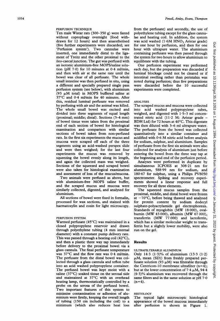
PERFUSION TECHNIQUETen male Wistar rats (300-350 g) were fastedwithout coprophagy overnight (food with-drawn for 12 hours) and then anaesthetised(five further experiments were discarded; see'Perfusion system'). Two cannulae wereinserted, one immediately distal to the liga-ment of Treitz and the other proximal to theileo-caecal junction. The gut was perfused withan isotonic aluminium-free MOPS/saline solu-tion (pH 7-0) for 10 minutes at 0-4 ml/min,and then with air at the same rate until thebowel was clear of all perfusate. The wholesmall intestine was then perfused in situ, usinga different and specially prepared single passperfusion system (see below), with aluminium(93 ,M total) in MOPS buffered saline at37°C and 0 4 ml/min for 40 minutes. Afterthis, residual luminal perfusate was removedby perfusing with air and the animal was killed.The whole small bowel was excised anddivided into three segments of equal length(proximal; middle; distal). Sections (3-4 mm)of bowel tissue were taken from the proximalend of each section of bowel for histologicalexamination and comparison with similarsections of bowel taken from non-perfusedrats. In the first six experiments the mucus andmucosa were scraped off each of the threesegments using an acid-washed perspex slideand were then weighed; for the last fourexperiments the mucus was removed bysqueezing the bowel evenly along its length,and again the collected mass was weighed.Sections of the squeezed and scraped bowelwere also taken for histological examinationand assessment of loss of the mucus/mucosa.Two animals were perfused as above, but
with aluminium-free MOPS saline buffer,and the scraped mucus and mucosa were
similarly collected, digested, and analysed foraluminium.
All sections of bowel were fixed in formalin,processed for wax sections, and stained withhaematoxylin and eosin for light microscopy.
PERFUSION SYSTEMWarmed perfusate (45°C) was maintained in aclosed polypropylene reservoir and drawnthrough polyethylene tubing (4 mm internaldiameter) with a constant pump delivery rate.This was passed through a heating coil (42°C),and then a plastic three way tap immediatelybefore delivery to the proximal bowel via a
glass cannula. The final perfusate temperaturewas 37°C and the flow rate was 0-4 ml/min.The perfusate from the distal bowel was col-lected through a glass cannula and teflon tubeinto an acid washed polypropylene container.The perfused bowel was kept moist with asaline (37°C) soaked tissue on the serosal sideand maintained at 37°C with an overheadheating lamp, thermostatically controlled by aprobe on the serosa of the perfused bowel.Two important features of this system tominimise contamination or adhesion of alu-minium were firstly, keeping the overall lengthof tubing (150 cm including the coil) to aminimum (which also reduces heat loss
from the perfusate) and secondly, the use ofpolyethylene tubing except for the glass cannu-lae and heating coil. In addition, the systemwas acid washed (1 6M HNO3 Aristar grade)for one hour by perfusion, and then for onehour with ultrapure water. The aluminiumcontaining perfusate was then passed throughthe system for two hours to allow aluminium toequilibrate with the tubing.One perfusion experiment was performed
each day, but the preparation was discarded ifluminal blockage could not be cleared or ifintestinal swelling rather than peristalsis wasnoted during perfusion; thus five preparationswere discarded before the 10 successfulexperiments were completed.
ANALYSESThe scraped mucus and mucosa were collectedinto acid washed polypropylene tubes,weighed, and digested with 1-5-3 ml concen-trated nitric acid (11 2 M: Aristar grade -BDH Ltd) for 72 hours at 40°C. This digestatewas then diluted with 3-6 ml ultrapure water.The perfusate from the bowel was collectedquantitatively into a similar container andacidified with 0-6 M nitric acid before analysisfor sodium, sulphur, and aluminium. Samplesof perfusate from the first six animals were alsocollected for analysis of aluminium just beforeentering the bowel from the three way tap atthe beginning and end of the perfusion period.
Analyses were performed in duplicate bysimultaneous ICPOES at 308-21 nm foraluminium, 588-99 nm for sodium, and180-67 for sulphur, using a Philips PV8050spectrometer. Spiking and recovery experi-ments showed a linear response and fullrecovery for all three elements.The squeezed mucus samples from the
proximal, middle, and distal bowel were frozenat -70°C, before being thawed and analysedfor protein content by sodium dodecylsulphate-polyacrylamide gel electrophoresis.Standards of myoglobin (MW 18 000), oval-bumin (MW 43 000), albumin (MW 67 000),transferrin (MW 77 000) and lactoferrin,which has a similar molecular weight to trans-ferrin but a slightly lower mobility, were alsorun on the gel.
Results
ULTRAFILTERABLE ALUMINIUMOnly 14-3 (1-3)% of aluminium (13-3 (1.2),uM, mean (SD)) from freshly prepared per-fusate solution (93 ,uM) was filterable throughthe Centricon-10 membrane ultrafilter (n=4);but at the lower concentration of 7-4 ,uM, 94-4(6&3)% aluminium was recovered through thesame filters and in the same solution at pH 7 0(n=4).
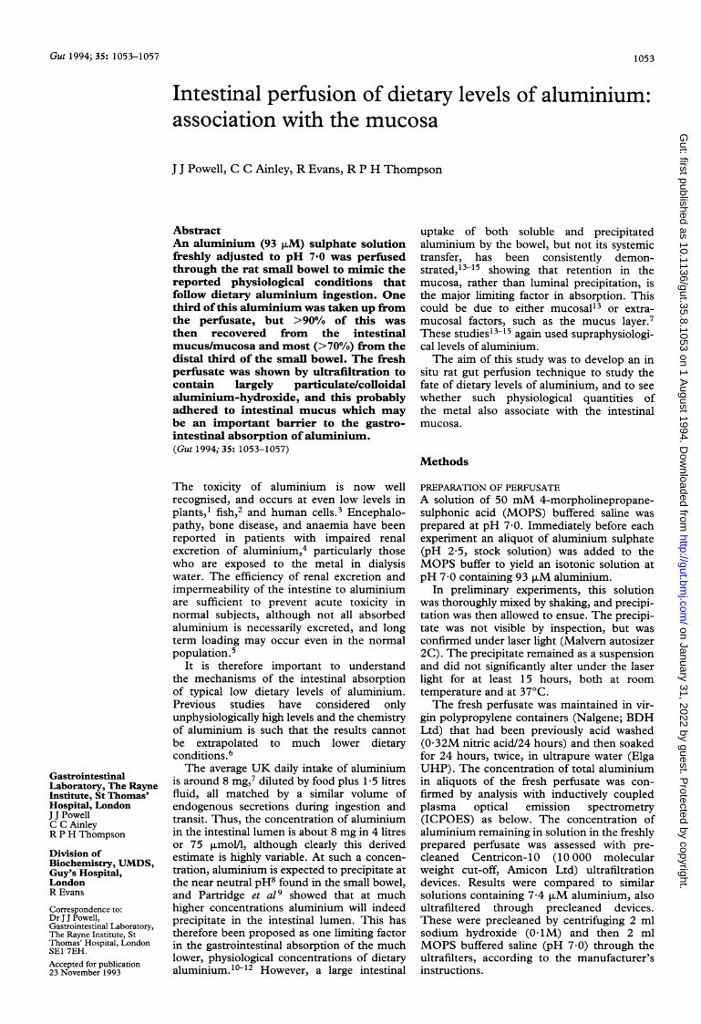
HISTOLOGYThe typical light microscopic histologicalappearance of the bowel mucosa immediatelyafter perfusion is shown in Figure 1.
1054
on January 31, 2022 by guest. Protected by copyright.
http://gut.bmj.com
/G
ut: first published as 10.1136/gut.35.8.1053 on 1 August 1994. D
ownloaded from

Intestinal perfusion of dietary levels of aluminium: association with the niucosa
Figure 1: Light microscopy section (original Figure 2: Light microscopy section (originalmagnification x 400) showing villi of the rat small bowel magnification x 100) showing the intestinal mucosa andfollowing successful perfusion; shedding of the tip of the lumen after removal of the luminal contents by squeezingmiddle villus is seen. the bowel. The mucosa remains intact but not all luminal
contents are removed; the residual is mainly mucus.
Compared with non-perfused control tissue,the mucosa was normal, except for someadditional sloughing of the villus tips (Fig 1).The mucosal scrape removed all mucosa, witha small amount of submucosa remaining onthe muscle layer. The squeezed mucosa wasintact (Fig 2), but occasionally some luminalmucus was still present indicating that squeez-ing did not completely remove all the mucus.
PERFUSATE CONCENTRATIONSOnce aluminium-containing perfusate hadbeen equilibrated with the perfusion appar-atus, there was no further loss of aluminiumfrom the perfusate by adhesion to the system(or sodium from the sodium chloride orsulphur from the MOPS buffer). In contrast,the percentage change in the perfusate concen-trations of aluminium, sodium, and sulphurafter perfusion through the small bowel areshown in Figure 3 (one sulphur atom is presentin one molecule of MOPS buffer).
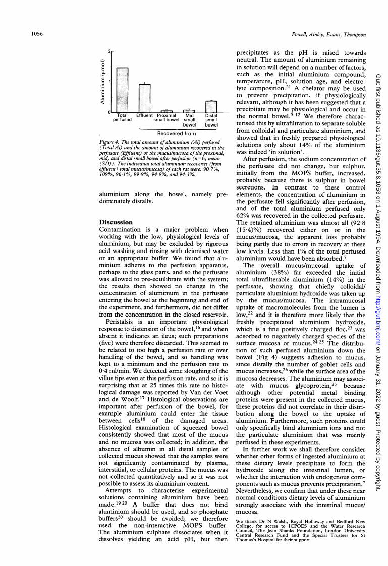
ALUMINIUM RECOVERYFigure 4 shows the distribution of aluminiumafter perfusion. The total aluminium perfusedwas 1-48 p.mol/experiment, and of this 62-2(6- 1)% (mean (SD)) was recovered in theeffluent; 92-8 (15-4)% (mean (SD)) of theunrecovered (37-8%) perfused aluminium wasdetected in the mucus/mucosal scrape, ofwhich 11-2 (4-8)% was in the proximal, 17-5(4-8)% in the mid, and 71-3 (5.3)% in thedistal segments (p<0 00l, Student's t test;distal v others). The total aluminiumrecovered, from perfusate plus all sections of
the intestinal mucus/mucosa, was 97 5 (6-4)%.Aluminium recovery from the mucus/mucosaof the two control perfusions was 0-008 and0-010 p.mol respectively.
POLYACRYLAMIDE GELSAlthough in all 12 mucus specimens weakbands were seen for substances that ran withmobilities close to those of transferrin andlactoferrin, their intensities were the same inthe three areas of the gut. In contrast, therewere strong bands for a substance thatmigrated with the same mobility as albumin inall specimens of mucus from the proximal andmiddle gut, but not from any mucus of thedistal gut. There were no protein bands thatran with the same intensity of distribution as
120
100'
80-co0)
,E 60-()
0) g
Ol VIIIIII[77777777/ II.77x7<7z%7 Y77777777A
Sodium Sulphur AluminiumFigure 3: The concentrations of aluminium, sodium, andsulphur remaining in the perfusate after intestinal perfusion,as a percentage of their concentrations in the perfusatebefore intestinal perfusion (n=6); mean (SD)).
1 055
on January 31, 2022 by guest. Protected by copyright.
http://gut.bmj.com
/G
ut: first published as 10.1136/gut.35.8.1053 on 1 August 1994. D
ownloaded from
Powell, Ainley, Evans, Thompson
0
C
Total Effluent Proximal Mid Distal
perfused small bowel small small
Recovered from
Figure 4: The total amount of aluminium (Al) perjfused(Total Al) and the amount of aluminium recovered in the
perfusate (Effluent) or the mucus/mucosa of the proximal,mid, and distal small bowel after perfusion (n =6; mean
(SD)). The individual total aluminium recoveries (fromeffluent+total mucus/mucosa) of each rat were: 90 7%,
109%, 96-1%, 99.90o, 94.99o~, and 94-3%.
aluminium along the bowel, namely pre-
dominately distally.
Discussion
Contamination is a major problem when
working with the low, physiological levels of
aluminium, but may be excluded by rigorousacid washing and rinsing with deionised water
or an appropriate buffer. We found that alu-minium adheres to the perfusion apparatus,perhaps to the glass parts, and so the perfusatewas allowed to pre-equilibrate with the system;
the results then showed no change in the
concentration of aluminium in the perfusate
entering the bowel at the beginning and end of
the experiment, and furthermore, did not differ
from the concentration in the closed reservoir.
Peristalsis is an important physiologicalresponse to distension of the bowel,t16 and whenabsent it indicates an ileus; such preparations
(five) were therefore discarded. This seemed to
be related to too high a perfusion rate or over
handling of the bowel, and so handling was
kept to a minimum and the perfusion rate to
0h4 mllmin. We detected some sloughing of thevillus tips even at this perfusion rate, and so it is
surprising that at 25 times this rate no histo-
logical damage was reported by Van der Voet
and de Woolf.17 Histological observations are
important after perfusion of the bowel; for
example aluminium could enter the tissuebetween cellst8 of the damaged areas.
Histological examination of squeezed bowel
consistently showed that most of the mucus
and no mucosa was collected; in addition, theabsence of albumin in all distal samples of
collected mucus showed that the samples were
not significantly contaminated by plasma,
interstitial, or cellular proteins. The mucus was
not collected quantitatively and so it was not
possible to assess its aluminium content.
Attempts to characterise experimentalsolutions containing aluminium have been
made.t9 20 A buffer that does not bindaluminium should be used, and so phosphatebuffers20 should be avoided; we thereforeused the non-interactive MOPS buffer.The aluminium sulphate dissociates when itdissolves yielding an acid pH, but then
precipitates as the pH is raised towardsneutral. The amount of aluminium remainingin solution will depend on a number of factors,such as the initial aluminium compound,temperature, pH, solution age, and electro-lyte composition.2" A chelator may be usedto prevent precipitation, if physiologicallyrelevant, although it has been suggested that aprecipitate may be physiological and occur inthe normal bowel.9-12 We therefore charac-terised this by ultrafiltration to separate solublefrom colloidal and particulate aluminium, andshowed that in freshly prepared physiologicalsolutions only about 14% of the aluminiumwas indeed 'in solution'.
After perfusion, the sodium concentration ofthe perfusate did not change, but sulphur,initially from the MOPS buffer, increased,probably because there is sulphur in bowelsecretions. In contrast to these controlelements, the concentration of aluminium inthe perfusate fell significantly after perfusion,and of the total aluminium perfused only62% was recovered in the collected perfusate.The retained aluminium was almost all (92.8(15A4)%) recovered either on or in themucus/mucosa, the apparent loss probablybeing partly due to errors in recovery at theselow levels. Less than 1% of the total perfusedaluminium would have been absorbed.7The overall mucus/mucosal uptake of
aluminium (38%) far exceeded the initialtotal ultrafilterable aluminium (14%) in theperfusate, showing that chiefly colloidal/particulate aluminium hydroxide was taken upby the mucus/mucosa. The intramucosaluptake of macromolecules from the lumen islow,22 and it is therefore more likely that thefreshly precipitated aluminium hydroxide,which is a fine positively charged floc,23 wasadsorbed to negatively charged species of thesurface mucosa or mucus.24 25 The distribu-tion of such perfused aluminium down thebowel (Fig 4) suggests adhesion to mucus,since distally the number of goblet cells andmucus increases,26 while the surface area of themucosa decreases. The aluminium may associ-ate with mucus glycoprotein,25 becausealthough other potential metal bindingproteins were present in the collected mucus,these proteins did not correlate in their distri-bution along the bowel to the uptake ofaluminium. Furthermore, such proteins couldonly specifically bind aluminium ions and notthe particulate aluminium that was mainlyperfused in these experiments.
In further work we shall therefore considerwhether other forms of ingested aluminium atthese dietary levels precipitate to form thehydroxide along the intestinal lumen, orwhether the interaction with endogenous com-ponents such as mucus prevents precipitation.7Nevertheless, we confirm that under these nearnormal conditions dietary levels of aluminiumstrongly associate with the intestinal mucus/mucosa.We thank Dr N Walsh, Royal Holloway and Bedford NewCollege, for access to ICPOES and the Water ResearchCouncil, The Jean Shanks Foundation, London UniversityCentral Research Fund and the Special Trustees for StThomas's Hospital for their support.
1 056
on January 31, 2022 by guest. Protected by copyright.
http://gut.bmj.com
/G
ut: first published as 10.1136/gut.35.8.1053 on 1 August 1994. D
ownloaded from
Intestinal perfusion of dietary levels of aluminium: association with the mucosa 1057
1 Jackson ML. Aluminium of acid soils in the food chain andsenility. Sci Total Environ 1983; 28: 269-76.
2 Birchall JD, Exley C, Chappell JS, Phillips MK. Acutetoxicity of aluminium to fish eliminated in silicon-richacid waters. Nature 1989; 338: 146-8.
3 Schofl C, Sanchez-Bueno A, Dixon CJ, Woods NM, LeeJAC, Cuthbertson KSR et al. Aluminium perturbsoscillatory phosphoinositide-mediated calcium signalingin hormone-stimulated hepatocytes. Biochem J 1990; 269:547-50.
4 Lindholm T, Thysell H, Ljunggren L, Divino JC,Schunnesson M, Stenstam M. Aluminium in patientswith uremia and patients with enteropathy. Nieren-Hochdruckkr 1983; 12: 192-7.
5 Powell JJ, Thompson RPH. Aluminium deposition in boneafter contamination of drinking water supply. Lancet1990; ii: 888 (letter).
6 Ganrot PO. Metabolism and possible health effects ofaluminium. Environ Health Perspect 1986; 65: 363-441.
7 Powell JJ, Thompson RPH. The chemistry of aluminium inthe gastrointestinal lumen and its uptake and absorption.Proc Nutr Soc 1993; 5: 241-53.
8 Martin BR. The chemistry of aluminium as related tobiology and medicine. Clin Chem 1986; 32: 1797-806.
9 Partridge N, Reigner F, White JL, Hem SL. Influence ofdietary constituents on intestinal absorption ofaluminium. Kidney Int 1989; 35: 1413-17.
10 Lote CH, Saunders H. Aluminium: gastrointestinalabsorption and renal excretion. Clin Sci 1991; 81: 289-95.
11 Stewart WK. Aluminium toxicity in individuals withchronic renal disease. In: (eds) Massey RC, Taylor D.Aluminium in food and the environment. London: RoyalSociety of Chemistry, 1989: 619.
12 Alfrey AC. Aluminium metabolism. Kidney Int 1986; 29(Suppl 18): S8-11.
13 Cochran M, Goddard G, Ramm G, Ludwigson N, MarshallJ, Halliday J. Absorbed aluminium is found with twocytosolic protein fractions, other than ferritin, in the ratduodenum. Gut 1993; 34: 643-6.
14 Van der Voet GB, Van Ginkel MF, De Wolff FA. Intestinalabsorption of aluminium in rats: stimulation by citric acid
and inhibition by Dinitrophenol. Toxicol Appl Pharmacol1989; 99: 90-7.
15 Feinroth M, Feinroth MV, Berlyne GM. Aluminiumabsorption in the rat everted gut sac. Miner ElectrolyteMetab 1982; 8: 29-35.
16 Granger ND, Barrowman JA, Kvietys PR. The smallintestine. In: Clinical gastrointestinal physiology:Philadelphia: WB Saunders Co, 1985: 141-207.
17 Van der Voet GB, de Wolff FA. The effect of di andtrivalent iron on the intestinal absorption of aluminium inrats. Toxicol Appl Pharmacol 1987; 90: 190-7.
18 Froment DH, Molitoris BA, Buddington B, Miller N,Alfrey AC. Site and mechanism of enhanced gastro-intestinal absorption of aluminium by citrate. Kidney Int1989; 36: 978-84.
19 Yokel RA, McNarmara PJ. Influence of renal impairment,chemical form and serum protein binding on intravenousand oral aluminium kinetics in the rabbit. Toxicol ApplPharmacol 1988; 95: 32-43.
20 Lote CJ, Wood JA, Saunders HC. Renal filtration,reabsorption and excretion of aluminium in the rat. ClinSci 1992; 82: 13-18.
21 Chen DTY. Solubility products of aluminium hydroxide invarious ionic solutions. Canadian J7ournal of Chemistry1973; 51: 3528-33.
22 O'Hagan DT. Intestinal translocation of particulates-implications for drug and antigen delivery. Advanced DrugDelivery Reviews 1990; 5: 265-85.
23 Arden TV. Water purification and recycling. In: ThompsonR. Ed. The modern inorganic chemicals industry. Specialpublication No 31. London: The Royal Society ofChemistry, 1982: 69-105.
24 Quaterman J. Metal absorption and the intestinal mucuslayer. Digestion 1987; 37: 1-9.
25 Crowther RS, Marriott C. Counter-ion binding to mucusglycoproteins. JPharm Pharmacol 1984; 36: 21-6.
26 Kulenkompff H. The structural basis of intestinalabsorption. In: Forth W, Rummel W. Eds. Pharma-cology of intestinal absorption: gastrointestinalabsorption of drugs. Vol 1; London: Pergamon, 1975:1-69.
on January 31, 2022 by guest. Protected by copyright.
http://gut.bmj.com
/G
ut: first published as 10.1136/gut.35.8.1053 on 1 August 1994. D
ownloaded from