Guidelines on Clinical Management of Dengue Fever / Dengue ... · DHF. The following are risk...
44
SLMH- EPID-05/03 Guidelines on Clinical Management of Dengue Fever / Dengue Haemorrhagic Fever Epidemiological Unit, Ministry of Health. 2005 Updated in JUNE 2009
Transcript of Guidelines on Clinical Management of Dengue Fever / Dengue ... · DHF. The following are risk...
SLMH- EPID-05/03
Guidelines on Clinical Management of
Dengue Fever / Dengue Haemorrhagic Fever
Epidemiological Unit, Ministry of Health.
2005 Updated in JUNE 2009
SLMH- EPID-05/03
Guidelines on Clinical Management of
Dengue / Dengue Haemorrhagic Fever
Prepared by the Sub-Committee of Technical Experts
on Clinical Management of DF/DHF
Epidemiological Unit, Ministry of Health. 2005 Updated in JUNE 2009
ii
Contributors 1. Dr. M.R.N. Abeysinghe Epidemiologist
2. Dr. Ranjani de Almeida Medical Officer in charge,
OPD, LRH, Colombo
3. Dr. M. Fernandopulle Consultant Paediatrician
4. Dr. D.H. Karunatilaka Consultant Paediatrician,
Lady Ridgeway Hospital,
Colombo.
5. Dr. Sujatha Ruwanpathirana Consultant Physician, NCTH,
Ragama.
6. Prof. Manouri Senanayaka Professor in Pediatrics,
Faculty of Medicine,
University of Colombo.
7. Dr. S.G. De Silva Consultant Physician, NHSL,
Colombo.
8. Prof. N.D. Warnasuriya Professor of Pediatrics,
Faculty of Medicine,
University of Sri Jayawardanepura.
9. Dr. J.S.D.K. Weeraman Consultant Paediatrician,
G.H. Kalutara.
10. Dr. Preethi Wijegoonawardene President, College of General
Physicians
11. Dr. Bandula Wijesiriwardana Consultant Physician, CSTH,
Kalubowila.
12. Dr. Padmakanthi Wijesuriya Consultant Paediatrician, NCTH,
Ragama.
iii
TABLE OF CONTENTS Contributors ... ... ... ... ... ... ... ii
Table of Contents ... ... ... ... ... ... iii
Foreword … … … … … … … … iv
Preface … … … … … … … … v
1. Dengue Illness – An Overview ... ... ... 1
2. Outpatient and First Contact Management ... ... 7
3. Management of Dengue Fever in the
Hospitalised Patient … … … … … 10
4. Coordination of Laboratory Investigation
for Management of DF/DHF ... ... 20
5. Dengue fever – Surveillance ... ... ... ... 21
6. References … … … … ... ... ... 24
Annexure (i) Leaflet for suspected Dengue Patients ... 25
Annexure (ii) Maintenance fluids for different body weights 26
Annexure (iii) Reduction of hourly maintenance rate ... 27
Annexure (iv) Management of DHF grade III & IV ... 28
Annexure (v) Dengue Special Surveillance format ... 29
Annexure (vi) Dengue Mortality Review – Procedure ... 31
Annexure (vii) Report on Death due to DF/DHF
- Institutional format ... ... ... 32
Annexure (viii) Field Investigation Report on
Death due to DF/DHF ... ... ... 37
iv
Foreword In the recent past we have witnessed a dramatic increase in the incidence of Dengue
Fever and its severe manifestations such as Dengue Haemorrhagic Fever and Dengue
Shock Syndrome globally as well as in Sri Lanka. As a result it has become a major
public health challenge. The case fatality rate due to DF/DHF remains low in the country
basically due to increase in the number of cases. However the number of deaths shows
an increase. It is felt that improvement in the clinical management could further reduce
the mortality due to this disease.
I hope that the guidelines on clinical management of Dengue/ Dengue Haemorrhagic
Fever prepared by the Epidemiological Unit with the contribution of experts in this field will
prove useful to clinicians for better case management and reduction of mortality due to
this disease.
Dr. H.A.P. Kahandaliyanage Director General of Health Services
v
Preface Dengue Fever has now become the most important communicable disease in Sri Lanka. Since the first reported outbreak of Dengue fever in 1965, there had been outbreaks on and off until the recent past affecting some densely populated areas. Since 1989 progressively large epidemics have been occuring more frequently involving a greater extent of the country so that it has become a major public health issue. Incidence of severe forms of the disease, namely Dengue Haemorrhagic Fever and Dengue Shock Syndrome is also on the rise leading to increase in mortality. Sri Lanka experienced the largest outbreak of Dengue Fever in 2004 with the highest recorded mortality in the recent past. Consequently dengue prevention and control activities were strengthened. Further to a Consultative Meeting of technical experts held at BMICH, Colombo on 14th August 2004 chaired by the Hon. Minister of Health, Nutrition and Uva Wellassa Development, several sub-committees were formulated to address the key issues with regard to Dengue control. This document was prepared by the sub-committee of technical experts on clinical management of DF/DHF with a view to assist clinicians in proper management of patients with the ultimate goal of preventing mortality due to this dreaded disease. I sincerely hope that this will be a very useful reference material among all other publications on this subject as it was prepared basically to address the issues currently faced by the dedicated Sri Lankan medical staff taking into consideration the immense difficulties they come across in patient management. I greatly appreciate the efforts of the experts who contributed for this document and I wish to thank the staff of the Epidemiological Unit specially Dr. Devika Mendis for the assistance extended in preparation of this document. Appreciation is extended to Naval Medical Research Unit 2 (NAMRU-2), Jakarta, Indonesia for their assistance in providing funds for this publication. Dr.M.R.N. Abeysinghe
Epidemiologist
1
1. DENGUE ILLNESS
An Overview Dengue illness is caused by any one of the four serotypes of Dengue virus – DEN 1-4.
This is considered to be the most important mosquito-borne disease of humans,
especially in the tropics.
Dengue Fever (DF), when due to primary dengue virus infection, is usually a mild
nonfatal disease. Clinical features are age dependant. Infants and young children
infected for the first time (primary infection) usually develop an undifferentiated fever with
or without a maculopapular rash. Older children and adults have either a mild febrile
illness or the classical incapacitating disease with high fever of abrupt onset (400C - 410C
/ 1030F – 1050F) sometimes biphasic with two or more of the following features – severe
headache (frontal), retro orbital pain, myalgia, bone / joint pain and rash (diffuse,
erythematous, maculopapular). Rarely haemorrhagic manifestations may occur.
The diagnosis of DF is likely if the above presentation occurs at the same location / time
as other confirmed DF cases.
The differential diagnosis includes other viral, bacterial and ricketsial diseases prevailing
in the area.
Virus isolation,serology, or PCR are needed for confirmation of the diagnosis.
Unlike DF, clinical features of Dengue Haemorrhagic Fever (DHF) which usually follows
a secondary infection are rather distinctive. Typically DHF is characterised by four major
clinical manifestations as follows;
1. High continuous fever for 2 – 7 days 2. Haemorrhagic tendency (positive tourniquet test, skin petechiae, easy
bruising, epistaxis, gingival bleeding and GI bleeding – haematemesis / malaena)
3. Hepatomegaly, usually soft and tender 4. Circulatory disturbance (shock in severe cases)
2
Thrombocytopenia (≤100,000/mm3) and Haemoconcentration (rising HCT of 20% or more) which represent the pathophysiologic changes of abnormal haemostasis and plasma leakage are constant findings. The clinical course of DHF is rather stereotypic. The incubation period is 5 – 8 days (range 3 – 14 days). The illness usually begins abruptly and has three phases, namely Febrile, Critical and Convalescent. In the acute febrile phase typically the patient has sudden rise in temperature accompanied by facial flushing, skin erythema, headache and muscle pain. Febrile convulsions may occur particularly in infants, as the temperature may be as high as 410C. Mild conjunctival injection and injected pharynx are common. Sore throat, rhinitis and cough are unusual. Anorexia, vomiting, abdominal pain are common and diarrhoea may occur. This phase resembles DF in many respects. Haemorrhagic manifestations are usually present. Massive GI bleeding may occur later as a result of prolonged shock leading to DIC. Haematuria is extremely rare. Tender hepatomegaly is often present but jaundice is not a feature. Critical phase, which lasts about 24 – 48 hours during which plasma leakage occurs, coincides or follows defervescence (becoming afebrile). Varying degrees of circulatory disturbances may develop and very close monitoring of the patient is necessary at this stage.
In mild DHF patients, changes in vital signs are minimal and transient, and recover spontaneously or after a brief period of treatment. In severe DHF patients the disease progresses rapidly into a stage of shock. Onset of shock is acute, generally occurring at the time of defervescence, which is usually on or after the 3rd day of illness (the shortest duration of fever is 2 days). In the early stages of shock patient often complains of acute right sided abdominal pain, becomes restless with subnormal temperature, sweating and cold clammy extremities. Rapid thready pulse, prolonged capillary refill time (over 2
seconds) and narrow pulse pressure (≤20mmHg) with characteristic high diastolic pressure (eg. 100/90, 110/90) are noted at this time. Hypotension is seen in late stages of shock. Right-sided pleural effusion and ascitis will occur. Despite being in shock, consciousness remains even if the patient is terminally ill. However, rarely neurological disturbances have been described.
3
The course of shock is short but life threatening. If proper treatment is not given the
patient deteriorates rapidly in to a stage of profound shock with undetectable pulse and
unrecordable blood pressure. Circumoral and peripheral cyanosis is noted and the skin
becomes blotchy, mottled and purplish. Without treatment the patient succumbs.
Prolonged shock is often complicated with metabolic acidosis and/or DIC and massive
bleeding. Severe bleeding most commonly occurs in the GI tract. Occasionally the
bleeding may be concealed. The most fatal haemorrhagic manifestation is intracranial
bleeding which leads to convulsions and coma. The critical period of plasma leakage and
shock rarely lasts longer than 48 hours. Even the patients with shock have a favourable
outcome if they receive proper treatment before reaching the irreversible stage of shock.
Rapid and often dramatic recovery is the rule with appropriate and timely intervention.
Infrequently unusual encephalitic signs are associated with electrolyte and metabolic
disturbances (hyponatraemia, hypocalcaemia and hypoglycaemia), intracranial
haemorrhage or hepatic failure (Reye or Reye like syndrome) and can give rise to a more
complicated course with a grave prognosis. Kidneys are rarely affected in dengue shock
syndrome and mostly associated with late stage of acute liver failure. Use of nephrotoxic
drugs, intravascular haemolysis due to G6PD deficiency and abnormal
haemoglobinopathies are some of the risk factors for developing acute renal failure. Dual
infections with other organisms present in the area have been reported as unusual
manifestations (eg; DHF and Leptospiorsis, DHF and common respiratory and
gastrointestinal infections).
The Convalescent phase is usually short and uneventful even in those with shock.
Diuresis ensues as shock resolves and the patient rapidly regains appetite. Some may
have a confluent petechial rash with characteristic scattered round areas of pale skin on
the extremities, which may be itchy. Bradycardia is a common finding.
The major pathophysiological hallmarks of DHF are the plasma leakage as a result of
increased vascular permeability and abnormal haemostasis. Hypovolaemic shock occurs
as a consequence of, and subsequent to a critical plasma volume loss. Abnormal
haemostasis including increased capillary fragility (positive tourniquet test and tendency
to bruise), impaired platelet function, thrombocytopenia and in most severe form,
4
disseminated intravascular coagulation contribute to varying degrees of haemorrhagic
diathesis. All available data strongly suggest the involvement of both immune systems
(cell mediated and humoral) and almost all haematological components in the
pathogenesis of DHF. The association of DHF with secondary dengue infection in older
children and primary infection in infants with passive dengue antibody from their mothers
led to propose the ‘two infection theory’, and the concept of antibody – dependent
immune enhancement (ADE). It was suggested that during the second infection with a
heterotypic dengue infection that failed to neutralise would instead enhance viral uptake
and replication in the mononuclear phagocytes. Such infected cells may then become a
target of an immune elimination mechanism, which can trigger the production of
mediators and activation of complement and coagulation cascade and eventually produce
DHF.
The following are risk factors for developing DHF / DSS
• Children are more prone to develop DHF / DSS than adults.
• DHF / DSS is associated more with well nourished than with under
nourished children.
• Primary infection in infants born to dengue immune mothers.
• Presence of underlying chronic illnesses (eg: heart disease, anaemia,
chronic liver disease)
The disease severity of DHF has been arbitrarily classified into four grades according to
the clinical hallmarks of bleeding and plasma leakage.
• Grade I - Only positive tourniquet test
• Grade II - Positive tourniquet test with spontaneous
superficial bleeding
• Grade III - Shock
• Grade IV - Profound shock with unrecordable
blood pressure and / or pulse
N.B. Every DHF patient must have evidence of plasma leakage and thrombocytopenia.
Final grading can be given only after the patient is afebrile for 2 days.
Non-shock DHF
DHF with shock
5
Occasionally DF cases, particularly those with unusual haemorrhage or
thrombocytopenia are misclassified as DHF Grade I or II. It should be emphasised that
thrombocytopenia with evidence of concurrent plasma leakage(rising Hct) are the two
essential findings in DHF that differentiate non – shock cases of DHF from DF.
Some significant laboratory findings occur in DHF and being aware of them is critical for
clinical diagnosis and management.
• In the peripheral blood, normal white cell count or leucopoenia with predominant
neutrophils is common initially and majority of them are immature forms. Towards
the end of the febrile phase there is a further reduction in the number of white
cells with simultaneous increase of lymphocytes with about 15-20% atypical
lymphocytes. This is usually observed one to two days before defervescence.
The white cell count helps to differentiate DF / DHF from bacterial infections and
predict the critical phase in DHF.
• Moderate to marked thrombocytopenia is a constant finding in DHF. The platelet
count drops rapidly to very low levels (≤100,000/mm3) shortly before or
simultaneously with the rise in haematocrit.
• Haematocrit determinations should be made more frequently after the platelet
count has fallen. The changes in the haematocrit are important for clinical
diagnosis of DHF.
• Evidence of plasma leakage includes a sudden rise in haematocrit(Hct) leading
to haemoconcentration (20% rise in Hct.). Hct levels correlate well with plasma
volume loss and disease severity. The level of haemoconcentration may be
equivocal when there is frank haemorrhage or early and excessive volume
replacement. For determination of Hct, it is important to draw blood from a vein at a distant site, away from the drip site in order to prevent haemodilution. At the peak of plasma leakage, right sided or bilateral pleural
effusions are seen on chest X ray. In the case of a mild leakage, pleural effusion
can be determined by right decubitus chest X ray.
• Elevation of aspartate amino transferase (SGOT),to a level which is about 2-3
times the level of alanine amino transferase (SGPT). However the values of both
these enzymes are usually less than 200 units. If the SGPT is over 200 units,
6
hepatic involvement is likely and changes in level of consciousness should be
monitored. Every patient with altered consciousness, especially those with restlessness, confusion and irritability should have his/her liver functions monitored.
• Clotting abnormalities are common in shock cases and the laboratory evidence of
DIC appears to be correlated with disease severity.
• The ESR is normal in DHF (≤ 20mm) together with normal CRP.
Differential diagnosis of DHF during the early febrile phase includes a wide spectrum of
viral, bacterial and parasitic infections, e.g. Typhoid fever, malaria, leptospirosis, viral
gastritis, acute gastroenteritis and acute tonsillitis. In a child presenting with acute onset
of high fever of 1 – 2 days, the finding of flushed face without coryza or any other
respiratory symptom should suggest the possibility of Dengue infection. A positive
tourniquet test increases the probability. As the disease progresses, enlargement of liver
which is usually soft and tender provide more support for a clinical diagnosis of DHF. The
diagnosis becomes certain when the platelet count drops shortly before or simultaneously
with a rise in Hct, which is unique to DHF. The presence of pleural effusion and/or ascitis
supports the diagnosis of DHF in a patient whose level of Hct is equivocal. Leucopenia
with a high proportion of atypical lymphocytes and a normal ESR helps to differentiate
DHF from bacterial infection and septic shock.
The major causes of death in DHF are,
• Prolonged shock leading to DIC and multi organ failure • Massive bleeding • Fluid overload leading to cardiac failure / pulmonary oedema • Acute liver failure with encephalopathy • Encephalitis • Myocarditis
With good clinical care, case fatality rate (CFR) should be around 0.5 – 1.0%, but
otherwise may vary up to 5%.
7
2. DENGUE FEVER -
OUTPATIENT AND FIRST CONTACT MANAGEMENT Assessment of patient on the 1st day of a fever during a dengue epidemic The early manifestations of dengue infection are high fever with significant myalgia and
arthralgia, possibly some erythema of the skin better noticed in the palms soles and
around the neck. Coryza and cough are not usually found. The practising physician
should exclude other common causes of fever. If another cause for fever is established,
act accordingly. Even if dengue fever is suspected, the patient need not be admitted at
this stage. If the patient or the guardians request admission, the physician should try to
convince them that admission is not necessary. A FBC at this stage will not help to rule
out or suspect dengue infection. Management consist of extra fluids, normal diet, rest and
the correct dose of paracetamol (60mg per kg per 24 hours). NSAIDS are
contraindicated. The patient should be reviewed on the 3rd day of fever.
A leaflet explaining the danger signs of dengue, which can be given to the
patient(Annexure i), and similar posters in the waiting room, are recommended.
Assessment of the patient on the 2nd day of fever. The assessment and the management is the same as on the 1st day. But the patient
should be reviewed in 24 hours.
Assessment of the patient on the 3rd day of fever The following should be actively looked for.
• Enlarged tender liver (essential to examine in the horizontal position)
• Check capillary filling time and the pulse. If the capillary filling time is > 2
seconds or the pulse is rapid or is of low volume check the blood pressure.
(measuring the BP is recommended in children over 5 years)

8
For children, normal values for vital signs are as follows.
Age Heart rate Blood pressure Systolic / Diastolic
0-3 months 100-150 65-85 / 45-55 3-6 months 90-120 70-90 / 50-65 6-12 months 80-100 80-100 / 55-65 1-4 years 70-110 90-105 / 55-70 4-6 years 65-110 95-110 / 60-75 6-12 years 60-95 100-120 / 60-75 > 12 years 55-85 110-135 / 65-85
• Examine for bleeding manifestations.
• Examine for cold extremities.
• Check for diminished air entry at the lung bases.
• Tourniquet test is not routinely recommended because of the time factor and
low yield rate. It can be done depending on the workload.
FBC is mandatory, if not, Hct and platelet count should be done.
It is recommended that MOO/OPD be trained to do the Hct and the facility is made
available at least in the large hospitals in areas where dengue fever is endemic.
The normal Hct can be taken as 45 for adults, 40 for children and 35 below 1 year.
A platelet count between 100,000 to 150,000 per cu.mm should alert the physician.
A patient with a platelet count below 100,000 per cu. mm should be admitted.
If the above findings are within normal limits and the patient is clinically well, the patient
may be sent home with the following warning messages. The patient should report back
immediately if any of the following findings are noticed.
• Significant abdominal pain • Black or Red coloured stool • Persistent vomiting • Coffee ground or Red coloured vomitus • Any other bleeding tendency • Cold extremities • Restlessness or drowsiness
If any of these finding are present the patient should be admitted.
9
In the absence of any of the above findings,
Review the patient in 24 hours and repeat PCV and platelet count. If patient is well and there is no fever, paracetamol should be stopped. If fever continues review daily till the 6th or 7th day with PCV and platelet counts. Serological tests for dengue are not essential for the clinical management except in a minority of cases where the diagnosis is in doubt. It is indicated to establish the diagnosis for epidemiological information.
Management on day 4 and 5 As long as the patient remains well with reasonably normal blood counts, they need not
be admitted. If any of the findings mentioned under day 3 as indications for admission are
found the patient should be admitted.
Even if one decided to admit the patient it is essential that the first contact doctor should
stabilise the patient at primary care before sending out for indoor care. This applies even
to the MOO/OPD at the major hospitals. The immediate treatment for stabilisation is
Hartman's solution or N – Saline bolus of 10ml per kg over 20 minutes. Establishment of
an ETU in the out patient department for this purpose is a useful step at least in the major
hospitals.
Good relationship between the ward staff and the OPD medical officer is essential. The
services of the registrar on call to the medical/paediatric casualty ward should be
available to the OPD officers when required. The OPD or the first contact doctor should in
turn communicate with the house officer on call to the ward prior to sending the patient to
the ward.

10
3. MANAGEMENT OF DENGUE HAEMORRHAGIC FEVER IN THE HOSPITALIZED PATIENT
Management of Dengue Haemorrhagic Fever is divided into three phases
according to the clinical course of the illness, with the durations as indicated.
• Febrile phase: 2-7 days • Critical / Leakage phase: 1-2 days • Convalescent phase: 1–5 days
FEBRILE PHASE
Antipyretics for fever • Paracetamol 60mg/kg/day in 4 divided doses. (1g every 6hrs in adults) • All NSAID’s through any route (oral, rectal, intramuscular) are contraindicated as
they may precipitate/aggravate gastrointestinal bleeding or Reye syndrome. • Tepid sponging if temperature remains high after a dose of Paracetemol.
Mosquito net or repellants may be used for the patient during day and night, to prevent
nosocomial spread.
Nutritional support • A balanced diet with adequate fluids. • If solids are refused, milk, fruit juices and ORS are recommended. • Plain water alone is inadequate as it may cause electrolyte imbalance.
• Black or red coloured food or drink should be avoided as it may be mistaken for
haematemesis in the event of vomiting.
Other supportive / symptomatic treatment • Vomiting: Domperidone IV 1mg/kg per day in 3 divided doses.(10mg/kg iv
tds in adults) • Febrile convulsions: Diazepam 0.5mg/kg rectally • Upper Gastro-intestinal bleeding: Ranitidine IV 1mg/kg/8hourly (50mg IV
tds in adults) or proton pump inhibitors - Pantoprazole.
11
Do not use antibiotics even in the presence of severe leucopenia.
Corticosteroids are ineffective in preventing shock in DHF and may
even be harmful by causing GI bleeding.
Daily haematocrit (Hct) estimation is mandatory. Twenty percent (20%) rise in Hct reflects
a significant plasma loss.
Oral Rehydration Solution should be given if the patient can take fluids orally.
I.V. fluid administration is considered only in those with dehydration due to severe or
persistent vomiting and refusal of all food and drink. It should be discontinued as soon as
the dehydration is corrected and when the oral intake is adequate. If IV fluids are to be
given more than a day, the amount should be the minimum required. Excessive IV fluids
given during this phase may cause fluid overload later in the illness.
All suspected dengue fever patients should be examined for following clinical changes at
least daily, beginning on the 3rd day of the illness until afebrile for at least 24 – 48 hrs
without the use of antipyretics.
Symptoms:
- History of bleeding into skin, nose, gums or passage of tarry
stools.
- Vomiting / refusal of food or drink / extreme thirst.
- Less fluid intake and poor urine output.
- Right sided upper abdominal pain.
- Poor appetite.
- Cold extremities - hands and feet.
- Refusal to sit up / postural giddiness.
12
Signs - Clinical deterioration with settling of fever
- Unexplained tachycardia.
- Tender enlarged liver.
- Cold clammy skin.
- Drowsiness / sleepiness.
- Behavioural changes / confusion
Record: Vital signs. Fluid intake and output.
Check: Platelet count and heamatocrit (Hct) daily from the 3rd day of illness. Hct will rise
before changes in pulse rate and blood pressure are noted.
A rise in Hct of 20% or more reflects a significant plasma loss.
Presence of leucopenia (WBC less than 5000/cumm) with 10 – 20% atypical
lymphocytes, usually predicts progression to critical phase within the next 24 hrs.
A platelet count less than 100,000/cumm also predicts that progression to critical phase is
imminent.
A platelet count less than 100,000/cumm with a rise in Hct over 20% indicates that
patient is in the critical phase.
DHF I & II CRITICAL/LEAKAGE PHASE (1 – 2 DAYS) – DHF without
shock
• Establish IV access
• Monitor vital signs (pulse rate and BP) every 2- 4 hrs.
• Monitor Hct at least twice daily
• Measure urine output every 2-4 hrs and record intake accurately
• Encourage oral fluid intake
• I.V. fluids are not mandatory if oral intake is adequate.
• If patient refuses oral fluids or there is severe vomiting, IV fluids are to be given.

13
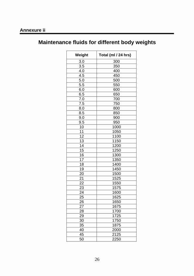
The total volume of IV fluids to be given for 24 hrs during this phase is as follows; For children – Maintenance (M) + 5% deficit
For adults - Maintenance x 2
The Maintenance (M) fluid needed for 24 hrs is calculated either using the
following formula or calculated using available tables. (annexure ii).
Use the ideal body weight (weight for age) to calculate the IV fluid requirement in obese/
over weight children.
The maximum weight for IV fluid calculation is 50 kg in adults and all overweight patients.
If the patient is taking orally, IV fluids should be omitted as early as possible.
As the rate of leakage of plasma is rapid during the first 6 – 12 hrs after its onset
the IV/ oral fluid replacement should parallel this rate and be guided by regular
measurement of Pulse rate, Blood pressure, Urine output and Hct.
CONVALESCENT PHASE (1 – 5 DAYS) - IV Fluid therapy can be stopped when Hct drops to 45% in adults, 40% in
children and 35% in infants.
- Return of appetite and diuresis are signs of recovery.
Body weight (kgs) Maintenance volume (ml) Administered over 24 hrs
< 10 100/Kg
10 – 20 1000 + 50 for each kg in excess of 10
> 20 1500 + 20 for each kg in excess of 20
14
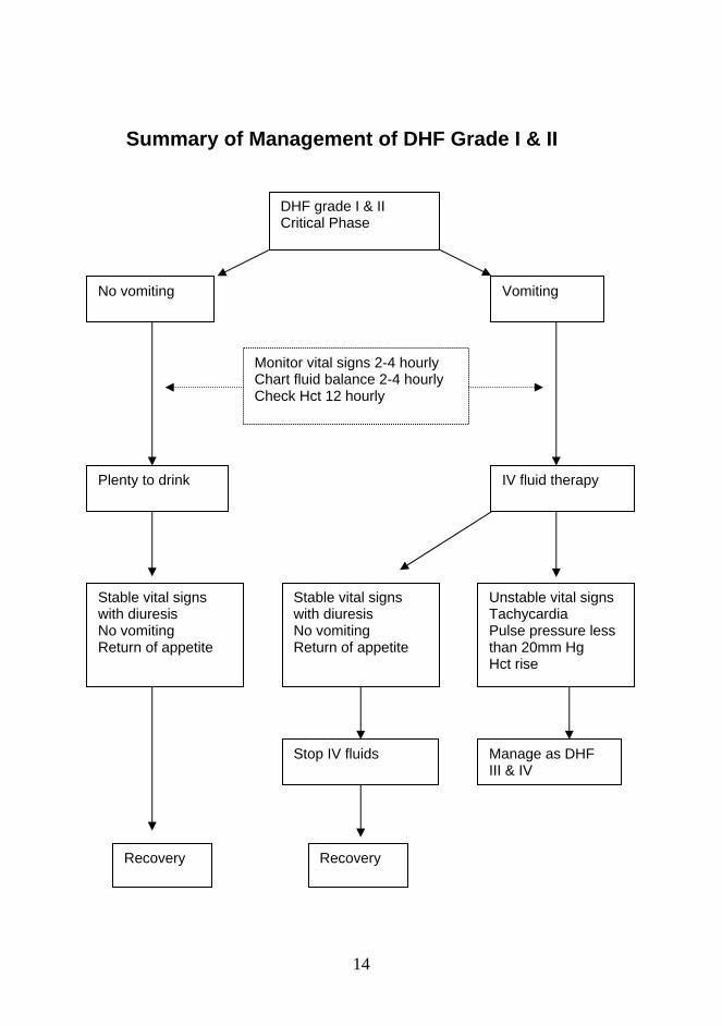
Summary of Management of DHF Grade I & II
DHF grade I & II Critical Phase
No vomiting Vomiting
Monitor vital signs 2-4 hourly Chart fluid balance 2-4 hourly Check Hct 12 hourly
Plenty to drink IV fluid therapy
Stable vital signs with diuresis No vomiting Return of appetite
Stable vital signs with diuresis No vomiting Return of appetite
Unstable vital signs Tachycardia Pulse pressure less than 20mm Hg Hct rise
Stop IV fluids Manage as DHF III & IV
Recovery Recovery
15
MANAGEMENT OF DHF III & IV CRITICAL / LEAKAGE PHASE (1 TO 2 DAYS) – DHF with shock
In this phase there is rapid leakage of plasma leading to shock. Hence IV fluid
replacement is mandatory. The volume used for replacement should be just sufficient to
maintain effective circulation during this critical period. As the rate of leakage of plasma
is not uniform (being more rapid during the first 6 – 12 hrs) the rate and volume of fluid
replacement should be adjusted according to the rate of plasma leakage. This should be
guided by the general condition, vital signs, urine output and Hct.
The type of fluid used should be isotonic with plasma. E.g. Crystalloid such as Hartmann
solution, normal Saline, N/2 saline + 5% Dextrose. In the case of massive leakage a
colloidal solution should be used in addition to crystalloids.
The recommended total volume of fluid needed by an adult in this phase is Maintenance
x 2 for 24 hrs. The maximum fluids needed by children for 24 hrs is approximately
maintenance plus 5% body weigh deficit (50ml / Kg / 24 hrs).
When leakage stops (by the end of 1 to 2 days) the IV fluid must be discontinued. Stable
vital signs (good volume pulse with wide pulse pressure, diuresis and stable Hct) are
good indicators to stop Intra Venous fluids.
Following management regimen is recommended in managing DHF III & IV. (Leakage
Phase)
• A Medical Officer should be in attendance all the time until the patient is stable.
• Ideally a specific area in the ward should be reserved for dengue patients.
• Monitor vital signs frequently, every 1 – 2 hrs during critical period / every 15min
during state of shock.
• Monitor Hct at least twice a day in the presence of overt shock.
• Special form for recording vital signs, Hct, intake / output and clinical findings
should be available at the bedside (important for adjusting the rate of IV fluids).
16
• Keep the patient flat in bed (strict bed rest should be advised) and give Oxygen
via face mask / nasal prongs.
• Arrest bleeding with appropriate technique. e.g. anterior nasal packing for
massive epistaxis.
• Avoid invasive procedures as much as possible. e.g. Nasogastric tube insertion
and deep neck vein cannulation.
• Vital signs specially Blood Pressure (BP), Pulse Pressure (PP) Capillary Refill
Time (CRT) have to be measured every 10 to 15 minutes until they are stable.
• An Intra Venous bolus of Hartmann solution or normal saline (10ml/Kg) should
be given immediately on detection of shock. The duration of bolus should not
exceed 20 minutes.
• While the fluid bolus is being infused, another Intra venous cannula should be
inserted simultaneously into another limb.
• Blood should be withdrawn for grouping / cross-matching, Hct and platelet
counts in uncomplicated DHF cases. In high risk or complicated DHF patients
other investigations such as liver function test, blood electrolyte (Na, Ca, K),
blood sugar, renal function tests, blood gas and coagulation screen (APPT, PT,
TT), an ECG and a Chest X-ray (Right decubitus view) should be done.
• Total amount of intra venous fluid needed for children during the leakage phase
is Maintenance + 50 ml/Kg/24hrs, for adults it is Maintenance x 2.
• I.V. boluses of Hartmann solution or normal saline (10ml/Kg) could be repeated
whenever pulse pressure is < 20mm. A maximum of three I.V. boluses can be
given.
• If there are no signs of improvement, I.V. boluses of colloid solution (plasma or
dextran 40) should be given, (10ml/Kg) up to a maximum of two I.V. boluses.
• When vital signs improve, as indicated by a fall in Hct and an increase in Urine
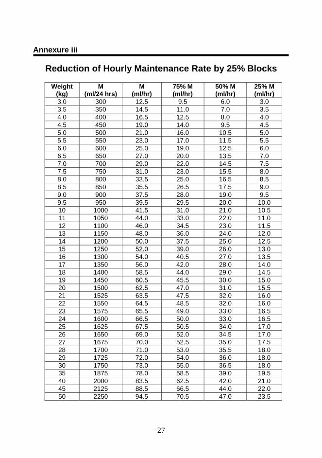
output, the rate of I.V. fluid replacement should be reduced by 25% of
maintenance (25% blocks) (annexure iii). The vital signs should be re-
assessed every hour with further reduction of the hourly rate by blocks of 25%.
I.V. Fluid therapy may be discontinued when the vital signs are stable and Hct
drops to 40%.
17
• Settling of tachycardia, good perfusion, return of appetite and diuresis indicate
recovery.
• In general, with early recognition and appropriate management of shock, rapid
and dramatic recovery is the rule.
• Refractory shock despite adequate volume replacement and a drop in Hct. (e.g.
from 50% to 40%) indicate significant internal bleeding and a need for blood
transfusion. Give fresh whole blood transfusion (10ml/Kg) or packed red cells
(5ml/Kg) and occasionally platelet-rich plasma in cases with significant bleeding
associated with thrombocytopenia.
• Correct metabolic and electrolyte disturbances. Metabolic acidosis can be
corrected with sodium bicarbonate. Electrolyte imbalances are usually found
during this phase particularly hyponatraemia and hypocalcaemia.
Hypokalaemia may occur during the convalescent phase. Hyponatraemia
results form inadequate intake of isotonic fluids and receiving hypotonic
solutions e.g. N/2 or N/5. If the patient has no convulsions, there is no need to
give hypertonic saline (3% sodium chloride) and Normal saline is sufficient.
Hypocalcaemia – results form leakage of Ca that follows albumin into the
pleural or peritoneal cavity. 10% Ca gluconate 1 ml /Kg/dose (maximum 10ml)
IV slowly is recommended only in complicated patients e.g. grade IV patients
with fluid overload.
Summary of management of this phase is given in annexure iv
Indications for administration of colloidal solutions
• Patients who receive adequate volume of crystalloid solution as recommended (2 – 3
boluses) and still have unstable vital signs with high Hct value.
• Patients who have signs of continued massive leakage of plasma e.g. periorbital
oedema, respiratory discomfort from massive pleural effusion and / or very tense
abdomen but yet the patient needs I.V. fluids as vital signs are not stable and high
Hct.
18
Indications for blood transfusion
Blood transfusion is indicated in the presence of blood loss, which can either be obvious
or concealed.
Significant obvious blood loss > 10% of total blood volume (6 –
8ml/Kg). Replace estimated loss with equal volume of blood (e.g.
in a 20 Kg patient, blood transfusion is indicated if he has lost 120
– 160 ml of blood). The total amount of blood transfusion is equal
to the volume of estimated blood loss.
Concealed internal bleeding, usually found in patients with
prolonged shock i.e. patients who received adequate amount of
IV fluids but still have unstable vital signs despite fall in Hct. (E.g.
Hct drop from 53% - 45% with unstable vital signs.)
Or
• Patient is in shock and treated for 6 hrs, but at the end of 6 hrs the
rate of IV fluid administration cannot be reduced.
Fresh whole blood 10 ml / Kg / dose or Packed red cells 5 ml / Kg / dose is given.
Indication for platelet transfusion Platelets are given only if significant bleeding occurs. (more than 6 – 8 ml /Kg)
There is no place for prophylactic platelet even with a count below 10,000 per cu mm if
there is no evidence of bleeding. If platelets are not available, fresh whole blood or
packed red cells transfusion is sufficient in patients with bleeding. When giving platelets,
be aware of large volume of platelet concentrate. Patients may get fluid overload from
rapid platelet transfusion. Platelet concentrates are used in only 0.4% of DHF patients in
specialized centres.
Convalescent Phase of DHF
About 48 hrs after defervescence most DHF patients enter the convalescent phase and
recover spontaneously with appropriate management. In adults recovery may be
prolonged with fatigue lasting 2 – 4 weeks. But in children, it is only about 5 days.
19
The following features indicate that the patient is in convalescent phase.
• Improved general condition and return of appetite
• Stable vital signs, strong slow pulse with wide pulse pressure
• Hct reduces to normal or sometimes below normal
• Diuresis
• Convalescent rash in around 20% of the DF & DHF cases
• Bradycardia
Management
• Discontinue IV fluids.
• Allow patient to rest. Traumatic or invasive procedures should not be carried out
e.g. intra-muscular injection.
• If patient still has no appetite check serum electrolyte as hypokalaemia may lead
to paralytic ileus. More potassium is lost during diuresis. Fruits or fruit juices are
recommended. KCl solution may be indicated if patients refuse to eat or drink
any fruits.
• Some patients who receive large amounts of IV fluids during the critical periods
may have fluid overload when re-absorption of extravasated plasma occurs in the
convalescent phase. Frusemide 1 – 2 ml / kg is helpful in this situation.
Indication for Discharge All following criteria should be fulfilled before discharge.
• No fever for at least 24 hrs without the use of antipyretics
• At least 2 days have lapsed after recovery from shock.
• Good general condition and improving appetite.
• Normal Hct at baseline level or around 38% - 40% when baseline value is not
known.
• Diuresis
• No distress from pleural effusion or ascites.
• Platelet count over 50,000 / cu mm
• No other complications
20
4. CO-ORDINATION OF LABORATORY INVESTIGATIONS FOR MANAGEMENT OF DF/ DHF
The following recommendations are made with regard to improvement of laboratory services for better case management at various levels of health care delivery. Teaching and provincial hospitals
Fully automated blood counters to be provided to all the teaching and provincial hospitals. (If these are available during the day, doing FBC will not be a problem even for OPD patients.)
After working hours, the existing semi-automated machines could be used. (As only a few MLTT would be allowed to handle the fully automated machines)
Strengthening the night lab staff during the epidemics of dengue fever. One extra person would be required to handle the specimens from dengue patients using the semi-automated machines.
When night lab support is not available or day lab support inadequate, microhaematocrits to be made available round the clock. Each medical and paediatric unit to be given one each or if there are financial constraints, one microhaematocrit should be kept in a place, which is accessible to all the units (for example, the ETU).
Base hospitals
Fully automated blood counters to be provided to large base hospitals. Until such time, at least semi-automated machines to be made available.
Strengthening the night lab staff during the epidemics of dengue fever. (One extra person would be required to handle the specimens from dengue patients using the semi-automated machines or manually)
When night lab support is not available or day lab support is inadequate, microheamatocrits to be made available round the clock. Medical and paediatric units to be given one each or if there are financial constraints, one microhaematocrits should be kept in a place, which is accessible to all the units. (For example, the ETU)
District Hospital and Peripheral Units
To make use of existing facilities.
21
5. DENGUE FEVER – SURVEILLANCE
Passive Surveillance; Notification DF/DHF is a notifiable disease in Sri Lanka and therefore along with the other
notifiable diseases, it should be notified to the respective MOH using the form H 544. It
is of utmost importance that notification should be done on clinical diagnosis and that it
should not be delayed until confirmation of the diagnosis, as early action has to be
taken by the MOH for prevention and control. Therefore, recording of the correct
address of the patient in the notification form is of paramount importance for field
investigation.
Special Surveillance Since the severity of the disease and mortality due to DF/DHF varies during
epidemics over the years, a special surveillance mechanism is useful to obtain the
information on clinical presentation, severity and outcome of the cases. Furthermore it
provides epidemiological information on confirmed cases, as data obtained by the
routine surveillance comprises of suspected cases of DF/DHF.
Special surveillance has been carried out in the field by the MOH/PHI of the
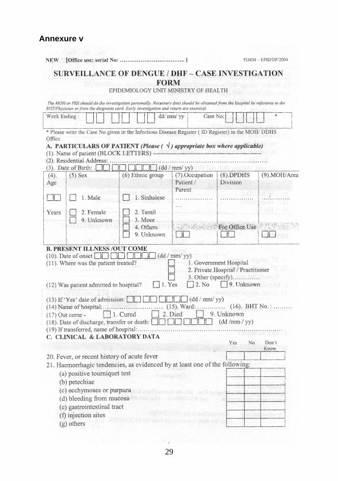
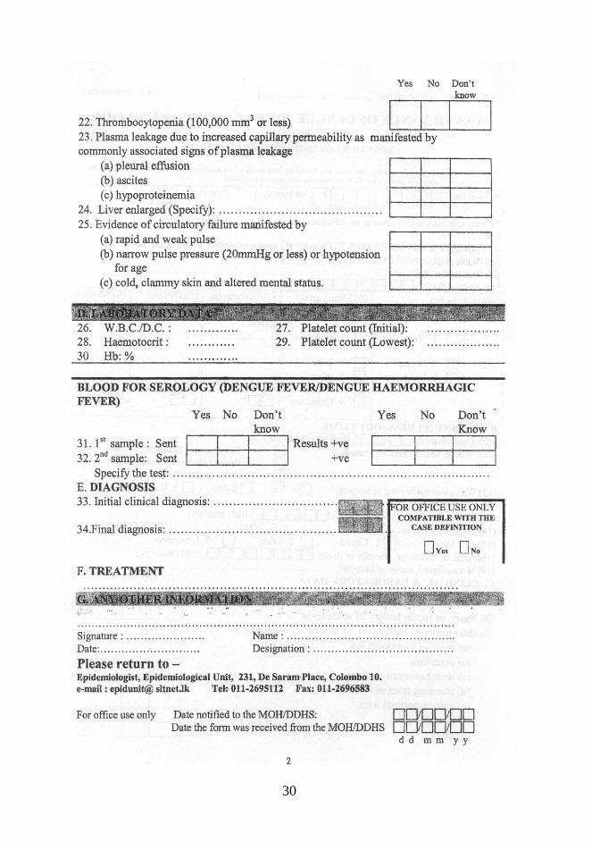
area utilizing a special format- form Epid/DF/2004 (Annexure v). However this
arrangement has not yielded expected results due to lack of data (clinial and
laboratory) with the patient/patient's family. To overcome this problem, it has been
decided to delegate this activity to the Medical Officer/Public Health or Infection
Control Nursing Officer, to be carried out in the hospital.
Sentinel Surveillance A sentinel reporting system has been established for DF/DHF, which gives an
indication of the trend of the disease in the areas concerned and acts as an early
warning system. Surveillance is carried out to obtain the number of fever cases
reporting to OPD of selected hospitals and number of suspected DF/DHF cases
admitted to these institutions.

22
Recommended guidelines for strengthening of surveillance of dengue fever are as
follows;
• Suspected dengue patients should be notified to the MOH of the patient's
area of residence with minimum delay by the medical practitioners.
Emphasis should be placed on providing the correct address of the
patient.
The importance of this activity needs to be discussed with the relevant
officers working at the admission counter, nursing officers maintaining the
admission register in the ward and the medical officers in the wards to
overcome the present shortcomings.
• Surveillance case definitions for ‘suspected dengue fever’ in the document
on Surveillance case definitions for notifiable diseases in Sri Lanka on
dengue fever is further clarified as follows.
An acute febrile illness of 3-7 days duration without a definitive /
alternative diagnosis with two or more of the following ;
headache, retro-orbital pain, myalgia, arthralgia, flushed extremities, tender hepatomegaly, rash, haemorrahagic manifestations, leucopenia, thrombocytopenia elevated Hct. Surveillance case definition of DHF is as follows;
A probable or confirmed case of dengue fever and haemorrhagic
tendencies evidenced by one or more of the following:
positive tourniquet test petechiae,ecchymoses or purpura bleeding; mucosa,gastrointestinal tract,injection sites or others haematemesis or malaena
And thrombocytopenia(100,000 cells per cu.mm)
And evidence of plasma leakage due to increased vascular
permeability manifested by one or more of the following - ≥ 20% rise in average haematocrit for age and sex
23
- ≥ 20% drop in the haematocrit following volume replacement treatment compared to baseline
- signs of plasma leakage ( pleural effusion, ascitis, hypoproteinaemia)
Surveillance case definition of Dengue Shock Syndrome;
All the above criteria plus evidence of circulatory failure manifested by
rapid and weak pulse,narrow pulse pressure (≤20 mm Hg)or hypotension
for age,cold clammy extremities and restlessnes.
• Patients diagnosed as cases of DF / DHF should be provided with a
diagnosis card by the clinicians where all significant findings are recorded.
Mortality Review With the objective of preventing deaths due to DF / DHF and improvement of
case management, mortality reviews need to be carried out at institutional level during
each quarter for deaths due to DF / DHF.(Annexure vi) To facilitate the timely surveillance activities, all deaths due to DF / DHF should
be notified to the MOH and Regional Epidemiologist of the area by telephone, fax or
telegram.
To obtain relevant data for the mortality review from the medical institutions
and field, the special formats (A & B) should be utilised respectively. (Annexure vii & viii)
The convenor of this meeting should be the Director of the relevant hospital and the
participants should be as follows.
From the institution • All clinicians (Paediatricians/Physicians) • Senior grade medical officers (SHO/Registrar) • Relevant house officers • JMO • Microbiologist/Histopathologist • Hospital Matron • Ward Sister in charge • Infection Control Nursing Officer
24
Other participants • Primary care Medical Officer/GP (if possible) who treated the patient
before admission
• MOH of the area
• Regional Epidemiologist
Information to be presented at the review are as follows ;
1. Clinical history, hospital course and laboratory investigations
2. Relevant haematological or pathological reports
3. Autopsy (pathological post-mortem) findings when cause of death is not
confirmed
4. Report on field investigation
5. Observations of the primary care level doctor
Recommendations and comments of the participants with regard to remedial measures should be summarised in the final report. Strict confidentiality should be maintained with regard to the report. Copies of the final report should be sent to the Epidemiologist.
References 1. Guidelines for Treatment of Dengue Fever/Dengue Haemorrhagic Fever in
Small Hospitals,World Health Organization Regional Office for South-East Asia
Region, New Delhi,1999.
2. Guidelines for Management of Dengue Haemorrhagic Fever, Sri Lanka
College of Paediatricians
3. Workshop on Case Management of Dengue Haemorrhagic Fever, June 2002,
Bangkok, Thailand.
4. Studies / Collaborative Studies on Dengue Infection/ Dengue Haemorrhagic
Fever, at Queen Sirikit National Institute of Child Health, Bangkok, Thailand.
25
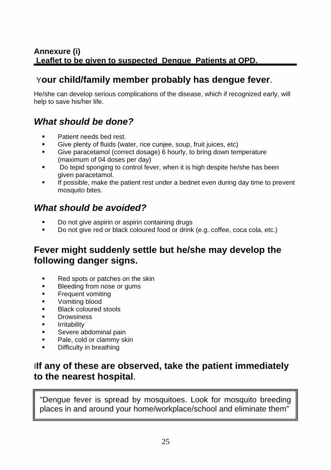
Annexure (i) Leaflet to be given to suspected Dengue Patients at OPD. Your child/family member probably has dengue fever. He/she can develop serious complications of the disease, which if recognized early, will help to save his/her life. What should be done?
Patient needs bed rest. Give plenty of fluids (water, rice cunjee, soup, fruit juices, etc) Give paracetamol (correct dosage) 6 hourly, to bring down temperature
(maximum of 04 doses per day) Do tepid sponging to control fever, when it is high despite he/she has been
given paracetamol. If possible, make the patient rest under a bednet even during day time to prevent
mosquito bites. What should be avoided?
Do not give aspirin or aspirin containing drugs Do not give red or black coloured food or drink (e.g. coffee, coca cola, etc.)
Fever might suddenly settle but he/she may develop the following danger signs.
Red spots or patches on the skin Bleeding from nose or gums Frequent vomiting Vomiting blood Black coloured stools Drowsiness Irritability Severe abdominal pain Pale, cold or clammy skin Difficulty in breathing
IIf any of these are observed, take the patient immediately to the nearest hospital.
"Dengue fever is spread by mosquitoes. Look for mosquito breeding places in and around your home/workplace/school and eliminate them”
26
Annexure ii
Maintenance fluids for different body weights
Weight Total (ml / 24 hrs) 3.0 300 3.5 350 4.0 400 4.5 450 5.0 500 5.5 550 6.0 600 6.5 650 7.0 700 7.5 750 8.0 800 8.5 850 9.0 900 9.5 950 10 1000 11 1050 12 1100 13 1150 14 1200 15 1250 16 1300 17 1350 18 1400 19 1450 20 1500 21 1525 22 1550 23 1575 24 1600 25 1625 26 1650 27 1675 28 1700 29 1725 30 1750 35 1875 40 2000 45 2125 50 2250
27
Annexure iii
Reduction of Hourly Maintenance Rate by 25% Blocks
Weight (kg)
M (ml/24 hrs)
M (ml/hr)
75% M (ml/hr)
50% M (ml/hr)
25% M (ml/hr)
3.0 300 12.5 9.5 6.0 3.0 3.5 350 14.5 11.0 7.0 3.5 4.0 400 16.5 12.5 8.0 4.0 4.5 450 19.0 14.0 9.5 4.5 5.0 500 21.0 16.0 10.5 5.0 5.5 550 23.0 17.0 11.5 5.5 6.0 600 25.0 19.0 12.5 6.0 6.5 650 27.0 20.0 13.5 7.0 7.0 700 29.0 22.0 14.5 7.5 7.5 750 31.0 23.0 15.5 8.0 8.0 800 33.5 25.0 16.5 8.5 8.5 850 35.5 26.5 17.5 9.0 9.0 900 37.5 28.0 19.0 9.5 9.5 950 39.5 29.5 20.0 10.0 10 1000 41.5 31.0 21.0 10.5 11 1050 44.0 33.0 22.0 11.0 12 1100 46.0 34.5 23.0 11.5 13 1150 48.0 36.0 24.0 12.0 14 1200 50.0 37.5 25.0 12.5 15 1250 52.0 39.0 26.0 13.0 16 1300 54.0 40.5 27.0 13.5 17 1350 56.0 42.0 28.0 14.0 18 1400 58.5 44.0 29.0 14.5 19 1450 60.5 45.5 30.0 15.0 20 1500 62.5 47.0 31.0 15.5 21 1525 63.5 47.5 32.0 16.0 22 1550 64.5 48.5 32.0 16.0 23 1575 65.5 49.0 33.0 16.5 24 1600 66.5 50.0 33.0 16.5 25 1625 67.5 50.5 34.0 17.0 26 1650 69.0 52.0 34.5 17.0 27 1675 70.0 52.5 35.0 17.5 28 1700 71.0 53.0 35.5 18.0 29 1725 72.0 54.0 36.0 18.0 30 1750 73.0 55.0 36.5 18.0 35 1875 78.0 58.5 39.0 19.5 40 2000 83.5 62.5 42.0 21.0 45 2125 88.5 66.5 44.0 22.0 50 2250 94.5 70.5 47.0 23.5
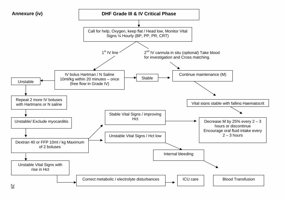
DHF Grade III & IV Critical Phase
Call for help, Oxygen, keep flat / Head low, Monitor Vital Signs ¼ Hourly (BP, PP, PR, CRT)
Unstable
IV bolus Hartman / N Saline 10ml/kg within 20 minutes – once
(free flow in Grade IV) Stable
Repeat 2 more IV boluses with Hartmans or N saline
Continue maintenance (M)
Dextran 40 or FFP 10ml / kg Maximum of 2 boluses
Vital signs stable with falling Haematocrit
Unstable Vital Signs with rise in Hct
Stable Vital Signs / improving Hct Decrease M by 25% every 2 – 3
hours or discontinue Encourage oral fluid intake every
2 – 3 hours Unstable Vital Signs / Hct low
Internal bleeding
Correct metabolic / electrolyte disturbances Blood Transfusion
1st IV line 2nd IV cannula in situ (optional) Take blood for investigation and Cross matching.
Unstable/ Exclude myocarditis
ICU care
Annexure (iv)
28
29
Annexure v
30
31
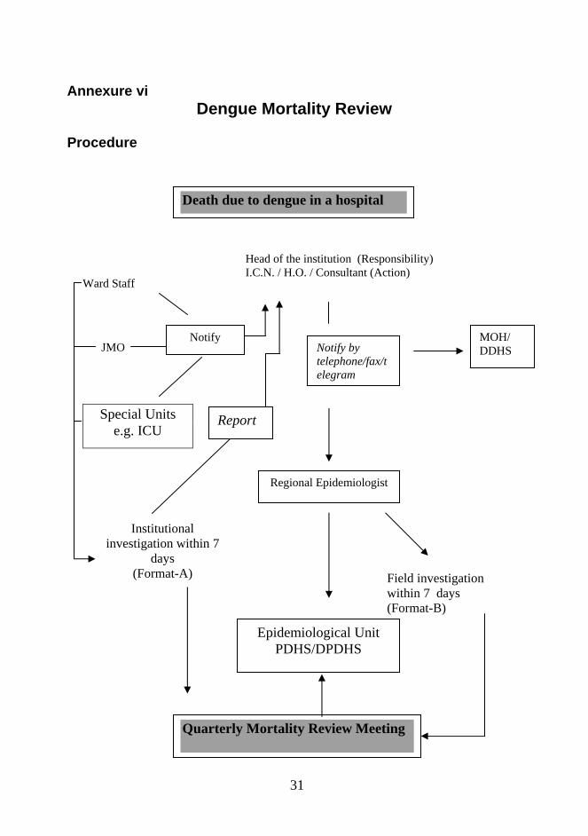
Annexure vi Dengue Mortality Review
Procedure
Death due to dengue in a hospital
Ward Staff
Head of the institution (Responsibility) I.C.N. / H.O. / Consultant (Action)
JMO
Special Units e.g. ICU
Institutional investigation within 7
days (Format-A)
Notify
Epidemiological Unit PDHS/DPDHS
MOH/ DDHS Notify by
telephone/fax/telegram
Regional Epidemiologist
Field investigation within 7 days (Format-B)
Quarterly Mortality Review Meeting
Report
32
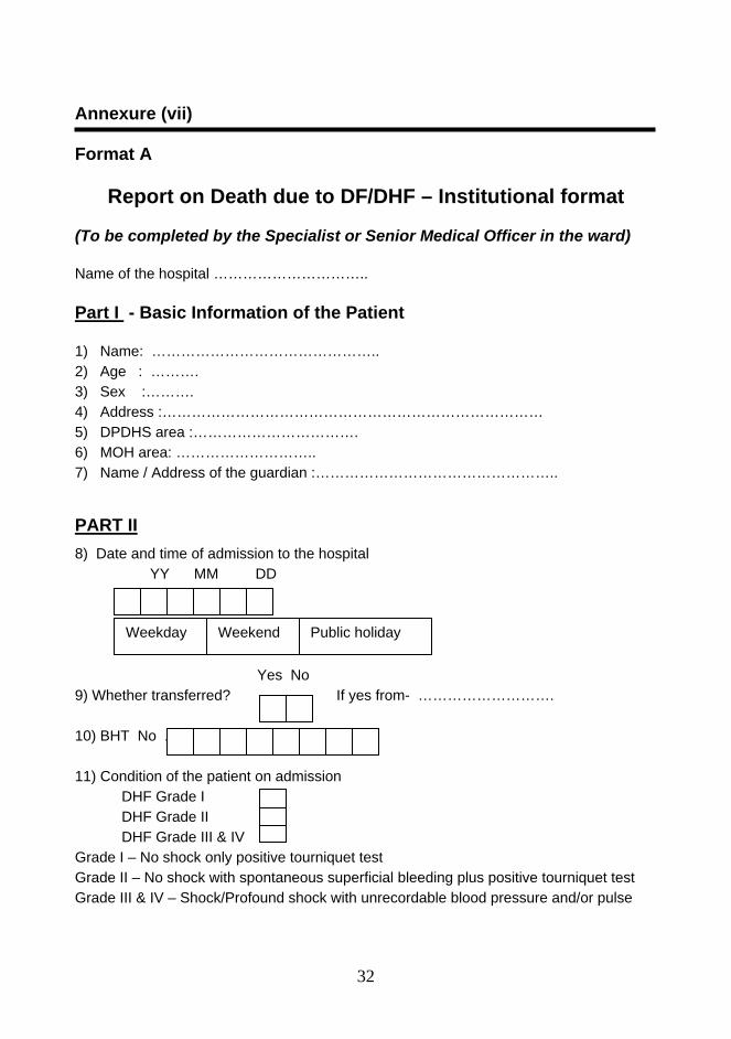
Annexure (vii) Format A
Report on Death due to DF/DHF – Institutional format (To be completed by the Specialist or Senior Medical Officer in the ward) Name of the hospital ………………………….. Part I - Basic Information of the Patient 1) Name: ……………………………………….. 2) Age : ………. 3) Sex :………. 4) Address :…………………………………………………………………… 5) DPDHS area :……………………………. 6) MOH area: ……………………….. 7) Name / Address of the guardian :………………………………………….. PART II 8) Date and time of admission to the hospital
YY MM DD
Yes No 9) Whether transferred? If yes from- ………………………. 10) BHT No . 11) Condition of the patient on admission
DHF Grade I DHF Grade II DHF Grade III & IV
Grade I – No shock only positive tourniquet test Grade II – No shock with spontaneous superficial bleeding plus positive tourniquet test Grade III & IV – Shock/Profound shock with unrecordable blood pressure and/or pulse
Weekday Public holiday Weekend
33
ys No
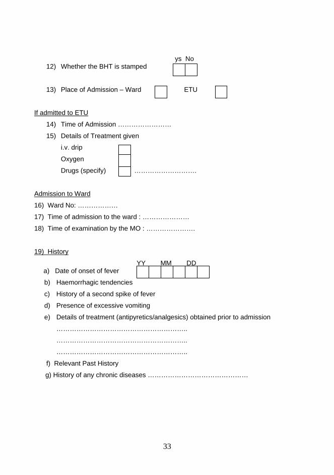
12) Whether the BHT is stamped ye
13) Place of Admission – Ward ETU
If admitted to ETU
14) Time of Admission ……………………
15) Details of Treatment given
i.v. drip
Oxygen
Drugs (specify) ……………………….
Admission to Ward
16) Ward No: ………………
17) Time of admission to the ward : …………………
18) Time of examination by the MO : ………………….
19) History
YY MM DD a) Date of onset of fever
b) Haemorrhagic tendencies
c) History of a second spike of fever
d) Presence of excessive vomiting
e) Details of treatment (antipyretics/analgesics) obtained prior to admission
…………………………………………………..
…………………………………………………..
…………………………………………………..
f) Relevant Past History
g) History of any chronic diseases ………………………………………
34
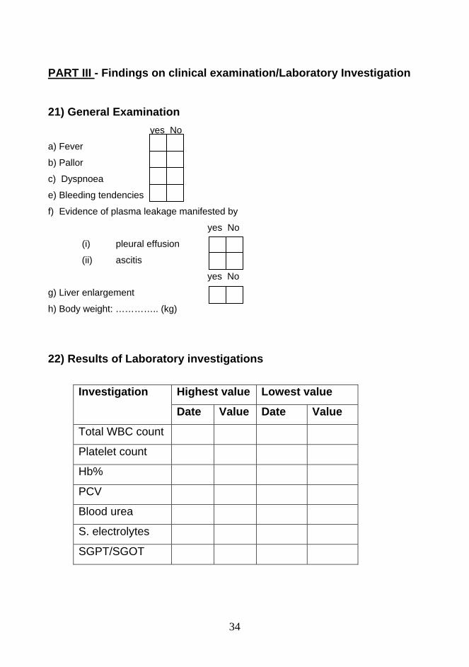
PART III - Findings on clinical examination/Laboratory Investigation 21) General Examination yes No
a) Fever
b) Pallor
c) Dyspnoea
e) Bleeding tendencies
f) Evidence of plasma leakage manifested by
yes No
(i) pleural effusion
(ii) ascitis
yes No
g) Liver enlargement
h) Body weight: ………….. (kg)
22) Results of Laboratory investigations
Highest value Lowest value Investigation
Date Value Date Value
Total WBC count
Platelet count
Hb%
PCV
Blood urea
S. electrolytes
SGPT/SGOT
35
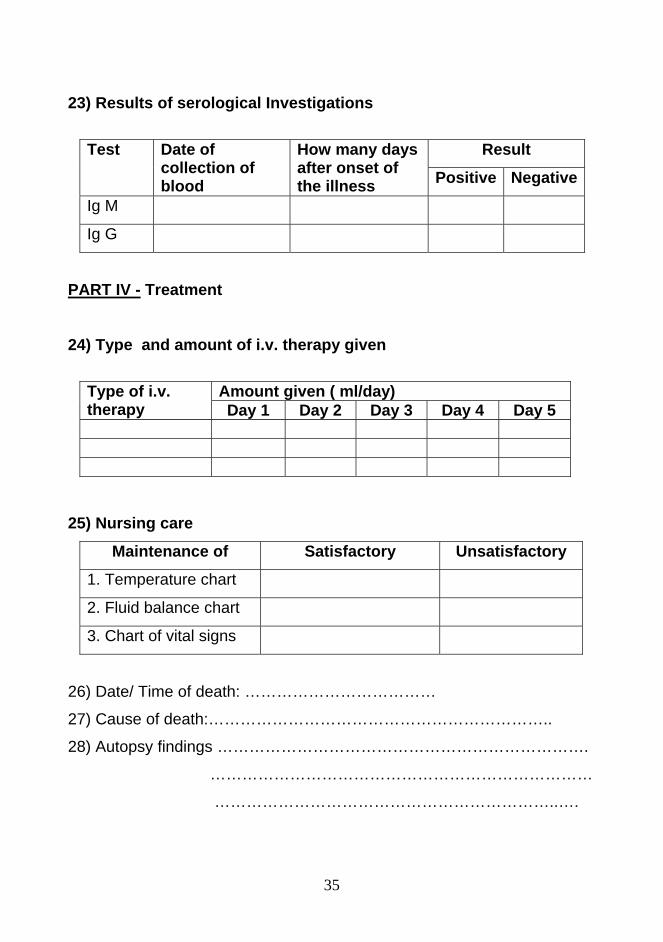
23) Results of serological Investigations
Result Test Date of collection of blood
How many days after onset of the illness Positive Negative
Ig M
Ig G
PART IV - Treatment 24) Type and amount of i.v. therapy given
Amount given ( ml/day) Type of i.v. therapy Day 1 Day 2 Day 3 Day 4 Day 5
25) Nursing care
Maintenance of Satisfactory Unsatisfactory
1. Temperature chart
2. Fluid balance chart
3. Chart of vital signs
26) Date/ Time of death: ………………………………
27) Cause of death:………………………………………………………..
28) Autopsy findings …………………………………………………………….
………………………………………………………………
………………………………………………………..….
36
29) Brief statement of events leading to death
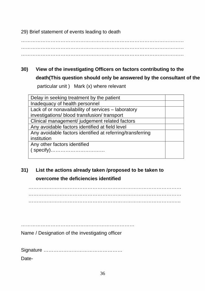
……………………………………………………………………………………………………………………………………………………………………………………………………………………………………………………………………… 30) View of the investigating Officers on factors contributing to the
death(This question should only be answered by the consultant of the particular unit ) Mark (x) where relevant
Delay in seeking treatment by the patient Inadequacy of health personnel Lack of or nonavailability of services – laboratory investigations/ blood transfusion/ transport
Clinical management/ judgement related factors Any avoidable factors identified at field level Any avoidable factors identified at referring/transferring institution
Any other factors identified ( specify)……………………………
31) List the actions already taken /proposed to be taken to
overcome the deficiencies identified
………………………………………………………………………………… ……………………………………………………………………………………………………………………………………………………………….…..
……………………………………………………………
Name / Designation of the investigating officer
Signature …………………………………………
Date-
37
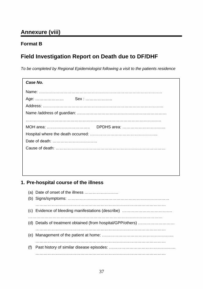
Annexure (viii) Format B Field Investigation Report on Death due to DF/DHF To be completed by Regional Epidemiologist following a visit to the patients residence 1. Pre-hospital course of the illness
(a) Date of onset of the illness ……………………. (b) Signs/symptoms: …………………………………………………………………
…………………………………………………………………………………… (c) Evidence of bleeding manifestations (describe) ………………………………. ……………………………………………………………………………………… (d) Details of treatment obtained (from hospital/GPP/others) ………………………
…………………………………………………………………………………… (e) Management of the patient at home: ……………………………………………..
…………………………………………………………………………………… (f) Past history of similar disease episodes: ………………………………………….
……………………………………………………………………………………
Case No. Name: ……………………………………………………………………………….
Age: ………………… Sex : ………………..
Address: ……………………………………………………………………………..
Name /address of guardian: …………………………………………………………
……………………………………………………………………………………….
MOH area: ………………………….. DPDHS area: …………………………..
Hospital where the death occurred: …………………………………………..
Date of death: ……………………………
Cause of death: ………………………………………………………………………
38
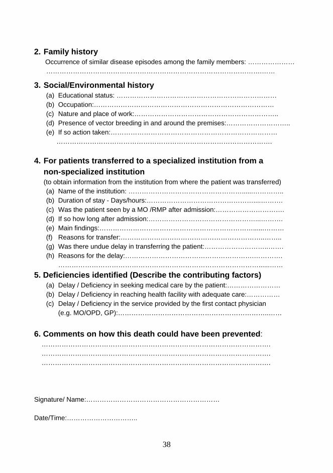
2. Family history Occurrence of similar disease episodes among the family members: …………………
…………………………………………………………………………………….……
3. Social/Environmental history (a) Educational status: ……………………………………………………………… (b) Occupation:……………………………………………………………………… (c) Nature and place of work:……………………………………………………….. (d) Presence of vector breeding in and around the premises:……………………….. (e) If so action taken:…………………………………………………………………
……………………………………………………………………………………. 4. For patients transferred to a specialized institution from a
non-specialized institution (to obtain information from the institution from where the patient was transferred) (a) Name of the institution: ……………………………………………......………….. (b) Duration of stay - Days/hours:…………………………………………....………. (c) Was the patient seen by a MO /RMP after admission:…………………………. (d) If so how long after admission:…………………………………………...………. (e) Main findings:……………………………………………………………......……… (f) Reasons for transfer:………………………………………………………..…….. (g) Was there undue delay in transferring the patient:………………………..……. (h) Reasons for the delay:………………………………………………….………….
………………………………………………………………………………......…… 5. Deficiencies identified (Describe the contributing factors)
(a) Delay / Deficiency in seeking medical care by the patient:…………………… (b) Delay / Deficiency in reaching health facility with adequate care:…………… (c) Delay / Deficiency in the service provided by the first contact physician
(e.g. MO/OPD, GP):…………………………………………………………..……
6. Comments on how this death could have been prevented: …………………………………………………………………………………………. …………………………………………………………………………………………. …………………………………………………………………………………………. Signature/ Name:…………………………………………………… Date/Time:…………………………..
![Dengue fever: Causes, complications, and vaccine strategies ......dengue fever (DF) to severe dengue hemorrhagic fever (DHF) and dengue shock syndrome (DSS) which may turn fatal [2].](https://static.fdocuments.net/doc/165x107/5f57aec821c68c72c60d0d52/dengue-fever-causes-complications-and-vaccine-strategies-dengue-fever.jpg)
![Complement-Mediated Neutralization of Dengue Virus ... · rhage and vascular permeability syndrome (dengue hemorrhagic fever/dengue shock syndrome [DHF/DSS]) (2). Although the ...](https://static.fdocuments.net/doc/165x107/5cae414a88c9938f4d8c97e1/complement-mediated-neutralization-of-dengue-virus-rhage-and-vascular-permeability.jpg)


![Dengue Fever/Severe Dengue Fever/Chikungunya Fever · Dengue fever and severe dengue (dengue hemorrhagic fever [DHF] and dengue shock syndrome [DSS]) are caused by any of four closely](https://static.fdocuments.net/doc/165x107/5e87bf3e7a86e85d3b149cd7/dengue-feversevere-dengue-feverchikungunya-dengue-fever-and-severe-dengue-dengue.jpg)