Guideline: Neonatal resuscitation
38
Maternity and Neonatal Clinical Guideline Queensland Health Neonatal resuscitation
Transcript of Guideline: Neonatal resuscitation
Maternity and Neonatal Clinical Guideline
Queensland Health
Neonatal resuscitation
Queensland Clinical Guideline: Neonatal resuscitation
Refer to online version, destroy printed copies after use Page 2 of 38
Document title: Neonatal resuscitation
Publication date: July 2016
Document number: MN16.5-V4-R21
Document supplement: The document supplement is integral to and should be read in conjunction with this guideline.
Amendments: Full version history is supplied in the document supplement.
Amendment date: September 2016 Replaces document: MN16.5-V3-R21 Author: Queensland Clinical Guidelines
Audience: Health professionals in Queensland public and private maternity and neonatal services
Review date: July 2021
Endorsed by: Queensland Clinical Guidelines Steering Committee Statewide Maternity and Neonatal Clinical Network (Queensland)
Contact: Email: [email protected] URL: www.health.qld.gov.au/qcg
Disclaimer This guideline is intended as a guide and provided for information purposes only. The information has been prepared using a multidisciplinary approach with reference to the best information and evidence available at the time of preparation. No assurance is given that the information is entirely complete, current, or accurate in every respect. The guideline is not a substitute for clinical judgement, knowledge and expertise, or medical advice. Variation from the guideline, taking into account individual circumstances may be appropriate. This guideline does not address all elements of standard practice and accepts that individual clinicians are responsible for: • Providing care within the context of locally available resources, expertise, and scope of practice • Supporting consumer rights and informed decision making in partnership with healthcare practitioners
including the right to decline intervention or ongoing management • Advising consumers of their choices in an environment that is culturally appropriate and which
enables comfortable and confidential discussion. This includes the use of interpreter services where necessary
• Ensuring informed consent is obtained prior to delivering care • Meeting all legislative requirements and professional standards • Applying standard precautions, and additional precautions as necessary, when delivering care • Documenting all care in accordance with mandatory and local requirements
Queensland Health disclaims, to the maximum extent permitted by law, all responsibility and all liability (including without limitation, liability in negligence) for all expenses, losses, damages and costs incurred for any reason associated with the use of this guideline, including the materials within or referred to throughout this document being in any way inaccurate, out of context, incomplete or unavailable.
© State of Queensland (Queensland Health) 2016
This work is licensed under a Creative Commons Attribution Non-Commercial No Derivatives 3.0 Australia licence. In essence, you are free to copy and communicate the work in its current form for non-commercial purposes, as long as you attribute Queensland Clinical Guidelines, Queensland Health and abide by the licence terms. You may not alter or adapt the work in any way. To view a copy of this licence, visit http://creativecommons.org/licenses/by-nc-nd/3.0/au/deed.en For further information contact Queensland Clinical Guidelines, RBWH Post Office, Herston Qld 4029, email [email protected], phone (07) 3131 6777. For permissions beyond the scope of this licence contact: Intellectual Property Officer, Queensland Health, GPO Box 48, Brisbane Qld 4001, email [email protected], phone (07) 3234 1479.
Queensland Clinical Guideline: Neonatal resuscitation
Refer to online version, destroy printed copies after use Page 3 of 38
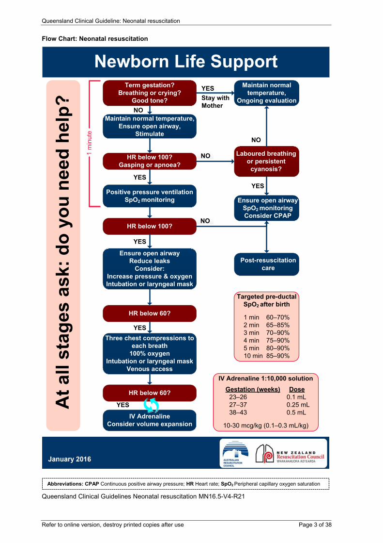
Flow Chart: Neonatal resuscitation
Queensland Clinical Guidelines Neonatal resuscitation MN16.5-V4-R21
Ensure open airwaySpO2 monitoringConsider CPAP
Term gestation?Breathing or crying?
Good tone?
Maintain normal temperature,Ensure open airway,
Stimulate
Positive pressure ventilationSpO2 monitoring
Ensure open airwayReduce leaks
Consider:Increase pressure & oxygenIntubation or laryngeal mask
Three chest compressions toeach breath
100% oxygenIntubation or laryngeal mask
Venous access
HR below 60?
IV AdrenalineConsider volume expansion
Post-resuscitationcare
Targeted pre-ductalSpO2 after birth
1 min 60–70%2 min 65–85%3 min 70–90%4 min 75–90%5 min 80–90%10 min 85–90%
IV Adrenaline 1:10,000 solutionGestation (weeks) Dose
23–26 0.1 mL27–37 0.25 mL38–43 0.5 mL
10-30 mcg/kg (0.1–0.3 mL/kg)
Ata
llst
ages
ask:
doyo
une
edhe
lp?
Newborn Life SupportMaintain normal
temperature,Ongoing evaluation
Laboured breathingor persistent
cyanosis?
HR below 60?
HR below 100?
HR below 100?Gasping or apnoea?
1m
inut
eYESStay withMother
YES
YES
YES
YES
YES
NO
NO
NO
NO
Abbreviations: CPAP Continuous positive airway pressure; HR Heart rate; SpO2 Peripheral capillary oxygen saturation
Queensland Clinical Guideline: Neonatal resuscitation
Refer to online version, destroy printed copies after use Page 4 of 38
Abbreviations
ANZCOR Australian and New Zealand Committee on Resuscitation bpm Beats per minute CDH Congenital diaphragmatic hernia CPAP Continuous positive airway pressure ETT Endotracheal tube FiO2 Fraction of inspired oxygen HIE Hypoxic ischaemic encephalopathy HR Heart rate IO Intraosseous IV Intravenous LMA Laryngeal mask airway NICU Neonatal intensive care unit PEEP Positive end expiratory pressure PIP Peak inspiratory pressure PPV Positive pressure ventilation PPROM Preterm prelabour rupture of membranes RhD Rh Blood Type D (Rh positive) SpO2 Peripheral capillary oxygen saturation UVC Umbilical venous catheter
Definitions
Acrocyanosis Blue hands and feet due to inadequate circulation of blood and oxygen to the extremities normally found in the first few hours after birth.
Cold stress Temperature between 36.0 °C and 36.4°C Corrected age Gestation plus postnatal age in weeks Hyperthermia Temperature greater than 37.5 °C Mild hypothermia Temperature between 35.5 °C and 35.9 °C Moderate hypothermia Temperature between 32.0 °C and 35.4°C
Queensland Clinical Guideline: Neonatal resuscitation
Refer to online version, destroy printed copies after use Page 5 of 38
Table of Contents
1 Introduction ..................................................................................................................................... 7 1.1 Clinical standards .................................................................................................................. 7 1.2 Clinicians................................................................................................................................ 8
2 Risk factors for neonatal resuscitation ........................................................................................... 9 3 Preparation: staff, equipment, medications and environment ...................................................... 10
3.1 Communication and information sharing ............................................................................. 11 4 Assessment and management of baby ........................................................................................ 12
4.1 Initial assessment and mangement ..................................................................................... 12 4.2 Subsequent assessment and management ........................................................................ 13 4.3 Oxygen saturation monitoring .............................................................................................. 14
4.3.1 Care of baby during resuscitation .................................................................................... 15 5 Airway management ..................................................................................................................... 16
5.1 General principles ................................................................................................................ 16 5.2 Airway and delivery devices ................................................................................................ 17
5.2.1 Delivery devices ............................................................................................................... 18 5.3 Supplemental Oxygen ......................................................................................................... 19 5.4 Oxygen/air mix ..................................................................................................................... 19 5.5 Positive pressure ventilation ................................................................................................ 20 5.6 Intubation ............................................................................................................................. 21
5.6.1 ETT size and length ......................................................................................................... 21 5.6.2 Endotracheal intubation ................................................................................................... 22
6 Chest compressions ..................................................................................................................... 23 7 Medication and fluids .................................................................................................................... 24
7.1 Adrenaline (epinephrine) ..................................................................................................... 25 7.2 Volume expanding fluids ...................................................................................................... 26
8 Special circumstances .................................................................................................................. 27 8.1 Preterm baby ....................................................................................................................... 27 8.2 Other special circumstances................................................................................................ 28 8.3 Congenital anomalies .......................................................................................................... 29
9 Care after resuscitation ................................................................................................................ 30 9.1 Cord blood sampling ............................................................................................................ 30
9.1.1 Umbilical artery cord blood gases.................................................................................... 30 9.2 Continuing clinical care ........................................................................................................ 31 9.3 Monitoring and management ............................................................................................... 32
10 Ethical considerations .................................................................................................................. 33 References .......................................................................................................................................... 34 Appendix A Equipment and medications for neonatal resuscitation ................................................... 35 Appendix B Oxygen/air mix and target oxygen saturation .................................................................. 36 Appendix C Endotracheal tube intubation ........................................................................................... 37 Acknowledgements .............................................................................................................................. 38
Queensland Clinical Guideline: Neonatal resuscitation
Refer to online version, destroy printed copies after use Page 6 of 38
List of Tables
Table 1 Clinical standards ..................................................................................................................... 7 Table 2 Clinicians .................................................................................................................................. 8 Table 3 Risk factors ............................................................................................................................... 9 Table 4 Preparation ............................................................................................................................. 10 Table 5 Communication and information sharing ................................................................................ 11 Table 6 Assessment and initial management at birth .......................................................................... 12 Table 7 Subsequent assessment and management ........................................................................... 13 Table 8 Oxygen saturation monitoring ................................................................................................. 14 Table 9 Care of baby ........................................................................................................................... 15 Table 10 General principles ................................................................................................................. 16 Table 11 Assist ventilation devices ...................................................................................................... 17 Table 12 Delivery devices.................................................................................................................... 18 Table 13 Supplemental oxygen ........................................................................................................... 19 Table 14 Suggested oxygen/air flows at 10 litres per minute .............................................................. 19 Table 15 Suggested oxygen/air flows at 8 litres per minute ................................................................ 19 Table 16 Positive pressure ventilation ................................................................................................. 20 Table 17 ETT and suction catheters sizes .......................................................................................... 21 Table 18 Endotracheal intubation ........................................................................................................ 22 Table 19 Chest compressions ............................................................................................................. 23 Table 20 Medication and fluids ............................................................................................................ 24 Table 21 Adrenaline (epinephrine) administration ............................................................................... 25 Table 22 Administration of volume expanding fluids ........................................................................... 26 Table 23 Preterm baby ........................................................................................................................ 27 Table 24 Other special circumstances ................................................................................................ 28 Table 25 Congenital anomalies ........................................................................................................... 29 Table 26 Cord blood gas sampling ...................................................................................................... 30 Table 27 Umbilical artery cord blood gases ........................................................................................ 30 Table 28 Continuing clinical care ......................................................................................................... 31 Table 29 Monitoring and management ................................................................................................ 32 Table 30 Ethical considerations ........................................................................................................... 33
Table of Figures
Figure 1 Rounded doses of IV adrenaline (epinephrine) 1:10,000 ..................................................... 25
Queensland Clinical Guideline: Neonatal resuscitation
Refer to online version, destroy printed copies after use Page 7 of 38
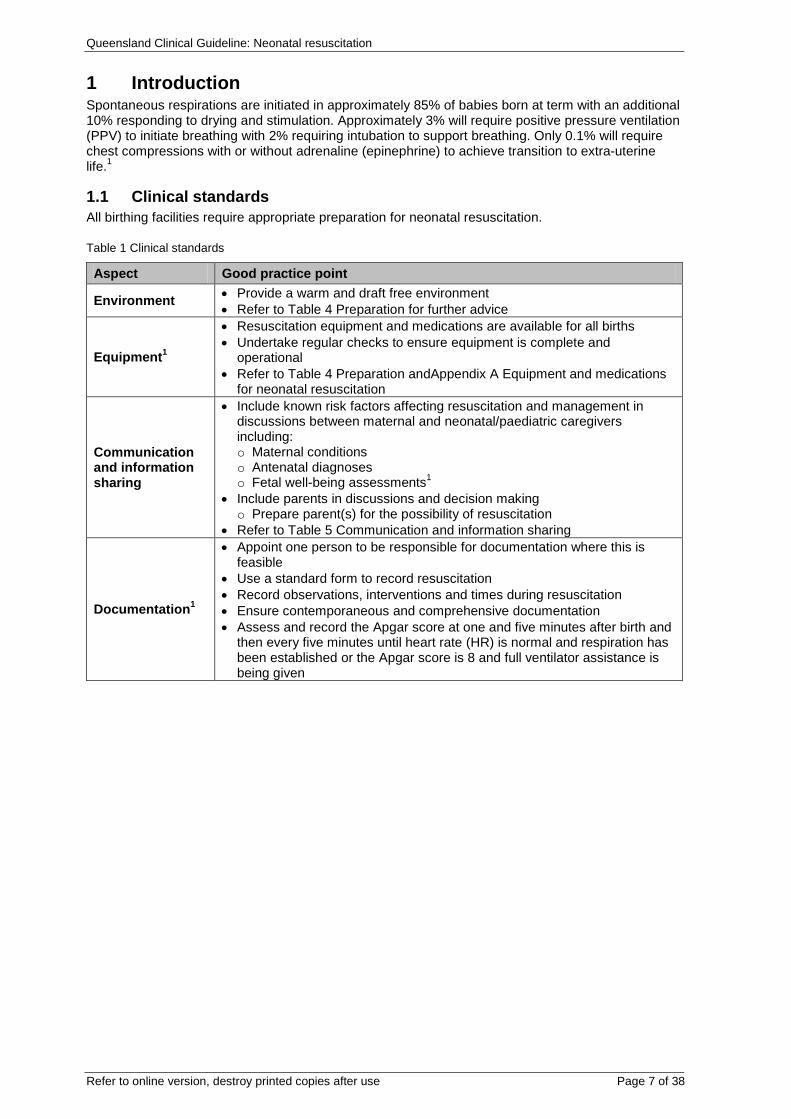
1 Introduction Spontaneous respirations are initiated in approximately 85% of babies born at term with an additional 10% responding to drying and stimulation. Approximately 3% will require positive pressure ventilation (PPV) to initiate breathing with 2% requiring intubation to support breathing. Only 0.1% will require chest compressions with or without adrenaline (epinephrine) to achieve transition to extra-uterine life.1
1.1 Clinical standards All birthing facilities require appropriate preparation for neonatal resuscitation.
Table 1 Clinical standards
Aspect Good practice point
Environment • Provide a warm and draft free environment • Refer to Table 4 Preparation for further advice
Equipment1
• Resuscitation equipment and medications are available for all births • Undertake regular checks to ensure equipment is complete and
operational • Refer to Table 4 Preparation andAppendix A Equipment and medications
for neonatal resuscitation
Communication and information sharing
• Include known risk factors affecting resuscitation and management in discussions between maternal and neonatal/paediatric caregivers including: o Maternal conditions o Antenatal diagnoses o Fetal well-being assessments1
• Include parents in discussions and decision making o Prepare parent(s) for the possibility of resuscitation
• Refer to Table 5 Communication and information sharing
Documentation1
• Appoint one person to be responsible for documentation where this is feasible
• Use a standard form to record resuscitation • Record observations, interventions and times during resuscitation • Ensure contemporaneous and comprehensive documentation • Assess and record the Apgar score at one and five minutes after birth and
then every five minutes until heart rate (HR) is normal and respiration has been established or the Apgar score is 8 and full ventilator assistance is being given
Queensland Clinical Guideline: Neonatal resuscitation
Refer to online version, destroy printed copies after use Page 8 of 38
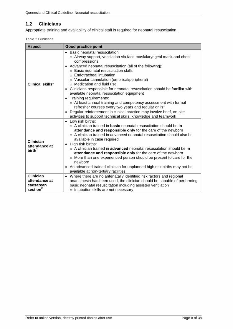
1.2 Clinicians Appropriate training and availability of clinical staff is required for neonatal resuscitation.
Table 2 Clinicians
Aspect Good practice point
Clinical skills1
• Basic neonatal resuscitation: o Airway support, ventilation via face mask/laryngeal mask and chest
compressions • Advanced neonatal resuscitation (all of the following): o Basic neonatal resuscitation skills o Endotracheal intubation o Vascular cannulation (umbilical/peripheral) o Medication and fluid use
• Clinicians responsible for neonatal resuscitation should be familiar with available neonatal resuscitation equipment
• Training requirements: o At least annual training and competency assessment with formal
refresher courses every two years and regular drills1 • Regular reinforcement in clinical practice may involve brief, on-site
activities to support technical skills, knowledge and teamwork
Clinician attendance at birth1
• Low risk births: o A clinician trained in basic neonatal resuscitation should be in
attendance and responsible only for the care of the newborn o A clinician trained in advanced neonatal resuscitation should also be
available in case required • High risk births: o A clinician trained in advanced neonatal resuscitation should be in
attendance and responsible only for the care of the newborn o More than one experienced person should be present to care for the
newborn • An advanced trained clinician for unplanned high risk births may not be
available at non-tertiary facilities Clinician attendance at caesarean section2
• Where there are no antenatally identified risk factors and regional anaesthesia has been used, the clinician should be capable of performing basic neonatal resuscitation including assisted ventilation o Intubation skills are not necessary
Queensland Clinical Guideline: Neonatal resuscitation
Refer to online version, destroy printed copies after use Page 9 of 38
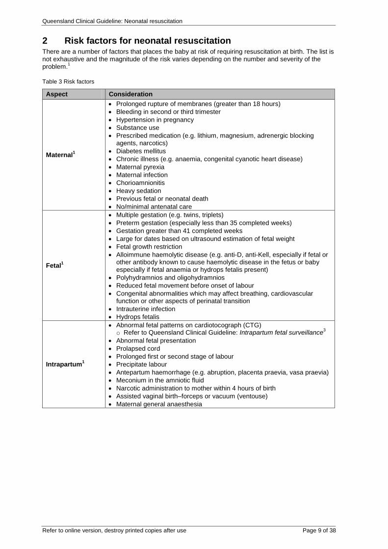
2 Risk factors for neonatal resuscitation There are a number of factors that places the baby at risk of requiring resuscitation at birth. The list is not exhaustive and the magnitude of the risk varies depending on the number and severity of the problem.1
Table 3 Risk factors
Aspect Consideration
Maternal1
• Prolonged rupture of membranes (greater than 18 hours) • Bleeding in second or third trimester • Hypertension in pregnancy • Substance use • Prescribed medication (e.g. lithium, magnesium, adrenergic blocking
agents, narcotics) • Diabetes mellitus • Chronic illness (e.g. anaemia, congenital cyanotic heart disease) • Maternal pyrexia • Maternal infection • Chorioamnionitis • Heavy sedation • Previous fetal or neonatal death • No/minimal antenatal care
Fetal1
• Multiple gestation (e.g. twins, triplets) • Preterm gestation (especially less than 35 completed weeks) • Gestation greater than 41 completed weeks • Large for dates based on ultrasound estimation of fetal weight • Fetal growth restriction • Alloimmune haemolytic disease (e.g. anti-D, anti-Kell, especially if fetal or
other antibody known to cause haemolytic disease in the fetus or baby especially if fetal anaemia or hydrops fetalis present)
• Polyhydramnios and oligohydramnios • Reduced fetal movement before onset of labour • Congenital abnormalities which may affect breathing, cardiovascular
function or other aspects of perinatal transition • Intrauterine infection • Hydrops fetalis
Intrapartum1
• Abnormal fetal patterns on cardiotocograph (CTG) o Refer to Queensland Clinical Guideline: Intrapartum fetal surveillance3
• Abnormal fetal presentation • Prolapsed cord • Prolonged first or second stage of labour • Precipitate labour • Antepartum haemorrhage (e.g. abruption, placenta praevia, vasa praevia) • Meconium in the amniotic fluid • Narcotic administration to mother within 4 hours of birth • Assisted vaginal birth–forceps or vacuum (ventouse) • Maternal general anaesthesia
Queensland Clinical Guideline: Neonatal resuscitation
Refer to online version, destroy printed copies after use Page 10 of 38
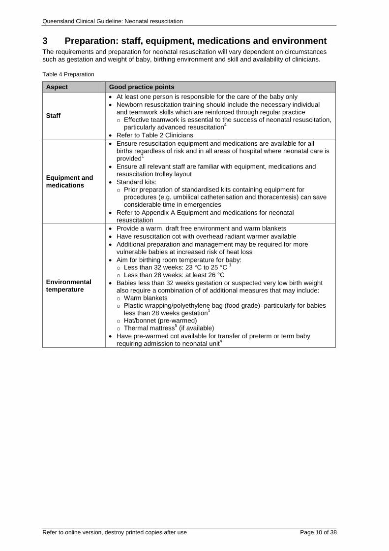
3 Preparation: staff, equipment, medications and environment The requirements and preparation for neonatal resuscitation will vary dependent on circumstances such as gestation and weight of baby, birthing environment and skill and availability of clinicians.
Table 4 Preparation
Aspect Good practice points
Staff
• At least one person is responsible for the care of the baby only • Newborn resuscitation training should include the necessary individual
and teamwork skills which are reinforced through regular practice o Effective teamwork is essential to the success of neonatal resuscitation,
particularly advanced resuscitation4 • Refer to Table 2 Clinicians
Equipment and medications
• Ensure resuscitation equipment and medications are available for all births regardless of risk and in all areas of hospital where neonatal care is provided1
• Ensure all relevant staff are familiar with equipment, medications and resuscitation trolley layout
• Standard kits: o Prior preparation of standardised kits containing equipment for
procedures (e.g. umbilical catheterisation and thoracentesis) can save considerable time in emergencies
• Refer to Appendix A Equipment and medications for neonatal resuscitation
Environmental temperature
• Provide a warm, draft free environment and warm blankets • Have resuscitation cot with overhead radiant warmer available • Additional preparation and management may be required for more
vulnerable babies at increased risk of heat loss • Aim for birthing room temperature for baby: o Less than 32 weeks: 23 °C to 25 °C 1 o Less than 28 weeks: at least 26 °C
• Babies less than 32 weeks gestation or suspected very low birth weight also require a combination of of additional measures that may include: o Warm blankets o Plastic wrapping/polyethylene bag (food grade)–particularly for babies
less than 28 weeks gestation1 o Hat/bonnet (pre-warmed) o Thermal mattress5 (if available)
• Have pre-warmed cot available for transfer of preterm or term baby requiring admission to neonatal unit4
Queensland Clinical Guideline: Neonatal resuscitation
Refer to online version, destroy printed copies after use Page 11 of 38
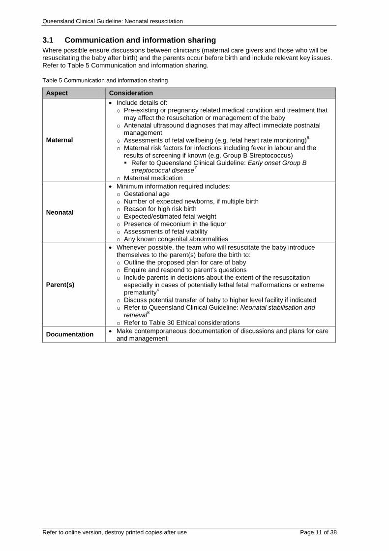
3.1 Communication and information sharing Where possible ensure discussions between clinicians (maternal care givers and those who will be resuscitating the baby after birth) and the parents occur before birth and include relevant key issues. Refer to Table 5 Communication and information sharing.
Table 5 Communication and information sharing
Aspect Consideration
Maternal
• Include details of: o Pre-existing or pregnancy related medical condition and treatment that
may affect the resuscitation or management of the baby o Antenatal ultrasound diagnoses that may affect immediate postnatal
management o Assessments of fetal wellbeing (e.g. fetal heart rate monitoring)6 o Maternal risk factors for infections including fever in labour and the
results of screening if known (e.g. Group B Streptococcus) Refer to Queensland Clinical Guideline: Early onset Group B
streptococcal disease7 o Maternal medication
Neonatal
• Minimum information required includes: o Gestational age o Number of expected newborns, if multiple birth o Reason for high risk birth o Expected/estimated fetal weight o Presence of meconium in the liquor o Assessments of fetal viability o Any known congenital abnormalities
Parent(s)
• Whenever possible, the team who will resuscitate the baby introduce themselves to the parent(s) before the birth to: o Outline the proposed plan for care of baby o Enquire and respond to parent’s questions o Include parents in decisions about the extent of the resuscitation
especially in cases of potentially lethal fetal malformations or extreme prematurity4
o Discuss potential transfer of baby to higher level facility if indicated o Refer to Queensland Clinical Guideline: Neonatal stabilisation and
retrieval8 o Refer to Table 30 Ethical considerations
Documentation • Make contemporaneous documentation of discussions and plans for care and management
Queensland Clinical Guideline: Neonatal resuscitation
Refer to online version, destroy printed copies after use Page 12 of 38
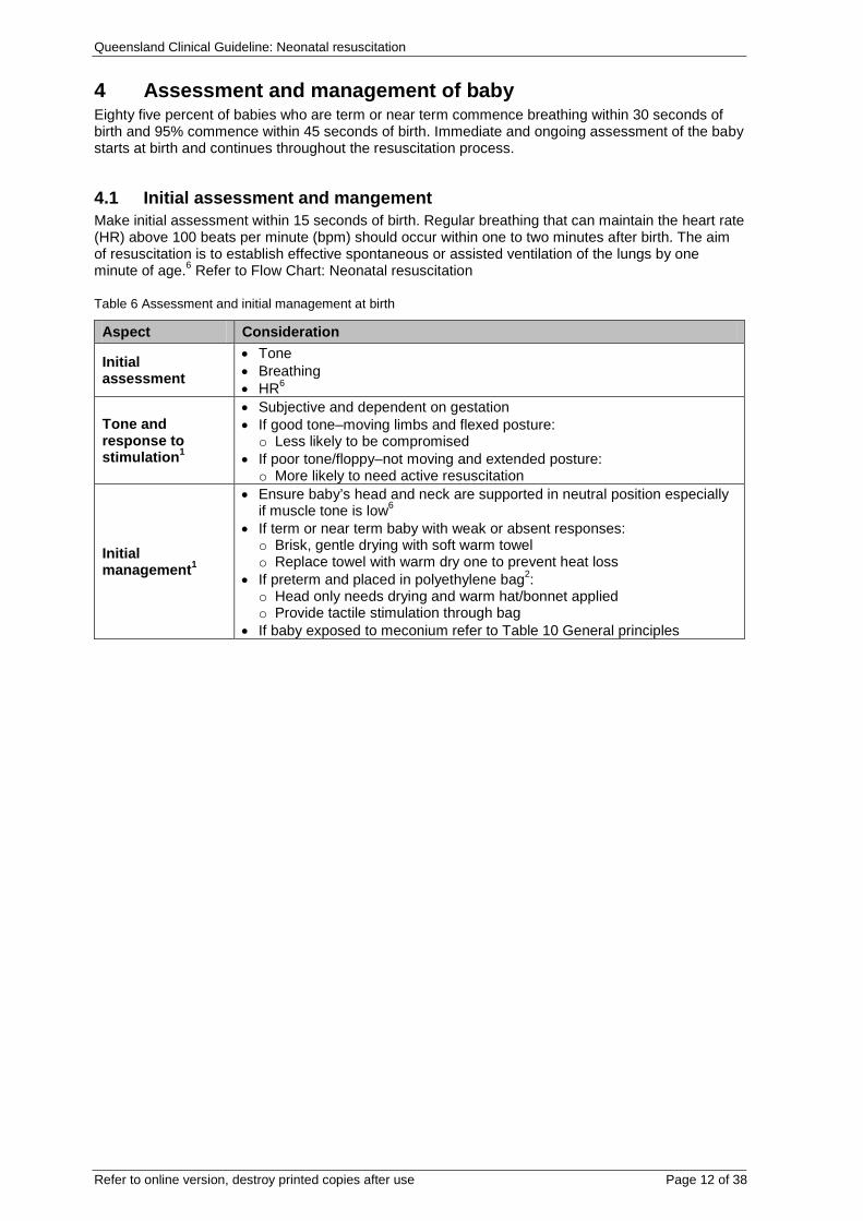
4 Assessment and management of baby Eighty five percent of babies who are term or near term commence breathing within 30 seconds of birth and 95% commence within 45 seconds of birth. Immediate and ongoing assessment of the baby starts at birth and continues throughout the resuscitation process.
4.1 Initial assessment and mangement Make initial assessment within 15 seconds of birth. Regular breathing that can maintain the heart rate (HR) above 100 beats per minute (bpm) should occur within one to two minutes after birth. The aim of resuscitation is to establish effective spontaneous or assisted ventilation of the lungs by one minute of age.6 Refer to Flow Chart: Neonatal resuscitation
Table 6 Assessment and initial management at birth
Aspect Consideration
Initial assessment
• Tone • Breathing • HR6
Tone and response to stimulation1
• Subjective and dependent on gestation • If good tone–moving limbs and flexed posture: o Less likely to be compromised
• If poor tone/floppy–not moving and extended posture: o More likely to need active resuscitation
Initial management1
• Ensure baby’s head and neck are supported in neutral position especially if muscle tone is low6
• If term or near term baby with weak or absent responses: o Brisk, gentle drying with soft warm towel o Replace towel with warm dry one to prevent heat loss
• If preterm and placed in polyethylene bag2: o Head only needs drying and warm hat/bonnet applied o Provide tactile stimulation through bag
• If baby exposed to meconium refer to Table 10 General principles
Queensland Clinical Guideline: Neonatal resuscitation
Refer to online version, destroy printed copies after use Page 13 of 38
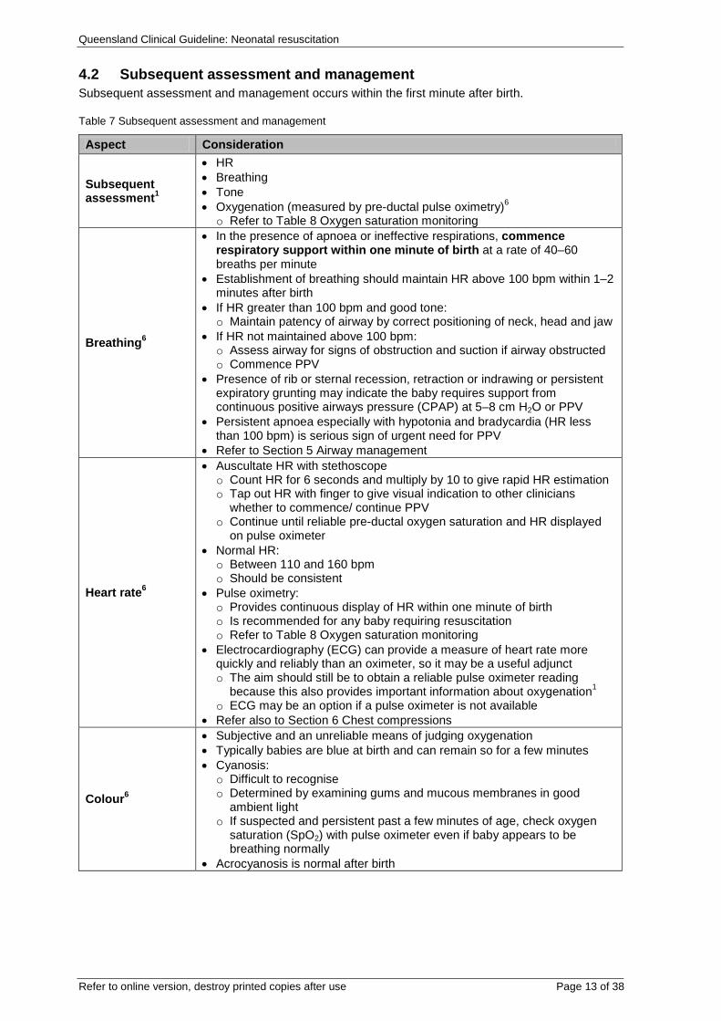
4.2 Subsequent assessment and management Subsequent assessment and management occurs within the first minute after birth.
Table 7 Subsequent assessment and management
Aspect Consideration
Subsequent assessment1
• HR • Breathing • Tone • Oxygenation (measured by pre-ductal pulse oximetry)6 o Refer to Table 8 Oxygen saturation monitoring
Breathing6
• In the presence of apnoea or ineffective respirations, commence respiratory support within one minute of birth at a rate of 40–60 breaths per minute
• Establishment of breathing should maintain HR above 100 bpm within 1–2 minutes after birth
• If HR greater than 100 bpm and good tone: o Maintain patency of airway by correct positioning of neck, head and jaw
• If HR not maintained above 100 bpm: o Assess airway for signs of obstruction and suction if airway obstructed o Commence PPV
• Presence of rib or sternal recession, retraction or indrawing or persistent expiratory grunting may indicate the baby requires support from continuous positive airways pressure (CPAP) at 5–8 cm H2O or PPV
• Persistent apnoea especially with hypotonia and bradycardia (HR less than 100 bpm) is serious sign of urgent need for PPV
• Refer to Section 5 Airway management
Heart rate6
• Auscultate HR with stethoscope o Count HR for 6 seconds and multiply by 10 to give rapid HR estimation o Tap out HR with finger to give visual indication to other clinicians
whether to commence/ continue PPV o Continue until reliable pre-ductal oxygen saturation and HR displayed
on pulse oximeter • Normal HR: o Between 110 and 160 bpm o Should be consistent
• Pulse oximetry: o Provides continuous display of HR within one minute of birth o Is recommended for any baby requiring resuscitation o Refer to Table 8 Oxygen saturation monitoring
• Electrocardiography (ECG) can provide a measure of heart rate more quickly and reliably than an oximeter, so it may be a useful adjunct o The aim should still be to obtain a reliable pulse oximeter reading
because this also provides important information about oxygenation1 o ECG may be an option if a pulse oximeter is not available
• Refer also to Section 6 Chest compressions
Colour6
• Subjective and an unreliable means of judging oxygenation • Typically babies are blue at birth and can remain so for a few minutes • Cyanosis: o Difficult to recognise o Determined by examining gums and mucous membranes in good
ambient light o If suspected and persistent past a few minutes of age, check oxygen
saturation (SpO2) with pulse oximeter even if baby appears to be breathing normally
• Acrocyanosis is normal after birth
Queensland Clinical Guideline: Neonatal resuscitation
Refer to online version, destroy printed copies after use Page 14 of 38
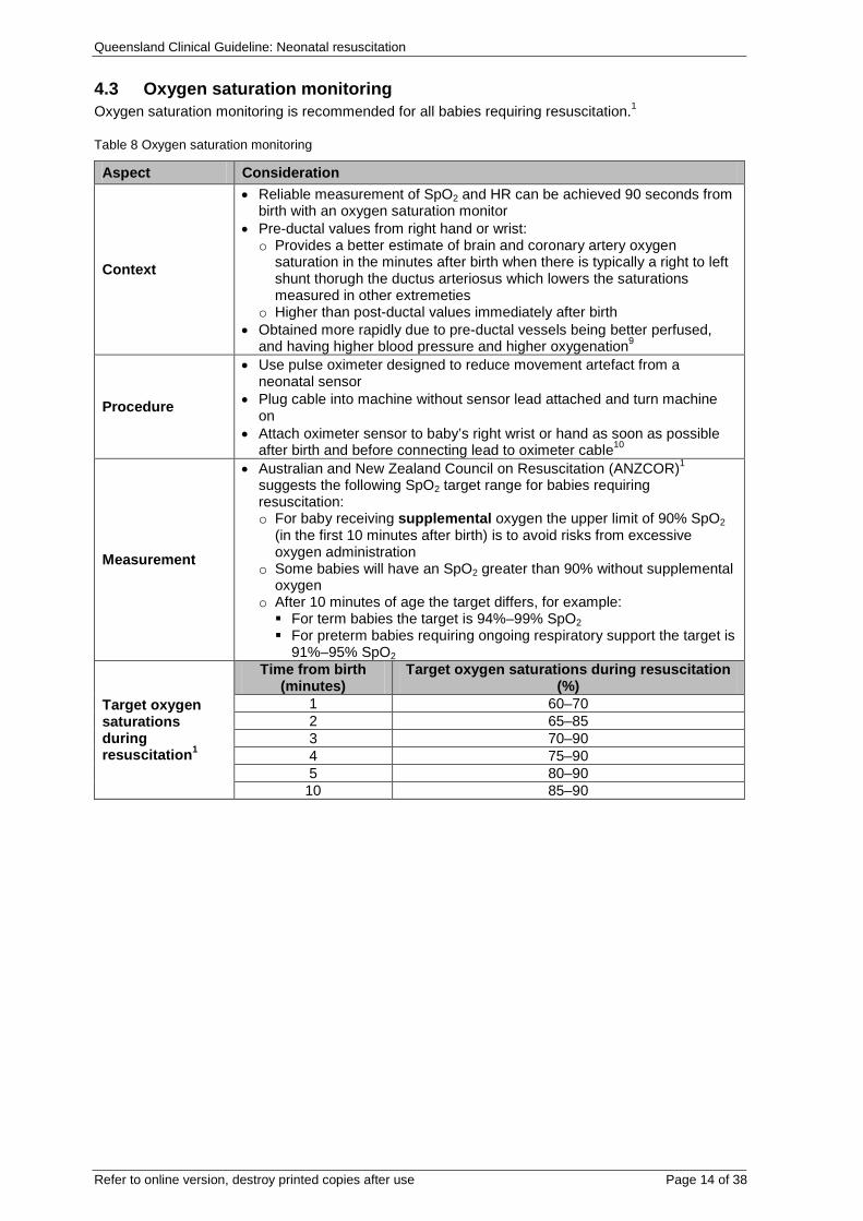
4.3 Oxygen saturation monitoring Oxygen saturation monitoring is recommended for all babies requiring resuscitation.1
Table 8 Oxygen saturation monitoring
Aspect Consideration
Context
• Reliable measurement of SpO2 and HR can be achieved 90 seconds from birth with an oxygen saturation monitor
• Pre-ductal values from right hand or wrist: o Provides a better estimate of brain and coronary artery oxygen
saturation in the minutes after birth when there is typically a right to left shunt thorugh the ductus arteriosus which lowers the saturations measured in other extremeties
o Higher than post-ductal values immediately after birth • Obtained more rapidly due to pre-ductal vessels being better perfused,
and having higher blood pressure and higher oxygenation9
Procedure
• Use pulse oximeter designed to reduce movement artefact from a neonatal sensor
• Plug cable into machine without sensor lead attached and turn machine on
• Attach oximeter sensor to baby’s right wrist or hand as soon as possible after birth and before connecting lead to oximeter cable10
Measurement
• Australian and New Zealand Council on Resuscitation (ANZCOR)1 suggests the following SpO2 target range for babies requiring resuscitation: o For baby receiving supplemental oxygen the upper limit of 90% SpO2
(in the first 10 minutes after birth) is to avoid risks from excessive oxygen administration
o Some babies will have an SpO2 greater than 90% without supplemental oxygen
o After 10 minutes of age the target differs, for example: For term babies the target is 94%–99% SpO2 For preterm babies requiring ongoing respiratory support the target is
91%–95% SpO2
Target oxygen saturations during resuscitation1
Time from birth (minutes)
Target oxygen saturations during resuscitation (%)
1 60–70 2 65–85 3 70–90 4 75–90 5 80–90
10 85–90
Queensland Clinical Guideline: Neonatal resuscitation
Refer to online version, destroy printed copies after use Page 15 of 38
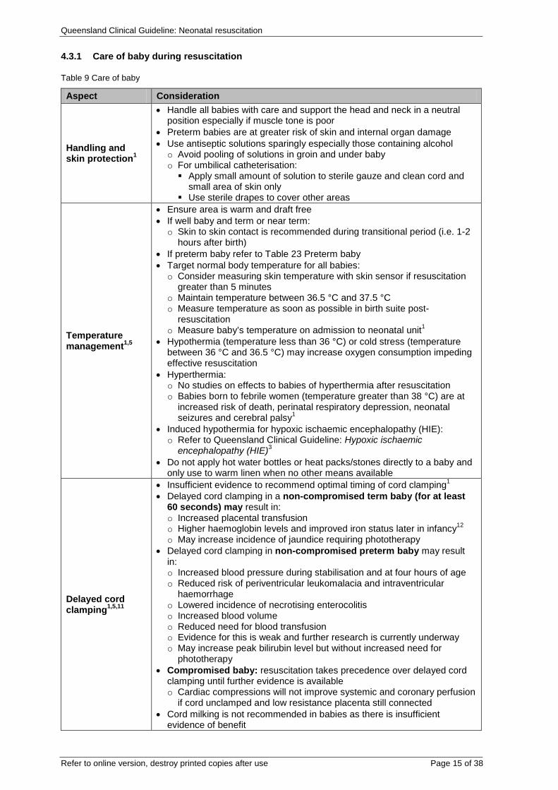
4.3.1 Care of baby during resuscitation
Table 9 Care of baby
Aspect Consideration
Handling and skin protection1
• Handle all babies with care and support the head and neck in a neutral position especially if muscle tone is poor
• Preterm babies are at greater risk of skin and internal organ damage • Use antiseptic solutions sparingly especially those containing alcohol o Avoid pooling of solutions in groin and under baby o For umbilical catheterisation: Apply small amount of solution to sterile gauze and clean cord and
small area of skin only Use sterile drapes to cover other areas
Temperature management1,5
• Ensure area is warm and draft free • If well baby and term or near term: o Skin to skin contact is recommended during transitional period (i.e. 1-2
hours after birth) • If preterm baby refer to Table 23 Preterm baby • Target normal body temperature for all babies: o Consider measuring skin temperature with skin sensor if resuscitation
greater than 5 minutes o Maintain temperature between 36.5 °C and 37.5 °C o Measure temperature as soon as possible in birth suite post-
resuscitation o Measure baby’s temperature on admission to neonatal unit1
• Hypothermia (temperature less than 36 °C) or cold stress (temperature between 36 °C and 36.5 °C) may increase oxygen consumption impeding effective resuscitation
• Hyperthermia: o No studies on effects to babies of hyperthermia after resuscitation o Babies born to febrile women (temperature greater than 38 °C) are at
increased risk of death, perinatal respiratory depression, neonatal seizures and cerebral palsy1
• Induced hypothermia for hypoxic ischaemic encephalopathy (HIE): o Refer to Queensland Clinical Guideline: Hypoxic ischaemic
encephalopathy (HIE)3 • Do not apply hot water bottles or heat packs/stones directly to a baby and
only use to warm linen when no other means available
Delayed cord clamping1,5,11
• Insufficient evidence to recommend optimal timing of cord clamping1 • Delayed cord clamping in a non-compromised term baby (for at least
60 seconds) may result in: o Increased placental transfusion o Higher haemoglobin levels and improved iron status later in infancy12 o May increase incidence of jaundice requiring phototherapy
• Delayed cord clamping in non-compromised preterm baby may result in: o Increased blood pressure during stabilisation and at four hours of age o Reduced risk of periventricular leukomalacia and intraventricular
haemorrhage o Lowered incidence of necrotising enterocolitis o Increased blood volume o Reduced need for blood transfusion o Evidence for this is weak and further research is currently underway o May increase peak bilirubin level but without increased need for
phototherapy • Compromised baby: resuscitation takes precedence over delayed cord
clamping until further evidence is available o Cardiac compressions will not improve systemic and coronary perfusion
if cord unclamped and low resistance placenta still connected • Cord milking is not recommended in babies as there is insufficient
evidence of benefit
Queensland Clinical Guideline: Neonatal resuscitation
Refer to online version, destroy printed copies after use Page 16 of 38
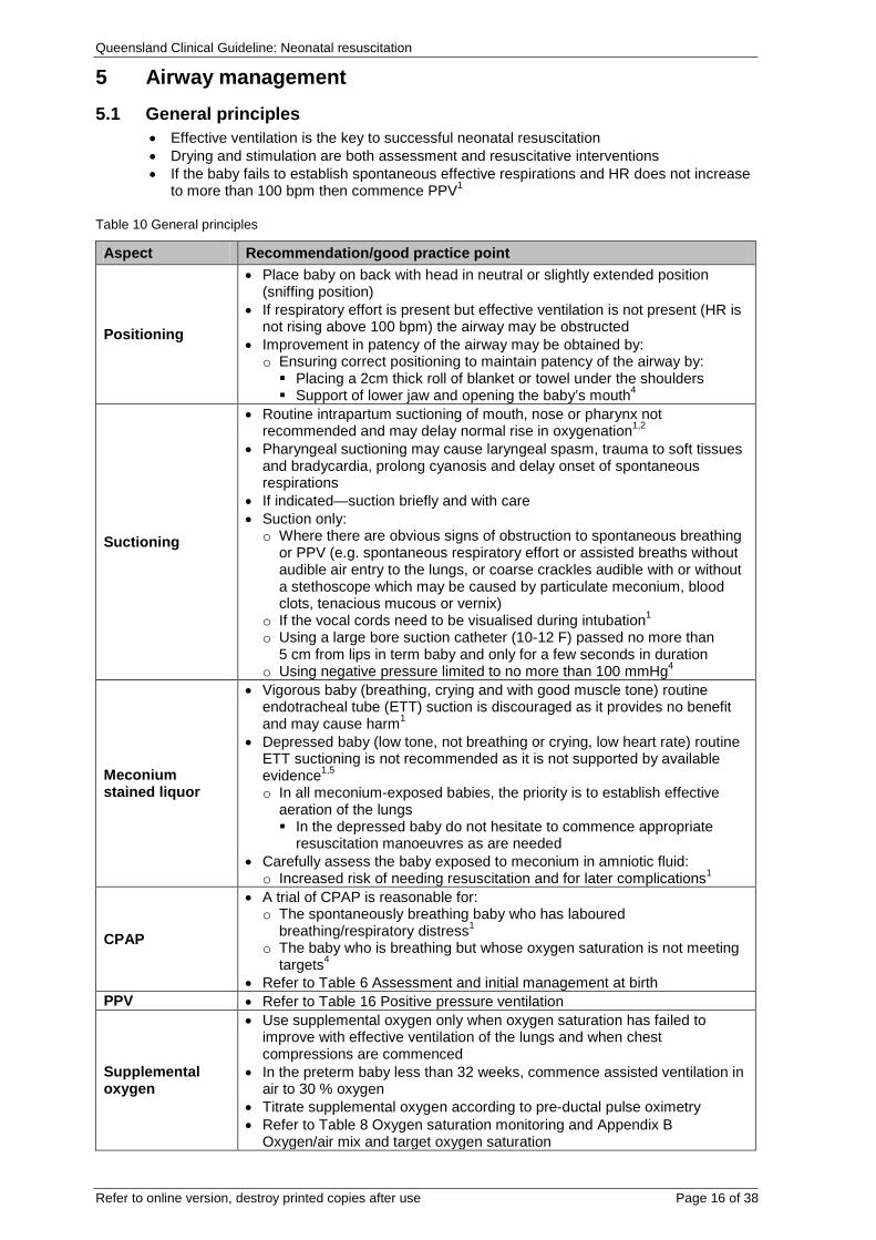
5 Airway management 5.1 General principles
• Effective ventilation is the key to successful neonatal resuscitation • Drying and stimulation are both assessment and resuscitative interventions • If the baby fails to establish spontaneous effective respirations and HR does not increase
to more than 100 bpm then commence PPV1
Table 10 General principles
Aspect Recommendation/good practice point
Positioning
• Place baby on back with head in neutral or slightly extended position (sniffing position)
• If respiratory effort is present but effective ventilation is not present (HR is not rising above 100 bpm) the airway may be obstructed
• Improvement in patency of the airway may be obtained by: o Ensuring correct positioning to maintain patency of the airway by: Placing a 2cm thick roll of blanket or towel under the shoulders Support of lower jaw and opening the baby’s mouth4
Suctioning
• Routine intrapartum suctioning of mouth, nose or pharynx not recommended and may delay normal rise in oxygenation1,2
• Pharyngeal suctioning may cause laryngeal spasm, trauma to soft tissues and bradycardia, prolong cyanosis and delay onset of spontaneous respirations
• If indicated—suction briefly and with care • Suction only: o Where there are obvious signs of obstruction to spontaneous breathing
or PPV (e.g. spontaneous respiratory effort or assisted breaths without audible air entry to the lungs, or coarse crackles audible with or without a stethoscope which may be caused by particulate meconium, blood clots, tenacious mucous or vernix)
o If the vocal cords need to be visualised during intubation1 o Using a large bore suction catheter (10-12 F) passed no more than
5 cm from lips in term baby and only for a few seconds in duration o Using negative pressure limited to no more than 100 mmHg4
Meconium stained liquor
• Vigorous baby (breathing, crying and with good muscle tone) routine endotracheal tube (ETT) suction is discouraged as it provides no benefit and may cause harm1
• Depressed baby (low tone, not breathing or crying, low heart rate) routine ETT suctioning is not recommended as it is not supported by available evidence1,5 o In all meconium-exposed babies, the priority is to establish effective
aeration of the lungs In the depressed baby do not hesitate to commence appropriate
resuscitation manoeuvres as are needed • Carefully assess the baby exposed to meconium in amniotic fluid: o Increased risk of needing resuscitation and for later complications1
CPAP
• A trial of CPAP is reasonable for: o The spontaneously breathing baby who has laboured
breathing/respiratory distress1 o The baby who is breathing but whose oxygen saturation is not meeting
targets4 • Refer to Table 6 Assessment and initial management at birth
PPV • Refer to Table 16 Positive pressure ventilation
Supplemental oxygen
• Use supplemental oxygen only when oxygen saturation has failed to improve with effective ventilation of the lungs and when chest compressions are commenced
• In the preterm baby less than 32 weeks, commence assisted ventilation in air to 30 % oxygen
• Titrate supplemental oxygen according to pre-ductal pulse oximetry • Refer to Table 8 Oxygen saturation monitoring and Appendix B
Oxygen/air mix and target oxygen saturation
Queensland Clinical Guideline: Neonatal resuscitation
Refer to online version, destroy printed copies after use Page 17 of 38
5.2 Airway and delivery devices
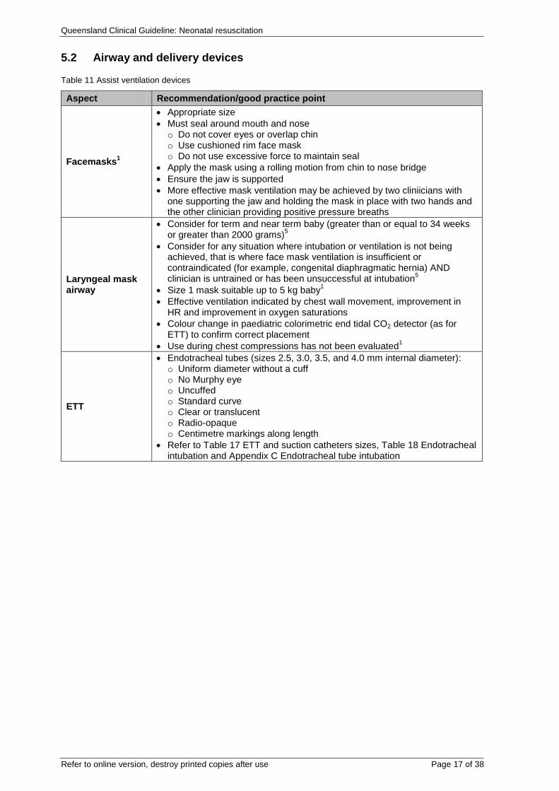
Table 11 Assist ventilation devices
Aspect Recommendation/good practice point
Facemasks1
• Appropriate size • Must seal around mouth and nose o Do not cover eyes or overlap chin o Use cushioned rim face mask o Do not use excessive force to maintain seal
• Apply the mask using a rolling motion from chin to nose bridge • Ensure the jaw is supported • More effective mask ventilation may be achieved by two cliniicians with
one supporting the jaw and holding the mask in place with two hands and the other clinician providing positive pressure breaths
Laryngeal mask airway
• Consider for term and near term baby (greater than or equal to 34 weeks or greater than 2000 grams)5
• Consider for any situation where intubation or ventilation is not being achieved, that is where face mask ventilation is insufficient or contraindicated (for example, congenital diaphragmatic hernia) AND clinician is untrained or has been unsuccessful at intubation5
• Size 1 mask suitable up to 5 kg baby1 • Effective ventilation indicated by chest wall movement, improvement in
HR and improvement in oxygen saturations • Colour change in paediatric colorimetric end tidal CO2 detector (as for
ETT) to confirm correct placement • Use during chest compressions has not been evaluated1
ETT
• Endotracheal tubes (sizes 2.5, 3.0, 3.5, and 4.0 mm internal diameter): o Uniform diameter without a cuff o No Murphy eye o Uncuffed o Standard curve o Clear or translucent o Radio-opaque o Centimetre markings along length
• Refer to Table 17 ETT and suction catheters sizes, Table 18 Endotracheal intubation and Appendix C Endotracheal tube intubation
Queensland Clinical Guideline: Neonatal resuscitation
Refer to online version, destroy printed copies after use Page 18 of 38
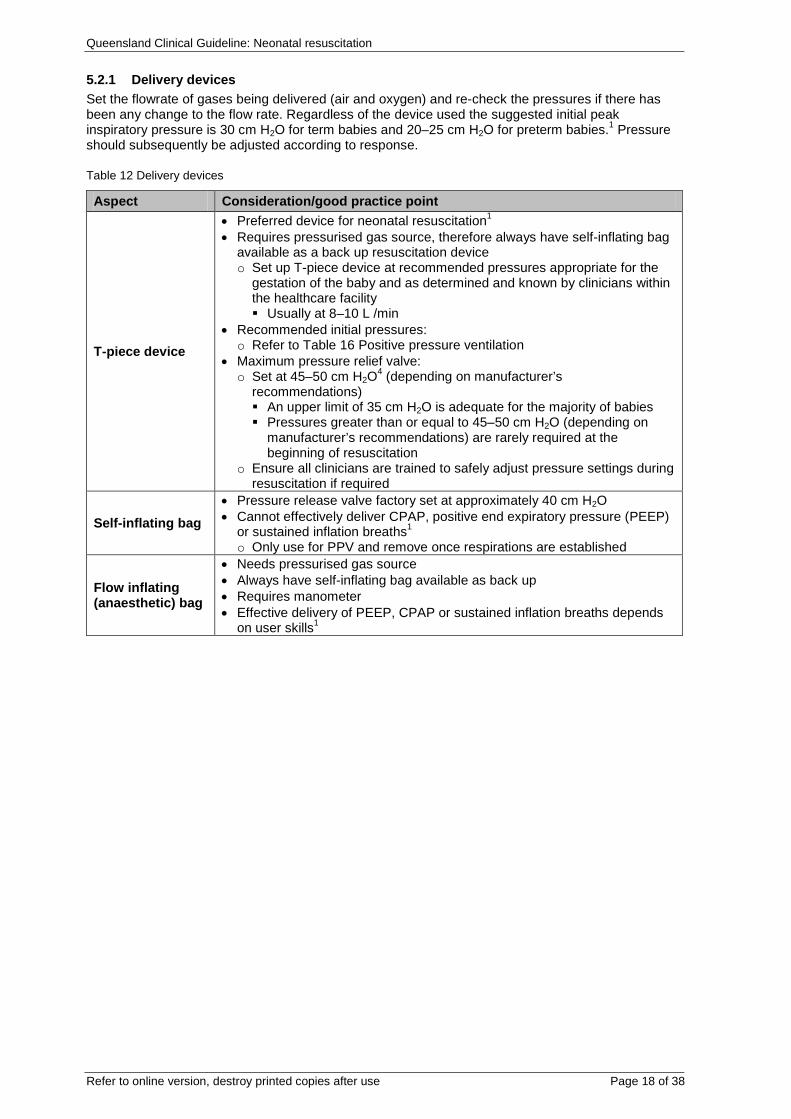
5.2.1 Delivery devices Set the flowrate of gases being delivered (air and oxygen) and re-check the pressures if there has been any change to the flow rate. Regardless of the device used the suggested initial peak inspiratory pressure is 30 cm H2O for term babies and 20–25 cm H2O for preterm babies.1 Pressure should subsequently be adjusted according to response.
Table 12 Delivery devices
Aspect Consideration/good practice point
T-piece device
• Preferred device for neonatal resuscitation1 • Requires pressurised gas source, therefore always have self-inflating bag
available as a back up resuscitation device o Set up T-piece device at recommended pressures appropriate for the
gestation of the baby and as determined and known by clinicians within the healthcare facility Usually at 8–10 L /min
• Recommended initial pressures: o Refer to Table 16 Positive pressure ventilation
• Maximum pressure relief valve: o Set at 45–50 cm H2O4 (depending on manufacturer’s
recommendations) An upper limit of 35 cm H2O is adequate for the majority of babies Pressures greater than or equal to 45–50 cm H2O (depending on
manufacturer’s recommendations) are rarely required at the beginning of resuscitation
o Ensure all clinicians are trained to safely adjust pressure settings during resuscitation if required
Self-inflating bag • Pressure release valve factory set at approximately 40 cm H2O • Cannot effectively deliver CPAP, positive end expiratory pressure (PEEP)
or sustained inflation breaths1 o Only use for PPV and remove once respirations are established
Flow inflating (anaesthetic) bag
• Needs pressurised gas source • Always have self-inflating bag available as back up • Requires manometer • Effective delivery of PEEP, CPAP or sustained inflation breaths depends
on user skills1
Queensland Clinical Guideline: Neonatal resuscitation
Refer to online version, destroy printed copies after use Page 19 of 38
5.3 Supplemental Oxygen
Table 13 Supplemental oxygen
Aspect Good practice point
Goal • Regardless of the gestation, aim for oxygen saturations consistent with a
healthy term baby1 • Refer to Table 8 Oxygen saturation monitoring and Appendix B
Oxygen/air mix and target oxygen saturation
Priority
• Ensure adequate lung inflation by auscultating lung fields • Only increase fractional inspired oxygen (FiO2) if CPAP or effective PPV
have failed to achieve the target oxygen saturation • Increase oxygen to 100% whilst chest compressions are occurring o Titrate oxygen to the recommended oxygen saturation targets, once
chest compressions have ceased
Term and near term baby1
• Use air initially and only administer oxygen to a baby whose SpO2 does not meet the lower target range despite effective respiratory support
• Begin to wean oxygen concentration when SpO2 reaches 90% if supplemental oxygen is being used
Preterm baby • Refer to Table 23 Preterm baby
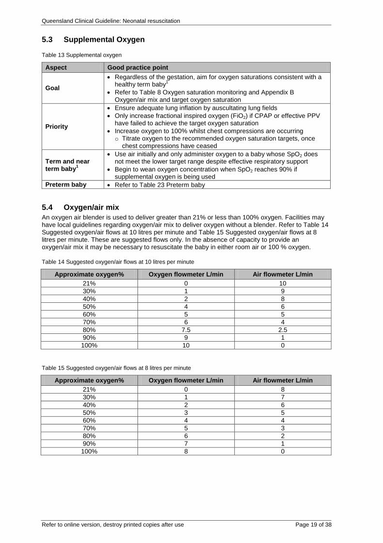
5.4 Oxygen/air mix An oxygen air blender is used to deliver greater than 21% or less than 100% oxygen. Facilities may have local guidelines regarding oxygen/air mix to deliver oxygen without a blender. Refer to Table 14 Suggested oxygen/air flows at 10 litres per minute and Table 15 Suggested oxygen/air flows at 8 litres per minute. These are suggested flows only. In the absence of capacity to provide an oxygen/air mix it may be necessary to resuscitate the baby in either room air or 100 % oxygen.
Table 14 Suggested oxygen/air flows at 10 litres per minute
Approximate oxygen% Oxygen flowmeter L/min Air flowmeter L/min 21% 0 10 30% 1 9 40% 2 8 50% 4 6 60% 5 5 70% 6 4 80% 7.5 2.5 90% 9 1 100% 10 0
Table 15 Suggested oxygen/air flows at 8 litres per minute
Approximate oxygen% Oxygen flowmeter L/min Air flowmeter L/min 21% 0 8 30% 1 7 40% 2 6 50% 3 5 60% 4 4 70% 5 3 80% 6 2 90% 7 1 100% 8 0
Queensland Clinical Guideline: Neonatal resuscitation
Refer to online version, destroy printed copies after use Page 20 of 38
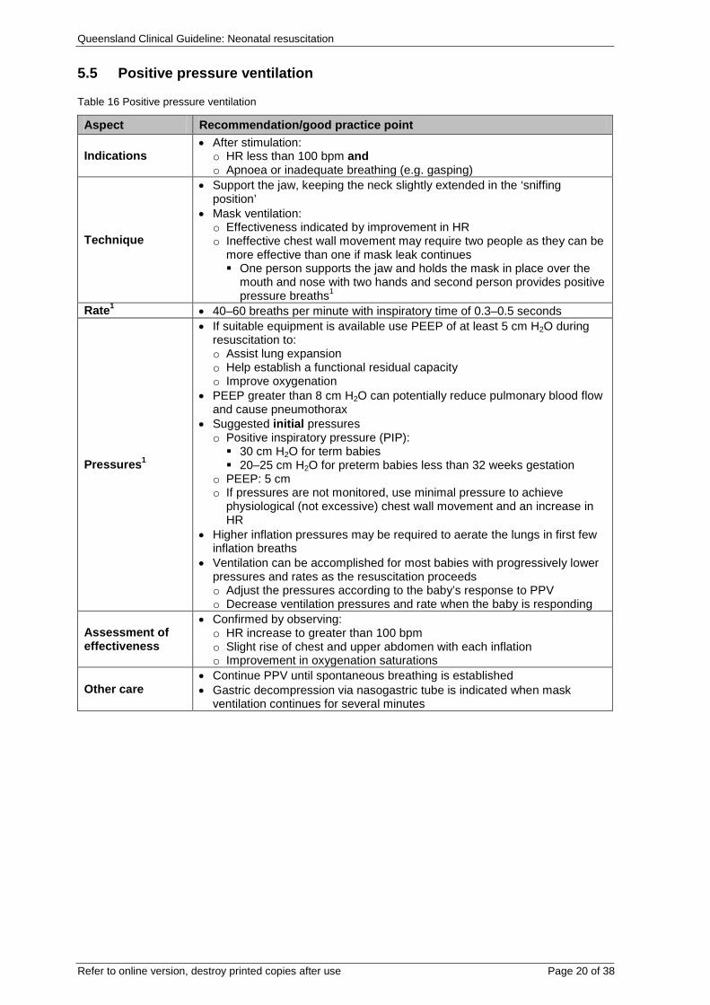
5.5 Positive pressure ventilation
Table 16 Positive pressure ventilation
Aspect Recommendation/good practice point
Indications • After stimulation: o HR less than 100 bpm and o Apnoea or inadequate breathing (e.g. gasping)
Technique
• Support the jaw, keeping the neck slightly extended in the ‘sniffing position’
• Mask ventilation: o Effectiveness indicated by improvement in HR o Ineffective chest wall movement may require two people as they can be
more effective than one if mask leak continues One person supports the jaw and holds the mask in place over the
mouth and nose with two hands and second person provides positive pressure breaths1
Rate1 • 40–60 breaths per minute with inspiratory time of 0.3–0.5 seconds
Pressures1
• If suitable equipment is available use PEEP of at least 5 cm H2O during resuscitation to: o Assist lung expansion o Help establish a functional residual capacity o Improve oxygenation
• PEEP greater than 8 cm H2O can potentially reduce pulmonary blood flow and cause pneumothorax
• Suggested initial pressures o Positive inspiratory pressure (PIP): 30 cm H2O for term babies 20–25 cm H2O for preterm babies less than 32 weeks gestation
o PEEP: 5 cm o If pressures are not monitored, use minimal pressure to achieve
physiological (not excessive) chest wall movement and an increase in HR
• Higher inflation pressures may be required to aerate the lungs in first few inflation breaths
• Ventilation can be accomplished for most babies with progressively lower pressures and rates as the resuscitation proceeds o Adjust the pressures according to the baby’s response to PPV o Decrease ventilation pressures and rate when the baby is responding
Assessment of effectiveness
• Confirmed by observing: o HR increase to greater than 100 bpm o Slight rise of chest and upper abdomen with each inflation o Improvement in oxygenation saturations
Other care • Continue PPV until spontaneous breathing is established • Gastric decompression via nasogastric tube is indicated when mask
ventilation continues for several minutes
Queensland Clinical Guideline: Neonatal resuscitation
Refer to online version, destroy printed copies after use Page 21 of 38
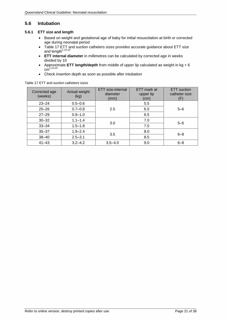
5.6 Intubation
5.6.1 ETT size and length • Based on weight and gestational age of baby for initial resuscitation at birth or corrected
age during neonatal period • Table 17 ETT and suction catheters sizes provides accurate guidance about ETT size
and length1,13,14 • ETT internal diameter in millimetres can be calculated by corrected age in weeks
divided by 10 • Approximate ETT length/depth from middle of upper lip calculated as weight in kg + 6
cm1,14,15 • Check insertion depth as soon as possible after intubation
Table 17 ETT and suction catheters sizes
Corrected age (weeks)
Actual weight (kg)
ETT size-internal diameter
(mm)
ETT mark at upper lip
(cm)
ETT suction catheter size
(F) 23–24 0.5–0.6
2.5 5.5
5–6 25–26 0.7–0.8 6.0 27–29 0.9–1.0 6.5 30–32 1.1–1.4
3.0 7.0
5–6 33–34 1.5–1.8 7.5 35–37 1.9–2.4
3.5 8.0
6–8 38–40 2.5–3.1 8.5 41–43 3.2–4.2 3.5–4.0 9.0 6–8
Queensland Clinical Guideline: Neonatal resuscitation
Refer to online version, destroy printed copies after use Page 22 of 38
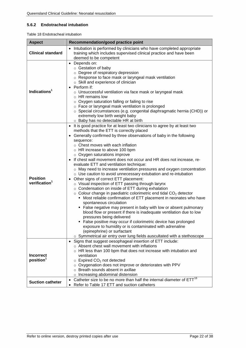
5.6.2 Endotracheal intubation
Table 18 Endotracheal intubation
Aspect Recommendation/good practice point
Clinical standard • Intubation is performed by clinicians who have completed appropriate
training which includes supervised clinical practice and have been deemed to be competent
Indications1
• Depends on: o Gestation of baby o Degree of respiratory depression o Response to face mask or laryngeal mask ventilation o Skill and experience of clinician
• Perform if: o Unsuccessful ventilation via face mask or laryngeal mask o HR remains low o Oxygen saturation falling or failing to rise o Face or laryngeal mask ventilation is prolonged o Special circumstances (e.g. congenital diaphragmatic hernia (CHD)) or
extremely low birth weight baby o Baby has no detectable HR at birth
Position verification1
• It is good practice for at least two clinicians to agree by at least two methods that the ETT is correctly placed
• Generally confirmed by three observations of baby in the following sequence: o Chest moves with each inflation o HR increase to above 100 bpm o Oxygen saturations improve
• If chest wall movement does not occur and HR does not increase, re-evaluate ETT and ventilation technique: o May need to increase ventilation pressures and oxygen concentration o Use caution to avoid unnecessary extubation and re-intubation
• Other signs of correct ETT placement: o Visual inspection of ETT passing through larynx o Condensation on inside of ETT during exhalation o Colour change in paediatric colorimetric end tidal CO2 detector Most reliable confirmation of ETT placement in neonates who have
spontaneous circulation False negative may present in baby with low or absent pulmonary
blood flow or present if there is inadequate ventilation due to low pressures being delivered
False positive may occur if colorimetric device has prolonged exposure to humidity or is contaminated with adrenaline (epinephrine) or surfactant
o Symmetrical air entry over lung fields auscultated with a stethoscope
Incorrect position1
• Signs that suggest oesophageal insertion of ETT include: o Absent chest wall movement with inflations o HR less than 100 bpm that does not increase with intubation and
ventilation o Expired CO2 not detected o Oxygenation does not improve or deteriorates with PPV o Breath sounds absent in axillae o Increasing abdominal distension
Suction catheter • Catheter size to be no more than half the internal diameter of ETT16 • Refer to Table 17 ETT and suction catheters
Queensland Clinical Guideline: Neonatal resuscitation
Refer to online version, destroy printed copies after use Page 23 of 38
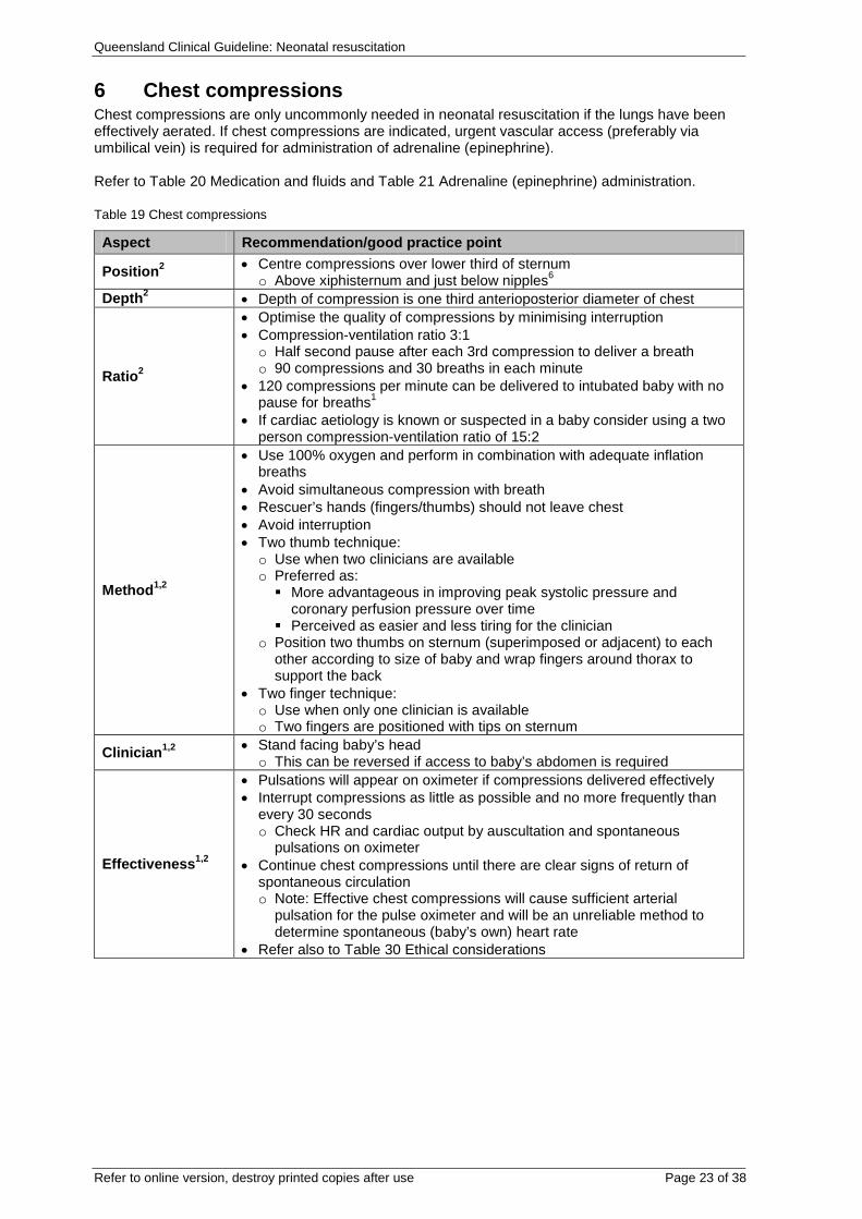
6 Chest compressions Chest compressions are only uncommonly needed in neonatal resuscitation if the lungs have been effectively aerated. If chest compressions are indicated, urgent vascular access (preferably via umbilical vein) is required for administration of adrenaline (epinephrine). Refer to Table 20 Medication and fluids and Table 21 Adrenaline (epinephrine) administration.
Table 19 Chest compressions
Aspect Recommendation/good practice point
Position2 • Centre compressions over lower third of sternum o Above xiphisternum and just below nipples6
Depth2 • Depth of compression is one third anterioposterior diameter of chest
Ratio2
• Optimise the quality of compressions by minimising interruption • Compression-ventilation ratio 3:1 o Half second pause after each 3rd compression to deliver a breath o 90 compressions and 30 breaths in each minute
• 120 compressions per minute can be delivered to intubated baby with no pause for breaths1
• If cardiac aetiology is known or suspected in a baby consider using a two person compression-ventilation ratio of 15:2
Method1,2
• Use 100% oxygen and perform in combination with adequate inflation breaths
• Avoid simultaneous compression with breath • Rescuer’s hands (fingers/thumbs) should not leave chest • Avoid interruption • Two thumb technique: o Use when two clinicians are available o Preferred as: More advantageous in improving peak systolic pressure and
coronary perfusion pressure over time Perceived as easier and less tiring for the clinician
o Position two thumbs on sternum (superimposed or adjacent) to each other according to size of baby and wrap fingers around thorax to support the back
• Two finger technique: o Use when only one clinician is available o Two fingers are positioned with tips on sternum
Clinician1,2 • Stand facing baby’s head o This can be reversed if access to baby’s abdomen is required
Effectiveness1,2
• Pulsations will appear on oximeter if compressions delivered effectively • Interrupt compressions as little as possible and no more frequently than
every 30 seconds o Check HR and cardiac output by auscultation and spontaneous
pulsations on oximeter • Continue chest compressions until there are clear signs of return of
spontaneous circulation o Note: Effective chest compressions will cause sufficient arterial
pulsation for the pulse oximeter and will be an unreliable method to determine spontaneous (baby’s own) heart rate
• Refer also to Table 30 Ethical considerations
Queensland Clinical Guideline: Neonatal resuscitation
Refer to online version, destroy printed copies after use Page 24 of 38
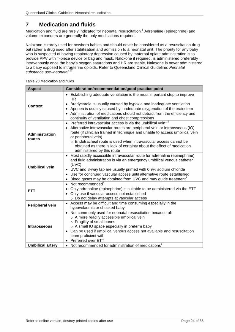
7 Medication and fluids Medication and fluid are rarely indicated for neonatal resuscitation.6 Adrenaline (epinephrine) and volume expanders are generally the only medications required. Naloxone is rarely used for newborn babies and should never be considered as a resuscitation drug but rather a drug used after stabilisation and admission to a neonatal unit. The priority for any baby who is suspected of having respiratory depression caused by maternal opiate administration is to provide PPV with T-piece device or bag and mask. Naloxone if required, is administered preferably intravenously once the baby’s oxygen saturations and HR are stable. Naloxone is never administered to a baby exposed to intrauterine opioids. Refer to Queensland Clinical Guideline: Perinatal substance use–neonatal.17
Table 20 Medication and fluids
Aspect Consideration/recommendation/good practice point
Context
• Establishing adequate ventilation is the most important step to improve HR
• Bradycardia is usually caused by hypoxia and inadequate ventilation • Apnoea is usually caused by inadequate oxygenation of the brainstem • Administration of medications should not detract from the efficiency and
continuity of ventilation and chest compressions
Administration routes
• Preferred intravascular access is via the umbilical vein1,2 • Alternative intravascular routes are peripheral vein or intraosseous (IO)
route (if clinician trained in technique and unable to access umbilical vein or peripheral vein) o Endotracheal route is used when intravascular access cannot be
obtained as there is lack of certainty about the effect of medication administered by this route
Umbilical vein
• Most rapidly accessible intravascular route for adrenaline (epinephrine) and fluid administration is via an emergency umbilical venous catheter (UVC)
• UVC and 3-way tap are usually primed with 0.9% sodium chloride • Use for continued vascular access until alternative route established • Blood gases may be obtained from UVC and may guide treatment1
ETT
• Not recommended1 • Only adrenaline (epinephrine) is suitable to be administered via the ETT • Only use if vascular access not established o Do not delay attempts at vascular access
Peripheral vein • Access may be difficult and time consuming especially in the hypovolaemic or shocked baby
Intraosseous
• Not commonly used for neonatal resuscitation because of: o A more readily accessible umbilical vein o Fragility of small bones o A small IO space especially in preterm baby
• Can be used if umbilical venous access not available and resuscitation team proficient with
• Preferred over ETT Umbilical artery • Not recommended for administration of medications1
Queensland Clinical Guideline: Neonatal resuscitation
Refer to online version, destroy printed copies after use Page 25 of 38
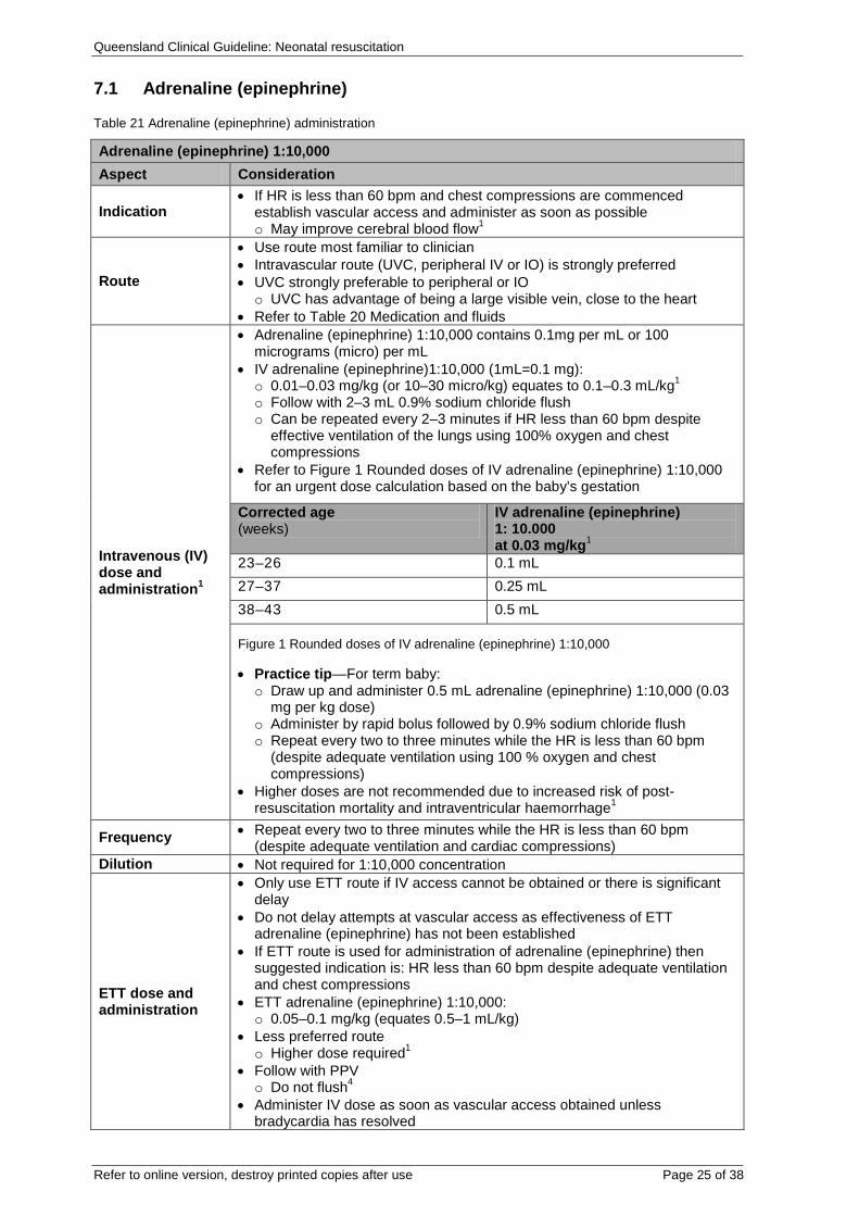
7.1 Adrenaline (epinephrine)
Table 21 Adrenaline (epinephrine) administration
Adrenaline (epinephrine) 1:10,000 Aspect Consideration
Indication • If HR is less than 60 bpm and chest compressions are commenced
establish vascular access and administer as soon as possible o May improve cerebral blood flow1
Route
• Use route most familiar to clinician • Intravascular route (UVC, peripheral IV or IO) is strongly preferred • UVC strongly preferable to peripheral or IO o UVC has advantage of being a large visible vein, close to the heart
• Refer to Table 20 Medication and fluids
Intravenous (IV) dose and administration1
• Adrenaline (epinephrine) 1:10,000 contains 0.1mg per mL or 100 micrograms (micro) per mL
• IV adrenaline (epinephrine)1:10,000 (1mL=0.1 mg): o 0.01–0.03 mg/kg (or 10–30 micro/kg) equates to 0.1–0.3 mL/kg1 o Follow with 2–3 mL 0.9% sodium chloride flush o Can be repeated every 2–3 minutes if HR less than 60 bpm despite
effective ventilation of the lungs using 100% oxygen and chest compressions
• Refer to Figure 1 Rounded doses of IV adrenaline (epinephrine) 1:10,000 for an urgent dose calculation based on the baby’s gestation
Corrected age (weeks)
IV adrenaline (epinephrine) 1: 10.000 at 0.03 mg/kg1
23–26 0.1 mL 27–37 0.25 mL 38–43 0.5 mL
Figure 1 Rounded doses of IV adrenaline (epinephrine) 1:10,000 • Practice tip—For term baby: o Draw up and administer 0.5 mL adrenaline (epinephrine) 1:10,000 (0.03
mg per kg dose) o Administer by rapid bolus followed by 0.9% sodium chloride flush o Repeat every two to three minutes while the HR is less than 60 bpm
(despite adequate ventilation using 100 % oxygen and chest compressions)
• Higher doses are not recommended due to increased risk of post-resuscitation mortality and intraventricular haemorrhage1
Frequency • Repeat every two to three minutes while the HR is less than 60 bpm (despite adequate ventilation and cardiac compressions)
Dilution • Not required for 1:10,000 concentration
ETT dose and administration
• Only use ETT route if IV access cannot be obtained or there is significant delay
• Do not delay attempts at vascular access as effectiveness of ETT adrenaline (epinephrine) has not been established
• If ETT route is used for administration of adrenaline (epinephrine) then suggested indication is: HR less than 60 bpm despite adequate ventilation and chest compressions
• ETT adrenaline (epinephrine) 1:10,000: o 0.05–0.1 mg/kg (equates 0.5–1 mL/kg)
• Less preferred route o Higher dose required1
• Follow with PPV o Do not flush4
• Administer IV dose as soon as vascular access obtained unless bradycardia has resolved
Queensland Clinical Guideline: Neonatal resuscitation
Refer to online version, destroy printed copies after use Page 26 of 38
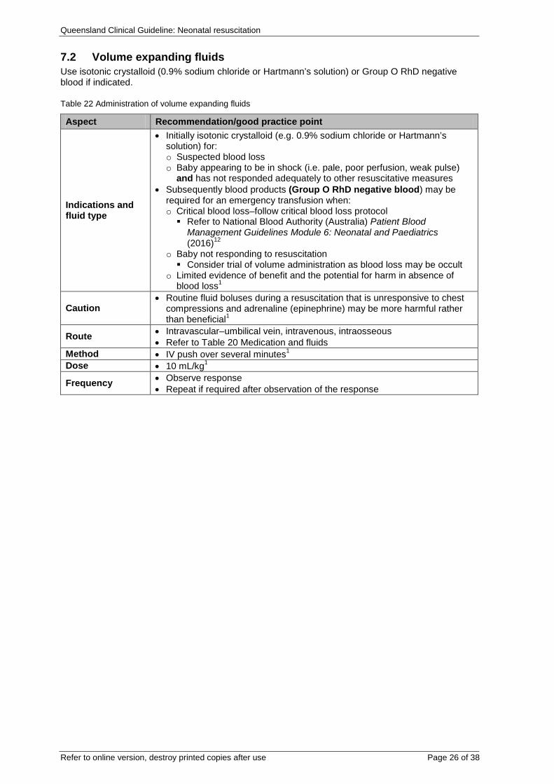
7.2 Volume expanding fluids Use isotonic crystalloid (0.9% sodium chloride or Hartmann’s solution) or Group O RhD negative blood if indicated.
Table 22 Administration of volume expanding fluids
Aspect Recommendation/good practice point
Indications and fluid type
• Initially isotonic crystalloid (e.g. 0.9% sodium chloride or Hartmann’s solution) for: o Suspected blood loss o Baby appearing to be in shock (i.e. pale, poor perfusion, weak pulse)
and has not responded adequately to other resuscitative measures • Subsequently blood products (Group O RhD negative blood) may be
required for an emergency transfusion when: o Critical blood loss–follow critical blood loss protocol Refer to National Blood Authority (Australia) Patient Blood
Management Guidelines Module 6: Neonatal and Paediatrics (2016)12
o Baby not responding to resuscitation Consider trial of volume administration as blood loss may be occult
o Limited evidence of benefit and the potential for harm in absence of blood loss1
Caution • Routine fluid boluses during a resuscitation that is unresponsive to chest
compressions and adrenaline (epinephrine) may be more harmful rather than beneficial1
Route • Intravascular–umbilical vein, intravenous, intraosseous • Refer to Table 20 Medication and fluids
Method • IV push over several minutes1 Dose • 10 mL/kg1
Frequency • Observe response • Repeat if required after observation of the response
Queensland Clinical Guideline: Neonatal resuscitation
Refer to online version, destroy printed copies after use Page 27 of 38
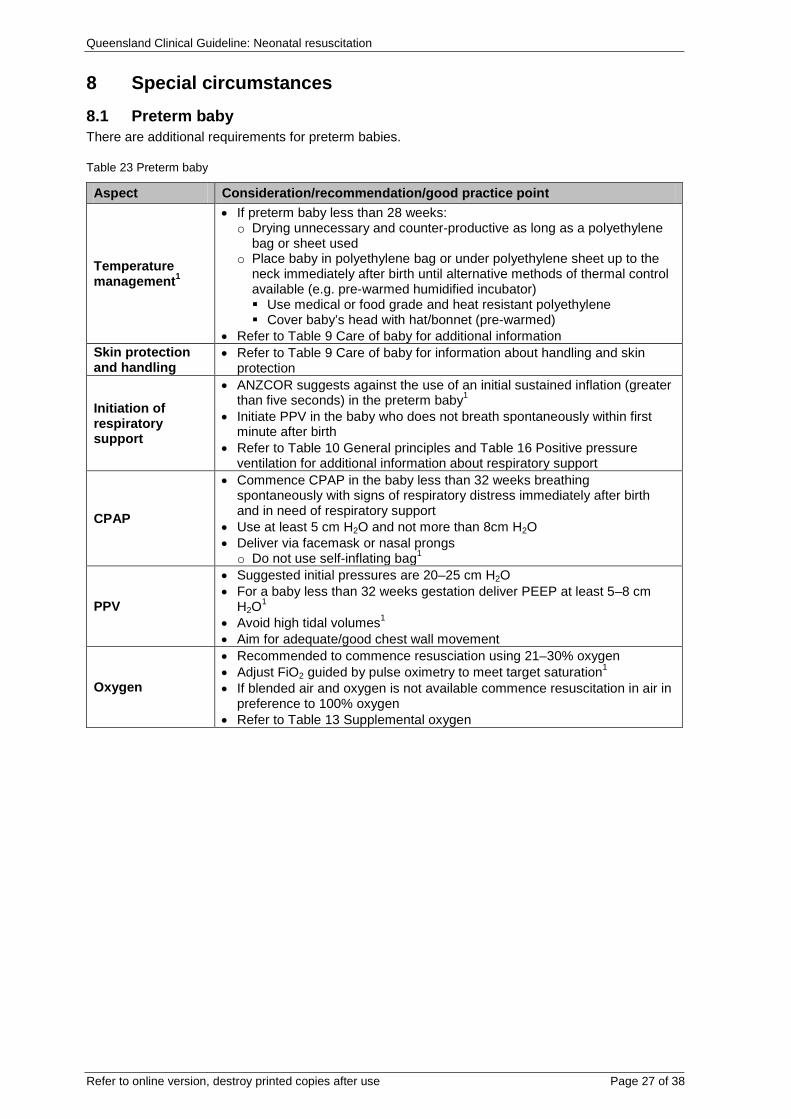
8 Special circumstances 8.1 Preterm baby There are additional requirements for preterm babies.
Table 23 Preterm baby
Aspect Consideration/recommendation/good practice point
Temperature management1
• If preterm baby less than 28 weeks: o Drying unnecessary and counter-productive as long as a polyethylene
bag or sheet used o Place baby in polyethylene bag or under polyethylene sheet up to the
neck immediately after birth until alternative methods of thermal control available (e.g. pre-warmed humidified incubator) Use medical or food grade and heat resistant polyethylene Cover baby’s head with hat/bonnet (pre-warmed)
• Refer to Table 9 Care of baby for additional information Skin protection and handling
• Refer to Table 9 Care of baby for information about handling and skin protection
Initiation of respiratory support
• ANZCOR suggests against the use of an initial sustained inflation (greater than five seconds) in the preterm baby1
• Initiate PPV in the baby who does not breath spontaneously within first minute after birth
• Refer to Table 10 General principles and Table 16 Positive pressure ventilation for additional information about respiratory support
CPAP
• Commence CPAP in the baby less than 32 weeks breathing spontaneously with signs of respiratory distress immediately after birth and in need of respiratory support
• Use at least 5 cm H2O and not more than 8cm H2O • Deliver via facemask or nasal prongs o Do not use self-inflating bag1
PPV
• Suggested initial pressures are 20–25 cm H2O • For a baby less than 32 weeks gestation deliver PEEP at least 5–8 cm
H2O1 • Avoid high tidal volumes1 • Aim for adequate/good chest wall movement
Oxygen
• Recommended to commence resusciation using 21–30% oxygen • Adjust FiO2 guided by pulse oximetry to meet target saturation1 • If blended air and oxygen is not available commence resuscitation in air in
preference to 100% oxygen • Refer to Table 13 Supplemental oxygen
Queensland Clinical Guideline: Neonatal resuscitation
Refer to online version, destroy printed copies after use Page 28 of 38
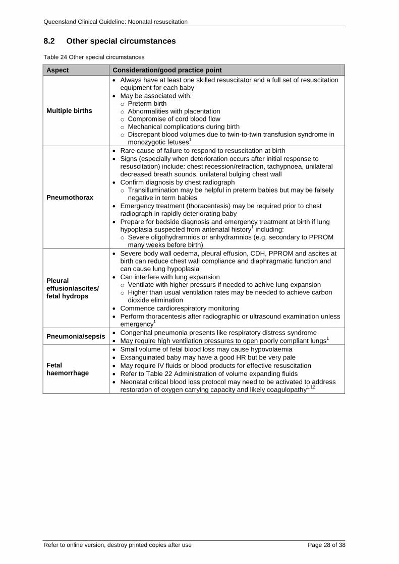
8.2 Other special circumstances
Table 24 Other special circumstances
Aspect Consideration/good practice point
Multiple births
• Always have at least one skilled resuscitator and a full set of resuscitation equipment for each baby
• May be associated with: o Preterm birth o Abnormalities with placentation o Compromise of cord blood flow o Mechanical complications during birth o Discrepant blood volumes due to twin-to-twin transfusion syndrome in
monozygotic fetuses1
Pneumothorax
• Rare cause of failure to respond to resuscitation at birth • Signs (especially when deterioration occurs after initial response to
resuscitation) include: chest recession/retraction, tachypnoea, unilateral decreased breath sounds, unilateral bulging chest wall
• Confirm diagnosis by chest radiograph o Transillumination may be helpful in preterm babies but may be falsely
negative in term babies • Emergency treatment (thoracentesis) may be required prior to chest
radiograph in rapidly deteriorating baby • Prepare for bedside diagnosis and emergency treatment at birth if lung
hypoplasia suspected from antenatal history1 including: o Severe oligohydramnios or anhydramnios (e.g. secondary to PPROM
many weeks before birth)
Pleural effusion/ascites/ fetal hydrops
• Severe body wall oedema, pleural effusion, CDH, PPROM and ascites at birth can reduce chest wall compliance and diaphragmatic function and can cause lung hypoplasia
• Can interfere with lung expansion o Ventilate with higher pressurs if needed to achive lung expansion o Higher than usual ventilation rates may be needed to achieve carbon
dioxide elimination • Commence cardiorespiratory monitoring • Perform thoracentesis after radiographic or ultrasound examination unless
emergency1
Pneumonia/sepsis • Congenital pneumonia presents like respiratory distress syndrome • May require high ventilation pressures to open poorly compliant lungs1
Fetal haemorrhage
• Small volume of fetal blood loss may cause hypovolaemia • Exsanguinated baby may have a good HR but be very pale • May require IV fluids or blood products for effective resuscitation • Refer to Table 22 Administration of volume expanding fluids • Neonatal critical blood loss protocol may need to be activated to address
restoration of oxygen carrying capacity and likely coagulopathy1,12
Queensland Clinical Guideline: Neonatal resuscitation
Refer to online version, destroy printed copies after use Page 29 of 38
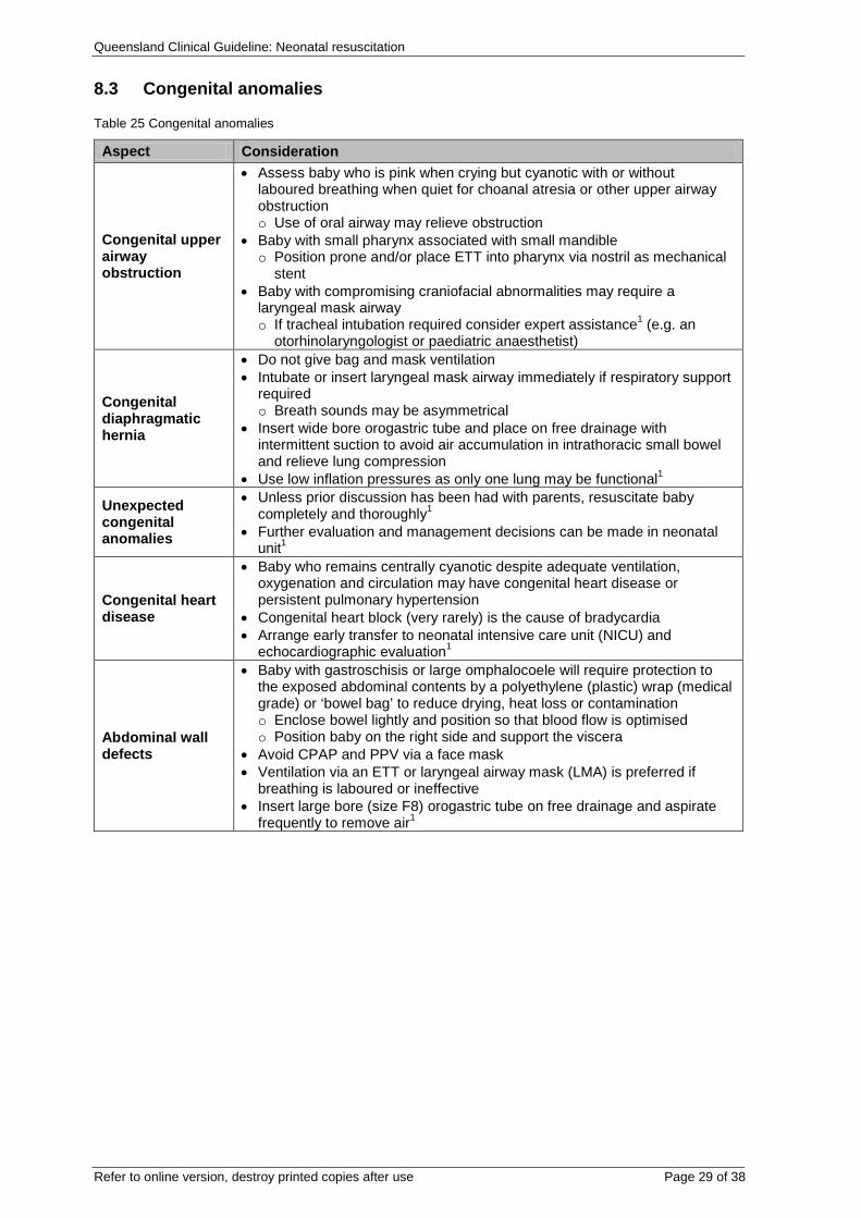
8.3 Congenital anomalies
Table 25 Congenital anomalies
Aspect Consideration
Congenital upper airway obstruction
• Assess baby who is pink when crying but cyanotic with or without laboured breathing when quiet for choanal atresia or other upper airway obstruction o Use of oral airway may relieve obstruction
• Baby with small pharynx associated with small mandible o Position prone and/or place ETT into pharynx via nostril as mechanical
stent • Baby with compromising craniofacial abnormalities may require a
laryngeal mask airway o If tracheal intubation required consider expert assistance1 (e.g. an
otorhinolaryngologist or paediatric anaesthetist)
Congenital diaphragmatic hernia
• Do not give bag and mask ventilation • Intubate or insert laryngeal mask airway immediately if respiratory support
required o Breath sounds may be asymmetrical
• Insert wide bore orogastric tube and place on free drainage with intermittent suction to avoid air accumulation in intrathoracic small bowel and relieve lung compression
• Use low inflation pressures as only one lung may be functional1
Unexpected congenital anomalies
• Unless prior discussion has been had with parents, resuscitate baby completely and thoroughly1
• Further evaluation and management decisions can be made in neonatal unit1
Congenital heart disease
• Baby who remains centrally cyanotic despite adequate ventilation, oxygenation and circulation may have congenital heart disease or persistent pulmonary hypertension
• Congenital heart block (very rarely) is the cause of bradycardia • Arrange early transfer to neonatal intensive care unit (NICU) and
echocardiographic evaluation1
Abdominal wall defects
• Baby with gastroschisis or large omphalocoele will require protection to the exposed abdominal contents by a polyethylene (plastic) wrap (medical grade) or ‘bowel bag’ to reduce drying, heat loss or contamination o Enclose bowel lightly and position so that blood flow is optimised o Position baby on the right side and support the viscera
• Avoid CPAP and PPV via a face mask • Ventilation via an ETT or laryngeal airway mask (LMA) is preferred if
breathing is laboured or ineffective • Insert large bore (size F8) orogastric tube on free drainage and aspirate
frequently to remove air1
Queensland Clinical Guideline: Neonatal resuscitation
Refer to online version, destroy printed copies after use Page 30 of 38
9 Care after resuscitation A baby with perinatal compromise or ongoing respiratory distress may have delayed or dysfunctional perinatal adaptation of the brain, heart, gastrointestinal tract, kidneys or other organs.1
9.1 Cord blood sampling
Table 26 Cord blood gas sampling
Aspect Consideration
Indications
• Baby requiring resuscitation to objectively assess the condition just before birth6
• Apgar score: o Less than 4 at one minute o Less than 7 at five minutes18
• When fetal blood sampling has occurred in labour1,3 • Refer to Queensland Clinical Guideline: Intrapartum fetal surveillance3
Procedure • Collect paired samples from umbilical vein and umbilical artery3 • Effect of delayed cord clamping on values uncertain6 but some research
has shown effect on acid-base parameters19,20
Interpretation
• If there is a low cord arterial pH the relative risk of neonatal encephalopathy is increased
• A normal pH does not rule out subsequent neonatal morbidity21 • Refer to Queensland Clinical Guideline: Hypoxic ischaemic
encephalopathy (HIE)22 • Refer to Table 27 Umbilical artery cord blood gases
Relative risk (RR) of neonatal morbidity (CI 95%)21 Arterial pH RR 7.06–7.10 2.22 (1.06–4.62) 7.01–7.05 3.63 (1.60–8.22)
Less than or equal to 7.0 18.20 (10.51–31.70)
Lactate
• There may be a marginally better predictability of morbidity using lactate measures ,23 however: o Minimal difference between lactate and pH predictability o Better data available for pH for longterm outcomes21
• Normal lactate thresholds have not been agreed
9.1.1 Umbilical artery cord blood gases Paired cord blood gases are the most objective means of assessing the condition of the baby just prior to birth. Paired cord samples from the umbilical vein and umbilical artery are advisable.1,3
Table 27 Umbilical artery cord blood gases
Umbilical artery1 2.5th percentile Mean 97.5th percentile pH 7.1 7.27 7.38
Base excess -11 -4 1 pO2 (mmHg) 6 17 30
pCO2 (mmHg) 35 52 74
Queensland Clinical Guideline: Neonatal resuscitation
Refer to online version, destroy printed copies after use Page 31 of 38
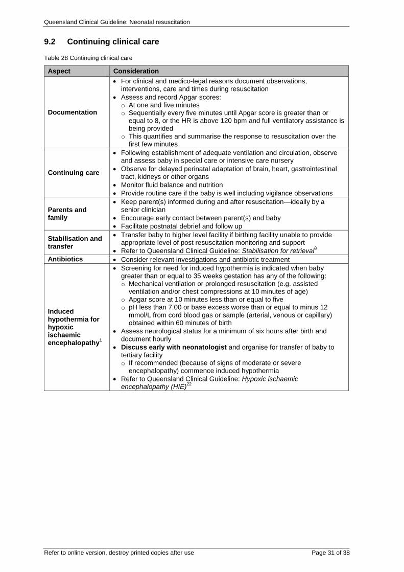
9.2 Continuing clinical care
Table 28 Continuing clinical care
Aspect Consideration
Documentation
• For clinical and medico-legal reasons document observations, interventions, care and times during resuscitation
• Assess and record Apgar scores: o At one and five minutes o Sequentially every five minutes until Apgar score is greater than or
equal to 8, or the HR is above 120 bpm and full ventilatory assistance is being provided
o This quantifies and summarise the response to resuscitation over the first few minutes
Continuing care
• Following establishment of adequate ventilation and circulation, observe and assess baby in special care or intensive care nursery
• Observe for delayed perinatal adaptation of brain, heart, gastrointestinal tract, kidneys or other organs
• Monitor fluid balance and nutrition • Provide routine care if the baby is well including vigilance observations
Parents and family
• Keep parent(s) informed during and after resuscitation—ideally by a senior clinician
• Encourage early contact between parent(s) and baby • Facilitate postnatal debrief and follow up
Stabilisation and transfer
• Transfer baby to higher level facility if birthing facility unable to provide appropriate level of post resuscitation monitoring and support
• Refer to Queensland Clinical Guideline: Stabilisation for retrieval8 Antibiotics • Consider relevant investigations and antibiotic treatment
Induced hypothermia for hypoxic ischaemic encephalopathy1
• Screening for need for induced hypothermia is indicated when baby greater than or equal to 35 weeks gestation has any of the following: o Mechanical ventilation or prolonged resuscitation (e.g. assisted
ventilation and/or chest compressions at 10 minutes of age) o Apgar score at 10 minutes less than or equal to five o pH less than 7.00 or base excess worse than or equal to minus 12
mmol/L from cord blood gas or sample (arterial, venous or capillary) obtained within 60 minutes of birth
• Assess neurological status for a minimum of six hours after birth and document hourly
• Discuss early with neonatologist and organise for transfer of baby to tertiary facility o If recommended (because of signs of moderate or severe
encephalopathy) commence induced hypothermia • Refer to Queensland Clinical Guideline: Hypoxic ischaemic
encephalopathy (HIE)22
Queensland Clinical Guideline: Neonatal resuscitation
Refer to online version, destroy printed copies after use Page 32 of 38
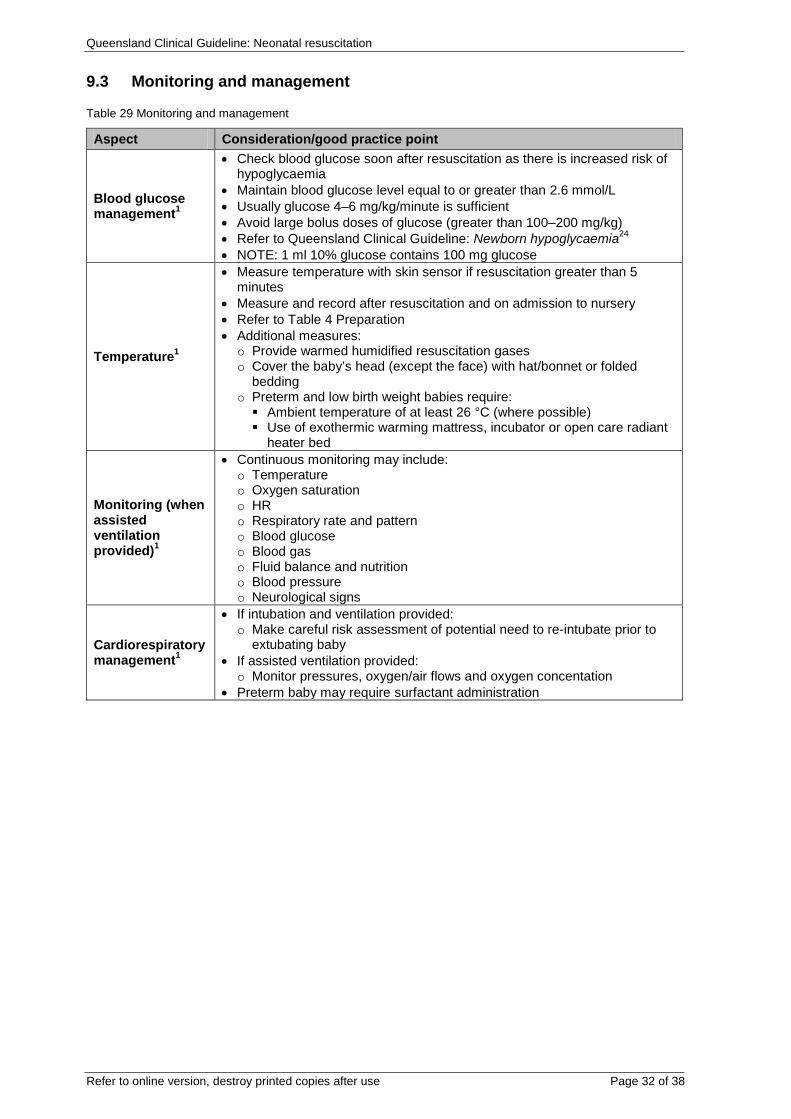
9.3 Monitoring and management
Table 29 Monitoring and management
Aspect Consideration/good practice point
Blood glucose management1
• Check blood glucose soon after resuscitation as there is increased risk of hypoglycaemia
• Maintain blood glucose level equal to or greater than 2.6 mmol/L • Usually glucose 4–6 mg/kg/minute is sufficient • Avoid large bolus doses of glucose (greater than 100–200 mg/kg) • Refer to Queensland Clinical Guideline: Newborn hypoglycaemia24 • NOTE: 1 ml 10% glucose contains 100 mg glucose
Temperature1
• Measure temperature with skin sensor if resuscitation greater than 5 minutes
• Measure and record after resuscitation and on admission to nursery • Refer to Table 4 Preparation • Additional measures: o Provide warmed humidified resuscitation gases o Cover the baby’s head (except the face) with hat/bonnet or folded
bedding o Preterm and low birth weight babies require: Ambient temperature of at least 26 °C (where possible) Use of exothermic warming mattress, incubator or open care radiant
heater bed
Monitoring (when assisted ventilation provided)1
• Continuous monitoring may include: o Temperature o Oxygen saturation o HR o Respiratory rate and pattern o Blood glucose o Blood gas o Fluid balance and nutrition o Blood pressure o Neurological signs
Cardiorespiratory management1
• If intubation and ventilation provided: o Make careful risk assessment of potential need to re-intubate prior to
extubating baby • If assisted ventilation provided: o Monitor pressures, oxygen/air flows and oxygen concentation
• Preterm baby may require surfactant administration
Queensland Clinical Guideline: Neonatal resuscitation
Refer to online version, destroy printed copies after use Page 33 of 38
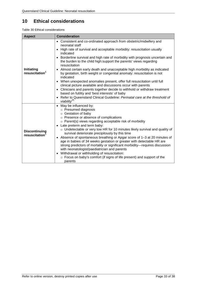
10 Ethical considerations Table 30 Ethical considerations
Aspect Consideration
Initiating resuscitation1
• Consistent and co-ordinated approach from obstetric/midwifery and neonatal staff
• High rate of survival and acceptable morbidity: resuscitation usually indicated
• Borderline survival and high rate of morbidity with prognosis uncertain and the burden to the child high:support the parents’ views regarding resuscitation
• Almost certain early death and unacceptable high morbidity as indicated by gestation, birth weight or congenital anomaly: resuscitation is not indicated
• When unexpected anomalies present, offer full resuscitation until full clinical picture available and discussions occur with parents
• Clinicians and parents together decide to withhold or withdraw treatment based on futility and ‘best interests’ of baby
• Refer to Queensland Clinical Guideline: Perinatal care at the threshold of viability25
Discontinuing resuscitation1
• May be influenced by: o Presumed diagnosis o Gestation of baby o Presence or absence of complications o Parent(s) views regarding acceptable risk of morbidity
• Late preterm and term baby: o Undetectable or very low HR for 10 minutes likely survival and quality of
survival deteriorate precipitously by this time • Absence of spontaneous breathing or Apgar score of 1–3 at 20 minutes of
age in babies of 34 weeks gestation or greater with detectable HR are strong predictors of mortality or significant morbidity—requires discussion with neonatologist/paediatrician and parents
• Withdrawal or withholding of resuscitation: o Focus on baby’s comfort (if signs of life present) and support of the
parents
Queensland Clinical Guideline: Neonatal resuscitation
Refer to online version, destroy printed copies after use Page 34 of 38
References 1. Australian and New Zealand Council on Resuscitation. Section 13 Neonatal Guidelines. 2016. 2. Perlman JM, Wylllie J, Kattwinkel J, Atkins DL, Chameides L, Goldsmith J, et al. Part 11: Neonatal Resuscitation: 2010 International Consensus on Cardiopulmonary Resuscitation and Emergency Cardiovascular Care Science With Treatment Recommendations. Circulation. 2010; 122(Suppl 2):S516–S538. 3. Queensland Clinical Guidelines. Intrapartum fetal surveillance Guideline No. MN 15.15-V4-R20. Queensland Health. Available from: http://www.health.qld.gov.au/qcg/. 2015. 4. Australian and New Zealand Council on Resuscitation. Section 13 - Neonatal Guidelines. 2010. 5. Perlman JM, Wyllie J, Kattwinkel J, Wyckoff MH, Aziz K, Guinsburg R, et al. Part 7: Neonatal Resuscitation: 2015 International Consensus on Cardiopulmonary Resuscitation and Emergency Cardiovascular Care Science with Treatment Recommendations. Circulation. 2015; 132(supp 1):S204-S241. 6. Australian and New Zealand Council on Resuscitation. Neonatal Life Support Flowchart. 2016. 7. Queensland Clinical Guidelines. Early onset Group B streptococcal disease Guideline No. MN10.20-V2-R15. 2010. 8. Queensland Clinical Guidelines. Neonatal stabilisation for retrieval. Guideline No. MN11.18-V1-R16. Queensland Health. Available from http://www.health.qld.gov.au/qcg/. 2011. 9. Dawson JA, Davis PG, O’Donnell CPF, Kamlin COF, Morley CJ. Pulse oximetry for monitoring infants in the delivery room: a review. Arch Dis Child Fetal Neonatal Edition. 2007; 92:F4-F7. 10. Dawson J, Morley C. Monitoring oxygen saturation and heart rate in the early neonatal period. Seminars in Fetal & Neonatal Medicine. 2010; 15:203-207. 11. McDonald SJ , Middleton P , Dowswell T , Morris PS Effect of timing of umbilical cord clamping of term infants on maternal and neonatal outcomes. Cochrane Database of Systematic Reviews 2013. 2013; Issue 7. Art. No.: CD004074. DOI:10.1002/14651858.CD004074.pub3. 12. National Blood Authority (Australia). Patient Blood Management Guidelines: Module 6–Neonatal and Paediatrics. Canberra. 2016. Available from: https://www.blood.gov.au/. 13. J Kattwinkel (Ed). Textbook of neonatal resuscitation. American Academy of Paediatrics and American Heart Association; 2011. 14. Kempley S, Moreiras J, Petrona F. Endotracheal tube length for neonatal intubation. Resuscitation. 2008; 77:369-373. 15. Peterson J, Johnson N, Deakins K, Wilson-Costello D, Jelovsek JE, Chatburn R. Accuracy of the 7-8-9 Rule for endotracheal tube placement inthe neonate. Journal of Perinatology. 2006; 26:333-336. 16. Russian C, Gaonzales JF, Henry NR. Suction catheter size: an assessment and comparison of 3 different calculation methods. Respiratory Care. 2014; 59(1):32-38. 17. Queensland Clinical Guidelines. Perinatal substance use-neonatal. Guideline No. MN38.15-V1-R21 Queensland Health. Available from http://www.health.qld.gov.au/qcg/. 2016. 18. Royal Australian and New Zealand College of Obstetricians and Gynaecologists. Intrapartum fetal surveillance clinical guideline. 2014. Available from: http://www.ranzcog.edu.au/. 19. De Paco C, Florido J, Garridao MC, Prados S, Navarrete L. Umbilical cord blood acid–base and gas analysis after early versus delayed cord clamping in neonates at term. Arch Gynecol Obstet. 2011; 283:1011-1014. 20. Wiberg N, Kallen K, Olofsson P. Delayed umbilical cord clamping at birth has effects on arterial and venous blood gases and lactate concentrations. BJOG. 2008; 115:697-703. 21. Yeh P, Emary K, Impey L. The relationship between umbilical cord arterial pH and serious adverse neonatal outcome: analysis of 51 519 consecutive validated samples. BJOG. 2012; 119:824-831. 22. Queensland Clinical Guidelines. Hypoxic Ischaemic Encephalopathy MN10.11-V5-R16, Queensland Health. Avavilable from http://www.health.qld.gov.au/qcg/. 2016. 23. Tuuli MG, Stout MJ, Shanks A, Odibo AO, Macones G, Cahill A. Umbilical cord arterial lactate compared with pH for predicting neonatal morbidity at term. Obstet Gynecol. 2014; 124(4):756-761. 24. Queensland Clinical Guidelines. Newborn hypoglycaemia Guideline No. MN13.8-V5-R18. Queensland Health. Available from http://www.health.qld.gov.au/qcg/. 2013. 25. Queensland Clinical Guidelines. Perinatal care at the threshold of viability. Guideline No. MN 14.32-V1-R19. Queensland Health. 2014. Available form http://www.health.qld.gov.au/qcg/. 2014.
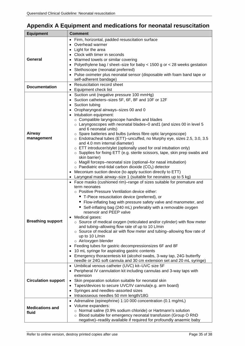
Queensland Clinical Guideline: Neonatal resuscitation
Refer to online version, destroy printed copies after use Page 35 of 38
Appendix A Equipment and medications for neonatal resuscitation Equipment Comment
General
• Firm, horizontal, padded resuscitation surface • Overhead warmer • Light for the area • Clock with timer in seconds • Warmed towels or similar covering • Polyethylene bag / sheet–size for baby < 1500 g or < 28 weeks gestation • Stethoscope (neonatal preferred) • Pulse oximeter plus neonatal sensor (disposable with foam band tape or
self-adherent bandage)
Documentation • Resuscitation record sheet • Equipment check list
Airway management
• Suction unit (negative pressure 100 mmHg) • Suction catheters–sizes 5F, 6F, 8F and 10F or 12F • Suction tubing • Oropharyngeal airways–sizes 00 and 0 • Intubation equipment: o Compatible laryngoscope handles and blades o Laryngoscopes with neonatal blades–0 and1 (and sizes 00 in level 5
and 6 neonatal units) o Spare batteries and bulbs (unless fibre optic laryngoscope) o Endotracheal tubes (ETT)–uncuffed, no Murphy eye, sizes 2.5, 3.0, 3.5
and 4.0 mm internal diameter) o ETT introducer/stylet (optionally used for oral intubation only) o Supplies for fixing ETT (e.g. sterile scissors, tape, skin prep swabs and
skin barrier) o Magill forceps–neonatal size (optional–for nasal intubation) o Paediatric end-tidal carbon dioxide (CO2) detector
• Meconium suction device (to apply suction directly to ETT) • Laryngeal mask airway–size 1 (suitable for neonates up to 5 kg)
Breathing support
• Face masks (cushioned rim)–range of sizes suitable for premature and term neonates o Positive Pressure Ventilation device either: T-Piece resuscitation device (preferred), or Flow-inflating bag with pressure safety valve and manometer, and Self-inflating bag (240 mL) preferably with a removable oxygen
reservoir and PEEP valve • Medical gases: o Source of medical oxygen (reticulated and/or cylinder) with flow meter
and tubing–allowing flow rate of up to 10 L/min o Source of medical air with flow meter and tubing–allowing flow rate of
up to 10 L/min o Air/oxygen blender
• Feeding tubes for gastric decompressionsizes 6F and 8F • 10 mL syringe for aspirating gastric contents • Emergency thoracentesis kit (alcohol swabs, 3-way tap, 24G butterfly
needle or 24G soft cannula and 30 cm extension set and 20 mL syringe)
Circulation support
• Umbilical venous catheter (UVC) kit–UVC size 5F • Peripheral IV cannulation kit including cannulas and 3-way taps with
extension • Skin preparation solution suitable for neonatal skin • Tapes/devices to secure UVC/IV cannula(e.g. arm board) • Syringes and needles–assorted sizes • Intraosseous needles 50 mm length/18G
Medications and fluid
• Adrenaline (epinephrine) 1:10 000 concentration (0.1 mg/mL) • Volume expanders: o Normal saline (0.9% sodium chloride) or Hartmann’s solution o Blood suitable for emergency neonatal transfusion (Group O RhD
negative)–readily available if required for profoundly anaemic baby
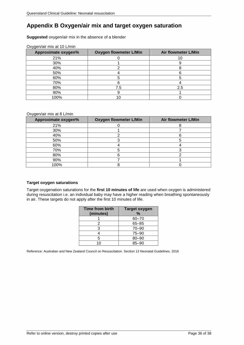
Queensland Clinical Guideline: Neonatal resuscitation
Refer to online version, destroy printed copies after use Page 36 of 38
Appendix B Oxygen/air mix and target oxygen saturation Suggested oxygen/air mix in the absence of a blender Oxygen/air mix at 10 L/min
Approximate oxygen% Oxygen flowmeter L/Min Air flowmeter L/Min 21% 0 10 30% 1 9 40% 2 8 50% 4 6 60% 5 5 70% 6 4 80% 7.5 2.5 90% 9 1 100% 10 0
Oxygen/air mix at 8 L/min Approximate oxygen% Oxygen flowmeter L/Min Air flowmeter L/Min
21% 0 8 30% 1 7 40% 2 6 50% 3 5 60% 4 4 70% 5 3 80% 6 2 90% 7 1 100% 8 0
Target oxygen saturations
Target oxygenation saturations for the first 10 minutes of life are used when oxygen is administered during resuscitation i.e. an individual baby may have a higher reading when breathing spontaneously in air. These targets do not apply after the first 10 minutes of life.
Time from birth (minutes)
Target oxygen %
1 60–70 2 65–85 3 70–90 4 75–90 5 80–90 10 85–90
Reference: Australian and New Zealand Council on Resuscitation. Section 13 Neonatal Guidelines. 2016
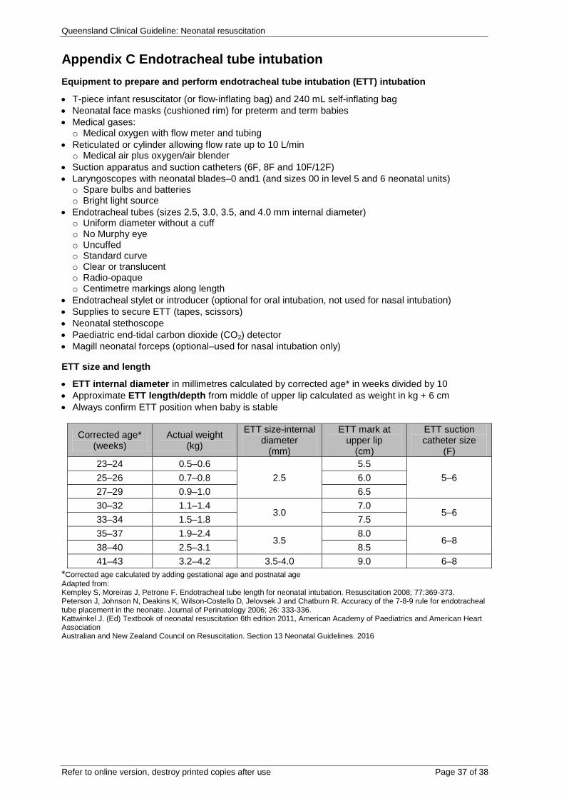
Queensland Clinical Guideline: Neonatal resuscitation
Refer to online version, destroy printed copies after use Page 37 of 38
Appendix C Endotracheal tube intubation Equipment to prepare and perform endotracheal tube intubation (ETT) intubation
• T-piece infant resuscitator (or flow-inflating bag) and 240 mL self-inflating bag • Neonatal face masks (cushioned rim) for preterm and term babies • Medical gases: o Medical oxygen with flow meter and tubing
• Reticulated or cylinder allowing flow rate up to 10 L/min o Medical air plus oxygen/air blender
• Suction apparatus and suction catheters (6F, 8F and 10F/12F) • Laryngoscopes with neonatal blades–0 and1 (and sizes 00 in level 5 and 6 neonatal units) o Spare bulbs and batteries o Bright light source
• Endotracheal tubes (sizes 2.5, 3.0, 3.5, and 4.0 mm internal diameter) o Uniform diameter without a cuff o No Murphy eye o Uncuffed o Standard curve o Clear or translucent o Radio-opaque o Centimetre markings along length
• Endotracheal stylet or introducer (optional for oral intubation, not used for nasal intubation) • Supplies to secure ETT (tapes, scissors) • Neonatal stethoscope • Paediatric end-tidal carbon dioxide (CO2) detector • Magill neonatal forceps (optional–used for nasal intubation only)
ETT size and length
• ETT internal diameter in millimetres calculated by corrected age* in weeks divided by 10 • Approximate ETT length/depth from middle of upper lip calculated as weight in kg + 6 cm • Always confirm ETT position when baby is stable
Corrected age* (weeks)
Actual weight (kg)
ETT size-internal diameter
(mm)
ETT mark at upper lip
(cm)
ETT suction catheter size
(F) 23–24 0.5–0.6
2.5 5.5
5–6 25–26 0.7–0.8 6.0 27–29 0.9–1.0 6.5 30–32 1.1–1.4
3.0 7.0
5–6 33–34 1.5–1.8 7.5 35–37 1.9–2.4
3.5 8.0
6–8 38–40 2.5–3.1 8.5 41–43 3.2–4.2 3.5-4.0 9.0 6–8
*Corrected age calculated by adding gestational age and postnatal age Adapted from: Kempley S, Moreiras J, Petrone F. Endotracheal tube length for neonatal intubation. Resuscitation 2008; 77:369-373. Peterson J, Johnson N, Deakins K, Wilson-Costello D, Jelovsek J and Chatburn R. Accuracy of the 7-8-9 rule for endotracheal tube placement in the neonate. Journal of Perinatology 2006; 26: 333-336. Kattwinkel J. (Ed) Textbook of neonatal resuscitation 6th edition 2011, American Academy of Paediatrics and American Heart Association Australian and New Zealand Council on Resuscitation. Section 13 Neonatal Guidelines. 2016
Queensland Clinical Guideline: Neonatal resuscitation
Refer to online version, destroy printed copies after use Page 38 of 38
Acknowledgements Queensland Clinical Guidelines gratefully acknowledge the contribution of Queensland clinicians and other stakeholders who participated throughout the guideline development process particularly:
Working Party Clinical Lead
Associate Professor Helen Liley, Neonatologist, Mater Health Services, South Brisbane
QCG Program Officer
Ms Stephanie Sutherns
Working Party Members
Mrs Kaye Amos-Fleming, Maternity Clinical Educator, The Sunshine Coast Private Hospital Mrs Seija Argyros, Neonatal Nurse Practitioner, Royal Brisbane Women's Hospital Dr Neil Bartels, General Practitioner, Brisbane & Rural and Remote Queensland Ms Liz Chappell, Neonatal Nurse Educator, Gold Coast University Hospital Ms Deborah Collins, Clinical Facilitator (Neonates), Logan Hospital Mrs Meshell Curtis, Nurse Educator, Royal Brisbane Women's Hospital Dr Hazel Dobinson, Rural General Practitioner, Children's Health Queensland Ms Judy Foote, RN (Child Health)/Midwife, Kirwan Child Youth and Family Health Service Dr John Gavranich, Director of Paediatrics, Ipswich Hospital Ms Tonya Gibbs, Nurse Educator, Nambour Hospital Mrs Helen Goodwin, Clinical Lecturer, University of Queensland Ms Tina Gray, Clinical Nurse, Hervey Bay Hospital Ms Jo Hardcastle, Clinical Midwifery Educator, The Wesley Hospital Ms Leah Hardiman, President, Maternity Choices Australia Ms Karen Hose, Neonatal Nurse Practitioner, Royal Brisbane Women's Hospital Dr Susan Ireland, Neonatologist, The Townsville Hospital Dr Vishal Kapoor, Neonatologist, Lady Cilento Children's Hospital Ms Cathy Krause, Clinical Nurse Midwife, St Vincent's Hospital Miss Christine Latimer, Project Officer, Neonatal Resuscitation and Stabilisation Statewide Education, The Townsville Hospital Mrs Megan Murphy, Clinical Nurse Consultant/Nurse Practitioner, The Townsville Hospital Mrs Anne Rashleigh, Clinical Facilitator (Midwifery), Redland Hospital Mrs Bernice Ross, Midwife/Lactation Consultant, Ipswich and Brisbane Dr Prasanna Shirkhedkar, Paediatrician, Caboolture Hospital Ms Alecia Staines, Consumer Representative, Maternity Consumer Network Mrs Rhonda Taylor, Clinical Midwifery Consultant, The Townsville Hospital Mrs Yoie Thomas, District Nurse/Midwifery Educator, South West Hospital and Health Service Mrs Deanna Ward, Clinical Educator (Midwifery & Neonatal Services), Mater Health Services North Queensland Dr Karen Whitfield, Senior Pharmacist, Royal Brisbane Women's Hospital
Queensland Clinical Guidelines Team
Associate Professor Rebecca Kimble, Director Ms Jacinta Lee, Manager Ms Lyndel Gray, Clinical Nurse Consultant Ms Stephanie Sutherns, Clinical Nurse Consultant Dr Brent Knack, Program Officer Steering Committee
Funding
This clinical guideline was funded by Healthcare Improvement Unit, Queensland Health