Guideline: Neonatal · PDF fileQueensland Maternity and Neonatal Clinical Guideline: Neonatal...
27
Neonatal resuscitation
Transcript of Guideline: Neonatal · PDF fileQueensland Maternity and Neonatal Clinical Guideline: Neonatal...

Neonatal resuscitation

Queensland Maternity and Neonatal Clinical Guideline: Neonatal resuscitation
Document title: Neonatal resuscitation
Publication date: October 2011
Document number: MN11.5-V2-R16
Document supplement
The document supplement is integral to and should be read in conjunction with this guideline
Replaces document: NN0911.5-V1-R11
Author: Queensland Maternity and Neonatal Clinical Guidelines Program
Audience: Health professionals in Queensland public and private maternity services
Review date: October 2016
Endorsed by: Statewide Maternity and Neonatal Clinical Network Queensland Health Patient Safety and Quality Executive Committee
Contact: Queensland Maternity and Neonatal Clinical Guidelines Program Email: [email protected] URL: http://www.health.qld.gov.au/qcg
Disclaimer These guidelines have been prepared to promote and facilitate standardisation and consistency of practice, using a multidisciplinary approach. Information in this guideline is current at time of publication. Queensland Health does not accept liability to any person for loss or damage incurred as a result of reliance upon the material contained in this guideline. Clinical material offered in this guideline does not replace or remove clinical judgement or the professional care and duty necessary for each specific patient case. Clinical care carried out in accordance with this guideline should be provided within the context of locally available resources and expertise. This Guideline does not address all elements of standard practice and assumes that individual clinicians are responsible to:
• Discuss care with consumers in an environment that is culturally appropriate and which enables respectful confidential discussion. This includes the use of interpreter services where necessary
• Advise consumers of their choice and ensure informed consent is obtained • Provide care within scope of practice, meet all legislative requirements and maintain
standards of professional conduct • Apply standard precautions and additional precautions as necessary, when delivering care • Document all care in accordance with mandatory and local requirements
This work is licensed under a Creative Commons Attribution Non-Commercial No Derivatives 2.5 Australia licence. To view a copy of this licence, visit http://creativecommons.org/licenses/by-nc-nd/2.5/au/
© State of Queensland (Queensland Health) 2010
In essence you are free to copy and communicate the work in its current form for non-commercial purposes, as long as you attribute the authors and abide by the licence terms. You may not alter or adapt the work in any way.
For permissions beyond the scope of this licence contact: Intellectual Property Officer, Queensland Health, GPO Box 48, Brisbane Qld 4001, email [email protected] , phone (07) 3234 1479. For further information contact Queensland Maternity and Neonatal Clinical Guidelines Program, RBWH Post Office, Herston Qld 4029, email [email protected] phone (07) 3131 6777.
Refer to online version, destroy printed copies after use Page 2 of 27

Queensland Maternity and Neonatal Clinical Guideline: Neonatal resuscitation
Flow Chart: Newborn life support
• Apply each set of activities in the flowchart for 30 seconds then assess response • Progress to the next step if the heart rate, breathing, tone and oxygenation do not improve or
the neonate is deteriorating • Continuing care after resuscitation may include cardiorespiratory management, blood
glucose management, antibiotics, induced hypothermia for hypoxic ischaemic encephalopathy, and stabilisation and transfer
Refer to online version, destroy printed copies after use Page 3 of 27
Queensland Maternity and Neonatal Clinical Guideline: Neonatal resuscitation
Refer to online version, destroy printed copies after use Page 4 of 27
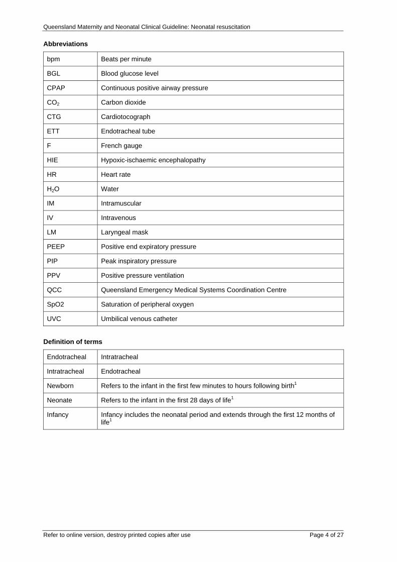
Abbreviations
bpm Beats per minute
BGL Blood glucose level
CPAP Continuous positive airway pressure
CO2 Carbon dioxide
CTG Cardiotocograph
ETT Endotracheal tube
F French gauge
HIE Hypoxic-ischaemic encephalopathy
HR Heart rate
H2O Water
IM Intramuscular
IV Intravenous
LM Laryngeal mask
PEEP Positive end expiratory pressure
PIP Peak inspiratory pressure
PPV Positive pressure ventilation
QCC Queensland Emergency Medical Systems Coordination Centre
SpO2 Saturation of peripheral oxygen
UVC Umbilical venous catheter
Definition of terms
Endotracheal Intratracheal
Intratracheal Endotracheal
Newborn Refers to the infant in the first few minutes to hours following birth1
Neonate Refers to the infant in the first 28 days of life1
Infancy Infancy includes the neonatal period and extends through the first 12 months of life1
Queensland Maternity and Neonatal Clinical Guideline: Neonatal resuscitation
Refer to online version, destroy printed copies after use Page 5 of 27
Table of Contents
1 Introduction.....................................................................................................................................6 1.1 Clinical standards ..................................................................................................................6
2 Anticipating the need for resuscitation ...........................................................................................7 2.1 Communication and information sharing ...............................................................................8 2.2 Equipment..............................................................................................................................8
2.2.1 Equipment checks .............................................................................................................8 3 Assessment of the newborn at birth ...............................................................................................9 4 Resuscitation management..........................................................................................................10
4.1 Positive pressure ventilation ................................................................................................10 4.2 Positive pressure ventilation delivery devices .....................................................................11 4.3 Supplemental oxygen administration...................................................................................12 4.4 Endotracheal intubation .......................................................................................................13
4.4.1 Intubation under specific circumstances..........................................................................14 4.5 Chest compressions ............................................................................................................15 4.6 Drugs and fluids...................................................................................................................16
4.6.1 Administration routes .......................................................................................................16 4.6.2 Adrenaline........................................................................................................................17 4.6.3 Volume expanding fluids..................................................................................................17
5 Care of the neonate after resuscitation ........................................................................................18 6 Ethical issues................................................................................................................................19 References ..........................................................................................................................................20 Appendix A: Sample form - Neonatal resuscitation record..................................................................21 Appendix B: Equipment required for neonatal resuscitation ...............................................................23 Appendix C: Endotracheal tube length and size and Adrenaline dosage by corrected gestation.......24 Appendix D: Laryngeal mask...............................................................................................................25 Appendix E: Use of a narcotic antagonist............................................................................................26 Acknowledgements..............................................................................................................................27 List of Tables
Table 1. Clinical standards recommended for facilities providing planned birthing services ................ 6 Table 2. Risk factors for neonatal resuscitation .................................................................................... 7 Table 3. Communication and information.............................................................................................. 8 Table 4. Assessment of the newborn at birth ........................................................................................ 9 Table 5. Positive pressure ventilation.................................................................................................. 10 Table 6. Positive pressure ventilation delivery devices ....................................................................... 11 Table 7. Supplemental oxygen ............................................................................................................ 12 Table 8. Target oxygen saturation....................................................................................................... 12 Table 9. Endotracheal intubation......................................................................................................... 13 Table 10. Intubation under specific circumstances ............................................................................. 14 Table 11. Combined chest compression and positive pressure ventilation ........................................ 15 Table 12. Administration routes........................................................................................................... 16 Table 13. Adrenaline administration .................................................................................................... 17 Table 14. Fluid administration ............................................................................................................. 17 Table 15. Continuing care of the neonate after resuscitation.............................................................. 18 Table 16. Ethical issues....................................................................................................................... 19

Queensland Maternity and Neonatal Clinical Guideline: Neonatal resuscitation
Refer to online version, destroy printed copies after use Page 6 of 27
1 Introduction Although the need for neonatal resuscitation can often be anticipated, on many occasions it is unexpected1:
• Most newborns are vigorous at birth1: o Approximately 10% will require some assistance at birth to begin breathing o Less than 1% will require extensive resuscitation
The guideline focus is on the neonatal period and particularly newborns. Paediatric resuscitation techniques may be used in term neonates after the newborn period and specifically in the situation of cardiac aetiology underlying their arrest.
1.1 Clinical standards Clinical standards recommended for maternity facilities providing planned birthing services are outlined in Table 1.
Table 1. Clinical standards recommended for facilities providing planned birthing services
Aspect Consideration
Resources1
• Required at all times: o A suitable place to resuscitate a newborn o Suitable neonatal resuscitation equipment o Clinicians trained in neonatal resuscitation
• Organised programs to develop and maintain the standards, skills and teamwork required for newborn resuscitation: o Training requires regular reinforcement in clinical practice and/or
refresher courses which should be undertaken at least annually • Debriefing services for clinicians:
o Regardless of seniority, resuscitations can be stressful o Reflection on practice provides a valuable learning opportunity
Clinical skill requirements1
• Clinicians trained in: o Basic neonatal resuscitation:
Airway support, ventilation via face mask and chest compressions o Advanced neonatal resuscitation:
All the skills of basic neonatal resuscitation Endotracheal intubation Vascular cannulation The use of drugs and fluids
• Clinicians responsible for neonatal resuscitation should be familiar with available neonatal resuscitation equipment
Clinician attendance at births1
• Low risk births: o A clinician trained in basic neonatal resuscitation should be in
attendance and responsible only for the care of the newborn o The Australian Resuscitation Council recommends a clinician trained in
advanced neonatal resuscitation should also be available • High risk births:
o A clinician trained in advanced neonatal resuscitation should be in attendance and responsible only for the care of the newborn
o More than one experienced person should be present to care for the newborn
• The Queensland Clinical Services Capability Framework describes the first principle as applying to all services and the second principle as applying to Level 3 services and above.2 Advanced neonatal resuscitation may not be possible in all Level 1 and 2 services
Documentation1
• Comprehensive and contemporaneous • When possible, one person should be appointed to document, the time,
interventions and newborn’s response during resuscitation • Use of a standardised neonatal resuscitation record is recommended [refer to
Appendix A: Sample form - Neonatal resuscitation record]

Queensland Maternity and Neonatal Clinical Guideline: Neonatal resuscitation
Refer to online version, destroy printed copies after use Page 7 of 27
2 Anticipating the need for resuscitation A variety of maternal, fetal and intrapartum circumstances can increase the risk of the newborn needing resuscitation at birth1 [refer to Table 2]
Table 2. Risk factors for neonatal resuscitation
Risk factors
Maternal1
• Prolonged rupture of membranes (greater than 18 hours) • Bleeding in second or third trimester • Pregnancy induced hypertension • Chronic hypertension • Substance abuse • Drug therapy (e.g. lithium, magnesium, adrenergic blocking agents,
narcotics) • Diabetes mellitus • Chronic illness (e.g. anaemia, cyanotic congenital heart disease) • Maternal pyrexia • Maternal infection • Chorioamnionitis • Heavy sedation • Previous fetal or neonatal death • No prenatal care
Fetal1
• Multiple gestation (e.g. twins, triplets) • Preterm gestation (especially less than 35 weeks) • Post term gestation (greater than 41 weeks) • Large for dates • Fetal growth restriction • Alloimmune haemolytic disease (e.g. anti-D, anti-Kell, especially if fetal
anaemia or hydrops fetalis present) • Polyhydramnios and oligohydramnios • Reduced fetal movement before onset of labour • Congenital abnormalities which may effect breathing, cardiovascular
function or other aspects of perinatal transition • Intrauterine infection • Hydrops fetalis
Intrapartum1
• Non reassuring fetal heart rate patterns on cardiotocograph (CTG) • Abnormal presentation • Prolapsed cord • Prolonged labour (or prolonged second stage of labour) • Precipitate labour • Antepartum haemorrhage (e.g. abruption, placenta praevia, vasa praevia) • Meconium in the amniotic fluid • Narcotic administration to mother within 4 hours of birth • Forceps birth • Vacuum-assisted (ventouse) birth • Maternal general anaesthesia

Queensland Maternity and Neonatal Clinical Guideline: Neonatal resuscitation
Refer to online version, destroy printed copies after use Page 8 of 27
2.1 Communication and information sharing Preparation for a high risk birth requires effective communication and information sharing and a consistent and coordinated approach between obstetric and neonatal clinicians [refer to Table 3].
Table 3. Communication and information
Aspect Consideration
Obstetric and neonatal clinicians
• Maternal information should include details of1: o Pre-existing or pregnancy related medical condition and treatment that
may affect the resuscitation or management of the newborn o Antenatal ultrasound diagnoses that may affect immediate postnatal
management o Assessments of fetal wellbeing (e.g. fetal heart rate monitoring)
• Minimum fetal information required by personnel responsible for the baby includes1: o Gestational age o Number of expected newborns, if multiple birth o Reason this is a high risk birth o Presence of meconium in the liquor o Assessments of fetal heart rate variability o Any known congenital abnormalities o Maternal risk factors for infections (including results of screening if known,
e.g. Group B Streptococcus) o Maternal medication
Parent(s)
• Whenever time permits, the neonatal team should introduce themselves to the parent(s) before the birth and should1: o Outline the proposed plan for the newborn’s care o Enquire and respond to parent’s questions
• In cases of potentially lethal fetal malformations or extreme prematurity, whenever possible, parents should be included in decisions about the extent of the resuscitation1
Documentation • Contemporaneous documentation of discussions and care plans
2.2 Equipment • A complete set of resuscitation equipment and drugs should always be readily available in
the areas of hospitals where newborns are born or receive neonatal care1 [refer to Appendix B: Equipment required for neonatal resuscitation]
2.2.1 Equipment checks • Facilities should maintain a clear record documenting the checking procedure for each set
of resuscitation equipment and drugs1 • Each set of resuscitation equipment and drugs should be checked:
o Before any resuscitation1 and o According to local hospital policy.1 Consider frequency of checking:
At least daily, preferably once per shift Following resuscitation To enable clinical staff familiarisation of layout in the emergency
situation

Queensland Maternity and Neonatal Clinical Guideline: Neonatal resuscitation
Refer to online version, destroy printed copies after use Page 9 of 27
3 Assessment of the newborn at birth Assessing the need to initiate and continue resuscitation should begin immediately after birth and proceed throughout the resuscitation.1 Evaluation and intervention (if required) are simultaneous processes, especially when more than one resuscitator is present.1 For clarity this process is described as a sequence of distinct steps1 [refer to Flowchart: Newborn life support]. Table 4 outlines important aspects of assessment at birth.1
Table 4. Assessment of the newborn at birth
Aspect Consideration
Cord clamping
• In the compromised newborn, the optimal timing of cord clamping remains unknown1
• The more severely compromised the newborn the more likely it is that resuscitation measures need to take priority over delayed cord clamping
Initial assessment • Tone • Breathing • Heart rate
Subsequent assessment
• Heart rate: o A prompt increase in heart rate remains the most sensitive indicator of
resuscitation efficacy1 • Breathing • Tone • Oxygenation:
o Preferably assessed using pulse oximetry
Temperature
• Hypothermia may increase oxygen consumption impeding effective resuscitation1
• Ensure area is warm and draft free1 • For term and near term newborn – dry newborn and remove wet linen1 • For very premature newborns:
o Increase ambient temperature to 260C1 o Placing the baby in a polyethylene bag (without drying) or under a
polyethylene sheet (food or medical grade, heat resistant) up to the neck, (immediately after birth until temperature is stable)1
Handling and skin protection
• All newborns should be handled gently (premature newborns are especially at risk of damage to skin and internal organs)1
• If emergency vascular access is required, antiseptic solution, particularly alcohol containing, should be applied sparingly1: o For umbilical catheterisation:
Apply only to the cord and less than 2 cm diameter of surrounding skin
Avoid pooling around the newborn’s groin and flanks
Pulse oximetry
• Oximetry is recommended when1: o The need for resuscitation is anticipated o Positive pressure ventilation (PPV) is administered for more than a
few breaths o Whenever persistent cyanosis is suspected during resuscitation or
any time after birth o When supplemental oxygen is administered
• The sensor should be placed on the newborn’s right hand or wrist and then connect the probe to the instrument.1 o This results in the quickest, accurate monitor display3
Supplemental oxygen
• Refer to Table 7

Queensland Maternity and Neonatal Clinical Guideline: Neonatal resuscitation
Refer to online version, destroy printed copies after use Page 10 of 27
4 Resuscitation management • Effective ventilation is the key to successful neonatal resuscitation1 • Drying and stimulation are both assessment and resuscitative interventions.1 If the
newborn fails to establish spontaneous effective respirations and heart rate does not increase to more than 100 beats per minute (bpm) then commence PPV1
• Effective teamwork is essential to the success of neonatal resuscitation, particularly advanced resuscitation
• Newborn resuscitation training should include the necessary individual and teamwork skills which are reinforced through regular practice1
4.1 Positive pressure ventilation • PPV may be delivered via a face mask, endotracheal tube (ETT) or laryngeal mask1 (LM)
[refer to Table 5 for important aspects of PPV1; Table 6 refers to devices for PPV1]
Table 5. Positive pressure ventilation
Aspect Consideration
Indications
• After stimulation commence PPV if: o Newborn’s heart rate is less than 100 bpm and o The newborn remains apnoeic or o Breathing is inadequate – gasping is not adequate breathing
Technique
• Ensuring the jaw is supported, keep the neck slightly extended in the ‘sniffing position’
• Two people may provide mask ventilation more effectively than one: o One person supports the jaw holding the mask in place with two hands o The other person provides positive pressure breaths
Rate • 40-60 breaths per minute
Pressures • For most newborns, ventilation can be accomplished with progressively lower pressures and rates as resuscitation proceeds
Mouth to mouth and nose
• Should be used when neonatal inflation devices are not available • Maternal blood and other body fluids should be wiped from the newborn’s face
to reduce the risk of infection to the resuscitator
Assessment of effectiveness
• Ventilation effectiveness is confirmed by observing: o Heart rate increasing to greater than 100 bpm o Slight rise of chest and upper abdomen with each inflation o Improving oxygenation (preferably assessed using pulse oximetry)
• Reassess technique if the: o Chest and abdomen do not rise with each inflation o Heart rate does not rise above 100 bpm o Oxygenation does not improve
• Consider tracheal intubation or use of laryngeal mask if face mask ventilation remains ineffective despite corrective intervention
PEEP during resuscitation
• If suitable equipment is available, positive end expiratory pressure (PEEP) (at least 5 cm H2O) should be used during resuscitation to: o Assist lung expansion o Help establish a functional residual capacity o Improve oxygenation
Continuous positive airway pressure (CPAP)1
• A trial of CPAP is reasonable for: o Spontaneously breathing newborns who have laboured breathing /
respiratory distress o Newborns who are breathing but whose saturations are not meeting
targets

Queensland Maternity and Neonatal Clinical Guideline: Neonatal resuscitation
Refer to online version, destroy printed copies after use Page 11 of 27
4.2 Positive pressure ventilation delivery devices
Table 6. Positive pressure ventilation delivery devices
Aspect Consideration
Acceptable delivery devices1
• T-piece device is preferred. Recommended initial pressures: o Peak inspiratory pressure (PIP):
Term newborn 30 cm H2O (water) Preterm newborn 20 – 25 cm H2O
o PEEP: 5 – 8 cm H2O o Maximum pressure relief valve: 50 cm H2O*
• Self inflating bag o Pressure release valve factory set at approximately 40 cm H2O
• Flow inflating (or anaesthetic) bag • Face masks should be:
o The appropriate size o Cushioned o Self-inflating bags cannot effectively deliver CPAP, PEEP or sustained
inflation breaths
T-piece device*
• Although a maximum pressure relief valve set at 50 cm H2O is recommended by the Australian Resuscitation Council1, a lower maximum pressure relief limit for e.g. 35 cm H2O will be adequate for the majority of babies receiving resuscitation.
• Within each hospital, neonatal T-piece devices should be set up using standard flow and pressure settings that are appropriate for newborns and well known to all clinicians
• All clinicians should be trained to safely adjust pressure settings during resuscitation if necessary.
• Rarely babies will need high peak inspiratory pressures of greater than or equal to 50 cm H2O at the commencement of ventilation

Queensland Maternity and Neonatal Clinical Guideline: Neonatal resuscitation
Refer to online version, destroy printed copies after use Page 12 of 27
4.3 Supplemental oxygen administration Supplemental oxygen administration should be provided only when oxygen saturation has failed to improve with effective ventilation of the lungs. Points for consideration are provided in Table 7.
Table 7. Supplemental oxygen
Aspect Consideration
Supplemental oxygen administration
• Regardless of gestation, the goal of oxygen administration is an oxygen saturation resembling that of a healthy term newborn1: o Assess the newborn’s response to supplemental oxygen o Reduce or cease supplemental oxygen if saturations are above 90%
at any time in the first 10 minutes, although it is acceptable and normal for some newborn saturations to rise above 90% in room air
o Table 8 below includes target saturations for newborns during resuscitation:
Table 8. Target oxygen saturation
Time from birth Target oxygen saturations
1 minute 60 – 70%
2 minutes 65 – 85%
3 minutes 70 – 90%
4 minutes 75 – 90%
5 minutes 80 – 90%
10 minutes 85 – 90%
• The priority is to ensure adequate lung inflation - only increase inspired oxygen concentration if PPV or CPAP have failed to achieve the target peripheral oxygen saturation (SpO2)1
• Term and near term newborns1: o Use air initially and only administer oxygen to newborns whose
saturations do not meet the lower target range despite respiratory support
o Reduce oxygen concentration if saturations reach 90% while supplemental oxygen is used
• Preterm newborns (less than 32 weeks)1: o Initially use room air or blended air and oxygen (at 30-50%) o Guided by pulse oximetry, adjust oxygen concentration according to
response o If blended air and oxygen are unavailable, initiate resuscitation with air

Queensland Maternity and Neonatal Clinical Guideline: Neonatal resuscitation
Refer to online version, destroy printed copies after use Page 13 of 27
4.4 Endotracheal intubation A decision to perform endotracheal intubation will depend on the newborn’s gestation, degree of respiratory depression, response to face mask ventilation, and the skill and experience of the resuscitator.1 [refer to Table 9]
Table 9. Endotracheal intubation
Aspect Consideration
Clinical standard1
• Neonatal endotracheal intubation should only be attempted by clinicians with appropriate training and experience in the procedure
• If there is no-one skilled at intubation present, continue PPV via a face mask or LM [refer to Table 10]
Indications1
• Tracheal suctioning in a non vigorous newborn exposed to meconium stained liquor4
• Unsuccessful ventilation via a face mask (e.g. heart rate remains low, oxygen saturation falling or failing to rise or prolonged)
• Special circumstances (e.g. diaphragmatic hernia, extremely low birth weight)
• Newborns born without a detectable heart rate • Administration of endotracheal medications (e.g. Adrenaline or artificial
surfactant) • Expected need for continued or prolonged ventilation
ETT size and insertion length1
• ETT size and insertion length are based on the newborn’s weight and corrected gestation [refer to Appendix C]
Position verification1
• ETT insertion depth must always be checked by comparing the markings on the tube with the formula or table [refer to Appendix C]
• Primarily correct ETT position is confirmed by effective ventilation demonstrated by: o Chest movement with each inflation o Heart rate increasing above 100 bpm o Improving oxygenation (oximetry is more accurate than visual
assessment) • Other signs to verify correct ETT placement include:
o Visual inspection of the ETT passing through the larynx o Misting in the ETT during expiration (usually visible for only the first
few exhaled breaths) o Colour change visible with an end-tidal carbon dioxide (CO2) detector
(the most reliable method in newborns who have spontaneous circulation):
False negatives may occur in newborns if there is very low or absent pulmonary blood flow
False positives may occur in devices contaminated by adrenaline or surfactant
o Hearing symmetrical breath sounds using a stethoscope placed on the upper chest. May be asymmetrical, despite optimal tube position, in some circumstances (e.g. pneumothorax, diaphragmatic hernia)
• A chest X-ray should be obtained to confirm ETT position once the newborn is stabilised4
Signs that ETT is not in the trachea
• No chest movement with inflations1 • A heart rate less than 100 bpm that does not increase soon after
intubation and inflation has started1 • No detection of expired CO2
1 • No improvement in oxygenation1 or sudden deterioration in oxygenation • The absence of breath sounds in the axillae1
Queensland Maternity and Neonatal Clinical Guideline: Neonatal resuscitation
Refer to online version, destroy printed copies after use Page 14 of 27
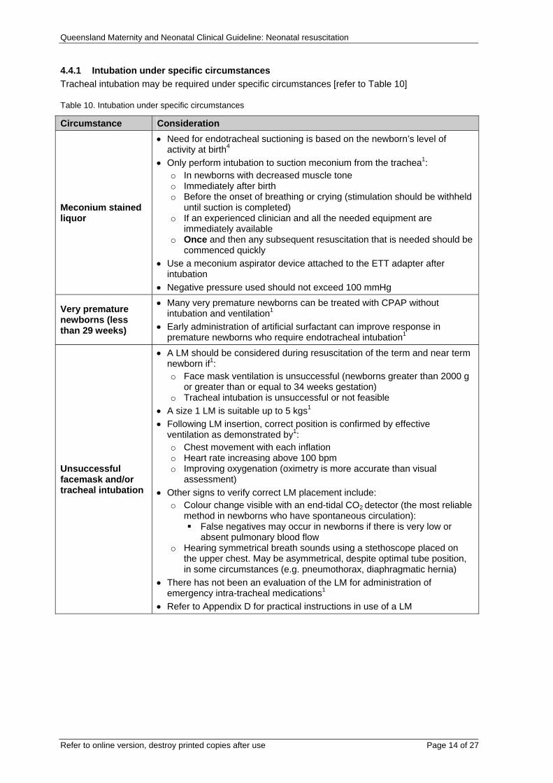
4.4.1 Intubation under specific circumstances Tracheal intubation may be required under specific circumstances [refer to Table 10]
Table 10. Intubation under specific circumstances
Circumstance Consideration
Meconium stained liquor
• Need for endotracheal suctioning is based on the newborn’s level of activity at birth4
• Only perform intubation to suction meconium from the trachea1: o In newborns with decreased muscle tone o Immediately after birth o Before the onset of breathing or crying (stimulation should be withheld
until suction is completed) o If an experienced clinician and all the needed equipment are
immediately available o Once and then any subsequent resuscitation that is needed should be
commenced quickly • Use a meconium aspirator device attached to the ETT adapter after
intubation • Negative pressure used should not exceed 100 mmHg
Very premature newborns (less than 29 weeks)
• Many very premature newborns can be treated with CPAP without intubation and ventilation1
• Early administration of artificial surfactant can improve response in premature newborns who require endotracheal intubation1
Unsuccessful facemask and/or tracheal intubation
• A LM should be considered during resuscitation of the term and near term newborn if1: o Face mask ventilation is unsuccessful (newborns greater than 2000 g
or greater than or equal to 34 weeks gestation) o Tracheal intubation is unsuccessful or not feasible
• A size 1 LM is suitable up to 5 kgs1 • Following LM insertion, correct position is confirmed by effective
ventilation as demonstrated by1: o Chest movement with each inflation o Heart rate increasing above 100 bpm o Improving oxygenation (oximetry is more accurate than visual
assessment) • Other signs to verify correct LM placement include:
o Colour change visible with an end-tidal CO2 detector (the most reliable method in newborns who have spontaneous circulation):
False negatives may occur in newborns if there is very low or absent pulmonary blood flow
o Hearing symmetrical breath sounds using a stethoscope placed on the upper chest. May be asymmetrical, despite optimal tube position, in some circumstances (e.g. pneumothorax, diaphragmatic hernia)
• There has not been an evaluation of the LM for administration of emergency intra-tracheal medications1
• Refer to Appendix D for practical instructions in use of a LM

Queensland Maternity and Neonatal Clinical Guideline: Neonatal resuscitation
Refer to online version, destroy printed copies after use Page 15 of 27
4.5 Chest compressions When adequate airway and breathing support are provided, cardiac compressions are rarely indicated for resuscitation of neonates. In neonates, cardiac output is heart rate dependent. If the heart rate is too slow, circulation will be inadequate to support tissue oxygenation.1 [refer to Table 11]
Table 11. Combined chest compression and positive pressure ventilation
Aspect Consideration Indication • Heart rate remains less than 60 bpm despite 30 seconds of effective PPV4 Supplemental oxygen
• Administer 100% oxygen
Technique
• Perform a chest compression each half second. In a half second pause after each 3rd compression, deliver a breath. This results in a 3:1 ratio with a total of 90 compressions and 30 breaths in each minute1
• Coordinate compressions and inflations to avoid simultaneous delivery of a compression and a breath
• Methods: o 2 thumb technique:
Strongly recommended when two clinicians are available. (Usually the resuscitator performing chest compressions faces the newborn’s head. The position can be reversed if access to the abdomen is required) or
o 2 finger technique – acceptable as an interim measure or where access is required to the lower chest and abdomen
• Ensure full chest expansion between compressions. However, the administrator’s hands should not leave the chest
• Effectively delivered chest compressions will be evident on a pulse oximeter
Duration
• Continue uninterrupted compressions for at least 30 seconds between each pause to assess improvement in spontaneous heart rate and cardiac output
• Continue chest compressions with as little interruption as possible until it is obvious that the heart rate is greater than 60 bpm
Assessment
• Improvement is evidenced by: o Audible heart rate on auscultation o Spontaneous pulsations on oximetry
Note: Chest compressions will also cause pulsations on the pulse oximeter
o Rise in oxygen saturation o Spontaneous movement or breaths

Queensland Maternity and Neonatal Clinical Guideline: Neonatal resuscitation
Refer to online version, destroy printed copies after use Page 16 of 27
4.6 Drugs and fluids Drugs and fluids are rarely indicated for resuscitation of newborns1:
• Bradycardia is usually caused by hypoxia and inadequate ventilation and apnoea by insufficient oxygenation of the brainstem. Establishing adequate ventilation is the most important step to improve heart rate, however if heart rate remains less than 60 bpm despite adequate ventilation, Adrenaline may be needed1
• Administration of drugs should not detract from the efficiency and continuity of ventilation and chest compressions
• For administration of Adrenaline and volume expanding fluids, refer to Table 13 and Table 14 below
• Refer to Appendix E for the rarely indicated use of a narcotic antagonist
4.6.1 Administration routes Preferred vascular access is via the umbilical vein.1,4 Alternative access is via an endotracheal tube, peripheral vein, or intraosseous lines. [refer to Table 12]
Table 12. Administration routes
Route Considerations1
Umbilical vein
• The umbilical vein is the most rapidly accessible intravascular route for Adrenaline and an umbilical venous catheter (UVC) can also be used for fluid administration
• A 3-way tap should be attached to the UVC and both primed with normal saline before use
• A UVC can provide continued vascular access until an alternative route is established
• Blood gases from the UVC may be useful in guiding treatment
Endotracheal tube
• Give only Adrenaline via the ETT: o ETT administration of Adrenaline is acceptable when the heart rate is
less than 60 bpm despite adequate ventilation and chest compressions and when there is no intravenous (IV) access1
o Administration of ETT Adrenaline should not delay attempts to obtain vascular access for administration of IV Adrenaline
Peripheral vein • Access can be difficult • Access may take too long
Intraosseous lines
• Not commonly used in newborns • Can be used if umbilical or venous access is not available • Consider if the resuscitator has greater experience with inserting
intraosseous lines
Queensland Maternity and Neonatal Clinical Guideline: Neonatal resuscitation
Refer to online version, destroy printed copies after use Page 17 of 27
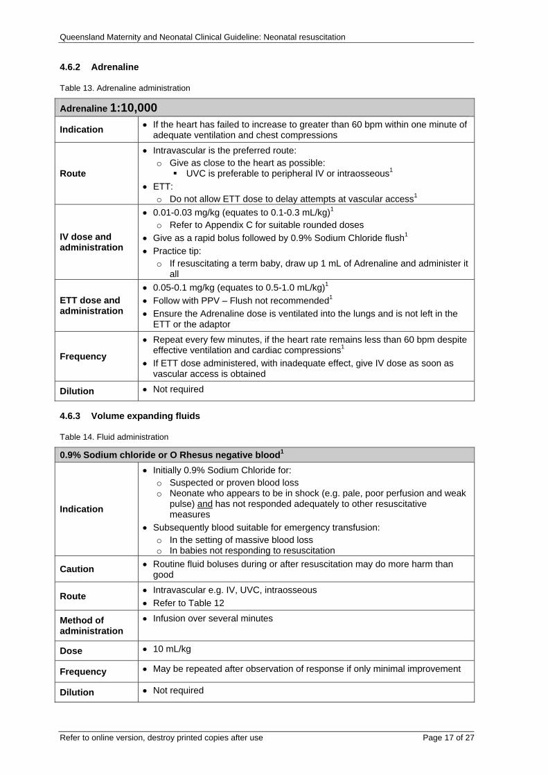
4.6.2 Adrenaline
Table 13. Adrenaline administration
Adrenaline 1:10,000
Indication • If the heart has failed to increase to greater than 60 bpm within one minute of adequate ventilation and chest compressions
Route
• Intravascular is the preferred route: o Give as close to the heart as possible:
UVC is preferable to peripheral IV or intraosseous1 • ETT:
o Do not allow ETT dose to delay attempts at vascular access1
IV dose and administration
• 0.01-0.03 mg/kg (equates to 0.1-0.3 mL/kg)1 o Refer to Appendix C for suitable rounded doses
• Give as a rapid bolus followed by 0.9% Sodium Chloride flush1 • Practice tip:
o If resuscitating a term baby, draw up 1 mL of Adrenaline and administer it all
ETT dose and administration
• 0.05-0.1 mg/kg (equates to 0.5-1.0 mL/kg)1 • Follow with PPV – Flush not recommended1 • Ensure the Adrenaline dose is ventilated into the lungs and is not left in the
ETT or the adaptor
Frequency
• Repeat every few minutes, if the heart rate remains less than 60 bpm despite effective ventilation and cardiac compressions1
• If ETT dose administered, with inadequate effect, give IV dose as soon as vascular access is obtained
Dilution • Not required
4.6.3 Volume expanding fluids
Table 14. Fluid administration
0.9% Sodium chloride or O Rhesus negative blood1
Indication
• Initially 0.9% Sodium Chloride for: o Suspected or proven blood loss o Neonate who appears to be in shock (e.g. pale, poor perfusion and weak
pulse) and has not responded adequately to other resuscitative measures
• Subsequently blood suitable for emergency transfusion: o In the setting of massive blood loss o In babies not responding to resuscitation
Caution • Routine fluid boluses during or after resuscitation may do more harm than good
Route • Intravascular e.g. IV, UVC, intraosseous • Refer to Table 12
Method of administration
• Infusion over several minutes
Dose • 10 mL/kg
Frequency • May be repeated after observation of response if only minimal improvement
Dilution • Not required

Queensland Maternity and Neonatal Clinical Guideline: Neonatal resuscitation
Refer to online version, destroy printed copies after use Page 18 of 27
5 Care of the neonate after resuscitation The neonate who has required resuscitation remains at risk and requires ongoing assessment.1 [refer to Table 151 and Guideline: Neonatal stabilisation5
Table 15. Continuing care of the neonate after resuscitation
Care Comments Location • Assess the need for admission to an intensive or special care nursery
Monitoring
• Monitoring required may include: o Oxygen saturation o Heart rate o Respiratory rate and pattern o Blood glucose measurement o Blood gas analysis o Fluid balance and nutrition o Blood pressure o Temperature o Neurological
Blood glucose management
• Neonates who require resuscitation are susceptible to hypoglycaemia • Check the blood glucose level (BGL) as soon as possible after
resuscitation • Maintain the BGL greater than 2.5 mmol/L • Avoid large bolus doses of glucose (greater than 100 – 200 mg/kg)
o 1 mL 10% Glucose contains 100 mg Glucose • Refer to Guideline: Neonatal hypoglycaemia and blood glucose level
monitoring6
Antibiotics
• The need for resuscitation can be a consequence of sepsis. Soon after resuscitation consider: o Relevant investigations o The need for antibiotics
Induced hypothermia for hypoxic-ischaemic encephalopathy
• Discuss any neonate with hypoxic-ischaemic encephalopathy (HIE) who is considered for induced hypothermia early with a Neonatologist
• Plans should be made for transfer (if required) and admission to a neonatal intensive care unit
• Refer to Guideline: Hypoxic-ischaemic encephalopathy6
Inter-hospital transfer
• If the birth facility is unable to provide an appropriate level of post resuscitation monitoring and support, transfer the neonate to an appropriate higher level facility
• To discuss the decision to transfer and to arrange transfer of the neonate to a higher level facility, contact Queensland Emergency Medical Systems Coordination Centre (QCC) by phoning 1300 799 127
Continuing care of the family
• Parent(s) find witnessing the resuscitation of their baby distressing regardless of outcome. Whenever possible: o Prepare parents for resuscitation if it is anticipated o Keep parent(s) informed during and after resuscitation. Ideally
information should be provided by a senior clinician o Facilitate early parental contact with their baby

Queensland Maternity and Neonatal Clinical Guideline: Neonatal resuscitation
Refer to online version, destroy printed copies after use Page 19 of 27
6 Ethical issues The birth of extremely premature neonates and those with severe congenital anomalies raise questions with parent(s) and among clinicians about initiation of resuscitation.1 [refer to Table 161].
Table 16. Ethical issues
Issue Consideration
Initiating resuscitation
• Where unexpected anomalies are present offer full resuscitation, ongoing care can be discussed with parent(s) after assessment of the neonate's condition.4 Examples of exceptions include neonates with4: o Anencephaly o Extreme prematurity for whom there is very little possibility of intact
survival
Discontinuing resuscitation
• Consider discontinuing resuscitation if the heart rate is undetectable and remains so for 10 minutes in a newly born baby1
• The decision to continue resuscitation beyond 10 minutes when there is no or very low heart rate may be influenced by1: o Presumed diagnosis o Gestation of neonate o Presence or absence of complications o Parent(s) previously expressed views regarding acceptable risk of
morbidity • When withdrawing or withholding resuscitation, care should be focused on
the neonate’s comfort and dignity1 • Autopsy may be appropriate after failed resuscitation.[refer to Guideline:
Stillbirth care7]

Queensland Maternity and Neonatal Clinical Guideline: Neonatal resuscitation
Refer to online version, destroy printed copies after use Page 20 of 27
References 1. Australian Resuscitation Council. Section 13 - Neonatal Guidelines. 2010 Dec [cited 2011 March 15]. Available from: http://www.resus.org.au/.
2. Queensland Health. Clinical Services Capability Framework for Public and Licensed Private Health Facilities v3.0. Brisbane: Queensland Government Department of Health. 2011.
3. O’Donnell CPF, Kamlin COF, Davis PG, Morley CJ. Obtaining pulse oximetry data in neonates: a randomised crossover study of sensor application techniques. Archives of disease in childhood. Fetal and neonatal edition. 2005; 90:F84-5.
4. Kattwinkel J, Perlman JM, Aziz K, Colby C, Fairchild K, Gallagher J, et al. Part 15: neonatal resuscitation: 2010 American Heart Association Guidelines for Cardiopulmonary Resuscitation and Emergency Cardiovascular Care. Circulation. 2010; 122(suppl3):S909-19.
5. Queensland Maternity and Neonatal Clinical Guidelines Program. Neonatal stabilisation. Guideline No. MN11.12-V1-R16. Queensland Health. 2011.
6. Queensland Maternity and Neonatal Clinical Guidelines Program. Neonatal hypoglycaemia and blood glucose level monitoring. Guideline No. MN10.8-V2-R12. Queensland Health. 2010.
7. Queensland Maternity and Neonatal Clinical Guidelines Program. Stillbirth care. Guideline No. MN11.24-V3-R16. Queensland Health. 2011.

Queensland Maternity and Neonatal Clinical Guideline: Neonatal resuscitation
Appendix A: Sample form - Neonatal resuscitation record
Refer to online version, destroy printed copies after use Page 21 of 27

Queensland Maternity and Neonatal Clinical Guideline: Neonatal resuscitation
Refer to online version, destroy printed copies after use Page 22 of 27

Queensland Maternity and Neonatal Clinical Guideline: Neonatal resuscitation
Refer to online version, destroy printed copies after use Page 23 of 27
Appendix B: Equipment required for neonatal resuscitation Equipment Comment
Standard kits1
• Prior preparation of standardised kits containing equipment for procedures (e.g. umbilical catheterisation) can save considerable time in emergencies
General1
• Firm padded resuscitation surface • Overhead warmer • Light for the area • Clock with timer in seconds • Warm towel or similar covering • Polyethylene bag or sheet, big enough for neonates less than 1500 g birth
weight • Stethoscope (neonatal preferred) • Pulse oximeter plus neonatal probe
Airway management1
• Suction apparatus and suction catheters (sizes 6F, 8F, and either 10F or 12F)
• Oropharyngeal airways (sizes 00 and 0) • Intubation equipment:
o Laryngoscopes with neonatal blades (sizes 00, 0, 1 ) o Spare batteries and bulbs o Compatible laryngoscope handles and blades o Endotracheal tubes (ETT) (uncuffed, no eye, sizes 2.5, 3.0, 3.5 and
4.0 mm internal diameter) o ETT introducer/stylet o Supplies for fixing ETT (e.g. sterile scissors, tape)
• End-tidal carbon dioxide (CO2) detector (to confirm intubation) • Meconium suction device (to apply suction directly to ETT) • Magill forceps (neonatal size - optional) • Laryngeal mask (size 1, suitable for neonates up to 5 kg)
Breathing support1
• Face masks (range of sizes suitable for premature and term neonates not Rendell Baker type)
• Positive pressure ventilation device either: o T-Piece resuscitation device (preferred), or o Flow-inflating bag with pressure safety valve and manometer, and o Self-inflating bag (240 mL) with a pressure release valve and a
removable oxygen reservoir • Medical gases:
o Source of medical oxygen (reticulated and/or cylinder allowing flow rate of up to 10 L/min) with flow meter and tubing
o Source of medical air plus air/oxygen blender • Feeding tubes for gastric decompression (sizes 6F, 8F)
Circulation support1
• Umbilical venous catheter (UVC) kit (including UVC sizes 3.5F and 5F) • Peripheral IV cannulation kit • Skin preparation solution suitable for neonate skin • Tapes/devices to secure UVC/IV cannula • Syringes and needles (assorted sizes) • Intraosseous needles (50 mm length)
Drugs and fluid1
• Adrenaline 1:10 000 concentration (0.1 mg/mL) • Volume expander:
o Normal saline (0.9% Sodium Chloride) o Blood suitable for emergency neonatal transfusion should be readily
available
Documentation1
• Resuscitation record sheet • Equipment check list

Queensland Maternity and Neonatal Clinical Guideline: Neonatal resuscitation
Appendix C: Endotracheal tube length and size and Adrenaline dosage by corrected gestation The following ETT references are sourced from the Australian Resuscitation Council, Section 13 - Neonatal Guidelines, 2010 [accessed 2011 March 15, http://www.resus.org.au/]. Endotracheal tube (ETT):
• Corrected gestation – gestation at birth plus postnatal age • ETT internal diameter in millimetres can be calculated as (gestational age in weeks divided
by 10): o 2.5 mm ETT for neonates less than 1 kg o 3.0 mm ETT for neonates 1 – 2 kg o 3.5 mm ETT for neonates 2 – 3 kg o 3.5 – 4.0 mm ETT for neonates greater than 3 kg
• Approximate ETT insertion depth from middle of upper lip can be calculated as (weight in kg + 6 cm)
Corrected gestation (weeks)
Actual weight
(kg)
ETT mark at upper lip
(cm)
ETT size - internal diameter
(mm)
ETT suction catheter size
(F)
23 – 24 0.5 – 0.6 5.5 2.5 5 or 6
25 – 26 0.7 – 0.8 6.0 2.5 5 or 6
27 – 29 0.9 – 1.0 6.5 2.5 5 or 6
30 – 32 1.1 – 1.4 7.0 3.0 6 or 8
33 – 34 1.5 – 1.8 7.5 3.0 6 or 8
35 – 37 1.9 – 2.4 8.0 3.5 8
38 – 40 2.5 – 3.1 8.5 3.5 8
41 – 43 3.2 – 4.2 9.0 3.5 – 4.0 8 or 10
Adrenaline:
Refer to online version, destroy printed copies after use Page 24 of 27
• Rounded IV Adrenaline doses are provided as a guide only
• The Australian Resuscitation Council, Section 13 - Neonatal Guidelines, 2010 [accessed 2011 September 5, http://www.resus.org.au/] recommends IV Adrenaline 0.01-0.03 mg/kg (equates to 0.1-0.3 mL/kg)
Corrected gestation (weeks)
IV Adrenaline 1:10,000
(mLs)
23 – 26 0.1
27 – 32 0.25
33 – 37 0.5
38 – 43 1
• Adrenaline can be repeated every 2-3 minutes if HR is less than 60 beats per minute
• If given via the less preferred ETT route, Adrenaline dosages should be three times higher
Queensland Maternity and Neonatal Clinical Guideline: Neonatal resuscitation
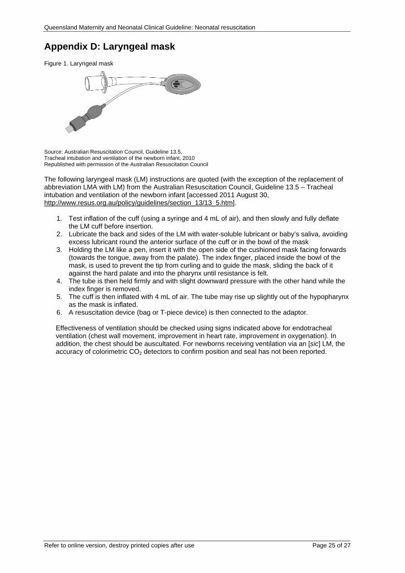
Appendix D: Laryngeal mask Figure 1. Laryngeal mask
Source: Australian Resuscitation Council, Guideline 13.5, Tracheal intubation and ventilation of the newborn infant, 2010 Republished with permission of the Australian Resuscitation Council The following laryngeal mask (LM) instructions are quoted (with the exception of the replacement of abbreviation LMA with LM) from the Australian Resuscitation Council, Guideline 13.5 – Tracheal intubation and ventilation of the newborn infant [accessed 2011 August 30, http://www.resus.org.au/policy/guidelines/section_13/13_5.htm].
1. Test inflation of the cuff (using a syringe and 4 mL of air), and then slowly and fully deflate the LM cuff before insertion.
2. Lubricate the back and sides of the LM with water-soluble lubricant or baby’s saliva, avoiding excess lubricant round the anterior surface of the cuff or in the bowl of the mask
3. Holding the LM like a pen, insert it with the open side of the cushioned mask facing forwards (towards the tongue, away from the palate). The index finger, placed inside the bowl of the mask, is used to prevent the tip from curling and to guide the mask, sliding the back of it against the hard palate and into the pharynx until resistance is felt.
4. The tube is then held firmly and with slight downward pressure with the other hand while the index finger is removed.
5. The cuff is then inflated with 4 mL of air. The tube may rise up slightly out of the hypopharynx as the mask is inflated.
6. A resuscitation device (bag or T-piece device) is then connected to the adaptor.
Effectiveness of ventilation should be checked using signs indicated above for endotracheal ventilation (chest wall movement, improvement in heart rate, improvement in oxygenation). In addition, the chest should be auscultated. For newborns receiving ventilation via an [sic] LM, the accuracy of colorimetric CO2 detectors to confirm position and seal has not been reported.
Refer to online version, destroy printed copies after use Page 25 of 27

Queensland Maternity and Neonatal Clinical Guideline: Neonatal resuscitation
Refer to online version, destroy printed copies after use Page 26 of 27
Appendix E: Use of a narcotic antagonist The following quotes are from the Neonatal Resuscitation Textbook 6th Edition 2011, American Heart Association/American Academy of Pediatrics, Lesson 7, page 247-248. “Narcotics given to the laboring mother to relieve pain may inhibit respiratory drive and activity in the newborn. In such cases, administration of naloxone (a narcotic antagonist) to the newborn will reverse the effects of narcotics on the baby.” Giving a narcotic antagonist is not the correct first therapy for a baby who is not breathing. The first corrective action is positive-pressure ventilation. The indications for giving naloxone to the baby require both of the following to be present:
• Continued respiratory depression after positive pressure ventilation has restored a normal heart rate and
• A history of maternal narcotic administration during labour “After naloxone administration, continue to administer positive pressure ventilation until the baby is breathing normally. The duration of action of the narcotic often exceeds naloxone. Therefore, observe the baby closely for recurrent respiratory depression, which may necessitate ongoing respiratory support. Caution: Do not give naloxone to the newborn of a mother who is suspected of being addicted to narcotics or is on methadone maintenance. This may result in the newborn having seizures.” Other drugs given to the mother, such as magnesium sulphate or non-narcotic analgesics or general anesthetics, also can depress respirations in the newborn; the effects of these drugs are not reversed by naloxone. If narcotics were not given to the mother or if naloxone does not result in restoring spontaneous respirations, transport the baby to the nursery for further evaluation and management while continuing to administer PPV and monitoring heart rate and pulse oximetry.”
Naloxone hydrochloride 0.4 mg/mL
Indication Persistent respiratory depression (after positive pressure ventilation has been provided) in a newborn whose mother has received an opiate analgesic in the 4 hours before birth
Route IV (including UVC) preferred Intramuscular (IM) acceptable but delayed onset of action
Method of administration
IV bolus followed by 0.9% Sodium Chloride flush
Dose 0.1 mg/kg = 0.25 mL/kg of 0.4 mg/mL stock solution
Frequency Duration of action of the narcotic can exceed that of naloxone, necessitating repeated doses of naloxone Recurrent respiratory depression may necessitate repeated doses of naloxone, or consideration of alternative diagnoses

Queensland Maternity and Neonatal Clinical Guideline: Neonatal resuscitation
Acknowledgements The Queensland Maternity and Neonatal Clinical Guidelines Program gratefully acknowledge the contribution of Queensland clinicians and other stakeholders who participated throughout the guideline development process particularly:
Working Party Clinical Lead
Dr Helen Liley, Neonatologist, Mater Health Services, Brisbane
Working Party Members
Mr Glen Alexander, Nurse Unit Manager, Logan Hospital
Dr Pita Birch, Neonatologist, Gold Coast Hospital
Dr David Cartwright, Neonatologist, Royal Brisbane and Women’s Hospital
Ms Liz Chappell, Neonatal Nurse, Gold Coast Hospital
Dr Mark Davies, Neonatologist, Royal Brisbane and Women’s Hospital
Dr Timothy Hong, Neonatologist, Gold Coast Hospital
Dr Luke Jardine, Neonatologist, Mater Health Services
Associate Professor Rebecca Kimble, Obstetrician, Royal Brisbane and Women’s Hospital
Ms Claudia Konig, Pharmacist, Royal Brisbane and Women’s Hospital
Ms Sam Lannan, Midwife, Nambour General Hospital
Mr Bruce Maybloom, Medical student, Bond University, Gold Coast
Ms Maryanne Payne, Neonatal Nurse, The Townsville Hospital
Dr Peter Schmidt, Neonatologist, Gold Coast Hospital
Ms Mary Tredinnick, Pharmacist, Royal Brisbane and Women’s Hospital
Program Team
Associate Professor Rebecca Kimble, Director, Queensland Maternity and Neonatal Clinical Guidelines Program
Ms Jacinta Lee, Program Manager, Queensland Maternity and Neonatal Clinical Guidelines Program
Ms Catherine van den Berg, Clinical Program Officer Queensland Maternity and Neonatal Clinical Guidelines Program
Ms Jackie Doolan, Clinical Program Officer, Queensland Maternity and Neonatal Clinical Guidelines Program
Ms Lyndel Gray, Clinical Program Officer, Queensland Maternity and Neonatal Clinical Guidelines Program
Steering Committee, Queensland Maternity and Neonatal Clinical Guidelines Program
Funding
This clinical guideline was funded by Queensland Health, Centre for Healthcare Improvement.
Refer to online version, destroy printed copies after use Page 27 of 27