Guide to Ophthalmic Equipment - Frank's Hospital … · ORBIS Guide to Ophthalmic Equipment ... The...
18
Guide to Ophthalmic Equipment For non-ophthalmic people Version 9.04.04
Transcript of Guide to Ophthalmic Equipment - Frank's Hospital … · ORBIS Guide to Ophthalmic Equipment ... The...

Guide to Ophthalmic Equipment
For non-ophthalmic people
Version 9.04.04

ORBIS Guide to Ophthalmic Equipment 2
Introduction Ophthalmologists and other eye care professionals use many devices to diagnose and treat eye problems. This guide introduces some of the more commonly used devices and is mainly written for persons who are not eye care professionals wishing to know what each device looks like and to understand how it is used. Part I will review some of the common devices used for examination and diagnosis. Part II will review devices used for the treatment of eye problems.
Versio
*Note: All prices mentioned in this guide are in US dollars and represent typical prices paid by US hospitals for equipment made in the US, Europe and Japan. Actual prices will depend on brand, model, discounts, desired features, purchaselocation, number of devices purchased, etc.
Inosuea mpsire Apa1w Apteabapc(ie
n
General Financial and Management Considerations for Owning Medical Equipment
addition to its original purchase price, medical equipment costs money to perate and to maintain during its life cycle. Installation of certain equipment ch as some lasers will involve initial additional costs for dedicated water and
lectrical supplies. Expensive consumables, which generally are not re-usable, re required for devices such as phaco machines and vitrectomy machines. Alledical devices, regardless of their complexity and ruggedness, require eriodic maintenance and corrective maintenance at some point. Even a mple device such as an ophthalmoscope requires ongoing costs for placement of bulbs and batteries, including rechargeable ones.
s a rule, equipment owners should budget anywhere from 5% to 10% of the urchase cost per year for each device for consumables, parts, maintenance, nd user training. The life cycle of a medical device can range between 5 and 5 years, depending on the ruggedness of the device and the environment in hich it is used.
ll eye care institutions should have a medical equipment management rogram to assure the maximum and most cost effective utilization of its chnology. This equipment management program may, depending on the
vailable resources and capacity of the institution, be handled by an in-house iomedical engineering department, by an outside service organization, or by n equipment maintenance service shared by several linked institutions. This rogram should include equipment inventory, preventive maintenance, orrective maintenance, emergency repair services, technology planning ncluding selection, procurement and retirement of equipment), training for quipment users and patient safety, among other functions.
06-29-04

ORBIS Guide to Ophthalmic Equipment 3
Version 06-29-04
General Considerations for Maintaining Ophthalmic Equipment Most ophthalmic diagnostic devices have optical components such as lenses, mirrors, and prisms. Many of these components have a special thin coating for filtering specific wavelengths of light, for reflecting light, or for reducing reflection. Great care must be exercised when removing dust and stains on optical components to avoid scratching or removing the surface coating. Dust and stainsbecome harder to clean when they accumulate and therefore periodic cleaningis recommended. However, excessive cleaning can lead to quick deterioration ofthe surface coating. Specific manufacturer instructions for frequency and methodof cleaning should be followed for each device. All ophthalmic equipment should be kept under dust covers when not in use. In regions with hot and humid climates, it is very common for fungus to grow on optical components such as lenses and mirrors. In its first stages, fungus would not be perceivable by the clinician. With time the fungus covers the lens surface in a web like manner. Initially there will be a very slight loss of image brightness, followed by decreased contrast due to light reflecting off the fungus. In its final stages, the fungus etches the outer coatings of the lens and image sharpness deteriorates. Removing fungus from lenses is extremely difficult and rarely yields good results. Ultraviolet radiation (sunlight or an ultraviolet lamp) or paraldehyde may be used to kill fungus. Once killed, the fungus may be easier to remove but the outer coatings of the lens will most likely have irreversible damage. Optics should be kept in a dry place with plenty of air circulation to prevent fungus growth. Air conditioners and dehumidifiers are very helpful in preventing fungus growth but if not available, the optics can be kept in a sealed container with packets of desiccant such as silica gel. Bulbs are common in most ophthalmic devices. When replacing bulbs, care should be taken to not touch them with bare fingers. Oils from the skin create hot spots on the bulb that can shorten the bulb’s life. Additionally, fingerprints can become etched into the bulb’s glass jacket and cause a shadow on the illumination field. Any maintenance that involves precise alignment of optics, or calibration of potentially dangerous forms of energy such as laser energy, should only be performed by manufacturer representatives or by qualified factory-trained personnel. The level of serviceability in the hospital for any device depends on theequipment design, the technology used, the level of support provided by the manufacturer, the available tools and test equipment, and the skills and training of the institution’s biomedical equipment personnel. All maintenance personnel must follow protective measures when testing and repairing lasers in order to reduce the possibility of exposure of the eye and skin to hazardous levels of laser radiation. One important measure is the use of proper laser safety glasses designed to filter the specific wavelengths and power of the laser being used. Lasers should not be used or tested in the presence of flammable anesthetics or other volatile substances or materials because of the serious risk of explosion or fire.
ORBIS Guide to Ophthalmic Equipment 4
Part I- Equipment for Examination, Diagnosis and Measurement
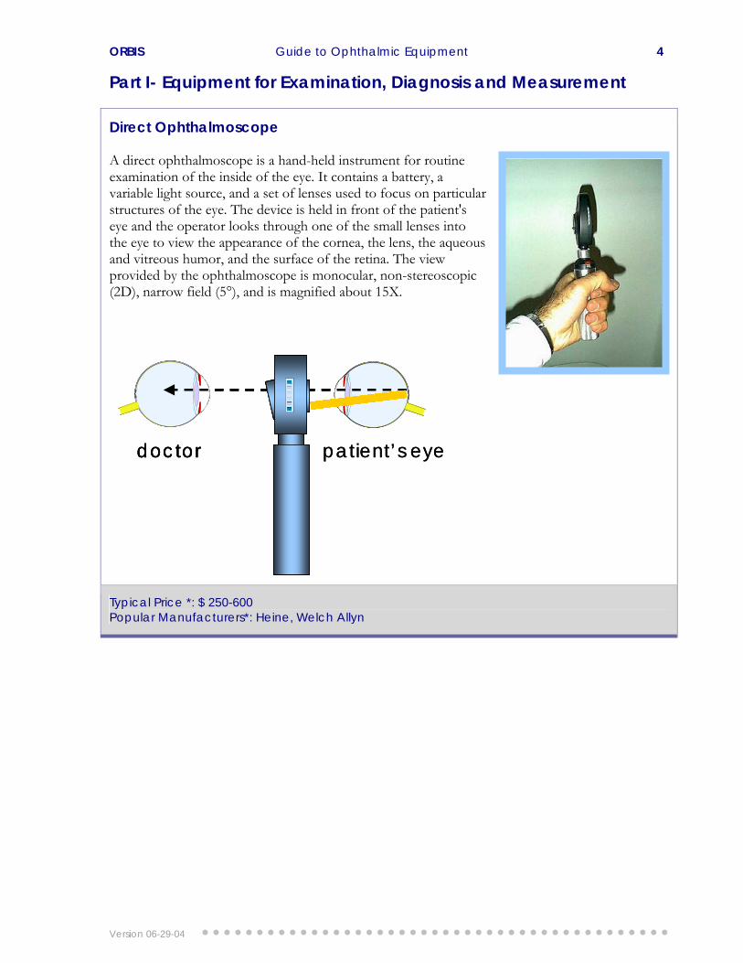
Direct Ophthalmoscope A direct ophthalmoscope is a hand-held instrument for routine examination of the inside of the eye. It contains a battery, a variable light source, and a set of lenses used to focus on pastructures of the eye. The device is held in front of the patient's eye and the operator looks through one of the small lenses into the eye to view the appearance of the cornea, the lens, the aqueoand vitreous humor, and the surface of the retina. The view provided by the ophthalmoscope is monocular, non-stereoscopic (2D), narrow field (5°), and is magnified
rticular
us
about 15X.
Typical Price *: $ 250-600 Popular Manufacturers*: Heine, Welch Allyn
doctor patient’s eyedoctordoctor patient’s eye
Version 06-29-04

ORBIS Guide to Ophthalmic Equipment 5
Indirect Ophthalmoscope A binocular indirect ophthalmoscope (BIO) is worn as a headset and is used in conjunction with a condensing aspheric lens held close to the patient’s eye. A BIO provides a much wider field of view (45°) than a direct ophthalmoscope and permits viewing of almost all the patient’s retina. The BIO is the viewing instrument of choice for retinal examinations. The view provided by the BIO is stereoscopic (3D), inverted, and illuminated with magnification of about 5X. Some BIOs have a buin video camera to permit eye care professionals in-training to view the examin
ilt-
ation on a screen.
Typical Price *: $1,000 to $2,000. $10,000 for video models Popular Manufacturers*: Heine, Keeler, Welch Allyn
~15mm
~60mm
hand-held lens
doctor
patient’s eye
light source
headset
~15mm
~60mm
hand-held lens
doctor
patient’s eye
light source
headset
Version 06-29-04

ORBIS Guide to Ophthalmic Equipment 6
Slit Lamp A slit lamp is a device designed specifically for examination of the external and internal anterior structures of the eye. Eye care professionals use slit lamps to identify diseases, spot foreign bodies, fit contact lenses, and visualize surgical laser procedures. The slit lamp is composed of a microscope and a light source. The microscbinocular, stereoscopic and has various magnification settings ranging from 6x to 40x. A special stage allows for a wide range of movemenof the microscope and positioning of the patient. The light source is the feature that makes this instrument so specific for examining the eye. The beam of light can be changed in intensity, heighwidth, direction, angle, and color. Most examinations are performed with the light beam set atmaximum height and narrow width thereby producing a slit of light, hence the name slit lamp. Some slit lamps have attachments for video cameras or digital still cameras for photographicdocumentation and telemed
ope is
t
t,
icine applications.
Typical Price *: $ 2,000-13,000 Popular Manufacturers*: Zeiss, Haag Streit, Marco, Topcon
doctor patient’s eye
light source
microscope
doctor patient’s eye
light source
microscope
Version 06-29-04

ORBIS Guide to Ophthalmic Equipment 7
Tonometer The eye maintains a fairly constant internal pressure to support its shape. This is known as intraocular pressure (IOP). The normal range of intraocular pressure is between 10 and 20 mmHg. Ophthalmic professionals use tonometers to measure IOP. An elevated IOP may indicate glaucoma. Tonometers come in three main types: Applanation, Non-contact and Schiotz. Applanation tonometers measure the force that is required to flatten the cornea in mmHg. They require the use of fluorescein dye and the cornea needs to be anesthetized. Most applanatitonometers come mounted on slit lamps. Non-contact tonometers obtain IOP without touching the eye and do not require anesthesia. The readings are taken after a soft puff of air is directed at the patient’s eye and the resulting corneal deformity is measured and converted to pressure. The Schiotz tonometer is a simple portable metallic device and is generally used in operating rooms. It consists of a footplate that is placed on the cornea and a central movable plunger that is fitted into a barrel. Attached to the plunger is a needle and scale for measurement. The reading on the scale is converted to mmHg by using a conversion card.
on
Typical Price *: $ 1,200 to $ 6,000 Popular Manufacturers*: Medtronic Xomed, Haag Streit, Perkins
F ~ IOP
FF
F ~ IOP
FFFF
Version 06-29-04

ORBIS Guide to Ophthalmic Equipment 8
Phoropter The phoropter, also called refractor, is a large and strange looking pair of glasses containing many lenses that can reproduce virtually any possible optical correction. The patient sits in a chair and looks into the phoropter, and views an eye chart approximately 20 feet away. The examiner moves different lenses in front of each eye, and asks the patient whether the vision is better or worse. The examiner can then make small increments of correction to establish the best-suited lens powers for the patient’s glasses.
Typical Price *: $ 2,000- 6,000 Popular Manufacturers*: Reichert, Topcon, Marco
Keratometer The Keratometer measures the curvature of the anterior central zone of the cornea, which is the chief refracting surface of the human eye. Measurements are made either in millimeters radius of curvature or in diopters. These measurements known as K readings are used for fitting contact lenses, evaluating corneal astigmatism and for calculating intraocular lens (IOL) power.
Typical Price *: 0,000 $ 1,200-1Popular Manufacturers*: B&L, Reichert
Version 06-29-04

ORBIS Guide to Ophthalmic Equipment 9
Diagnostic Ultrasound Ultrasonography involves the use of reflected sound waves from tissue interfaces to draw an acoustic picture of a structure. Ultrasonic scanners are used in ophthalmology in two modes: A mode and B mode (also known as A-scan and B-scan respectively). In A mode they measure the axial length of the eye. The eye measures between 21 and 26 mm in length. This measurement is used for calculating the power of the IOL that should be implanted after the removal of a cataract. In B mode they provide a two dimensional image of the interior structures of the eye which permits detection of retinal detachments, foreign bodies and tumors. This is especially useful when the light path of the eye is obstructed by a cloudy cataract or by blood in vitreous, for instance, and viewing the interior of the eye cannot be accomplished using conventional optical instruments. Some of the most recent models of B-scan mach
the
ines have software that assembles 3D images.
Typical Price *: $5,000 to $15,000 for A-scan, $10,000 to $35,000 for A/B-scan Popular Manufacturers*: Quantel Medical, Alcon, Sonomed, and OTI
Cornea Lens Retina A-scanCornea Lens Retina A-scan
Cornea Lens Retina
B-scan
Cornea Lens Retina
B-scan
Version 06-29-04

ORBIS Guide to Ophthalmic Equipment 10
Fundus Camera A fundus camera, also know as retinal camera, is an instrument designed for taking pictures of the back of the eye, or fundus. These images are used tdocument ocular conditions (e.g., glaucoma, diabetes, hypertension, etc.). In the case of retinopathy, fundus photography documentation helps the doctor keep a database of the progression of the disease and facilitate its management and control. The camera is often used in fluorescein angiography, a test where fluorescein dye is injected into a patient and a fundus camera is used to take pictures of the retina to reveal retinal circulation.
o
diabetic
A fundus camera is a specialized low power microscope with an attached camera. Its optical design is based on the indirect ophthalmoscope. The retina can be photographed directly since the pupil is used as both an entrance and exit for the fundus camera's illuminating and imaging light rays. The patient sits at the fundus camera with their chin in a chin rest and their forehead against the bar. An ophthalmic photographer focuses and aligns the fundus camera. A flash fires as the photographer presses the shutter release, creating a fundus photograph.
Many current fundus cameras can produce retinal images in digital form, providing a host of uses that greatly expands their value. With film-based cameras, there is the ongoing cost of the film and it’s processing. This limits their use to only the “essential” diagnostic needs while digital fundus cameras can be used as often as desired and can be interfaced with a computer for storage of the retinal images as graphic files. These files can then be archived, edited, printed or sent to other eye care specialists through a local computer network or over the Internet.
Typical Price *: $15,000 to $60,000 Popular Manufacturers*: Canon, Topcon, Kowa, Zeiss
camera
patient’s eye
light source
objective
focus
Semi-transparent mirror
mirror camera
patient’s eye
light source
objective
focus
Semi-transparent mirror
mirror
Version 06-29-04

ORBIS Guide to Ophthalmic Equipment 11
Part II- Equipment for Treatment
Operating Microscope Eye surgeons use operating microscopes for procedures that require high magnification and variablefocusing. The operating microscope has features such as pedal-controlled motorized focusing, motorizezoom magnification, and motorized lateral and longitudinal (x-y) positioning. These allow the surgeon to concentrate on the surgery rather than on manipulating
d
the microscope.
es is ve
h,
ocal
5X eyepiece is 75 to 200 mm.
rough
can
ond et for an assistant
rgeon.
A set of articulated arms connects the microscope head assembly to a mobile floor stand, wall mount, or ceiling mount. The lens system consists of eyepiece lenses, magnification lenses, and objective lenses. The magnification of operating microscope eyepiec
typically 8X to 20X. Objectilenses are described by their working distance or focal lengtwhich is the focused distancefrom the objective lens to the viewed object. The typical flength of objective lenses for eye surgery using a 12.1 Light from a halogen light source is directed into the tubethrough prisms or fiber optic cables and shines through the objective lens onto the operating field. The light beam is reflected from the operating field ththe objective lens and the magnification changer drum to the eyepieces. The surgeonthen see the image of the operating field. A beam splitter allows the image to be directed through prisms to photographic or video cameras, or to a seceyepiece ssu
Typical Price *: $5,000 to $80,000 Popular Manufacturers*: Zeiss, Topcon, Leica,
Version 06-29-04
ORBIS Guide to Ophthalmic Equipment 12
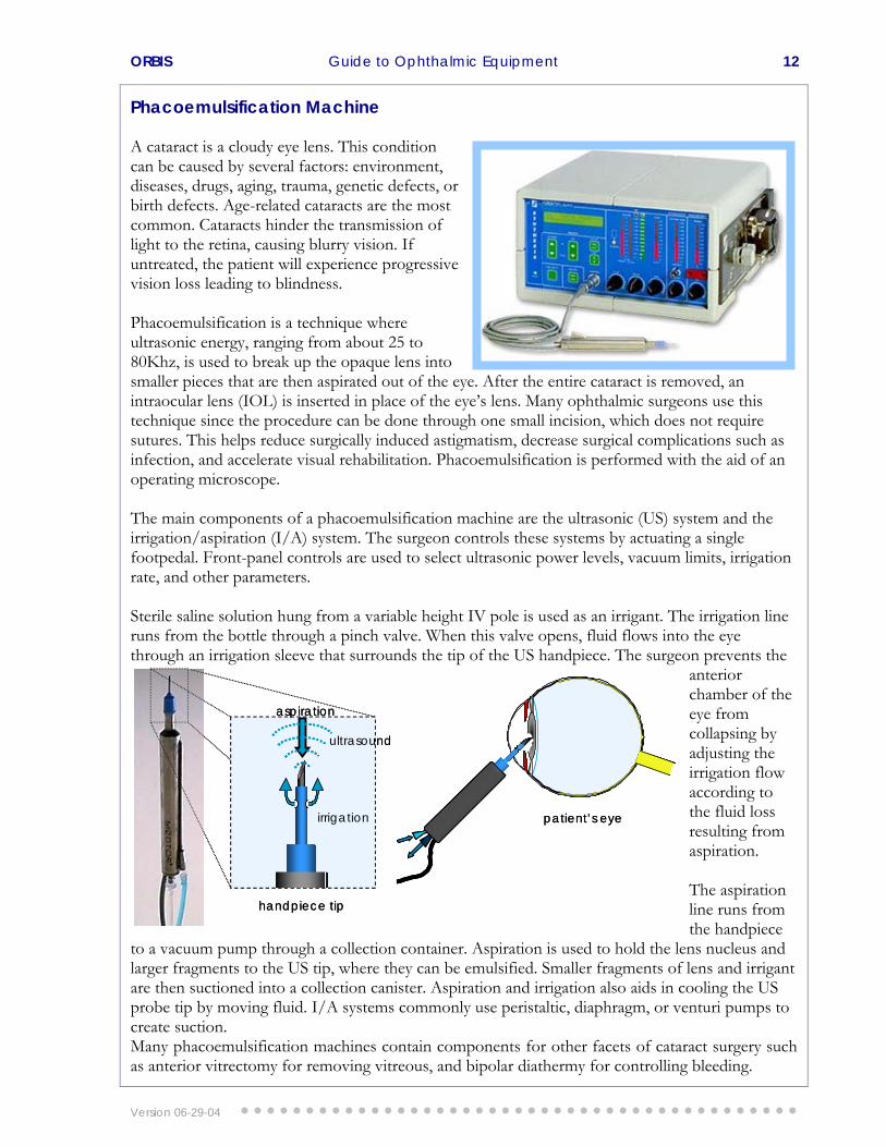
Phacoemulsification Machine A cataract is a cloudy eye lens. This condition can be caused by several factors: environmediseases, drugs, aging, trauma, genetic defects, or birth defects. Age-related cataracts are the most common. Cataracts hinder the transmission of light to the retina, causing blurry vision. If untreated, the patient will experience progressive vision loss leading to blindness.
nt,
Phacoemulsification is a technique where ultrasonic energy, ranging from about 25 to 80Khz, is used to break up the opaque lens into smaller pieces that are then aspirated out of the eye. After the entire cataract is removed, an intraocular lens (IOL) is inserted in place of the eye’s lens. Many ophthalmic surgeons use this technique since the procedure can be done through one small incision, which does not require sutures. This helps reduce surgically induced astigmatism, decrease surgical complications such as infection, and accelerate visual rehabilitation. Phacoemulsification is performed with the aid of an operating microscope. The main components of a phacoemulsification machine are the ultrasonic (US) system and the irrigation/aspiration (I/A) system. The surgeon controls these systems by actuating a single footpedal. Front-panel controls are used to select ultrasonic power levels, vacuum limits, irrigation rate, and other parameters. Sterile saline solution hung from a variable height IV pole is used as an irrigant. The irrigation line runs from the bottle through a pinch valve. When this valve opens, fluid flows into the eye through an irrigation sleeve that surrounds the tip of the US handpiece. The surgeon prevents the
anterior chamber of the eye from collapsing by adjusting the irrigation flow according to the fluid loss resulting from aspiration. The aspiration line runs from the handpiece
to a vacuum pump through a collection container. Aspiration is used to hold the lens nucleus and larger fragments to the US tip, where they can be emulsified. Smaller fragments of lens and irrigant are then suctioned into a collection canister. Aspiration and irrigation also aids in cooling the US probe tip by moving fluid. I/A systems commonly use peristaltic, diaphragm, or venturi pumps to create suction. Many phacoemulsification machines contain components for other facets of cataract surgery such as anterior vitrectomy for removing vitreous, and bipolar diathermy for controlling bleeding.
irrigation
aspiration
ultrasound
handpiece tip
patient’s eyeirrigation
aspiration
ultrasound
handpiece tip
patient’s eye
Version 06-29-04

ORBIS Guide to Ophthalmic Equipment 13
It is common to see phacoemulsification and vitrectomy machines integrated into a single system.
Typical Price *: $15,000 to $100,000 Popular Manufacturers*: Alcon, Bausch & Lomb, Oertli, AMO
IV Fluid
Irrig
atio
n
Aspi
ratio
n
Osc
illatio
n Si
gnal
Controls
Roller Pump
footswitch
collection canister
handpiece
IV Fluid
Irrig
atio
n
Aspi
ratio
n
Osc
illatio
n Si
gnal
Controls
Roller Pump
footswitch
collection canister
handpiece
Version 06-29-04

ORBIS Guide to Ophthalmic Equipment 14
Vitrectomy Machine Vitreous is a clear, jelly-like substance that fills the inside of the eye. Since vitreous is normally clear, light rays are able to travel through it and reach the retina. However, any variation in the consistency, color or structure of the vitreous can hinder transmission of light to the retina, affecting vision. Vitrectomy is a procedure in which the suremoves cloudy vitreous from the eye and replaces it with a clear solution. Light can then pass through this clear fluid, restoring normal sight.
rgeon
piration
se
A vitrectomy is performed with the aid of an operating microscope and a contact lens that is placed on the patient’s cornea. This allows a clear view of the vitreous cavity and retina at various magnifications. Vitrectomy machines have the following main functions: vitreous cutting, irrigation, aspiration, and illumination. Cutting the vitreous is accomplished by a small handpiece containing a guillotine, oscillating or rotating cutter. Pulses of compressed air mechanically actuate the cutter. Some vitrectomy machines require connection to an external compressed air source, while others have an internal pump. Cutting is performed in the adjustable range of 60 to 2000
cuts per minute. The sliced vitreous is aspirated through the handpiece, which is connected to a suction line that carries the fragments to acollection canister. Assystems commonly uperistaltic, diaphragm, or venturi pumps. An irrigation line runs from an IV bottle with sterile saline solution through a pinch valve to the handpiece. When the pinch valve opens, fluid flows into the eye. A light probe that is inserted through a tiny incision in the eye provides illumination for the procedure. The light probe is coupled via a fiber optic cable to a high intensity halogen light source housed inside the machine. The surgeon controls the vitrectomy machine using a footpedal. Front-panel controls are used to select cutting rates, vacuum limits, irrigation rate, light intensity and other parameters. It is common to see phacoemulsification and vitrectomy functions integrated into a single machine.
Typical Price *: $15,000 to $100,000 Popular Manufacturers*: Alcon, Bausch & Lomb, Oertli, AMO
cutting
suctionsuction
cutting mechanism
patient’s eye
vitreous
cutting
suctionsuction
cutting mechanism
patient’s eye
vitreous
Version 06-29-04

ORBIS Guide to Ophthalmic Equipment 15
Cryo Surgical Unit Cryosurgery is the use of extreme cold to treat a variety of conditions. In ophthalmology, cryosurgery is used to treat conditions such as retinal detachment, trichiasis (ingrown eyelashes), glaucoma and cataract extraction among others. Cryo surgical units (CSU) apply a refrigerant (cryogen) to withdraw heat from target tissue through contact with a cryogen-cooled probe. The effect is to freeze the surrounding tissue so that it dies. In the tissue immediately beyond the killed zone a degree of coagulation occurs thus limiting the resulting bleeding. The surgeon controls the freezing by activating a pedal that releases the cryogen from a pressurized tank into the probe. Compressed nitrous oxide (N2O) and carbon dioxide (CO2) are used as cryogens in ophthalmology. When these gases expand into the probe, they cause the tip of the probe to cool. The lowest probe-tip temperatures that can be attained with these gases are -89° and -79°C, respectively. A variety of interchangeable probes with different tip sizes and shapes are available for specific types of surgery.
Ophthalmic cryosurgery is used for cataract removal in developing countries, but its use in developed countries has decreased over the past decade. Intracapsular Cataract extraction (ICCE) has been replaced by extracapsular extraction, using either phacoemulsification or irrigation/aspiration techniques that leave the posterior lens capsule intact and allow implantation of a prosthetic lens behind the iris; cryosurgical reattachment of the retina has been supplanted by laser photocoagulation and other techniques. However, cyclocryotherapy (freezing of the ciliary body to reduce ciliary process secretion of aqueous humor, thereby lowering interocular pressure and halting damage to the ocular nerve) is still being used to treat advanced cases of open-angle glaucoma in patients with limited functional vision, and cryosurgery is the preferred method of treatment for trichiasis and basal cell carcinomas of the lid and periocular region because it yields superior cosmetic results. Cryosurgery is also used experimentally in
the treatment of corneal herpes (herpetickeratitis) and retinopathy in premature infants.
Typical Price *: $3,000 to $12,000 Popular Manufacturers*: Keeler, Cooper Surgical, Erbe
Version 06-29-04

ORBIS Guide to Ophthalmic Equipment 16
Ophthalmic Lasers Ophthalmic lasers allow precise treatment of a range of eye problems with little risk of infection. Many laser procedures are relatively pain free and can be performed on an outpatient basis. The combination of safety, accuracy, and relative low cost, make lasers very useful ophthalmic tools.
iation is mitted as the atoms return to their original energy levels.
obe that is inserted into the eye), slitlamp, perating microscope and indirect ophthalmoscope.
nction f the molecular composition of the tissue and the wavelength and power of the laser light.
to destroy normal blood vessels so that hemorrhage or scarring will not damage central vision.
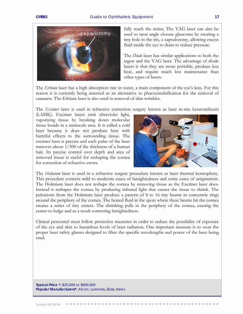
turned. The YAG laser is commonly used to vaporize a portion of the capsule, allowing light to
The word Laser is an acronym for light amplification by stimulated emission of radiation. Laser light is coherent (wavelengths are in phase in space and time), monochromatic (one color or wavelength), and collimated (light is emitted as a narrow beam in a specific direction). Laser beams are produced by the excitation of atoms to a higher than usual energy state. Laser rade The main components of a laser system are the laser tube, the pump or excitation source, the power supply, and a cooling unit. Laser energy is delivered to eye structures using one of several delivery systems: endoprobe (a small fiber optic pro Different types of lasers emit specific wavelengths of light and are used to treat specific eye problems. Lasers are usually named according to the active material used. For instance, an argon laser contains argon gas as its active material, while the YAG laser contains a solid material made up of yttrium, aluminum, and garnet. The effects that lasers have on eye tissues are both a fuo The argon laser emits blue-green wavelengths, which are absorbed by the cells under the retina and by the red hemoglobin in blood. However, blue-green wavelengths can pass through the fluid inside the eye without causing damage. For this reason, the argon laser is used extensively in the treatment of diabetic retinopathy, a severe disorder of the retina that causes blood vessels to leak. The argon laser can burn and seal these blood vessels. Retinal detachment is another serious eye problem that can be treated by the argon laser. The laser is used to weld the detached retina to the underlying choroid layer of the eye. Some forms of glaucoma, a leading cause of blindness, may also be treated with argon lasers. For instance, angle closure glaucoma can be treated by using an argon laser to create a tiny hole in the iris, allowing excess fluid inside the eye to drain to reduce pressure. Macular degeneration, a severe condition that affects central vision in older people, is sometimes treated with an argon or krypton laser. In this treatment, the laser is usedab The YAG laser generates short-pulsed, high-energy light beams to cut, perforate, or fragment tissue. This laser may also be called a neodymium-YAG or ND-YAG laser. Many people have the misconception that a YAG laser is used to remove cataracts. This misunderstanding happens because up to two thirds of cataract patients develop a condition known as posterior capsular opacification, a clouding of the lens capsule months after cataract surgery. This gradual loss of vision is similar to the symptoms of a cataract, causing people to believe that their cataract hasre
Version 06-29-04
ORBIS Guide to Ophthalmic Equipment 17
fully reach the retina. The YAG laser can also be used to treat angle closure glaucoma by creating a
ny hole in the iris, a capsulotomy, allowing excess
more portable, produce less eat, and require much less maintenance than
lsification for the removal of ataracts. The Erbium laser is also used in removal of skin wrinkles.
ping the cornea r correction of refractive errors.
the periphery of the cornea, causing the enter to bulge and as a result correcting farsightedness.
laser safety glasses designed to filter the specific wavelengths and power of the laser being used.
tifluid inside the eye to drain to reduce pressure. The Diode laser has similar applications to both the argon and the YAG laser. The advantage of diode lasers is that they are hother types of lasers.
The Erbium laser has a high absorption rate in water, a main component of the eye's lens. For this reason it is currently being assessed as an alternative to phacoemuc The Excimer laser is used in refractive correction s(LASIK). Excimer lasers emit ultraviolet light, vaporizing tissue by breaking down molecular tissue bonds in a miniscule area. It is called a cold laser because it does not produce heat with harmful effects to the surrounding tissue. The excimer laser is precise and each pulse of the laser removes about 1/500 of the thickness of a human hair. Its precise control over depth and area of removed tissue is useful for reshafo The Holmium laser is used in a refractive surgery procedure known as laser thermal keratoplasty. This procedure corrects mild to moderate cases of farsightedness and some cases of astigmatism. The Holmium laser does not reshape the cornea by removing tissue as the Excimer laser does. Instead it reshapes the cornea by producing infrared light that causes the tissue to shrink. The pulsations from the Holmium laser produce a pattern of 8 to 16 tiny beams in concentric rings around the periphery of the cornea. The heated fluid in the spots where these beams hit the cornea creates a series of tiny craters. The shrinking pulls in c Clinical personnel must follow protective measures in order to reduce the possibility of exposure of the eye and skin to hazardous levels of laser radiation. One important measure is to wear the proper
Typical Price *: $25,000 to $600,000 Popular Manufacturers*: Alcon, Lumenis, Zeiss, Iridex
capsulotomy
urgery known as laser in-situ keratomileusis
Version 06-29-04

ORBIS Guide to Ophthalmic Equipment 18
APPENDIX- Diagram of the Eye
Version 06-29-04