


Growth suppression of human hepatocellular carcinoma ... · growth suppression of human...
17
GROWTH SUPPRESSION OF HUMAN HEPATOCELLULAR CARCINOMA XENOGRAFTS BY A MONOCLONAL ANTIBODY CH12 DIRECTED TO EPIDERMAL GROWTH FACTOR RECEPTOR VARIANT III # Hua Jiang 1, *, Huamao Wang 1, 2, *, Zhonghua Tan 3 , Suwen Hu 1 , Hai Wang, Bizhi Shi 1 , Lin Yang 1 , Peiyong Li 3 , Jianren Gu 1 , Hongyang Wang 1, 4 , Zonghai Li 1 From 1 State Key Laboratory of Oncogenes and Related Genes, Shanghai Cancer Institute, Medical School of Shanghai Jiaotong University, Shanghai 200032, China, 2 Institute of Bioengineering, Zhejiang Academy of Medical Science, Hangzhou 310013, China, 3 Ruijin Hospital of Shanghai Jiaotong University, Shanghai 200030, China and 4 International Cooperation Laboratory on Signal Transduction, Eastern Hepatobiliary Surgery Institute, Second Military Medical University, Shanghai, 200438, China Running head: A monoclonal anti-EGFRvIII antibody for cancer therapy *These authors contributed equally to this work. Address correspondence to: Zonghai Li, State Key Laboratory of Oncogenes and Related Genes, Shanghai Cancer Institute, Shanghai Jiao Tong University School of Medicine, No. 25/Ln. 2200, Xie Tu Road, Shanghai 200032, China. Tel.: (86)-21-64436601; fax: (86)-21-64432027. E-mail addresses: [email protected] The abbreviations used are: HCC, hepatocellular carcinoma; EGFR, epidermal growth factor receptor; ADCC, antibody-dependent cell-mediated cytotoxicity; CDC, complement dependent cytotoxicity; EGFRvIII, epidermal growth factor receptor variant III; HNSCC, head and neck squamous cell carcinoma. Human hepatocellular carcinoma (HCC) is considered difficult to cure because its resistant to radio- and chemotherapy and high recurrence rate after curative liver resection. Epidermal growth factor receptor variant III (EGFRvIII) has shown to express in HCC tissues and cell lines. This study describes efficacy of an anti-EGFRvIII monoclonal antibody (mAb CH12) for the treatment of HCC xenografts with EGFRvIII expression and the underlying mechanism of EGFRvIII as an oncogene in HCC. The results demonstrated that CH12 bound preferentially to EGFRvIII with a dissociation constant (Kd) of 1.346 nm/L. In addition, CH12 induces strong antibody-dependent cellular cytotoxicity (ADCC) and complement-dependent cytotoxicity (CDC) in Huh7-EGFRvIII (with exogenous expression of EGFRvIII) and SMMC-7721 (with endogenous expression of EGFRvIII) cells. Notably, CH12 significantly inhibited the growth of Huh7-EGFRvIII and SMMC-7721 xenografts in vivo with a growth inhibition ratio much higher than C225, a FDA (US Food and Drug Administration)-approved anti-EGFR antibody. Treatment of the two HCC xenografts with CH12 significantly suppressed tumor proliferation and angiogenesis. Mechanistically, in vivo treatment with CH12 reduced the phosphorylation of constitutively active EGFRvIII, Akt and ERK. Down-regulation of the apoptotic protectors Bcl-X L , Bcl-2 and the cell cycle regulator cyclin D1, as well as up-regulation of the cell-cycle inhibitor p27, were also observed after in vivo CH12 treatment. Collectively, these results indicate that the monoclonal antibody CH12 is a promising therapeutic agent for HCC with EGFRvIII expression. 1 http://www.jbc.org/cgi/doi/10.1074/jbc.M110.192252 The latest version is at JBC Papers in Press. Published on December 16, 2010 as Manuscript M110.192252 Copyright 2010 by The American Society for Biochemistry and Molecular Biology, Inc. by guest on June 16, 2018 http://www.jbc.org/ Downloaded from
-
Upload
truongkhuong -
Category
Documents
-
view
215 -
download
1
Transcript of Growth suppression of human hepatocellular carcinoma ... · growth suppression of human...

GROWTH SUPPRESSION OF HUMAN HEPATOCELLULAR CARCINOMA XENOGRAFTS BY A MONOCLONAL ANTIBODY CH12 DIRECTED TO
EPIDERMAL GROWTH FACTOR RECEPTOR VARIANT III# Hua Jiang1,*, Huamao Wang1, 2,*, Zhonghua Tan3, Suwen Hu1, Hai Wang, Bizhi Shi1, Lin
Yang1, Peiyong Li3, Jianren Gu1, Hongyang Wang1, 4, Zonghai Li1
From 1State Key Laboratory of Oncogenes and Related Genes, Shanghai Cancer Institute, Medical School of Shanghai Jiaotong University, Shanghai 200032, China, 2Institute of Bioengineering, Zhejiang Academy of Medical Science, Hangzhou 310013, China, 3Ruijin Hospital of Shanghai
Jiaotong University, Shanghai 200030, China and 4International Cooperation Laboratory on Signal Transduction, Eastern Hepatobiliary Surgery Institute, Second Military Medical University,
Shanghai, 200438, China Running head: A monoclonal anti-EGFRvIII antibody for cancer therapy
*These authors contributed equally to this work. Address correspondence to: Zonghai Li, State Key Laboratory of Oncogenes and Related Genes, Shanghai Cancer Institute, Shanghai Jiao Tong University School of Medicine, No. 25/Ln. 2200, Xie Tu Road, Shanghai 200032, China. Tel.: (86)-21-64436601; fax: (86)-21-64432027. E-mail addresses: [email protected] The abbreviations used are: HCC, hepatocellular carcinoma; EGFR, epidermal growth factor receptor; ADCC, antibody-dependent cell-mediated cytotoxicity; CDC, complement dependent cytotoxicity; EGFRvIII, epidermal growth factor receptor variant III; HNSCC, head and neck squamous cell carcinoma.
Human hepatocellular carcinoma (HCC) is considered difficult to cure because its resistant to radio- and chemotherapy and high recurrence rate after curative liver resection. Epidermal growth factor receptor variant III (EGFRvIII) has shown to express in HCC tissues and cell lines. This study describes efficacy of an anti-EGFRvIII monoclonal antibody (mAb CH12) for the treatment of HCC xenografts with EGFRvIII expression and the underlying mechanism of EGFRvIII as an oncogene in HCC. The results demonstrated that CH12 bound preferentially to EGFRvIII with a dissociation constant (Kd) of 1.346 nm/L. In addition, CH12 induces strong antibody-dependent cellular cytotoxicity (ADCC) and complement-dependent cytotoxicity (CDC) in Huh7-EGFRvIII (with exogenous expression of EGFRvIII) and SMMC-7721 (with endogenous
expression of EGFRvIII) cells. Notably, CH12 significantly inhibited the growth of Huh7-EGFRvIII and SMMC-7721 xenografts in vivo with a growth inhibition ratio much higher than C225, a FDA (US Food and Drug Administration)-approved anti-EGFR antibody. Treatment of the two HCC xenografts with CH12 significantly suppressed tumor proliferation and angiogenesis. Mechanistically, in vivo treatment with CH12 reduced the phosphorylation of constitutively active EGFRvIII, Akt and ERK. Down-regulation of the apoptotic protectors Bcl-XL, Bcl-2 and the cell cycle regulator cyclin D1, as well as up-regulation of the cell-cycle inhibitor p27, were also observed after in vivo CH12 treatment. Collectively, these results indicate that the monoclonal antibody CH12 is a promising therapeutic agent for HCC with EGFRvIII expression.
1
http://www.jbc.org/cgi/doi/10.1074/jbc.M110.192252The latest version is at JBC Papers in Press. Published on December 16, 2010 as Manuscript M110.192252
Copyright 2010 by The American Society for Biochemistry and Molecular Biology, Inc.
by guest on June 16, 2018http://w
ww
.jbc.org/D
ownloaded from

Hepatocellular carcinoma (HCC) is the fifth most common cancer and the third most common cause of cancer-related death in the world (1). The cancer is usually diagnosed at a stage when the disease is already advanced and incurable. Surgery is the most effective treatment for HCC. However, tumor recurrence after a curative liver resection is high (2, 3). Adjuvant chemotherapy has not significantly improved HCC patients’ survival (3). Sorafenib is the only targeted therapy approved by the FDA to treat HCC. Although sorafenib showed good tolerability in the studied populations, most patients in clinical practice suffer from underlying liver cirrhosis with impaired metabolic function and experience dose-limiting toxicities with the need to reduce the overall dose of sorafenib subsequently (4).
The epidermal growth factor receptor (EGFR) has been successfully targeted for cancer therapy (5). EGFR expression in HCC has been reported (6). EGFR expression has been suggested to contribute to the aggressive growth characteristics of HCC tumors (7-9). EGFR overexpression has been demonstrated to be positively correlated with early tumor recurrence and a negative prognostic factor in poorly differentiated HCCs (6, 10). Hence, EGFR represent a promising target for developing innovative HCC treatment strategies. However, despite the clinical success of several EGFR targeting therapies including the monoclonal antibody cetuximab (C225, Erbitux) (11, 12) in the treatment of colon cancer and head and neck squamous cell carcinoma (13, 14), no obvious responses were observed in HCC patients after treatment with cetuximab (15).
The presence of EGFR gene mutations may account for the limited clinical response to EGFR targeting therapies such as cetuximab in HCC patients. Among the EGFR mutations, EGFRvIII is the most commonly described
mutation which is a ligand-independent, constitutively active variant with in-frame deletion of exon 2 – exon 7 resulting in the deletion of amino acids 6–273 in the extracellular domain and the generation of a glycine at the fusion point (16). EGFRvIII expression has been detected in glioma, non–small cell lung carcinoma, breast cancer, HNSCC, ovarian carcinoma as well as hepatocellular carcinoma (17-20). Recently, we also observed its expression in liver cancer cell lines such as SMMC-7721(21). EGFRvIII mRNA has also been observed in the serum of HCC patients (22). Expression of EGFRvIII can promote tumor cell growth in vitro and in vivo (20, 21, 23). Collectively, these results suggest that EGFRvIII is a potential therapeutic target for HCC. To explore whether EGFRvIII-targeting therapy is a new option for HCC patients, here we report the development of a monoclonal antibody directed to EGFRvIII for the treatment of HCC xenografts in vivo.
EXPERIMENTAL PROCEDURES
Cell culture- The human hepatocellular carcinoma cell lines Huh-7 (expressing low levels of endogenous wtEGFR); Huh7-EGFR (Huh-7 cells with exogenous EGFR overexpression); Huh7-EGFRvIII (Huh-7 cells with exogenous EGFRvIII overexpression (21) and SMMC-7721 (Chinese Academy of Science, Shanghai, China) were maintained in DMEM supplemented with 10% fetal bovine serum in a humidified atmosphere of 95% air and 5% CO2 at 37°C.
FACS analysis- 1×10⁶ cells were collected by centrifugation and incubated with 20μg/ml primary antibody, in phosphate-buffered saline containing 1% newborn calf serum for 45 min at 4 °C. After being washed with cold phosphate-buffered saline, cells were incubated for an additional 45 min at 4 °C with a fluorescein (FITC)-conjugated goat
2
by guest on June 16, 2018http://w
ww
.jbc.org/D
ownloaded from

anti-human antibody (Kang-Chen Bio-tech, Shanghai, China) in the dark. For each sample, at least 10,000 cells were analyzed by FACS cytometry (Beckman Coulter Epics Altra, Miami, FL) and MultiCycle AV for Windows 5.0 (Phoenix Flow Systems, San Diego, CA).
Immunological effector functions- ADCC was performed as previously described (24). Hepatocellular carcinoma cells Huh-7, Huh7-EGFRvIII and SMMC-7721 were used as target cells. Effector cells (peripheral blood mononucleocytes) were freshly prepared and added to the target cells to achieve E:T ratios of 100:1. For the CDC assay, target cells were plated at a density of ~2000 cells/well, with varying amounts of the CH12 antibody (0.01–10 μg/ml) in microtiter plates. Human complement, prepared from the blood of normal, healthy volunteers, was added to each well and incubated at 37°C for 4h. The percent cytotoxicity of the antibody was calculated as follows: 1- (number of viable cells in well treated with antibody and complement/ number of viable cells in well treated with the isotype control antibody and complement) ×100%. The assay was performed in triplicates.
Radiolabelling and binding affinity assay- IODO-GENTM(Sigma, St. Louis, MO, USA) was used to iodinate CH12 with iodine-125 (125I) (Amersham, Piscataway, NJ, USA). Radiolabelling was performed on the day of injection into mice. Prior to injection, the percentage (%) of unbound radionuclide content was determined by instant thin layer chromatography (ITLC), and binding ability of the final radiolabelled product was tested using a Huh7-EGFRvIII cell binding (Lindmo) assay as detailed below. Scatchard analysis was used to determine the binding constant (Ka) and number of antibody molecules bound per cell for 125I-labelled antibody.
In vivo antitumor effects- Tumor volumes were measured as previously described (21).
Mice were manipulated and housed according to protocols approved by the Shanghai Medical Experimental Animal Care Commission.
Western blot analysis- Immunoblotting experiments were performed according to standard procedures (21). The following antibodies were used: 12H23, anti-phospho- EGFR (Tyr 1068) (Abcam, Cambridge, UK) and anti-GAPDH (Kang-Chen Bio-tech, Shanghai, China). Anti-phospho-Akt (Thr 308), Akt (H-136), anti-phospho-ERK, anti-ERK1, anti-cyclin D1, anti-Bcl-2, anti- Bcl-XL and anti-p27 were all obtained from Santa Cruz Biotechnology.
Immunohistochemical analysis- To assess angiogenesis and cell proliferation in tumors, formalin-fixed paraffin embedded tumor tissues were immunostained using a monoclonal rat anti-mouse CD34 (Abcam, Cambridge, UK) and anti-Ki-67 (Santa Cruz Biotechnology, Santa Cruz, CA ). After deparaffinization and rehydration, the tissue sections were incubated with 3% hydrogen peroxide in methanol to quench endogenous peroxidase. The sections were blocked for 30 min with 1% BSA and incubated overnight with the primary antibody at 4°C. The sections were then washed with PBS and incubated with a HRP-conjugated secondary antibody for 30 min. The products were then visualized using a diaminobenzidine staining kit (TIANGEN Biotech, Beijing, China) and counterstained in haematoxylin.
As a measure of proliferation, the Ki-67 labeling index was determined as the ratio of labeled nuclei: total nuclei in high-power (×400) fields. Approximately 2000 nuclei were counted in each case by systematic random sampling.
Microvessel density (MVD) was determined by measuring the number of stained microvessels in each section from six mice of each group as described (25). The
3
by guest on June 16, 2018http://w
ww
.jbc.org/D
ownloaded from

mean microvessel count of the six most vascular areas was taken as the MVD, which was expressed as the absolute number of microvessels per 0.74 mm2 (×200 field).
TUNEL assay- The tumor tissues sections were firstly de-paraffinized and rehydrated and were then incubated with proteinase K (20μg/ml) for 20 min at 37°C. After several washes with PBS, the sections were incubated with TUNEL assay buffer (Beyotime Biotechnology, Nanjing, China) for 1h at 37°C in the dark. Then the slides were rinsed in PBS three times and visualized under a Zeiss LSM confocal microscope (Carl Zeiss, Jena, Germany). TUNEL-positive cells were counted at ×400 magnification. The apoptotic index was calculated as a ratio of apoptotic cell number: total cell number in each field.
Statistical analysis- All data are presented as the mean ± standard error and were analyzed by the Student’s t test. P < 0.05 was considered statistically significant.
RESULTS
Selection of anti-EGFRvIII mAbs- The protocol for preparation of the hybridomas against EGFRvIII is shown in the supplemental data. One of the hybridoma clones named 12H23 showed preferential binding with EGFRvIII and overexpressed EGFR by FACS analysis (Fig. S1). Importantly, 12H23 can significantly inhibit the growth of Huh7-EGFRvIII xenografts in vivo with a higher growth inhibition ratio than the anti-EGFR antibody C225 (Fig. S2). At the end of the study, the inhibition ratio of the C225 treated and 12H23-treated groups were 30% and 63.4%, respectively (Fig. S2). To analyze binding epitope of 12H23, several EGFR-derived recombinant proteins were expressed in E. coli (Fig S3 A and B). The antibody binds to N12-S1 (EGFR, cysteine rich region, 161G to 313C) and EGFRvIIIex (EGFR, 1L to 623A with amino acids 6-273
deletion) but not to N12-S2 (EGFR cysteine rich region, 458G to 614T) recombinant protein in ELISA assay. After comparing the amino acid composition of N12-S1 and that of EGFRvIIIex, 12H23 is suggested to bind to the same epitope as that of mAb 806 (24). To fine map the binding epitope, several peptides related to CC16 (287CGADSYEMEEDGVRKC302) were fused with the N12 peptide and ELISA assay confirmed that 12H23 bind to the same epitope as that of mAb 806 (Fig. S3C). Based on the binding specificity and antitumor activity, we decided to construct antibody 12H23 as a mouse-human chimera to minimize immunogenicity for potential use in clinical trials. The resultant mouse-human chimera antibody named CH12 contains the variable regions of the heavy and light chains of 12H23 linked to human gamma-1 and kappa constant regions. CH12 was produced in a CHO-DG44 cell line by the procedure described in the supplementary methods.
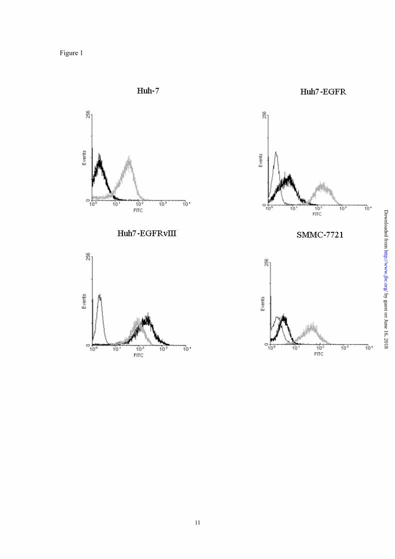
Binding specificity and affinity of CH12- A FACS assay was used to test the binding specificity of CH12. C225 (cetuximab), a chimeric monoclonal IgG1 antibody was included as a control. The results indicated that C225 but not CH12 was able to bind to Huh-7 cells (Fig. 1A). CH12 binds with higher ratio to Huh7-EGFRvIII cells than to Huh7-EGFR cells while C225 binds to the two cell lines at a similar ratio, indicating the preferential binding of CH12 to EGFRvIII (Fig. 1B-C). We have previously described that SMMC-7721 cells expressed endogenous EGFRvIII (21). FACS analysis showed that CH12 binds to SMMC-7721 cells, although with a lower ratio than to Huh7-EGFRvIII cells (Fig. 1D), suggesting that the EGFRvIII may be expressed at a low density on SMMC-7721 cells. The binding affinity of CH12 to EGFRvIII was assessed by radiolabelling with 125I in the presence of
4
by guest on June 16, 2018http://w
ww
.jbc.org/D
ownloaded from

unlabeled antibodies. Ka value for the radiolabelled antibody was 1.346×10⁹M-1.
Immune effector function- The ADCC and CDC activities of CH12 on target cells were shown in Figure 2. High cytotoxicity was observed with CH12 against Huh7-EGFRvIII cells with 45.3% cytotoxicity at 0.1μg/ml CH12 (Fig. 2A). A 29.4% cytotoxicity was observed targeting SMMC-7721 at 1μg/ml of CH12 (Fig. 2A). The results of the CDC analyses showed that CH12 also have high-specific cytotoxicity on Huh7-EGFRvIII cells. Cytotoxicity on Huh7-EGFRvIII and SMMC-7721 cells was 32.7% and 21.1% cytotoxicity, respectively, at 1µg/ml CH12 (Fig. 2B). For the EGFRvIII negative Huh-7 cells, no ADCC and CDC activities were induced by CH12.
CH12 significantly inhibits the growth of Huh7-EGFRvIII and SMMC-7721 xenografts- The therapeutic potential of CH12 was first examined in established Huh7-EGFRvIII HCC tumor models; and C225 was included as a control. As shown in Figures 3A and B, CH12 significantly inhibited the growth of Huh7-EGFRvIII xenografts at day 29 after inoculation (CH12 vs vehicle, P<0.05). Interestingly, when compared to the C225 control, CH12 also showed significant growth inhibition in Huh7-EGFRvIII HCC and the inhibitory ratios of C225 and CH12 at day 50 were 32.9% and 64.5%, respectively (CH12 vs C225, P<0.05).
An established SMMC-7721 tumor model was also used to evaluate the growth inhibition rate of CH12. As shown in Figures 3C and D, CH12 significantly inhibited the growth of SMMC-7721 tumor xenografts at day 43 (P<0.05). CH12 also showed higher growth inhibition than C225 at day 50 (P<0.05). The inhibitory ratios of C225 and CH12 54 d after inoculation were 46.9% and 65.0%, respectively.
CH12 treatment reduced proliferation and
angiogenesis and enhanced apoptosis in Huh7-EGFRvIII tumors- To elucidate the mechanism underlying the in vivo growth suppression caused by CH12, we determined the proliferation rate of tumors from control or treated mice. In both tumor models, the proliferative index, measured by Ki-67 staining of the CH12 -treated tumors, was significantly lower than that of the vehicle control tumors (p<0.01) or the C225-treated tumors (p<0.05; Fig. 4A-B). In addition, analysis of the apoptotic index through TUNEL staining demonstrated a significant increase in the number of apoptotic cells in CH12-treated tumors as compared with the vehicle control tumors (p<0.01; Fig. 5). The number of apoptotic cells in CH12-treated Huh7-EGFRvIII tumors was also significantly higher than that in C225-treated Huh7-EGFRvIII tumors (p<0.01; Fig. 5). In SMMC-7721 xenografts, CH12 also induced more apoptotic cells than that of C225, although the difference is not statistically significant.
The extent of tumor vascularization was analyzed by immunostaining of tumors from treated and control specimens for CD34. To quantify tumor vascularization, MVD was determined by scoring the number of stained microvessels. In Huh7-EGFRvIII tumor models, CH12-treated tumors showed 40% less MVD than did vehicle control tumors and 35% than did C225-treated tumors (p<0.05; Fig. 4C-D). In SMMC-7721 tumor models, CH12-treated tumors showed 53% less MVD than did vehicle control tumors and 48% than did C225-treated tumors (p<0.01; Fig. 4C-D).
CH12 treatment reduces EGFRvIII autophosphorylation and down-regulates Bcl-XL and Bcl-2 expression in Huh7-EGFRvIII and SMMC-7721 HCC tumors- To understand the molecular events occurring in CH12-treated tumors, some key signal molecules in the EGFR pathway were
5
by guest on June 16, 2018http://w
ww
.jbc.org/D
ownloaded from

examined by Western blot analysis (Fig. 6). Because the constitutively active kinase activity and autophosphorylation of the carboxyl terminus of EGFRvIII are essential for its biological functions (17, 26), we determined the EGFRvIII phosphorylation status in tumors from treated and control animals. CH12 treatment dramatically reduced EGFRvIII autophosphorylation, although receptor levels were only slightly decreased in the xenografts. Phosphorylation of Akt and ERK, the two most important downstream signal transductors of EGFRvIII was also examined. CH12 treatment reduced both Akt and ERK phosphorylation in Huh7-EGFRvIII and SMMC-7721 cell lines, although the phosphorylation level of ERK were downregulated to a greater degree in SMMC-7721 cells than in Huh7-EGFRvIII cells (Fig.6).
Since CH12 significantly inhibited the proliferation of HCC, two cell cycle regulators cylinD1 and p27 were examined. As shown in Figure 6, cyclin D1 was significantly down-regulated by CH12-treatment. The expression of cyclin D1 in CH12 treatment group was weaker than that in C225 treatment group. CH12 treatment potently up-regulated the expression of p27 (Fig. 6).
Mechanism underlying the apoptosis promotion effect of CH12 treatment was also studied. Therefore,the expression level of Bcl-XL and Bcl-2 were examined. The results shown in Figure 6 clearly indicate that Bcl-XL and Bcl-2 were significantly down-regulated in CH12-treated tumors. However, C225-treated tumors showed almost no reduction of Bcl-XL and Bcl-2 expression (Fig. 6).
DISCUSSION
One limitation of antibodies targeting the wild-type (wt) EGFR is their significant uptake in normal tissues such as the liver and
skin, which often causes side effects such as skin rash and gastrointestinal toxicity. To avoid this problem, Johns TG, et al developed mAb 806 which binding to a specific epitope of EGFR (27). In a phase I clinical study, ch806 displayed excellent targeting of tumor sites in all patients with no evidence of normal tissue uptake, and no significant toxicity (28). However, the highest dosage level of ch806 in the phase I clinical trial was 40mg/m2. The authors mentioned that further dose escalation was not performed due to the limited amount of cGMP ch806 available for the trial (28), suggesting the difficulty in production of ch806. In this study, human-mouse chimeric antibody of 12H23 (CH12) displayed almost the same binding affinity to EGFRvIII as ch806 (29). Interestingly, we found that the yield of CH12 is approximately 5-fold higher than that of ch806 (data not shown). Additionally, in an in vivo biodistribution assay, 24 h after antibody injection, the percentage of the injected radioactive dose (ID) per gram of tissue (%ID g-1) of l25I-CH12 in Huh7-EGFRvIII xenografts are highest among the tested solid organs, indicating its accumulation in the tumor tissues (Fig. S4). Therefore, we decided to use CH12 to treat HCC.
Intriguingly, CH12 displayed significant ADCC and CDC effects on Huh7-EGFRvIII cells as well as SMMC-7721 cells, which bound weakly to CH12 in a FACS assay. Importantly, CH12 showed strong anti-tumor effect on Huh7-EGFRvIII and SMMC-7721 xenografts. We also tested the anti-HCC effect of the FDA-approved antibody C225. The results indicated that C225 displayed limited growth-inhibition effect on HCC xenografts with EGFRvIII expression. Thus, the EGFRvIII expression may be a reason for the minimal effect of C225 in the clinical trial.
Further study on the mechanism underlying the growth inhibition effect indicated that
6
by guest on June 16, 2018http://w
ww
.jbc.org/D
ownloaded from

CH12 treatment can suppress angiogenesis, promote apoptosis and decrease proliferation. Cyclin D1 is involved in regulating the G1/S transition and was observed to be amplified in HCC (30-32). p27 is a cdk inhibitor which binds to cyclin-CDK complexes and facilitates the inhibition of the catalytic activity of cdk, ultimately inducing G1 cell cycle arrest. It has been suggested that p27 loss occurs early in the carcinogenesis process. The more aggressive, metastasizing cancers tend to lack p27 expression (33). High p27 expression, correlated with prolonged survival, is a favorable independent prognostic parameter for HCC (33, 34). Thus, the decreased proliferation in HCC xenografts caused by CH12 treatment should be ascribed to the down-regulation of cyclin D1 expression and up-regulation of p27 expression. It has been demonstrated that the EGFRvIII can induce up-regulation of Bcl-XL (35), which has been observed to overexpress in a subset of HCC and demonstrated as a significant prognostic factor for disease progression in human HCC (36, 37). Previous study indicated that EGFR inhibitor gefitinib can suppress the expression of anti-apoptotic Bcl-2 and Bcl-XL, further rendering HCC cells prone to apoptosis (38). Therefore, the weakened expression of Bcl-2
and Bcl-XL should contribute to the apoptosis-promoting effect of CH12.
As we know, antibodies are often used with chemotherapeutic drugs in clinic. Previous report indicated that combining cetuximab with TKIs (erlotinib or AG1024) or the HMG-CoA-reductase inhibitor fluvastatin or doxorubicin resulted in synergistic antiproliferative effects on HepG2 and Huh-7 hepatocellular cancer cells (39). Considering that EGFRvIII expression can decrease the sensitivity of HCC cell lines to the chemotherapeutic drug 5-fluorouracil (21), CH12 which inhibits the signal transduction of EGFRvIII may be combined with 5-fluorouracil to enhance the antitumor effects.
CH12 can also possible to be combined with sorafenib, which inhibits Raf-1, b-Raf, vascular endothelial growth factor receptors(VEGFRs)1, 2 and 3, and PDGFR-β (40) for HCC treatment. This combination may significantly reduce the dosage of sorafenib and thus the side effects sorafenib induced. Taken together, our study demonstrated that the monoclonal antibody CH12 developed here has potential to be used as therapeutics for HCC with EGFRvIII expression.
REFERENCES
1. Llovet, J. M., Burroughs, A., and Bruix, J. (2003) Lancet 362(9399), 1907-1917 2. Carr, B. I. (2004) Gastroenterology 127(5 Suppl 1), S218-224 3. Zhu, A. X. (2006) The oncologist 11(7), 790-800 4. Worns, M. A., Weinmann, A., Pfingst, K., Schulte-Sasse, C., Messow, C. M.,
Schulze-Bergkamen, H., Teufel, A., Schuchmann, M., Kanzler, S., Duber, C., Otto, G., and Galle, P. R. (2009) Journal of clinical gastroenterology 43(5), 489-495
5. Gainet, M., Guardiola, E., Dufresne, A., and Pivot, X. (2003) Cancer Radiother 7(3), 195-199 6. Daveau, M., Scotte, M., Francois, A., Coulouarn, C., Ros, G., Tallet, Y., Hiron, M., Hellot, M.
F., and Salier, J. P. (2003) Molecular carcinogenesis 36(3), 130-141 7. DeCicco, L. A., Kong, J., and Ringer, D. P. (1997) Cancer letters 111(1-2), 149-156 8. Kira, S., Nakanishi, T., Suemori, S., Kitamoto, M., Watanabe, Y., and Kajiyama, G. (1997)
Liver 17(4), 177-182
7
by guest on June 16, 2018http://w
ww
.jbc.org/D
ownloaded from

9. Moser, G. J., Wolf, D. C., and Goldsworthy, T. L. (1997) Toxicologic pathology 25(3), 275-283 10. Zhao, Y. N., Cao, J., Wu, F. X., Ou, C., Yuan, W. P., Mo, Q. G., Wei, W., Li, Y., Su, J. J., and
Liang, A. M. (2004) Ai zheng = Aizheng = Chinese journal of cancer 23(7), 762-766 11. Mendelsohn, J., and Baselga, J. (2003) J Clin Oncol 21(14), 2787-2799 12. Nygren, P., Sorbye, H., Osterlund, P., and Pfeiffer, P. (2005) Acta oncologica (Stockholm,
Sweden) 44(3), 203-217 13. Harari, P. M. (2004) Endocrine-related cancer 11(4), 689-708 14. Huang, S., Armstrong, E. A., Benavente, S., Chinnaiyan, P., and Harari, P. M. (2004) Cancer
research 64(15), 5355-5362 15. Yu, X. T., Zhu, S. N., Xu, Z. D., Hu, X. Q., Zhu, T. F., Chen, J. Q., and Lu, S. L. (2007)
Journal of cancer research and clinical oncology 133(3), 145-152 16. Kuan, C. T., Wikstrand, C. J., and Bigner, D. D. (2001) Endocrine-related cancer 8(2), 83-96 17. Moscatello, D. K., Holgado-Madruga, M., Godwin, A. K., Ramirez, G., Gunn, G., Zoltick, P.
W., Biegel, J. A., Hayes, R. L., and Wong, A. J. (1995) Cancer research 55(23), 5536-5539 18. Okamoto, I., Kenyon, L. C., Emlet, D. R., Mori, T., Sasaki, J., Hirosako, S., Ichikawa, Y.,
Kishi, H., Godwin, A. K., Yoshioka, M., Suga, M., Matsumoto, M., and Wong, A. J. (2003) Cancer science 94(1), 50-56
19. Ou, C., Wu, F. X., Luo, Y., Cao, J., Zhao, Y. N., Yuan, W. P., Li, Y., and Su, J. J. (2005) Ai zheng = Aizheng = Chinese journal of cancer 24(2), 166-169
20. Sok, J. C., Coppelli, F. M., Thomas, S. M., Lango, M. N., Xi, S., Hunt, J. L., Freilino, M. L., Graner, M. W., Wikstrand, C. J., Bigner, D. D., Gooding, W. E., Furnari, F. B., and Grandis, J. R. (2006) Clin Cancer Res 12(17), 5064-5073
21. Wang, H., Jiang, H., Zhou, M., Xu, Z., Liu, S., Shi, B., Yao, X., Yao, M., Gu, J., and Li, Z. (2009) Cancer letters 279(1), 30-38
22. Zhou, M., Gong, B., Gu, J., and Li, Z. Liver Int 30(6), 925-927 23. Nishikawa, R., Ji, X. D., Harmon, R. C., Lazar, C. S., Gill, G. N., Cavenee, W. K., and Huang,
H. J. (1994) Proceedings of the National Academy of Sciences of the United States of America 91(16), 7727-7731
24. Yang, L., Jiang, H., Shi, B., Wang, H., Li, J., Wang, H., Yao, M., and Li, Z. Cancer Immunol Immunother 59(10), 1481-1487
25. Poon, R. T., Ng, I. O., Lau, C., Yu, W. C., Yang, Z. F., Fan, S. T., and Wong, J. (2002) J Clin Oncol 20(7), 1775-1785
26. Luwor, R. B., Johns, T. G., Murone, C., Huang, H. J., Cavenee, W. K., Ritter, G., Old, L. J., Burgess, A. W., and Scott, A. M. (2001) Cancer research 61(14), 5355-5361
27. Johns, T. G., Adams, T. E., Cochran, J. R., Hall, N. E., Hoyne, P. A., Olsen, M. J., Kim, Y. S., Rothacker, J., Nice, E. C., Walker, F., Ritter, G., Jungbluth, A. A., Old, L. J., Ward, C. W., Burgess, A. W., Wittrup, K. D., and Scott, A. M. (2004) The Journal of biological chemistry 279(29), 30375-30384
28. Scott, A. M., Lee, F. T., Tebbutt, N., Herbertson, R., Gill, S. S., Liu, Z., Skrinos, E., Murone, C., Saunder, T. H., Chappell, B., Papenfuss, A. T., Poon, A. M., Hopkins, W., Smyth, F. E., MacGregor, D., Cher, L. M., Jungbluth, A. A., Ritter, G., Brechbiel, M. W., Murphy, R., Burgess, A. W., Hoffman, E. W., Johns, T. G., and Old, L. J. (2007) Proceedings of the National Academy of Sciences of the United States of America 104(10), 4071-4076
29. Panousis C, R. V., Johns TG, Renner C, Liu Z, Cartwright G, Lee FT, Wang D, Gan H,, and
8
by guest on June 16, 2018http://w
ww
.jbc.org/D
ownloaded from

Cao D, K. A., Smyth FE, Brechbiel MW, Burgess AW, Old LJ and Scott AM. ( 2005) Br J Cancer 92, 1069-1077
30. Zhang, Y. J., Jiang, W., Chen, C. J., Lee, C. S., Kahn, S. M., Santella, R. M., and Weinstein, I. B. (1993) Biochemical and biophysical research communications 196(2), 1010-1016
31. Hunter, T., and Pines, J. (1994) Cell 79(4), 573-582 32. Nishida, N., Fukuda, Y., Komeda, T., Kita, R., Sando, T., Furukawa, M., Amenomori, M.,
Shibagaki, I., Nakao, K., Ikenaga, M., and et al. (1994) Cancer research 54(12), 3107-3110 33. Ito, Y., Matsuura, N., Sakon, M., Miyoshi, E., Noda, K., Takeda, T., Umeshita, K., Nagano, H.,
Nakamori, S., Dono, K., Tsujimoto, M., Nakahara, M., Nakao, K., Taniguchi, N., and Monden, M. (1999) Hepatology (Baltimore, Md 30(1), 90-99
34. Fiorentino, M., Altimari, A., D'Errico, A., Cukor, B., Barozzi, C., Loda, M., and Grigioni, W. F. (2000) Clin Cancer Res 6(10), 3966-3972
35. Nagane, M., Coufal, F., Lin, H., Bogler, O., Cavenee, W. K., and Huang, H. J. (1996) Cancer research 56(21), 5079-5086
36. Takehara, T., Liu, X., Fujimoto, J., Friedman, S. L., and Takahashi, H. (2001) Hepatology (Baltimore, Md 34(1), 55-61
37. Watanabe, J., Kushihata, F., Honda, K., Sugita, A., Tateishi, N., Mominoki, K., Matsuda, S., and Kobayashi, N. (2004) Surgery 135(6), 604-612
38. Hopfner, M., Sutter, A. P., Huether, A., Schuppan, D., Zeitz, M., and Scherubl, H. (2004) Journal of hepatology 41(6), 1008-1016
39. Huether, A., Hopfner, M., Baradari, V., Schuppan, D., and Scherubl, H. (2005) Biochemical pharmacology 70(11), 1568-1578
40. Faloppi, L., Scartozzi, M., Maccaroni, E., Di Pietro Paolo, M., Berardi, R., Del Prete, M., and Cascinu, S. Cancer treatment reviews
FOOTNOTES
#We greatly thank Dr. Zhiqiang An at the University of Texas Health Science Center at Houston for critical reading of the manuscript. This study is supported by Supporting Program of the “Eleventh Five-year Plan” for Sci & Tech Research of China (Grant No. 2008ZX10002-024 and 2009ZX09103-701), National Basic Research Program (Grant No. 2010CB529902),National High Technology Research and Development Program of China (Grant No. 2007AA021203),Research fund of the state key laboratory of oncogenes and related genes (Grant No. 91-09-05), National Natural Science Foundation (Grant No. 30901820) and Doctoral Program Foundation of Institutions of Higher Education of China (Grant No. 20090073120109).
FIGURE LEGENDS
Figure 1. FACS analysis of parental and transfected Huh-7 hepatocellular carcinoma cell lines stably expressing wt EGFR (Huh7-EGFR) or mutant EGFR (Huh7-EGFRvIII) and SMMC-7721 cells. Cells were incubated with CH12 (black line) and C225 (gray line) followed by FITC-conjugated goat anti-human IgG (H+L) antibody. The negative control (irrelevant antibody) fluorescence is plotted on each panel (white line).
Figure 2. Immune effector function of chimeric antibody CH12. (A) Antibody-dependent cellular cytotoxicity on Huh-7, SMMC-7721 and Huh7-EGFRvIII cells at an effector:target cell ratio of 100:1 and antibody concentrations ranging from 0.01 to 10 μg/ml. (B) Complement-dependent
9
by guest on June 16, 2018http://w
ww
.jbc.org/D
ownloaded from

cytotoxicity activity of 0.01–10 μg/ml antibody on Huh-7, SMMC-7721 and Huh7-EGFRvIII cells. The mean percent cytotoxicity of triplicate determinations is presented (Bars=SD).
Figure 3. Antitumor effects of CH12 and C225 on Huh7-EGFRvIII or SMMC-7721 xenografts in established models. Huh7-EGFRvIII or SMMC-7721 cells (3 × 10⁶) were subcutaneously injected into 4- to 6-week-old nude mice when tumors had reached a mean tumor volume of 200 mm3. The mice were then randomly allocated into three groups (n = 6) and treated with (i) vehicle (sterile PBS), (ii) 25 mg/kg C225 in sterile PBS or (iii) 25 mg/kg CH12 in sterile PBS. Injections were administered intraperitoneally three times per week for 2 weeks. * CH12 group versus control group (P < 0.05); ** CH12 group versus C225 group (P < 0.05). In (A) and (C), the data are expressed as mean tumor volumes. In (B) and (D), the data are expressed as the percentage inhibition of tumor growth. (Bars=SE)
Figure 4. CH12 treatment leads to less growth and vasculogenesis compared to C225 in SMMC-7721 and Huh7-EGFRvIII xenograft tumors. (A) Tumor sections were stained for Ki-67. The cell proliferative index was assessed as the percentage of total cells that were Ki-67 positive from six randomly selected high-power fields (×400) in xenografts from six mice of each group. (C) Tumor sections were immunostained with anti-CD34 antibody. MVD values were analyzed by measuring the number of stained microvessels, as described in ‘Materials and Methods’, from six randomly selected fields (×200) in xenografts from six mice of each group. Data are the mean ± SE. All data were analyzed by the Student’s t test. P < 0.05 was considered statistically significant.
Figure 5. CH12 treatment leads to an increase in apoptosis compared to C225 in SMMC-7721 and Huh7-EGFRvIII xenograft tumors. Apoptotic cells were detected using the TUNEL assay. The apoptotic index was assessed by the ratio of TUNEL-positive cells:total number of cells from six randomly selected high-power fields (×400) in xenografts from six mice of each group. Data are the mean ± SE. All data were analyzed by Student’s t test. P < 0.05 was considered statistically significant.
Figure 6. Mechanisms of antitumor activity following treatment with CH12 and C225. Established SMMC-7721(A) and Huh7-EGFRvIII (B) xenografts treated with PBS, CH12 or C225 as single agents were excised and prepared by homogenization in cell lysis buffer. Tumor lysates (30 μg) were then subjected to SDS-polyacrylamide gels and immunoblotted for total EGFR, phospho-EGFR (Tyr1068), total Akt, phospho-Akt, total ERK, phospho-ERK, Cyclin D1, Bcl-XL, Bcl-2 and p27, as indicated.
10
by guest on June 16, 2018http://w
ww
.jbc.org/D
ownloaded from

Peiyong Li, Jianren Gu, Hongyang Wang and Zonghai LiHua Jiang, Huamao Wang, Zhonghua Tan, Suwen Hu, Hai Wang, Bizhi Shi, Lin Yang,
IIImonoclonal antibody CH12 directed to epidermal growth factor receptor variant
Growth suppression of human hepatocellular carcinoma xenografts by a
published online December 16, 2010J. Biol. Chem.
10.1074/jbc.M110.192252Access the most updated version of this article at doi:
Alerts:
When a correction for this article is posted•
When this article is cited•
to choose from all of JBC's e-mail alertsClick here
Supplemental material:
http://www.jbc.org/content/suppl/2010/12/16/M110.192252.DC1
by guest on June 16, 2018http://w
ww
.jbc.org/D
ownloaded from


