Growth mode and strain effect on relaxor ferroelectric ...
11
Growth mode and strain effect on relaxor ferroelectric domains in epitaxial 0.67Pb(Mg 1/3 Nb 2/ 3 )O 3 –0.33PbTiO 3 /SrRuO 3 heterostructures† Jamal Belhadi, * a Ur ˇ ska Gabor, a Hana Ur ˇ si ˇ c, b Nina Daneu, a Jieun Kim, c Zishen Tian, c Gertjan Koster, ad Lane W. Martin c and Matja ˇ z Spreitzer a Controlling the growth of complex relaxor ferroelectric thin films and understanding the relationship between biaxial strain–structural domain characteristics are desirable for designing materials with a high electromechanical response. For this purpose, epitaxial thin films free of extended defects and secondary phases are urgently needed. Here, we used optimized growth parameters and target compositions to obtain epitaxial (40–45 nm) 0.67Pb(Mg 1/3 Nb 2/3 )O 3 –0.33PbTiO 3 /(20 nm) SrRuO 3 (PMN–33PT/SRO) heterostructures using pulsed-laser deposition (PLD) on singly terminated SrTiO 3 (STO) and ReScO 3 (RSO) substrates with Re ¼ Dy, Tb, Gd, Sm, and Nd. In situ reflection high-energy electron diffraction (RHEED) and high-resolution X-ray diffraction (HR-XRD) analysis confirmed high-quality and single- phase thin films with smooth 2D surfaces. High-resolution scanning transmission electron microscopy (HR-STEM) revealed sharp interfaces and homogeneous strain further confirming the epitaxial cube-on- cube growth mode of the PMN–33PT/SRO heterostructures. The combined XRD reciprocal space maps (RSMs) and piezoresponse force microscopy (PFM) analysis revealed that the domain structure of the PMN–33PT heterostructures is sensitive to the applied compressive strain. From the RSM patterns, an evolution from a butterfly-shaped diffraction pattern for mildly strained PMN–33PT layers, which is evidence of stabilization of relaxor domains, to disc-shaped diffraction patterns for high compressive strains with a highly distorted tetragonal structure, is observed. The PFM amplitude and phase of the PMN–33PT thin films confirmed the relaxor-like for a strain state below 1.13%, while for higher compressive strain (1.9%) the irregularly shaped and poled ferroelectric domains were observed. Interestingly, the PFM phase hysteresis loops of the PMN–33PT heterostructures grown on the SSO substrates (strain state of 0.8%) exhibited an enhanced coercive field which is about two times larger than that of the thin films grown on GSO and NSO substrates. The obtained results show that epitaxial strain engineering could serve as an effective approach for tailoring and enhancing the functional properties in relaxor ferroelectrics. I. Introduction Oxide relaxor ferroelectrics with a perovskite structure are complex materials with remarkable dielectric and piezoelectric properties which are attributed to the coexistence of polar nanoregions (PNRs) and nanoscale inhomogeneities with normal ferroelectric properties. 1–3 In their bulk form, relaxor ferroelectrics have been intensively studied by both experi- mental and theoretical methods due to the great number of potential applications, such as in energy harvesting devices, medical devices, information storage devices, etc. 2,4,5 The (1 x) Pb(Mg 1/3 Nb 2/3 )O 3 –xPbTiO 3 (PMN–PT) solid solution, composed of the relaxor PMN (rhombohedral phase) and classical ferro- electric PT (tetragonal phase), is one of the most studied relaxor ferroelectrics having received considerable interest from both the scientic and industrial communities due to its extraordi- nary piezoelectric properties (ultrahigh piezoelectric coefficient, d 33 > 1500 pC N 1 ) and electromechanical coupling factor (k 33 > 0.9) when approaching the morphotropic phase boundary (MPB). 6–9 Recent studies using experimental (e.g., neutron and X-ray scattering measurements) and theoretical (e.g., molecular- dynamics simulations) methods showed that the structural description at the nanometer scale in the PMN–PT system is non-trivial. The exact origin of the giant piezoelectric response a Advanced Materials Department, Joˇ zef Stefan Institute, Jamova cesta 39, 1000, Ljubljana, Slovenia. E-mail: [email protected] b Electronic Ceramics Department, Joˇ zef Stefan Institute, Jamova cesta 39, 1000, Ljubljana, Slovenia c Department of Materials Science and Engineering, University of California, Berkeley, CA 94720, USA d MESA+ Institute for Nanotechnology, University of Twente, 7500 AE Enschede, The Netherlands † Electronic supplementary information (ESI) available. See DOI: 10.1039/d0ra10107a Cite this: RSC Adv. , 2021, 11, 1222 Received 30th November 2020 Accepted 21st December 2020 DOI: 10.1039/d0ra10107a rsc.li/rsc-advances 1222 | RSC Adv. , 2021, 11, 1222–1232 © 2021 The Author(s). Published by the Royal Society of Chemistry RSC Advances PAPER Open Access Article. Published on 04 January 2021. Downloaded on 12/7/2021 12:53:07 PM. This article is licensed under a Creative Commons Attribution-NonCommercial 3.0 Unported Licence. View Article Online View Journal | View Issue
Transcript of Growth mode and strain effect on relaxor ferroelectric ...

RSC Advances
PAPER
Ope
n A
cces
s A
rtic
le. P
ublis
hed
on 0
4 Ja
nuar
y 20
21. D
ownl
oade
d on
12/
7/20
21 1
2:53
:07
PM.
Thi
s ar
ticle
is li
cens
ed u
nder
a C
reat
ive
Com
mon
s A
ttrib
utio
n-N
onC
omm
erci
al 3
.0 U
npor
ted
Lic
ence
.
View Article OnlineView Journal | View Issue
Growth mode an
aAdvanced Materials Department, Jozef St
Ljubljana, Slovenia. E-mail: jamal.belhadi@bElectronic Ceramics Department, Jozef St
Ljubljana, SloveniacDepartment of Materials Science and Engin
CA 94720, USAdMESA+ Institute for Nanotechnology, Univ
Netherlands
† Electronic supplementary informa10.1039/d0ra10107a
Cite this: RSC Adv., 2021, 11, 1222
Received 30th November 2020Accepted 21st December 2020
DOI: 10.1039/d0ra10107a
rsc.li/rsc-advances
1222 | RSC Adv., 2021, 11, 1222–1232
d strain effect on relaxorferroelectric domains in epitaxial 0.67Pb(Mg1/3Nb2/
3)O3–0.33PbTiO3/SrRuO3 heterostructures†
Jamal Belhadi, *a Urska Gabor, a Hana Ursic, b Nina Daneu,a Jieun Kim,c
Zishen Tian,c Gertjan Koster,ad Lane W. Martin c and Matjaz Spreitzer a
Controlling the growth of complex relaxor ferroelectric thin films and understanding the relationship
between biaxial strain–structural domain characteristics are desirable for designing materials with a high
electromechanical response. For this purpose, epitaxial thin films free of extended defects and secondary
phases are urgently needed. Here, we used optimized growth parameters and target compositions to
obtain epitaxial (40–45 nm) 0.67Pb(Mg1/3Nb2/3)O3–0.33PbTiO3/(20 nm) SrRuO3 (PMN–33PT/SRO)
heterostructures using pulsed-laser deposition (PLD) on singly terminated SrTiO3 (STO) and ReScO3
(RSO) substrates with Re ¼ Dy, Tb, Gd, Sm, and Nd. In situ reflection high-energy electron diffraction
(RHEED) and high-resolution X-ray diffraction (HR-XRD) analysis confirmed high-quality and single-
phase thin films with smooth 2D surfaces. High-resolution scanning transmission electron microscopy
(HR-STEM) revealed sharp interfaces and homogeneous strain further confirming the epitaxial cube-on-
cube growth mode of the PMN–33PT/SRO heterostructures. The combined XRD reciprocal space maps
(RSMs) and piezoresponse force microscopy (PFM) analysis revealed that the domain structure of the
PMN–33PT heterostructures is sensitive to the applied compressive strain. From the RSM patterns, an
evolution from a butterfly-shaped diffraction pattern for mildly strained PMN–33PT layers, which is
evidence of stabilization of relaxor domains, to disc-shaped diffraction patterns for high compressive
strains with a highly distorted tetragonal structure, is observed. The PFM amplitude and phase of the
PMN–33PT thin films confirmed the relaxor-like for a strain state below �1.13%, while for higher
compressive strain (�1.9%) the irregularly shaped and poled ferroelectric domains were observed.
Interestingly, the PFM phase hysteresis loops of the PMN–33PT heterostructures grown on the SSO
substrates (strain state of �0.8%) exhibited an enhanced coercive field which is about two times larger
than that of the thin films grown on GSO and NSO substrates. The obtained results show that epitaxial
strain engineering could serve as an effective approach for tailoring and enhancing the functional
properties in relaxor ferroelectrics.
I. Introduction
Oxide relaxor ferroelectrics with a perovskite structure arecomplex materials with remarkable dielectric and piezoelectricproperties which are attributed to the coexistence of polarnanoregions (PNRs) and nanoscale inhomogeneities withnormal ferroelectric properties.1–3 In their bulk form, relaxor
efan Institute, Jamova cesta 39, 1000,
ijs.si
efan Institute, Jamova cesta 39, 1000,
eering, University of California, Berkeley,
ersity of Twente, 7500 AE Enschede, The
tion (ESI) available. See DOI:
ferroelectrics have been intensively studied by both experi-mental and theoretical methods due to the great number ofpotential applications, such as in energy harvesting devices,medical devices, information storage devices, etc.2,4,5 The (1� x)Pb(Mg1/3Nb2/3)O3–xPbTiO3 (PMN–PT) solid solution, composedof the relaxor PMN (rhombohedral phase) and classical ferro-electric PT (tetragonal phase), is one of the most studied relaxorferroelectrics having received considerable interest from boththe scientic and industrial communities due to its extraordi-nary piezoelectric properties (ultrahigh piezoelectric coefficient,d33 > 1500 pC N�1) and electromechanical coupling factor (k33 >0.9) when approaching the morphotropic phase boundary(MPB).6–9 Recent studies using experimental (e.g., neutron andX-ray scattering measurements) and theoretical (e.g., molecular-dynamics simulations) methods showed that the structuraldescription at the nanometer scale in the PMN–PT system isnon-trivial. The exact origin of the giant piezoelectric response
© 2021 The Author(s). Published by the Royal Society of Chemistry

Paper RSC Advances
Ope
n A
cces
s A
rtic
le. P
ublis
hed
on 0
4 Ja
nuar
y 20
21. D
ownl
oade
d on
12/
7/20
21 1
2:53
:07
PM.
Thi
s ar
ticle
is li
cens
ed u
nder
a C
reat
ive
Com
mon
s A
ttrib
utio
n-N
onC
omm
erci
al 3
.0 U
npor
ted
Lic
ence
.View Article Online
in PMN–PT is still under debate due to the high complexity ofthe microstructure and the local polar order/disorder at theMPB.10–15 Despite the great attention received by PMN–PT inthin lm form, in particular for energy harvesting, energystorage and cooling applications,16–21 there is still a lack ofknowledge regarding epitaxial thin lms, particularly concern-ing correlations between the local atomic structure/strain andphysical properties.10,22–27 Thus, understanding of the domainsin relaxor ferroelectric thin lms and their evolution underexternal parameters such as epitaxial strain is crucial for prac-tical applications. While strain engineering in ferroelectric andmultiferroic thin lms is known to be a powerful route tocontrol, tune, and enhance the functional properties and alsocreate/induce new exotic properties that do not exist in bulk,28–30
few studies have been done on the effect of strain in relaxorferroelectrics.10,23,31,32 Note that control of the epitaxial strain atthe atomic level and the profound understanding of its effect onthe structural characteristics require samples of high quality,free from inactive pyrochlore phases. However, due to thecompositional complexity of PMN–PT, the synthesis of pyro-chlore free phases is known to be challenging. Pulsed-laserdeposition (PLD) is one of the most used techniques for theepitaxial growth of a wide range of ferroic oxides and hetero-structures.33–35 The high volatility of lead at high depositiontemperatures (usually >500 �C; the temperature necessary forthe formation of the perovskite phase) promotes the formationof the pyrochlore phases which results in high leakage currents,among other effects. Using PLD, different strategies have beenconsidered to minimize the evaporation of lead at hightemperatures and to produce pure PMN–PT lms. These strat-egies are based essentially on the optimization of growthparameters (temperature of the substrate, oxygen pressure,energy density, laser frequency, etc.), the use of substrates witha higher miscut, or by employing targets with leadexcess.16,21,36–38 Recently, by examining the chemical composi-tion of epitaxial PMN–33PT lms, Gabor et al. reported that thelms prepared from targets with PbO excess exhibit a deciencyin magnesium concentration.21 The magnesium decit wasbelieved to maintain the macroscopic electroneutrality, other-wise violated by the surplus of lead. We should note that theloss of magnesium at high temperatures due to the re-evaporation from lms was also reported.38
In this context, the principal aim of the present work is thegrowth of high-quality epitaxial relaxor ferroelectric PMN–33PTthin lms, which, in turn, enables the investigation of the roleof biaxial mist strain in driving changes to the domain struc-tures and piezoelectric response of the functional layer. In orderto avoid the aforementioned issues with lead and magnesiumdeciency and to produce stoichiometric PMN–33PT thin lms,20 mol% PbO and 10 mol% MgO excess were used in the targetin the present study. In addition, one has to take into accountthat the growth quality of the lms strongly depends on thesubstrate quality, which needs to be sufficiently high to solelyinvestigate the effect of the strain. In this work, the PMN–33PTthin lms (40–45 nm) were grown on atomically smooth andsingly-terminated oxide single-crystalline SrTiO3 (STO) andReScO3 (Re ¼ Dy, Tb, Gd, Sm, and Nd) (RSO) substrates (with
© 2021 The Author(s). Published by the Royal Society of Chemistry
a rocking curve value < 0.05�). These substrates permit theapplication of a wide range of compressive strains on the PMN–33PT lms from �2.90% (STO substrate) to �0.12% (NSOsubstrate). Finally, achieving high quality functional PMN–PTheterostructure capacitors also depends on the nature andcrystalline quality of an electrode layer. Here, a high-qualitySrRuO3 (SRO) layer was used as a bottom electrode. Thechoice of SRO is motivated by its epitaxial growth and smoothsurface on a large number of perovskite single-crystallinesubstrates.39 Besides, SRO presents good electrical conduc-tivity and high chemical stability which makes it an idealcandidate for the bottom electrode in epitaxial perovskite het-erostructure device fabrication.40 In order to control theepitaxial growth mode at the unit cell level of the depositedmaterials, in situ high-pressure reection high-energy electrondiffraction (RHEED) was used; rst for the SRO on singlyterminated STO and RSO substrates and then for the PMN–33PT layer on the SRO/STO and SRO/RSO templates.
The effect of the strain on the domain structures andmicrostructures was analyzed using high-resolution X-raydiffraction (HR-XRD) and high-resolution transmission elec-tron microscopy (HR-TEM). To the best of our knowledge, noHR-TEM analysis of the epitaxial fully strained PMN–33PT lmsgrown on RSO substrates has been reported so far. The ferro-electric and piezoelectric responses and the domain switchingof the PMN–33PT layers were investigated using piezoresponseforce microscopy and the properties were correlated to thestrain state and the structural characteristics of the material.
II. Materials and methodsSample preparation
The PMN–33PT polycrystalline target with 20 mol% PbO and10 mol% MgO excess was prepared in-house using the colum-bite route. The detailed preparation procedure is describedelsewhere.41 The SRO target for the bottom electrode isa commercial target purchased from Beijing Goodwill MetalTechnology. The (�45–50 nm) PMN–33PT/(�20 nm) SRO het-erostructures were grown on single terminated substrates byPLD (Twente Solid State Technology, TSST) using a KrF excimerlaser. The PMN–33PT thin lms were grown under 0.27 mbar ofoxygen pressure (pO2) at a heater temperature of 570 �C whilethe SRO bottom electrode layer was grown under 0.13 mbar ofpO2 at 585 �C. The uence and pulse frequency of the laser werexed at 2.25 J cm�2 and 4 Hz for the ablation of the PMN–33PTwhile for SRO they were xed at 2.5 J cm�2 and 2 Hz, respec-tively. The target-to-substrate distance was kept constant at55 mm for all depositions. Aer the PMN–33PT depositions, thesamples were cooled to room temperature at a rate of10 �C min�1 in 600 mbar O2.
Structural characterization
The X-ray q–2q patterns, rocking curves (u scan), X-ray reec-tivity (XRR), and reciprocal space mapping (RSM) were per-formed with the use of a high-resolution X-ray diffractometer(Empyrean, Malvern PANalytical). All XRD measurements were
RSC Adv., 2021, 11, 1222–1232 | 1223
RSC Advances Paper
Ope
n A
cces
s A
rtic
le. P
ublis
hed
on 0
4 Ja
nuar
y 20
21. D
ownl
oade
d on
12/
7/20
21 1
2:53
:07
PM.
Thi
s ar
ticle
is li
cens
ed u
nder
a C
reat
ive
Com
mon
s A
ttrib
utio
n-N
onC
omm
erci
al 3
.0 U
npor
ted
Lic
ence
.View Article Online
done using a double-bounce Ge (2 2 0) hybrid monochromatoron the incident beam and a PIXcel3D detector on the diffractedbeam. The diffracted beam in the q–2q scans and RSMs wascaptured in 1D mode while 0D mode was used for XRR androcking curves. An in situ RHEED system (STAIB Instruments)operating at an accelerating voltage of 30 kV was used tomonitor the surface quality and the crystallinity of thesubstrates and the SRO and PMN–33PT layers. The RHEEDpatterns and real-time evolution of the intensity were analyzedusing kSA 400 soware (k-Space Associates).
The surface structure morphology and the roughness of thesubstrates and as-grown thin lms were studied by atomic forcemicroscopy (AFM, Veeco Dimension 3100 SPM) using siliconprobes (OTESPA-R3, Bruker).
Samples for scanning transmission electron microscopy(STEM) were prepared using the conventional approach for thepreparation of cross-sections. The samples were thinned inplan parallel down to a thickness of 10–15 mm using a tripodprecision polishing machine. The thinned section was gluedonto the copper holder and ion-milled to perforation ina precision polishing ion-milling system (PIPS, Gatan, Pleas-anton, USA). The TEM analyses were performed using a 200 kVprobe-aberration corrected atomic-resolution scanning trans-mission electron microscope (JEOL ARM200 CF, Jeol Ltd.,Tokyo, Japan) equipped with an energy dispersive X-ray spec-trometer (Jeol Centurio 100) and Gatan Quantum ER DualEELS spectrometer for the analyses of chemical composition.
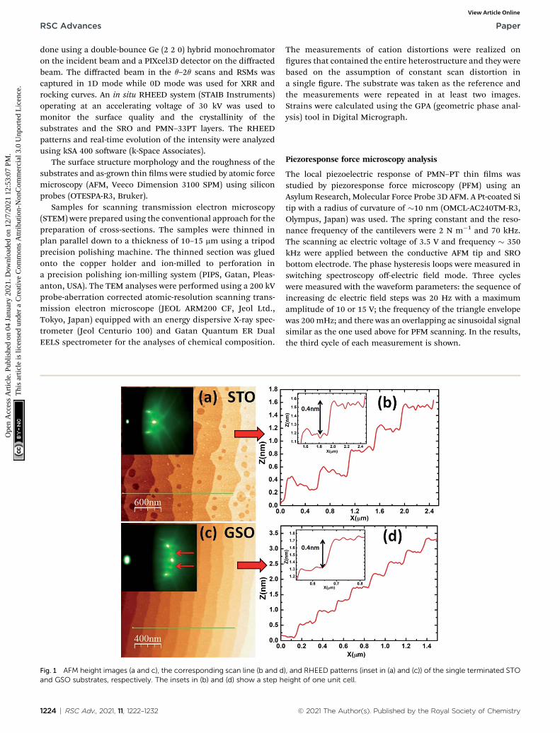
Fig. 1 AFM height images (a and c), the corresponding scan line (b and dand GSO substrates, respectively. The insets in (b) and (d) show a step h
1224 | RSC Adv., 2021, 11, 1222–1232
The measurements of cation distortions were realized ongures that contained the entire heterostructure and they werebased on the assumption of constant scan distortion ina single gure. The substrate was taken as the reference andthe measurements were repeated in at least two images.Strains were calculated using the GPA (geometric phase anal-ysis) tool in Digital Micrograph.
Piezoresponse force microscopy analysis
The local piezoelectric response of PMN–PT thin lms wasstudied by piezoresponse force microscopy (PFM) using anAsylum Research, Molecular Force Probe 3D AFM. A Pt-coated Sitip with a radius of curvature of �10 nm (OMCL-AC240TM-R3,Olympus, Japan) was used. The spring constant and the reso-nance frequency of the cantilevers were 2 N m�1 and 70 kHz.The scanning ac electric voltage of 3.5 V and frequency � 350kHz were applied between the conductive AFM tip and SRObottom electrode. The phase hysteresis loops were measured inswitching spectroscopy off-electric eld mode. Three cycleswere measured with the waveform parameters: the sequence ofincreasing dc electric eld steps was 20 Hz with a maximumamplitude of 10 or 15 V; the frequency of the triangle envelopewas 200 mHz; and there was an overlapping ac sinusoidal signalsimilar as the one used above for PFM scanning. In the results,the third cycle of each measurement is shown.
), and RHEED patterns (inset in (a) and (c)) of the single terminated STOeight of one unit cell.
© 2021 The Author(s). Published by the Royal Society of Chemistry

Paper RSC Advances
Ope
n A
cces
s A
rtic
le. P
ublis
hed
on 0
4 Ja
nuar
y 20
21. D
ownl
oade
d on
12/
7/20
21 1
2:53
:07
PM.
Thi
s ar
ticle
is li
cens
ed u
nder
a C
reat
ive
Com
mon
s A
ttrib
utio
n-N
onC
omm
erci
al 3
.0 U
npor
ted
Lic
ence
.View Article Online
III. Results and discussionGrowth control of PMN–33PT/SRO on singly terminatedsubstrates
The growth of epitaxial thin lms with atomic precision andsharp interfaces requires atomically at and singly terminatedsubstrates. In this work, a ScO2-terminated surface for theReScO3 (Re ¼ Dy, Tb, Gd, Sm, and Nd) substrates was achievedby annealing at high temperatures followed by chemical etchingusing a NaOH-deionized water solution.42 For the STOsubstrates, a TiO2 terminated surface was obtained aer etchingwith buffered-HF solution followed by annealing at hightemperature.43
The examples of AFM topography and the RHEED patterns ofthe treated STO and GSO substrates are shown in Fig. 1. Thepatterns show well-dened step edges with a height of one unitcell, conrming the single TiO2 and ScO2 surface terminationsof the STO and ReScO3 substrates, respectively. Furthermore,the RHEED patterns (insets in Fig. 1(a and c)) of both substratetypes recorded at room temperature in vacuum show sharp andnarrow diffraction spots with the presence of Kikuchi lineswhich suggests crystalline and atomically at surfaces. TheRHEED patterns for the ReScO3 substrates present additionalhalf-order spots (indicated by arrows) which are evidence oforthorhombic symmetry.
Note that controlling the growth at the atomic level for boththe SRO bottom electrode layer and PMN–33PT is key forobtaining the high-quality lms and understanding theirgrowth mechanism. Here, we used high pressure in situ RHEEDto investigate the real-time growth evolution of the studiedheterostructures.
In Fig. 2(a), an example is shown for the real-time specularRHEED spot intensity evolution recorded during the growth of
Fig. 2 Time dependence of the RHEED intensity of the specular spot dur(b) PMN–33PT on different SRO templates. The inset in (a) shows the RHRHEED patterns at a different stage of the growth for PMN–33PT grown
© 2021 The Author(s). Published by the Royal Society of Chemistry
SRO on the SSO substrate with a ScO2-terminated surface. Ascan be seen from Fig. 2(a), clear RHEED oscillations were ob-tained for the SRO grown on SSO up to a thickness of about20 nm giving evidence of a 2D (layer-by-layer) growth mode. TheRHEED pattern recorded aer the deposition of SRO (inset inFig. 2(a)) exhibits sharp diffraction spots suggesting a verysmooth 2D surface and high epitaxial quality which is crucialfor the growth of functional PMN–33PT layers.
The real-time RHEED intensity evolution during the growthof PMN–33PT on SRO/STO and SRO/ReScO3 templates is dis-played in Fig. 2(b). The rst step of the growth of PMN–33PTlayer is characterized by a decrease in the intensity of thespecular RHEED spot followed by clear oscillations (maximum 7oscillations for PMN–33PT/SRO/GSO which corresponds toabout 3 nm of the deposited PMN–33PT layer) and, then, theintensity remains constant with increasing deposition timesuggesting a transition from a 2D layer-by-layer to a step-owgrowth mode. Then, several RHEED images were taken ata different stage of growth (Fig. 2(c)). The RHEED patterns havesharp lines and insignicant intensity variations until thegrowth end (2500 pulses), and it conrms a smooth surface and2D-growth mode. Note that similar RHEED patterns wereobserved on all studied PMN–33PT layers grown on differentsubstrates. The deposition rate was estimated from RHEEDoscillations and it is found to be in the range of 0.0172–0.0197 nm per pulse. The thickness estimated from this depo-sition rate, matches well with the total thicknesses obtainedfrom the X-ray reectivity (Fig. S3 in the ESI†) and TEM analysis.This is a proof of the negligible change in the deposition rateduring the growth.
At room temperature, the surface quality of all as-grownPMN–33PT lms was checked using RHEED and AFM (Fig. S1in the ESI†). The obtained RHEED patterns show sharp streaks
ing the growth of (a) SRO on ScO2 single terminated SSO substrate andEED pattern recorded after the deposition of SRO layer. (c) Example ofSRO/SSO.
RSC Adv., 2021, 11, 1222–1232 | 1225

Fig. 3 The diagram presents a comparison of the pseudocubic lattice constants and the lattice mismatch between STO and ReScO3 (Dy–Nd)substrates and PMN–33PT and SRO.
RSC Advances Paper
Ope
n A
cces
s A
rtic
le. P
ublis
hed
on 0
4 Ja
nuar
y 20
21. D
ownl
oade
d on
12/
7/20
21 1
2:53
:07
PM.
Thi
s ar
ticle
is li
cens
ed u
nder
a C
reat
ive
Com
mon
s A
ttrib
utio
n-N
onC
omm
erci
al 3
.0 U
npor
ted
Lic
ence
.View Article Online
with the presence of diffraction spots evidencing a smoothsurface and high crystalline quality of the PMN–33PT layers.The obtained root mean square (RMS) roughness from AFMtopography was found to be in the order of the unit cell height(varying from 0.267 nm for PMN–33PT/SRO/SSO to 0.486 nm forPMN–33PT/SRO/STO) conrming the smooth surface of allsamples.
Strain effect and structural investigation using HR-XRD andHR-STEM
As mentioned above, the STO and ReScO3 substrates applydifferent epitaxial strain states upon the subsequent lms(Fig. 3). As can be seen from the gure, SRO experiencesa compressive strain when grown on STO (�0.64%) anda tensile strain when grown on the ReScO3 substrates rangingfrom �0.64 to +2.06% while PMN–33PT is compressivelystrained in the range from �2.9% (STO) to �0.12% (NSO).
To determine the crystalline quality and study the effect ofthe strain on the structural characteristics of PMN–33PT/SROheterostructures, the high-resolution XRD (q–2q, rockingcurve, and RSM) and XRR were carried out. According to the q–
Fig. 4 (a) q–2q XRD patterns of PMN–33PT/SRO/(STO and ReScO3) he(001) diffraction peak.
1226 | RSC Adv., 2021, 11, 1222–1232
2q patterns of all the studied samples, all heterostructures aresingle-phase and pyrochlore-free. The presence of Laue fringesaround the main Bragg peaks is indicative of epitaxial high-quality thin-lm growth (Fig. 4(a) and S2 in the ESI†). As wecan see from Fig. 4(b), the rocking curves obtained for all thePMN–33PT thin lms have low full width-at-half-maximum(FWHM) values (Du < 0.06�) that indicates a relatively low-level mosaicity and conrms the high crystalline quality of theheterostructures. Note that the SRO layers also present very lowmosaicity with FWHM values of 0.05–0.08� (not shown here).The XRR patterns of all PMN–33PT/SRO heterostructures(Fig. S3 in the ESI†) show the presence of clear nite Kiesigfringes, which indicates high interface quality and smoothsurfaces. The obtained roughness from the simulation of theXRR patterns is below 0.4 nm for the SRO layers and close to thevalues obtained from AFM for the PMN–33PT layers.
Except for the PMN–33PT/SRO/STO and PMN–33PT/SRO/NSO heterostructures, one can see from Fig. 4(a) that the out-of-plane lattice parameter of PMN–33PT increases (2q of thediffracted peaks shis to lower angles) when the latticemismatch increases which is consistent with an increase in thein-plane compressive strain. The HR-XRD RSM studies were
terostructures and (b) the rocking curves of PMN–33PT layers around
© 2021 The Author(s). Published by the Royal Society of Chemistry

Paper RSC Advances
Ope
n A
cces
s A
rtic
le. P
ublis
hed
on 0
4 Ja
nuar
y 20
21. D
ownl
oade
d on
12/
7/20
21 1
2:53
:07
PM.
Thi
s ar
ticle
is li
cens
ed u
nder
a C
reat
ive
Com
mon
s A
ttrib
utio
n-N
onC
omm
erci
al 3
.0 U
npor
ted
Lic
ence
.View Article Online
also performed to obtain information about the in-plane cellparameters, strain state and domain structures of the PMN–33PT layers. The selected RSMs around symmetric 002 andasymmetric 013/0�13 reections for PMN–33PT/SRO underdifferent strain states grown on DSO, GSO, and SSO are shownin Fig. 5(a–f). The RSMs of the other PMN–33PT/SRO hetero-structures are shown in Fig. S4 in the ESI.† First, the obtainedresults indicate that all SRO layers are fully strained (same Qx asthe substrate), except for SRO/NSO (Fig. S4(b)†), where the in-plane cell parameter of SRO layer is different from the NSOsubstrate. The second observation is that the PMN–33PT layersgrown on SRO/DSO, TSO, GSO, and SSO are coherently strained,while the PMN–33PT layers grown on SRO/STO (Fig. S4(a)†) andSRO/NSO (Fig. S4(b)†) are relaxed and partially relaxed,respectively, with respect to the substrate lattice. The partialstrain relaxation in the SRO layer grown on NSO and strainrelaxation of the PMN–33PT layer grown on SRO/STO is relatedto the large epitaxial lattice mismatch with the substrate ofabout 2% and �2.9%, respectively. The partial strain relaxation
Fig. 5 (002) and (013) RSMs of PMN–33PT/SRO/DSO heterostructure shoheterostructures showing the butterfly-like shape (c–f).
© 2021 The Author(s). Published by the Royal Society of Chemistry
of the PMN–33PT layer grown on SRO/NSO is due to the bottomSRO layer which is not fully strained. Noting that, by reducingthe thickness of SRO or by using a bottom electrode with a smalllattice mismatch with NSO substrate (e.g., Ba0.5Sr0.5RuO3),a fully strained PMN–33PT could be grown on NSO substrate.10
It is intriguing to note from Fig. 5 that the fully strainedPMN–33PT layers present a progressive change of the RSMreection from a disc-like shape for highly strained lms(Fig. 5(a and b)) to a classic buttery-like shape for mildlystrained lms grown on GSO and SSO substrates (Fig. 5(c–f)).The presence of such a buttery-like shaped pattern in PMN–33PT lms is likely evidence of the presence of relaxor nano-domain structures.10,26 However, the disappearance of thebuttery-like shape for the highly strained lm (PMN–33PT/SRO/DSO) is an indication of a change in the polar domainsand can be explained by the reduction of the relaxor domainsand the disorder within the PMN–33PT structure.
Recently, Kim et al. investigated the effect of biaxial-strain in55 nm PMN–32PT/Ba0.5Sr0.5RuO3/GSO, SSO, and NSO and
wing the disk-like shape (a and b) and PMN–33PT/SRO/(GSO and SSO)
RSC Adv., 2021, 11, 1222–1232 | 1227
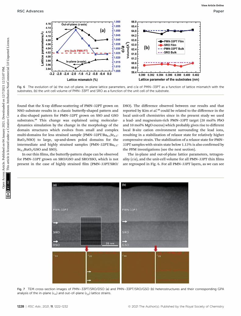
Fig. 6 The evolution of (a) the out-of-plane, in-plane lattice parameters, and c/a of PMN–33PT as a function of lattice mismatch with thesubstrates, (b) the unit cell volume of PMN–33PT and SRO as a function of the unit cell of the substrate.
RSC Advances Paper
Ope
n A
cces
s A
rtic
le. P
ublis
hed
on 0
4 Ja
nuar
y 20
21. D
ownl
oade
d on
12/
7/20
21 1
2:53
:07
PM.
Thi
s ar
ticle
is li
cens
ed u
nder
a C
reat
ive
Com
mon
s A
ttrib
utio
n-N
onC
omm
erci
al 3
.0 U
npor
ted
Lic
ence
.View Article Online
found that the X-ray diffuse-scattering of PMN–32PT grown onNSO substrate results in a classic buttery-shaped pattern anda disc-shaped pattern for PMN–32PT grown on SSO and GSOsubstrates.10 This change was explained using molecular-dynamics simulation by the change in the morphology of thedomain structures which evolves from small and complexmulti-domains for less strained sample (PMN–32PT/Ba0.5Sr0.5-RuO3/NSO) to large, up-and-down poled domains for theintermediate and highly strained samples (PMN–32PT/Ba0.5-Sr0.5RuO3/GSO and SSO).
In our thin lms, the buttery-pattern shape can be observedfor PMN–33PT grown on SRO/GSO and SRO/SSO, which is notpresent in the case of highly strained lm (PMN–33PT/SRO/
Fig. 7 TEM cross-section images of PMN–33PT/SRO/DSO (a) and PManalysis of the in-plane (3xx) and out-of-plane (3yy) lattice strains.
1228 | RSC Adv., 2021, 11, 1222–1232
DSO). The difference observed between our results and thatreported by Kim et al.10 could be related to the difference in thelocal unit-cell chemistries since in the present study we useda lead- and magnesium-rich PMN–33PT target (20 mol% PbOand 10 mol%MgO excess) which probably gives rise to differentlocal B-site cation environment surrounding the lead ions,resulting in a stabilization of relaxor state for relatively highercompressive strain. The stabilization of a relaxor state for PMN–33PT samples with strain state below 1.13% is also conrmed bythe PFM investigations (see the next section).
The in-plane and out-of-plane lattice parameters, tetragon-ality (c/a), and the unit-cell volume for all PMN–33PT thin lmsare regrouped in Fig. 6. For all PMN–33PT layers, as we can see
N–33PT/SRO/GSO (b) heterostructures and their corresponding GPA
© 2021 The Author(s). Published by the Royal Society of Chemistry
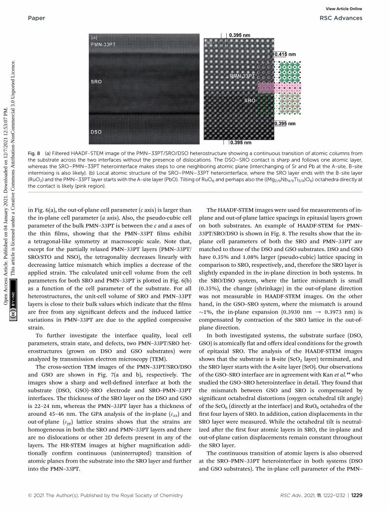
Fig. 8 (a) Filtered HAADF-STEM image of the PMN–33PT/SRO/DSO heterostructure showing a continuous transition of atomic columns fromthe substrate across the two interfaces without the presence of dislocations. The DSO–SRO contact is sharp and follows one atomic layer,whereas the SRO–PMN–33PT heterointerface makes steps to one neighboring atomic plane (interchanging of Sr and Pb at the A-site, B-siteintermixing is also likely). (b) Local atomic structure of the SRO–PMN–33PT heterointerface, where the SRO layer ends with the B-site layer(RuO2) and the PMN–33PT layer starts with the A-site layer (PbO). Tilting of RuO6 and perhaps also the ((Mg2/9Nb4/9Ti3/9)O6) octahedra directly atthe contact is likely (pink region).
Paper RSC Advances
Ope
n A
cces
s A
rtic
le. P
ublis
hed
on 0
4 Ja
nuar
y 20
21. D
ownl
oade
d on
12/
7/20
21 1
2:53
:07
PM.
Thi
s ar
ticle
is li
cens
ed u
nder
a C
reat
ive
Com
mon
s A
ttrib
utio
n-N
onC
omm
erci
al 3
.0 U
npor
ted
Lic
ence
.View Article Online
in Fig. 6(a), the out-of-plane cell parameter (c axis) is larger thanthe in-plane cell parameter (a axis). Also, the pseudo-cubic cellparameter of the bulk PMN–33PT is between the c and a axes ofthe thin lms, showing that the PMN–33PT lms exhibita tetragonal-like symmetry at macroscopic scale. Note that,except for the partially relaxed PMN–33PT layers (PMN–33PT/SRO/STO and NSO), the tetragonality decreases linearly withdecreasing lattice mismatch which implies a decrease of theapplied strain. The calculated unit-cell volume from the cellparameters for both SRO and PMN–33PT is plotted in Fig. 6(b)as a function of the cell parameter of the substrate. For allheterostructures, the unit-cell volume of SRO and PMN–33PTlayers is close to their bulk values which indicate that the lmsare free from any signicant defects and the induced latticevariations in PMN–33PT are due to the applied compressivestrain.
To further investigate the interface quality, local cellparameters, strain state, and defects, two PMN–33PT/SRO het-erostructures (grown on DSO and GSO substrates) wereanalyzed by transmission electron microscopy (TEM).
The cross-section TEM images of the PMN–33PT/SRO/DSOand GSO are shown in Fig. 7(a and b), respectively. Theimages show a sharp and well-dened interface at both thesubstrate (DSO, GSO)–SRO electrode and SRO–PMN–33PTinterfaces. The thickness of the SRO layer on the DSO and GSOis 22–24 nm, whereas the PMN–33PT layer has a thickness ofaround 45–46 nm. The GPA analysis of the in-plane (3xx) andout-of-plane (3yy) lattice strains shows that the strains arehomogeneous in both the SRO and PMN–33PT layers and thereare no dislocations or other 2D defects present in any of thelayers. The HR-STEM images at higher magnication addi-tionally conrm continuous (uninterrupted) transition ofatomic planes from the substrate into the SRO layer and furtherinto the PMN–33PT.
© 2021 The Author(s). Published by the Royal Society of Chemistry
The HAADF-STEM images were used for measurements of in-plane and out-of-plane lattice spacings in epitaxial layers grownon both substrates. An example of HAADF-STEM for PMN–33PT/SRO/DSO is shown in Fig. 8. The results show that the in-plane cell parameters of both the SRO and PMN–33PT arematched to those of the DSO and GSO substrates. DSO and GSOhave 0.35% and 1.08% larger (pseudo-cubic) lattice spacing incomparison to SRO, respectively, and, therefore the SRO layer isslightly expanded in the in-plane direction in both systems. Inthe SRO/DSO system, where the lattice mismatch is small(0.35%), the change (shrinkage) in the out-of-plane directionwas not measurable in HAADF-STEM images. On the otherhand, in the GSO–SRO system, where the mismatch is around�1%, the in-plane expansion (0.3930 nm / 0.3973 nm) iscompensated by contraction of the SRO lattice in the out-of-plane direction.
In both investigated systems, the substrate surface (DSO,GSO) is atomically at and offers ideal conditions for the growthof epitaxial SRO. The analysis of the HAADF-STEM imagesshows that the substrate is B-site (ScO2 layer) terminated, andthe SRO layer starts with the A-site layer (SrO). Our observationsof the GSO–SRO interface are in agreement with Kan et al.44 whostudied the GSO–SRO heterointerface in detail. They found thatthe mismatch between GSO and SRO is compensated bysignicant octahedral distortions (oxygen octahedral tilt angle)of the ScO6 (directly at the interface) and RuO6 octahedra of therst four layers of SRO. In addition, cation displacements in theSRO layer were measured. While the octahedral tilt is neutral-ized aer the rst four atomic layers in SRO, the in-plane andout-of-plane cation displacements remain constant throughoutthe SRO layer.
The continuous transition of atomic layers is also observedat the SRO–PMN–33PT heterointerface in both systems (DSOand GSO substrates). The in-plane cell parameter of the PMN–
RSC Adv., 2021, 11, 1222–1232 | 1229

Fig. 9 Topography (a–d) height and (e–h) deflection images, PFM out-of-plane (i–l) amplitude, and (m–p) phase images of PMN–33PT thin filmson different substrates. The crosses in (i–l) correspond to the measurement points of the local PFM phase hysteresis loops shown in panels (q–t).
RSC Advances Paper
Ope
n A
cces
s A
rtic
le. P
ublis
hed
on 0
4 Ja
nuar
y 20
21. D
ownl
oade
d on
12/
7/20
21 1
2:53
:07
PM.
Thi
s ar
ticle
is li
cens
ed u
nder
a C
reat
ive
Com
mon
s A
ttrib
utio
n-N
onC
omm
erci
al 3
.0 U
npor
ted
Lic
ence
.View Article Online
33PT is coherent with SRO, meaning that the PMN–33PT layer iscontracted from 0.4018 nm to 0.395 nm and 0.397 nm insystems with DSO and GSO, respectively. The shrinkage is notaccompanied by the formation of mist dislocations but iscompensated by expansion in the out-of-plane direction asalready indicated by XRD results. The out-of-plane cell param-eter values measured from HAADF-STEM images are 0.415 nmon DSO and 0.409 nm on GSO and they are in good agreementwith those obtained by XRD. The HAADF-STEM images of theSRO–PMN–33PT contact show that the interface is stepped, withsteps limited to one atomic plane, where a certain degree ofcation mixing is expected. The formation of steps is also aneffective mechanism for compensation of lattice mismatch.Also, at this heterointerface, the compensation of octahedral tiltin the SRO layer is expected.
Investigation of piezoelectric performance by PFM
The local piezoelectric performance was investigated by pie-zoresponse force microscopy in PMN–33PT lms on NSO, SSO,GSO, and DSO substrates. The topography, deection, and PFMamplitude and phase images are shown in Fig. 9. The PMN–33PT lms with a compressive strain below �1.13% grown onNSO, SSO, and GSO did not show piezoelectric response, onlynoise is observed. These results could indicate the relaxor-likebehavior similar to that in other lead-based relaxors duringthe rst few PFM scans,45 but, also, the paraelectric behaviour isnot excluded. According to the literature,41 in PMN–PT bulkceramics and thicker lms, the compositions above x ¼ 0.30possess ferroelectric-like behaviour. However, when decreasingthe average grain size, relaxor-like behavior prevails.46
1230 | RSC Adv., 2021, 11, 1222–1232
On the other hand, in the lms grown on DSO substrates,piezoelectric response is observed (Fig. 9(l and p)), most prob-ably due to the largest compressive strain (�1.9%) induced inthese lms, as conrmed by the XRD RSMs results. A fewhundred nanometre-sized irregularly shaped ferroelectricdomains are present. A similar domain structure was observedalso in thicker PMN–33PT lms prepared by PLD.41 Theseresults indicate that the large compressive stress as well asapplication of dc elds induce the relaxor-to-ferroelectriccrossover in a few tens of nanometre-thick relaxor-like PMN–33PT lms. When applying larger electric elds, theferroelectric-like behaviour appears in all samples, which isevident from the local PFM phase hysteresis loops shown inFig. 9(q–t).
Interestingly, the PFM hysteresis loops (Fig. 9(q–t)) show thatPMN–33PT grown on SSO substrate with a strain state of��0.8% exhibits a large coercive eld (Fig. 9(r)). Note that thiscoercive eld is about two times larger than that of the PMN–33PT thin lms grown on GSO and NSO substrates. Theseresults are in agreement with the recent study reported by Kimet al.10 in which enhanced electromechanical response in PMN–32PT/Ba0.5Sr0.5RuO3/SSO with an increase in the saturationpolarization and the coercive eld was observed. Using themolecular-dynamics simulations, the authors showed that,while the compressive strain drives a tendency toward moreferroelectric-like order, certain unit cells become more disor-dered at some critical applied strain.
The obtained results demonstrate that the effect of the strainin relaxor ferroelectric thin lms manifests differentlycompared to the classical ferroelectrics and the stabilization of
© 2021 The Author(s). Published by the Royal Society of Chemistry

Paper RSC Advances
Ope
n A
cces
s A
rtic
le. P
ublis
hed
on 0
4 Ja
nuar
y 20
21. D
ownl
oade
d on
12/
7/20
21 1
2:53
:07
PM.
Thi
s ar
ticle
is li
cens
ed u
nder
a C
reat
ive
Com
mon
s A
ttrib
utio
n-N
onC
omm
erci
al 3
.0 U
npor
ted
Lic
ence
.View Article Online
different unit-cell ordering could be used as an effective way fortailoring and enhancing the functional properties in relaxorferroelectrics.
IV. Conclusion
In the present study, we reported the growth of the epitaxial45 nm PMN–33PT/20 nm SRO heterostructures using PLD andinvestigated the effect of compressive strain on the domainstructures and piezoelectric response of PMN–33PT. By usinga well-dened substrate surface (STO and ReScO3 substrateswith Re ¼ Dy, Tb, Gd, Sm, and Nd), the high quality and stoi-chiometric heterostructures were obtained. In situ high-pressure RHEED was used to control the real-time growth ofSRO and PMN–33PT layers revealing a 2D growth mode.Combining HR-XRD and HR-STEM investigations, a fullystrained PMN–33PT/SRO heterostructures were revealed onDSO, TSO, GSO, and SSO substrates with sharp interfaces con-rming the 2D cube-on-cube growthmode. In addition, the GPAanalysis showed homogeneous strain within the hetero-structures with the absence of defects. The HR-XRD RSMs ofPMN–33PT layers revealed a progressive evolution of thedomain structures from buttery-shaped reciprocal space mapsto disc-shaped patterns with increasing compressive biaxialstrain. These changes were explained by the stabilization ofrelaxor domains for low strain states which evolves to moredistorted ferroelectric domains for the highest achieved strainstates. The piezoelectric force microscopy studies revealedtypical relaxor-like behaviour for a strain state below �1.13%while for higher compressive strain irregularly shaped andpoled ferroelectric domains were observed in agreement withthe RSMs results. Interestingly, the local PFM piezoelectricphase hysteresis loops revealed a large coercive eld for PMN–33PT/SRO/SSO heterostructure (strain state of ��0.8%). Notingthat, the results obtained in the present study demonstrate thatthe domain structures and piezoelectric performances inrelaxor ferroelectric thin lms are sensitive to applied externalstrain which could be used to tailor or enhance the functionalproperties.
Conflicts of interest
There are no conicts to declare.
Acknowledgements
This research is nanced by the Slovenian Research Agency[grant numbers J2-9237, J2-2510, N2-0149, P2-0091, and P2-0105]. L. W. M. acknowledges support from the NationalScience Foundation under Grant DMR-1708615.
References
1 R. A. Cowley, S. N. Gvasaliya, S. G. Lushnikov, B. Roessli andG. M. Rotaru, Relaxing with relaxors: a review of relaxorferroelectrics, Adv. Phys., 2011, 60, 229–327.
© 2021 The Author(s). Published by the Royal Society of Chemistry
2 H. Fu and R. E. Cohen, Polarization rotation mechanism forultrahigh electromechanical response in single-crystalpiezoelectrics, Nature, 2000, 403, 281.
3 Z. Kutnjak, J. Petzelt and R. Blinc, The giantelectromechanical response in ferroelectric relaxors asa critical phenomenon, Nature, 2006, 441, 956–959.
4 M. Ahart, et al., Origin of morphotropic phase boundaries inferroelectrics, Nature, 2008, 451, 545–548.
5 J.-M. Kiat, et al., Monoclinic structure of unpoledmorphotropic high piezoelectric PMN-PT and PZN-PTcompounds, Phys. Rev. B: Condens. Matter Mater. Phys.,2002, 65, 064106.
6 Z.-G. Ye, High-Performance Piezoelectric Single Crystals ofComplex Perovskite Solid Solutions, MRS Bull., 2009, 34,277–283.
7 C. He, et al., Composition and orientation dependence ofhigh electric-eld-induced strain in Pb(In1/2Nb1/2)O3–
Pb(Mg1/3Nb2/3)O3–PbTiO3 single crystals, J. Appl. Phys.,2012, 112, 126102.
8 F. Li, et al., Ultrahigh piezoelectricity in ferroelectricceramics by design, Nat. Mater., 2018, 17, 349.
9 F. Li, S. Zhang, Z. Xu and L.-Q. Chen, The Contributions ofPolar Nanoregions to the Dielectric and PiezoelectricResponses in Domain-Engineered Relaxor-PbTiO3 Crystals,Adv. Funct. Mater., 2017, 27, 1700310.
10 J. Kim, et al., Epitaxial Strain Control of Relaxor FerroelectricPhase Evolution, Adv. Mater., 2019, 31, 1901060.
11 M. J. Krogstad, et al., The relation of local order to materialproperties in relaxor ferroelectrics, Nat. Mater., 2018, 17,718–724.
12 H. Takenaka, I. Grinberg, S. Liu and A. M. Rappe, Slush-likepolar structures in single-crystal relaxors, Nature, 2017, 546,391–395.
13 H. Takenaka, I. Grinberg and A. M. Rappe, Seeing the forestand the trees, Nat. Mater., 2018, 17, 657–658.
14 A. Kumar, et al., Atomic-resolution electron microscopy ofnanoscale local structure in lead-based relaxorferroelectrics, Nat. Mater., 2020, 1–6, DOI: 10.1038/s41563-020-0794-5.
15 M. Otonicar, et al., Connecting the Multiscale Structure withMacroscopic Response of Relaxor Ferroelectrics, Adv. Funct.Mater., 2020, 2006823.
16 S. H. Baek, et al., Giant piezoelectricity on Si for hyperactiveMEMS, Science, 2011, 334, 958–961.
17 S. Pandya, et al., Pyroelectric energy conversion with largeenergy and power density in relaxor ferroelectric thinlms, Nat. Mater., 2018, 17, 432–438.
18 A. S. Mischenko, Q. Zhang, J. F. Scott, R. W. Whatmore andN. D. Mathur, Giant Electrocaloric Effect in Thin-FilmPbZr0.95Ti0.05O3, Science, 2006, 311, 1270–1271.
19 M. Mietschke, et al., Inuence of the polarization anisotropyon the electrocaloric effect in epitaxial PMN-PT thin lms, J.Appl. Phys., 2016, 120, 114102.
20 X. Wang, L. Zhang, X. Hao and S. An, High energy-storageperformance of 0.9Pb(Mg1/3Nb2/3)O3-0.1PbTiO3 relaxorferroelectric thin lms prepared by RF magnetronsputtering, Mater. Res. Bull., 2015, 65, 73–79.
RSC Adv., 2021, 11, 1222–1232 | 1231
RSC Advances Paper
Ope
n A
cces
s A
rtic
le. P
ublis
hed
on 0
4 Ja
nuar
y 20
21. D
ownl
oade
d on
12/
7/20
21 1
2:53
:07
PM.
Thi
s ar
ticle
is li
cens
ed u
nder
a C
reat
ive
Com
mon
s A
ttrib
utio
n-N
onC
omm
erci
al 3
.0 U
npor
ted
Lic
ence
.View Article Online
21 U. Gabor, et al., Stabilization of the perovskite phase in PMN-PT epitaxial thin lms via increased interface roughness,Appl. Surf. Sci., 2020, 513, 145787.
22 E. A. Eliseev and M. D. Glinchuk, Static properties of relaxorferroelectric thin lms, J. Appl. Phys., 2007, 102, 104110.
23 P. Miao, et al., Ferroelectricity and Self-Polarization inUltrathin Relaxor Ferroelectric Films, Sci. Rep., 2016, 6,19965.
24 A. Fernandez, J. Kim, D. Meyers, S. Saremi and L. W. Martin,Finite-size effects in lead scandium tantalate relaxor thinlms, Phys. Rev. B, 2020, 101, 094102.
25 S. Jiao, et al., Growth and electrical properties of epitaxial0.7Pb(Mg1/3Nb2/3)O3-0.3PbTiO3 thin lm by pulsed laserdeposition, J. Mater. Sci.: Mater. Electron., 2018, 29, 6779–6784.
26 S. Saremi, J. Kim, A. Ghosh, D. Meyers and L. W. Martin,Defect-Induced (Dis)Order in Relaxor Ferroelectric ThinFilms, Phys. Rev. Lett., 2019, 123, 207602.
27 M. D. Nguyen, E. P. Houwman, M. T. Do and G. Rijnders,Relaxor-ferroelectric thin lm heterostructure with largeimprint for high energy-storage performance at lowoperating voltage, Energy Storage Materials, 2020, 25, 193–201.
28 A. R. Damodaran, et al., New modalities of strain-control offerroelectric thin lms, J. Phys.: Condens. Matter, 2016, 28,263001.
29 M. Dawber, K. M. Rabe and J. F. Scott, Physics of thin-lmferroelectric oxides, Rev. Mod. Phys., 2005, 77, 1083–1130.
30 L. W. Martin and A. M. Rappe, Thin-lm ferroelectricmaterials and their applications, Nat. Rev. Mater., 2017, 2,16087.
31 S. Prosandeev, D. Wang and L. Bellaiche, Properties ofEpitaxial Films Made of Relaxor Ferroelectrics, Phys. Rev.Lett., 2013, 111, 247602.
32 M. Tyunina, J. Levoska, P.-E. Janolin and A. Dejneka, Low-temperature relaxor state induced by epitaxial compressionin PbSc0.5Nb0.5O3 lms, Phys. Rev. B: Condens. MatterMater. Phys., 2013, 87, 224107.
33 D. H. A. Blank, J. M. Dekkers and A. J. H. M. Rijnders, Pulsedlaser deposition in Twente: from research tool towardsindustrial deposition, J. Phys. D: Appl. Phys., 2014, 47,034006.
34 M. D. Nguyen, R. Tiggelaar, T. Aukes, G. Rijnders andG. Roelof, Wafer-scale growth of highly textured
1232 | RSC Adv., 2021, 11, 1222–1232
piezoelectric thin lms by pulsed laser deposition formicro-scale sensors and actuators, J. Phys.: Conf. Ser., 2017,922, 012022.
35 J. Belhadi, F. Ravaux, H. Bouyanf, M. Jouiad and M. ElMarssi, Quantication and mapping of elastic strains inferroelectric [BaZrO3]x/[BaTiO3](1-x) superlattices, Appl. Surf.Sci., 2020, 512, 145761.
36 M. Boota, E. P. Houwman, M. D. Nguyen, G. Lanzara andG. Rijnders, Effect of fabrication conditions on phaseformation and properties of epitaxial (PbMg1/3Nb2/3O3)0.67-(PbTiO3)0.33 thin lms on (001) SrTiO3, AIP Adv., 2016, 6,055303.
37 P. Chekhonin, et al., Effect of substrate miscut on themicrostructure in epitaxial Pb(Mg1/3Nb2/3)O3-PbTiO3 thinlms, Mater. Charact., 2017, 234–241.
38 J. Wang, K. H. Wong, H. L. W. Chan and C. L. Choy,Composition control and electrical properties of PMN-PTthin lms around the morphotropic boundary, Appl. Phys.A, 2004, 79, 551–556.
39 G. Koster, et al., Structure, physical properties, andapplications of SrRuO3 thin lms, Rev. Mod. Phys., 2012,84, 253–298.
40 K. S. Takahashi, et al., Inverse tunnel magnetoresistance inall-perovskite junctions of La0.7Sr0.3MnO3/SrTiO3/SrRuO3,Phys. Rev. B: Condens. Matter Mater. Phys., 2003, 67, 094413.
41 U. Gabor, et al., Structural peculiarities of 0.67Pb(Mg1/3Nb2/3)O3–0.33PbTiO3 thin lms grown directly on SrTiO3
substrates, J. Eur. Ceram. Soc., 2018, 38, 4453–4462.42 J. E. Kleibeuker, et al., Atomically Dened Rare-Earth
Scandate Crystal Surfaces, Adv. Funct. Mater., 2010, 20,3490–3496.
43 G. Koster, B. L. Kropman, G. J. H. M. Rijnders, D. H. A. Blankand H. Rogalla, Quasi-ideal strontium titanate crystalsurfaces through formation of strontium hydroxide, Appl.Phys. Lett., 1998, 73, 2920–2922.
44 D. Kan, R. Aso, H. Kurata and Y. Shimakawa, Epitaxial straineffect in tetragonal SrRuO3 thin lms, J. Appl. Phys., 2013,113, 173912.
45 U. Prah, et al., Strengthened relaxor behavior in (1�x)Pb(Fe0.5Nb0.5)O3 – xBiFeO3, J. Mater. Chem. C, 2020, 8,3452–3462.
46 M. Alguero, et al., Size effect in morphotropic phaseboundary Pb(Mg1/3Nb2/3)O3–PbTiO3, Appl. Phys. Lett., 2007,91, 112905.
© 2021 The Author(s). Published by the Royal Society of Chemistry