
GREEN Diethanolamine (DEA) and Related DEA …...TOXNET Search Statements – DEA Family of...
104
GREEN Diethanolamine (DEA) and Related DEA-Containing Ingredients CIR EXPERT PANEL MEETING MARCH 3-4, 2011
Transcript of GREEN Diethanolamine (DEA) and Related DEA …...TOXNET Search Statements – DEA Family of...
GREEN
Diethanolamine (DEA) and Related DEA-Containing Ingredients
CIR EXPERT PANEL MEETING
MARCH 3-4, 2011
Memorandum
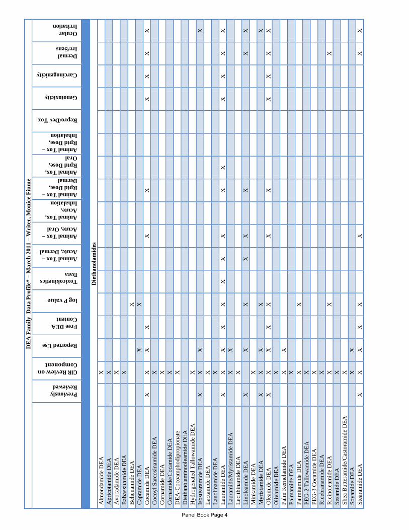
To: CIR Expert Panel Members and Liaisons From: Monice M. Fiume MMF Senior Scientific Analyst/Writer Date: February 10, 2011 Subject: Re-Review of Diethanolamine (DEA) and Related DEA-Containing Ingredients At the December Panel meeting, the Panel made the decision to reopen the safety assessment of Triethanolamine (TEA), Diethanolamine, and Monoethanolamine (MEA). That decision was based on the need to incorporate new data, but most importantly, on the benefit of separating the ethanolamines, and having each of these ingredients be in its own report with a family of related ingredients created for each. The re-review of DEA and 68 DEA-containing ingredients is being submitted for your review. In considering the potential safety issues with DEA-containing ingredients, it was reasoned that, were they to penetrate the skin, the toxicity of most concern would be the DEA moiety. The acid salt ingre-dients, DEA Myristate, for example, would be expected to dissociate into DEA and the corresponding acid. The covalent DEA ingredients, such as cocamide DEA, do not readily dissociate into DEA and the other component. However, in the case of these covalent ingredients, DEA may be of concern as an impurity and/or metabolite. Since this is the first time the groupings are being presented to the Panel, there is an opportunity to make a further determination whether this family of ingredients is appropriate as currently grouped. If it is not, the Panel can make changes. The safety of 8 of the ingredients included in this re-review, as currently grouped, has been reviewed previously by the CIR. Summary information from the existing safety assessments is included in the current re-review document. Additionally, many of the ingredients included in this re-review include a component that has been reviewed by the CIR. For example, DEA-Isostearate is the DEA salt of isostearic acid; isostearic acid has been reviewed by the CIR. Table 2 provides the conclusions from the CIR reports on all the component ingredients. Finally, many of the ingredients are lacking safety data. The Panel should consider any existing CIR reports that can be used to determine the safety of ingredients that dissociate. For those that do not

dissociate, the Panel should consider whether the impurity level of DEA can be used as a determining factor in considering safety. As a reminder, NTP studies have results indicating clear evidence of carcinogenicity in mice for DEA and some DEA fatty acid esters. The Panel determined that the mode of action of DEA carcinogenesis in mice was understood and the penetration was sufficiently well-characterized, such that the carcinogeni-city findings in mice were considered to have no relevance to human health from the use of cosmetics containing DEA. This re-review is the first of the three ethanolamine reports being presented. The re-reviews on TEA and MEA will be presented at later meetings. Also included for your review are previous CIR reports about ingredients discussed in this report.
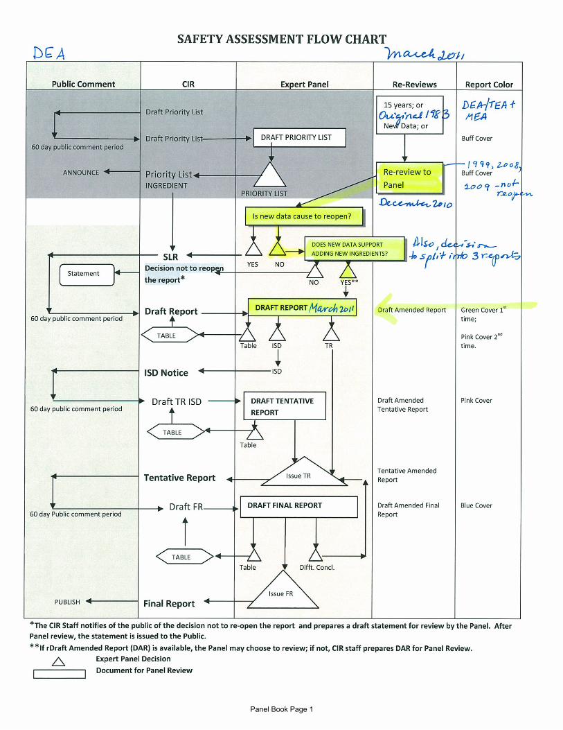
Panel Book Page 1

TEA, DEA, MEA HISTORY
Original Report: In 1983, the Expert Panel determined that these ingredients were safe for use in cosmetic formulations designed for discontinuous, brief use followed by thorough rinsing from the surface of the skin. In products intended for prolonged contact with the skin, the concentration of ethanolamines should not exceed 5%. Ethanolamine (MEA) should be used only in rinse-off products. Triethanolamine (TEA) and diethanolamine (DEA) should not be used in products containing N-nitrosating agents. June 1999: discussed NTP carcinogenicity results; presentations were made by Dr. Lehman-McKeeman and Dr. Stott June 2008: presentation was made by Dr. Stott; Acetamide MEA was discussed, with reference to the MEA, DEA, TEA report June 2009: discussed DEA carcinogenicity; the DEA report was not be reopened December 2010: formal rereview package was presented to the Panel; report was split into 3 separate documents – DEA, TEA, and MEA, add additional ingredients will be added to each report March 2011: the RR of DEA was presented to the Panel, including the new ingredient subgroups
Panel Book Page 2
DE
A F
amily
Dat
a Pr
ofile
* –
Mar
ch 2
011
– W
rite
r, M
onic
e Fi
ume
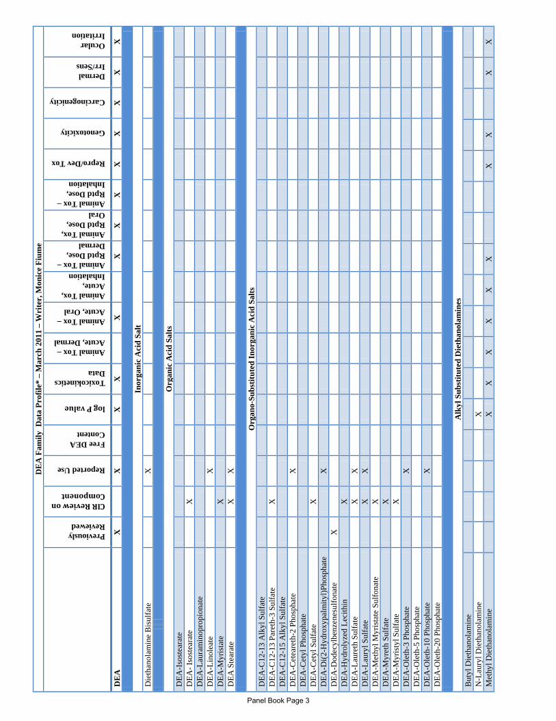
Previously Reviewed
CIR Review on Component
Reported Use
Free DEA Content
log P value
Toxicokinetics Data
Animal Tox – Acute, Dermal
Animal Tox – Acute, Oral
Animal Tox, Acute, Inhalation Animal Tox – Rptd Dose, Dermal Animal Tox, Rptd Dose, Oral Animal Tox – Rptd Dose, Inhalation
Repro/Dev Tox
Genotoxicity
Carcinogenicity
Dermal Irr/Sens
Ocular Irritation
DE
A
X
X
X
X
X
X
X
X
X
X
X
X
X
In
orga
nic
Aci
d Sa
lt D
ieth
anol
amin
e B
isul
fate
X
O
rgan
ic A
cid
Salts
D
EA-I
sost
eara
te
D
EA- I
sost
eara
te
X
DEA
-Lau
ram
inop
ropi
onat
e
DEA
-Lin
olea
te
X
DEA
-Myr
ista
te
X
DEA
Ste
arat
e
X
X
O
rgan
o-Su
bstit
uted
Inor
gani
c A
cid
Salts
D
EA-C
12-1
3 A
lkyl
Sul
fate
DEA
-C12
-13
Pare
th-3
Sul
fate
X
D
EA-C
12-1
5 A
lkyl
Sul
fate
DEA
-Cet
eare
th-2
Pho
spha
te
X
DEA
-Cet
yl P
hosp
hate
DEA
-Cet
yl S
ulfa
te
X
DEA
-Di(2
-Hyd
roxy
palm
ityl)P
hosp
hate
X
D
EA-D
odec
ylbe
nzen
esul
fona
te
X
DEA
-Hyd
roly
zed
Leci
thin
X
D
EA-L
aure
th S
ulfa
te
X
X
D
EA-L
aury
l Sul
fate
X
X
DEA
-Met
hyl M
yris
tate
Sul
fona
te
X
DEA
-Myr
eth
Sulfa
te
X
DEA
-Myr
isty
l Sul
fate
X
D
EA-O
leth
-3 P
hosp
hate
X
D
EA-O
leth
-5 P
hosp
hate
DEA
-Ole
th-1
0 Ph
osph
ate
X
DEA
-Ole
th-2
0 Ph
osph
ate
Alk
yl S
ubst
itute
d D
ieth
anol
amin
es
But
yl D
ieth
anol
amin
e
N-L
aury
l Die
than
olam
ine
X
Met
hyl D
ieth
anol
amin
e
X
X
X
X
X
X
X
X
X
X
Panel Book Page 3
DE
A F
amily
Dat
a Pr
ofile
* –
Mar
ch 2
011
– W
rite
r, M
onic
e Fi
ume
Previously Reviewed
CIR Review on Component
Reported Use
Free DEA Content
log P value
Toxicokinetics Data
Animal Tox – Acute, Dermal
Animal Tox – Acute, Oral
Animal Tox, Acute, Inhalation Animal Tox – Rptd Dose, Dermal Animal Tox, Rptd Dose, Oral Animal Tox – Rptd Dose, Inhalation
Repro/Dev Tox
Genotoxicity
Carcinogenicity
Dermal Irr/Sens
Ocular Irritation
D
ieth
anol
amid
es
Alm
onda
mid
e D
EA
X
Apr
icot
amid
e D
EA
X
Avo
cada
mid
e D
EA
X
Baba
ssua
mid
e DE
A
X
B
ehen
amid
e D
EA
X
Cap
ram
ide
DEA
X
X
Coc
amid
e D
EA
X
X
X
X
X
X
X
X
X
X
C
ocoy
l Sar
cosi
nam
ide
DEA
X
C
orna
mid
e D
EA
X
Cor
nam
ide/
Coc
amid
e D
EA
X
DEA
-Coc
oam
phod
ipro
pion
ate
X
Die
than
olam
inoo
leam
ide
DEA
Hyd
roge
nate
d Ta
llow
amid
e D
EA
X
Isos
tear
amid
e D
EA
X
X
X
X
La
ctam
ide
DEA
X
La
nolin
amid
e D
EA
X
Laur
amid
e D
EA
X
X
X
X
X
X
X
X
X
X
X
X
X
X
La
uram
ide/
Myr
ista
mid
e D
EA
X
X
Le
cith
inam
ide
DEA
X
Li
nole
amid
e D
EA
X
X
X
X
X
X
X
X
M
inka
mid
e D
EA
X
Myr
ista
mid
e D
EA
X
X
X
X
X
Ole
amid
e D
EA
X
X
X
X
X
X
X
X
X
X
X
Oliv
amid
e DE
A
X
Pa
lm K
erne
lam
ide
DEA
X
X
Palm
amid
e D
EA
X
Palm
itam
ide
DEA
X
X
PEG
-2 T
allo
wam
ide
DEA
X
PE
G-3
Coc
amid
e D
EA
X
Ric
ebra
nam
ide
DEA
X
R
icin
olea
mid
e D
EA
X
X
X
Sesa
mid
e D
EA
X
Shea
But
tera
mid
e/C
asto
ram
ide
DEA
X
So
yam
ide
DEA
X
X
Stea
ram
ide
DEA
X
X
X
X
X
X
X
X
Panel Book Page 4

DE
A F
amily
Dat
a Pr
ofile
* –
Mar
ch 2
011
– W
rite
r, M
onic
e Fi
ume
Previously Reviewed
CIR Review on Component
Reported Use
Free DEA Content
log P value
Toxicokinetics Data
Animal Tox – Acute, Dermal
Animal Tox – Acute, Oral
Animal Tox, Acute, Inhalation Animal Tox – Rptd Dose, Dermal Animal Tox, Rptd Dose, Oral Animal Tox – Rptd Dose, Inhalation
Repro/Dev Tox
Genotoxicity
Carcinogenicity
Dermal Irr/Sens
Ocular Irritation
Stea
ram
ide
DEA
-Dis
tear
ate
X
Stea
ram
idoe
thyl
Die
than
olam
ine
St
eara
mid
oeth
yl D
ieth
anol
amin
e H
Cl
Ta
llam
ide
DEA
X
Ta
llow
amid
e D
EA
U
ndec
ylen
amid
e D
EA
X
X
W
heat
Ger
mam
ide
DEA
X
*“
X”
indi
cate
s tha
t dat
a w
ere
avai
labl
e in
a c
ateg
ory
for t
he in
gred
ient
Panel Book Page 5

DE
A S
earc
h In
fo
N
LM
EU
FD
A
Ch
em
Po
rta
l
#
use
s co
nc
da
ta
To
xlin
e-
Pu
bm
ed
Mis
c
NLM
N
TIS
R
eg
istr
y
NT
IS
Me
rck
E
U
SC
CS
EC
E-
TO
C
SID
S
IAR
C
NT
P
EA
FU
S
OT
C
HP
VIS
IU
CLI
D
da
ta s
et
da
te s
ea
rch
ed
1
-7&
12
-11
1
1-2
3
1/1
1
1-2
5-1
1
1
-25
1
-25
1-2
5-1
1
1-2
5-1
1
DE
A
11
1-4
2-2
39
2
35
x
x
II
x x
x
DE
A B
isu
lfa
te
59
21
9-5
6-6
x
II
DE
A-M
yri
sta
te
53
40
4-3
9-0
x
II
DE
A S
tea
rate
n
o
DE
A-I
sost
ea
rate
II
DE
A-L
ino
lea
te
59
23
1-4
2-4
x
II
DE
A-L
au
ram
ino
pro
pio
na
te
65
10
4-3
6-1
x
II
DE
A-L
au
ryl S
ulf
ate
14
3-0
0-0
x
II
DE
A-C
12
-13
Alk
yl S
ulf
ate
II
DE
A-M
yri
styl S
ulf
ate
65
10
4-6
1-2
x
II
DE
A-C
12
-15
Alk
yl S
ulf
ate
II
DE
A-C
ety
l Su
lfa
te
51
54
1-5
1-6
x
II
DE
A-L
au
reth
Su
lfa
te
58
85
5-3
6-0
x
II
DE
A-C
12
-13
Pa
reth
-3 S
ulf
ate
II
DE
A-M
yre
th S
ulf
ate
II
DE
A-D
od
ecy
lbe
nze
ne
Su
lfo
na
te
26
54
5-5
3-9
x
II
DE
A-M
eth
yl M
yri
sta
te S
ulf
on
ate
64
13
1-3
6-8
II
DE
A-C
ety
l Ph
osp
ha
te
61
69
3-4
1-2
x
II
DE
A-C
ete
are
th-2
Ph
osp
ha
te
II
DE
A-O
leth
-3 P
ho
sph
ate
58
85
5-6
3-3
II
DE
A-O
leth
-5 P
ho
sph
ate
58
85
5-6
3-3
x
II
DE
A-O
leth
-10
Ph
osp
ha
te
58
85
5-6
3-3
II
DE
A-O
leth
-20
Ph
osp
ha
te
58
85
5-6
3-3
II
DE
A-H
yd
roly
zed
Le
cith
in
II
DE
A-D
i(2
-Hyd
roxy
pa
lmit
yl)
-
Ph
osp
ha
te[
no
Panel Book Page 6

N
LM
EU
FD
A
Ch
em
Po
rta
l
#
use
s co
nc
da
ta
To
xlin
e-
Pu
bm
ed
Mis
c
NLM
N
TIS
R
eg
istr
y
NT
IS
Me
rck
E
U
SC
CS
EC
E-
TO
C
SID
S
IAR
C
NT
P
EA
FU
S
OT
C
HP
VIS
IU
CLI
D
da
ta s
et
Me
thyl D
ieth
an
ola
min
e
[10
5-5
9-9
]
x
no
x
Bu
tyl D
ieth
an
ola
min
e
10
2-7
9-4
x
X
N-L
au
ryl D
ieth
an
ola
min
e
[15
41
-67
-9 ]
x
II
I
Ca
pra
mid
e D
EA
13
6-2
6-5
x
II
I
Un
de
cyle
na
mid
e D
EA
60
23
9-6
8-1
; 2
53
77
-64
-4
x
III
Lau
ram
ide
DE
A
12
0-4
0-1
x
II
I
x
x
Myri
sta
mid
e D
EA
75
45
-23
-5
x
III
Lau
ram
ide
/ M
yri
sta
mid
e D
EA
II
I
Pa
lmit
am
ide
DE
A
75
45
-24
-6
x
III
Ste
ara
mid
e D
EA
93
-82
-3
x
III
Be
he
na
mid
e D
EA
70
49
6-3
9-8
x
II
I
Lact
am
ide
DE
A
III
Iso
ste
ara
mid
e D
EA
52
79
4-7
9-3
x
X
Ole
am
ide
DE
A
52
99
-69
-4;
93
-83
-4
x
III
x
x
Lin
ole
am
ide
DE
A
56
86
3-0
2-6
x
II
I
Alm
on
da
mid
e D
EA
12
40
46
-18
-0
x
III
Ap
rico
tam
ide
DE
A
18
51
23
-36
-8
x
III
Avo
cad
am
ide
DE
A
12
40
46
-21
-5
x
III
Ba
ba
ssu
am
ide
DE
A
12
40
46
-24
-8
x
III
Co
cam
ide
DE
A
61
79
1-3
1-9
x
II
I
x
x x
Co
rna
mid
e D
EA
II
I
Co
rna
mid
e/
Co
cam
ide
DE
A
III
Hyd
rog
en
ate
d T
allo
wa
mid
e D
EA
68
44
0-3
2-4
x
II
I
Lan
olin
am
ide
DE
A
[85
40
8-8
8-4
]
x
II
I
Leci
thin
am
ide
DE
A
III
Min
ka
mid
e D
EA
12
40
46
-27
-1
x
III
Panel Book Page 7

N
LM
EU
FD
A
Ch
em
Po
rta
l
#
use
s co
nc
da
ta
To
xlin
e-
Pu
bm
ed
Mis
c
NLM
N
TIS
R
eg
istr
y
NT
IS
Me
rck
E
U
SC
CS
EC
E-
TO
C
SID
S
IAR
C
NT
P
EA
FU
S
OT
C
HP
VIS
IU
CLI
D
da
ta s
et
Oliv
am
ide
DE
A
12
40
46
-30
-6
x
III
Pa
lm K
ern
ela
mid
e D
EA
73
80
7-1
5-5
x
II
I
Pa
lma
mid
e D
EA
II
I
Ric
eb
ran
am
ide
DE
A
III
Ric
ino
lea
mid
e D
EA
40
71
6-4
2-5
x
II
I
Se
sam
ide
DE
A
12
40
46
-35
-1
x
III
Sh
ea
Bu
tte
ram
ide
/Ca
sto
ram
ide
DE
A
X
So
ya
mid
e D
EA
68
42
5-4
7-8
x
II
I
x
Ta
llam
ide
DE
A
68
15
5-2
0-4
x
II
I
x
Ta
llow
am
ide
DE
A
68
14
0-0
8-9
x
II
I
Wh
ea
t G
erm
am
ide
DE
A
12
40
46
-39
-5
x
III
PE
G-2
Ta
llow
am
ide
DE
A
X
PE
G-3
Co
cam
ide
DE
A
X
Ste
ara
mid
oe
thyl
Die
tha
no
lam
ine
X
Ste
ara
mid
oe
thyl
Die
tha
no
lam
ine
HC
l
X
DE
A-C
oco
am
ph
od
ipro
pio
na
te
no
Die
tha
no
lam
ino
ole
am
ide
DE
A
X
Ste
ara
mid
e D
EA
-Dis
tea
rate
X
Co
coyl S
arc
osi
na
mid
e D
EA
68
93
8-0
5-6
x
X
Re
fere
nce
s O
rde
red
Wh
it –
NT
IS –
Panel Book Page 8
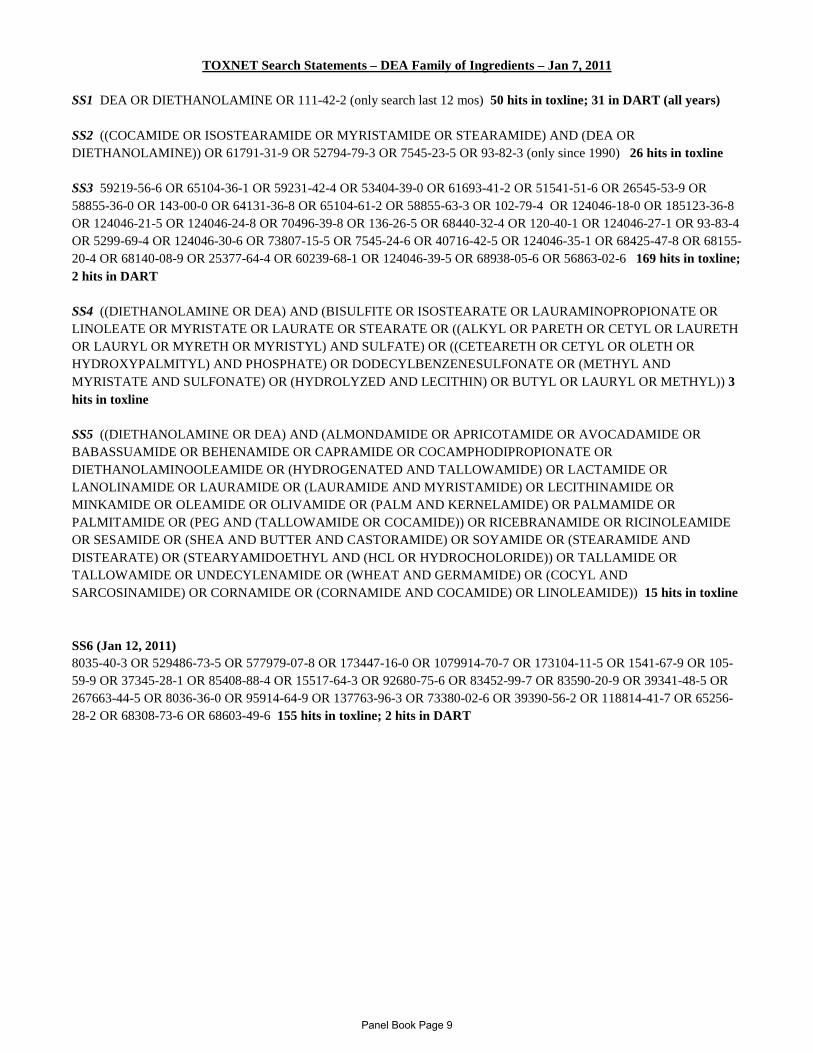
TOXNET Search Statements – DEA Family of Ingredients – Jan 7, 2011 SS1 DEA OR DIETHANOLAMINE OR 111-42-2 (only search last 12 mos) 50 hits in toxline; 31 in DART (all years) SS2 ((COCAMIDE OR ISOSTEARAMIDE OR MYRISTAMIDE OR STEARAMIDE) AND (DEA OR DIETHANOLAMINE)) OR 61791-31-9 OR 52794-79-3 OR 7545-23-5 OR 93-82-3 (only since 1990) 26 hits in toxline SS3 59219-56-6 OR 65104-36-1 OR 59231-42-4 OR 53404-39-0 OR 61693-41-2 OR 51541-51-6 OR 26545-53-9 OR 58855-36-0 OR 143-00-0 OR 64131-36-8 OR 65104-61-2 OR 58855-63-3 OR 102-79-4 OR 124046-18-0 OR 185123-36-8 OR 124046-21-5 OR 124046-24-8 OR 70496-39-8 OR 136-26-5 OR 68440-32-4 OR 120-40-1 OR 124046-27-1 OR 93-83-4 OR 5299-69-4 OR 124046-30-6 OR 73807-15-5 OR 7545-24-6 OR 40716-42-5 OR 124046-35-1 OR 68425-47-8 OR 68155-20-4 OR 68140-08-9 OR 25377-64-4 OR 60239-68-1 OR 124046-39-5 OR 68938-05-6 OR 56863-02-6 169 hits in toxline; 2 hits in DART SS4 ((DIETHANOLAMINE OR DEA) AND (BISULFITE OR ISOSTEARATE OR LAURAMINOPROPIONATE OR LINOLEATE OR MYRISTATE OR LAURATE OR STEARATE OR ((ALKYL OR PARETH OR CETYL OR LAURETH OR LAURYL OR MYRETH OR MYRISTYL) AND SULFATE) OR ((CETEARETH OR CETYL OR OLETH OR HYDROXYPALMITYL) AND PHOSPHATE) OR DODECYLBENZENESULFONATE OR (METHYL AND MYRISTATE AND SULFONATE) OR (HYDROLYZED AND LECITHIN) OR BUTYL OR LAURYL OR METHYL)) 3 hits in toxline SS5 ((DIETHANOLAMINE OR DEA) AND (ALMONDAMIDE OR APRICOTAMIDE OR AVOCADAMIDE OR BABASSUAMIDE OR BEHENAMIDE OR CAPRAMIDE OR COCAMPHODIPROPIONATE OR DIETHANOLAMINOOLEAMIDE OR (HYDROGENATED AND TALLOWAMIDE) OR LACTAMIDE OR LANOLINAMIDE OR LAURAMIDE OR (LAURAMIDE AND MYRISTAMIDE) OR LECITHINAMIDE OR MINKAMIDE OR OLEAMIDE OR OLIVAMIDE OR (PALM AND KERNELAMIDE) OR PALMAMIDE OR PALMITAMIDE OR (PEG AND (TALLOWAMIDE OR COCAMIDE)) OR RICEBRANAMIDE OR RICINOLEAMIDE OR SESAMIDE OR (SHEA AND BUTTER AND CASTORAMIDE) OR SOYAMIDE OR (STEARAMIDE AND DISTEARATE) OR (STEARYAMIDOETHYL AND (HCL OR HYDROCHOLORIDE)) OR TALLAMIDE OR TALLOWAMIDE OR UNDECYLENAMIDE OR (WHEAT AND GERMAMIDE) OR (COCYL AND SARCOSINAMIDE) OR CORNAMIDE OR (CORNAMIDE AND COCAMIDE) OR LINOLEAMIDE)) 15 hits in toxline SS6 (Jan 12, 2011) 8035-40-3 OR 529486-73-5 OR 577979-07-8 OR 173447-16-0 OR 1079914-70-7 OR 173104-11-5 OR 1541-67-9 OR 105-59-9 OR 37345-28-1 OR 85408-88-4 OR 15517-64-3 OR 92680-75-6 OR 83452-99-7 OR 83590-20-9 OR 39341-48-5 OR 267663-44-5 OR 8036-36-0 OR 95914-64-9 OR 137763-96-3 OR 73380-02-6 OR 39390-56-2 OR 118814-41-7 OR 65256-28-2 OR 68308-73-6 OR 68603-49-6 155 hits in toxline; 2 hits in DART
Panel Book Page 9

DR. BERGFELD: Thank you. So, the
motion's been made to reopen and it's been
seconded. Any further discussion?
DR. MARKS: And with the intent -- and
we'll -- as Paul mentioned earlier, at least for
our team the intent was to add methylene glycol,
but as we work through the report, we'll decide
whether or not we want to continue that.
DR. BERGFELD: All right. Call for the
question, all those in favor, please indicate by
raising your hand?
Thank you. Unanimous. Then moving on
to the second to the last ingredient which is the
MEA/DEA/TEA. Dr. Belsito?
DR. BELSITO: Yes, this is a re-review
of the document and it's gone through a number of
iterations. The initial was 1983, and since that
time there have been a number of discussions
regarding DEA. However, it's really time that we
look at the original report which contained all
three. And when we -- when my team looked at the
data we really felt that perhaps with the
FULL PANEL - December 2010
exception of opening it to reassess MEA and
changing it to our current way of stating that we
had limited it to rinse off products because of
irritation, to the current way of stating, could
be used in leave-ons if formulated not to be
irritating, it was really no reason to open the
document.
However, the reason to open it would be
that there are a number of MEA, DEA, and TEA
compounds that could be tagged onto this quite
easily that we haven't reviewed. So we are
recommending that, A, the report be split into
three different reports: An MEA, a DEA, and a TEA
report; and that all of the related cosmetic MEAs,
DEAs, and TEAs be included in each of those
reports. And that's a motion.
DR. BERGFELD: Motion to reopen and
split it into three different ingredient groups
has been made.
DR. MARKS: Second.
DR. BERGFELD: Second. Any further
discussion about reopening? John?
DR. BAILEY: Yeah, I agree, but I think
that it's really important how these groups are
going to be constituted. And I would like to see
the proposed group as soon as possible and then we
will refer that to our Science and Support
Committee just to make sure that they're
comfortable with the way the group is put
together. You know, there was some, I wouldn't
say concern, but some interest in making sure that
these groups are as rational and logical as
possible, so we would need to get those as soon as
we can.
DR. BERGFELD: Alan?
DR. ANDERSEN: Yeah, we will most
certainly get the potential add-ons out ASAP. I
see a primary focus of the March meeting on
receiving that input from industry, receiving the
input from the panel as the panel gets the
opportunity to look at those groupings, and
negotiating what actually should be done as
add-ons. So, I don't know that we're -- I mean,
unless we hit the nail perfectly on the head,
there's going to be some negotiating in March.
DR. BERGFELD: Call for the question
then to reopen, all those in favor please indicate
by raising your hands?
Thank you. It's reopened. And then
moving to the last ingredient to be considered
this morning, human umbilical extract, Dr. Marks?
DR. MARKS: In 2002, the CIR published
its final safety assessments in the ingredients
derived from human and animal placentas and
umbilical cords with a conclusion that the
available data were insufficient to support the
safety. We recently had correspondence from a
company specifically concerning use of human
umbilical extract in cosmetic products. They
supplied some data, but when you look at our
insufficient data needs from the original safety
assessment, really those data needs were not met
and so our team moves not to reopen this safety
assessment.
DR. BERGFELD: Second? Is that a
second? Comment?
Panel Book Page 10

CIR Meeting day 1 of 2 (Breakout Session) Page: 14
Anderson Court Reporting -- 703-519-7180 -- www.andersonreporting.net
1 the risk assessment pages that you're looking at
2 will substantially make up the summary that you're
3 going to see at the next meeting.
4 DR. BELSITO: Okay, good. Anything else
5 on this? No? Okay. So moving on to the next
6 one, it's the re- review of MEA, DEA, and TEA.
7 And Alan has essentially already stolen my
8 thunder, which is basically how many salts and
9 esters of these can we make into super families?
10 And so I'm thinking we should be reopen it not
11 only to add those in, but I think our conclusion
12 that MEA should not be used in leave-on products
13 is based upon irritation. And we've taken a
14 different step now to say "when formulated not to
15 be irritating," so that conclusion may not be
16 correct either as it stands. So I would say that
17 we reopen the documents and take Alan's, split
18 them into three and add the salts and esters of
19 the MEAs, DEAs, and TEAs so that we get everything
20 that's out there.
21 DR. ANDERSEN: I think with that
22 strategy what you could expect to see at the next
BELSITO TEAM - December 2010CIR Meeting day 1 of 2 (Breakout Session) Page: 15
Anderson Court Reporting -- 703-519-7180 -- www.andersonreporting.net
1 meeting would be three more, three separate and
2 more comprehensive documents that list the Organic
3 Acid Salts that could conceptually be included and
4 then examines the question of going on to, let's
5 see, MEAs, for example, the DEA list. There is
6 yet a second group that takes off on the fact that
7 we've already reviewed cocamide DEA, lauramide
8 DEA, which are not -- may not technically be
9 considered as salts, but we'll look at forming
10 those groups as well. Monice and Bart have
11 already done a great deal of homework on this, and
12 are kind of ready to package that, but we just
13 kind of finished it last week and it seemed
14 disingenuous to dump all of that on the Panel for
15 this meeting. So if for all sorts of reasons, it
16 seems appropriate to reopen these, then we can
17 take the next step at the next meeting.
18 DR. BELSITO: Is everyone in agreement
19 of splitting them into three separate documents
20 when we do that?
21 DR. SNYDER: Yes.
22 DR. LIEBLER: Fine with it.
CIR Meeting day 1 of 2 (Breakout Session) Page: 16
Anderson Court Reporting -- 703-519-7180 -- www.andersonreporting.net
1 DR. BELSITO: Okay, any comments?
2 DR. LIEBLER: I guess I had
3 misinterpreted the cover memo, and I thought that
4 the main reason to discuss these was the
5 appearance of new data on carcinogenicity. So
6 really that's not the main issue here.
7 DR. SNYDER: No.
8 DR. LIEBLER: Okay.
9 MS. FIUME: Originally --
10 DR. ANDERSEN: I think, in fact, it's an
11 old issue at this point in terms of DEA
12 carcinogenesis. At this point in time arguably
13 explained process of choline metabolism in mice,
14 and it's not hugely relevant.
15 DR. LIEBLER: Right, so based on all of
16 that, I said don't reopen these, but I agree with
17 the reason now to reopen.
18 DR. BELSITO: Any other comments? Okay,
19 dicarboxylic acid. Okay, so in August we issued a
20 tentative report for the twelve dicarboxylic
21 acids, 44 diesters, finding them safe in present
22 practice of use and concentration. There was one
CIR Meeting day 1 of 2 ( Main Session) Page: 147
Anderson Court Reporting -- 703-519-7180 -- www.andersonreporting.net
1 HIV, it put together all sorts of stuff, and we
2 started to separate the two boilerplates out, and
3 some time next year, we will be bringing to the
4 panel all of the boilerplates for boilerplate
5 re-review so we can go through and make sure
6 they're currently up to date. We felt there were
7 more than enough agenda items on this meeting to
8 not do it starting with this meeting.
9 DR. MARKS: Thank you. Okay, onto the
10 next ingredient or ingredients. We're in the MEA,
11 DEA, TEA re- review. There's quite a history of
12 these ingredients, and I think where we're at at
13 this point is do we reopen, do we separate it out
14 into three different reports, do we put them
15 together? And I'll open it up for discussion.
16 And then, also, we should talk about if we reopen,
17 do we reopen it to add salts and simple esters,
18 also? And to further comment, and, Tom, I'd asked
19 you about the nitrosamine formulation concern, and
20 Ron's, where DEA has been banned in the EU and
21 Canada, plus it's salts and MEA and TEA has had
22 restrictions. So, let's go ahead and decide
MARKS TEAM - December 2010
Panel Book Page 11
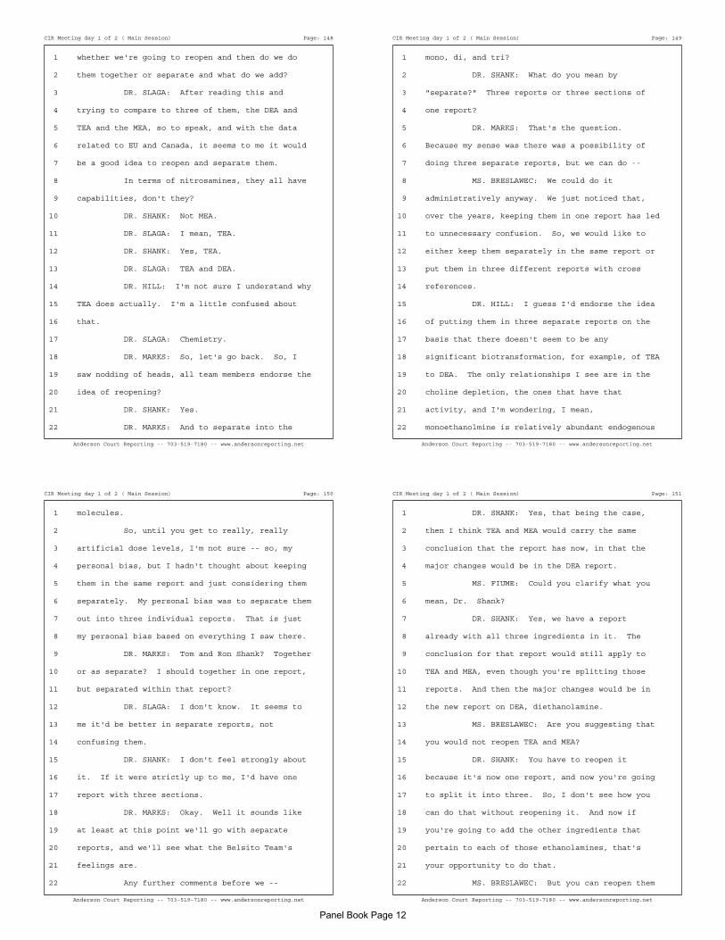
CIR Meeting day 1 of 2 ( Main Session) Page: 148
Anderson Court Reporting -- 703-519-7180 -- www.andersonreporting.net
1 whether we're going to reopen and then do we do
2 them together or separate and what do we add?
3 DR. SLAGA: After reading this and
4 trying to compare to three of them, the DEA and
5 TEA and the MEA, so to speak, and with the data
6 related to EU and Canada, it seems to me it would
7 be a good idea to reopen and separate them.
8 In terms of nitrosamines, they all have
9 capabilities, don't they?
10 DR. SHANK: Not MEA.
11 DR. SLAGA: I mean, TEA.
12 DR. SHANK: Yes, TEA.
13 DR. SLAGA: TEA and DEA.
14 DR. HILL: I'm not sure I understand why
15 TEA does actually. I'm a little confused about
16 that.
17 DR. SLAGA: Chemistry.
18 DR. MARKS: So, let's go back. So, I
19 saw nodding of heads, all team members endorse the
20 idea of reopening?
21 DR. SHANK: Yes.
22 DR. MARKS: And to separate into the
CIR Meeting day 1 of 2 ( Main Session) Page: 149
Anderson Court Reporting -- 703-519-7180 -- www.andersonreporting.net
1 mono, di, and tri?
2 DR. SHANK: What do you mean by
3 "separate?" Three reports or three sections of
4 one report?
5 DR. MARKS: That's the question.
6 Because my sense was there was a possibility of
7 doing three separate reports, but we can do --
8 MS. BRESLAWEC: We could do it
9 administratively anyway. We just noticed that,
10 over the years, keeping them in one report has led
11 to unnecessary confusion. So, we would like to
12 either keep them separately in the same report or
13 put them in three different reports with cross
14 references.
15 DR. HILL: I guess I'd endorse the idea
16 of putting them in three separate reports on the
17 basis that there doesn't seem to be any
18 significant biotransformation, for example, of TEA
19 to DEA. The only relationships I see are in the
20 choline depletion, the ones that have that
21 activity, and I'm wondering, I mean,
22 monoethanolmine is relatively abundant endogenous
CIR Meeting day 1 of 2 ( Main Session) Page: 150
Anderson Court Reporting -- 703-519-7180 -- www.andersonreporting.net
1 molecules.
2 So, until you get to really, really
3 artificial dose levels, I'm not sure -- so, my
4 personal bias, but I hadn't thought about keeping
5 them in the same report and just considering them
6 separately. My personal bias was to separate them
7 out into three individual reports. That is just
8 my personal bias based on everything I saw there.
9 DR. MARKS: Tom and Ron Shank? Together
10 or as separate? I should together in one report,
11 but separated within that report?
12 DR. SLAGA: I don't know. It seems to
13 me it'd be better in separate reports, not
14 confusing them.
15 DR. SHANK: I don't feel strongly about
16 it. If it were strictly up to me, I'd have one
17 report with three sections.
18 DR. MARKS: Okay. Well it sounds like
19 at least at this point we'll go with separate
20 reports, and we'll see what the Belsito Team's
21 feelings are.
22 Any further comments before we --
CIR Meeting day 1 of 2 ( Main Session) Page: 151
Anderson Court Reporting -- 703-519-7180 -- www.andersonreporting.net
1 DR. SHANK: Yes, that being the case,
2 then I think TEA and MEA would carry the same
3 conclusion that the report has now, in that the
4 major changes would be in the DEA report.
5 MS. FIUME: Could you clarify what you
6 mean, Dr. Shank?
7 DR. SHANK: Yes, we have a report
8 already with all three ingredients in it. The
9 conclusion for that report would still apply to
10 TEA and MEA, even though you're splitting those
11 reports. And then the major changes would be in
12 the new report on DEA, diethanolamine.
13 MS. BRESLAWEC: Are you suggesting that
14 you would not reopen TEA and MEA?
15 DR. SHANK: You have to reopen it
16 because it's now one report, and now you're going
17 to split it into three. So, I don't see how you
18 can do that without reopening it. And now if
19 you're going to add the other ingredients that
20 pertain to each of those ethanolamines, that's
21 your opportunity to do that.
22 MS. BRESLAWEC: But you can reopen them
Panel Book Page 12

CIR Meeting day 1 of 2 ( Main Session) Page: 152
Anderson Court Reporting -- 703-519-7180 -- www.andersonreporting.net
1 just to add ingredients, which makes it a little
2 more expedient. DEA, it seems you're suggesting
3 to reopen to reconsider the conclusion, perhaps?
4 DR. SHANK: Correct.
5 DR. SLAGA: Yes.
6 DR. SHANK: How do you reopen? You're
7 creating three new reports. So, you're not
8 reopening DEA, you're not reopening the current
9 report. You're splitting it.
10 DR. MARKS: Yes --
11 DR. SHANK: How do you do that
12 procedurally? What words you use --
13 MS. BRESLAWEC: I think it's something
14 that we would do administratively.
15 DR. MARKS: No, that's a good point,
16 Ron, because in 1983, these were grouped together.
17 So, you're reopening that report, but if we decide
18 to do three separate reports, we're not reopening
19 them in that; we're reopening to separate it. So,
20 I guess administratively, you have to make sure
21 that that's not a problem with the CIR guidelines.
22 But, if there are, it seems to me just as we've
CIR Meeting day 1 of 2 ( Main Session) Page: 153
Anderson Court Reporting -- 703-519-7180 -- www.andersonreporting.net
1 done with other reports; we've had major sections
2 within the report. There will be. Well, the
3 conclusion will just deal with it.
4 DR. BAILEY: And couldn't this also --
5 in splitting these, wouldn't it be logical to
6 include adding the other alkanolamines within that
7 group, like a diethanol. I mean, it would be
8 dialkonalamines because there are some in the
9 dictionary now.
10 MS. BRESLAWEC: We've actually prepared,
11 and, Bart, maybe you'd like to come up here, as
12 well, but we've started looking at possible
13 add-ons for all there, MEA, DEA, and TEA, and we
14 are approaching it very systematically. There are
15 groups that seem to us to be natural add-ons, like
16 organic acid salts, for example, and then there
17 are groups that are related, but may be a little
18 far out or groups that are related, but probably
19 should be considered on their own. We're not
20 ready to present those groups for discussion right
21 now, but we have started the process, and we have
22 quite a bit of information on it, but it's
CIR Meeting day 1 of 2 ( Main Session) Page: 154
Anderson Court Reporting -- 703-519-7180 -- www.andersonreporting.net
1 something that warrants more preparation before
2 it's presented to you all for discussion.
3 So, yes, we would like to consider
4 reopening all three reports for the potential of
5 adding new ingredients.
6 DR. MARKS: Halyna, how much do you see
7 in having separate reports that you're now going
8 to have a lot of refer to the other report to
9 support that the safety of the other ingredients.
10 Like Ron says TEA and MEA, the same conclusions.
11 So, does that make sense to separate them out if
12 we're going to be using data from one to support
13 the other? And, I, again, am looking forward in
14 terms of if there's going to be a lot of data
15 that's shared in all three reports, and does it
16 make sense to have there separate reports?
17 DR. BOYER: For each of the three
18 chemicals, there is a lot of chemical-specific
19 information. So, it doesn't need to be a lot of
20 cross-reference and so forth. And DEA actually
21 stands out when you look at that data and the
22 mechanistic information that's been published and
CIR Meeting day 1 of 2 ( Main Session) Page: 155
Anderson Court Reporting -- 703-519-7180 -- www.andersonreporting.net
1 so forth. So, I think from that perspective, it
2 certainly makes sense to separate them.
3 DR. HILL: Excuse me. And in regards to
4 potentially expanding the groups, I would just say
5 that I strongly suspect that there's going to be,
6 particularly with DEA, there's some toxicology
7 issues that might pertain to it that might not
8 pertain to anything even related. Now, amides of
9 DEA at some point, but those are really widely,
10 heavily used for cosmetic ingredients, and I think
11 moving in that direction would be right now with
12 great caution in my estimation because I think
13 there might not be that much to worry about.
14 DR. BOYER: Right.
15 DR. HILL: And, so, if you tag related
16 to something where there clearly is a problem --
17 well, I say "clearly is a problem," seems to be a
18 problem. Don't know in humans, but you might be
19 creating a problem where there wasn't one before.
20 DR. MARKS: I think, again, for the
21 stenographers, that was Dr. Boyer who was
22 commenting earlier, correct?
Panel Book Page 13
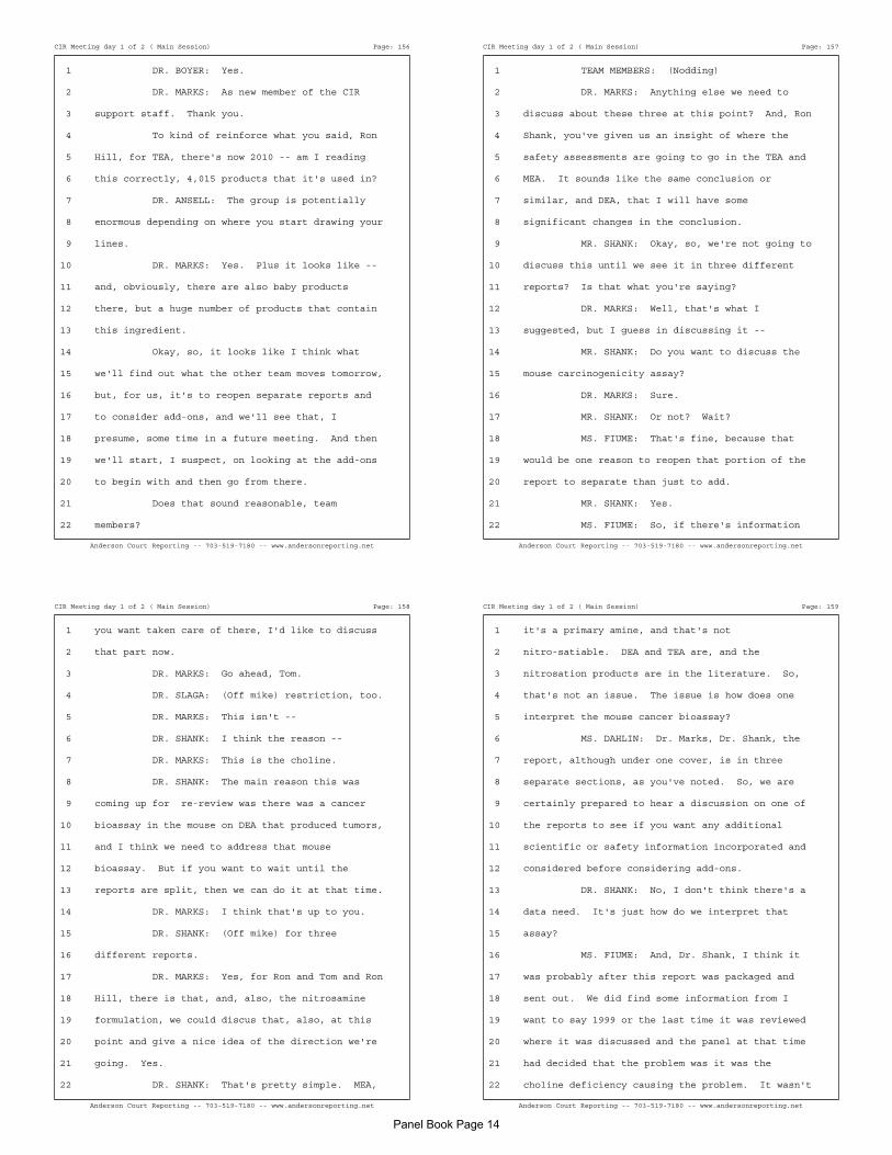
CIR Meeting day 1 of 2 ( Main Session) Page: 156
Anderson Court Reporting -- 703-519-7180 -- www.andersonreporting.net
1 DR. BOYER: Yes.
2 DR. MARKS: As new member of the CIR
3 support staff. Thank you.
4 To kind of reinforce what you said, Ron
5 Hill, for TEA, there's now 2010 -- am I reading
6 this correctly, 4,015 products that it's used in?
7 DR. ANSELL: The group is potentially
8 enormous depending on where you start drawing your
9 lines.
10 DR. MARKS: Yes. Plus it looks like --
11 and, obviously, there are also baby products
12 there, but a huge number of products that contain
13 this ingredient.
14 Okay, so, it looks like I think what
15 we'll find out what the other team moves tomorrow,
16 but, for us, it's to reopen separate reports and
17 to consider add-ons, and we'll see that, I
18 presume, some time in a future meeting. And then
19 we'll start, I suspect, on looking at the add-ons
20 to begin with and then go from there.
21 Does that sound reasonable, team
22 members?
CIR Meeting day 1 of 2 ( Main Session) Page: 157
Anderson Court Reporting -- 703-519-7180 -- www.andersonreporting.net
1 TEAM MEMBERS: (Nodding)
2 DR. MARKS: Anything else we need to
3 discuss about these three at this point? And, Ron
4 Shank, you've given us an insight of where the
5 safety assessments are going to go in the TEA and
6 MEA. It sounds like the same conclusion or
7 similar, and DEA, that I will have some
8 significant changes in the conclusion.
9 MR. SHANK: Okay, so, we're not going to
10 discuss this until we see it in three different
11 reports? Is that what you're saying?
12 DR. MARKS: Well, that's what I
13 suggested, but I guess in discussing it --
14 MR. SHANK: Do you want to discuss the
15 mouse carcinogenicity assay?
16 DR. MARKS: Sure.
17 MR. SHANK: Or not? Wait?
18 MS. FIUME: That's fine, because that
19 would be one reason to reopen that portion of the
20 report to separate than just to add.
21 MR. SHANK: Yes.
22 MS. FIUME: So, if there's information
CIR Meeting day 1 of 2 ( Main Session) Page: 158
Anderson Court Reporting -- 703-519-7180 -- www.andersonreporting.net
1 you want taken care of there, I'd like to discuss
2 that part now.
3 DR. MARKS: Go ahead, Tom.
4 DR. SLAGA: (Off mike) restriction, too.
5 DR. MARKS: This isn't --
6 DR. SHANK: I think the reason --
7 DR. MARKS: This is the choline.
8 DR. SHANK: The main reason this was
9 coming up for re-review was there was a cancer
10 bioassay in the mouse on DEA that produced tumors,
11 and I think we need to address that mouse
12 bioassay. But if you want to wait until the
13 reports are split, then we can do it at that time.
14 DR. MARKS: I think that's up to you.
15 DR. SHANK: (Off mike) for three
16 different reports.
17 DR. MARKS: Yes, for Ron and Tom and Ron
18 Hill, there is that, and, also, the nitrosamine
19 formulation, we could discus that, also, at this
20 point and give a nice idea of the direction we're
21 going. Yes.
22 DR. SHANK: That's pretty simple. MEA,
CIR Meeting day 1 of 2 ( Main Session) Page: 159
Anderson Court Reporting -- 703-519-7180 -- www.andersonreporting.net
1 it's a primary amine, and that's not
2 nitro-satiable. DEA and TEA are, and the
3 nitrosation products are in the literature. So,
4 that's not an issue. The issue is how does one
5 interpret the mouse cancer bioassay?
6 MS. DAHLIN: Dr. Marks, Dr. Shank, the
7 report, although under one cover, is in three
8 separate sections, as you've noted. So, we are
9 certainly prepared to hear a discussion on one of
10 the reports to see if you want any additional
11 scientific or safety information incorporated and
12 considered before considering add-ons.
13 DR. SHANK: No, I don't think there's a
14 data need. It's just how do we interpret that
15 assay?
16 MS. FIUME: And, Dr. Shank, I think it
17 was probably after this report was packaged and
18 sent out. We did find some information from I
19 want to say 1999 or the last time it was reviewed
20 where it was discussed and the panel at that time
21 had decided that the problem was it was the
22 choline deficiency causing the problem. It wasn't
Panel Book Page 14
CIR Meeting day 1 of 2 ( Main Session) Page: 160
Anderson Court Reporting -- 703-519-7180 -- www.andersonreporting.net
1 the DEA, it was the choline, and there was a
2 discussion. So, I will capture that, as well, it
3 was just discovered after it came out. But if you
4 don't agree what may have been said at that time,
5 then I'll capture something differently or look
6 for different information.
7 DR. SHANK: Okay, I'd have to read that,
8 but I was on the panel at that time, so, I
9 shouldn't make the same argument all over again.
10 We don't need to discuss that now, and we'll see
11 what we said 11 years ago.
12 DR. MARKS: Well, basically in 2008, the
13 panel agreed that the NTP findings of
14 carcinogenesis in the mouse for DEA and certain
15 DEA fatty acid esters was related to choline
16 (inaudible) and not relevant to human health.
17 Tom, is that your recollection?
18 DR. SLAGA: (Nodding)
19 DR. MARKS: I think that's how we dealt
20 with the mouse carcinogenicity.
21 DR. HILL: But I had a question based on
22 information that was in both presentations at the
CIR Meeting day 1 of 2 ( Main Session) Page: 161
Anderson Court Reporting -- 703-519-7180 -- www.andersonreporting.net
1 1999 meeting. Of course, that was long before my
2 participation. Both Dr. Lehman-McKeeman, I don't
3 know if I'm saying her name right, and Dr. Stott
4 mentioned that DEA is incorporated in ceramides
5 and possibly sphingomyelins, and then in the
6 discussion of DEA, that whole possible mechanism
7 is dropped, and because I guess there's a
8 pharmacologist in our department who's working on
9 that and effects on cancer stem cells and
10 apoptosis, I want to know if that thread of
11 biology has continued or people have just ignored
12 those pieces of information which came from
13 industry source presentations. Whether there's
14 been any follow-up whatsoever on that biology.
15 And that's one of the reasons why I was looking to
16 see this split was because there may be an issue
17 with DEA biology that doesn't show up at all that
18 shouldn't be an issue with TEA, that shouldn't be
19 an issue with monoethanolmine, but it very well
20 might be a big issue with DEA and only DEA.
21 DR. BOYER: Well, that mechanism seems
22 to certainly distinguish DEA from the other two.
CIR Meeting day 1 of 2 ( Main Session) Page: 162
Anderson Court Reporting -- 703-519-7180 -- www.andersonreporting.net
1 As far as I know, there has been no significant
2 progress in terms of developing information to
3 interpret or to determine the importance of those
4 observations, the observation that DEA seems to be
5 incorporated into possible lipids. And there's a
6 lot of speculation about what could happen and how
7 that mechanism might explain some of the toxic
8 effects not necessarily the carcinogenicity.
9 DR. HILL: Well, ceramides have a strong
10 role to play in regulating apoptotic pathways, as
11 well as proliferative pathways, and these were
12 mentioned in two different presentations by two
13 independent labs. So, I guess I'm raising it now
14 so that in mining the literature, whatever might
15 be out there, you will be attuned to looking for
16 anything.
17 DR. BOYER: Absolutely.
18 DR. HILL: (off mike)
19 DR. BOYER: Right.
20 DR. HILL: And I'm not thinking that
21 this is at all relevant in any of the amides of
22 DEA because I doubt that DEA is significantly
CIR Meeting day 1 of 2 ( Main Session) Page: 163
Anderson Court Reporting -- 703-519-7180 -- www.andersonreporting.net
1 generated from those amides. But I think it might
2 be something specific to DEA, which I guess is
3 really not used much at all at this point. I get
4 the sense.
5 DR. BOYER: Right.
6 DR. HILL: But it would be clean if
7 those three were dealt with separately then in
8 going to -- because I can envision language in
9 something that's reviewed that's structurally
10 similar, like the kinds of ingredients you were
11 suggesting to expand to. The panel has previously
12 reviewed DEA. We note the structural similarity,
13 but the specific toxicological issues pertaining
14 to that compound don't pertain to any of these,
15 and here's why.
16 DR. BOYER: Yes.
17 DR. HILL: And, so, it would be very
18 clean to be able to refer to that single report
19 and not give issue with the other two that I don't
20 think have any same issues at all.
21 DR. MARKS: Would you like to, since
22 there are three separate reports within this
Panel Book Page 15
CIR Meeting day 1 of 2 ( Main Session) Page: 164
Anderson Court Reporting -- 703-519-7180 -- www.andersonreporting.net
1 document, should we, again, sort of have a preview
2 of what's coming down the road, take a look at
3 them individually? I think that the conclusion in
4 1983 -- it's going to be a little interesting if
5 we keep the same wording. So, his conclusion that
6 TEA, DEA, and MEA are safe for use in cosmetic
7 formulations designed for discontinuous brief use
8 followed by thorough rinsing from the surface of
9 the skin. And products intended for prolonged
10 contact with the skin, the concentration
11 ethanolamines should not exceed 5 percent, MEA
12 should only be used in products that do not
13 contain nitrosating agents.
14 So, I know the TEA and the MEA, Ron, you
15 suggested this same conclusion or something
16 similar is going to be okay. The DEA, there's
17 going to be changes.
18 Do you want to go through these
19 individually now? We dealt with the mouse, I
20 think, where the choline metabolism not relevant
21 to the human. We disused the nitrosamine
22 formulation concern left to deal with the ban in
CIR Meeting day 1 of 2 ( Main Session) Page: 165
Anderson Court Reporting -- 703-519-7180 -- www.andersonreporting.net
1 EU and Canada. Or restricted.
2 DR. SLAGA: Wasn't it suggested to have
3 two reports instead of three? I mean, I thought
4 that's what you were thinking, too. No?
5 DR. SHANK: No, my suggestion was one
6 report with three sections. But we all decided
7 three individual reports. I think.
8 DR. SLAGA: I thought you meant that you
9 wanted to have TEA and MEA combined because they
10 all have the same conclusions.
11 DR. ANDERSEN: I wanted all three
12 combined. One single report with three sections.
13 But that's a minority opinion.
14 DR. HILL: Well, if there's nothing to
15 ceramides and if there's nothing to more than
16 choline deficiency in that particular assay then
17 you could keep them combined. I guess in my mind,
18 it's somewhat dependent on the toxicology here.
19 MS. BRESLAWEC: We really would prefer
20 separating them out in one form or another because
21 it's caused a lot of confusion when we've looked
22 at derivatives or components that contain DEA or
CIR Meeting day 1 of 2 ( Main Session) Page: 166
Anderson Court Reporting -- 703-519-7180 -- www.andersonreporting.net
1 MEA or TEA. Administratively, it's very difficult
2 to deal with them in the same report. So, whether
3 it's one report with three sections, we're fine
4 with that, or three separate reports, we're fine
5 with that, as long as each of the ingredients are
6 handled separately.
7 DR. MARKS: Well, certainly, if they
8 were all in the same report, we wouldn't be
9 dealing with taking a combined report in 1983 and
10 now re-reviewing it and creating three separate
11 reports.
12 Ron Hill and Tom, does it matter to you
13 whether they all be combined in the one report and
14 three sections or three separate reports?
15 DR. SLAGA: Really, it's the same thing.
16 DR. MARKS: Yes, except we have to know
17 which way we're going to go as we proceed. Should
18 we wait and see what the Belsito Team says? I can
19 see there's not a strong --
20 DR. SLAGA: -- (Off mike) six reports.
21 DR. MARKS: Yes, six. I can see there's
22 not a great strong feeling one way or another, as
CIR Meeting day 1 of 2 ( Main Session) Page: 167
Anderson Court Reporting -- 703-519-7180 -- www.andersonreporting.net
1 long as it's separated. So, we'll just say
2 separated in either3 separate reports or within
3 one.
4 Anything more in terms of looking at
5 these individual ones before we come back to this
6 in a future meeting? If there anything else you
7 wanted, Monice, to get any directions?
8 MS. FIUME: I just wanted to make sure
9 so from my understanding, what we will bring back
10 at the next meeting is three reports with what we
11 feel were the proper add-on ingredients that you
12 are more than welcome to take out, but this way,
13 we'd at least have it prepared for you as what we
14 think the next iteration of the reports are.
15 Is that correct?
16 DR. MARKS: Yes. Are there any data
17 needs for these individual ones at this point, and
18 is there enough in this report in terms of the
19 data? Certainly from irritation and
20 sensitization, I thought it was fine. Is there
21 anything else in terms of data needs?
22 Ron, you had mentioned one concern you
Panel Book Page 16
CIR Meeting day 1 of 2 ( Main Session) Page: 168
Anderson Court Reporting -- 703-519-7180 -- www.andersonreporting.net
1 had, but --
2 DR. HILL: It was just an information,
3 sort of see if there's anything out there request.
4 Not a data need.
5 DR. MARKS: So, it sounds like the main
6 thing we're going to do next time is clarify the
7 discussion concerning the mouse and concerning the
8 nitrosamine formation for each of these as
9 separate ingredients, and then decide on the
10 add-ons, but in terms of data, it seems like we're
11 okay at this point. Is that --
12 DR. BRESLAWEC: Just to clarify one
13 thing, they'll be draft amended reports that
14 you'll get next time.
15 MS. FIUME: And then the only other
16 thing I was going to say is in Wave 2, you should
17 have received what the original re-review summary
18 was for DEA. So, I will pull from that
19 information, as well, that includes some of your
20 decision-making or conclusion as to why it went
21 the way it did.
22 DR. MARKS: Anything else?
Panel Book Page 17

June 2009 – Belsito Team Then our next item is DEA carcinogenesis. This is I think essentially being prompted because of issues of neurotoxicity data and we have these two papers by Craciunescu and by Craciunescu again looking at reports of DEA altering neurogenesis and inducing apoptosis and in fetal mice hippocampus, and then the other about dose response effects of dermally applied DEA neurogenesis. In the dermal paper even the authors acknowledged that the dermal penetration in the detectable plasma levels are such that they are far below the concentrations associated with perturbed brain development in mice. Then of course we had the prior issues that we had dealt with in the report about species difference, absorption differences and effects on choline metabolism that aren't really pertinent to humans. It wasn't clear to me what we're supposed to be doing with this, whether this was just an update to decide to open or not reopen. DR. ANDERSEN: I think my intent, and let's see if it matches what anybody actually read, is we had focused on the question of DEA carcinogenesis. There are now ample data both mechanistic, et cetera, to demonstrate that the positive findings are indeed species specific and they relate to choline metabolism. The summary that you have puts all of that together and on being published would resolve that question. The Panel would be on record as saying we buy into the choline metabolism as being the mechanistic cause and confirming that these materials as used in cosmetics do not present a risk of carcinogenesis. That was the purpose of summarizing all of that history. What we had not talked about in any way, shape or form were these new data on neurotoxicity and the question that is on the table is what should we do about those data. Should it cause another round of review or are you comfortable enough saying that it doesn't need to be reopened? When we had round after round for example with thalates and another new set of data came in, we did briefly reopen to consider those data. It turned out there was no formal need to reopen, but at least briefly we considered it. Were those data not there, it would just be a re-review summary and that would be the end of it. What do you think makes sense to do now that we have two studies with neurotox endpoints? DR. BELSITO: I think we addressed the species difference and the choline metabolism in our last re-review of something with DEA. DR. ANDERSEN: Yes. DR. BELSITO: In this case you have the same author who reports the effects on mice coming back and saying when you dermally apply it which is the relevant thing in cosmetics, there is no effect on the mice. So it's not like we have to defend the cosmetic use. The same author who had made these reports I think has defended it for us. I'm not sure that we really need to do anything with it. That was my feeling. It's not like we have to say this author reported it in mice but now you have to look at dermal absorption and you have to look at choline metabolism. He's already done that for us. DR. LIEBLER: Particularly in view of the fact that the neurotox effect is rationalized in terms of the choline metabolism effect as well. DR. SNYDER: I think the only issue is that we're kind of stuck because we're in the middle of a re-review and I really don't like not having it appear as though we didn't consider it these reports even though we understand that the mechanism is already known and we've already addressed the mechanism but it may not be readily apparently clear to the reader how we addressed that. DR. BELSITO: Was it time for a DEA re-review or did this just get accelerated because of these reports? I thought we just did it. DR. ANDERSEN: We just did it. This is the summary of what we did last year. We haven't had time to preparing the summary. DR. BELSITO: Because I thought we had already issued that and now we're looking at it again because of new data. DR. ANDERSEN: No. DR. BERGFELD: Don't you think you'd just add this to the paragraph on page 2 as to the choline to just update the reference? DR. BELSITO: Yes, I would just do that. DR. BERGFELD: I think it's still an unusual summary because it also includes the safety assessments of the others, and there are several of them which we haven't done particularly in the past. This is a new entry, or maybe once before. While we're discussing those I wondered if we couldn't go back and look at the citation that appears at the end of each paragraph under those ingredients containing N-nitrosating agents is one statement, another one is in which N-nitroso compounds are formed containing nitrosating agents. It seems to me we're talking about the same thing there even though we've used different terminology. DR. ANDERSEN: That's correct. Over time there have been three different ways that we've phrased it. The intention in all three cases is the same. Yes, we could develop a single language. My intent in presenting Table 1 was to simply capture what's in the documents as opposed to unifying them. DR. BERGFELD: I thought that was a good idea actually. DR. ANDERSEN: But it certainly brings into great relief the fact that we've used different phraseology over time. There's no question about that.
Panel Book Page 18
DR. BELSITO: I would agree with Wilma's comment. I was confused. I thought we had already re-reviewed it and passed on it. If this is just our final review of the document, I don't think it changes anything. Just put statements as to these papers and include the author's conclusion which I think exonerates us. On the paper though I just have one question. On the second page, the fifth line down sort of in the middle it says, "In studies with multiple (3)", I'm assuming that there were three studies or was it three body lotion doses, and I think by putting the number in parenthesis now that we're using numbers for references also could be confusing as to whether that was the reference. Was it three studies with different body lotion doses, was it one study with three different body lotions? That needs to just be clarified. DR. ANDERSEN: It was the latter I'm pretty sure, but I'll confirm that. And, yes, we have to be careful about how we do stuff now that we're presenting it differently. DR. BERGFELD: On page 1 if I could interrupt, the second paragraph, "In addition, work done at the FDA." Were they reviews? What kind of work was done? What is the work done? DR. ANDERSEN: That was work that Bob Bronaugh presented during the discussion on his data on DEA penetration. DR. BERGFELD: Is there another way of stating that other than "work done"? DR. ANDERSEN: Yes, there. DR. BELSITO: And "data presented by"? DR. ANDERSEN: Yes, and that should have a reference as well. DR. BERGFELD: Could I ask a question? Are we going to have a book with all these summary statements somewhere so we could reflect on a format and the changes in the format? DR. ANDERSEN: Soon. That is one of the tasks that Halyna Breslawec has taken on and she's busily working on it. She was almost ready to put it on the agenda for this meeting but not quite, so a discussion of our precedent files you can expect depending on whether September's agenda is really heavy or not, but I'd like to get it on the September agenda. DR. BAILEY: So are you talking about consistency for the nitrosamine statement or just consistency of the format? DR. BERGFELD: All of it. DR. ANDERSEN: Everything. We're looking at soup to nuts. DR. BERGFELD: Particularly in these. We are beginning to enlarge these re-review summaries when we don't reopen and they are developing into a fair amount of text. I'd just to take a look at what we've been doing. DR. ANDERSEN: Thank you.
June 2009 – Marks Team DR. MARKS: -- see, let's move on. Since Wilbur isn't here, we have a couple other things to do before the cyclomethicones. Next in the agenda I have is the re-review summary of DEA carcinogenesis and then we happened to get a couple papers on the issue of neurogenesis and neurotoxicity with DEA. So let's just start first with this -- DR. ANDERSEN: Not -- in addition of neurogenesis. DR. MARKS: Yes. Right. DR. ANDERSEN: I mean if it actually increased neurogenesis -- SPEAKER: You'd want some. SPEAKER: (off mike) DR. HILL: Well, not in utero, however. DR. MARKS: So -- any rate -- Alan asked us to look over this re-review and summary and how does it appear? Tom, you're the one I have -- how do you like the summary? Or do you have any -- any suggestions? DR. SLAGA: Well, I think the summary is in good shape -- I mean other than a few typos and that type of thing. The papers -- on the one hand at higher concentrations, you could have some apoptosis as well as some inhibition of neurogenesis. But in a small human study they did at (off mike) -- that was -- it had no effect on the brain development in the mouse at the levels they looked at it. The mouse was a much higher level than you would find in any cosmetics -- much higher. So -- to me -- I don't think it's an issue. DR. ANDERSEN: Well, in the metabolic issues, mouse versus human are still there in terms of colene deficiency, etc. DR. SLAGA: Right. Yeah -- no -- that colene part is definitely in amounts that doesn't seem to be a relationship with human. DR. ANDERSEN: Yeah, I was just concerned that our entire focus has been on DEA and DEA fatty acid carcinogenesis and I think that issue was nicely resolved -- DR. SLAGA: Yeah -- resolved. DR. ANDERSEN: -- and then this hit of something we hadn't really talked about. DR. SLAGA: We probably emphasized it a little too much, but at the same time it's in the literature a lot and it's good to
Panel Book Page 19

eliminate if there is a concern. DR. SHANK: The neurotoxicity should be added to the review (off mike). DR. SLAGA: Yeah, to the review. But not (off mike). DR. ANDERSEN: (off mike) DR. SHANK: Acknowledge it. That's all. DR. ANDERSEN: Just include a sentence that says it exists. DR. SHANK: Okay. DR. MARKS: Okay. And in -- DR. ANDERSEN: Thank you. DR. MARKS: -- with that you include the 2009 use of concentration table, correct? Alan, (off mike)? DR. ANDERSEN: Well, that -- I decided not to do that for this summary because the focus was on simply the question of carcinogenesis. DR. MARKS: Okay. DR. ANDERSEN: It didn't focus on the question of use concentrations at all. You didn't -- we never talked about that. The issue was is this stuff carcinogenic -- I'm sorry. Does it present a carcinogenic risk to humans? And the answer was no. So we didn't go on to talk about use concentration. There was an absence of a hazard, so exposure wasn't so important. We have to go -- Carol would have to go resurvey to get valid data for use and I'm not sure that's worth it. DR. MARKS: Okay. I just -- DR. ANDERSEN: I think if -- you know -- brining it up tomorrow and let's see if anybody else is concerned. If so, then we can -- it's just a matter of doing another survey. DR. SLAGA: Nothing to it. DR. MARKS: No -- yeah -- I was just putting it in the format of what we usually do with a re-review. DR. ANDERSEN: It's different -- (off mike) it's different on purpose. DR. MARKS: Yeah. Okay. Next is the hair dye epidemiology. We heard the presentation this morning by Julie Skare. Skare?
June 2009 – Full Panel DR. BERGFELD: I do. All right, then we'll move on. The last ingredient is a re-review summary of the DEA carcinogenicity, and that is Dr. Belsito. DR. BELSITO: Yes. This was a re-review we did the last time, and in the interim there were two reports that surfaced, one on diethanolamine altering neurogenesis and inducing apoptosis in fetal mouse hippocampus and then by the same author is the dose response effective dermally applied diethanolamine on neurogenesis. And while we found these interesting, they're really not relevant for several reasons. One, we know that these effects are due to choline and there's a difference between murine and human metabolism. But even the authors in the second paper applying a commercially available skin lotion noted that the concentrations that were absorbed were far, far below the concentrations that would exert any effect. So, while we felt it was important that we add these references to the document just to show that where everything is up to date in Washington that it didn't really change that re-review summary and to add them and go ahead with it, issue it as final. DR. BERGFELD: Was it my understanding that you were going to add that to the paragraph on 2, which describes the choline metabolism as well? DR. BELSITO: Yes. DR. BERGFELD: And so your motion is -- DR. BELSITO: To go ahead with this as a final re- review with this, the simple addition of those two references. DR. BERGFELD: And agree -- DR. MARKS: Yea, our team concurs with that. DR. BERGFELD: Any further discussion then? Seeing none, I'll call for the vote. All those in favor of proceeding? Thank you. Unanimous.
June 2009 – Executive Summary Re-review Summary of DEA Carcinogenesis The Expert Panel reviewed and approved the summary of its earlier re-review decision to not reopen the safety
Panel Book Page 20

assessment of Diethanolamine (DEA), Cocamide DEA, Cocamide MEA, Isostearamide DEA & MEA, Linoleamide DEA, Myristamide DEA & MEA, Oleamide DEA, and Stearamide DEA & MEA. This re-review was unusual in that it focused on an endpoint, carcinogenesis. The CIR Expert Panel reviewed the large body of data developed since studies conducted by the NTP reported clear evidence that dermal exposure to DEA and Cocamide DEA were hepatocarcinogenic in male and female mice. The body of work now available has demonstrated that DEA affects mouse choline metabolism, which leads to cellular choline depletion, which leads to altered DNA methylation, which leads to altered gene expression (increased DNA synthesis and oncogene expression, reduced tumor suppressor gene expression and apotosis). At this meeting, the Expert Panel also considered two newer studies that reported effects on neurogenesis in mice, noting that the mechanism of action likely involves the effect of DEA on mouse choline metabolism.
June 2008 – Presentation by Dr. Stott Minutes summarizing presentation Dr. William Stott, representing the Alkanolamines Panel of the American Chemistry Council, reviewed the large body of data developed since the point 10 years ago when studies conducted by the NTP reported clear evidence that DEA and Cocamide DEA were carcinogenic in male and female mice. The work has demonstrated that DEA affects mouse choline metabolism, which leads to cellular choline depletion, which leads to altered DNA methylation, which leads to altered gene expression (increased DNA synthesis and oncogene expression, reduced tumor suppressor gene expression and apoptosis). In work done at FDA and elsewhere, dermal penetration of DEA in personal care product vehicles was found to be significantly higher through mouse skin than either rat or human skin. In addition, the activity of choline oxidase, which is hundreds of times higher in the mouse compared to humans, suggesting that humans are resistant to choline deficiency — choline oxidase levels in the rat, however, are even higher than in the mouse. Overall, the available data support that DEA carcinogenesis in the mouse is related to choline depression and the effect is reversible and threshold-based. Given the known resistance in humans to choline deficiency, these data do not suggest a human health risk from the use of DEA and DEA fatty acids in cosmetic products.
June 2008 – Acetamide MEA Belsito Team (Valerie’s Notes) Dr. Belsito: Why was there a 7.5% limit? This is inconsistent with the MEA/TEA/DEA report.
If reopened to add, the issues with MEA in leave on products versus rinse off products will come up Dr. Eisenmann: Would MEA have its own group instead? Dr. Belsito: MEA ingredients as a group? Dr. Andersen: The MEA report was not addressed in the Acetamide MEA report. Dr. Eisenmann: There is a 2-gen. study going on. Dr. Andersen: Can be tabled to wait for the results. Dr. Belsito: Table until the results come in. What ingredients should be added? Any data needs? Format as Sodium Cetearyl Sulfate afterwards. Create an Alkonolamide MEA family report. Marks Team (Valerie’s Notes) Dr. Slaga/Shank: Do not reopen. Dr. Shank: No add-ons due to the limitation in the original conclusion and sensitization. Dr. Bergfeld: Agree. Dr. Shank: The add-ons may have different absorption and sensitization due to the addition of fatty acid esters, which can increase penetration. Dr. Bailey: MEA ingredients have few uses. Dr. Marks: These add-ons are not no-brainers. Do not reopen/add.
Panel Book Page 21
June 2008 – Full Panel Chairman’s Opening Remarks Dr. Bergfeld welcomed the attendees to the 107th meeting of the CIR Expert Panel. She noted that the following three presentations were made on the preceding day: (1) The presentation by Dr. Bill Stott was on DEA carcinogenicity. The data presented assured the Panel that there is no human risk that is associated with having these chemicals in cosmetics. It was learned that the mouse model that the Panel was somewhat worried about, i.e., due to liver tumors, is not relevant to humans. Acetamide MEA Dr. Marks stated that a CIR Final Report with the following conclusion on this ingredient was published in 1993: On the basis of the data presented in this report, the CIR Expert Panel concludes that Acetamide MEA is safe as a cosmetic ingredient at concentrations not to exceed 7.5% in “leave-on” products and is safe in the present practices of use in “rinse-off” products. Cosmetic formulations containing Acetamide MEA should not contain nitrosating agents or significant amounts of free acetamide. Dr. Marks said that this conclusion was reaffirmed by his Team and, thus, this safety assessment should not be reopened. He added that his Team considered the list of chemically similar ingredients that was provided and determined that, in light of the 7.5% concentration limit, actual data on all of the chemically similar ingredients would be needed if a decision to reopen the Final Report were made. Dr. Belsito said that his Team determined that the Final Report should be reopened, taking into consideration the following information: The CIR Final Report on MEA states that this ingredient should not be used in leave-on products, and it was noted that Acetamide MEA breaks down to MEA. After further review of the Final Report on Acetamide MEA to determine the basis for the 7.5% concentration limit for leave-on products, it was determined that, in the absence of leave-on product uses, it was necessary to establish a concentration limit for Acetamide MEA in these products. The 7.5% concentration limit is actually the highest concentration that was tested in skin sensitization studies. Dr. Belsito stated that his Team determined that the Final Report on Acetamide MEA should be reopened and that MEA should be added to this document along with other MEA ingredients selected from the list that was provided. Dr. Shank noted that MEA is one of 3 ingredients included in the published CIR Final Report on TEA, DEA, and MEA. Dr. Belsito said that the plan is to incorporate all of the data on MEA from this Final Report into the reopened document on Acetamide MEA, creating an MEA ingredient family. Dr. Shank said that MEA belongs in the primary document on ethanolamines, and not in the document on a secondary ingredient such as Acetamide MEA. Dr. Belsito noted that an MEA ingredient family is being created, consisting of MEA (lead ingredient), Acetamide MEA, and selected MEA ingredients. The plan is to reopen MEA, but not the Final Report on Acetamide MEA, and add Acetamide MEA and MEA ingredients selected from the list that was provided. Dr. Bailey said that he believes that Acetamide MEA is a very stable compound and would not break down very easily. He added that the idea that consumers would be exposed to MEA that has been released from Acetamide MEA is probably not true. Dr. Bailey recommended that the CIR Final Report on MEA, DEA, and TEA remain as a separate document and not be amended. He questioned the need to reopen the Final Report on Acetamide MEA because this ingredient is a stable compound and noted that this report should remain as a separate document. He added that the notion of grouping is reasonable, because the smaller molecule is going to be different from the longer-chain MEA derivatives (also very stable compounds). Dr. Belsito wanted to know if Dr. Bailey also thinks that, because Acetamide MEA is a short-chain compound, the Panel should not recommend the addition of other ingredients to this safety assessment. Dr. Bailey agreed that this approach would be reasonable because the longer-chain MEA derivatives would be somewhat different, both chemically and biologically. Dr. Belsito said that, typically, greater toxicity is associated with the shorter chain compounds. Dr. Shank noted that the fatty acid ester component would increase dermal penetration, making compounds more lipid-soluble. Dr. Bronaugh agreed. Dr. Snyder wanted to know whether there are any issues that are related to ingredient use in new product categories. Dr. Belsito said that Acetamide MEA is being used in some sprays/aerosol fixatives, and that the Panel’s boilerplate relating to particle size and inhalation exposure could be incorporated. He added that use concentrations have not increased. Dr. Snyder said that there would likely be a new concentration limit if the Panel receives new data on Acetamide MEA indicating negative sensitization at higher concentrations.
Panel Book Page 22
Dr. Belsito said that a statement expressing the following points should be incorporated into the discussion: The Panel is aware of the fact that MEA may be used in rinse-off, but not leave-on, products, but agrees that Acetamide MEA would be stable on the skin. Therefore, there are no safety concerns, particularly in light of the sensitization data in the final safety assessment on MEA. Dr. Belsito said that the preceding statement is needed because, otherwise, it appears that the Panel has issued contradictory opinions. The CIR Final Report on MEA states that this ingredient should not be used in leave-on products, yet the Expert Panel established a concentration limit for Acetamide MEA in leave-on products. Dr. Marks added that the discussion should also contain a statement explaining why the safety assessment on Acetamide MEA was not expanded to include chemically similar ingredients. This statement could read as follows: The Panel considered chemically similar ingredients, but, because of skin sensitization and increased skin penetration issues, the decision not to expand this group was made. The Panel voted unanimously in favor of not reopening the CIR Final Report on Acetamide MEA. Dr. Belsito said that if the Panel is not going to reopen this document, then there is no need to reopen the CIR Final Report on TEA, DEA, and MEA. Miscellaneous from the Minutes – Near adjournment Dr. Bergfeld stated that the presentations on diethanolamine carcinogenesis and hair dye epidemiology from the preceding day will be captured in the minutes. In response to Dr. Snyder’s concern, Dr. Andersen stated that CIR will formally undertake a re-review summary, for the Panel’s consideration, that will describe all of the DEA fatty acids that have been reviewed and all of the available data on DEA. Dr. Andersen added that the overall plan is to prepare a short re-review summary that could be published and made available on-line, as the final denouement of the Panel’s consideration of DEA. Dr. Andersen noted that Jonathon Busch, with the American Chemistry Council, has agreed to provide his extensive bibliography and make sure that CIR has all of the relevant information that needs to be captured. This will be presented to the Panel with a summary that captures the Panel’s questions that were addressed to Dr. Stott on the preceding day and the Panel’s comments to the effect that DEA carcinogenicity may be a mouse phenomenon and not relevant to DEA use in cosmetics (i.e., human exposure).
Panel Book Page 23
COSMETIC INGREDIENT REVIEW
ANNOUNCEMENTS
Dr. William Stott, representing the Alkanolamines Panel of the American Chemistry Council, reviewed the large body of data developed since the point 10 years ago when studies conducted by the NTP reported clear evidence that DEA and Cocamide DEA were carcinogenic in male and female mice. The work has demonstrated that DEA affects mouse choline metabolism, which leads to cellular choline depletion, which leads to altered DNA methylation, which leads to altered gene expression (increased DNA synthesis and oncogene expression, reduced tumor suppressor gene expression and apotosis).
In work done at FDA and elsewhere, dermal penetration of DEA in personal care product vehicles was found to be significantly higher through mouse skin than either rat or human skin. In addition, the activity of choline oxidase, which is hundreds of times higher in the mouse compared to humans, suggesting that humans are resistant to choline deficiency — choline oxidase levels in the rat, however, are even higher than in the mouse. Overall, the available data support that DEA carcinogenesis in the mouse is related to choline depression and the effect is reversible and threshold-based. Given the known resistance in humans to choline deficiency, these data do not suggest a human health risk from the use of DEA and DEA fatty acids in cosmetic products.
Panel Book Page 24
June 1999 Meeting Industry Presentation Diethanolamine - Update on Research Dr. Loretz stated that industry has conducted research in an effort to understand the mechanism of action of DEA in the NTP carcinogenicity study that was completed. Specifically, she noted that the Ethanolamines Panel of the Chemical Manufacturers Association (CMA) and Procter and Gamble Company have been involved in this research program. Today’s speakers are Dr. William Stott (with Dow Chemical Company) and Dr. Lois Lehman-McKeeman (with Proctor and Gamble). The slide presentations by both speakers are inserted at the end of the minutes. Dr. Lehman-McKeeman’s presentation is included below. I will discuss the results of ongoing research regarding the toxic effects of DEA. Particularly, I am going to focus on the work that is being done in my laboratory at Procter and Gamble. DEA is a secondary amine that is widely used in commerce; particularly, it is used in the synthesis of long-chain fatty acids for a variety of consumer products. The issue that brings us all here to discuss this research is the fact that in a long-term bioassay, DEA has been shown to be carcinogenic, specifically, in mice. This was a two-year dermal study in which mice were treated with 40, 80, or 160 mg/kg/day. Essentially, the tumor incidence was 100%, regardless of the dose that was administered. In these livers, there was also an observed increase in the actual multiplicity of tumors. So, as opposed to a single tumor per liver, DEA-treated mice typically exhibited four to six tumors per liver. It should be noted that, in this particular study (B6C3F1 mouse strain), there was a very high spontaneous rate of tumor formation. The rate, in this particular study, in the control group was between 65 and - 35 - 70%. DEA also produced some incidence of kidney tumors in mice, particularly in males. In direct contrast to the effects in mice, dermal application in rats produced absolutely no carcinogenic effects. The dosage applied to the rats was significantly lower than that observed in the mouse. Actually, rats respond somewhat differently to DEA. It has been shown that the absorption across the skin is far less in a rat, and DEA is much more irritating to the skin of rats. In combination with this carcinogenicity data, it has also been shown in a fairly comprehensive battery of tests (particularly those conducted by NTP) that DEA is in no way DNA reactive. So, it appears that this is some kind of non-genotoxic event, leading to tumor formation. For the purpose of this talk, I would really like to focus on the hypothesis that we developed and the results that we have obtained in testing this hypothesis to explain the formation of these liver tumors in mice. Based on the structural similarity of DEA to phospholipid precursors, including ethanolamine and choline, we postulated that DEA treatment may, in fact, be disrupting choline homeostasis in some way that produces a biochemical condition that is similar to choline deficiency. If this were the case, then, the DEA-induced liver tumors may be resulting from chronic choline deficiency. I think that we all recognize that choline is an important precursor in the synthesis of a variety of essential cellular components, including phosphatidylcholine (phospholipid) and acetylcholine (neurotransmitter). It is also incorporated into agents such as sphingomyelin and liposhingomyelin, both of which are functionally important in signal transduction mechanisms, as well as platelet activating factor. So, with respect to the latter, it can contribute to allergic and inflammatory reactions. The significance of a relationship with choline deficiency or the perturbation of choline homeostasis is that choline deficiency, in and of itself, is known to be carcinogenic in rodents. Importantly, this is the only single nutritional deficiently that is known to be cancer-causing in rodents. The mechanism of this carcinogenic effect is not fully characterized. But, given the involvement of choline in so many fundamental processes, it has been postulated and shown that a variety of cellular changes occur in the presence of deranged choline homeostasis (i.e., cell proliferation, changes in methylation patterns of genes, activation of protein kinase C, etc.). To test this hypothesis, we chose to first evaluate it in a rapidly proliferating cell type, specifically the Chinese hamster ovary (CHO) cell. The CHO cell was chosen for the following two reasons: (1) As a proliferating cell, we felt that it would be sensitive to these effects. (2) Phosphatidylcholine synthesis and choline homeostasis are very well characterized and understood in the CHO cell. So, we felt that if these experiments demonstrated an effect in this area, it would argue that there was some plausibility to this hypothesis. If, on the other hand, the CHO cell did not show an effect, this would tell us that this is not a credible hypothesis. So, we simply did experiments with standard media which often used a CHO cell culture that contained 100 μM choline in the medium. We exposed these cells to DEA and then used 33P phosphate to label the phospholipid pool. The cells
Panel Book Page 25
were extracted after a 48 h culture and subjected to a TLC separation that allowed us to isolate and separate the major phospholipids in the cells. Elution of the phophatidylcholine is indicated on the autoradiograph. We found that at test concentrations of 20 to 1,000 μg/ml, there was basically no cytotoxicity until we reached a concentration of 1,000 μg/ml. So, there was really no change in cell number or in the total phosphate incorporated into those cells. However, we found a concentration-dependent reduction in the utilization of 33P into the phosphatidylcholine band. So, this was indicative of the fact that DEA was indeed disrupting phosphatidylcholine homeostasis in these cells. This effect can occur in two ways, and this schematic (in slide presentation) simply summarizes the ways in which phosphatidylcholine synthesis is regulated in the CHO cell. Free choline is taken up into the cell and ultimately incorporated into the phosphotidylcholine phospholipids. First of all, the uptake of choline can occur in one of two ways, either by an energydependent facilitated transport or by simple diffusion at higher concentrations. Once taken up, it is phosphorylated and phosphocholine is the intracellular storage pool of choline. There is another way that these cells can incorporate phosphatidylcholine, and that is to actually take it up from the serum that exists in the media, which would then require hydrolysis to liberate the free choline. So, DEA would disrupt the uptake of choline into the cell, and, secondarily, it could also be affecting the incorporation of choline into these precursors, yielding phosphatidylcholine. So, we looked at both the uptake and the utilization of choline in the presence of DEA. To look at the uptake of choline, we simply tracked tritiated choline that was placed in the media. This is the time course (see slide presentation) over which choline is taken up, and we are focusing on the energy dependent transport. You can see that it begins to plateau after approximately 20 min. So, we chose to look at a time point of 10 min. of exposure to DEA and choline, and those results are shown here. I think that it is obvious that what we found was that at concentrations of 50 μg/ml and higher, DEA was blocking more than 90% of the uptake of free choline into the CHO cell. So, this argues that one of the mechanisms by which DEA is disrupting choline homeostasis is very specific in terms of how it is being taken up into the cell. The second thing that we looked at was the utilization of choline into the phospholipid pool. The way that we looked at the effects of DEA on that utilization was to simply determine whether DEA in and of itself was being incorporated into phospholipids as a functional head group. Those results are shown here (see slide presentation). In this case, we cultured the CHO cells with a 14C-labeled DEA for 48 h. This is the standard 33P extraction that was done simultaneously. You can see that DEA, once again, was being incorporated into the phospholipid pool. In fact, we tested this at a concentration of 500 μg/ml, and found that 20% of the total DEA ended up in the phospholipid fraction. At the moment, we are trying to identify the phospholipid tails in this fraction. This clearly demonstrates that the cells are actually utilizing DEA in place of or in addition to the natural lipid head groups. Finally, to look at this mechanism, what we wanted to discern was whether this was a reversible or an irreversible effect. So, we did two separate experiments to look at reversibility of the effect. The first one, shown on the left (see slide presentation), is a time-dependent experiment. Again, I am looking at the percent of the total phospholipid synthesis that is phosphatidylcholine. This is our standard result with a concentration of DEA of 500 μg/ml. We can see that there is a marked reduction in the 48 h incubation experiment. If, however, we culture the cells in the presence of DEA for 48 h, then remove the DEA and allow the cells to continue to grow for an additional 24 h, we begin to see that they did, in fact, recover and that the phosphatidylcholine synthesis was returning to normal. The other experiment that we did was to determine whether an excess of choline would prevent the changes that were shown previously. Choline (30 mM) completely overcame the effect of DEA, and there was no change in phosphatidylcholine synthesis. So, these experiments indicate that this is, in fact, a reversible phenomenon. I mentioned at the outset that in a series of standard genetic toxicity tests, DEA was uniformly negative. However, there have been some data published with the Syrian hamster embryo (SHE) cell system in which morphological transformation is evaluated, indicating that DEA does cause morphological transformation of the SHE cells. This assay has been developed and validated against approximately 200 chemicals. It is considered that cell transformation in the SHE cell is indicative of the ultimate carcinogenic potential of a chemical. These data (see slide presentation) simply summarize the concordance, sensitivity, and specificity of this assay. The results obtained using this cell system are approximately 85% predictive of the NTP bioassay results. These are the results (see slide presentation) that were published in a paper that was done in the validation experiment in concert with the NTP. These experiments were done prior to the completion of the DEA bioassay and were used to predict the carcinogenic potential of this chemical. These data were published by a colleague of mine, Dr. Bob Ledet, and Procter and
Panel Book Page 26
Gamble. MTF represents morphological transformation, and you can see that DEA increased the transformation frequency in these cells. The prediction was that DEA would be carcinogenic. Given all of the work that we have done in the CHO cell and recognizing these data, we basically stepped back from them and said, does DEA affect the SHE cell in a manner that is consistent with what is seen in the CHO cell? We are in the process of completing these experiments. These are results of an experiment in which we supplemented SHE cells with choline in the presence of 500 μg/ml DEA. Basically, we saw the same effect in the SHE cell that was seen in the CHO cell. That is, DEA inhibited phosphatidylcholine synthesis by more than 50%. In the presence of exogenous choline, we again inhibited or prevented that reduction from taking place. With that as background, we postulated that if there were anything to this choline hypothesis (i.e., mechanism involving the disruption of choline homeostasis), then the supplementation of these SHE cells in a standard assay would prevent the transformation from taking place. So, we did that experiment. Due to some toxicity, we lowered the concentration a bit. In the presence of the normal medium, we once again reproduced the transformation potential of DEA in the SHE cell. When we supplemented with choline, you can see that we totally prevented this transformation from taking place (see slide presentation). We did not affect toxicity in these cells nor did we alter the transformation frequency of a positive control that is always used in this assay (benzo[a]pyrene). You will notice that these controls are somewhat different. The historical control transformation frequency is between 0 and 0.6%. So, they don’t consider these to be any different. But, when you compare between the cholines-upplemented and control medium, you can see that there is clearly an elimination of the transformation potential. So, we take these results as being fundamentally important and definitely supportive of the fact that DEA is disrupting choline homeostasis. That clearly has an effect on cell transformation, and, likely, on the carcinogenic potential of this chemical. We are now relating what we have seen in these cultured cells to the in vivo experience and the in vivo effects of this chemical. At the moment, I have virtually no data to show you in this regard. However, based on work that has been done by Bill Spunger, we do know that DEA does produce biochemical changes that mimic choline deficiency in the mouse. One of the things that we are focusing on in our laboratory is to specifically ask why DEA is not carcinogenic in rats. The question is, does DEA produce choline deficiency in the rat? If it does not produce choline deficiency or mimic those changes in the rat, then I think that we have the basis for a species-specific effect related to these biochemical perturbations. If DEA does affect choline homeostasis in the rat, then, we may be looking at the sensitivity of the B6C3F1 mouse in this liver in responding to chemicals like this. The second thing that we are going to look at a little more closely is the dose response relationship for these changes in the mouse, again, recognizing that there is no dose response relationship in the carcinogenicity bioassay. There is also the question of whether the mechanism that has been deduced from cell culture carries over into the whole animal. Specifically, can we show that DEA does inhibit choline uptake into cells, particularly hepatocytes? If these changes are competitive and reversible, then there should be a critical concentration that is required to cause these effects, fundamentally important to a risk assessment process. Lastly, I want to introduce the concept of species differences that may come into play here. This diagram (see slide presentation) summarizes the interaction between the choline and phosphatidylcholine synthesis pathway, and the one-carbon pool in the utilization of methionine and S-adenosyl methionine. Fundamentally, one of the points that I would like to make from this slide is that there are actually significant species differences in the way that many of these intermediates are utilized. In the rodent, there is a very high activity of choline oxidase enzyme that oxidizes choline to betaine. Fundamentally, in rats and mice, if you don’t use it, you lose it. It immediately oxidizes this. There are data dating back to 1960 from Manny Farber’s laboratory. More recently, the oxidase that catalyzes this reaction has been cloned. What is recognized is that humans are inherently very slow at this oxidation, such that it has been postulated (since the early 1960's) that there are species differences in sensitivity to developing choline deficiency. This ability to inactivate or remove choline from the pathway in the rodent is believed to be one of the factors that really drives them to be very sensitive to these changes. So, as we go through this program (I haven’t mentioned humans today, but we recognize this fundamental biochemical difference) and generate data, the critical question that we will be addressing is the human relevance of these findings. [End of Dr. Lehman-McKeeman’s presentation]
Panel Book Page 27

Dr. Slaga asked if there is really any difference between mice and rats. Dr. Lehman-McKeeman said that there is no difference between the two. She said that this is why understanding why that rat doesn’t show these changes is critical to really positioning how good this hypothesis is. Dr. Bailey wanted to know if Dr. Lehman-McKeeman said that choline deficiency in mice also leads to liver tumors. Dr. Lehman-McKeeman said that choline deficiency clearly leads to liver tumors in mice and rats. Dr. Belsito recalled that Dr. Lehman-McKeeman had said that rat skin is more easily irritated than mouse skin, but that the skin of the rat is less absorptive. He viewed this as a possible simple solution to the difference between mice and rats that was observed in the carcinogenicity bioassay. Dr. Lehman-McKeeman said that the possible simple solution is that this is a function of the dose. She also said that there are data that show that the penetration across the rat skin is far less when compared to the mouse. Dr. Belsito asked whether any in vitro studies have been done, such as those that have been done using Chinese and Syrian hamster cells, on rat cells to see whether the same effects on phosphatidylcholine in vitro were observed. Dr. Lehman-McKeeman said that this type of study has not been done. She also said that the other part to that is to really address what that difference is in terms of exposure and how significant that is. Dr. McEwen wanted to know if the difference between rat and mouse skin sensitivity that was mentioned is based on data from other experiments or on data from the experiment that she discussed. Dr. Lehman-McKeeman said that it is indicated in the NTP bioassay report that the changes in the rat skin were extreme during the 91-day and two-year studies. She also said that her laboratory just completed a two-week experiment in which irritation was observed in the rat. Dr. McEwen wanted to know whether this observation is due to the fact that mice can groom, but that rats cannot. Dr. Lehman-McKeeman said that she does not know whether it is due to the fact that the mouse licks the material off of its back or whether it is a function of the rapidity and completeness with which this chemical penetrates mouse skin. Dr. McEwen said that it is not known whether the same kinds of skin effects would have been observed in the mouse, had the animal been properly collared and rendered unable to groom. Dr. Lehman-McKeeman agreed with Dr. McEwen’s comment. However, in light of the comment, she also said that she believes that DEA penetrates the skin quickly and effectively and, therefore, does not remain on the surface. Dr. Slaga agreed that DEA is not DNA-reactive, but also recalled that it gives rise to free radicals. Additionally, he said that choline deficiency will modify DNA bases and increase free radicals. Dr. Lehman-McKeeman said that she has not seen any data suggesting that DEA gives rise to free radicals. She said that there has been some emphasis on whether or not, as a secondary amine, it can be nitrosated to a nitrosamine. Dr. Slaga said that there have been several published studies on a series of compounds in which the mouse was compared with the rat. He said that, in general, many compounds that are not DNA-reactive will lead to free radical damage in the mouse, but will not in the rat. For example, dieldrin causes tumors in the mouse liver but not in the rat. Dr. Slaga said that so many different compounds fall into this category, and that this may be the case with DEA. Dr. Lehman-McKeeman said that Dr. Slaga’s point is well taken, and that it is possible that her laboratory may be addressing this phenomenon.
Panel Book Page 28
The presentation by Dr. William Stott, on behalf of the Chemical Manufacturers Association Ethanolamines Panel, is included below. Along with some funding from CTFA, I have been sponsoring several studies (work in progress). When we looked at the tumor data, we said that the data resemble the effects of a nitrosamine and wanted to know whether there is an alternate method that could be promoting these effects. Our research program is focused on the following three areas: (1) potential nitrosamine formation in vivo in mice, (2) potential choline deficiency scenario, alone or promoting the effect, and (3) once we had identified what we thought might be a mechanism suggesting a mode of action, we felt that it would be important to do interspecies comparisons. In the first experiment, B6C3F1 mice (strain used in NTP bioassay) received a carcinogenic dose of 100 mg/kg DEA for two weeks by three different means. A skin painting study was performed with the intention of mimicking the NTP study. This study was conducted at Battelle-Columbus, using their methodology, with ethanol as a vehicle. The dosages applied to the skin were in the range of 1.7 ml/kg. Next, a second skin painting administration identical to the first (except for the use of daisy collars on the animals) was performed. We collected blood and urine samples and looked for NDELA (N-nitrosodiethanolamine) detection limits. This experiment was followed up with a second experiment, which was the same as the preceding one, with the exception that nitrite intake (in drinking water) was evaluated. Blood samples were analyzed for DEA and NDELA. Livers were submitted for analysis of phospholipids, choline, phosphocholine, and a number of compounds. Blood levels were determined in animals from the following four dose groups: (1) dermal, with collar; (2) dermal, no collar (dermal + oral); and (3) oral gavage. A nice dose response was observed, a 30% increase in DEA in moving from use of the collar to no collar (see slide presentation). The next experiment was a double gavage experiment (see slide presentation). This is not nitrite in the drinking water. However, it also is not nitrite added in the same syringe with the DEA. There was a fair dose response. This represents my estimation of what the NTP animals would have received primarily by the drinking water (bolus and non-bolus administration). This is NDELA concentration in the blood (in ppb) and in the ingesta. This is the dose level that we chose to administer in sodium nitrite (40 mg/kg) via the animal’s water (non-bolus dose). Results indicated that it was below detection levels for practically everything. The preceding results moved us to explore our second hypothesis, choline deficiency. It doesn’t rule out NDELA, but it certainly takes it out of a category of here is your primary suspect. The choline-methionine-C1 pool is fairly well known. There is dietary intake of choline and it can also be manufactured from phosphatidylcholine (see slide presentation). Phosphatidylcholine is a product of the trimethylation of ethanolamine. Phosphocholine was observed to decrease (approximately 80%) within two weeks of administration of a choline-deficient diet. DEA administration resulted in decreased hepatic and renal phosphatidylcholine levels. The DEA ended up primarily in ceramides. Choline deficiency does a number of things in animals. Most of this work has been done using rats because they develop a fatty liver (which is typical of a human condition). Mice don’t appear to develop fatty liver. Rats subjected to choline deficiency also develop tumors. Choline deficiency also has the following effects: promotes tumor formation following initiation by genotoxins, increased cell proliferation and lipid peroxidation, hypomethylation of DNA, and decreased hematopoiesis and phosphatidylcholine in erythrocyte membranes. At two weeks post-administration of DEA, phosphocholine (the primary storage pool for choline in rodents) and choline decreased in rats, indicating choline deficiency, and sphingomyelin (i.e., ceramide) increased (see slide presentation). DEA can be incorporated into ceramide. Minimal decreases in phosphatidylcholine and phosphatidylethanolamine were also observed in rats after DEA administration. Regarding work in progress, B6C3F1 mice are dosed with DEA via oral gavage for four weeks. In blood, we are looking for indicators of hepatotoxicity. In the liver, we are looking for levels of the following: phospholipids (phosphatidylcholine and phosphatidylethanolamine), ceramide, sphingomyelin, diacylglycerol, choline, and phosphocholine. At Dow, Inc., we will be doing a gap junction intercellular communication assay in vivo. We will focus on protein kinase C activity and concentrations. Specifically, we will focus on two isoforms that have been shown to be induced by choline deficiency. We will use a PCNA method (an ELISA technique) for the indication of cell proliferation. We are also collecting urine and doing cross stains by an ELISA. There is a planned further experiment in which B6C3F1 mice will be exposed similarly. Hepatocellular proliferation will
Panel Book Page 29
be evaluated via BrdU immunohistochemical staining and morphometrics. The protein kinase C (PKC) phosphorylation cascade is significant (see slide presentation) in the biochemistry of the cell. Ceramide is believed to turn-off PKC by a number of interactions with enzymes controlling the DAG - phosphatidic acid pathway. Levels of the following chemicals in the phosphorylation cascade will be measured: 1,2- sn-DAG, P-choline, phosphatidylcholine, PKC, and sphingomyelin (a ceramide). PKC activity is linked to cell proliferation and phosphorylation, and is basically a benchmark of phorbol ester-type promotion. Ceramides are the primary metabolic pool that DEA is going to end up in. In summary, the CMA - CTFA research has, we feel, demonstrated choline deficiency in mice in vivo. There is no consistent NDELA formation in vivo that we can detect using a fairly sensitive methodology (detection limit = ppb). We have observed a 30% higher blood DEA level when you allow access to that dosing site in the animal. This is the direction of our research plan: cell proliferation and morphometrics, gap junction work, lipid peroxidation work, PKC activity and concentration. I failed to mention that a liver section will be sent to Procter and Gamble for S-adenosyl methionine (SAM) measurements. Should we observe a decrease in SAM, we will quickly move into DNA methylation status work. Finally, once we have identified what we feel are some key markers, we want to begin some in vitro interspecies comparisons of hepatocyte effects of DEA. [End of Dr. Stott’s presentation] Dr. Belsito said that he did not understand the significance of measuring urinary cross stains. Dr. Stott said that this is an early measure of oxidative stress of lipid oxidation in the membranes. Dr. Klaassen wanted to know the status of FDA’s involvement with DEA. Dr. Bailey said that DEA remains a central issue at FDA and that FDA is moving to complete its risk assessment before the end of this calendar year. He also said that the issue of a secondary mechanism is an intriguing one; however, from a risk assessment point of view, the data that would be needed to convincingly establish a secondary mechanism would have to be very complete. Dr. Klaassen said that some very interesting hypotheses are being tested, and that he would hope that enough time is provided such that some drastic decisions don’t have to be made before the results are made available. Dr. Bailey said that the same presentation was made last week before FDA’s toxicologists. He added that the issue of DEA is a very important public health issue that FDA wants to resolve as quickly as possible. Dr. Klaassen wanted to know if concern about DEA relates only to its presence in cosmetics. Dr. Bailey said that virtually all products are covered (foods, drugs, and cosmetics). He said that there is not that much direct use of DEA, but that the real issue relates to its presence as a contaminant in the various conjugates (all of which were studied by NTP) as well as triethanolamine. DEA is a contaminant of triethanolamine. In response to Dr. Belsito’s question, Dr. Bailey confirmed that FDA’s risk assessment is based on the NTP data. Dr. Bailey also said that if additional data are received that could change this, they will be considered. He reiterated that the standard for demonstrating secondary mechanisms has to be very convincing.
Panel Book Page 30
Draft Amended Report Diethanolamine (DEA) and Related DEA-Containing
Ingredients as Used in Cosmetics
March 4, 2010 The 2011 Cosmetic Ingredient Review Expert Panel members are: Chairman, Wilma F. Bergfeld, M.D., F.A.C.P.; Donald V. Belsito, M.D.; Curtis D. Klaassen, Ph.D.; Daniel C. Liebler, Ph.D.; Ronald A Hill, Ph.D. James G. Marks, Jr., M.D.; Ronald C. Shank, Ph.D.; Thomas J. Slaga, Ph.D.; and Paul W. Snyder, D.V.M., Ph.D. The CIR Director is F. Alan Andersen, Ph.D. This report was prepared by Monice Fiume, Senior Scientific Analyst/Writer.
© Cosmetic Ingredient Review 1101 17th Street, NW, Suite 412 " Washington, DC 20036-4702 " ph 202.331.0651 " fax 202.331.0088 "
Panel Book Page 31
ii
Table of Contents
Introduction ...................................................................................................................................................................................................... 1 Chemistry ......................................................................................................................................................................................................... 2
Definition and Structure .............................................................................................................................................................................. 2 Method of Manufacture ............................................................................................................................................................................... 4 Analytical Methods ..................................................................................................................................................................................... 4 Impurities .................................................................................................................................................................................................... 4
Use .................................................................................................................................................................................................................... 5 Cosmetic ..................................................................................................................................................................................................... 5 Non-Cosmetic ............................................................................................................................................................................................. 6
Toxicokinetics .................................................................................................................................................................................................. 6 Absorption, Distribution, Metabolism and Excretion .................................................................................................................................. 6
In-Vitro .................................................................................................................................................................................................. 7 Dermal .................................................................................................................................................................................................... 9
Non-Human ...................................................................................................................................................................................... 9 Human ............................................................................................................................................................................................ 11
Oral ...................................................................................................................................................................................................... 11 Non-Human .................................................................................................................................................................................... 11
Intravenous ........................................................................................................................................................................................... 12 Non-Human .................................................................................................................................................................................... 12
N-Nitrosodiethanolamine (NDELA) Formation ........................................................................................................................................ 13 Toxicological Studies ..................................................................................................................................................................................... 14
Acute (Single Dose) Toxicity .................................................................................................................................................................... 14 Dermal .................................................................................................................................................................................................. 14 Oral ...................................................................................................................................................................................................... 14 Inhalation ............................................................................................................................................................................................. 15 Other .................................................................................................................................................................................................... 15
Repeated Dose Toxicity ............................................................................................................................................................................ 15 Dermal .................................................................................................................................................................................................. 15 Oral ...................................................................................................................................................................................................... 19 Inhalation ............................................................................................................................................................................................. 21
Reproductive and Developmental Toxicity .................................................................................................................................................... 22 Dermal .................................................................................................................................................................................................. 22 Oral ...................................................................................................................................................................................................... 23 Inhalation ............................................................................................................................................................................................. 24
Effect on Hippocampal Neurogenesis and Apoptosis ............................................................................................................................... 24 Genotoxicity ................................................................................................................................................................................................... 25
In Vitro ...................................................................................................................................................................................................... 25 Carcinogenicity ............................................................................................................................................................................................... 26
Dermal .................................................................................................................................................................................................. 26 Possible Mode of Action for Carcinogenic Effects ................................................................................................................................... 29
Irritation and Sensitization .............................................................................................................................................................................. 29 Irritation .................................................................................................................................................................................................... 30
Skin ...................................................................................................................................................................................................... 30 Non-Human .................................................................................................................................................................................... 30 Human ............................................................................................................................................................................................ 31
Mucosal ................................................................................................................................................................................................ 31 In Vitro ........................................................................................................................................................................................... 31 Non-Human .................................................................................................................................................................................... 31
Sensitization .............................................................................................................................................................................................. 32 Non-Human .......................................................................................................................................................................................... 32 Human .................................................................................................................................................................................................. 33
Co-Reactivity .................................................................................................................................................................................. 33 Provocative Testing ........................................................................................................................................................................ 34
Phototoxicity/Photosensitivity ................................................................................................................................................................... 34 Human .................................................................................................................................................................................................. 34 Case Studies ......................................................................................................................................................................................... 34
Miscellaneous Studies .................................................................................................................................................................................... 34 Inhibition of Choline Uptake ..................................................................................................................................................................... 34
Occupational Exposure ................................................................................................................................................................................... 35 Summary ........................................................................................................................................................................................................ 35 Discussion....................................................................................................................................................................................................... 38 Tables ............................................................................................................................................................................................................. 39
Table 1. Definitions and Structures .......................................................................................................................................................... 39 Table 2. Conclusions of previously reviewed ingredients and components ............................................................................................. 46
Panel Book Page 32

Table of Contents (continued)
iii
Table 3. Physical and chemical properties ............................................................................................................................................... 49 Table 4a. Historical and current frequency and concentration of use according to duration and type of exposure .................................. 51 Table 4b. Frequency (2010) and concentration of use according to duration and type of exposure ......................................................... 52 Table 4c. Ingredients not reported to be in use ......................................................................................................................................... 54 Table 5. Status for use in Europe according to the EC CosIng Database ................................................................................................. 55 Table 6. Conclusions of NTP dermal carcinogenicity studies .................................................................................................................. 56
References ...................................................................................................................................................................................................... 57
Panel Book Page 33
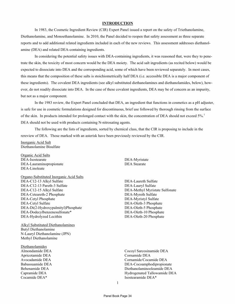
1
INTRODUCTION
In 1983, the Cosmetic Ingredient Review (CIR) Expert Panel issued a report on the safety of Triethanolamine,
Diethanolamine, and Monoethanolamine. In 2010, the Panel decided to reopen that safety assessment as three separate
reports and to add additional related ingredients included in each of the new reviews. This assessment addresses diethanol-
amine (DEA) and related DEA-containing ingredients.
In considering the potential safety issues with DEA-containing ingredients, it was reasoned that, were they to pene-
trate the skin, the toxicity of most concern would be the DEA moiety. The acid salt ingredients (as recited below) would be
expected to dissociate into DEA and the corresponding acid, some of which have been reviewed separately. In most cases,
this means that the composition of these salts is stoichiometrically half DEA (i.e. accessible DEA is a major component of
these ingredients). The covalent DEA ingredients (see alkyl substituted diethanolamines and diethanolamides, below), how-
ever, do not readily dissociate into DEA. In the case of these covalent ingredients, DEA may be of concern as an impurity,
but not as a major component.
In the 1983 review, the Expert Panel concluded that DEA, an ingredient that functions in cosmetics as a pH adjuster,
is safe for use in cosmetic formulations designed for discontinuous, brief use followed by thorough rinsing from the surface
of the skin. In products intended for prolonged contact with the skin, the concentration of DEA should not exceed 5%.1
DEA should not be used with products containing N-nitrosating agents.
The following are the lists of ingredients, sorted by chemical class, that the CIR is proposing to include in the
rereview of DEA. Those marked with an asterisk have been previously reviewed by the CIR.
Inorganic Acid Salt Diethanolamine Bisulfate Organic Acid Salts DEA-Isostearate DEA-Lauraminopropionate DEA-Linoleate
DEA-Myristate DEA Stearate
Organo-Substituted Inorganic Acid Salts DEA-C12-13 Alkyl Sulfate DEA-C12-13 Pareth-3 Sulfate DEA-C12-15 Alkyl Sulfate DEA-Ceteareth-2 Phosphate DEA-Cetyl Phosphate DEA-Cetyl Sulfate DEA-Di(2-Hydroxypalmityl)Phosphate DEA-Dodecylbenzenesulfonate* DEA-Hydrolyzed Lecithin
DEA-Laureth Sulfate DEA-Lauryl Sulfate DEA-Methyl Myristate Sulfonate DEA-Myreth Sulfate DEA-Myristyl Sulfate DEA-Oleth-3 Phosphate DEA-Oleth-5 Phosphate DEA-Oleth-10 Phosphate DEA-Oleth-20 Phosphate
Alkyl Substituted Diethanolamines Butyl Diethanolamine N-Lauryl Diethanolamine (JPN) Methyl Diethanolamine Diethanolamides Almondamide DEA Apricotamide DEA Avocadamide DEA Babassuamide DEA Behenamide DEA Capramide DEA Cocamide DEA*
Cocoyl Sarcosinamide DEA Cornamide DEA Cornamide/Cocamide DEA DEA-Cocoamphodipropionate Diethanolaminooleamide DEA Hydrogenated Tallowamide DEA Isostearamide DEA*
Panel Book Page 34

2
Lactamide DEA Lanolinamide DEA Lauramide DEA* Lauramide/Myristamide DEA Lecithinamide DEA Linoleamide DEA* Minkamide DEA Myristamide DEA* Oleamide DEA Olivamide DEA* Palm Kernelamide DEA Palmamide DEA Palmitamide DEA PEG-2 Tallowamide DEA
PEG-3 Cocamide DEA Ricebranamide DEA Ricinoleamide DEA Sesamide DEA Shea Butteramide/Castoramide DEA Soyamide DEA Stearamide DEA* Stearamide DEA-Distearate Stearamidoethyl Diethanolamine Stearamidoethyl Diethanolamine HCl Tallamide DEA Tallowamide DEA Undecylenamide DEA Wheat Germamide DEA
Lauramide DEA, linoleamide DEA, and oleamide DEA have previously been reviewed by the Expert Panel. In
1986, the Panel concluded that these ingredients are safe as used, and that they should not be used in products containing
nitrosating agents.2 Cocamide DEA was also included in that 1986 assessment; an amended report on cocamide DEA was
issued in 1996.3 In 1996, the Panel concluded cocamide DEA is safe as used in rinse-off products and safe at concentrations
≤10% in leave-on cosmetic products. Cocamide DEA should not be used as an ingredient in cosmetic products in which N-
nitroso compounds are formed. In 1995, the Expert Panel concluded that isostearamide DEA, myristamide DEA, and
stearamide DEA are safe for use in rinse-off products.4 In leave-on products, these ingredients are safe for use at
concentrations that will limit the release of free ethanolamines to 5%, with a maximum use concentration of 40%. In 2009,
the Expert Panel concluded that DEA-dodecylbenzenesulfonate is safe as used when formulated to be non-irritating.5
This family of 69 ingredients, which includes previously reviewed ingredients, has been created to provide a single
comprehensive review of related DEA-containing ingredients. While the ingredients in each subgroup listed above were pre-
sented alphabetically, the order in the report will follow ingredient groupings and chain length, and is provided in Table 1.
The ingredients now included in this review consist of DEA and one or more component. The safety of many of
these components has been reviewed by the CIR. The conclusions of the previously reviewed ingredients, and of the compo-
nents that have been reviewed, are provided in Table 2.
CHEMISTRY
Definition and Structure
DEA is an amino alcohol. DEA is produced commercially by aminating ethylene oxide with ammonia. The replace-ment of two hydrogens of ammonia with ethanol groups produces DEA. DEA contains small amounts of triethanol-amine (TEA) and ethanolamine (MEA).
HONH
OH
Figure 1. DEA
DEA is structurally similar to choline and ethanolamine.6
DEA is reactive and bifunctional, combining the properties of alcohols and amines. At temperatures of 140°-160°C, DEA will react with fatty acids to form ethanolamides. The reaction of ethanolamines and sulfuric acid produces sulfates and, under anhydrous conditions, DEA may react with carbon dioxide to form carbamates. DEA can act as an antioxidant in the autoxidation of fats of both animal and vegetable origin. From the Final Report on the Safety Assessment of Triethanolamine, Diethanolamine, and Monoethanolamine1
Panel Book Page 35
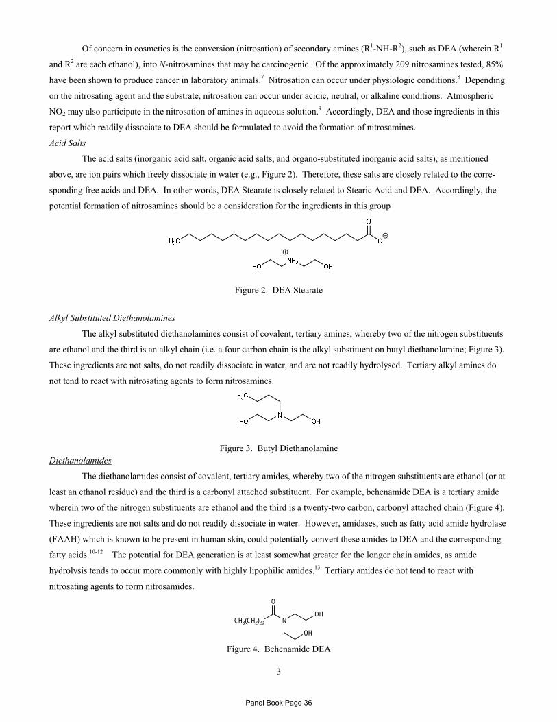
3
Of concern in cosmetics is the conversion (nitrosation) of secondary amines (R1-NH-R2), such as DEA (wherein R1
and R2 are each ethanol), into N-nitrosamines that may be carcinogenic. Of the approximately 209 nitrosamines tested, 85%
have been shown to produce cancer in laboratory animals.7 Nitrosation can occur under physiologic conditions.8 Depending
on the nitrosating agent and the substrate, nitrosation can occur under acidic, neutral, or alkaline conditions. Atmospheric
NO2 may also participate in the nitrosation of amines in aqueous solution.9 Accordingly, DEA and those ingredients in this
report which readily dissociate to DEA should be formulated to avoid the formation of nitrosamines.
Acid Salts
The acid salts (inorganic acid salt, organic acid salts, and organo-substituted inorganic acid salts), as mentioned
above, are ion pairs which freely dissociate in water (e.g., Figure 2). Therefore, these salts are closely related to the corre-
sponding free acids and DEA. In other words, DEA Stearate is closely related to Stearic Acid and DEA. Accordingly, the
potential formation of nitrosamines should be a consideration for the ingredients in this group
Figure 2. DEA Stearate
Alkyl Substituted Diethanolamines
The alkyl substituted diethanolamines consist of covalent, tertiary amines, whereby two of the nitrogen substituents
are ethanol and the third is an alkyl chain (i.e. a four carbon chain is the alkyl substituent on butyl diethanolamine; Figure 3).
These ingredients are not salts, do not readily dissociate in water, and are not readily hydrolysed. Tertiary alkyl amines do
not tend to react with nitrosating agents to form nitrosamines.
Figure 3. Butyl Diethanolamine Diethanolamides
The diethanolamides consist of covalent, tertiary amides, whereby two of the nitrogen substituents are ethanol (or at
least an ethanol residue) and the third is a carbonyl attached substituent. For example, behenamide DEA is a tertiary amide
wherein two of the nitrogen substituents are ethanol and the third is a twenty-two carbon, carbonyl attached chain (Figure 4).
These ingredients are not salts and do not readily dissociate in water. However, amidases, such as fatty acid amide hydrolase
(FAAH) which is known to be present in human skin, could potentially convert these amides to DEA and the corresponding
fatty acids.10-12 The potential for DEA generation is at least somewhat greater for the longer chain amides, as amide
hydrolysis tends to occur more commonly with highly lipophilic amides.13 Tertiary amides do not tend to react with
nitrosating agents to form nitrosamides.
N
O
OH
OH
CH3(CH2)20
Figure 4. Behenamide DEA
Panel Book Page 36
4
The structures and definitions of DEA and all related ingredients are provided in Table 1, and available chemical
and physical properties are provided in Table 3.
Method of Manufacture
Method of manufacture data of on most of the ingredients included in this assessment were not found. The
information that was available is follows.
Diethanolamine
DEA is produced by reacting 2 moles of ethylene oxide with 1 mole of ammonia.14 Typically, ethylene oxide is
reacted with ammonia in a batch process to produce a crude mixture of approximately one-third each MEA, DEA, and TEA.
The crude mixture is later separated by distillation.
Diethanolamides
Lauramide, oleamide, linoleamide, and cocamide DEA are produced by a condensation reaction at a 1:1 or 1:2 molar ratio of a mixture of lauric and myristic acid (for lauramide DEA), oleic acid (oleamide DEA), linoleic acid or its methyl ester (linoleamide DEA), or methyl cocoate, coconut oil, whole coconut acids, or stripped coconut fatty acids (cocamide DEA) to DEA. From the Final Report on the Safety Assessment of Cocamide, Lauramide, Linoleamide, and Oleamide DEA2
Lauramide DEA
Lauramide DEA is produced by the condensation of lauric acid methyl ester with DEA at elevated temperature and
in the presence of a catalyst.15
Cocamide DEA
Cocamide DEA has been produced by the reaction of refined coconut oil with diethanolamide in the presence of sodium methoxide (catalyst), yielding cocamide DEA, 10% glycerine, and 5% coconut fatty acid amide. From the Amended Final Report on the Safety Assessment of Cocamide DEA.3
Analytical Methods
The amount of DEA in fatty acid diethanolamides was determined using a gas chromatographic method with flame
ionization detection.16
Impurities
Diethanolamine
Dow Chemical Company reports that DEA is commercially available with a minimum purity of 99.3%, containing
0.45% max. MEA and 0.25% max TEA.17
Diethanolamides
In the manufacture of the 1:2 mixture of fatty acid to DEA, ethylene glycol and free DEA residues are present. The 1:1 mixture contains much less free amine. Alkanolamides manufactured by base-catalyzed condensation of DEA, and the methyl ester of long chain fatty acids are susceptible to nitrosamine formation. From the Final Report on the Safety Assessment of Cocamide, Lauramide, Linoleamide, and Oleamide DEA2
Lauramide DEA
Various grades of Lauramide DEA are available for cosmetic use. The free amine value is 10-35 (sic). From the Final Report on the Safety Assessment of Cocamide, Lauramide, Linoleamide, and Oleamide DEA2
In National Toxicology Program (NTP) studies, the purity of lauramide DEA was approximately 90% for lauric
acid DEA condensate, with approximately 5% amine (probably DEA) and approximately 5% other organic impurities.15
NDELA was detected at a concentration of 3600 ppb. However, the report also stated that, based on data provided by the
manufacturer, lauramide DEA contained 0.83% free DEA by weight, and approximately 9% other organic impurities.
Commercial samples of lauramide DEA were analyzed for DEA.16 The amount of DEA in the 9 samples ranged
from 1.2-12.4%. NDELA was not found in any of the samples.
Panel Book Page 37

5
Stearamide DEA
Stearamide DEA is characterized by 9-12% free fatty acids (as oleic acid) and 2-6% free amines (as DEA). From the Final Report on Isostearamide DEA & MEA, Myristamide DEA & MEA, and Stearamide DEA & MEA4
Oleamide DEA
Oleamide DEA contains 6.0-7.5% free fatty acids (as oleic acid). From the Final Report on the Safety Assessment of Cocamide, Lauramide, Linoleamide, and Oleamide DEA2
In NTP studies of oleamide DEA, the oleic acid DEA condensate content was 47.5%.18 Impurities were identified
as other fatty acid alkanolamides (approximately 30%), other fatty acids, and unidentified impurities. Free DEA was
estimated at 0.19%. NDELA was detected at a concentration of 68 ppb.
Linoleamide DEA
Commercial sample of linoleamide DEA were analyzed for DEA, and 4.3-5.0% was detected.16 NDELA was not
found in any of the samples.
Cocamide DEA
Various grades of cocamide DEA are available. Alkanolamines manufactured by base-catalyzed condensation of DEA and the methyl ester of long chain fatty acids are susceptible to nitrosamine formation. Cocamide DEA contains 4.0-8.5% free DEA. From the Amended Final Report on the Safety Assessment of Cocamide DEA.3
In NTP studies, cocamide DEA contained approximately 18.2% free DEA by wt, alkanolamides of unsaturated
acids, and amine salts of the acids. NDELA was detected at a concentration of 219 ppb.19
Commercial samples of cocamide DEA were analyzed for DEA.16 The amount of DEA in the 9 samples ranged
from 3.2-14.0%. NDELA was not found in any of the samples.
USE
Cosmetic
DEA functions in cosmetics as a pH adjuster.20 While a few of the other ingredients might function as a pH
adjuster, the majority have other functions, including surfactant, emulsifying agent, viscosity increasing agent, hair or skin
conditioning agents, foam booster, or antistatic agent.
In 1981, according to data provided through the Food and Drug Administration (FDA) Voluntary Cosmetic Regis-
tration Program (VCRP), DEA was used in 18 formulations, and all but one of those products were rinse-off formulations.1
Twelve uses were in hair coloring products. Products containing DEA were used at concentrations of ≤5%. VCRP data
obtained in 2010 indicate that DEA is used in 30 formulations; 15 are leave-on formulations and 15 are rinse-off, and 13 uses
are in non-coloring hair formulations.21 According to data submitted by Industry in response to a survey conducted by the
Personal Care Products Council (Council), DEA is used at concentrations of 0.0008-0.3%.22 The highest concentration used
in leave-on products is 0.06%, in moisturizing products. The complete current and historical use data for DEA and other
previously reviewed ingredients are provided in Table 4a. The use data on ingredients being reviewed for the first time are
provided in Table 4b. Ingredients not reported to be in use, according to VCRP data obtained in 2010, are listed in Table 4c.
According to the Council, “DEA per se is rarely if ever used in personal care products.”23 The potential for expo-
sure to DEA exists from the use of alkanolamides of DEA (which are condensation products of DEA and fatty acids, i.e.,
diethanolamides).
Some of the ingredients included in this assessment are present in aerosolized products, and potential effects on the
lungs of aerosolized products containing this ingredient are of concern. The Expert Panel has previously determined that
Panel Book Page 38
6
because the size of aerosol particles used in hair sprays are greater than 10µm in diameter, they are deposited in the naso-
pharyngeal region and are not inhalable.
According to the opinion of the Scientific Committee on Consumer Safety of the European Commission (EC),
dialkanolamines (e.g. DEA) and their salts (i.e., the acid salts listed previously) are on the list of substances which must not
form part of the composition of cosmetic products.24 According to the Commission’s opinion paper, amines occur only in
their salt form in all cosmetic products. This is because all amines are alkaline compounds which are always neutralized by
an acid component to produce their salts. There is concern about the potential for nitrosamine formation; in principle,
secondary amines are potential precursors of nitrosamines. The ingredients that are included in Annex II of the EC CosIng
database (the list of substances prohibited in cosmetic products) based on this opinion are listed in Table 5
Fatty acid dialkanolamines (i.e., the alkyl substituted diethanolamines) are listed in Annex III of the EC CosIng
database, which is a list of substances cosmetic products must not contain except subject to the restrictions laid down. The
restrictions for these ingredients are: maximum secondary amine content of 0.5% in the finished product; do not use with
nitrosating systems; maximum secondary amine content of 5% for raw materials; maximum nitrosamine content of 50 µg/kg;
and keep in nitrite free containers. The ingredients listed in Annex III with these restrictions, as well as EC information for
all other ingredients included in this report, are also provided in Table 5.
In Canada, DEA is completely prohibited as per the Cosmetic Hotlist; Health Canada prohibits the use of dialkanol-
amines (e.g., DEA).25 This prohibition is based on the EU prohibition in the Cosmetics Regulation, Annex II. The use of
DEA in product formulations in Canada is being investigated due to reported use at concentrations of ≤3%. (Health Canada,
personal communication).
Non-Cosmetic
Many of the ingredients included in this safety assessment have use as indirect food additives.26
Diethanolamine
DEA is used in the manufacture of emulsifiers and dispersing agents for textile specialties, agricultural chemicals, waxes, mineral and vegetable oils, paraffin, polishes, cutting oils, petroleum demulsifiers, and cement additives. It is an intermedi-ate for resins, plasticizers, and rubber chemicals. DEA is used as a lubricant in the textile industry, a humectant and softening agent for hides, as an alkalizing agent and surfactant in pharmaceuticals, as an absorbent for acid gases, and in organic syntheses. From the Final Report on the Safety Assessment of Triethanolamine, Diethanolamine, and Monoethanolamine1
Methyl Diethanolamine
Methyl diethanolamine is used as a flocculent monomer in water treatment, as a sweetening agent in natural gas
treatment, as a catalyst in urethane coating, and as a chemical intermediate in the manufacture of textile lubricants (fabric
softener) and analgesic pharmaceuticals.27
Cocamide DEA
Cocamide DEA is used as a corrosion inhibitor in metalworking fluids and in polishing agents. From the Amended Final Report on the Safety Assessment of Cocamide DEA3
TOXICOKINETICS
Absorption, Distribution, Metabolism and Excretion
In vitro absorption studies were performed using mouse, rat, and human skin. In in vitro studies using mouse and rat skin, 1.3 and 0.04%, respectively, of the applied dose of undiluted [14C]DEA was absorbed. In studies using human skin samples, the absorption of undiluted DEA, as well as concentrations of <1% DEA in combination with fatty acid dialkanolamides, was less than 1% of the applied dose. Penetration of DEA in aqueous solutions was greater than when DEA was undiluted. In studies using human liver slices, DEA was absorbed; the aqueous-extractable radioactivity was
Panel Book Page 39
7
primarily unchanged DEA, while analysis of the organic extracts suggested that DEA was incorporated into ceramides, and slowly methylated. Lauramide DEA was better absorbed in liver slices, and while the absorbed radioactivity was mostly unchanged lauramide DEA, 18-42% was present in the form of metabolites.
In dermal studies with DEA, methyl DEA, and lauramide DEA, the applied doses were generally well absorbed through mouse and/or rat skin, and absorption increased with duration of exposure. In the tissues, the liver generally had the greatest disposition of radioactivity. Urine was the principal route of elimination. Upon dosing with methyl DEA, primarily metabolites, not unchanged methyl DEA, were found in the urine. Lauramide DEA absorption was not dose dependent and the parent compound and the half-acid amide metabolites were detected in the plasma, and disposition did not vary with time.
In oral studies, DEA accumulated in the tissues, with the greatest disposition being in the liver; radioactivity was primarily as unchanged DEA. Urinary excretion was also primarily as unchanged DEA. In a repeated-dose study, stead-state for bioaccumulation occurred after 4 wks; however, DEA continued to bioaccumulate in blood throughout dosing. With lauramide DEA, 79% of the dose was excreted in the urine 72 h after dosing. Four percent of the dose was recovered in the tissue. After 6 hrs, only very polar metabolites, thought to be carboxylic acids, were found in the urine.
In vitro percutaneous absorption studies of cosmetic preparations containing free DEA up to 0.6% showed some penetration occurred in human skin.
Mice exposed orally to sodium nitrate were dosed orally and dermally with 4 mg/kg DEA. A small amount of NDELA was formed following a single oral dose of DEA. No NDELA was detected following dermal dosing with DEA.
In-Vitro
Diethanolamine
The in vitro absorption of 2 mg/cm2 [14C]DEA was determined using fuzzy rat skin.28 A total of 1.4% of the applied
dose was absorbed over 24 h, with 1.9% of the dose remaining in both the stratum corneum and viable dermis/epidermis.
Values were similar at 72 h.
Three full thickness skin preparations from CD rats, CD-1 mice, New Zealand White (NZW) rabbits, and 6 from
female mammoplasty patients, were used to compare the dermal penetration of DEA through the skin of different species.29
[14C]DEA (96.5% purity; sp. act. 15.0 mCi/mmol) was applied to the skin sample undiluted, or as an aq. solution, at a dose of
20 mg/cm2. Dose volumes of 35 µl undiluted [14C]DEA or 95 µl of the aq. solution (37% w/w) were applied to the exposed
surface of the skin (1.77 cm2). These volumes maintained an infinite dose during the 6 h exposure period. Skin sample
integrity was confirmed with the use of a reference chemical, [14C]ethanol. With undiluted DEA, the cumulative dose
absorbed was 0.04% in rat skin, 1.30% in mouse skin, 0.02% in rabbit skin, and 0.08% in human skin. With aq. DEA, an
increase in the cumulative dose absorbed was seen in all species: 0.56% in rat skin, 6.68% in mouse skin, 2.81% in rabbit
skin, and 0.23% in human skin.
Penetration of undiluted and aq. DEA was greater through mouse skin than any other skin sample. With undiluted
DEA, penetration was similar for rat, rabbit, and human skin samples. Penetration of DEA was greater for an aqueous
solution than for undiluted DEA. The researchers hypothesized that this may be attributable to elevated skin hydration
caused by the application of the aqueous solution to the skin.
The percutaneous absorption of DEA in cosmetic formulations spiked with [14C]DEA (95-99% purity) was exam-
ined using viable human skin.30 Two shampoo formulations, both with a [14C]DEA dose of 0.49 µCi were applied as a 1:6
aq. dilution for 5 min; one shampoo contained cocamide DEA with a concentration of 0.092% free DEA, and the other con-
tained lauramide DEA with a concentration of 0.28% free DEA. The amount applied of the shampoo was 1.2 mg (diluted
1:6), and the dose was 1.9 mg/cm2. (The DEA dose was 4.2 µg/cm2 for the first formulation, and 7.7 µg/cm2 for the second
shampoo formulation.) Two hair dye formulations, spiked with 0.49 or 0.43 µCi DEA, were applied for 30 min. One hair
dye contained lauramide DEA and the other contained cocamide and oleamide DEA. The estimated concentration of free
Panel Book Page 40
8
DEA in the hair dye products was 0.61%. An application of 11.2 mg was applied to the skin samples, with a dose of 17.5
mg/cm2. (The DEA dose was 109.3 µg/cm2 for the first formulation, and 108.9 µg/cm2 for the second hair dye formulation.)
Additionally, 3 mg/cm2 of two body lotions (amount applied not stated) with 0.13 or 0.12 µCi DEA was applied for 24 h.
(The DEA dose was 1.0 µg/cm2 for the first formulation, and 1.2 µg/cm2 for the second lotion formulation.) These products
contained 0.0155% TEA, with 0.020% free DEA.
Very little DEA was found in the receptor fluid, with only 0.1% of the applied dose recovered in the receptor fluid
of the shampoo and hair dye formulations. While the amount absorbed was similar for these two product types, the distribu-
tion and localization were different, with most of the DEA (62-68%) penetrating from the shampoos being localized in the
stratum corneum, and that from the hair dyes (52-64%) being found in deeper epidermal and dermal layers. With the lotion,
0.6-1.2% of the dose was recovered in the receptor fluid. Penetration from the lotions differed from each other. With the
first lotion, 15.4% of the applied dose penetrated, with 0.6% found in the receptor fluid and 14.8% in the skin; approximately
65% of the DEA in the skin was in the epidermis and dermis. With the second lotion formulation, only 7.8% of the applied
dose penetrated, with 1.2% found in the receptor fluid and 6.6% in the skin; approximately 56% of the DEA in the skin was
in the epidermis and dermis.
The researchers examined whether DEA was binding to skin. DEA did not appear to be covalently bound to skin
proteins, and extending the times before analysis to 48 and 72 h did not result in any statistically significant difference when
compared to the values obtained after 24 h. Repeat application studies with viable skin did not produce a significant change
of dose absorbed into receptor fluid. However, with non-viable skin, absorption into the receptor fluid from the lotion in-
creased each day, from 0.6% on the first day to 2.6% on the third day; testing showed that the skin barrier did not remain in-
tact for the entire 72 h. Using a shampoo and a lotion formulation, non-viable skin gave penetration values similar to viable
skin. The researchers concluded that most of the DEA that penetrated was not available for systemic absorption.
Another study examining the percutaneous penetration of seven cosmetic formulations spiked with radiolabeled and
unlabeled DEA was performed, mimicking simulated use conditions using fresh human skin samples.31 Two shampoos both
contained 0.98% DEA and 4.02% cocamide DEA, and two additional shampoo formulations both contained 0.25% DEA and
4.75% lauramide DEA; all were applied as a 1:10 aq dilution and a dose of 100 µl/cm2 (equivalent to 100 mg/cm2) for 10
min. A bubble bath containing 0.25% DEA and 4.75% lauramide DEA, applied as a 1:300 dilution and a dose of 100 µl/cm2
(equivalent to 100 mg/cm2) for 30 min, a moisturizer containing 0.008% DEA and 2% TEA, applied at a dose of 5 mg/cm2
for 48 h, and a semi-permanent hair dye containing 0.075% DEA and 1.42% lauramide DEA and an oxidative hair dye con-
taining 0.25% DEA and 4.74% lauramide DEA, both applied at a dose of 100 mg/cm2 for 30 min, were also used. Very little
DEA penetrated the skin; 0.011-0.034% of the applied dose of the shampoo formulations penetrated, 0.024-0.063% of the
applied dose of the hair dye formulations, and 0.508% of the bubble bath formulation penetrated the skin. The moisturizer
formulation was also applied to frozen skin samples. A total of 0.605% of the applied dose penetrated fresh skin, while
0.456% penetrated frozen skin.
The researchers also examined the penetration of a simple aq. 1% solution of DEA through fresh and frozen human
skin samples. The cumulative 24 h percutaneous absorption was approximately 5-fold greater in frozen skin compared to
fresh skin, i.e., 8.87 µg/cm2 compared to 1.73 µg/cm2. The 24-h cumulative penetration values represented 0.433 and
0.086% of the dose for frozen and fresh skin, respectively. The amounts of DEA remaining on and in the skin were 1.68 and
1.14% of the applied dose recovered from the frozen and fresh skin samples, respectively. The researchers further investigat-
ed the distribution of [14C]DEA between aqueous and lipid fractions of viable skin strata. The radioactivity on the skin
Panel Book Page 41
9
samples from 2 donors (n=12) was determined after a 24 h exposure. At 24 h, the cumulative permeation value was 0.405%
of the applied dose for one donor (abdominal skin) and 0.067% for the other (breast skin). (The researchers stated that it may
be significant that one sample was abdominal skin and the other was breast skin.) Tape-strip profiles for both donors ap-
peared to indicate that the DEA had become evenly distributed throughout the stratum corneum. The majority of DEA was
recovered in aqueous extract, as opposed to organic extract, of the epidermal and dermal tissue, which suggested to the
researchers that the material was in the free state and not associated with the lipid fraction.
Human liver slices were incubated with 1 mM [14C]DEA (>97% purity).32 After 4 and 12 h, 11 and 29% of the
DEA, respectively, absorbed into the liver slices. The radioactivity was comprised mostly of DEA (85-97%); four other
metabolites were present at low concentrations. The liver slices were fractionated into aqueous, organic, and pellet fractions.
The aqueous-extractable radioactivity was comprised primarily of DEA, with up to four other components. DEA-derived
radioactivity in the organic extracts was predominately (>90%) comprised of phospholipids containing non-methylated head-
groups. DEA was readily absorbed and incorporated into ceramides, forming mostly ceramide-phosphodiethanolamine, and
it was slowly methylated therein.
Lauramide DEA
Human liver slices, and liver slices from diethylhexyl phthalate-(DEHP) induced and untreated male F344 rats, were
incubated with [14C]lauramide DEA.33 Lauramide DEA “partitioned well” into the liver slices, and approximately 70% of the
radioactivity absorbed into the slices in 4 h. The absorbed radioactivity was present mostly as lauramide DEA. In the ex-
tracts, 18-42% of the radioactivity was present in the form of metabolites. The analytes present in the incubation media in-
cluded half-acid amides, parent lauramide DEA, and three other metabolites that are products of ω- and ω-1 to 4 hydroxyla-
tion.
The in vitro metabolism of [14C]lauramide DEA, randomly labeled on the DEA moiety, was examined in liver and
kidney microsomes from rats and humans to determine the extent of hydroxylation, and to determine the products formed.34
Incubation of lauramide DEA with liver microsomes from control and DEHP-treated rats produced two major high perform-
ance liquid chromatography (HPLC) peaks that were identified as 11-hydroxy- and 12-hydroxy-lauramide DEA. Treatment
with DEHP increased the 12-hydroxylation rate 5-fold, while the 11-hydroxylase activity was unchanged. Upon comparison
of lauramide DEA hydroxylation rates from human liver microsomes with those from rat liver and kidney microsomes, the
lauramide DEA 12-hydroxylase activity in human liver was similar to the rate found in liver microsomes of control rats. The
rate was 3 times greater than that observed in rat kidney microsomes.
Dermal
Non-Human
Diethanolamine
[14C]DEA was applied to a 2 cm2 area of the intrascapular region of male F344/N rats at a dose of 2.1, 7.6, or 27.5
mg/kg bw.35,36 After 48 h, 2.9, 10.5, or 16.2% of the dose, respectively, was absorbed. It was shown that a 10-fold increase
in the concentration of DEA resulted in a 450-fold increase in the rate of absorption.
A single occluded dose of [14C]DEA was applied to a 19.5 cm2 area on the back of rats for 6 h, and the treated site of
50% of the animals was rinsed.37 In the animals that were not rinsed, 80% of the dose was found in the wrappings, and 3.6%
found in the skin. In rinsed animals, rinsates from the wrappings contained 58% of the dose, and from the skin contained
26% of the dose. Unrinsed animals absorbed 1.4% of the dose, while those that were rinsed absorbed 0.64%. The majority
of the radioactivity was found in the carcass, liver, and kidneys.
Panel Book Page 42
10
The researchers also conducted a repeated-dose percutaneous-absorption study in which as a pre-exposure, 1500
mg/kg/day non-radiolabeled DEA was applied occlusively under a 2 in x 2 in gauze square to the backs of rats 6 h/day for 3
or 6 days. [14C]DEA, 1500 mg/kg/day, was then applied for 3-6 consecutive days under occlusion; each dose was left in
contact with the skin for 48 h. Totals of 21 and 41% of the dose were absorbed by the animals dosed for 3 and 6 days,
respectively. The majority of the recovered radioactivity was in the wrapping. The carcass, liver, and kidneys contained
most of the radioactivity in the animals. Totals of 4.3 and 13% of the radioactivity were recovered in the urine of animals
dosed with non-radiolabeled DEA for 3 and 6 days, respectively. Pre-exposure to DEA resulted in a 1.5- to 3.0-fold increase
in the absorption rate.
Groups of 4-5 male B6C3F1 mice were used in dermal studies of the absorption, distribution, metabolism, and
excretion (ADME) of DEA.38 The total volume for mice was 15 µl/dose, and the dose was applied to a 1 cm2 area of skin
using a non-occlusive covering. Absorption through mouse skin was greater than through rat skin. At 48 h after dermal
application of 8 and 23 mg/kg [14C]DEA, 26.8 and 33.8% of the dose was absorbed in the mice. A statistically significant
increase in the amount absorbed after dosing with 81 mg/kg, 58.1%, was observed. The amount of radioactivity found at the
site of application was only 4.0, 3.1,and 2.2% of the dose following application of 8, 23, and 81 mg/kg [14C]DEA,
respectively, and the amount excreted in the urine 48 h after application was 7.5%, 10.4, and 16.4%, respectively. The
tissue/blood ratio was greatest in the liver and kidneys.
Groups of 4-5 male Fischer 344 rats were used to evaluate the ADME of DEA (99% purity) following dermal
administration.38 The dose, which contained 6-20 µCi radiolabel, unlabeled DEA, and ethanol, for a total volume of 25 µl
per dose, was applied to a 2 cm2 area of skin under a non-occlusive covering. Absorption increased significantly with
increasing dose. After 48 h, only 2.9% of the radioactivity was absorbed following dermal application of 2.1 mg/kg, while
10.5 and 16.2% of the radioactivity was absorbed with doses of 7.6 and 27.5 mg/kg, respectively. Of the amount absorbed,
1.2, 4.3, and 4.5% of the radioactivity was recovered in the skin at the dose site following application of 2.1, 7.6, and 27.5
mg/kg [14C]DEA, respectively. The greatest tissue/blood ratios were found in the liver and lung.
DEA, 80 mg/kg bw in acetone, was applied to a 2 cm2 area on the backs 7 female C57BL/6 mice for 11 days.39
DEA and its methylated metabolites accumulated in the liver and plasma of mice. Also, a statistically significantly decrease
in hepatic concentrations of choline and its metabolites were reported.
Methyl Diethanolamine
A dose of 500 mg/kg, uniformly labeled, [14C]methyl DEA, 25 µCi, was applied to a 2 cm x 4 cm area of 4 male,
and a 2 cm x 3 cm area of 4 female, Fischer 344 rats.40 The occlusive patches were applied for 6 or 72 h; the number of ani-
mals used per duration was not specified. With the 6-h exposure, 17-21% of the applied dose was absorbed. With the 72 h
exposure, 41-50% of the applied dose was absorbed. The greatest amounts of radioactivity were found in the livers and kid-
neys of animals dosed for 6 and 72 h. The principal route of elimination was the urine, but the rates of urinary excretion were
slow. With the 6 h exposure, approximately 2.5-4.75% of the total dose was excreted in the urine in 72 h, and with the 72 h
exposure, approximately 7.5-8.5% of the total dose was excreted. The major component in the urine was metabolites, rather
than unchanged methyl DEA. In the animals dosed for 6 h, radioactivity was systemically available after the dose was
washed. The concentrations of radioactivity in the plasma in the plasma of animals of this group were greatest at 60 h, with
values of 2.6-4.2 µg/g. In the animals dosed for 72 h, the amounts of radioactivity in the plasma were also greatest at 60 h,
with values of 8.5-10.0 µg/g. Unchanged methyl DEA was available at 72 h for both exposure groups.
Panel Book Page 43
11
Lauramide DEA
Groups of four male B6C3F1 mice and four F344 rats were dosed dermally with randomly labeled [14C]lauramide
DEA, 2-17 µCi/dose for mice and 16-118 µCi/dose for rats.33 The vehicle was ethanol. A non-occlusive application was
made to a 0.5 in2 or 1 in2 area of skin of mice and rats, respectively. At the end of the study, the excised skin was rinsed with
ethanol. Mice were dosed with 5-800 mg/kg [14C]lauramide DEA. At 72 h, 50-70% of the applied radioactivity was ab-
sorbed, and absorption was similar for all the doses. Approximately 32-55% of the radioactivity was excreted in the urine.
In rats dosed with 25 or 400 mg/kg lauramide DEA, 21-26% of the radioactivity penetrated the skin in 72 h, and 3-
5% was recovered at the site of application. Approximately 20-24% of the radioactivity was recovered in the urine. The
tissue/blood ratio was greatest in the liver and kidney. Lauramide DEA and the half-acid amide metabolites were detected in
the plasma, with maximum levels found 24 h after dosing.
The researchers also applied 25 mg/kg/day lauramide DEA, 5 days/wk for 3 wks, to a group of 5 rats. Disposition
did not vary much at the different collection time points.
Human
Three female subjects applied a lotion containing 1.8 mg DEA/g lotion to their entire body 2x/day for 1 mo.39
Blood samples were collected 1 day prior to the start of dosing, at 1 wk, and at 1 mo. Two of the subjects completed 1 mo of
application, while the third completed 3 wks. Application of the DEA-containing lotion for 1 mo resulted in increased
concentrations of DEA and dimethyl DEA in plasma, when compared to blank samples. (As a reference point, however, it
was calculated that the concentration of DEA and metabolites in the plasma of humans after a few weeks of application of the
lotion were only 0.5-1% of the concentrations achieved in mice, where 80 mg/kg/day was applied for 11 days.)
Oral
Non-Human
Diethanolamine
Four male Fischer 344 rats were dosed orally by gavage with 7 mg/kg aq. [14C]DEA to examine the ADME of DEA
(>97% purity).32 The amount of radioactivity excreted in the urine at 24 and 48 h was 9 and 22%, respectively, and the
amount in the feces was 1.6 and 2.4%, respectively. At 48 h after dosing, 27% of the radioactivity accumulated in the liver,
5% in the kidneys, and 0.32, 0.27, 0.19, and 0.18% in the spleen, brain, heart, and blood, respectively. As measured in the
liver and the brain, 87-89% of the radioactivity distributed into the aq. phase, and 70-80% of that total radioactivity was as
unchanged DEA. In the liver, the remaining radioactivity in the aqueous phase was distributed between three methylated
metabolite fractions, while in the brain, only one minor, non-methylated metabolite was present in the aqueous extract.
With repeat oral dosing with [14C]DEA, radioactivity continued to accumulate in tissues, reaching steady states at 4-
8 wks. The levels of DEA equivalents in the blood, brain, and liver were much higher following 8 weeks of repeated oral
doses of 7 mg/kg/day, when compared to the single dose post-48-h values. Again, DEA was the major radioactive compo-
nent. Using HPLC, almost all of the organic-extractable hepatic radioactivity eluted with the phosphatidylcholine fraction,
with greater than 95% of the material in the form of phospholipids containing an N,N-dimethyl-DEA headgroup. In the
brain, the entire organic-extractable radioactivity eluted in the phosphatidylethanolamine fraction, and it was almost entirely
comprised of DEA-containing headgroups.
According to the researchers, the results of this study demonstrated that DEA is O-phosphorylated and N-methyl-
ated, and that these metabolites are incorporated as the polar headgroups in aberrant phospholipids. The researchers felt this
was evidence that DEA and MEA, a naturally-occurring alkanolamine, share common biochemical pathways of transforma-
Panel Book Page 44
12
tion. Retention and bioaccumulation of DEA-derived radioactivity was attributed partly to aberrant phospholipids being
incorporated into tissues, most probably in cell membranes.
Groups of 3-5 male Fischer 344 rats were used to evaluate the ADME of a single oral administration of [14C]DEA
(99% purity).38 The dose administered contained 2-200 µCi radiolabeled, unlabeled DEA, and the amount of water needed to
achieve a target dose of 5 ml/kg bw. Gastrointestinal absorption was nearly complete after doses up to 200 mg/kg DEA. At
48 h after dosing with 7 mg/kg, 22% of the radioactivity was excreted in the urine and 2.4% in the feces, primarily as
unchanged DEA. Radioactivity was not detected in carbon dioxide. Excretion was mostly unchanged DEA. A total of 57%
of the radioactivity was found in the body 48 h after dosing with 7 mg/kg [14C]DEA. Disposition of radioactivity was
greatest in the liver (27.3%), muscle (16.3%), skin (5.1%), and kidneys (5%); only 0.2% of the radioactivity was found in the
blood after 48 h. Dose did not affect distribution in the tissues.
The researchers then evaluated the ADME of DEA upon repeat oral exposure. Four rats were dosed orally with 7
mg/kg/day [14C]DEA for 5 days. Approximately 40% of the total radioactivity was excreted during dosing. At 48 h after the
last dose, a total of 42% of the radioactivity was found in the tissues, with the greatest distribution in the liver (18.2%),
muscle (12.4%), kidneys (5.56%), and skin (4.18%). To further investigate DEA bioaccumulation, rats were dosed with 7
mg/kg/day DEA, 5 days/wk, for 2, 4, or 8 wks. During the last week of each dosing period, 69, 79, and 92% of the dose was
excreted, respectively. After 8 wks of dosing, most of the radioactivity was recovered as unchanged DEA; however there
were also significant amounts of poorly retained metabolites, N-methylDEA, and another metabolite that was tentatively
identified as a quaternized lactone. The % radioactivity recovered in the liver after 2, 4, and 8 wks of dosing was 12.3, 7.9,
and 4.12%, respectively. It was estimated that steady-state for bioaccumulation occurred after 4 wks of repeat dosing, except
for the blood, which continued to bioaccumulate DEA throughout dosing. Clearance of bioaccumulated DEA appears to be a
first-order process, with a whole body elimination half-life of ~6 days.
Lauramide DEA
Three male F344 rats were dosed orally with randomly labeled [14C]lauramide DEA, 16-18 µCi/dose, formulated to
give a target dose of 5 ml/kg bw.33 After oral dosing with 1000 mg/kg [14C]lauramide DEA, approximately 10, 60,and 79%
of the dose was recovered in the urine after 6, 24 h, and 72 h, respectively. Approximately 4% of the dose was recovered in
the tissues after 72 h, with almost 3% found in adipose tissue and 1.3% in the liver. After 6 h, no DEA, DEA metabolites, or
unchanged lauramide DEA were present in the urine; only very polar metabolites were found. The researchers postulated
that the metabolites were carboxylic acids, and that the acid function was formed from the lauryl chain.
Intravenous
Non-Human
Diethanolamine
Groups of Sprague-Dawley rats (number per group not specified) were given an intravenous (i.v.) dose of 10 or 100
mg/kg DEA in physiological saline.37 Animals were killed 96 h after dosing. Peak blood concentrations appeared 5 min after
dosing. Elimination from the blood was biphasic. Totals of 25 and 36% of the dose were excreted in the urine with 10 and
100 mg/kg DEA, respectively.
Groups of 3-5 male Fischer 344 rats were used to evaluate the ADME of [14C]DEA (99% purity) in phosphate-
buffered saline after i.v. administration.38 After 48 h, 28% of the dose was excreted in the urine, 0.6% in the feces, and only
0.2% in carbon dioxide. Using HPLC, it was determined that most of the radioactivity in the urine was present as unchanged
DEA. A total of 54% of the dose was found in the body 48 h after dosing with 7 mg/kg [14C]DEA, and as with oral dosing,
Panel Book Page 45
13
the greatest disposition was found in the liver (27%), muscle (15%), skin (4.5%), and kidneys (4%). Only 0.2% of the
radioactivity was found in the blood after 48 h.
Groups of 5 female Sprague-Dawley rats were dosed i.v., via the cannulated jugular vein, with 10 or 100 mg/kg
[14C]DEA (97.4% purity).41 The dose volume was 2 ml/kg, and each rat received ~4.2 µCi 14C. Blood samples were taken at
various intervals up to 84 h after dosing. The peak concentrations of radioactivity in both the plasma and the red blood cells
were observed 5 min after dosing. Clearance of radioactivity from the plasma was calculated to be approximately 50 and 93
ml/h/kg for the low and high dose, respectively. In blood, these values were 84 and 242 ml/h/kg, respectively
Urine and feces were collected for 96 h after dosing. During this time, the major route of excretion was urinary; 25
and 36% of the dose was recovered in the urine. Urinary excretion was rapid at the high dose level, with 23% of the dose
recovered in the first 12 h. Only 8.5% of the dose was recovered with the low dose during this time frame. The majority of
the radioactivity was recovered in the carcass; 35 and 28% for the low and high dose, respectively. At 96 h after dosing with
10 and 100 mg/kg DEA, approximately 21 and 17% of the dose was recovered in the liver, 7 and 5% in the kidneys, and 5
and 5% in the skin, respectively. The researchers stated that because the majority of the applied dose (administered radio-
activity) was recovered in the tissues, particularly in the liver and kidneys, this indicated a propensity for bioaccumulation.
There was some evidence that the bioaccumulation was dose-dependent.
Methyl Diethanolamine
Groups of 4 male Fischer 344 rats were given a single i.v. dose of 50 or 500 mg/kg, uniformly labeled, [14C]methyl
DEA, 10 µCi, at a dose volume of 2 ml/kg. The principal route of elimination was the urine, but the rates of urinary excre-
tion were slow. In the 50 mg/kg group, approximately 60% of the total dose was excreted in the urine in 72 h; with the 500
mg/kg dose, approximately 67.5% of the total dose was excreted. The major components in the urine of animals dosed with
50 mg/kg were metabolites. Conversely, in urine of animals dosed with 500 mg/kg, the major component was unchanged
methyl DEA. In the plasma, a rapid distribution phase was followed by a slower elimination phase; 9 and 44% of the total
radioactivity in the plasma was unchanged methyl DEA at 1 h after dosing with 50 and 500 mg/kg, respectively.
Lauramide DEA
Three male B6C3F1 mice and four F344 rats were dosed intravenously with randomly labeled [14C]lauramide DEA,
3-5 µCi and 16-17 µCi, respectively, formulated to deliver a target dose of 4 ml/kg in mice and 1 ml/kg in rats.33 The dose
for mice was 50 mg/kg and for rats was 25 mg/kg. In mice, lauramide DEA was quickly metabolized and eliminated. At 24
h after dosing, approximately 95% of the dose was excreted, with 90% found in the urine. The highest concentrations and
total amounts of the lauramide DEA were in adipose tissue.
In rats, 50% of the dose was excreted in the urine within the first 6 h, and more than 80% was excreted in the urine
by 24 h. The rats were killed at 72 h after dosing, and only 3% of the dose was recovered in the tissues; 1% of the dose was
in the adipose tissue and 0.67% was found in the liver.
N-Nitrosodiethanolamine (NDELA) Formation
The formation of NDELA upon dermal dosing with DEA (99.7% purity), with and without supplemental oral
sodium nitrite, was determined in male B6C3F1 mice.42 Groups of 5-6 mice were dosed orally with aq. DEA or dermally
with DEA in acetone. DEA, 160 mg/kg/day, was applied for 7 days/wk for 2 wks. One group of mice dosed dermally was
allowed access to the application site, while the other was not. Studies were performed both with and without ~40 mg/kg/day
supplemental sodium nitrite in drinking water. NDELA was not found in the urine or blood of mice dosed with DEA without
nitrite or in the blood or gastric contents of those given supplemental nitrite with DEA.
Panel Book Page 46
14
NDELA formation from DEA (>99% purity) and nitrite was also examined in another study.43 Female B6C3F1
mice were dosed dermally or orally with 4 mg/kg DEA, in conjunction with oral exposure to sodium nitrite. Following 7
days of dermal dosing, no NDELA was detected in the blood, ingesta, or urine of test, vehicle control, or sodium nitrite
control mice. (The limits of detection for the blood, ingesta, and urine were 0.001, 0.006, and 0.47 µg/ml, respectively.)
With a single oral dose, NDELA was formed in all of the animals; the amounts of NDELA detected in the blood and ingesta
of mice 2 h post-dosing were very small; 0.008 ± 0.003 µg/g and 0.424 ± 0.374 µg/g, respectively.
TOXICOLOGICAL STUDIES
Acute dermal testing with methyl diethanolamine,50% lauramide DEA, and undiluted and 10% aq linoleamide DEA, acute oral testing with DEA, methyl DEA, butyl DEA, and several fatty acid diethanolamides, and acute inhalation testing with methyl DEA did not result in significant toxicity.
In repeat dermal testing with DEA, lauramide DEA, and cocamide DEA in mice and/or rats, irritation was observed at the site of application. Increases in liver and kidney weights were observed in most studies, while decreases in body weight were observed sporadically. The LOAEL for DEA in a 2-wk study in mice was 160 mg/kg bw. Repeat dermal dosing with methyl DEA in rats also caused skin lesions, but it did not seem to affect liver weights or body weights, and an increase in kidney weights was observed in 1 of 3 studies. The NOEL for methyl DEA in a 13-wk study in rats was 100 mg/kg day. A formulation containing 3% linoleamide DEA was not a cumulative systemic toxicant in a 13-wk dermal study.
In repeat oral testing with DEA, increases in liver and kidney weights and decreases in body weights were seen in mice and rats. Deaths, believed to be test-article related, occurred in most of the studies, and included a mouse given 100 mg/kg DEA by gavage. With repeat oral dosing of lauramide DEA, the NOEL was 250 mg/kg/day in one study using rats. The NOEL for Beagle dogs fed lauramide DEA for 12 wks was 5000 ppm.
In inhalation studies with DEA in rats, liver and kidney weights were again increased. In 13-wk studies with ≤400 mg/m3DEA, microscopic effects were observed in the larynx. The 90-day NOAEC was 1.5 mg/m3 DEA.
Acute (Single Dose) Toxicity
Dermal
Methyl Diethanolamine
The percutaneous LD50 of undiluted methyl DEA was determined in NZW rabbits, using occlusive patches.27 The
LD50 values were 9.85 ml/kg (10.2 g/kg) for males and 10.9 ml/kg (11.3 g/kg) for females. In other studies, the dermal LD50
in rabbits ranged from 6-11.3 g/kg bw.44
Lauramide DEA
In an acute dermal toxicity study using guinea pigs, 50% lauramide DEA in corn oil was non-toxic. From the Final Report on the Safety Assessment of Cocamide, Lauramide, Linoleamide, and Oleamide DEA2
Linoleamide DEA
Linoleamide DEA, tested as 10% aq. and undiluted, was nontoxic in acute studies with guinea pigs. From the Final Report on the Safety Assessment of Cocamide, Lauramide, Linoleamide, and Oleamide DEA2
Oral
Diethanolamine
The acute oral toxicity of DEA was determined using guinea pigs and rats. Using groups of 2-3 guinea pigs, all guinea pigs survived dosing with 1 g/kg, but none survived dosing with 3 g/kg DEA gum arabic solution. Using groups of 5 rats, the oral LD50 of undiluted DEA was 0.71-0.80 ml/kg, and for a group of 6 rats, the LD50 of DEA in water was 1.82 g/kg. Using 90-120 rats, 20% aq. DEA had an acute LD50 of 1.41-2.83 g/kg, based on results of testing performed over a 10 yr time period. With groups of 10 rats, a hair preparation containing 1.6% DEA had an LD50 of 14.1 g/kg when diluted and 12.9 ml/kg when undiluted. From the Final Report on the Safety Assessment of Triethanolamine, Diethanolamine, and Monoethanolamine1
Panel Book Page 47
15
Methyl Diethanolamine
Groups of 5 male and 5 female Sprague-Dawley rats were used to determine the LD50 value for undiluted methyl
DEA. The oral LD50 for male and female rats was 1.87 ml/kg (1.95 g/kg).27 In other oral studies in rats, the LD50 ranged
from 1.95-4.78 g/kg bw.44
Butyl Diethanolamine
The oral LD50 of butyl DEA in rats was 4.25 g/kg.45
Lauramide DEA
In rats, the oral LD50 of 25% lauramide DEA in corn oil was >5 g/kg, of 10% aq. was 2.7 g/kg, of a shampoo formulation containing 8% lauramide DEA was 9.63 g/kg, and of a bubble bath containing 6% lauramide DEA was >15 g/kg. From the Final Report on the Safety Assessment of Cocamide, Lauramide, Linoleamide, and Oleamide DEA2
Stearamide DEA
The oral LD50 of a mixture containing 35-40% stearamide DEA was >20 g/kg in CFW mice. From the Final Report on Isostearamide DEA & MEA, Myristamide DEA & MEA, and Stearamide DEA & MEA4
Oleamide DEA
In rats, the oral LD50 of undiluted oleamide DEA was 12.4 ml/kg. From the Final Report on the Safety Assessment of Cocamide, Lauramide, Linoleamide, and Oleamide DEA2
Linoleamide DEA
In rats, the oral LD50 of undiluted and 10% aq. linoleamide DEA was >5 g/kg, and the LD50 of a product containing 1.5% linoleamide in formulation was 3.16 g/kg. From the Final Report on the Safety Assessment of Cocamide, Lauramide, Linoleamide, and Oleamide DEA2
Cocamide DEA
The acute oral toxicity of cocamide DEA was determined using groups of 3 male and 3 female Wistar rats. Three
or more animals per group died with doses of ≥6.3 g/kg.46
Inhalation
Methyl Diethanolamine
Five male and 5 female Sprague-Dawley albino rats were exposed for 6 h to a saturated methyl DEA vapor.27 None
of the rats died.
Other
Diethanolamine
The intraperitoneal (i.p.) LD50 of DEA was 2.3 g/kg for mice. From the Final Report on the Safety Assessment of Triethanolamine, Diethanolamine, and Monoethanolamine1
Methyl Diethanolamine
Reported i.p. LD50 values for methyl DEA in mice ranged from 0.5-0.67 g/kg bw.44
Repeated Dose Toxicity
Dermal
Diethanolamine
In a 13-wk study, 1 mg/kg of a hair dye formulation containing 2.0% DEA was applied to the backs of 12 rabbits for 1 h, twice weekly. The test site skin was abraded for half of the animals. No systemic toxicity was observed, and there was no histomorphologic evidence of toxicity From the Final Report on the Safety Assessment of Triethanolamine, Diethanolamine, and Monoethanolamine1 DEA was applied dermally to groups of 5 male and 5 female B6C3F1 mice, 5x/wk for 2 wks, at doses of 0-2500
mg/kg bw in 95% ethanol.47 All of the male and 3 of the female high-dose test animals died during the study. Ulceration,
Panel Book Page 48
16
irritation, and crusting were observed at the application site of male mice of the 1250 and 2500 mg/kg groups and females of
the 2500 mg/kg group. Microscopically, moderate to marked epidermal ulceration and inflammation were observed in these
animals. Ulcerative necrosis extended into the underlying dermis. Minimally severe acanthosis, without inflammation, was
seen in the 160, 320, and 630 mg/kg dose groups. Absolute and relative liver weights increased in a dose-dependent manner
in males and females. The lowest observable adverse effect level (LOAEL) was 160 mg/kg bw.
Repeated dermal exposure of DEA (99.6% purity) in 96% ethanol was applied to the shaved backs of groups of
B6C3F1 mice for the following time periods: 8 males and 8 females were dosed daily with 0 or 160 mg/kg bw (dose volume
2.13 ml/kg bw) for 1 wk, followed by a 3-wk recovery period; groups of 10 males were dosed 5 days/wk with 0 or 160 mg/kg
bw for 1, 4, or 13 wks; and groups of 8 males were dosed 5 days/wk with 0-1250 mg/kg for 1 or 13 wks.48 With the last
dosing scheme, application of 630 and 1250 mg/kg DEA was discontinued, and the animals killed, after 1 wk due to severe
skin lesions; the high dose was then 160 mg/kg. In the other animals, including controls, some erythema and/or focal crust
formation was observed and attributed to the procedure and/or the vehicle, but not to DEA. No DEA-related deaths were
observed. Body weights were not affected by dosing. Statistically significant increases in liver weights were seen in mice
dosed with ≥10 mg/kg bw/day DEA for 1 or more weeks. Microscopically in the liver, eosinophilia was found in animals
dosed with ≥40 mg/kg DEA for 1 or more weeks, and hepatocellular giant cells were seen in animals dosed with 160 mg/kg
for 13 wks.
Unoccluded dermal applications of 0-600 mg/ml DEA (purity >99%) in acetone were applied to the backs of 10
male and 10 female B6C3F1 mice, 5 days/wk for 13 wks, at doses of 0-1250 mg/kg bw.49 (The area of the dose site was not
provided.) Two male mice and 4 female mice dosed with 1250 mg/kg DEA were killed in moribund condition. Final mean
body weights of male mice dosed with 1250 mg/kg were statistically significantly decreased compared to controls. Clinical
signs of toxicity, observed in males and females dosed with 630 and 1250 mg/kg DEA, were irritation, crust formation, and
thickening at the application site. Ulceration and inflammation were observed in the 630 and 1250 mg/kg dose groups.
Dose-dependent increases in absolute and relative liver weights, associated with hepatocellular cytological changes, were
observed, and hepatocellular necrosis was seen in males dosed with 320-1250 mg/kg DEA. Absolute kidney weights were
statistically significantly increased in males and females of all test groups and relative kidney weights were increased in
males of all test groups and females dosed with 630 or 1250 mg/kg DEA; nephropathy was not found. Heart weights were
increased in high dose males and females, and degeneration was reported.
Unoccluded dermal applications of 0-500 mg/ml DEA (purity >99%) in 95% ethanol were applied to the backs of 10
male and 10 female F344/N rats, 5 days/wk for 13 wks, at doses of 0-500 mg/kg bw.50 (The area of the dose site was not
provided.) One male and 2 females given 500 mg/kg died or was killed in moribund condition during the study. Clinical
signs of toxicity, observed in males and females given 125-500 mg/kg DEA, were irritation and crusting at the application
site. Final mean body weights were statistically significantly decreased in males dosed with 250 and 500 mg/kg and females
dosed with 125-500 mg/kg DEA. Increases in absolute and relative kidney weights were observed with increased incidence
of renal lesions. Increases in absolute and relative liver weights were not accompanied by an increase in hepatic lesions.
Demyelination in the medulla oblongata was seen in all high dose animals and 7 females given 250 mg/kg DEA; this lesion
was minimal in severity.
Methyl Diethanolamine
The dermal toxicity of methyl DEA was evaluated in a two 9-day studies using groups of 20 male and 20 female
Fischer 344 rats.51 In both studies, nine 6-hr occlusive applications using a 2” x 2” gauze pad were made to a shaved area of
Panel Book Page 49
17
the trunk over an 11-day period. The animals were killed the day after dosing termination. In the first study, the rats were
dosed with 0, 260, 1040, or 2080 mg/kg/day in deionized water. The dose volume was 2.0, 0.25, 1.0, and 2.0 ml/kg/day,
respectively; methyl DEA was undiluted. None of the animals died during the study, and no clinical signs of toxicity were
observed. Transient, barely perceptible erythema was observed for one male and one female of the mid and high dose
groups, and barely perceptible edema was observed for on female of the low dose group on day 5. Dose-related exfoliation,
excoriation, and necrosis were observed, and fissuring were observed in female rats. Body weight gains were reduced in a
dose-related fashion, being statistically significant for males. Feed consumption was also reduced dose-dependently in
males, being statistically significant in the mid and high dose groups. Females of the high dose group had statistically signifi-
cant decreases in hemoglobin concentration, hematocrit, and mean corpuscular hemoglobin, with increased segmented
neutrophils. Absolute kidney and adrenal gland weights were statistically significantly increased in females of the 2080
mg/kg group, and relative kidneys to body weights were statistically significantly increased in the 1040 and 2080 mg/kg
groups
In the second study, the rats were dosed with 0, 100, 500, or 750 mg/kg/day methyl DEA. The dose volume was 1.0
ml/kg/day for all doses.. None of the animals died during the study, and no clinical signs of toxicity were observed. Similar
dermal results were reported. In this study, there were no statistically significant differences in body weights or feed con-
sumption between treated and control animals, and there were no differences in hematology. No treatment-related differ-
ences in organ weights were found for any dose. At the application site, dose-related increases in the incidence and severity
of acanthosis and hyperkeratosis were observed, and multifocal dermatitis and exocytosis of polymorphonuclear leukocytes
into the stratum corneum were observed.
The researchers also performed a 13 wk study in which Fischer 344 rats received 65 occlusive applications of
methyl DEA, 6 h/day, 5 days/wk. Twenty male and 20 female rats were dosed with 0 or 750 mg/kg/day, and groups of 10
male and 10 female rats were dosed with 100 or 250 mg/kg/day. Patches were applied as described previously. All rats of
the low and mid dose groups, and 10 rats/gender of the control and high dose groups, were killed the day after dosing
termination. The remaining control and high dose animals were killed after a 4-wk recovery period. As in the 9-day studies,
no animals died and no clinical signs of toxicity were observed. Slight, transient, erythema was only seen in the high dose
group. Dose-related incidences of desquamation, excoriation, ulceration, necrosis, and eschar were observed.
Microscopically, acanthosis, hyperkeratosis, and parakeratosis were observed at the site of application. There were no signi-
ficant differences in body weights, and no effects on organ weights. The no-observed effect level (NOEL) was 100
mg/kg/day.
Lauramide DEA
The dermal toxicity of lauramide DEA was evaluated in two 13-wk studies using Sprague-Dawley rats. A 0.45% aq. solu-tion of a cream cleanser containing 4.0% lauramide DEA, tested in 15 females, and a medicated liquid cleanser containing 5.0% lauramide DEA, tested in 10 males and 10 females, did not have any systemic toxic effects. From the Final Report on the Safety Assessment of Cocamide, Lauramide, Linoleamide, and Oleamide DEA2
The dermal toxicity of lauramide DEA (90% purity; 0.83% free DEA by wt) was evaluated in mice and rats.
Groups of 10 male and 10 female B6C3F1 mice were dosed with 0-800 mg/kg bw lauramide DEA in ethanol, 5 days/wk, for
14 wks.15 All animals survived until study termination. Dermal irritation was observed at the application site in males and
females dosed with 400 or 800 mg/kg lauramide DEA. Final mean body weights and mean body weight gains were similar
for test and control animals. The absolute kidney weights of males of the 10, 400 and 800 mg/kg bw groups, the relative
kidney to body weights of all dosed males, and the liver weights of females of the 200, 400, and 800 mg/kg bw groups, were
Panel Book Page 50
18
significantly greater than those of the control mice. The absolute thymus weights of males of the 400 and 800 mg/kg groups
were significantly less than those of the controls. There were no significant differences in reproductive tissue evaluation or
estrous cycle between the treated and control groups. At the application site, incidences of non-neoplastic lesions of the skin,
including hyperplasia of the epidermis and sebaceous gland, chronic inflammation, parakeratosis, and ulceration, were in-
creased in males and females dosed with ≥200 mg/kg lauramide DEA..
Groups of 20 male and 20 female F344/N rats were administered 0-400 mg/kg bw lauramide DEA in ethanol, 5
days/wk for 14 wks; 10 rats per group were used for clinical pathology.15 All animals survived until study termination. Der-
mal irritation was observed at the application site of males dosed with ≥100 mg/kg and in females dosed with 200 or 400
mg/kg lauramide DEA. Final mean body weights and mean body weight gains of males of the 200 and 400 mg/kg bw group
were significantly less than those of the control group. Kidney weights of females dosed with 200 or 400 mg/kg bw were
significantly greater, and absolute liver weights of males dosed 400 mg/kg lauramide DEA were significantly less, than those
of the control groups. There were no significant differences in reproductive tissue evaluation or estrous cycle between the
treated and control groups. At the application site, incidences of non-neoplastic lesions of the skin, including hyperplasia of
the epidermis and sebaceous gland, chronic inflammation, parakeratosis, and ulceration, were significantly increased with
increasing dose.
Oleamide DEA
The dermal toxicity of oleamide DEA (47.5% oleic acid DEA condensate content; 0.19% free DEA) was evaluated
using mice and rats. Groups of 10 male and 10 female B6C3F1 mice were dosed with 0-800 mg/kg bw oleamide DEA in
ethanol (0-320 mg/ml), 5 days/wk, for 13 wks.18 All animals, except one high dose male, survived until study termination.
Final mean body weighs and body weight gains of males of the 800 mg/kg group and females of the 400 mg/kg group were
significantly less than those of controls. Dermal irritation was observed at the application site of all treated males and most
females dosed with ≥100 mg/kg oleamide DEA. Lesions included epidermal hyperplasia, parakeratosis, suppurative epider-
mal and chronic active dermal inflammation, sebaceous gland hypertrophy, and ulcer; severity generally increased with in-
creased dose. Heart weights of females of the 200 mg/kg and males and females of the 400 and 800 mg/kg groups, kidney
weights of males of the 50, 100, and 400 mg/kg groups, and liver weights of all dose groups were significantly greater than
those of controls. The incidences of hematopoietic cell proliferation of the spleen of males of the 800 mg/kg group and
females of the 400 and 800 mg/kg groups were significantly greater than the controls. Sperm motility and vaginal cytology
parameters of dosed mice were similar to those of the controls.
Groups of 20 male and 20 female F344/N rats were administered 0-400 mg/kg bw oleamide DEA in ethanol (0-485
mg/ml) for 5 days/wk for 13 wks; 10 rats per group were used for clinical chemistry and hematology evaluation.18 All ani-
mals survived until study termination. Dermal irritation was observed at the application site of most males dosed with ≥100
mg/kg and all females dosed with ≥50 mg/kg oleamide DEA. Lesions included epidermal hyperplasia, parakeratosis, suppur-
ative epidermal and chronic active dermal inflammation, and sebaceous gland hypertrophy; severity general increased with
increased dose. The final mean body weights and mean body weight gains of males of the 200 and 400 mg/kg groups and
mean body weight gains of females of the 400 mg/kg group were significantly less than controls; some associated lower
organ weights were observed. Kidney weights were significantly greater for females of the 200 and 400 mg/kg groups as
compared to controls. Some increases in segmented neutrophil counts and alkaline phosphatase concentrations were report-
ed. There were no biologically significant differences in sperm motility or vaginal cytology parameters between treated and
control rats.
Panel Book Page 51

19
Linoleamide DEA
The dermal toxicity of a shampoo formulation containing 3.0% linoleamide DEA was evaluated in a 13-wk study. The test article was applied as a 2.5% solution, a 25% solution, or a 25% solution that was rinsed after 15 min, to groups of 10 male and 10 female Sprague-Dawley rats. Dermal irritation was observed, but the formulation containing 3% linoleamide DEA was not a cumulative systemic toxicant. From the Final Report on the Safety Assessment of Cocamide, Lauramide, Linoleamide, and Oleamide DEA2
Cocamide DEA
The dermal toxicity of cocamide DEA (containing 18.2% free DEA by wt) was evaluated using mice and rats.
Groups of 10 male and 10 female B6C3F1 mice were dosed with 0-800 mg/kg bw cocamide DEA in ethanol (0-320 mg/ml),
5 days/wk, for 14 wks.19 All animals survived until study termination. Dermal irritation was observed at the application site
of males and females of the 800 mg/kg dose group. Epidermal and sebaceous gland hyperplasia, parakeratosis, chronic active
inflammation, and ulcer were observed; severity general increased with increased dose. Final mean body weights and mean
body weight gains were similar for test and control animals. The absolute and relative liver and kidney weights to body
weights of males and females of the 800 mg/kg group, relative liver weights to body weights of females of the 400 mg/kg
group, and absolute and relative lung weights to body weights of females of the 800 mg/kg group were significantly greater
than for those of the controls. The epididymal spermatozoal concentration was significantly greater in males of the 800
mg/kg dose group.
Groups of 20 male and 20 female F344/N rats were dosed dermally with 0-400 mg/kg/bw cocamide DEA in ethanol
(0-485 mg/ml), 5 days/wk, for 14 wks; 10 rats per group were used for clinical chemistry and hematology evaluation.19 All
animals survived until study termination. Dermal irritation was observed at the application site of 2 males and one female of
the 100 mg/kg group and nearly all males and females of the 200 and 400 mg/kg dose groups. Lesions included epidermal
and sebaceous gland hyperplasia, parakeratosis, chronic active inflammation, and ulcer; incidence and severity general in-
creased with increasing dose. Final mean body weights and mean body weight gains of males and females of the 200 and
400 mg groups were significantly less than those of the controls. Kidney weights of females of the 50 mg/kg group were sig-
nificantly greater than those of the controls. Decreases in epididymal weights in 200 and 400 mg/kg males were attributed to
decreased body weights. Changes in some hematology and clinical chemistry parameters were noted, and the researchers
stated there was an indication of altered lipid metabolism, as evidenced by decreased cholesterol and triglyceride concentra-
tions. The incidences of renal tubule regeneration were greater in females of the 100 dose group, and the incidences and
severities were greater in females of the 200 and 400 mg/kg dose groups, as compared to controls.
Oral
Diethanolamine
Oral studies were conducted in which neonatal rats were dosed with 1-3 mM/kg/day DEA, as a neutralized salt, on days 5-15 after birth, male rats were dosed with 4 mg/ml neutralized DEA in drinking water for 7 wks, and rats were fed 0-0.68 g/kg/day DEA in feed for 90 days. Repeated oral ingestion of DEA produced evidence of hepatic and renal damage. Deaths occurred in the 7 wk and 90 day studies. Administration of neutralized DEA in the drinking water at doses of 490 mg/kg/day for 3 days or of 160 mg/kg/day for 1 wk produced alterations of hepatic mitochondrial function. Oral and administration of DEA my affect, directly or indirectly, the serum enzyme levels, isozyme patterns, and concentrations of some amino acids and urea in the male rat liver and kidney. Repeated DEA administration in the drinking water increased male rat hepatic mitochondrial ATPase and altered mitochondrial structure and function. From the Final Report on the Safety Assessment of Triethanolamine, Diethanolamine, Monoethanolamine1 Female CD-1 mice, 3 per group, were dosed orally, by gavage, with 10-1000 mg/kg bw DEA in distilled water for 7
days.52 One animal of the 100 mg/kg group died, and the death was considered test article-related. (Two deaths in the 1000
mg/kg group were attributed to gavage error.)
Panel Book Page 52

20
Groups of 48 female B6C3F1 mice were dosed orally, by gavage, with 0-600 mg/kg bw DEA in distilled water for
14 days.53 No effect on body weight gain was observed for any group. Liver weights were increased in a dose-dependent
manner; no effects were seen in thymus, spleen, or kidney weights. DEA exposure resulted in an increased in the number of
B-cells and a decreased in the number of CD4+CD8- (18%) T-cell subset; total T-cells and other T-cell subsets were not
affected. A dose-dependent decreased in the antibody-forming response to sheep erythrocytes was observed with 600 mg/kg
DEA, and a dose-dependent decrease in the cytotoxic T-lymphocyte response was observed, which was statistically
significant at the lowest dose. The natural killer cell response was not affected.
In a 13-wk study, 10 male and 10 female B6C3F1 mice were dosed orally, by gavage, 5 times/wk, with 0-800 mg/kg
DEA in deionized water.54 Two males of the high dose group died during the study. Body weights and body weight gains of
high dose males were reduced. No clinical signs of toxicity were noted. Treatment-related renal lesions were found, but
details were not provided.
Groups of 10 male and 10 female B6C3F1 mice were given 0-10,000 ppm DEA (>99% purity) in the drinking water
for 13 wks.49 The pH of the solution was adjusted to 7.4. All males and females of the 5000 and 10,000 ppm groups and 3
females of the 2500 ppm group died during the study. Body weight gains were decreased in males of the 2500 ppm group
and females of the 1250 and 2500 ppm groups. No significant gross effects were noted at necropsy. Statistically significant,
dose-dependent, increases in absolute and relative liver weights were observed, with hepatocellular cytological alterations
and necrosis in animals given ≥2500 pm DEA. Absolute and relative kidney weights also increased dose-dependently, with
statistically significant increases in mice given 1250 or 2500 ppm DEA, and neuropathy was observed in all male test groups
and female test groups given 2500 or 5000 ppm DEA. Heart weights were increased in female mice given 2500 ppm DEA,
and relative heart weights were increased in males of the 2500 ppm group and females of the 1250 and 2500 ppm groups.
Groups of 48 F344 female rats were dosed orally, by gavage, with 0-200 mg/kg bw DEA in distilled water for 14
days.55 Rats exposed to 10 and 200 mg/kg DEA had significant decreases in body weight and/or body weight gains. Liver
and kidney weights were increased in a dose-dependent manner. Exposure to DEA did not alter the number of B-cells, T-
cells, or T-cell subsets. The proliferative response to allogenic cells, as measured by the mixed leukocyte response, was
increased in a dose-dependent manner; the increase in the high dose was statistically significant. Natural killer cell response
and cytotoxicity of resident macrophages were decreased in DEA-treated animals.
Groups of 10 male albino rats were given feed containing 0-1000 mg/kg bw/day DEA (99% purity) for 32 days.56
Nine of the 10 high dose animals died during the study. All test animals had decreased hemoglobin and hematocrits, with an
increased white blood cell count. The relative liver weights of animals fed 0.01 and 0.1% DEA, and the absolute liver
weights of animals of the 0.1% group, were increased. In a repeat 30-day study using the same procedure and dose levels, 7
of the 10 high dose animals died, and the remaining 3 high-dose animals killed, prior to study termination; body weights and
feed consumption were significantly decreased in this group. Again, hemoglobin and hematocrit were decreased in the 0.1%
group, and the hemoglobin value was reduced in the 0.1% group.
Groups of 10 male F344/N rats were given 0-5000 ppm and 10 female F344/N rats were given 0-2500 ppm DEA in
the drinking water for 13 wks.50 Two males of the high dose group died during the study. The following dose-related effects
were observed: decreases in body weight gains in males and females; hematological effects; increases in kidney weights ac-
companied by renal lesions; and increases in relative liver weights in males and females. Demyelination of the medulla of
the brain and of the spinal cord was observed in all males of the 2500 and 5000 ppm groups and all females of the 1250 and
2500 ppm groups.
Panel Book Page 53

21
To determine whether repeated dermal administration of DEA induced cell proliferation and/or apoptosis in the
livers of mice, male and female B6C3F1/Crl mice were dosed dermally with 0 and 160 mg/kg bw DEA (99.6% purity) in
96% ethanol for 1 wk followed by a 3-wk recovery period, and male mice were exposed dermally to 0 or 160 mg/kg for 1, 4,
or 13 wks.48 A dose response relationship was examined with application of 0-160 mg/kg DEA to male mice for 1 or 13
wks. After 1 wk of dosing, increased cell proliferation in the liver was observed in males and females; this effect was
reversible. Repeated application of ≥10 mg/kg DEA to male B6C3F1 mice caused increased liver cell proliferation. DEA
had no effect on the number of apoptotic cells in the liver.
Lauramide DEA
The oral toxicity of lauramide DEA was evaluated in two 13-wk dietary studies. In the first study, 0-2% lauramide DEA was evaluated using groups of 15 male and 15 female SPF rats. A reduction in growth was associated with reduced feed intake at dose of ≥0.5% lauramide DEA. There were no treatment-related gross or microscopic lesions. The no-effect dose was 0.1% lauramide DEA. In the second study, groups of 20 male and 20 female Wistar rats were fed 0-250 mg/kg/day. No adverse effects were reported, and the no-effect dose for rats was 250 mg/kg/day. Groups of 4 male and 5 female Beagle dogs were fed 0-5000 ppm lauramide DEA for 12 wks. No adverse effects were reported, and the no-effect dose for dogs was 5000 ppm lauramide DEA. From the Final Report on the Safety Assessment of Cocamide, Lauramide, Linoleamide, and Oleamide DEA2
Inhalation
Diethanolamine
Short-term inhalation of 200 ppm DEA vapor or 1400 ppm DEA aerosols produced respiratory difficulties and some deaths in male rats. Inhalation of 25 ppm for 216 continuous hours resulted in increased liver and kidney weights, while exposure of male rats to 6 ppm DEA following a “workday” schedule for 13 wks caused decreased growth rate, increased lung and kidney weights, and some deaths. From the Final Report on the Safety Assessment of Triethanolamine, Diethanolamine, and Monoethanolamine1 In a 2-wk dose-range finding inhalation study, 10 male and 10 female Wistar rats were exposed, nose only, to target
concentrations of 0-400 mg/m3 DEA (>99% pure) for 6 h/day, 5 days/wk.57 Mass median aerodynamic diameter (MMAD)
was 0.6-1.9 µm. Test article-related effects were not seen with 100 or 200 mg/m3 DEA. With 400 mg/m3 DEA, decreased
body weights and body weight gains were seen in males and relative and absolute liver weights were seen in females. Micro-
scopically, no effects were seen in the respiratory tract; the larynx was not examined.
Based on the results of the dose-range finding study, target concentrations of 0, 15, 150, and 400 mg/m3 DEA were
used in a 90-day study, in which groups of 13 male and 13 female rats were exposed via inhalation to 65 exposures, 6-h/day 5
days/wk. The MMAD was 0.6-0.7 µm. A functional observational battery was conducted using 10 rats/gender/group. Body
weight gains were reduced in males of the high dose group. Some statistically significant effects on clinical chemistry values
were seen in the mid and high dose group. Males and females of the high dose group had statistically significant increases in
blood content in the urine, and males of the mid and high dose groups excreted significantly elevated amounts of renal tubu-
lar epithelial cells. Relative liver weights were statistically significantly increased in males and females of the high dose
group and females of the mid-dose group, and relative kidney weights were statistically significantly increased in males and
females of the mid and high dose groups. Focal squamous metaplasia of the ventral laryngeal epithelium was observed in
test animals of all groups, and a concentration-dependent increase in laryngeal squamous hyperplasia, and in the incidence
and severity of local inflammation of the larynx and trachea, were observed. There were no indications of neurotoxicological
effects.
In a third study, test groups of 10 male and 10 female Wistar rats were exposed to target concentrations of 0, 1.5, 3,
and 8 mg/m3 DEA using the same dosing schedule as above, and recovery groups of 10 females were exposed to 3 or 8
mg/m3, with a post-exposure period of 3 mos. No dose-related clinical signs were observed. Liver weights of test, but not
Panel Book Page 54
22
recovery, females dosed with 8 mg/m3 were statistically significantly increased. Laryngeal effects were similar to those
described above. No microscopic changes were observed in the upper respiratory tract of recovery animals. The 90-day no
observed effect concentration (NOAEC) was determined to be 1.5 mg/m3 DEA.
In inhalation studies, Sprague-Dawley rats, Hartley guinea pigs and Beagle dogs (number per species and sex not
specified) were each exposed to 0.5 ppm DEA for 6 h/day, 5days/wk, for a total of 45 exposures.56 All animals survived
until study termination. There were no clinical signs of toxicity, and no evidence of irritation. No gross or microscopic
lesions were observed at necropsy.
REPRODUCTIVE AND DEVELOPMENTAL TOXICITY
In a study in which gravid mice were dosed with 0-320 mg/kg DEA from day 6 of gestation through PND 21, no effects on skeletal formation were observed, but dose-dependent effects on some growth and developmental parameters were observed. In a study in which parental mice were treated with DEA for 4 wks prior to dosing, sperm motility was decreased in a dose-dependent manner. In rats and rabbits, dermal dosing with up to 1500 mg/kg, and 350 mg/kg DEA, respectively, during gestation, did not have any fetotoxic or teratogenic effects. The NOEL for embryonal/fetal toxicity was 380 mg/kg/day for rats and 350 mg/kg/day for rabbits. In dermal reproductive studies with up to 1000 mg/kg/day methyl DEA in rats, the NOEL for developmental toxicity was 1000 mg/kg/day.
In an oral reproductive study in which rats were dosed with up to 1200 mg/kg/day DEA on days 6-15 of gestation, maternal mortality was observed at doses of ≥50 mg/kg; the NOEL for embryonal/fetal toxicity was 200 mg/kg/day. In a study in which gravid rats were dosed orally with up to 300 mg/kg/day DEA, the dams of the 300 mg/kg group were killed due to excessive toxicity; the LD50 was calculated to be 218 mg/kg. The LOAEL for both maternal toxicity and teratogenicity was 125 mg/kg/day.
In a reproductive study in which rats were exposed by inhalation to DEA on days 6-15 of gestation, the NOAEC for both maternal and developmental toxicity was 0.05 mg/l, and the NOAEC for teratogenicity was .0.2 g/ml
Dermal
Diethanolamine
Hair dyes containing up to 2% DEA were applied topically to the shaved skin of groups of 20 gravid rats on days, 1, 4, 7, 10, 13, 16, and 19 of gestation, and the rats were killed on day 20 of gestation. No developmental or reproductive effects were observed. From the Final Report on the Safety Assessment of Triethanolamine, Diethanolamine, and Monoethanolamine1 A reproductive and developmental toxicity study was performed in which 0-320 mg/kg DEA (98.5% purity) in
ethanol was applied to a 2 cm2 area on the back of 15 male C57BL/6 mice, 15 per group, for 4 wks.58 (It was not stated
whether the test area was covered.) These males were then mated with untreated females. In the parental male mice, sperm
motility was significantly decreased in a dose-dependent manner. In male pups, a significant decrease in epididymis weight
was seen in the 80 mg/kg group at postnatal day (PND) 21, and reductions in male reproductive weights of high-dose pups
was seen at PND 70. There were no significant differences in skeletal formation, and differences in growth and development
parameters were not significant.
Doses of 0-320 mg/kg DEA (98.5% purity) in ethanol were applied to a 2 cm2 area on the backs of groups of 10
gravid female C57BL/6 mice on day 6 of gestation through PND 21. The body weights of male and female pups of the high
dose group were statistically significantly decreased compared to controls. No specific differences in organ weights were ob-
served in pups of the test groups as compared to controls. There were no significant differences in skeletal formation. Some
dose-dependent effects on growth and developmental parameters were noted. Sperm motility was decreased in male pups,
but this result was not statistically significant.
CD rats and NZW rabbits were used to evaluate the potential of DEA (≥99.4% purity) to produce developmental
toxicity with dermal exposure.59 Groups of 25 gravid CD rats were dosed dermally with 150-1500 mg/kg/day DEA in deion-
Panel Book Page 55
23
ized water, 6 h/day, on days 6-15 of gestation, under an occlusive covering. Dosing volume was 4 ml/kg/day. Control
animals were dosed with vehicle only. Teratogenic effects were not seen at any dose. Dermal application of 500 mg/kg DEA
resulted in alterations of maternal hematological parameters, but did not affect embryonal/fetal development. Other signs of
maternal toxicity were seen at this dose and with 1500 mg/kg/day. The NOEL for embryonal/fetal toxicity was estimated to
be 380 mg/kg/day, which incorporates an adjustment for the 10-24% deficit in expected dose that occurred on days 12-15 of
gestation.
Groups of 15 mated rabbits were dosed dermally with 35-350 mg/kg/day DEA in deionized water, 6 h/day, on days
6-18 of gestation, under an occlusive covering. Dosing volume was 2 ml/kg/day, and controls were dosed with vehicle only.
Dermal administration of 350 mg/kg/day DEA produced severe skin irritation in rabbits, and signs of maternal toxicity were
observed at this dose. No developmental toxicity was observed, and there was no evidence of teratogenicity at any dose. The
NOEL for maternal toxicity of DEA in rabbits was 35 mg/kg/day, and the embryonal/fetal NOEL was 350 mg/kg/day DEA.
Methyl Diethanolamine
Five groups of 8 gravid female CD rats were used in a dermal dose-range finding study with methyl DEA in water
(99.5% purity).60 Doses of 0-1000 mg/kg/day, at a volume of 4 ml/kg, were applied to a 20 cm2 shaved area of the back with
an occlusive patch on days 6-15 of gestation, and the dams were killed on day 21 of gestation. Significant dermal irritation,
including exfoliation, excoriation, crusting, and ecchymoses, was observed at the dosing site of dams of the 750 and 1000
mg/kg groups. Erythema increased in severity during dosing; no erythema was observed at 4 days after termination of dos-
ing. Barely perceptible to moderate edema was also observed in several dams of these groups. There was no effect on mater-
nal body weights, body weight gains, and liver or kidney weights. There were no signs of reproductive or fetal toxicity, and
there was no increased incidence of malformations.
Groups of 25 gravid female CD rats were used in the definitive study, and doses of 0, 250, 500, or 1000 mg/kg/day
were applied using the same procedure described previously. Dermal reactions, including erythema, exfoliation, and crusting
in the 500 mg/kg group and erythema, exfoliation, excoriation, crusting, ecchymoses, necrosis, and edema in the 1000 mg/kg
group, were observed at the site of application. There was no effect on maternal body weights, body weight gains, or liver or
kidney weights; erythrocyte and hematocrit count were decreased in the 1000 mg/kg group. There were no signs of repro-
ductive or fetal toxicity, and there was no increased incidence of malformations. The NOELs for maternal toxicity was 250
mg/kg/day and for developmental toxicity was 1000 mg/kg/day.
Oral
Diethanolamine
In a Chernoff-Kavlock screening test, groups of 4 gravid CD-1 mice were dosed orally, by gavage, with 0-2605
mg/kg bw DEA in distilled water on days 6-15 of gestation.52 Two, 3, and 4/4 animals of the 720, 1370, and 2605 mg/kg
dose groups died during the study. Rough hair coats were observed at all dose levels. Group of 50 female gravid CD-1
female mice were dosed orally, by gavage, with 0 or 450 mg/kg bw DEA in distilled water on days 6-15 of gestation. No
animals died during the study. The reproductive index and average number of live litters on day 0 were not affected by
dosing, but the average number of live litters on day 3 was decreased. Mean body weights and body weight gains of pups
were also decreased on PND 3.
Gravid Sprague-Dawley rats were dosed orally, by gavage, with 50-1200 mg/kg/day DEA on days 6-15 of gesta-
tion.61 Maternal mortality was observed at doses of 50-1200 mg/kg/day. At doses of 50 and 200 mg/kg/day, no differences
in gross developmental endpoints were found between test and control animals. The NOEL for embryonal/fetal toxicity was
Panel Book Page 56
24
200 mg/kg/day DEA. However, maternal weight gains were significantly decreased at that dose. (Additional details were
not provided.)
Groups of 12 gravid female Sprague-Dawley rats were dosed orally, by gavage, with 50-300 mg/kg bw/day DEA
(>98% purity) distilled water on days 6-19 of gestation, while controls were dosed with vehicle only.62 Dosing volume was 5
ml/kg. Surviving dams and pups were killed on PND 21. All females dosed with 300 mg/kg DEA were killed prior to study
termination due to excessive toxicity. Toxicity was also observed for one dam dosed with 200 mg/kg, and only 5 dams of the
250 mg/kg group delivered live litters and survived until study termination. The calculated LD50 was 218 mg/kg bw/day. No
significant maternal or developmental toxicity was seen with 50 mg/kg bw/day DEA. Signs of maternal and developmental
toxicity were seen at doses of ≥125 mg/kg bw/day and included decreased maternal weight gains, increased kidney weights in
dams, increased post-implantation and postnatal mortality, and reduced live pup weights. The no observable adverse effect
level for maternal toxicity and teratogenicity was 0.05 mg/l, and the LOAEL for these parameters were 125 mg/kg/day.
Inhalation
Diethanolamine
In a range-finding study, groups of 10 gravid Wistar rats were exposed, nose-only, to target concentrations of 0.1-
0.4 mg /l DEA, 6 h/day, on days 6-15 of gestation.63 (DEA purity was >98.7%.) All animals survived until study termina-
tion. Relative liver weights were increased in animals of the 0.2 mg/l group, and absolute and relative liver weights were in-
creased in animals of the 0.4 mg/l group. No treatment-related effects were observed with 0.1 mg/l DEA.
Groups of 25 gravid Wistar rats were exposed, nose-only, to target concentrations of 0.01-0.2 mg DEA aerosol/l air,
6 h/day, on days 6-15 of gestation.64,65 (DEA purity >98.7%.) Maternal toxicity, as indicated by vaginal hemorrhage, was
seen at the highest dose level. No treatment-related malformations were observed at 0.2 mg/m3. The NOAEC for both
maternal and developmental toxicity was 0.05 mg/l, and the NOAEC for teratogenicity was >0.2 mg/l, which was the highest
dose tested in this study.
Effect on Hippocampal Neurogenesis and Apoptosis
Diethanolamine
The effect of DEA on neurogenesis was investigated using C57BL/6 mice.66 DEA, 0-640 mg/kg bw in ethanol, was
applied to a 2 cm2 area on the backs of gravid female mice, 6 per group, on days 7-17 of gestation. A dose-related decrease
in litter size was observed at doses >80 mg/kg, and the decrease was statistically significant at doses of 160-640 mg/kg
bw/day. The livers of the maternal mice were analyzed on day 17 of gestation, and hepatic concentrations of choline and its
metabolites were statistically significantly decreased. In the fetal brain, treatment with 80 mg/kg bw/day DEA diminished
the proportion of cells that were in the mitotic phase to 50% of controls. The number of apoptotic cells of the hippocampal
area was >70% higher in fetuses of DEA-treated mice compared to controls. The researchers stated that the effect observed
in the mouse fetal brain after administration of DEA was likely to be secondary to diminished choline levels. The researchers
also hypothesized that a potential mechanism for the effect of choline deficiency, and maybe DEA, on progenitor cell
proliferation and apoptosis involves abnormal methylation of promoter regions of genes.
The doses used in the above study were based on expected concentrations of 1-25% DEA in cosmetic formulations.
However, based on comments from the Cosmetic, Toiletry, and Fragrance Association (now known as the Personal Care
Products Council) indicating the over-estimation of the amount of DEA contained in consumer products,23 the researchers
tested lower doses to establish a dose-response relationship. Groups of 7 mice were dosed dermally with 0-80 mg/kg bw
DEA (purity >99.5%) as described previously, with the exception that acetone was used as the vehicle. While the results
Panel Book Page 57
25
reported in the earlier study with 80 mg/kg bw DEA were confirmed, no differences were seen between treated and control
groups with <80 mg/kg bw.
In a study to identify the potential mechanism for the alterations described above, mouse neural precursor cells were
treated in vitro with DEA.67 Cells exposed to 3 mM DEA had less cell proliferation at 48 h and had increased apoptosis at 72
h. DEA treatment decreased choline uptake into the cells, resulting in diminished choline and phosphocholine. A three-fold
increase in choline concentration prevented the effects of DEA exposure on cell proliferation and apoptosis; intracellular
phosphocholine levels remained low. The researcher hypothesized that DEA interferes with choline transport and choline
phosphorylation in neural precursor cells. Additionally, it was suggested that DEA acts by altering intracellular choline
availability.
GENOTOXICITY
DEA, methyl DEA, oleamide DEA, and cocamide DEA were, generally, non-genotoxic in a number of assays. Ex-ceptions were positive results for DEA in an in vitro assay of DNA strand break in isolated rat, hamster, and pig hepatocytes, the induction of SCEs in CHO cells by lauramide DEA, and an increase in the frequency of micronucleated erythrocytes in mice by cocamide DEA.
In Vitro
Diethanolamine
DEA, with and without metabolic activation using liver preparations from rats induced with a polychlorinated biphenyl mixture, was not mutagenic to Salmonella typhimurium TA100 or TA1535. From the Final Report on the Safety Assessment of Triethanolamine, Diethanolamine, and Monoethanolamine1
With or without metabolic activation, DEA was not mutagenic in Ames test using S. typhimurium TA98, TA100,
TA1535, or TA1537, was negative in a mouse lymphoma assay, and did not induce sister chromatid exchanges (SCEs) or
chromosomal aberrations in a Chinese hamster ovary (CHO) cell cytogenetic assay.35 DEA was not clastogenic in a mouse
micronucleus test. Positive results were reported in an in vitro assay for induction of DNA single-strand breaks in isolated
hepatocytes for rats, hamsters (both at ≥25 µmol/tube), and pigs (≥12.5 µmol/tube).
In studies examining species selectivity of effects caused by DEA, increases in DNA synthesis were observed in
mouse and rat, but not human, hepatocytes following treatment with DEA.68 Additionally, when the hepatocytes were
incubated in medium containing reduced choline, DNA synthesis was increased in mouse and rat hepatocytes, but not human
hepatocytes. Conversely, choline supplementation reduced DEA-induced DNA synthesis in mouse and rat hepatocytes.
Methyl Diethanolamine
The genotoxic potential of methyl DEA was evaluated in an Ames test, the CHO/HGPRT forward mutation test, an
SCE in CHO cells, with and without metabolic activation, and in an in vivo micronucleus test in Swiss-Webster mice.69
Methyl DEA did not produce any significant response in any of these assays.
Lauramide DEA
Lauramide DEA was not mutagenic or genotoxic in multiple Ames assays, a DNA damage assay using Bacillus subtilis, an in vitro transformation assay using Syrian golden hamster embryo cells, or an in vivo transformation assay using hamster embryo cells. Lauramide DEA was mutagenic in the spot test with two strains of S. typhimurium, but quantitative results were not provided. From the Final Report on the Safety Assessment of Cocamide, Lauramide, Linoleamide, and Oleamide DEA2
Lauramide DEA (0.3-1000 µg/plate) was not mutagenic in the Ames test with or without metabolic activation, and it
was negative in a L5178Y mouse lymphoma assay, did not increase the number of chromosomal aberrations in CHO cells,
Panel Book Page 58
26
with or without metabolic activation, and was not clastogenic in a mouse micronucleus test.15 Lauramide DEA induced SCEs
in CHO cells, in the presence and absence of metabolic activation.
Oleamide DEA
Oleamide DEA was not mutagenic in an Ames test (0.1-200 µg/plate) with or without metabolic activation, and it
did not induce mutations in L5178Y mouse lymphoma cells, with or without metabolic activation.18
Cocamide DEA
Cocamide DEA was not mutagenic in an Ames assay, did not induce mutations in L5178Y mouse lymphoma cells,
SCEs or chromosomal aberrations in CHO cells; all tests were performed with and without metabolic activation.19 However,
at the end of a 14-wk repeated dose study (described earlier), significant increases in the frequencies of micronucleated
normochromatic erythrocytes were found in peripheral blood of male and female mice.
CARCINOGENICITY
The carcinogenic potential of dermally applied DEA and lauramide, oleamide, and cocamide DEA was evaluated by the NTP in B6C3F1 mice and F344/N rats. The doses tested are included in parentheses. DEA produced clear evidence of carcinogenic activity in male and female mice (0-160 mg/kg) and no evidence in male and female rats (0-64 mg/kg); laura-mide DEA produced some evidence of carcinogenic activity in female mice (0-200 mg/kg) and no evidence in male mice or male and female rats (0-100 mg/kg); oleamide DEA produced no evidence of carcinogenic activity in male or female mice (0-30 mg/kg) or male or female rats (0-100 mg/kg); and cocamide DEA produced clear evidence of carcinogenic activity in male and female mice (0-200 mg/kg), equivocal evidence in female rats (0-100 mg/kg),and no evidence in male rats (0-100 mg/kg). According to the IARC Working Group, their overall evaluation is that there is inadequate evidence in humans for the carcinogenicity of DEA and that DEA is not classifiable as to its carcinogenicity in humans. Because DEA is not mutagenic or clastogenic, a non-genotoxic mode of tumorigenic action is indicated. A plausible mode of action for DEA carcinogenicity in rodents involves cellular choline deficiency.
Dermal
Table 6 summarizes the conclusions of the NTP dermal studies on DEA, lauramide DEA, oleamide DEA, and
cocamide DEA.
Diethanolamine
The NTP evaluated the carcinogenic potential of DEA (>99% purity) in ethanol using B6C3F1 mice and F344/N
rats.35 Groups of 50 male and 50 female mice were dosed dermally with 0-160 mg/kg/day, 5 days/wk, for 103 wks. Survival
of dosed females, but not males, was significantly decreased in a dose-dependent manner. Mean body weights of test animals
were decreased at various intervals throughout the study. In male mice, the incidences of hepatocellular adenoma and of
hepatocellular adenoma and carcinoma (combined) in all dose groups, and the incidence of hepatocellular carcinoma and
hepatoblastoma in the 80 and 160 mg/kg group, were statistically significantly increased compared to controls. In female
mice, the incidence of hepatocellular neoplasms was significantly increased. Male mice also had a dose-related increase in
the incidences of renal tubule hyperplasia and renal tubule adenoma or carcinoma (combined), and an increase in the
incidence of renal tubule adenoma. In male and female mice, incidences of thyroid gland follicular cell hyperplasia were
increased. Hyperkeratosis, acanthosis, and exudate were treatment-related changes observed at the application site. It was
concluded that there was clear evidence of carcinogenic activity of DEA in male and female B6C3F1 mice based on increased
incidences of liver neoplasms in males and females and increased incidences of renal tubule neoplasms in males.
In the rats, groups of 50 males were dosed dermally with 0-64 mg/kg bw DEA, and groups of 50 females with 0-32
mg/kg bw, 5 days/wk, for 103 wks. The only treatment-related clinical finding was irritation at the application site. Minimal
to mild non-neoplastic lesions were found at the site of application of dosed males and females. The incidence and severity
Panel Book Page 59

27
of nephropathy in dosed females, but not males, was significantly greater in the treated groups compared to controls. There
was no evidence of carcinogenic activity of DEA in male or female F344/rats.
The carcinogenic potential of DEA was evaluated using a Tg·AC (zetaglobin v-Ha-ras) transgenic mouse model.70,71
Groups of 10-15 female homozygous mice were dosed dermally with 0-20 mg DEA/mouse in 95% ethanol, 5x/wk, for 20
wks. DEA was inactive in Tg·AC mice.
According to a review of DEA by the International Agency for Research on Cancer (IARC) Working Group, there is
inadequate evidence in humans, for the carcinogenicity of DEA.72 There is limited evidence in experimental animals for the
carcinogenicity of DEA. The overall evaluation of the IARC is that DEA is not classifiable as to its carcinogenicity to
humans (Group 3).
Lauramide DEA
The NTP evaluated the carcinogenic potential of lauramide DEA (90% purity; 0.83% free DEA by wt) using
B6C3F1 mice and F344/N rats.15 Groups of 50 male and 50 female mice were dosed dermally with 0, 100, or 200 mg/kg/day
DEA in ethanol (0, 50, or 100 mg/ml, respectively), 5 days/wk, for 105-106 wks. There were no clinical findings attributable
to lauramide DEA. In female mice, the incidences of hepatocellular adenoma and carcinoma (combined) were significantly
increased in all dose groups, of hepatocellular adenoma was significantly increased in females of the 100 mg/kg group, and
of eosinophilic foci was significantly increased in the 200 mg/kg group. The incidences of these lesions in male mice were
not significantly different from controls. Incidences of non-neoplastic lesions of the skin at the site of application were
significantly increased in treated males and females; the lesions were mostly epidermal and sebaceous gland hyperplasia.
The incidence of focal hyperplasia of thyroid gland follicular cells was significantly greater in males of the 200 mg/kg group
compared to controls; there were not corresponding increases in the incidences of follicular cell neoplasms. There was no
evidence of carcinogenic activity in male mice, and there was some evidence of carcinogenic activity in female B6C3F1 mice.
This conclusion for female mice was based on increased incidences of hepatocellular neoplasms; these researchers stated
these increases were associated with free DEA, which was present as a contaminant.
Groups of 50 male and 50 female rats were dosed dermally with 0, 50, or 100 mg/kg bw lauramide DEA in ethanol
(0, 85, or 170 mg/ml, respectively), 5 days/wk, for 104-105 wks. Survival and mean body weights of test animals were simi-
lar to controls. The only treatment-related clinical finding was minimal to moderate irritation at the application site; epider-
mal and sebaceous gland hyperplasia, hyperkeratosis, and chronic inflammation were significantly increased compared to
controls. The incidence of neoplasms was similar for treated and control rats. The incidence of forestomach ulcer in the 100
mg/kg group males, of inflammation of the nasal mucosa in all test males, and of chronic inflammation of the liver in 100
mg/kg females was significantly lower than in the controls. There was no evidence of carcinogenic activity of lauramide
DEA in male or female F344/rats.
Oleamide DEA
The NTP also examined the carcinogenic potential of dermally applied oleamide DEA (47.5% oleic acid DEA
condensate content; 0.19% free DEA) using B6C3F1 mice and F344/N rats.18 Groups of 55 male and 55 female mice were
dosed dermally with 0, 15, or 30 mg/kg oleamide DEA in ethanol (0, 7.5, or 15 mg/ml, respectively), 5 days/wk, for 105 wks;
5 males and 5 females per group were used for a 3-mos interim evaluation. Survival was similar for treated and control mice.
Mean body weights of females of the 30 mg/kg group were less than controls as of wk 76 of the study. Increased incidence
of dermal irritation was observed at the application site of males of the 30 mg/kg dose group. The incidences of epidermal
and sebaceous gland hyperplasia were significantly increased in all males and female dose groups, as compared to controls, at
Panel Book Page 60
28
both the 3-mos and 2-yr evaluation. Additional dermal lesions were observed, but a dose-related increase in neoplasms was
not observed. The incidence of malignant lymphoma in female mice increased with increasing dose, and the increase was
significant in the high dose group. However, the researchers noted that the incidence in the high-dose group was similar to
the incidences observed in other studies that used ethanol as the vehicle. There was no evidence of carcinogenic activity in
male or female mice dosed dermally with ≤30 mg/kg oleamide DEA.
The researchers also dosed dermally groups of 50 male and 50 female rats with 0, 50, or 100 mg/kg oleamide DEA
in ethanol (0, 85, or 170 mg/ml, respectively), 5 days/wk, for 104 wks. Survival was similar for treated and control rats.
Mean body weights of males of the 100 mg/kg group were slightly less than controls throughout the study, while in the
females of this dose group, a decrease in body weights was observed from wk 24 on. Mild to moderate irrigation was ob-
served at the application site of doses rats. Skin lesions observed at the application site, including, significant increases in
epidermal and sebaceous hyperplasia, were considered indicative of local irritation, with no neoplastic or preneoplastic
changes. The researchers did not consider increased incidences of lesions in the forestomach, testis, and thyroid gland test
article-related. There was no evidence of carcinogenic activity in male or female rats dosed dermally with ≤100 mg/kg
oleamide DEA.
Cocamide DEA
The carcinogenic potential of dermally applied cocamide DEA (containing 18.2% free DEA by wt) was also assayed
by the NTP, using B6C3F1 mice and F344/N rats.19 Groups of 50 male and 50 female mice were dosed dermally with 0,
100, or 200 mg/kg cocamide DEA in ethanol, 5 days/wk, for 104-105 wks. There were no significant differences in survival
between the test animals and the controls. Mean body weights of 100 and 200 mg/kg females were less than controls from
wks 93 and 77, respectively. Dermal irritation was observed at the application site of 200 mg/kg males. The incidences of
epidermal and sebaceous gland hyperplasia and hyperkeratosis were significantly greater in all dose groups compared to the
controls, and the incidences of ulceration, in 200 mg/kg males and inflammation and parakeratosis in 200 mg/kg females
were increased.. The incidences of hepatic neoplasms were significantly greater in dosed male and female mice compared to
controls. The incidences of eosinophilic foci in dosed groups of males were increased compared to controls. The incidence
of nephropathy was significantly less than that of the controls. The incidences of renal tubule adenoma and of renal tubule
adenoma or carcinoma (combined) in 200 mg/kg males were significantly greater than controls and exceeded the historical
control ranges for these neoplasms. In the thyroid gland, the incidences of follicular cell hyperplasia in all dosed groups of
males and females were significantly greater than the controls. The researchers concluded there was clear evidence of car-
cinogenic activity in male B6C3F1 mice, based on increased incidences of hepatic and renal tubule neoplasms, and in female
B6C3F1 mice, based on increased incidences of hepatic neoplasms. The researchers stated these increases were associated
with the concentration of free DEA present as a contaminant in the DEA condensate.
In the rats, groups of 50 males and 50 females were dosed dermally with 0, 50, or 100 mg/kg bw cocamide DEA in
ethanol (0, 85, or 170 mg/ml, respectively), 5 days/wk for 104 wks. Survival and mean body weights were similar in test and
control animals. Dermal irritation was observed at the application site of 100 mg/kg females. The incidences of epidermal
and sebaceous gland hyperplasia, parakeratosis, and hyperkeratosis were significantly greater in all dose groups compared to
the controls; the severity of the lesions generally increased with increasing dose and ranged from minimal to mild. Inci-
dences of renal tubule hyperplasia in dosed females and of renal tubule adenoma or carcinoma (combined) in females of the
50 mg/kg group were significantly greater than in the controls Incidences of nephropathy were similar between test and
control rats; severity in females increased with increasing dose. In the forestomach, the incidences of chronic, active inflam-
Panel Book Page 61

29
mation, epithelial hyperplasia, and epithelial ulcer were significantly increased in 100 mg/kg females. The incidence of pan-
creatic acinar atrophy was significantly greater in the 100 mg/kg males than in the controls. The researchers concluded there
was no evidence of carcinogenic activity in male F344/N rats dosed dermally with 50 or 100 mg/kg cocamide DEA. There
was equivocal evidence of carcinogenic activity in female F344/N rats, based on a marginal increase in the incidences of
renal tubule neoplasms.
Possible Mode of Action for Carcinogenic Effects
A non-genotoxic mode of tumorigenic action is indicated, because DEA is not mutagenic or clastogenic. Choline
deficiency has been shown to increase spontaneous carcinogenesis in rodents, and choline deficiency may promote liver
tumor formation.6 Since disposition data indicate that DEA is less readily absorbed across rat skin than mouse skin, resulting
in lower blood and tissue levels of DEA in rats than in mice, it is suggested that, in rats, the levels of DEA that occur are not
high enough to markedly alter choline homeostasis. If true, species differences observed in tumor susceptibility could be a
function of the internal dose of DEA. Alternatively, species differences in tumor susceptibility may explain the increased
incidence of hepatocarcinogenesis in B6C3F1 mice compared to rats exposed to DEA.. Additionally, rats and mice are
reported to be much more susceptible to choline deficiency than humans.73
A species-selective inhibition of gap junctional intercellular communication by DEA in mouse and rat, but not
human, hepatocytes with medium containing reduced choline concentrations provided additional support that the mechanism
for DEA-induced carcinogenicity in rodents involves cellular choline deficiency.74 Also, Bachman et al. have hypothesized
the DEA-induced choline deficiency leads to altered DNA methylation patterns, which facilitates tumorigenesis.75
Two other hypotheses as to the mode of action of DEA carcinogenicity as possible alternatives to the intracellular
choline deficiency hypothesis have been proposed.76 One involves the nitrosation of DEA to NDELA and the other the for-
mation of DEA-containing phospholipids. The researcher did not find these likely for the following reasons. Regarding the
first alternate hypothesis, nitrosation to NDELA; NDELA was not detected in mouse plasma or urine after cotreatment of
mice with DEA and nitrite. In regards to the second, altered phospholipids; while DEA has been shown to be incorporated
into phospholipids, without qualitative or quantitative differences between rats and mice, carcinogenic effects are seen only
in mice, making it unlikely that incorporation of DEA into the phospholipids is a major determinant of carcinogenic response.
Leung et al reviewed the information available and also felt that choline deficiency is the mechanism responsible for liver
tumor promotion in mice.77
The localization of β-catenin protein in hepatocellular neoplasms and hepatoblastomas in B6C3F1 mice exposed
dermally to 0-160 mg/kg bw DEA for 2 yrs were characterized, and genetic alterations in the Catnb and H-ras genes were
evaluated.78 A lack of H-ras mutations in hepatocellular neoplasms and hepatoblastomas led the researchers to suggest that
the signal transduction pathway is not involved in the development of liver tumors following DEA administration.
IRRITATION AND SENSITIZATION
Undiluted DEA was moderately irritating to rabbit skin, and methyl DEA was non to slightly irritating to rabbit skin. The dermal irritation of fatty acid diethanolamides, in non-human and human testing, varied greatly with formulation and test conditions. Similar observations were made with ocular irritation testing of DEA and fatty acid ethanolamides. Un-diluted methyl DEA was moderately irritating to rabbit eyes.
DEA and methyl DEA were not sensitizers in guinea pig maximization studies. DEA and lauramide DEA, and lino-leamide DEA were not sensitizers in humans. Cocamide DEA, 0.01-10%, produced positive results in provocative sensitiza-tion studies. Lauramide DEA was not phototoxic in humans.
Panel Book Page 62
30
Irritation
Skin
Non-Human
Diethanolamine
The primary skin irritation potential of DEA was determined using rabbits. Undiluted DEA, applied to an unspecified number of rabbits using 10 open applications of 0.1 ml to the ears and 10 semi-occluded applications to the abdomen, was moderately irritating. No irritation was observed with 10% aq. DEA following the same protocol. Using groups of 6 rabbits, application of 30 and 50% DEA using semi-occlusive patches to intact and abraded skin produced essentially no irritation.
From the Final Report on the Safety Assessment of Triethanolamine, Diethanolamine, and Monoethanolamine1
Methyl Diethanolamine
Occlusive patches with 0.5 ml undiluted methyl DEA were applied to a shaved area of the trunk of NZW rabbits
(number not specified) for 4 h.27 Methyl DEA produced mild erythema and edema, which subsided after 2 days; a few
scattered ecchymoses were also observed, without necrosis.
The primary skin irritation of methyl DEA was evaluated by applying 0.01 ml of the test substance to the skin of 5
albino rabbits.44 Methyl DEA was slightly irritating to rabbit skin, with a score of 2/10. Other sources reported that methyl
DEA was not irritating to rabbit skin.44
Lauramide DEA
The dermal irritation potential of lauramide DEA was evaluated in numerous tests using rabbits and guinea pigs. In immer-sion tests using guinea pigs, lauramide DEA, applied as 0.1-0.5% aq solutions, was minimally to mildly irritating in , a shampoo formulation containing 8% lauramide DEA, tested as a 0.5% solution, was a slight irritant, and a bubble bath containing 6% lauramide DEA, tested as a 0.5% aq. solution, was practically non-irritating.. In rabbits, lauramide DEA, tested as a 1.25-10% aq solution, was practically non- to slightly irritating, while a 20% aq. solution was a severe irritant. In a cumulative irritation test using rabbits, a 1% aq. solution of lauramide DEA was not an irritant, a 5% solution was a moderate irritant, and a 25% solution was a severe irritant. Liquid soap formulations containing 10% lauramide DEA ranged from mildly to severely irritating in rabbit skin. From the Final Report on the Safety Assessment of Cocamide, Lauramide, Linoleamide, and Oleamide DEA2
Stearamide DEA
A mixture containing 35-40% stearamide DEA had a primary irritation score of 0 in a dermal study using rabbits. From the Final Report on Isostearamide DEA & MEA, Myristamide DEA & MEA, and Stearamide DEA & MEA4
Oleamide DEA
Oleamide DEA, tested at 5 and 70% in propylene glycol, was mildly and moderately irritating, respectively, to rabbit skin. From the Final Report on the Safety Assessment of Cocamide, Lauramide, Linoleamide, and Oleamide DEA2
Linoleamide DEA
Linoleamide DEA, tested as a 0.1-0.5% aq., was non- to slightly irritating in immersion tests with guinea pigs, and a for-mulation containing 1.5% linoleamide DEA, tested as a 0.5% aq. solution, was a slight irritant in an immersion test. In pri-mary irritation tests using rabbits, 5-10% aq. linoleamide DEA was non to mildly irritating, while an aq. solution of 20% linoleamide DEA was a severe dermal irritant in rabbits.. A formulations containing 1.5% linoleamide DEA, tested as a 2.5% aq. solution, was a minimal dermal irritant in rabbits. From the Final Report on the Safety Assessment of Cocamide, Lauramide, Linoleamide, and Oleamide DEA2
Cocamide DEA
The irritation potential of a solution containing 10% cocamide DEA and 20% sodium lauryl sulfate was evaluated in 15 subjects in conjunction with 5 other cosmetic-grade surfactant solutions. Adverse reactions were not observed. The re-searchers concluded that skin irritation was not simply related to the total concentration of the surfactants in contact with the skin, but rather the combination of surfactants present. From the Amended Final Report on the Safety Assessment of Cocamide DEA3
Panel Book Page 63
31
Ricinoleamide DEA
The surfactant glyceryl ricinoleate + ricinoleamide DEA was evaluated for dermal irritation in a Draize test using
NZW rabbits.79 A semi-occlusive patch with 0.5 g of the test material was applied to a 6 cm2 shaved site on the dorsal area of
the trunk for 4 h. No signs of irritation were observed, and the surfactant was non-irritating to rabbit skin.
Human
Diethanolamine
In a study in which an undiluted formulation containing 1.6% DEA was applied to the back of 12 female subjects for 23 h/day for 21 days, the formulation was considered an experimental cumulative irritant. No irritation was reported during the induction phase of sensitization studies for a formulation containing 2% DEA, tested as a 10% solution in distilled water (165 subjects), for a formulation containing 2.7% DEA, tested undiluted (100 subjects), or for a formulation contain-ing 1.6% DEA, tested undiluted (25 subjects). From the Final Report on the Safety Assessment of Triethanolamine, Diethanolamine, and Monoethanolamine.1
Lauramide DEA
Numerous studies were conducted in humans to evaluate the dermal irritation potential of lauramide DEA. In primary irri-tation tests (single patch) using 17-19 subjects of a shampoo containing 8% and a bubble bath containing 6% lauramide DEA, both tested as a 1.25% aq solution, and an unspecified product containing 5% tested as a 1% aq. solution, minimal to mild irritation was observed. In three cumulative irritation, soap chamber, tests using 12-15 subjects, liquid soap formula-tions containing 10% lauramide DEA and tested as 8% aq solutions were essentially non- to mildly irritating. In a 21-day cumulative irritation study, a medicated liquid soap containing 5% lauramide DEA, tested as a 25% solution, was a moder-ate skin irritant. In use studies, a liquid soap containing 10% lauramide DEA, evaluated in 114 subjects for 4 wks, was minimally irritating under normal use and an acne liquid cleanser containing 5% lauramide DEA, evaluated in 50 subjects with twice daily use for 6 wks, was a mild irritant. From the Final Report on the Safety Assessment of Cocamide, Lauramide, Linoleamide, and Oleamide DEA2
Linoleamide DEA
In a primary irritation (single patch) study, a product containing 1.5% linoleamide DEA, tested as a 1.25% aq. solution in 20 subjects, was a mild skin irritant. From the Final Report on the Safety Assessment of Cocamide, Lauramide, Linoleamide, and Oleamide DEA2
Cocamide DEA
An aq. solution of 12.5 mmol/l cocamide DEA was applied to the forearm of 15 volunteers.80 Twice a day, 5
days/wk, 0.3 ml of the test material was applied for 45 min/exposure, using a plastic chamber, for a total of 28 applications.
The mean transepidermal water loss (TEWL) with cocamide DEA was 7.0 g/m2 l; as a point of comparison, the TEWL with
sodium lauryl sulfate was 15.2 g/m2 l.
Mucosal
In Vitro
Myristamide DEA
The irritation potential of various concentrations of myristamide DEA was evaluated in a neutral red assay. The IC50 values in Chinese hamster fibroblast V79 cells, rabbit corneal cells, and human epidermal keratinocytes were 15.2, 23.9, and 6.2 µg/ml, respectively. The DS20 (concentration predicted to produce a Draize score of 20/110) was 14.4% w/w myristamide DEA. From the Final Report on Isostearamide DEA & MEA, Myristamide DEA & MEA, and Stearamide DEA & MEA4
Cocamide DEA
The ocular irritation potential of cocamide DEA was evaluated in the MTT (not defined) cytotoxicity assay, and the
irritation classification was compared to the results of a Draize test.81 In the MTT assay, a 10% solution was classified as a
non- to minimal ocular irritant. This result was similar to a non-irritant score obtained in the Draize test.
Non-Human
Diethanolamine
The ocular irritation potential of 30-100% DEA was evaluated using rabbits. As a 30% aq. solution, DEA was
Panel Book Page 64

32
essentially non-irritating, while a 50% aq. solution was a severe irritant. Instillation of 0.02 ml undiluted DEA produced severe injury to rabbit eyes. A hair preparation containing 1.6% DEA had a maximum avg. irritation score of 0.7/110 for rinsed and unrinsed eyes. From the Final Report on the Safety Assessment of Triethanolamine, Diethanolamine, and Monoethanolamine1
Methyl Diethanolamine
The ocular irritation potential of methyl DEA was evaluated by instilling undiluted test material into the conjuncti-
val sac of rabbits.44 Methyl DEA was moderately irritating to rabbit eyes, with a score of 4/10. In a study in which 0.005 ml
was instilled into the conjunctival sac of 6 rabbits, slight to moderate conjunctival irritation was observed in all 6 rabbits.27
Iritis was seen in all rabbits, and corneal opacity was observed in the eye of one rabbit. All effects resolved by day 3.
Lauramide DEA
Five ocular irritation studies were performed in rabbits with lauramide DEA at concentrations of 1-25% One percent aq. lauramide DEA was mildly irritating, 5% was slightly to moderately irritating, 10-20% was moderately irritating, and 25% was moderately to severely irritating. One bubble bath formulations containing 6% lauramide DEA was practically non-irritating, while another was moderately irritating, and three shampoo formulations containing 8% lauramide DEA were non- to moderately irritating. In a mucous membrane irritation test, a soap containing 10% lauramide DEA was significant-ly more irritating than water to vaginal mucosa of rabbits. From the Final Report on the Safety Assessment of Cocamide, Lauramide, Linoleamide, and Oleamide DEA2
Stearamide DEA
A mixture containing 35-40% stearamide DEA was not-irritating to rabbit eyes. From the Final Report on Isostearamide DEA & MEA, Myristamide DEA & MEA, and Stearamide DEA & MEA4
Isostearamide DEA
A formulation containing 8.0% isostearamide DEA was a moderate irritant in rabbit eyes. From the Final Report on Isostearamide DEA & MEA, Myristamide DEA & MEA, and Stearamide DEA & MEA4
Oleamide DEA
Undiluted oleamide DEA was practically non-irritating to rabbit eyes. From the Final Report on the Safety Assessment of Cocamide, Lauramide, Linoleamide, and Oleamide DEA2
Linoleamide DEA
Linoleamide DEA, 10% aq, was practically non-irritating to rabbit eyes, while the undiluted test article was minimally to moderately irritating. A product containing 1.5% linoleamide DEA, applied as a 25% aq solution, and a formulation con-taining 15%, were moderate eye irritants in rabbits, while a formulation containing 15% , applied as a 25% aq. solution, was mildly irritating. From the Final Report on the Safety Assessment of Cocamide, Lauramide, Linoleamide, and Oleamide DEA2
Cocamide DEA
A substance composed of >64% cocamide DEA and <29% DEA was a severe irritant in rabbit eyes. From the Amended Final Report on the Safety Assessment of Cocamide DEA3
Ricinoleamide DEA
The surfactant glyceryl ricinoleate + ricinoleamide DEA was evaluated for ocular irritation using NZW rabbits.79
No signs of irritation were observed, and the surfactant was a non-irritant.
Sensitization
Non-Human
Diethanolamine
DEA had an EC3 value of 40% in a mouse local lymph node assay (OECD guideline 429), resulting in a categoriza-
tion of weak potency of for skin sensitization.82
Panel Book Page 65

33
In a maximization study using 15 Dunkin-Hartley guinea pigs, intradermal and epicutaneous induction used 1% and
17.6% aq. DEA, respectively, after 10% sodium lauryl sulfate pre-treatment.83 At challenge with 0.7, 3.5, or 7% DEA, 1/15
animals reacted to the lowest and highest challenge concentrations after 2, but not 3 days. In a second maximization test us-
ing 20 Himalayan spotted guinea pigs, the intradermal induction, epicutaneous induction, and epicutaneous challenge con-
centrations were 5%, 75%, and 25% DEA in physiological saline, respectively.83,84 Freund’s complete adjuvant (FCA) was
used at intradermal induction. Two animals had mild erythema at day 1, and one animals had mild erythema at day 2. DEA
was not a sensitizer.
Methyl Diethanolamine
The sensitization potential of methyl DEA was evaluated in a guinea pig maximization study using 10 male and 10
female Dunkin Hartley guinea pigs.85 FCA and sodium lauryl sulfate were used in the study. A concentration of 5% in
propylene glycol was used for the intradermal induction, and undiluted methyl DEA was used for the topical induction and at
challenge. Dermal responses were seen at challenge in both test and control animals; therefore, a re-challenge was performed
using 10 and 50% methyl DEA. No dermal responses were observed at re-challenge, and methyl DEA was not considered a
sensitizer in guinea pigs.
Human
Diethanolamine
Formulations containing 1.6 and 2.7% DEA, tested undiluted, and a formulations containing 2% DEA, tested at a 10% solution in distilled water, were not sensitizing in clinical studies. From the Final Report on the Safety Assessment of Triethanolamine, Diethanolamine, and Monoethanolamine1
Lauramide DEA
Six repeat insult patch tests (RIPTs) using 41-159 subjects were performed on formulations containing 4-10% lauramide DEA, as 0.25-1.25% solutions. Lauramide DEA was not a sensitizer in any of the studies. However, a liquid soap containing 10% lauramide DEA, tested as a 1% aq. solution on 159 subjects, a shampoo containing 8%, tested as a 0.5% aq. solution in 99 subjects, and a skin cleanser containing 5%, tested as a 0.25% aq. solution in 86 subjects, all had reactions that were considered irritating. From the Final Report on the Safety Assessment of Cocamide, Lauramide, Linoleamide, and Oleamide DEA2
Linoleamide DEA
In an RIPT conducted with 100% linoleamide DEA on 100 subjects, no irritation or sensitization reactions were observed. A dandruff shampoo containing 1.5% linoleamide DEA, tested as a 1% aq. solution in a RIPT using 101 subjects, was an irritant, but not a sensitizer. From the Final Report on the Safety Assessment of Cocamide, Lauramide, Linoleamide, and Oleamide DEA2
Cocamide DEA
Numerous studies were performed in provocative studies, mostly using patients with occupational exposure to cocamide DEA, to evaluate the sensitization potential of cocamide DEA. Concentrations of 0.01-10% were tested. Positive results were seen in all eight studies. However, during their Discussion, the Panel noted that there is a need to recognize that while occupational exposure to cocamide DEA can result in sensitization, cosmetic use does not present the same concerns. From the Amended Final Report on the Safety Assessment of Cocamide DEA3
Co-Reactivity
Cocamide DEA
Thirty-five patients that had positive patch tests to cocamidopropyl betaine, amidoamine, or both, were tested for co-
reactivity with cocamide DEA.86 Two of the patients (5.7%) had positive reactions to cocamide DEA.
Panel Book Page 66
34
Provocative Testing
Diethanolamine
Over a 15-yr period, provocative patch testing using DEA was performed on 8791 patients.83 There were 157
(1.8%) positive reactions to DEA, and most of the reactions (129; 1.4%) were weak positives. There were 17 (0.2%) irritant
reactions reported. Cosensitization was reported; 77% of the patients that reacted to DEA also tested positive to MEA.
Occupational sensitization was reported; of 7112 male patients, 1.0% that did not work in the metal industry had positive
reactions to DEA, as opposed to 3.1 and 7.5% of those working in the metal industry and those exposed to water-based
metalworking fluids, respectively.
Phototoxicity/Photosensitivity
Human
Lauramide DEA
A liquid soap containing 10% lauramide DEA, tested as a 10% aq. solution in 25 subjects, was not phototoxic. In a photo-sensitivity study of a liquid soap containing 10% lauramide DEA, tested as a 1% aq. solution in 25 subjects, slight irritation was seen in 9 subjects at induction and 4 at challenge, but the test substance was not a photosensitizer. From the Final Report on the Safety Assessment of Cocamide, Lauramide, Linoleamide, and Oleamide DEA2
Case Studies
Undecylenamide DEA
One patient with dermatitis of the hands and axillae had positive test reaction to a liquid soap.87 Testing with
undecylenamide DEA, an ingredient in the soap, at 0.1 and 1% aq., gave positive reactions. In 10 control subjects, testing
with 0.1% undecylenamide DEA was negative.
Cocamide DEA
One patient with dermatitis on the hands and face, and two with dermatitis on the hands and forearms, were patch
tested using the North American Contact Dermatitis Group standard tray and some additional chemicals.88 The three patients
had either personal or industrial exposure to cocamide DEA-containing products. All three had positive patch test results
(2+) to cocamide DEA, and two had reactions to several other chemicals. In all patients, the dermatitis cleared with avoid-
ance of DEA-containing products.
MISCELLANEOUS STUDIES
Diethanolamine
In male albino rats, following repeated oral administration of 320 mg/kg/day in drinking water of radiolabeled DEA, a decrease was seen in the amount of choline incorporated in the liver and the kidneys after 1, 2, and 3 wks as compared to 0 wks. MEA and choline phospholipid derivatives were synthesized faster and in greater amounts, and were catabolized faster than DEA phospholipid derivatives. This was not seen with a single 250 mg/kg injection of DEA. From the Final Report on the Safety Assessment of Triethanolamine, Diethanolamine, and Monoethanolamine1
Inhibition of Choline Uptake
Diethanolamine
The ability of DEA to alter cellular choline levels was examined in a number of studies. In the study described
previously in which B6C3F1 mice were dosed orally and dermally with 160 mg/kg/day DEA in conjunction with oral sodium
nitrite, a pronounced decrease in choline and its metabolites was observed in the livers.42 The smallest decreases were seen
in the mice that were dosed dermally and not allowed access to the test site, and the greatest increase was seen in the mice
dosed orally.
Panel Book Page 67
35
In Syrian hamster embryo (SHE) cells, DEA inhibited choline uptake at concentrations ≥50 µg/ml, reaching a
maximum 80% inhibition at 250-500 µg/ml.6 DEA also reduced phosphatidylcholine in the phospholipds, was incorporated
into SHE lipids, and transformed SHE cells in a concentration-dependent manner. Excess choline blocked these biochemical
effects and inhibited cell transformation, and the researchers hypothesized there is a relationship between the effect of DEA
on intracellular choline availability and utilization and its ability to transform cells. In a study performed to test the hypothe-
sis that DEA treatment could produce biochemical changes consistent with choline deficiency in mice, it was found that DEA
treatment caused a number of biochemical changes consistent with choline deficiency in mice.89 Hepatic concentrations of S-
adenosymethionine (SAM) decreased. Biochemical changes were seen without fatty livers, an observation often associated
with choline deficiency.
To study the dose-response, reversibility and strain-dependence of DEA effects, B6C3F1 mice were dosed dermally
with DEA in ethanol for 4 wks.89 Control animals were either not dosed or dosed with ethanol only. The pattern of changes
observed in choline metabolites after DEA treatment was very similar to that observed in choline-deficient mice, and the
NOEL for DEA-induced changes in choline homeostasis was 10 mg/kg/day. Fatty livers were not observed. (Lehman-
McKeeman hypothesized that the lack of fatty livers is the result of an age-dependence mechanism.76) The reactions were
dose-dependent, strain-dependent, and reversible. Dermal application of 95% ethanol decreased hepatic betaine levels,
suggesting that used of ethanol as a vehicle for dermal application of DEA could exacerbate the biochemical effects of DEA.
OCCUPATIONAL EXPOSURE
Diethanolamine
The National Institute for Occupational Safety and Health recommended exposure limits time-weight average for
DEA is 3 ppm (15 mg/m3).90 The Occupational Safety and Health Administration does not have a permissible exposure limit
for DEA.
SUMMARY
This report assesses the safety of DEA and 68 additional DEA-containing ingredients as used in cosmetics. Some
of these ingredients have been previously reviewed by the CIR, and are included here to create a report on the complete
family of ingredients.
The acid salt ingredients would be expected to dissociate into DEA and the corresponding acid. The covalent DEA
ingredients, however, are not salts and do not readily dissociate in water. However, amidases, such as fatty acid amide
hydrolase which is known to be present in human skin, could potentially convert the diethanolamides to DEA and the corre-
sponding fatty acids. In the case of these covalent ingredients, DEA may be of concern as an impurity, but not as a major
component.
Because the acid salts can dissociate, there is a potential of the formation of nitrosamines. However, tertiary alkyl
amines do not tend to react with nitrosating agents to form nitrosamines, and tertiary amides do not tend to react with
nitrosating agents to form nitrosamides.
DEA functions in cosmetic formulations as a pH adjuster. While a few of the other ingredients function as a pH
adjuster, the majority have other functions, including surfactant, emulsifying agent, viscosity increasing agent, hair or skin
conditioning agents, foam booster, or antistatic agent.
DEA typically contains some amount of MEA or TEA; according to one supplier, DEA has a minimum purity of
99.3%, with 0.045% max MEA and 0.25% max TEA. The diethanolamides generally have some amount of free DEA, and
Panel Book Page 68
36
that amount can vary greatly be ingredient. For example, it was estimated that oleamide DEA contained 0.19% free DEA,
while cocamide DEA contained 18.2% free DEA by weight. NDELA may also be present in DEA or DEA-containing
ingredients.
In 2010, DEA was reported to be used in 30 formulations at concentrations of 0.0008-0.3%. The highest leave-on
concentration reported was 0.06%. The ingredients with the greatest reported frequency of use were cocamide DEA and
lauramide DEA, with 850 and 545 reported used in 2010; the majority of the uses were in rinse-off products. In Europe,
dialkanolamines and their salts (i.e., DEA and the acid salts) are on the list of substances which must not form part of the
composition of cosmetic products, and in Canada, the use of dialkanolamines is prohibited, based on the European ruling.
Fatty acid dialkanolamines (i.e., the alkyl substituted diethanolamines) are allowed in use for use in products in Europe, with
restrictions.
In vitro absorption studies were performed using mouse, rat, and human skin. In in vitro studies using mouse and rat
skin, 1.3 and 0.04%, respectively, of the applied dose of undiluted [14C]DEA was absorbed. In studies using human skin
samples, the absorption of undiluted DEA, as well as concentrations of <1% DEA in combination with fatty acid
dialkanolamides, was less than 1% of the applied dose. Penetration of DEA in aqueous solutions was greater than when DEA
was undiluted. In studies using human liver slices, DEA was absorbed; the aqueous-extractable radioactivity was primarily
unchanged DEA, while analysis of the organic extracts suggested that DEA was incorporated into ceramides, and slowly
methylated. Lauramide DEA was better absorbed in liver slices, and while the absorbed radioactivity was mostly unchanged
lauramide DEA, 18-42% was present in the form of metabolites.
In dermal studies with DEA, methyl DEA, and lauramide DEA, the applied doses were generally well absorbed
through mouse and/or rat skin, and absorption increased with duration of exposure. In the tissues, the liver generally had the
greatest disposition of radioactivity. Urine was the principal route of elimination. Upon dosing with methyl DEA, primarily
metabolites, not unchanged methyl DEA, were found in the urine. Lauramide DEA absorption was not dose dependent and
the parent compound and the half-acid amide metabolites were detected in the plasma, and disposition did not vary with time.
In oral studies, DEA accumulated in the tissues, with the greatest disposition being in the liver; radioactivity was
primarily as unchanged DEA. Urinary excretion was also primarily as unchanged DEA. In a repeated-dose study, stead-state
for bioaccumulation occurred after 4 wks; however, DEA continued to bioaccumulate in blood throughout dosing. With
lauramide DEA, 79% of the dose was excreted in the urine 72 h after dosing. Four percent of the dose was recovered in the
tissue. After 6 hrs, only very polar metabolites, thought to be carboxylic acids, were found in the urine.
In vitro percutaneous absorption studies of cosmetic preparations containing free DEA up to 0.6% showed some
penetration occurred in human skin.
Mice exposed orally to sodium nitrate were dosed orally and dermally with 4 mg/kg DEA. A small amount of
NDELA was formed following a single oral dose of DEA. No NDELA was detected following dermal dosing with DEA.
Acute dermal testing with methyl diethanolamine,50% lauramide DEA, and undiluted and 10% aq linoleamide
DEA, acute oral testing with DEA, methyl DEA, butyl DEA, and several fatty acid diethanolamides, and acute inhalation
testing with methyl DEA did not result in significant toxicity.
In repeat dermal testing with DEA, lauramide DEA, and cocamide DEA in mice and/or rats, irritation was observed
at the site of application. Increases in liver and kidney weights were observed in most studies, while decreases in body
weight were observed sporadically. The LOAEL for DEA in a 2-wk study in mice was 160 mg/kg bw. Repeat dermal
dosing with methyl DEA in rats also caused skin lesions, but it did not seem to affect liver weights or body weights, and an
Panel Book Page 69

37
increase in kidney weights was observed in 1 of 3 studies. The NOEL for methyl DEA in a 13-wk study in rats was 100
mg/kg day. A formulation containing 3% linoleamide DEA was not a cumulative systemic toxicant in a 13-wk dermal study.
In repeat oral testing with DEA, increases in liver and kidney weights and decreases in body weights were seen in
mice and rats. Deaths, believed to be test-article related, occurred in most of the studies, and included a mouse given 100
mg/kg DEA by gavage. With repeat oral dosing of lauramide DEA, the NOEL was 250 mg/kg/day in one study using rats.
The NOEL for Beagle dogs fed lauramide DEA for 12 wks was 5000 ppm.
In inhalation studies with DEA in rats, liver and kidney weights were again increased. In 13-wk studies with ≤400
mg/m3DEA, microscopic effects were observed in the larynx. The 90-day NOAEC was 1.5 mg/m3 DEA.
In a study in which gravid mice were dosed with 0-320 mg/kg DEA from day 6 of gestation through PND 21, no
effects on skeletal formation were observed, but dose-dependent effects on some growth and developmental parameters were
observed. In a study in which parental mice were treated with DEA for 4 wks prior to dosing, sperm motility was decreased
in a dose-dependent manner. In rats and rabbits, dermal dosing with up to 1500 mg/kg, and 350 mg/kg DEA, respectively,
during gestation, did not have any fetotoxic or teratogenic effects. The NOEL for embryonal/fetal toxicity was 380
mg/kg/day for rats and 350 mg/kg/day for rabbits. In dermal reproductive studies with up to 1000 mg/kg/day methyl DEA in
rats, the NOEL for developmental toxicity was 1000 mg/kg/day.
In an oral reproductive study in which rats were dosed with up to 1200 mg/kg/day DEA on days 6-15 of gestation,
maternal mortality was observed at doses of ≥50 mg/kg; the NOEL for embryonal/fetal toxicity was 200 mg/kg/day. In a
study in which gravid rats were dosed orally with up to 300 mg/kg/day DEA, the dams of the 300 mg/kg group were killed
due to excessive toxicity; the LD50 was calculated to be 218 mg/kg. The LOAEL for both maternal toxicity and
teratogenicity was 125 mg/kg/day.
In a reproductive study in which rats were exposed by inhalation to DEA on days 6-15 of gestation , the NOAEC for
both maternal and developmental toxicity was 0.05 mg/l, and the NOAEC for teratogenicity was .0.2 g/ml.
DEA, methyl DEA, oleamide DEA, and cocamide DEA were, generally, non-genotoxic in a number of assays. Ex-
ceptions were positive results for DEA in an in vitro assay of DNA strand break in isolated rat, hamster, and pig hepatocytes,
the induction of SCEs in CHO cells by lauramide DEA, and an increase in the frequency of micronucleated erythrocytes in
mice by cocamide DEA
The carcinogenic potential of dermally applied DEA and lauramide, oleamide, and cocamide DEA was evaluated by
the NTP in B6C3F1 mice and F344/N rats. The doses tested are included in parentheses. DEA produced clear evidence of
carcinogenic activity in male and female mice (0-160 mg/kg) and no evidence in male and female rats (0-64 mg/kg); laura-
mide DEA produced some evidence of carcinogenic activity in female mice (0-200 mg/kg) and no evidence in male mice or
male and female rats (0-100 mg/kg); oleamide DEA produced no evidence of carcinogenic activity in male or female mice
(0-30 mg/kg) or male or female rats (0-100 mg/kg;, and cocamide DEA produced clear evidence of carcinogenic activity in
male and female mice (0-200 mg/kg), equivocal evidence in female rats (0-100 mg/kg),and no evidence in male rats (0-100
mg/kg). According to the IARC Working Group, their overall evaluation is that there is inadequate evidence in humans for
the carcinogenicity of DEA and that DEA is not classifiable as to its carcinogenicity in humans. Because DEA is not
mutagenic or clastogenic, a non-genotoxic mode of tumorigenic action is indicated. A plausible mode of action for DEA
carcinogenicity in rodents involves cellular choline deficiency.
Undiluted DEA was moderately irritating to rabbit skin, and methyl DEA was non- to slightly irritating to rabbit
skin The dermal irritation fatty acid diethanolamides, in non-human and human testing, varied greatly with formulation and
Panel Book Page 70

38
test conditions. Similar observations were made with ocular irritation testing of DEA and fatty acid ethanolamides. Undilut-
ed methyl DEA was moderately irritating to rabbit eyes.
DEA and methyl DEA were not sensitizers in guinea pig maximization studies. DEA and lauramide DEA, and lino-
leamide DEA were not sensitizers in humans. Cocamide DEA, 0.01-10%, produced positive results in provocative sensitiza-
tion studies. Lauramide DEA was not phototoxic in humans.
DISCUSSION
primarily to be developed at the Panel meeting, but to include…
In 1983, the Expert Panel reviewed the safety of DEA in an assessment that also included TEA and MEA. The CIR
Expert Panel has determined that DEA, TEA, and MEA should be updated separately to allow incorporation of new data, and
to add related ingredients. Accordingly, this report assesses the safety of DEA and 68 DEA-containing ingredients.
The potential adverse effects of inhaled aerosols depend on the specific chemical species, the concentration and the
duration of the exposure and their site of deposition within the respiratory system. In practice, aerosols should have at least
99% of their particle diameters in the 10 – 110 µm range and the mean particle diameter in a typical aerosol spray has been
reported as ~38 µm. Particles with an aerodynamic diameter of ≤ 10 µm are respirable. Inhalation data are available for
DEA, but even in the absence of inhalation toxicity data, the Panel determined that DEA and related DEA-containing ingre-
dients can be used safely in aerosol products, because the product size is not respirable.
Panel Book Page 71

39
TABLES
Table 1. Definitions and Structures Ingredient CAS No. Definition Function(s) Formula/structure
Diethanolamine and inorganic salt Diethanolamine 111-42-2
a secondary amine with two ethanol functional groups
pH adjuster HO
NHOH
Diethanolamine Bisulfate 59219-56-6
the diethanolamine salt of sulfuric acid
Buffering agent; pH adjuster
HONH2
OH
HSO4
Diethanolamine organic acid salts DEA-Myristate 53404-39-0
the diethanolamine salt of myristic acid
Surf. - Cleansing Ag.
DEA Stearate the diethanolamine salt of stearic acid
In VCRP/not in Council Database
DEA-Isostearate the diethanolamine salt of isostearic acid
Surf. - Cleansing Ag.
One example of an “iso”
DEA-Linoleate 59231-42-4
diethanolamine salt of linoleic acid
Surf. - Cleansing Ag.
DEA-Lauraminopropionate 65104-36-1
the diethanolamine salt of lauraminopro-pionic acid
Hair Cond. Ag. Surf. - Foam Boosters
Diethanolamine organo-substituted inorganic acid salts -Alkyl sulfate esters DEA-Lauryl Sulfate 143-00-0
the diethanolamine salt of lauryl sulfate
Surf. - Cleansing Ag.
HONH2
OH
O S O
O
O
CH3(CH2)11
DEA-C12-13 Alkyl Sulfate
the diethanolamine salt of the sulfate of C12-13 alcohols
Surf. - Cleansing Ag.
HONH2
OH
O S O
O
O
R
wherein R is a 12 to 13 carbon alkyl chain
DEA-Myristyl Sulfate 65104-61-2
the diethanolamine salt of myristyl sulfate
Surf. - Cleansing Ag.
HONH2
OH
O S O
O
O
CH3(CH2)13
DEA-C12-15 Alkyl Sulfate
the diethanolamine salt of the sulfate of C12-15 alcohols
Surf. - Cleansing Ag.
wherein R is a 12 to 15 carbon alkyl chain
Panel Book Page 72
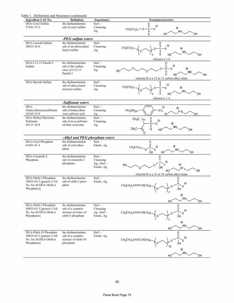
Table 1. Definitions and Structures (continued)
40
Ingredient CAS No. Definition Function(s) Formula/structure DEA-Cetyl Sulfate 51541-51-6
the diethanolamine salt of cetyl sulfate
Surf. - Cleansing Ag.
HONH2
OH
O S O
O
O
CH3(CH2)15
-PEG sulfate esters
DEA-Laureth Sulfate 58855-36-0
the diethanolamine salt of an ethoxylated lauryl sulfate
Surf. - Cleansing Ag.
HONH2
OH
O S O
O
OO
CH3(CH2)11
n wherein n 1-4
DEA-C12-13 Pareth-3 Sulfate
the diethanolamine salt of the sulfate ester of C12-13 Pareth-3
Surf. - Cleansing Ag.
wherein R is a 12 to 13 carbon alkyl chain
DEA-Myreth Sulfate the diethanolamine salt of ethoxylated myristyl sulfate
Surf. - Cleansing Ag.
HONH2
OH
O S O
O
OO
CH3(CH2)13
n wherein n 1-4
-Sulfonate esters
DEA-Dodecylbenzenesulfonate 26545-53-9
the diethanolamine salt of dodecylben-zene sulfonic acid
Surf. - Cleansing Ag.
DEA-Methyl Myristate Sulfonate 64131-36-8
the diethanolamine salt of an α-sulfonat-ed fatty acid ester
Surf. - Cleansing Ag.
-Alkyl and PEG phosphate estersDEA-Cetyl Phosphate 61693-41-2
the diethanolamine salt of cetyl phos-phate
Surf. - Emuls. Ag.
HONH2
OH
P
O
OOH
OCH3(CH2)15
DEA-Ceteareth-2 Phosphate
the diethanolamine salt of ceteareth-2 phosphate
Surf. - Cleansing Ag.; Surf. - Emuls. Ag.
wherein R is a 16 or 18 carbon alkyl chain
DEA-Oleth-3 Phosphate 58855-63-3 [generic CAS No. for all DEA-Oleth-n Phosphates]
the diethanolamine salt of oleth-3 phos-phate
Surf. - Emuls. Ag.
HONH2
OH
OO
CH(CH2)8
3
P
O
OHOCH3(CH2)7CH
DEA-Oleth-5 Phosphate 58855-63-3 [generic CAS No. for all DEA-Oleth-n Phosphates]
the diethanolamine salt of a complex mixture of esters of oleth-5 phosphate
Surf. - Cleansing Ag.; Surf. - Emuls. Ag.
HONH2
OH
OO
CH(CH2)8
5
P
O
OHOCH3(CH2)7CH
DEA-Oleth-10 Phosphate 58855-63-3 [generic CAS No. for all DEA-Oleth-n Phosphates]
the diethanolamine salt of a complex mixture of oleth-10 phosphate
Surf. - Emuls. Ag.
HONH2
OH
OO
CH(CH2)8
10
P
O
OHOCH3(CH2)7CH
Panel Book Page 73
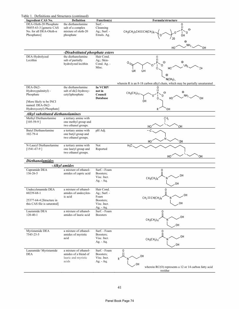
Table 1. Definitions and Structures (continued)
41
Ingredient CAS No. Definition Function(s) Formula/structure DEA-Oleth-20 Phosphate 58855-63-3 [generic CAS No. for all DEA-Oleth-n Phosphates]
the diethanolamine salt of a complex mixture of oleth-20 phosphate
Surf. - Cleansing Ag.; Surf. - Emuls. Ag.
HONH2
OH
OO
CH(CH2)8
20
P
O
OHOCH3(CH2)7CH
-Disubstituted phosphate estersDEA-Hydrolyzed Lecithin
the diethanolamine salt of partially hydrolyzed lecithin
Hair Cond. Ag.; Skin-Cond. Ag. - Misc.
wherein R is an 8-18 carbon alkyl chain, which may be partially unsaturated
DEA-Di(2-Hydroxypalmityl) -Phosphate [More likely to be INCI named: DEA-Di(2-Hydroxycetyl) Phosphate]
the diethanolamine salt of di(2-hydroxy-cetyl)phosphate
In VCRP/ not in Council Database HO
NH2OH
P
O
OO
OCH3(CH2)13
OH
OHR
Alkyl substituted diethanolamines Methyl Diethanolamine [105-59-9 ]
a tertiary amine with one methyl group and two ethanol groups.
Butyl Diethanolamine 102-79-4
a tertiary amine with one butyl group and two ethanol groups.
pH Adj.
N-Lauryl Diethanolamine [1541-67-9 ]
a tertiary amine with one lauryl group and two ethanol groups.
Not Reported
Diethanolamides -Alkyl amides Capramide DEA 136-26-5
a mixture of ethanol-amides of capric acid
Surf. - Foam Boosters; Visc. Incr. Ag. - Aq.
N
O
OH
OH
CH3(CH2)8
Undecylenamide DEA 60239-68-1 25377-64-4 [Structure in this CAS file is saturated]
a mixture of ethanol-amides of undecylen-ic acid
Hair Cond. Ag.; Surf. - Foam Boosters; Visc. Incr. Ag. - Aq.
N
O
OH
OH
CH(CH2)8CH2
Lauramide DEA 120-40-1
a mixture of ethanol-amides of lauric acid
Surf. - Foam Boosters
N
O
OH
OH
CH3(CH2)10
Myristamide DEA 7545-23-5
a mixture of ethanol-amides of myristic acid
Surf. - Foam Boosters; Visc. Incr. Ag. - Aq.
N
O
OH
OH
CH3(CH2)12
Lauramide/ Myristamide DEA
a mixture of ethanol-amides of a blend of lauric and myristic acids
Surf. - Foam Boosters; Visc. Incr. Ag. - Aq.
N
O
OH
OH
R
wherein RC(O) represents a 12 or 14 carbon fatty acid residue
Panel Book Page 74
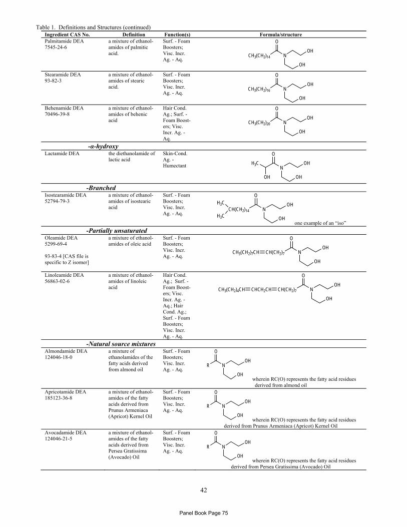
Table 1. Definitions and Structures (continued)
42
Ingredient CAS No. Definition Function(s) Formula/structure Palmitamide DEA 7545-24-6
a mixture of ethanol-amides of palmitic acid.
Surf. - Foam Boosters; Visc. Incr. Ag. - Aq.
N
O
OH
OH
CH3(CH2)14
Stearamide DEA 93-82-3
a mixture of ethanol-amides of stearic acid.
Surf. - Foam Boosters; Visc. Incr. Ag. - Aq.
N
O
OH
OH
CH3(CH2)16
Behenamide DEA 70496-39-8
a mixture of ethanol-amides of behenic acid
Hair Cond. Ag.; Surf. - Foam Boost-ers; Visc. Incr. Ag. - Aq.
N
O
OH
OH
CH3(CH2)20
-α-hydroxy Lactamide DEA the diethanolamide of
lactic acid Skin-Cond. Ag. - Humectant N
O
OH
OH
H3C
OH
-Branched Isostearamide DEA 52794-79-3
a mixture of ethanol-amides of isostearic acid
Surf. - Foam Boosters; Visc. Incr. Ag. - Aq.
N
O
OH
OH
CH(CH2)14
H3C
H3Cone example of an “iso”
-Partially unsaturated Oleamide DEA 5299-69-4 93-83-4 [CAS file is specific to Z isomer]
a mixture of ethanol-amides of oleic acid
Surf. - Foam Boosters; Visc. Incr. Ag. - Aq.
N
O
OH
OH
CH(CH2)7CH3(CH2)7CH
Linoleamide DEA 56863-02-6
a mixture of ethanol-amides of linoleic acid
Hair Cond. Ag.; Surf. - Foam Boost-ers; Visc. Incr. Ag. - Aq.; Hair Cond. Ag.; Surf. - Foam Boosters; Visc. Incr. Ag. - Aq.
N
O
OH
OH
CH(CH2)7CHCH2CHCH3(CH2)4CH
-Natural source mixtures Almondamide DEA 124046-18-0
a mixture of ethanolamides of the fatty acids derived from almond oil
Surf. - Foam Boosters; Visc. Incr. Ag. - Aq.
N
O
OH
OH
R
wherein RC(O) represents the fatty acid residues derived from almond oil
Apricotamide DEA 185123-36-8
a mixture of ethanol-amides of the fatty acids derived from Prunus Armeniaca (Apricot) Kernel Oil
Surf. - Foam Boosters; Visc. Incr. Ag. - Aq.
N
O
OH
OH
R
wherein RC(O) represents the fatty acid residues derived from Prunus Armeniaca (Apricot) Kernel Oil
Avocadamide DEA 124046-21-5
a mixture of ethanol-amides of the fatty acids derived from Persea Gratissima (Avocado) Oil
Surf. - Foam Boosters; Visc. Incr. Ag. - Aq.
N
O
OH
OH
R
wherein RC(O) represents the fatty acid residues derived from Persea Gratissima (Avocado) Oil
Panel Book Page 75
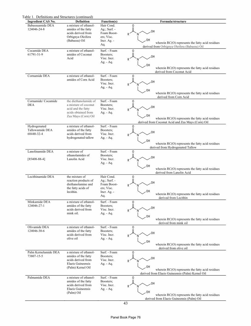
Table 1. Definitions and Structures (continued)
43
Ingredient CAS No. Definition Function(s) Formula/structure Babassuamide DEA 124046-24-8
a mixture of ethanol-amides of the fatty acids derived from Orbignya Oleifera (Babassu) Oil
Hair Cond. Ag.; Surf. - Foam Boost-ers; Visc. Incr. Ag. - Aq.
N
O
OH
OH
R
wherein RC(O) represents the fatty acid residues derived from Orbignya Oleifera (Babassu) Oil
Cocamide DEA 61791-31-9
a mixture of ethanol-amides of Coconut Acid
Surf. - Foam Boosters; Visc. Incr. Ag. - Aq.
N
O
OH
OH
R
wherein RC(O) represents the fatty acid residues derived from Coconut Acid
Cornamide DEA a mixture of ethanol-amides of Corn Acid
Surf. - Foam Boosters; Visc. Incr. Ag. - Aq.
N
O
OH
OH
R
wherein RC(O) represents the fatty acid residues derived from Corn Acid
Cornamide/ Cocamide DEA
the diethanolamide of a mixture of coconut acid and the fatty acids obtained from Zea Mays (Corn) Oil
Surf. - Foam Boosters; Visc. Incr. Ag. - Aq.
N
O
OH
OH
R
wherein RC(O) represents the fatty acid residues derived from Coconut Acid and Zea Mays (Corn) Oil
Hydrogenated Tallowamide DEA 68440-32-4
a mixture of ethanol-amides of the fatty acids derived from hydrogenated tallow
Surf. - Foam Boosters; Visc. Incr. Ag. - Aq.
N
O
OH
OH
R
wherein RC(O) represents the fatty acid residues derived from Hydrogenated Tallow
Lanolinamide DEA [85408-88-4]
a mixture of ethanolamides of Lanolin Acid
Surf. - Foam Boosters; Visc. Incr. Ag. - Aq.
N
O
OH
OH
R
wherein RC(O) represents the fatty acid residues derived from Lanolin Acid
Lecithinamide DEA the mixture of reaction products of diethanolamine and the fatty acids of lecithin.
Hair Cond. Ag.; Surf. - Foam Boost-ers; Visc. Incr. Ag. - Aq.
N
O
OH
OH
R
wherein RC(O) represents the fatty acid residues derived from Lecithin
Minkamide DEA 124046-27-1
a mixture of ethanol-amides of the fatty acids derived from mink oil.
Surf. - Foam Boosters; Visc. Incr. Ag. - Aq.
N
O
OH
OH
R
wherein RC(O) represents the fatty acid residues derived from mink oil
Olivamide DEA 124046-30-6
a mixture of ethanol-amides of the fatty acids derived from olive oil
Surf. - Foam Boosters; Visc. Incr. Ag. - Aq.
N
O
OH
OH
R
wherein RC(O) represents the fatty acid residues derived from olive oil
Palm Kernelamide DEA 73807-15-5
a mixture of ethanol-amides of the fatty acids derived from Elaeis Guineensis (Palm) Kernel Oil
Surf. - Foam Boosters; Visc. Incr. Ag. - Aq.
N
O
OH
OH
R
wherein RC(O) represents the fatty acid residues derived from Elaeis Guineensis (Palm) Kernel Oil
Palmamide DEA a mixture of ethanol-amides of the fatty acids derived from Elaeis Guineensis (Palm) Oil
Surf. - Foam Boosters; Visc. Incr. Ag. - Aq.
N
O
OH
OH
R
wherein RC(O) represents the fatty acid residues derived from Elaeis Guineensis (Palm) Oil
Panel Book Page 76
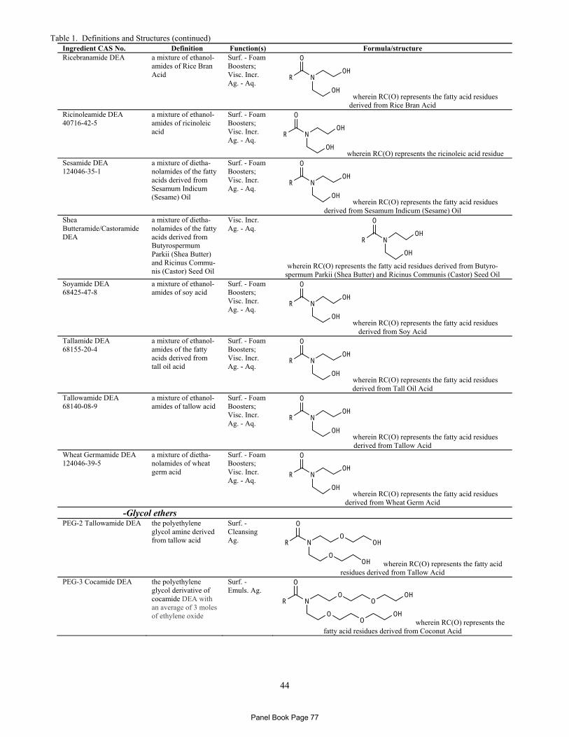
Table 1. Definitions and Structures (continued)
44
Ingredient CAS No. Definition Function(s) Formula/structure Ricebranamide DEA a mixture of ethanol-
amides of Rice Bran Acid
Surf. - Foam Boosters; Visc. Incr. Ag. - Aq.
N
O
OH
OH
R
wherein RC(O) represents the fatty acid residues derived from Rice Bran Acid
Ricinoleamide DEA 40716-42-5
a mixture of ethanol-amides of ricinoleic acid
Surf. - Foam Boosters; Visc. Incr. Ag. - Aq.
N
O
OH
OH
R
wherein RC(O) represents the ricinoleic acid residue Sesamide DEA 124046-35-1
a mixture of dietha-nolamides of the fatty acids derived from Sesamum Indicum (Sesame) Oil
Surf. - Foam Boosters; Visc. Incr. Ag. - Aq.
N
O
OH
OH
R
wherein RC(O) represents the fatty acid residues derived from Sesamum Indicum (Sesame) Oil
Shea Butteramide/Castoramide DEA
a mixture of dietha-nolamides of the fatty acids derived from Butyrospermum Parkii (Shea Butter) and Ricinus Commu-nis (Castor) Seed Oil
Visc. Incr. Ag. - Aq.
N
O
OH
OH
R
wherein RC(O) represents the fatty acid residues derived from Butyro-
spermum Parkii (Shea Butter) and Ricinus Communis (Castor) Seed Oil Soyamide DEA 68425-47-8
a mixture of ethanol-amides of soy acid
Surf. - Foam Boosters; Visc. Incr. Ag. - Aq.
N
O
OH
OH
R
wherein RC(O) represents the fatty acid residues derived from Soy Acid
Tallamide DEA 68155-20-4
a mixture of ethanol-amides of the fatty acids derived from tall oil acid
Surf. - Foam Boosters; Visc. Incr. Ag. - Aq.
N
O
OH
OH
R
wherein RC(O) represents the fatty acid residues derived from Tall Oil Acid
Tallowamide DEA 68140-08-9
a mixture of ethanol-amides of tallow acid
Surf. - Foam Boosters; Visc. Incr. Ag. - Aq.
N
O
OH
OH
R
wherein RC(O) represents the fatty acid residues derived from Tallow Acid
Wheat Germamide DEA 124046-39-5
a mixture of dietha-nolamides of wheat germ acid
Surf. - Foam Boosters; Visc. Incr. Ag. - Aq.
N
O
OH
OH
R
wherein RC(O) represents the fatty acid residues derived from Wheat Germ Acid
-Glycol ethers PEG-2 Tallowamide DEA the polyethylene
glycol amine derived from tallow acid
Surf. - Cleansing Ag. N
O
O
R
O
OH
OH wherein RC(O) represents the fatty acid residues derived from Tallow Acid
PEG-3 Cocamide DEA the polyethylene glycol derivative of cocamide DEA with an average of 3 moles of ethylene oxide
Surf. - Emuls. Ag.
NO
O
R
O
OOH
OOH
wherein RC(O) represents the fatty acid residues derived from Coconut Acid
Panel Book Page 77
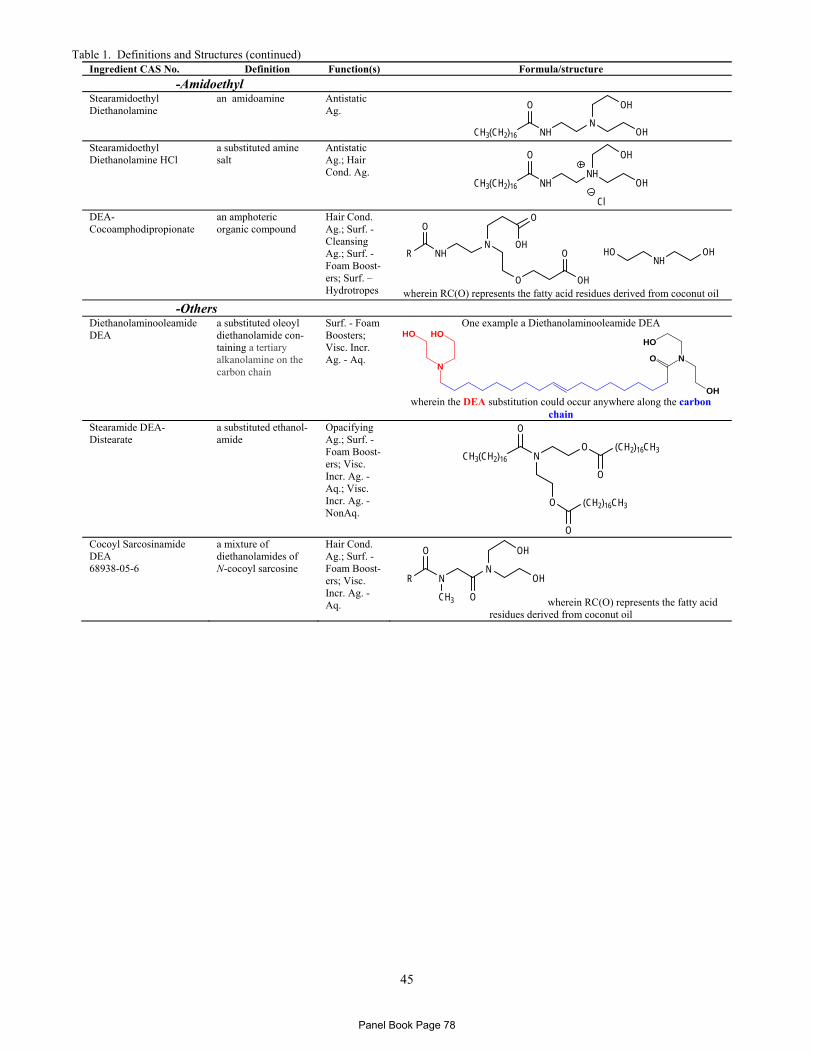
Table 1. Definitions and Structures (continued)
45
Ingredient CAS No. Definition Function(s) Formula/structure
-Amidoethyl Stearamidoethyl Diethanolamine
an amidoamine Antistatic Ag.
NH
O
NCH3(CH2)16
OH
OH Stearamidoethyl Diethanolamine HCl
a substituted amine salt
Antistatic Ag.; Hair Cond. Ag.
NH
O
NHCH3(CH2)16
OH
OH
Cl DEA-Cocoamphodipropionate
an amphoteric organic compound
Hair Cond. Ag.; Surf. - Cleansing Ag.; Surf. - Foam Boost-ers; Surf. – Hydrotropes
NH
O
NR
O OH
ONH
OHHO
O
OH
wherein RC(O) represents the fatty acid residues derived from coconut oil
-Others Diethanolaminooleamide DEA
a substituted oleoyl diethanolamide con-taining a tertiary alkanolamine on the carbon chain
Surf. - Foam Boosters; Visc. Incr. Ag. - Aq.
One example a Diethanolaminooleamide DEA
O N
OH
HO
N
HO HO
wherein the DEA substitution could occur anywhere along the carbon
chain Stearamide DEA-Distearate
a substituted ethanol-amide
Opacifying Ag.; Surf. - Foam Boost-ers; Visc. Incr. Ag. - Aq.; Visc. Incr. Ag. - NonAq.
N
O
OCH3(CH2)16
(CH2)16CH3
O
O
(CH2)16CH3
O
Cocoyl Sarcosinamide DEA 68938-05-6
a mixture of diethanolamides of N-cocoyl sarcosine
Hair Cond. Ag.; Surf. - Foam Boost-ers; Visc. Incr. Ag. - Aq.
N
CH3
O
RN
O
OH
OH
wherein RC(O) represents the fatty acid residues derived from coconut oil
Panel Book Page 78
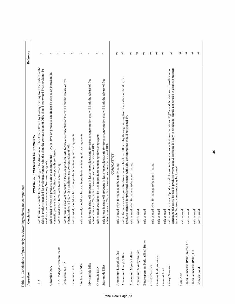
46
Tab
le 2
. C
oncl
usio
ns o
f pr
evio
usly
rev
iew
ed in
gred
ient
s an
d co
mpo
nent
s
Ingr
edie
nt
Con
clu
sion
R
efer
ence
PR
EV
IOU
SL
Y R
EV
IEW
ED
IN
GR
ED
IEN
TS
DE
A
safe
for
use
in c
osm
etic
for
mul
atio
ns d
esig
ned
for
disc
onti
nuou
s, b
rief
use
fol
low
ed b
y th
orou
gh r
insi
ng f
rom
the
surf
ace
of th
e sk
in; i
n pr
oduc
ts in
tend
ed f
or p
rolo
nged
con
tact
wit
h th
e sk
in, t
he c
once
ntra
tion
of
DE
A s
houl
d no
t exc
eed
5%; s
houl
d no
t be
used
wit
h pr
oduc
ts c
onta
inin
g N
-nit
rosa
ting
age
nts.
1
Coc
amid
e D
EA
sa
fe a
s us
ed in
rin
se-o
ff p
rodu
cts;
saf
e at
con
cent
rati
ons
≤10
% in
leav
e-on
pro
duct
s; s
houl
d no
t be
used
as
an in
gred
ient
in
cosm
etic
pro
duct
s in
whi
ch N
-nit
roso
com
poun
ds a
re f
orm
ed
3
DE
A D
odec
ylbe
nzen
esul
fona
te
safe
as
used
whe
n fo
rmul
ated
to b
e no
n-ir
rita
ting
5
Isos
tear
amid
e D
EA
sa
fe f
or u
se in
rin
se-o
ff p
rodu
cts;
in le
ave-
on p
rodu
cts,
saf
e fo
r us
e at
a c
once
ntra
tion
that
wil
l lim
it th
e re
leas
e of
fre
e et
hano
lam
ines
to 5
%, w
ith
a m
axim
um u
se c
once
ntra
tion
of
40%
4
Lau
ram
ide
DE
A
safe
as
used
; sho
uld
not b
e us
ed in
pro
duct
s co
ntai
ning
nit
rosa
ting
age
nts
2
Lin
olea
mid
e D
EA
sa
fe a
s us
ed; s
houl
d no
t be
used
in p
rodu
cts
cont
aini
ng n
itro
sati
ng a
gent
s 2
Myr
ista
mid
e D
EA
sa
fe f
or u
se in
rin
se-o
ff p
rodu
cts;
in le
ave-
on p
rodu
cts,
saf
e fo
r us
e at
a c
once
ntra
tion
that
wil
l lim
it th
e re
leas
e of
fre
e et
hano
lam
ines
to 5
%, w
ith
a m
axim
um u
se c
once
ntra
tion
of
40%
4
Ole
amid
e D
EA
sa
fe a
s us
ed; s
houl
d no
t be
used
in p
rodu
cts
cont
aini
ng n
itro
sati
ng a
gent
s 2
Ste
aram
ide
DE
A
safe
for
use
in r
inse
-off
pro
duct
s; in
leav
e-on
pro
duct
s, s
afe
for
use
at a
con
cent
rati
on th
at w
ill l
imit
the
rele
ase
of f
ree
etha
nola
min
es to
5%
, wit
h a
max
imum
use
con
cent
rati
on o
f 40
%
4
CO
MP
ON
EN
TS
Am
mon
ium
Lau
reth
Sul
fate
sa
fe a
s us
ed w
hen
form
ulat
ed to
be
non-
irri
tati
ng
91
Am
mon
ium
Lau
ryl S
ulfa
te
safe
in f
orm
ulat
ions
des
igne
d fo
r di
scon
tinu
ous,
bri
ef u
se f
ollo
wed
by
thor
ough
rin
sing
fro
m th
e su
rfac
e of
the
skin
; in
prod
ucts
inte
nded
for
pro
long
ed c
onta
ct w
ith
skin
, con
cent
rati
ons
shou
ld n
ot e
xcee
d 1%
92
Am
mon
ium
Myr
eth
Sul
fate
sa
fe a
s us
ed w
hen
form
ulat
ed to
be
non-
irri
tati
ng
91
Am
mon
ium
Myr
isty
l Sul
fate
sa
fe a
s us
ed
93
But
yros
perm
um P
arki
i (S
hea)
But
ter
safe
as
used
94
C12
-13
Par
eth-
3 sa
fe a
s us
ed w
hen
form
ulat
ed to
be
non-
irri
tati
ng
95
Coc
oam
phod
ipro
pion
ate
safe
as
used
96
C
ocon
ut A
cid
safe
as
used
94
C
ocoy
l Sar
cosi
ne
safe
as
used
in r
inse
-off
pro
duct
s, s
afe
for
use
in le
ave-
on p
rodu
cts
at c
once
ntra
tion
s of
≤5%
, and
the
data
wer
e in
suff
icie
nt to
de
term
ine
the
safe
ty f
or u
se in
pro
duct
s w
here
coc
oyl s
arco
sine
is li
kely
to b
e in
hale
d; s
houl
d no
t be
used
in c
osm
etic
pro
duct
s in
whi
ch N
-nit
roso
com
poun
ds m
ay b
e fo
rmed
97
Cor
n A
cid
safe
as
used
94
E
laei
s G
uine
ensi
s (P
alm
) K
erne
l Oil
sa
fe a
s us
ed
94
Ela
eis
Gui
neen
sis
(Pal
m)
Oil
sa
fe a
s us
ed
94
Isos
tear
ic A
cid
safe
as
used
98
Panel Book Page 79
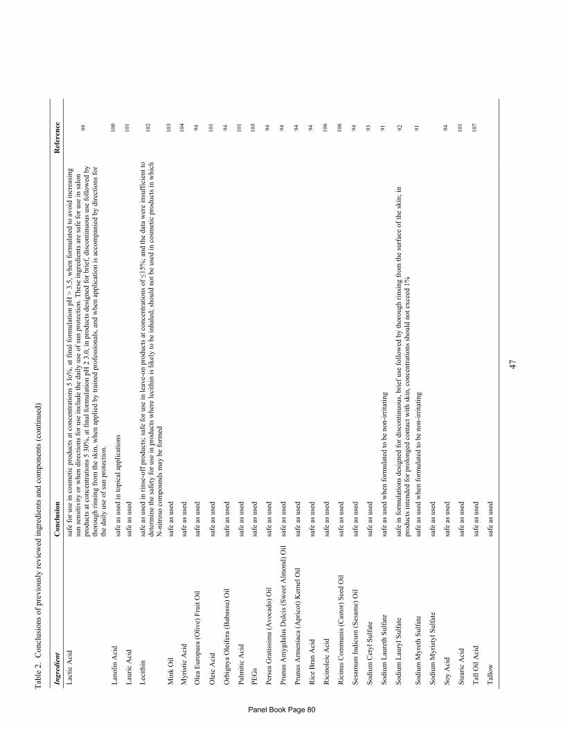
Tab
le 2
. C
oncl
usio
ns o
f pr
evio
usly
rev
iew
ed in
gred
ient
s an
d co
mpo
nent
s (c
onti
nued
)
47
Ingr
edie
nt
Con
clu
sion
R
efer
ence
Lac
tic
Aci
d sa
fe f
or u
se in
cos
met
ic p
rodu
cts
at c
once
ntra
tion
s 5
lo%
, at f
inal
for
mul
atio
n pH
> 3
.5, w
hen
form
ulat
ed to
avo
id in
crea
sing
su
n se
nsit
ivit
y or
whe
n di
rect
ions
for
use
incl
ude
the
dail
y us
e of
sun
pro
tect
ion.
The
se in
gred
ient
s ar
e sa
fe f
or u
se in
sal
on
prod
ucts
at c
once
ntra
tion
s 5
30%
, at f
inal
for
mul
atio
n pH
2 3
.0, i
n pr
oduc
ts d
esig
ned
for
brie
f, d
isco
ntin
uous
use
fol
low
ed b
y th
orou
gh r
insi
ng f
rom
the
skin
, whe
n ap
plie
d by
trai
ned
prof
essi
onal
s, a
nd w
hen
appl
icat
ion
is a
ccom
pani
ed b
y di
rect
ions
for
th
e da
ily
use
of s
un p
rote
ctio
n.
99
Lan
olin
Aci
d sa
fe a
s us
ed in
topi
cal a
ppli
cati
ons
100
Lau
ric
Aci
d sa
fe a
s us
ed
101
Lec
ithi
n sa
fe a
s us
ed in
rin
se-o
ff p
rodu
cts;
saf
e fo
r us
e in
leav
e-on
pro
duct
s at
con
cent
rati
ons
of ≤
15%
; and
the
data
wer
e in
suff
icie
nt to
de
term
ine
the
safe
ty f
or u
se in
pro
duct
s w
here
leci
thin
is li
kely
to b
e in
hale
d; s
houl
d no
t be
used
in c
osm
etic
pro
duct
s in
whi
ch
N-n
itro
so c
ompo
unds
may
be
form
ed
102
Min
k O
il
safe
as
used
10
3 M
yris
tic
Aci
d sa
fe a
s us
ed
104
Ole
a E
urop
aea
(Oli
ve)
Fru
it O
il
safe
as
used
94
O
leic
Aci
d sa
fe a
s us
ed
101
Orb
igny
a O
leif
era
(Bab
assu
) O
il
safe
as
used
94
P
alm
itic
Aci
d sa
fe a
s us
ed
101
PE
Gs
safe
as
used
10
5 P
erse
a G
rati
ssim
a (A
voca
do)
Oil
sa
fe a
s us
ed
94
Pru
nus
Am
ygda
lus
Dul
cis
(Sw
eet A
lmon
d) O
il
safe
as
used
94
P
runu
s A
rmen
iaca
(A
pric
ot)
Ker
nel O
il
safe
as
used
94
R
ice
Bra
n A
cid
safe
as
used
94
R
icin
olei
c A
cid
safe
as
used
10
6 R
icin
us C
omm
unis
(C
asto
r) S
eed
Oil
sa
fe a
s us
ed
106
Ses
amum
Ind
icum
(S
esam
e) O
il
safe
as
used
94
S
odiu
m C
etyl
Sul
fate
sa
fe a
s us
ed
93
Sod
ium
Lau
reth
Sul
fate
sa
fe a
s us
ed w
hen
form
ulat
ed to
be
non-
irri
tati
ng
91
Sod
ium
Lau
ryl S
ulfa
te
safe
in f
orm
ulat
ions
des
igne
d fo
r di
scon
tinu
ous,
bri
ef u
se f
ollo
wed
by
thor
ough
rin
sing
fro
m th
e su
rfac
e of
the
skin
; in
prod
ucts
inte
nded
for
pro
long
ed c
onta
ct w
ith
skin
, con
cent
rati
ons
shou
ld n
ot e
xcee
d 1%
92
Sod
ium
Myr
eth
Sul
fate
sa
fe a
s us
ed w
hen
form
ulat
ed to
be
non-
irri
tati
ng
91
Sod
ium
Myr
isty
l Sul
fate
sa
fe a
s us
ed
Soy
Aci
d sa
fe a
s us
ed
94
Ste
aric
Aci
d sa
fe a
s us
ed
101
Tal
l Oil
Aci
d sa
fe a
s us
ed
107
Tal
low
sa
fe a
s us
ed
Panel Book Page 80

Tab
le 2
. C
oncl
usio
ns o
f pr
evio
usly
rev
iew
ed in
gred
ient
s an
d co
mpo
nent
s (c
onti
nued
)
48
Ingr
edie
nt
Con
clu
sion
R
efer
ence
Whe
at G
erm
Aci
d sa
fe a
s us
ed
94
Zea
May
s (C
orn)
Oil
sa
fe a
s us
ed
94
Panel Book Page 81
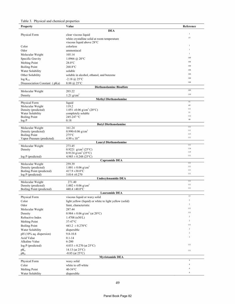
49
Table 3. Physical and chemical properties
Property Value Reference
DEA Physical Form clear viscous liquid 1 white crystalline solid at room temperature
viscous liquid above 28°C
57
Color colorless 1 Odor ammoniacal 1 Molecular Weight 105.14 1 Specific Gravity 1.0966 @ 20°C 108 Melting Point 28.0°C 108 Boiling Point 268.8°C 108 Water Solubility soluble 108 Other Solubility soluble in alcohol, ethanol, and benzene 108 log Kow -2.18 @ 25°C 108 Disassociation Constant ( pKa) 8.88 @ 25°C 108
Diethanolamine Bisulfate Molecular Weight 203.22 109 Density 1.21 g/cm3 110
Methyl Diethanolamine Physical Form liquid 51 Molecular Weight 119.2 69 Density (predicted) 1.051 ±0.06 g/cm3 (20°C) 111 Water Solubility completely soluble 51 Boiling Point 245-247 °C 112 log P 0.18 40
Butyl Diethanolamine Molecular Weight 161.24 111 Density (predicted) 0.990±0.06 g/cm3 111 Boiling Point 275°C 113 Vapor Pressure (predicted) 4.99 x 10⁻⁴ 111
Lauryl Diethanolamine Molecular Weight 273.45 111 Density 0.9221 g/cm3 (25°C)
0.9124 g/cm3 (25°C)
114 115
log P (predicted) 4.985 ± 0.248 (25°C) 111 Capramide DEA
Molecular Weight 259.39 111 Density (predicted) 1.001 ± 0.06 g/cm3 111 Boiling Point (predicted) 417.9 ±30.0°C 111 log P (predicted) 3.014 ±0.270 111
Undecylenamide DEA Molecular Weight 271.40 111 Density (predicted) 1.002 ± 0.06 g/cm3 111 Boiling Point (predicted) 440.4 ±40.0°C 111
Lauramide DEA Physical Form viscous liquid or waxy solid 15 Color light yellow (liquid) or white to light yellow (solid) 2 Odor faint, characteristic 2 Molecular Weight 287.44 111 Density 0.984 ± 0.06 g/cm3 (at 20°C) 111 Refractive Index 1.4708 (n30/L) 2 Melting Point 37-47°C 2 Boiling Point 443.2 ± 0.270°C 111 Water Solubility dispersible 2 pH (10% aq. dispersion) 9.8-10.8 2 Acid Value Alkaline Value
0.1-14 6-200
2
log P (predicted) 4.033 ± 0.270 (at 25°C) 111 pKa pKb
14.13 (at 25°C) -0.85 (at 25°C)
111
Myristamide DEA Physical Form waxy solid 4 Color white to off-white 4 Melting Point 40-54°C 4 Water Solubility dispersible 4
Panel Book Page 82

Table 3. Physical and chemical properties (continued)
50
Property Value Reference
Other Solubility soluble in alcohol, chlorinated hydrocarbons, and aromatic hydrocarbons; dispersible in mineral spirits, kerosene, white mineral oils, and natural fats and oils
4
pH (10% aq. dispersion) 9.5-10.5 4 log P (predicted) 5.025±0.270 111 Acid Value Alkaline Value
1 (max) 26-50
4
Palmitamide DEA Molecular Weight 343.54 111 Density (predicted) 0.959 ± 0.06 g/cm3 (20°C) 111 Boiling Point (predicted) 492.5 ±30.0°C 111 log P (predicted) 6.071 ±0.270 111
Stearamide DEA Physical Form wax-like solid 4 Color white to pale yellow 4 Molecular Weight 371.60 111 Density (predicted) 0.959 ± 0.06 g/cm3 (20°C) 111 pH (1% aq. dispersion) 9-10 4 log P (predicted) 7.090 ±0.270 111
Behenamide DEA Molecular Weight 427.70 111 Density (predicted) 0.935 ± 0.06 g/cm3 (20°C) 111 Boiling Point (predicted) 562.1 ±30.0°C 111 log P (predicted) 9.128 ±0.270 111
Oleamide DEA Physical Form liquid 2 Color amber 2 Molecular Weight 387.68 18 Specific Gravity 0.99 (25/25°C) 2 Phase Transition congeals at -8°C 2 Boiling Point (predicted) 525.6 ±45.0°C 111 Water Solubility dispersible 2 Other Solubility soluble in alcohols, glycols, ketones, esters, benzenes, chlorinated solvents, and
aliphatic hydrocarbons 2
pH 9-10 2 log P (predicted) 6.681 ±0.275 111
Linoleamide DEA Physical Form syrup-like liquid or waxlike mass 2 Color light yellow (liquid) or white to yellow (mass) 2 Odor characteristic 2 Specific Gravity 0.972-0.982 (25°/25°C) 2 Water Solubility slightly soluble 2 Boiling Point (predicted) 525.6 ±50.0°C 111 Other Solubility soluble in ethanol, propylene glycol, and glycerin; insoluble in mineral oil 2 Acid Value Alkaline Value
2.0 (max) 25-50 (calculated as DEA)
2
log P (predicted) 6.277 ±0.275 111 Cocamide DEA
Physical Form clear viscous liquid 3,19 Color amber or yellow 3,19,19 Odor faint coconut 3 Molecular Weight 280-290 19 Melting Point 23-35°C Water Solubility soluble in water 3 pH (10% aq. solution) 9.5-10.5 3 Acid Value 3.0 max 3
Ricinoleamide DEA Molecular Weight 385.58 111 Density (predicted) 1.007± 0.06 g/cm3 (20°C) 111 Boiling Point (predicted) 560.5 ±50.0°C 111 log P (predicted) 4.867 ±0.289 111
Panel Book Page 83

51
Tab
le 4
a. H
isto
rica
l and
cur
rent
fre
quen
cy a
nd c
once
ntra
tion
of
use
acco
rdin
g to
dur
atio
n an
d ty
pe o
f ex
posu
re
#
of U
ses
Con
c. o
f Use
(%
) #
of U
ses
Con
c. o
f Use
(%
) #
of U
ses
Con
c. o
f Use
(%
) #
of U
ses
Con
c. o
f Use
(%
)
1981
1 20
1021
19
811
2010
22
1981
2 20
1021
19
812
2011
# 19
95
2010
21
1995
20
11
1995
20
1021
19
95
2011
DE
A
Lau
ram
ide
DE
A
Myr
ista
mid
e D
EA
S
tear
amid
e D
EA
T
otal
s*
18
30
≤5
0.00
8-0.
3 60
4 54
5 ≤5
0
6 N
R
2-15
19
9 2-
15
D
ura
tion
of
Use
L
eave
-On
1 15
1-
5 0.
008-
0.06
17
27
1-
10
N
R
NR
15
13
8 15
Rin
se O
ff
13
15
≤5
0.00
9-0.
3 47
9 47
9 1-
50
5
NR
2-
6
6 1
2-6
D
ilut
ed fo
r U
se
4 N
R
NR
N
R
108
39
≤50
1
NR
N
R
N
R
NR
N
R
E
xpos
ure
Typ
e
E
ye A
rea
NR
N
R
NR
N
R
2 N
R
0.1-
10
N
R
NR
N
R
N
R
NR
N
R
P
ossi
ble
Inge
stio
n N
R
NR
N
R
NR
N
R
NR
N
R
N
R
NR
N
R
N
R
NR
N
R
In
hala
tion
N
R
3 N
R
NR
2
16
1-25
NR
N
R
NR
NR
N
R
NR
Der
mal
Con
tact
4
15
≤0.1
0.
009-
0.06
21
0 18
5 ≤5
0
2 N
R
5
14
8 5
D
eodo
rant
(un
dera
rm)
NR
N
R
NR
N
R
NR
1
NR
NR
N
R
15
N
R
1 15
Hai
r -
Non
-Col
orin
g 1
13
1-5
0.03
-0.3
29
5 14
4 0.
1-50
4 N
R
2-6
5
NR
2-
6
Hai
r-C
olor
ing
12
2 1-
5 N
R
96
216
0.1-
10
N
R
NR
N
R
N
R
NR
N
R
N
ail
1 N
R
NR
N
R
4 N
R
1-5
N
R
NR
N
R
N
R
NR
N
R
M
ucou
s M
embr
ane
NR
5
≤0.1
0.
009
108
118
≤50
1
NR
N
R
1
NR
N
R
B
ath
Pro
duct
s 4
NR
N
R
NR
56
39
0.
1-25
1 N
R
NR
NR
N
R
NR
Bab
y P
rodu
cts
NR
N
R
NR
N
R
13
NR
0.
1-10
NR
N
R
NR
NR
N
R
NR
# of
Use
s C
onc.
of U
se (
%)
# of
Use
s C
onc.
of U
se (
%)
# of
Use
s C
onc.
of U
se (
%)
# of
Use
s C
onc.
of U
se (
%)
19
95
2010
21
1995
20
11
1981
2 20
1021
19
812
2011
19
812
2010
21
1981
2 20
11
1994
3 20
1021
19
943
2011
Isos
tear
amid
e D
EA
O
leam
ide
DE
A
Lin
olea
mid
e D
EA
C
ocam
ide
DE
A
T
otal
s 23
2
2-15
121
13
≤25
92
13
3 ≤1
0
745*
* 85
0 N
A
D
ura
tion
of
Use
L
eave
-On
17
2 15
4 3
1-10
2 3
1-5
36
43
N
A
R
inse
-Off
3
NR
2-
6
112
2 ≤2
5
83
120
≤10
60
4 73
4 N
A
D
ilut
ed fo
r U
se
3 N
R
NR
5 8
≤5
7
10
1-10
76
73
NA
Exp
osu
re T
ype
Eye
Are
a 1
NR
N
R
N
R
NR
N
R
N
R
NR
N
R
N
R
1 N
A
P
ossi
ble
Inge
stio
n N
R
NR
N
R
N
R
NR
N
R
N
R
NR
N
R
N
R
NR
N
A
In
hala
tion
N
R
NR
N
R
N
R
NR
N
R
N
R
1 N
R
1
NR
N
A
D
erm
al C
onta
ct
23
2 5
10
12
≤1
0
17
24
1-10
199
354
NA
Deo
dora
nt (
unde
rarm
) N
R
NR
15
NR
N
R
NR
NR
N
R
NR
4 N
R
NA
Hai
r -
Non
-Col
orin
g 3
NR
2-
6
12
1 ≤2
5
29
4 ≤1
0
277
259
NA
Hai
r-C
olor
ing
NR
N
R
NR
99
NR
≤1
0
45
105
1-10
236
237
NA
Nai
l N
R
NR
N
R
N
R
NR
N
R
N
R
NR
N
R
2
NR
N
A
M
ucou
s M
embr
ane
NR
N
R
NR
5 N
R
≤5
7
9 1-
10
81
20
8 N
A
B
ath
Pro
duct
s 3
NR
N
R
N
R
8 N
R
2
10
1-5
46
57
N
A
B
aby
Pro
duct
s N
R
NR
N
R
N
R
NR
N
R
N
R
NR
N
R
1
13
NA
* B
ecau
se e
ach
ingr
edie
nt m
ay b
e us
ed in
cos
met
ics
wit
h m
ulti
ple
expo
sure
type
s, th
e su
m o
f al
l exp
osur
e ty
pes
my
not e
qual
the
sum
of
tota
l use
s.
# C
once
ntra
tion
of
use
surv
ey in
pro
cess
. N
R –
no
repo
rted
use
s
Panel Book Page 84

52
Table 4b. Frequency (2010) and concentration of use according to duration and type of exposure
Diethanolamine Bisulfate DEA Stearate DEA Linoleate
# of Uses21 Conc of Use (%)# # of Uses21 Conc of Use (%) # of Uses21 Conc of Use (%)
Totals* 6 1 1
Duration of Use
Leave-On NR 1 NR
Rinse-Off 6 NR 1
Diluted for Use NR NR NR
Exposure Type
Eye Area NR NR NR
Possible Ingestion NR NR NR
Inhalation NR NR NR
Dermal Contact 6 1 NR
Deodorant (underarm) NR NR NR
Hair - Non-Coloring NR NR 1
Hair-Coloring NR NR NR
Nail NR NR NR
Mucous Membrane 6 NR NR
Bath Products NR NR NR
Baby Products NR NR NR
DEA Lauryl Sulfate DEA Laureth Sulfate DEA-Cetyl Phosphate
# of Uses21 Conc of Use (%)# # of Uses21 Conc of Use (%) # of Uses21 Conc of Use (%)
Totals* 3 1 50 Duration of Use Leave-On NR NR 46
Rinse Off 3 NR 4
Diluted for Use NR 1 NR
Exposure Type
Eye Area NR NR 5
Possible Ingestion NR NR NR
Inhalation NR NR NR
Dermal Contact 3 1 48
Deodorant (underarm) NR NR NR
Hair - Non-Coloring NR NR NR
Hair-Coloring NR NR NR
Nail NR NR NR
Mucous Membrane 1 NR NR
Bath Products NR 1 NR
Baby Products NR NR NR
DEA-Oleth-3 Phosphate DEA-Oleth-10 Phosphate DEA-(Di(2-Hydroxypalmityl)
Phosphate
# of Uses21 Conc of Use (%)# # of Uses21 Conc of Use (%) # of Uses21 Conc of Use (%)
Totals* 7 8 3
Duration of Use
Leave-On 5 6 2
Rinse-Off 2 2 1
Diluted for Use NR NR NR
Exposure Type
Eye Area NR NR NR
Possible Ingestion NR NR NR
Inhalation NR NR NR
Dermal Contact 2 NR 3
Deodorant (underarm) NR NR NR
Hair - Non-Coloring 5 8 NR
Hair-Coloring NR NR NR
Nail NR NR NR
Mucous Membrane NR NR NR
Bath Products NR NR NR
Baby Products NR NR NR
Panel Book Page 85
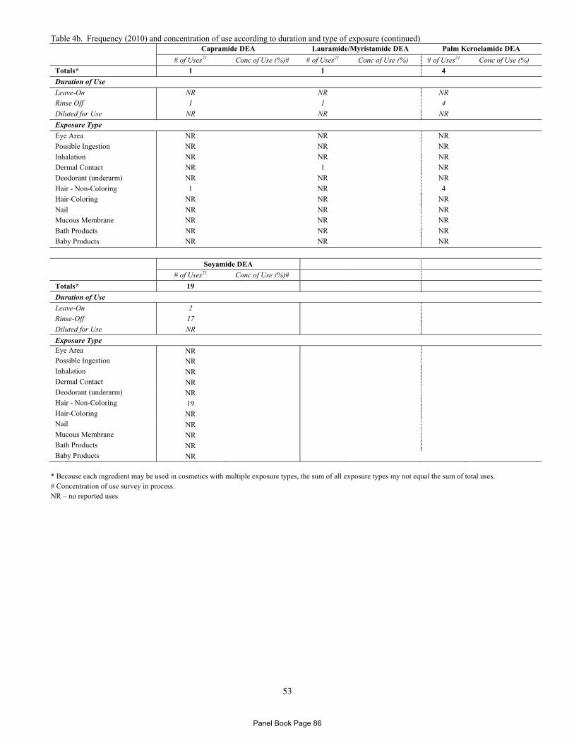
Table 4b. Frequency (2010) and concentration of use according to duration and type of exposure (continued)
53
Capramide DEA Lauramide/Myristamide DEA Palm Kernelamide DEA
# of Uses21 Conc of Use (%)# # of Uses21 Conc of Use (%) # of Uses21 Conc of Use (%)
Totals* 1 1 4
Duration of Use
Leave-On NR NR NR
Rinse Off 1 1 4
Diluted for Use NR NR NR
Exposure Type
Eye Area NR NR NR
Possible Ingestion NR NR NR
Inhalation NR NR NR
Dermal Contact NR 1 NR
Deodorant (underarm) NR NR NR
Hair - Non-Coloring 1 NR 4
Hair-Coloring NR NR NR
Nail NR NR NR
Mucous Membrane NR NR NR
Bath Products NR NR NR
Baby Products NR NR NR
Soyamide DEA
# of Uses21 Conc of Use (%)#
Totals* 19
Duration of Use
Leave-On 2
Rinse-Off 17
Diluted for Use NR
Exposure Type Eye Area NR Possible Ingestion NR Inhalation NR Dermal Contact NR Deodorant (underarm) NR Hair - Non-Coloring 19 Hair-Coloring NR Nail NR Mucous Membrane NR Bath Products NR Baby Products NR
* Because each ingredient may be used in cosmetics with multiple exposure types, the sum of all exposure types my not equal the sum of total uses. # Concentration of use survey in process. NR – no reported uses
Panel Book Page 86
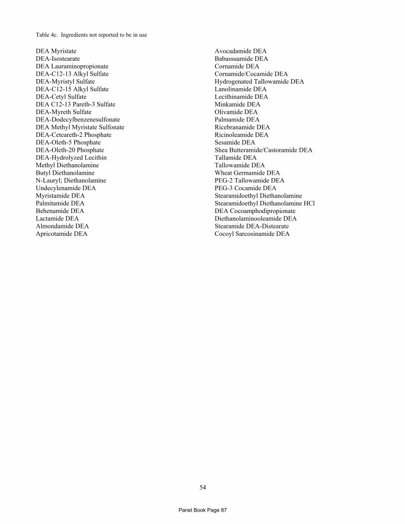
54
Table 4c. Ingredients not reported to be in use
DEA Myristate DEA-Isostearate DEA Lauraminopropionate DEA-C12-13 Alkyl Sulfate DEA-Myristyl Sulfate DEA-C12-15 Alkyl Sulfate DEA-Cetyl Sulfate DEA C12-13 Pareth-3 Sulfate DEA-Myreth Sulfate DEA-Dodecylbenzenesulfonate DEA Methyl Myristate Sulfonate DEA-Ceteareth-2 Phosphate DEA-Oleth-5 Phosphate DEA-Oleth-20 Phosphate DEA-Hydrolyzed Lecithin Methyl Diethanolamine Butyl Diethanolamine N-Lauryl; Diethanolamine Undecylenamide DEA Myristamide DEA Palmitamide DEA Behenamide DEA Lactamide DEA Almondamide DEA Apricotamide DEA
Avocadamide DEA Babassuamide DEA Cornamide DEA Cornamide/Cocamide DEA Hydrogenated Tallowamide DEA Lanolinamide DEA Lecithinamide DEA Minkamide DEA Olivamide DEA Palmamide DEA Ricebranamide DEA Ricinoleamide DEA Sesamide DEA Shea Butteramide/Castoramide DEA Tallamide DEA Tallowamide DEA Wheat Germamide DEA PEG-2 Tallowamide DEA PEG-3 Cocamide DEA Stearamidoethyl Diethanolamine Stearamidoethyl Diethanolamine HCl DEA Cocoamphodipropionate Diethanolaminooleamide DEA Stearamide DEA-Distearate Cocoyl Sarcosinamide DEA
Panel Book Page 87

55
Table 5. Status for use in Europe according to the EC CosIng Database Dialkanolamines and Their Salts (i.e., DEA and its acid salts) – listed in Annex II - prohibitedDEA Diethanolamine Bisulfate DEA-Myristate DEA-Isostearate DE-Linoleate DEA-Lauraminopropionate DEA-Lauryl Sulfate DEA-C12-13 Alkyl Sulfate DEA-Myristyl Sulfate DEA-C12-15 Alkyl Sulfate DEA-Cetyl Sulfate DEA-Laureth Sulfate
DEA-C12-13 Pareth-3 Sulfate DEA-Myreth Sulfate DEA-Dodecylbenzenesulfonate DEA-Methyl Myristate DEA-Cetyl Phosphate DEA-Ceteareth-2 Phosphate DEA-Oleth-3 Phosphate DEA-Oleth-5 Phosphate DEA-Oleth-10 Phosphate DEA-Oleth-20 Phosphate DEA Hydrolyzed Lecithin
Fatty Acid Dialkanolamides (i.e., the alkyl substituted diethanolamines) – listed in Annex III - restrictions N-Lauryl Diethanolamine Capramide DEA Undecylenamide DEA Lauramide DEA Myristamide DEA Lauramide/Myristamide DEA Palmitamide DEA Stearamide DEA Behenamide DEA Isostearamide DEA Oleamide DEA Linoleamide DEA Almondamide DEA Apricotamide DEA Avocadamide DEA Babassuamide DEA Cocamide DEA
Cornamide DEA Cornamide/Cocamide DEA Hydrogenated Tallowamide DEA Lanolinamide DEA Lecithinamide DEA Minkamide DEA Olivamide DEA Palm Kernelamide DEA Palmamide DEA Ricebranamide DEA Ricinoleamide DEA Sesamide DEA Soyamide DEA Tallamide DEA Tallowamide DEA Wheat Germamide DEA
Listed in EC Inventory – Annex III (restrictions) Butyl Diethanolamine (maximum of 2.5% in ready for use preparations; do not use with nitrosating systems; minimum purity 99%; maximum secondary amine content in raw material, 0.5%; maximum nitrosamine content, 50 µg/kg; keep in nitrite-free containers) In EC Inventory – no annex specified Lactamide DEA Shea Butteramide/Castoramide DEA PEG-2 Tallowamide DEA PEG-3 Cocamide DEA Stearamidoethyl Diethanolamine
Stearamidoethyl Diethanolamine HCl Diethanolaminooleamide DEA Stearamide DEA-Distearate Cocoyl Sarcosinamide DEA
Not Listed in EC Inventory DEA Stearate (also not in INCI Dictionary) DEA-Di(2-Hydroxypalmityl)Phosphate (also not in INCI Dictionary) Methyl Diethanolamine DEA Cocoamphodipropionate
Panel Book Page 88
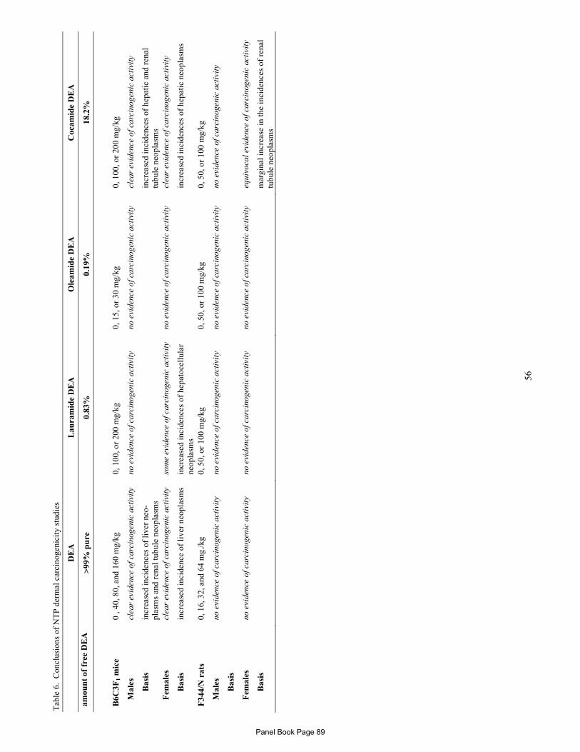
56
Tab
le 6
. C
oncl
usio
ns o
f N
TP
der
mal
car
cino
geni
city
stu
dies
D
EA
L
aura
mid
e D
EA
O
leam
ide
DE
A
Coc
amid
e D
EA
amou
nt
of f
ree
DE
A
>99
% p
ure
0.
83%
0.
19%
18
.2%
B6C
3F1
mic
e 0
, 40,
80,
and
160
mg/
kg
0, 1
00, o
r 20
0 m
g/kg
0,
15,
or
30 m
g/kg
0,
100
, or
200
mg/
kg
M
ales
cl
ear
evid
ence
of c
arci
noge
nic
acti
vity
no
evi
denc
e of
car
cino
geni
c ac
tivi
ty
no e
vide
nce
of c
arci
noge
nic
acti
vity
cl
ear
evid
ence
of c
arci
noge
nic
acti
vity
Bas
is
incr
ease
d in
cide
nces
of
live
r ne
o-pl
asm
s an
d re
nal t
ubul
e ne
opla
sms
incr
ease
d in
cide
nces
of
hepa
tic
and
rena
l tu
bule
neo
plas
ms
F
emal
es
clea
r ev
iden
ce o
f car
cino
geni
c ac
tivi
ty
som
e ev
iden
ce o
f car
cino
geni
c ac
tivi
ty
no e
vide
nce
of c
arci
noge
nic
acti
vity
cl
ear
evid
ence
of c
arci
noge
nic
acti
vity
Bas
is
incr
ease
d in
cide
nce
of li
ver
neop
lasm
s in
crea
sed
inci
denc
es o
f he
pato
cell
ular
ne
opla
sms
in
crea
sed
inci
denc
es o
f he
pati
c ne
opla
sms
F34
4/N
rat
s 0,
16,
32,
and
64
mg.
/kg
0, 5
0, o
r 10
0 m
g/kg
0,
50,
or
100
mg/
kg
0, 5
0, o
r 10
0 m
g/kg
M
ales
no
evi
denc
e of
car
cino
geni
c ac
tivi
ty
no e
vide
nce
of c
arci
noge
nic
acti
vity
no
evi
denc
e of
car
cino
geni
c ac
tivi
ty
no e
vide
nce
of c
arci
noge
nic
acti
vity
Bas
is
F
emal
es
no e
vide
nce
of c
arci
noge
nic
acti
vity
no
evi
denc
e of
car
cino
geni
c ac
tivi
ty
no e
vide
nce
of c
arci
noge
nic
acti
vity
eq
uivo
cal e
vide
nce
of c
arci
noge
nic
acti
vity
Bas
is
m
argi
nal i
ncre
ase
in th
e in
cide
nces
of
rena
l tu
bule
neo
plas
ms
Panel Book Page 89
57
REFERENCES
1. Elder RL (ed). Final Report on the Safety Assessment of Triethanolamine, Diethanolamine, and Monoethanolamine. J Am Coll Toxicol. 1983;2:(7).
2. Elder RL (ed). Final report on the safety assessment of cocamide DEA, lauramide DEA, linoleamide DEA, and oleamide DEA. J Am Coll Toxicol. 1986;5:(5):415-454.
3. Andersen FA (ed). Amended final report on the safety assessment of cocamide DEA. J Am Coll Toxicol. 1996;15:(6):527-542.
4. Pang S. Isostearamide DEA & MEA, Myristamide DEA & MEA, Stearmide DEA & MEA. 1995. Available from the CIR, 1101 17th Street, NW, Ste 412, Washington DC 20036. http://cir-safety.org.
5. Becker LC. Amended safety assessment of dodecylbenzenesulfonate, decylbenzenesulfonate, and tridecylbenezenesulfonate salts as used in cosmetics. 2009. Available from the CIR, 1101 17th Street, NW, Ste 412, Washington DC 20036. http://cir-safety.org.
6. Lehman-McKeeman LD and Gamsky EA. Choline supplementation inhibits diethanolamine-induced morphological transfomation in Syrian hamster embryo cells: Evidence for a carcinogenic mechanism. Toxicol Sci. 2000;55:303-310.
7. Toxicity and carcinogenicity of N-nitroso compounds. 1981. Chapter: 1. Mycotoxins and N-Nitroso Compounds:Environmental Risks. Shank RC. Boca Raton, FL: CRC Press, Inc.
8. Rostkowski K, Zwierzl K, Ró¿añski J, Moniuszko-Jakoniuk J, and Roszczenko A. Formation and Metabolism of N-Nitrosamines. Polish Journal of Environmental Studies. 1998;7:(6):321-325.
9. Challis BC, Shuker DE, Fine DH, Goff EU, and Hoffman GA. Amine nitration and nitrosation by gaseous nitrogen dioxide. IARC Sci Publ. 1982;41:11-20.
10. Bíró T, Tóth BI, Haskó G, Paus R, and Pacher P. The endocannabinoid system of the skin in health and disease: novel perspectives and therapeutic opportunities. Trends Pharmacol Sci. 2009;30:(8):411-420.
11. Bisogno T, De Petrocellis L, and Di Marzo V. Fatty acid hydrolase, an enzyme with many bioactive substrates. Possible therapeutic implications. Current Pharmaceutical Design. 2002;8:125-133.
12. Gray GM, Tabiowo A, and Trotter MD. Studies on the soluble membrane-bound amino acid 2-naphthylamidases in pig and human epidermis. Biochem J. 1977;161:667-675.
13. Lemke TL. Review of organic functional groups: introduction to medicinal organic chemistry. 4th ed. 2003.
14. Dow Chemical Company. The alkanolamines handbook. 1988. Midland, MI: The Dow Chemical Company.Secondary reference in Knaak et al. 1997.
15. National Toxicology Program. NTP Technical report on the toxicology and carcinogenesis studies of lauric acid diethanolamine condensate (CAS No. 120-40-1) in F344/N rats and B6C3F1 mice. (Dermal studies.) NTP TR 480. 1999.
16. Chou, H. J. Determination of diethanolamine and N-nitrosodiethanolamine in fatty acid diethanolamides. J Assoc Off Anal Chem Intl. 1998;81:(5):943-947.
17. Dow Chemical Company. Sales Specification: Diethanolamine. 1998. Secondary reference in IARC 2000.
18. National Toxicology Program. NTP Technical report on the toxicology and carcinogenesis studies of oleic acid diethanolamine condensate (CAS No. 93-83-4) in F344/N rats and B6C3F1 mice. (Dermal studies.) NTP TR 481. 1999.
19. National Toxicology Program. NTP Technical report on the toxoicology and carcinogenesis studies of coconut oil acid diethanolamine condensate (CAS No. 68603-42-9) in F344/N rats and B6C3F1 mice. (Dermal studies.) NTP TR 479. 2001.
20. Gottschalck T.E. and Bailey, J. E. eds. International Cosmetic Ingredient Dictionary and Handbook. Washington, DC: Personal Care Products Council, 2010.
Panel Book Page 90
58
21. Food and Drug Administration (FDA). Frequency of use of cosmetic ingredients. FDA Database. 2010. Washington, DC: FDA.Updated May 4.
22. Personal Care Products Council. Concentration of use data - submitted by industry in response to a Council survey. 10-13-2010. Unpublished data submitted by the Council.
23. Bailey J. DEA in consumer products is safe. FASEB J. 2007;21:(1):295.
24. European Commission. Cosing Database. Annex II. List of substances which must not form part of the composition of cosmetic products. Secondary alkyl- and alkanolamines and their salts, including diethanolamine. http://ec.europa.eu/consumers/cosmetics/cosing/index.cfm?fuseaction=search.details&id=28757. 2001.
25. Health Canada.Cosmetic Ingredient Hotlist. 2010. http://www.hc-sc.gc.ca/cps-spc/person/cosmet/info-ind-prof/_hot-list-critique/hotlist-liste_1-eng.php. Accessed 10-29-2010.
26. Food and Drug Administration.Everything Added to Food in the United States (EAFUS). 5-27-2010. http://www.fda.gov/Food/FoodIngredientsPackaging/ucm115326.htm. Accessed 1-30-2011.
27. Ballantyne B and Leung H-W. Acute toxicity and primary irritancy of alkylalkanolamines. Vet Human Toxicol. 1992;38:(6):422-426.
28. Yourick JJ, Marks A, Lockhead RY, and Bronaugh RL. Diethanolamine (DEA) in vitro dermal absorption in Fuzzy rat skin. Unpublished manuscript. Secndary reference in OECD 2008.
29. Sun JD, Beskitt JL, Tallant MJ, and Frantz SW. In vitro skin penetration of monoethanolamine and diethanolamine using excised skin from rats, mice, rabbits, and humans. J Toxicol -- Cut & Ocular Toxicol. 1996;15:(2):131-146.
30. Kraeling MEK, Yourick JJ, and Bronaugh RL. In vitro human skin penetration of diethanolamine. Food Chem Toxicol. 2004;42:1553-1561.
31. Brain KR, Walter KA, Green DM, Brain S, Loretz LJ, Sharma RK, and Dressler WE. Percutaneous penetration of diethanolamine through human skin in vitro: Application from cosmetic vehicles. Food Chem Toxicol. 2005;43:681-690.
32. Mathews JM, Garner CE, and Matthews HB. Metabolism, bioaccumulation, and incorporation of diethanolamine into phospholipids. Chem Res Toxicol. 1995;8:(5):625-633.
33. Mathews JM, DeCosta K, and Thomas BF. Lauramide diethanolamine absorption, metabolism, and disposition in rats and mice after oral, intravenous, and dermal administration. Drug Metabolism and Disposition. 1996;24:(7):702-710.
34. Merdink, J., Decosta, K., Mathews, J. M., Jones, C. B., Okita, J. R., and Okita, R. T. Hydroxylation of lauramide diethanolamine by liver microsomes. Drug Metabolism and Disposition. 1996;24:(2):180-186.
35. National Toxicology Program. Toxicology and carcinogenesis studies of diethanolamine (CAS No. 111-42-2) in F344/N rats and B6C3F1 mice. (Dermal studies.) NTP TR 478. 1999.
36. Mathews JM and Jeffcoat AR. Absorption and disposition of diethanolamine (DEA) in rats and mice after oral , dermal, and intravenous administration. RTI/3662/00-12P. 1991. Secondary reference in Knaak et al 1997.
37. Waechter JM, Bormett GA, and Stewart HS. Diethanolamine: pharmacokinetics in Sprague-Dawley rats following dermal or intravenous administration. 1995. Secondary reference in Knaak et al. 1997.
38. Mathews JM, Garner CE, Black SL, and Matthews HB. Diethanolamine absorption, metabolism and disposition in rat and mouse following oral, intravenous and dermal administration. Xenobiotica. 1997;27:(7):733-746.
39. Craciunescu CN, Niculescu MD, Guo Z, Johnson AR, Fischer L, and Zeisel SH. Dose response effects of dermally applied diethanolamine on neurogenesis in fetal mouse hippocampus and potential exposure of humans. Toxicol Sci. 2009;107:(1):220-226.
40. Leung H-W, Ballantyne B, and Frantz SW. Pharmacokinetics of N-methyldiethanolamine following acute cutaneous and intravenous dosing in the rat. J Toxicol -- Cut & Ocular Toxicol. 1996;15:(4):343-353.
Panel Book Page 91
59
41. Mendrala AL, Waechter JM, Bormett GA, Bartels MJ, and Stott WT. The pharmacokinetics of diethanolamine in Sprague-Dawley rats following intravenous administration. Food Chem Toxicol. 2001;39:931-939.
42. Stott WT, Bartels MJ, Brzak KA, Mar M-H, Markham DA, Thorton CM, and Zeisel SH. Potential mechanisms of tumorigenic action of diethanolamine in mice. Toxicol Lett. 2000;114:67-75.
43. Saghir SA, Brzak A, Markham DA, Bartels MJ, and Stott WT. Investigation of the formation of N-nitrosodiethanolamine in B6C3F1 mice following topical administration of triethanolamine. REg Toxicol Pharmacol. 2005;43:10-18.
44. European Commission.IUCLID Data set for methyl diethanolamine. 2-18-2000. http://ecb.jrc.ec.europa.eu/esis/index.php?GENRE=CASNO&ENTREE=105-59-9. Accessed 1-25-2011.
45. Smyth HF. Arch Indus Hyg OccupMed. 1954;10:61-68.
46. Consumer Product Testing.Acute oral toxicity study on cocamide DEA in rats. 6-12-2002. http://iaspub.epa.gov/oppthpv/document_api.download?FILE=Robust Summary 1.pdf. Accessed 1-25-2011.
47. National Toxicology Program. Toxicity studies of diethanolamine (CAS No. 111-42-2) administered topically and in drinking water to F344/N rats and B6C3F1 mice. Tech. Rep. Series No. 20. 1992. Seconary reference in OECD 2008.
48. Mellert W, Kaufmann W, Rossbacher R, and van Ravenzwaay B. Investigations on cell proliferation in B6C3F1 mouse liver by diethanolamine. Food Chem Toxicol. 2004;42:127-134.
49. Melnick RL, Mahler J, Bucher JR, Hejtmancik M, Singer A, and Persing RL. Toxicity of diethanolamine. 2. Drinking water and topical application exposures in B6C3F1 mice. J Appl Toxicol. 1994;14:(1):11-19.
50. Melnick RL, Mahler J, Bucher JR, Thompson M, Hejtmancik M, Ryan MJ, and Mezza LE. Toxicity of diethanolamine. 1. Drinking waer and topical application exposures in F344 rats. J Appl Toxicol. 1994;14:(1):1-9.
51. Werley MS, Ballantyne B, Leung H-W, Hermansky SJ, and Fowler EH. Acute and subchronic repeated cutaneous application of N-methyldiethanolamine in the Fischer 344 rat. J Toxicol -- Cut & Ocular Toxicol. 1997;16:157-171.
52. Environmental Health Research and Testing. Screening of priority chemicals for reproductive hazards. Monoethanolamin, diethanolamine, and triethanolamine, ETOX-85-1002. PB98-139067. 4-14-1987. Secondary reference in OECD 2008.
53. Munson AE, White KL, and McCay JA. Immunotoxicity of diethanolamine in female B6C3F1 mice. Report to the NTP. 1992. Secondary reference in OECD 2008.
54. Gulf South Research Institiute. Subchronic test of diethanolamine (C55174) in B6C3F1 mice and Fischer 344 rats. 5-9-1980. Secondary reference in OECD 2008.
55. Munson AE, White KL, and McCay JA. Immunotoxicity of diethanolamine in female Fischer 344 rats. Report to the NTP. 1992. Secondary reference in OECD 2008.
56. Eastman Kodak Company. Health and safety studies for diethanolamine, with cover letter. TSCATS, OTC 0516742, Doc ID 86-890000205. 1989. Secondary reference in OECD 2008.
57. Gamer AO, Rossbacher R, Kaufmann W, and van Ravenzwaay B. The inhalation toxicity of di- and triethanolamine upon repeated exposure. Food Chem Toxicol. 2008;46:2173-2183.
58. Lee ID, Lee GS, Moon HJ, Seok JH, Yang JY, Chae SY, Lee YW, Kim SM, Kim SH, and Jeong SY. A study of the reproductive and developmental toxicity of diethanolamine. (Unofficial translation.). The Annual Report of KFDA. 2007. 11:1802-1824. 8EHQ-0409-17501A; Submission by Dow Chemical Co.
59. Marty MS, Neeper-Bradely TL, Neptun DA, and Carney EW. Developmental toxicity of diethanolamine applied cutaneously to CD rats and New Zealand White rabbits. REg Toxicol Pharmacol. 1999;30:169-181.
60. Leung W-W and Ballantyne B. Developmental toxicity study with N-methyldiethanolamine by repeated cutaneous application to CD rats. J Toxicol -- Cut & Ocular Toxicol. 1998;17:(4):179-190.
Panel Book Page 92
60
61. Gulati DK, Grimes LK, Heindel J, and Schwetz BA. Range finding studies: Developmental toxicity of diethanolamine when administered via gavage in CD Sprague-Dawley rats, NTP 89-RF/DT-002. 1990. Secondary reference in Marty et al. 1999.
62. Center for Life Science and Technology. Final study report. Developmental toxicity screen for diethanolamine (CAS No. 111-42-2) administered by gavage to Sprague-Dawley (CD) rats on gestational days 6 through 19: Evaluation of dams and pups through postnatal day 21. 12-22-1999. Prepared for the National Toxicology Program; NTP Study No. TER-96-001.
63. BASF AG. Range-finding study on the prenatal inhalation toxicity of diethanolamin in pregnant rats. BASF Project No 11R0233/90011. 8-20-1991. Secondary reference in OECD 2008.
64. Gamer AO, Hellwig J, and Hildebrand B. Study of the prenatal toxicity of diethanolamin n rats after inhalation, Project No. 31R0233/90010; BASF. 1993.
65. BASF AG. Study of the prenatal inhalation toxicity of diethanolamine in rats after inhalation. BASF Project No. 31R0233/90010. 7-8-1993. Secondary reference in OECD 2008.
66. Craciunescu CN, Wu R, and Zeisel SH. Diethanolamine alters neurogenesis and induces apoptosis in fetal mouse hippocampus. FASEB J. 2006;20:1635-1640.
67. Niculescu MD, Wu R, Guo Z, da Costa KA, and Zeisel SH. Diethanolamine alters proliferation and choline metabolism in mouse neural precursor cells. Toxicol Sci. 2007;96:(2):321-326.
68. Kamendulis LM and Klaunig JE. Species differences in the induction of hepatocellular DNA synthesis by diethanolamine. Toxicol Sci. 2005;87:(2):328-336.
69. Leung H-W and Ballantyne B. Evaluation of the genotoxic potential of alkylalkanolamines. Mutat Res. 1997;393:7-15.
70. Tennant RW, French JE, and Spalding JW. Identifying chemical carcinogens and assessing potential risk in short-term bioassays using transgenic mouse models. Environ Health Perspect. 1995;103:942-950.
71. Spalding JW, French J, Stasiewicz S, Furedi-Machacek M, Conner F, Tice RR, and Tennant RW. Responses of transgenic mouse lines p53+/- and Tg·AC to agents tested in conventional carcinogenicity bioassays. Toxicol Sci. 2000;53:213-223.
72. International Agency for Research on Cancer (IARC). Diethanolamine. IARC Monographs on the evaluation of the carcinogenic risk of chemicals to humans . 2000;77:349-379.
73. Zeisel SH and Blusztajn JK. Choline and human nutrition. Annu Rev Nutr. 1994;14:269-296.
74. Kamendulis LM, Smith DJ, and Klaunig JE. Species differences in the inhibition of gap junctional intercellular communication (GJIC) by diethanolamine. Toxicologist. 2004. 78:(S-1):
75. Bachman AN, Kamednulis LM, and Goodman JI. Diethanolamine and phenobarbital produce an altered pattern of methylation in GC-rich regions of DNA in B6C3F1 mouse hepatocytes similar to that resulting from choline deficiency. Toxicol Sci. 2006;90:(2):317-325.
76. Lehman-McKeeman LD. Incorporating mechanistic data into risk assessment. Toxicology. 2002;181-182:271-274.
77. Leung H-W, Kamendulis LM, and Stott WT. Review of carcinogenic activity of diethanolamine and evidence of choline deficiency as a plausible mode of action. REg Toxicol Pharmacol. 2005;43:260-271.
78. Hayashi S-M, Ton VT, Hong H-HL, Irwin RD, Haseman JK, Devereux TR, and Sills RC. Genetic alterations in the Catnb gene but not the H-ras gene in hepatocellular neoplasms and hepatoblastomas of B6C3Fa mice following exposure to diethanolamine for 2 years. Chemico-Biological Interactions. 2003. 146:251-261. Secondary reference in OECD 2008.
79. Corsini, E., Marinovich, M., Marabini, L., Chiesara, E., and Galli, C. L. Interleukin-1 production after treatment with non-ionic surfactants in a murine keratinocytes cell line. Toxicology in Vitro. 1994;8:(3):361-369.
80. Tupker, R. A., Pinnagoda, J., Coenraads, P.-J., and Nater, J. P. The influence of repeated exposure to surfactants on the human skin as determined by transepidermal water loss and visual scoring. Contact Dermatitis. 1989;20:(2):108-114.
Panel Book Page 93
61
81. Stern, M., Klausner, M., Alvarado, R., Renskers, K., and Dickens, M. Evaluation of the EpiOcular tissue model as an alternative to the Draize eye irritation test. Toxicology in Vitro. 1998;12:(4):455-461.
82. Kimber I, Basketter DA, Butler M, Gamer A, Garrigue JL, Gerberick GR, Newsome C, Steiling W, and Vohr HW. Classification of contact allergens to potency. Proposals, Food Chemical Toxciol. 2003. 41:1799-1809.
83. Lessmann H, Uter W, Schnuch A, and Geier J. Skin sensitizing properties of ethanolamines mono-, di-, and triethanolamine. Data analysis of a multicentre surveillance network (IVDK) and review of literature. Contact Derm. 2009;60:243-255.
84. RCC. Contact hypersensitivity to Diethanolamin, rein in albino Guinea pigs, Maximization test, RCC Project 264903. Unpublished report. 1990. Secondary reference in OECD 1990.
85. Leung H-W and Blaszcak DL. The skin sensitization potential of four alkylalkanolamines. Vet Human Toxicol. 1998;40:65-67.
86. Brey NL and Fowler JF. Relevance of positive patch-test reactions tp cocamidopropyl betaine and amidoamine. Dermatitis. 2004;15:(1):7-9.
87. Christersson, S. and Wrangsjo, K. Contact allergy to undecylenamide diethanolamide in a liquid soap. Contact Dermatitis. 1992;27:(3):191-192.
88. Fowler JF. Allergy to cocamide DEA. Am J of Contact Derm. 1998;9:(1):40-41.
89. Lehman-McKeeman LD, Gamsky EA, Hicks SM, Vassalo JD, Mar M-H, and Zeisel SH. Diethanolamine induces hepatic choline deficiency in mice. Toxicol Sci. 2002;67:38-45.
90. NIOSH Pocket Guide to Chemical Hazards. 11-18-2010. http://www.cdc.gov/niosh/npg/npgd0208.html. Accessed 1-16-2011.
91. Robinson VC, Bergfeld WF, Belsito DV, Hill RA, Klaassen CD, Marks JG, Shank RC, Slaga TJ, Snyder PW, and Andersen FA. Final report of the amended safety assessment of sodium laureth sulfate and related salts of sulfated ethoxylated alcohols. Int J Toxicol. 2010;29:(Supple 3):151S-161S.
92. -Elder RL (ed). Final report on the safety assessment of sodium lauryl sulfate and ammonium lauryl sulfate. J Am Coll Toxicol. 1983;2:(7):127-181.
93. Fiume M, Bergfeld WF, Belsito DV, Klaassen CD, Marks JG, Shank RC, Slaga TJ, Snyder PW, and Andersen FA. Final report on the safety assessment of sodium cetearyl sulfate and related alkyl sulfates as used in cosmetics. Int J Toxicol. 2010;29:(Suppl 2):115S-132S.
94. Burnett CL and Fiume MM. Tentative report of the CIR Expert Panel on the safety of plant-derived fatty acid oils and used in cosmetics. 12-20-2010. Available from the CIR, 1101 17th Street, NW, Ste 412, Washington DC 20036. http://cir-safety.org.
95. Fiume MM and Heldreth BA. CIR Expert Panel final amended report on alkyl PEG ethers as used in cosmetics. 2011. Available from the CIR, 1101 17th Street, NW, Ste 412, Washington DC 20036. http://cir-safety.org.
96. Elder RL (ed). Final report on the safety assessment of cocamphoacetate, cocoamphopropionate, cocoamphodiacetate, and cocoamphodipropionate. J Am Coll Toxicol. 1990;9:(2):121-142.
97. Andersen FA (ed). Final report on the safety assessment of cocoyl sarcosine, lauroyl sarcosine, myristoyl sarcosine, oleoyl sarcosin, stearoyl sarcosine, sodium cocoyl sarcosinate, sodium lauroyl sarcosinate, sodium myristoyl sarcosinate, ammonium cocoyl sarcosinate, and ammonium lauroyl sarcosinate. Int J Toxicol. 2001;20:(Suppl 1):1-14.
98. Elder RL (ed). Final report on the safety assessment of isostearic acid. J Am Coll Toxicol. 1986;2:(7):61-74.
99. Andersen FA (ed). Final report on the safety assessment of glycolic acid, ammonium, calcium, potassium, and sodium glycolates, methyl, ethyl, propyl, and butyl glycolates, and lactic acid, ammonium, calcium, potassium, sodium, and TEA-lactates, methyl, ethyl, isoprpyl, and butyl lactates, and lauryl, myristyl, and cetyl lactates. Int J Toxicol. 1998;17:(Suppl 1):1-241.
100. Elder RL (ed). Final report on the safety assessment for acetylated lanolin alcohol and related compounds. JEPT. 1980;4:(4):63-92.
Panel Book Page 94
62
101. Elder RL (ed). Final report on the safety assessment of oleic acid, lauric acid, palmitic acid, myristic acid, and stearic acid. J Am Coll Toxicol. 1987;6:(3):321-401.
102. Andersen FA (ed). Final report on the safety assessment of lecithin and hydrogenated lecithin. Int J Toxicol. 2001;20:(Suppl 1):21-45.
103. Andersen FA (ed). Final amended report on the safety of mink oil. Int J Toxicol. 2005;24:(Suppl 3):57-64.
104. Becker LC, Bergfeld WF, Belsito DV, Hill RA, Klaassen CD, Marks JG, Shank RC, Slaga TJ, Snyder PW, and Andersen FA. Final report on the amended safety assessment of myristic acid and its salts and esters as used in cosmetics. Int J Toxicol. 2010;29:(Suppl 3):162S-186S.
105. Andersen FA. Final Report of the CIR Expert Panel - Amended Safety Assessment of Triethylene Glycol and Polyethylene Glycols (PEGs)-4, -6, -7, -8, -9, -10, -12, -14, -16, -18, -20, -32, -33, -40, -45, -55, -60, -75, -80, -90, -100, -135, -150, -180, -200, -220, -240, -350, -400, -450, -500, -800, -2M, -5M, -7M, -9M, -14M, -20M, -23M, -25M, -45M, -65M, -90M, -115M, -160M and -180M and any PEGs = 4 as used in Cosmetics. 6-29-2010. Available from the CIR, 1101 17th Street, NW, Ste 412, Washington DC 20036. http://cir-safety.org.
106. Andersen FA (ed). Final Report on the Safety Assessment of Ricinus Communis (Castor) Seed Oil, Hydrogenated Castor Oil, Glyceryl Ricinoleate, Glyceryl Ricinoleate SE, Ricinoleic Acid, Potassium Ricinoleate, Sodium Ricinoleate, Zinc Ricinoleate, Cetyl Ricinoleate, Ethyl Ricinoleate, Glycol Ricinoleate, Isopropyl Ricinoleate, Methyl Ricinoleate, and Octyldodecyl Ricinoleate. Int J Toxicol. 2011;26:(Suppl 3):31-77.
107. Robinson V, Bergfeld WF, Belsito DV, Klaassen CD, Marks JG, Shank RC, Slaga TJ, and Andersen FA. Amended safety assessment of tall oil acid, sodium tallate, potasstium tallate, and ammonium tallate. Int J Toxicol. 2009;28:(Suppl 3):252S-258S.
108. Toxicology of mono-, di-, and triethanolamine. 1997. Ware GW. New York: Springer-Verlag.
109. Environmental Protection Agency.Substance Registry Services - Ethanol, 2,2'-iminobis-, sulfate (1:1) (salt). 1-30-2011. http://iaspub.epa.gov/sor_internet/registry/substreg/searchandretrieve/advancedsearch/externalSearch.do?p_type=SRSITN&p_value=314260. Accessed 1-30-2011.
110. Zhao C. Journal of Physical Chemistry B. 2008. 112:(23):6923-3936. Obtained from CAPLUS.
111. Advanced Chemistry Development (ACD/Labs). Advanced Chemistry Development software v11.02. 2011. ((C) 1994-2011 ACD/Labs).
112. Nikolaev AF. Zhurnal Obshchei Khimii. 1963;33:391-394.
113. Syracuse Research Corporation. PhysProp data. 2011. Syracuse, NY:
114. Nikawitz EJ. US 2541088. 1951. Obtained from CAPLUS.
115. Komori S. Technology Reports of the Osaka University. 1956;6:387-391.
Panel Book Page 95
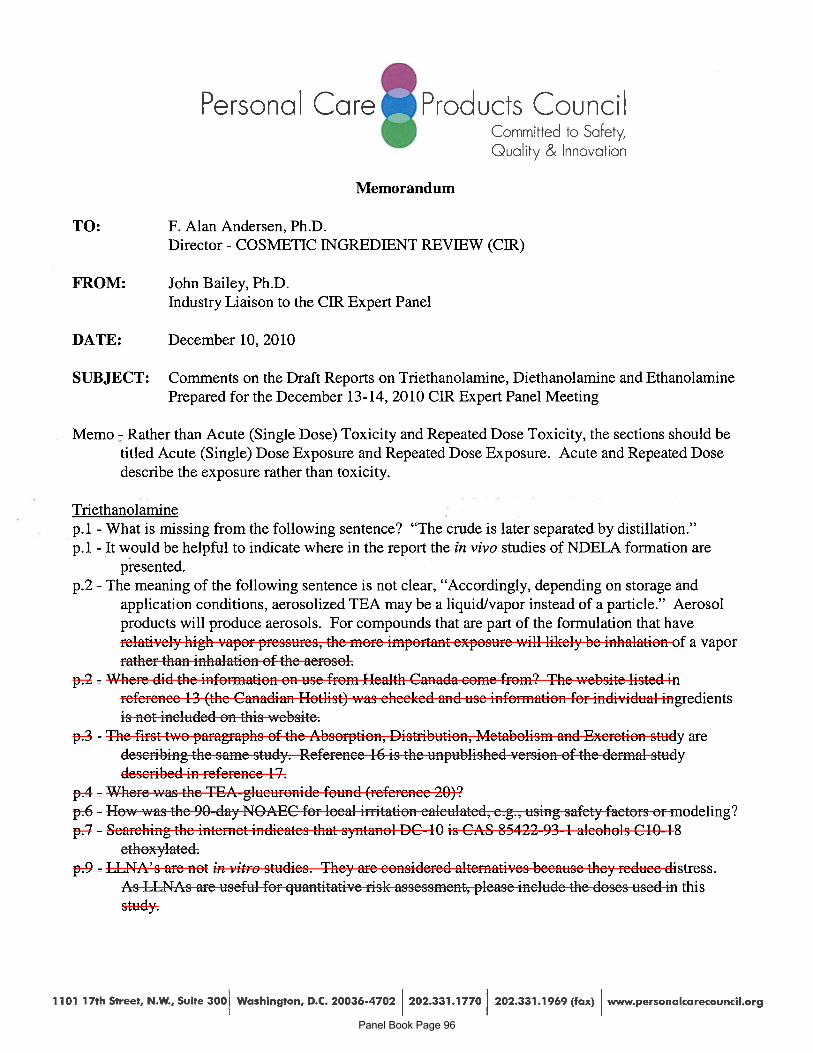
Personal Care Products CouncilCommitted to Safety,Qualify & Innovation
Memorandum
TO: F. Alan Andersen, Ph.D.Director - COSMETIC INGREDIENT REVIEW (CIR)
FROM: John Bailey, Ph.D.Industry Liaison to the CIR Expert Panel
DATE: December 10, 2010
SUBJECT: Comments on the Draft Reports on Triethanolamine, Diethanolamine and EthanolaminePrepared for the December 13-14, 2010 CIR Expert Panel Meeting
Memo - Rather than Acute (Single Dose) Toxicity and Repeated Dose Toxicity, the sections should betitled Acute (Single) Dose Exposure and Repeated Dose Exposure. Acute and Repeated Dosedescribe the exposure rather than toxicity.
Triethanolaminep.1 - What is missing from the following sentence? “The crude is later separated by distillation.”p.1 - It would be helpful to indicate where in the report the in vivo studies of NDELA formation are
presented.p.2 - The meaning of the following sentence is not clear, “Accordingly, depending on storage and
application conditions, aerosolized TEA may be a liquidlvapor instead of a particle.” Aerosolproducts will produce aerosols. For compounds that are part of the formulation that haverelatively high vapor pressures, the more important exposure will likely be inhalation of a vaporrather than inhalation of the aerosol.
p.2 - Where did the information on use from Health Canada come from? The website listed inreference 13 (the Canadian Hotlist) was checked and use information for individual ingredientsis not included on this website.
p.3 - The first two paragraphs of the Absorption, Distribution, Metabolism and Excretion study aredescribing the same study. Reference 16 is the unpublished version of the dermal studydescribed in reference 17.
p.4- - Where was the TEA-glucuronide found (reference 20)?p.6 - How was the 90-day NOAEC for local irritation calculated, e.g., using safety factors or modeling?p.7 - Searching the internet indicates that syntanol DC-b is CAS 85422-93-1 alcohols Cb0-18
ethoxylated.p.9 - LLNA’s are not in vitro studies. They are considered alternatives because they reduce distress.
As LLNAs are useful for quantitative risk assessment., please include the doses used in thisstudy.
1101 17th Street, N.W., Suite 3OO Washington, D.C. 20036-4702 202.331.1770 202.331.1969 (fax) www.personalcarecouncil.org Panel Book Page 96
mmf
Cross-Out
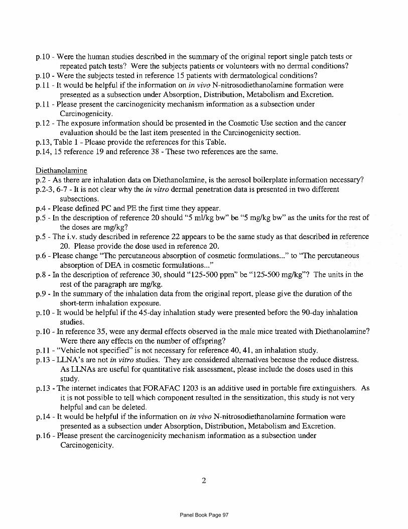
p.10 - Were the human studies described in the summary of the original report single patch tests orrepeated patch tests? Were the subjects patients or volunteers with no dermal conditions?
p.10 - Were the subjects tested in reference 15 patients with dermatological conditions?p.11 - It would be helpful if the information on in vivo N-nitrosodiethanolamine formation were
presented as a subsection under Absorption, Distribution, Metabolism and Excretion.p.11 - Please present the carcinogenicity mechanism information as a subsection under
Carcinogenicity.p.12 - The exposure information should be presented in the Cosmetic Use section and the cancer
evaluation should be the last item presented in the Carcinogenicity section.p.13, Table 1 - Please provide the references for this Table.p.14, 15 reference 19 and reference 38 - These two references are the same.
Diethanolaminep.2 - As there are inhalation data on Diethanolamine, is the aerosol boilerplate information necessary?p.2-3, 6-7 - It is not clear why the in vitro dermal penetration data is presented in two different
subsections.p.4 - Please defined PC and PE the first time they appear.p.S - In the description of reference 20 should “5 mi/kg bw” be “5 mg/kg bw” as the units for the rest of
the doses are mg/kg?p.S - The i.v. study described in reference 22 appears to be the same study as that described in reference
20. Please provide the dose used in reference 20. =
p.6 - Please change “The percutaneous absorption of cosmetic formulations...” to “The percutaneousabsorption of DEA in cosmetic formulations...”
p.8 - In the description of reference 30, should “125-500 ppm” be “125-500 mg/kg”? The units in therest of the paragraph are mg/kg.
p.9 - In the summary of the inhalation data from the original report, please give the duration of theshort-term inhalation exposure.
p.10 - It would be helpful if the 45-day inhalation study were presented before the 90-day inhalationstudies.
p.10 - In reference 35, were any dermal effects observed in the male mice treated with Diethanolamine?Were there any effects on the number of offspring?
p.11 - “Vehicle not specified” is not necessary for reference 40, 41, an inhalation study.p.13 - LLNA’s are not in vitro studies. They are considered alternatives because the reduce distress.
As LLNAs are useful for quantitative risk assessment, please include the doses used in thisstudy.
p.13 - The internet indicates that FORAFAC 1203 is an additive used in portable fire extinguishers. Asit is not possible to tell which component resulted in the sensitization, this study is not veryhelpful and can be deleted.
p.14 - It would be helpful if the information on in vivo N-nitrosodiethanolamine formation werepresented as a subsection under Absorption, Distribution, Metabolism and Excretion.
p.16 - Please present the carcinogenicity mechanism information as a subsection underCarcinogenicity.
2
Panel Book Page 97
mmf
Cross-Out
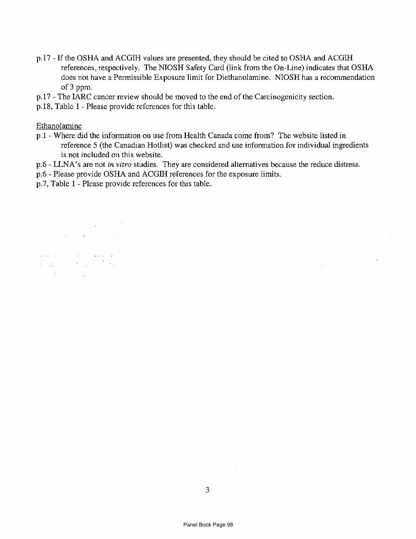
p.17 - If the OSHA and ACGIH values are presented, they should be cited to OSHA and ACGIHreferences, respectively. The NIOSH Safety Card (link from the On-Line) indicates that OSHAdoes not have a Permissible Exposure limit for Diethanolamine. NIOSH has a recommendationof 3 ppm.
p.17 - The IARC cancer review should be moved to the end of the Carcinogenicity section.p.18, Table 1 - Please provide references for this table.
Ethanolaminep.1 - Where did the information on use from Health Canada come from? The website listed in
reference 5 (the Canadian Hotlist) was checked and use information for individual ingredientsis not included on this website.
p.6 - LLNA’s are not in vitro studies. They are considered alternatives because the reduce distress.p.6 - Please provide OSHA and ACGIII references for the exposure limits.p.7, Table 1 - Please provide references for this table.
3
Panel Book Page 98
mmf
Cross-Out
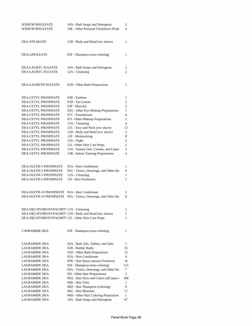
SODIUM BISULFATE 10A - Bath Soaps and Detergents 2SODIUM BISULFATE 10E - Other Personal Cleanliness Produ 4
DEA-STEARATE 12D - Body and Hand (exc shave) 1
DEA-LINOLEATE 05F - Shampoos (non-coloring) 1
DEA-LAURYL SULFATE 10A - Bath Soaps and Detergents 1DEA-LAURYL SULFATE 12A - Cleansing 2
DEA-LAURETH SULFATE 02D - Other Bath Preparations 1
DEA-CETYL PHOSPHATE 03B - Eyeliner 1DEA-CETYL PHOSPHATE 03D - Eye Lotion 1DEA-CETYL PHOSPHATE 03F - Mascara 2DEA-CETYL PHOSPHATE 03G - Other Eye Makeup Preparations 1DEA-CETYL PHOSPHATE 07C - Foundations 4DEA-CETYL PHOSPHATE 07I - Other Makeup Preparations 2DEA-CETYL PHOSPHATE 12A - Cleansing 4DEA-CETYL PHOSPHATE 12C - Face and Neck (exc shave) 13DEA-CETYL PHOSPHATE 12D - Body and Hand (exc shave) 3DEA-CETYL PHOSPHATE 12F - Moisturizing 8DEA-CETYL PHOSPHATE 12G - Night 2DEA-CETYL PHOSPHATE 12J - Other Skin Care Preps 2DEA-CETYL PHOSPHATE 13A - Suntan Gels, Creams, and Liquid 3DEA-CETYL PHOSPHATE 13B - Indoor Tanning Preparations 4
DEA-OLETH-3 PHOSPHATE 05A - Hair Conditioner 1DEA-OLETH-3 PHOSPHATE 05G - Tonics, Dressings, and Other Hai 4DEA-OLETH-3 PHOSPHATE 12A - Cleansing 1DEA-OLETH-3 PHOSPHATE 12I - Skin Fresheners 1
DEA-OLETH-10 PHOSPHATE 05A - Hair Conditioner 2DEA-OLETH-10 PHOSPHATE 05G - Tonics, Dressings, and Other Hai 6
DEA-DI(2-HYDROXYPALMITY12A - Cleansing 1DEA-DI(2-HYDROXYPALMITY12D - Body and Hand (exc shave) 1DEA-DI(2-HYDROXYPALMITY12J - Other Skin Care Preps 1
CAPRAMIDE DEA 05F - Shampoos (non-coloring) 1
LAURAMIDE DEA 02A - Bath Oils, Tablets, and Salts 1LAURAMIDE DEA 02B - Bubble Baths 25LAURAMIDE DEA 02D - Other Bath Preparations 13LAURAMIDE DEA 05A - Hair Conditioner 4LAURAMIDE DEA 05B - Hair Spray (aerosol fixatives) 16LAURAMIDE DEA 05F - Shampoos (non-coloring) 115LAURAMIDE DEA 05G - Tonics, Dressings, and Other Hai 7LAURAMIDE DEA 05I - Other Hair Preparations 2LAURAMIDE DEA 06A - Hair Dyes and Colors (all types r 188LAURAMIDE DEA 06B - Hair Tints 1LAURAMIDE DEA 06D - Hair Shampoos (coloring) 9LAURAMIDE DEA 06G - Hair Bleaches 17LAURAMIDE DEA 06H - Other Hair Coloring Preparation 1LAURAMIDE DEA 10A - Bath Soaps and Detergents 67
Panel Book Page 99

LAURAMIDE DEA 10B - Deodorants (underarm) 1LAURAMIDE DEA 10E - Other Personal Cleanliness Produ 51LAURAMIDE DEA 12A - Cleansing 25LAURAMIDE DEA 12H - Paste Masks (mud packs) 1LAURAMIDE DEA 12J - Other Skin Care Preps 1
LAURAMIDE/MYRISTAMIDE D12A - Cleansing 1
STEARAMIDE DEA 05A - Hair Conditioner 1STEARAMIDE DEA 12D - Body and Hand (exc shave) 8
ISOSTEARAMIDE DEA 07C - Foundations 2
OLEAMIDE DEA 02B - Bubble Baths 8OLEAMIDE DEA 05F - Shampoos (non-coloring) 1OLEAMIDE DEA 12A - Cleansing 1OLEAMIDE DEA 12D - Body and Hand (exc shave) 1OLEAMIDE DEA 12F - Moisturizing 2
LINOLEAMIDE DEA 02A - Bath Oils, Tablets, and Salts 1LINOLEAMIDE DEA 02B - Bubble Baths 3LINOLEAMIDE DEA 02C - Bath Capsules 1LINOLEAMIDE DEA 02D - Other Bath Preparations 5LINOLEAMIDE DEA 04E - Other Fragrance Preparation 1LINOLEAMIDE DEA 05F - Shampoos (non-coloring) 4LINOLEAMIDE DEA 06A - Hair Dyes and Colors (all types r 105LINOLEAMIDE DEA 10A - Bath Soaps and Detergents 7LINOLEAMIDE DEA 10C - Douches 1LINOLEAMIDE DEA 10E - Other Personal Cleanliness Produ 1LINOLEAMIDE DEA 12A - Cleansing 2LINOLEAMIDE DEA 12D - Body and Hand (exc shave) 1LINOLEAMIDE DEA 12J - Other Skin Care Preps 1
COCAMIDE DEA 01A - Baby Shampoos 6COCAMIDE DEA 01C - Other Baby Products 7COCAMIDE DEA 02A - Bath Oils, Tablets, and Salts 5COCAMIDE DEA 02B - Bubble Baths 45COCAMIDE DEA 02D - Other Bath Preparations 23COCAMIDE DEA 03E - Eye Makeup Remover 1COCAMIDE DEA 04E - Other Fragrance Preparation 1COCAMIDE DEA 05A - Hair Conditioner 1COCAMIDE DEA 05E - Rinses (non-coloring) 1COCAMIDE DEA 05F - Shampoos (non-coloring) 239COCAMIDE DEA 05G - Tonics, Dressings, and Other Hai 7COCAMIDE DEA 05H - Wave Sets 1COCAMIDE DEA 05I - Other Hair Preparations 4COCAMIDE DEA 06A - Hair Dyes and Colors (all types r 233COCAMIDE DEA 06D - Hair Shampoos (coloring) 3COCAMIDE DEA 06G - Hair Bleaches 1COCAMIDE DEA 07I - Other Makeup Preparations 1COCAMIDE DEA 10A - Bath Soaps and Detergents 151COCAMIDE DEA 10E - Other Personal Cleanliness Produ 57COCAMIDE DEA 11E - Shaving Cream 1COCAMIDE DEA 11F - Shaving Soap 2COCAMIDE DEA 12A - Cleansing 36COCAMIDE DEA 12C - Face and Neck (exc shave) 7COCAMIDE DEA 12D - Body and Hand (exc shave) 9COCAMIDE DEA 12H - Paste Masks (mud packs) 1COCAMIDE DEA 12J - Other Skin Care Preps 7
Panel Book Page 100

PALM KERNELAMIDE DEA 05F - Shampoos (non-coloring) 4
SOYAMIDE DEA 05A - Hair Conditioner 3SOYAMIDE DEA 05B - Hair Spray (aerosol fixatives) 4SOYAMIDE DEA 05C - Hair Straighteners 5SOYAMIDE DEA 05F - Shampoos (non-coloring) 5SOYAMIDE DEA 05G - Tonics, Dressings, and Other Hai 2
Panel Book Page 101


