Graphitic carbon nitride prepared from urea as a ...
7
1806 Graphitic carbon nitride prepared from urea as a photo- catalyst for visible-light carbon dioxide reduction with the aid of a mononuclear ruthenium(II) complex Kazuhiko Maeda *1 , Daehyeon An 1 , Ryo Kuriki 1,2 , Daling Lu 3 and Osamu Ishitani 1 Full Research Paper Open Access Address: 1 Department of Chemistry, School of Science, Tokyo Institute of Technology, 2-12-1-NE-2 Ookayama, Meguro-ku, Tokyo 152-8550, Japan, 2 Japan Society for the Promotion of Science, Kojimachi Business Center Building, 5-3-1, Kojimachi, Chiyoda-ku, Tokyo 102-0083, Japan and 3 Suzukakedai Materials Analysis Division, Technical Department, Tokyo Institute of Technology, 4259 Nagatsuta-cho, Midori-ku, Yokohama 226-8503, Japan Email: Kazuhiko Maeda * - [email protected] * Corresponding author Keywords: artificial photosynthesis; heterogeneous photocatalysis; hybrid material; metal complexes; solar fuels Beilstein J. Org. Chem. 2018, 14, 1806–1812. doi:10.3762/bjoc.14.153 Received: 21 April 2018 Accepted: 26 June 2018 Published: 17 July 2018 This article is part of the thematic issue "Photoredox catalysis for novel organic reactions". Guest Editor: M. Antonietti © 2018 Maeda et al.; licensee Beilstein-Institut. License and terms: see end of document. Abstract Graphitic carbon nitride (g-C 3 N 4 ) was synthesized by heating urea at different temperatures (773–923 K) in air, and was examined as a photocatalyst for CO 2 reduction. With increasing synthesis temperature, the conversion of urea into g-C 3 N 4 was facilitated, as confirmed by X-ray diffraction, FTIR spectroscopy and elemental analysis. The as-synthesized g-C 3 N 4 samples, further modified with Ag nanoparticles, were capable of reducing CO 2 into formate under visible light (λ > 400 nm) in the presence of triethanolamine as an electron donor, with the aid of a molecular Ru(II) cocatalyst (RuP). The CO 2 reduction activity was im- proved by increasing the synthesis temperature of g-C 3 N 4 , with the maximum activity obtained at 873–923 K. This trend was also consistent with that observed in photocatalytic H 2 evolution using Pt-loaded g-C 3 N 4 . The photocatalytic activities of RuP/g-C 3 N 4 for CO 2 reduction and H 2 evolution were thus shown to be strongly associated with the generation of the crystallized g-C 3 N 4 phase. 1806 Introduction Carbon nitride is one of the oldest synthetic polymers [1], and has several allotropes. Among them, graphitic carbon nitride (g-C 3 N 4 ) is the most stable form and is an emerging material as an organic semiconductor photocatalyst active for various kinds of reactions such as water splitting, CO 2 reduction, and degra- dation of harmful organic compounds, because of its non-toxic, stable, and earth-abundant nature [2-7]. Our group has developed photocatalytic CO 2 reduction systems using g-C 3 N 4 -based materials, in combination with functional
Transcript of Graphitic carbon nitride prepared from urea as a ...

1806
Graphitic carbon nitride prepared from urea as a photo-catalyst for visible-light carbon dioxide reduction withthe aid of a mononuclear ruthenium(II) complexKazuhiko Maeda*1, Daehyeon An1, Ryo Kuriki1,2, Daling Lu3 and Osamu Ishitani1
Full Research Paper Open Access
Address:1Department of Chemistry, School of Science, Tokyo Institute ofTechnology, 2-12-1-NE-2 Ookayama, Meguro-ku, Tokyo 152-8550,Japan, 2Japan Society for the Promotion of Science, KojimachiBusiness Center Building, 5-3-1, Kojimachi, Chiyoda-ku, Tokyo102-0083, Japan and 3Suzukakedai Materials AnalysisDivision, Technical Department, Tokyo Institute of Technology, 4259Nagatsuta-cho, Midori-ku, Yokohama 226-8503, Japan
Email:Kazuhiko Maeda* - [email protected]
* Corresponding author
Keywords:artificial photosynthesis; heterogeneous photocatalysis; hybridmaterial; metal complexes; solar fuels
Beilstein J. Org. Chem. 2018, 14, 1806–1812.doi:10.3762/bjoc.14.153
Received: 21 April 2018Accepted: 26 June 2018Published: 17 July 2018
This article is part of the thematic issue "Photoredox catalysis for novelorganic reactions".
Guest Editor: M. Antonietti
© 2018 Maeda et al.; licensee Beilstein-Institut.License and terms: see end of document.
AbstractGraphitic carbon nitride (g-C3N4) was synthesized by heating urea at different temperatures (773–923 K) in air, and was examined
as a photocatalyst for CO2 reduction. With increasing synthesis temperature, the conversion of urea into g-C3N4 was facilitated, as
confirmed by X-ray diffraction, FTIR spectroscopy and elemental analysis. The as-synthesized g-C3N4 samples, further modified
with Ag nanoparticles, were capable of reducing CO2 into formate under visible light (λ > 400 nm) in the presence of
triethanolamine as an electron donor, with the aid of a molecular Ru(II) cocatalyst (RuP). The CO2 reduction activity was im-
proved by increasing the synthesis temperature of g-C3N4, with the maximum activity obtained at 873–923 K. This trend was also
consistent with that observed in photocatalytic H2 evolution using Pt-loaded g-C3N4. The photocatalytic activities of RuP/g-C3N4
for CO2 reduction and H2 evolution were thus shown to be strongly associated with the generation of the crystallized g-C3N4 phase.
1806
IntroductionCarbon nitride is one of the oldest synthetic polymers [1], and
has several allotropes. Among them, graphitic carbon nitride
(g-C3N4) is the most stable form and is an emerging material as
an organic semiconductor photocatalyst active for various kinds
of reactions such as water splitting, CO2 reduction, and degra-
dation of harmful organic compounds, because of its non-toxic,
stable, and earth-abundant nature [2-7].
Our group has developed photocatalytic CO2 reduction systems
using g-C3N4-based materials, in combination with functional
Beilstein J. Org. Chem. 2018, 14, 1806–1812.
1807
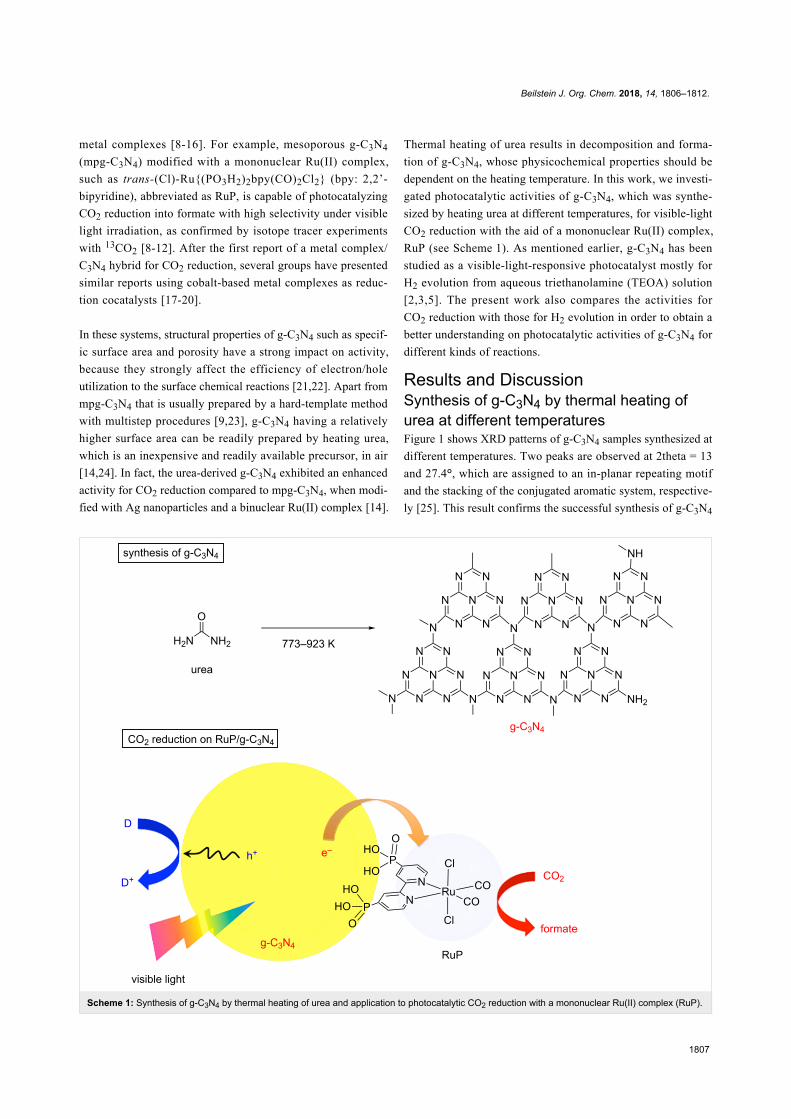
Scheme 1: Synthesis of g-C3N4 by thermal heating of urea and application to photocatalytic CO2 reduction with a mononuclear Ru(II) complex (RuP).
metal complexes [8-16]. For example, mesoporous g-C3N4
(mpg-C3N4) modified with a mononuclear Ru(II) complex,
such as trans-(Cl)-Ru{(PO3H2)2bpy(CO)2Cl2} (bpy: 2,2’-
bipyridine), abbreviated as RuP, is capable of photocatalyzing
CO2 reduction into formate with high selectivity under visible
light irradiation, as confirmed by isotope tracer experiments
with 13CO2 [8-12]. After the first report of a metal complex/
C3N4 hybrid for CO2 reduction, several groups have presented
similar reports using cobalt-based metal complexes as reduc-
tion cocatalysts [17-20].
In these systems, structural properties of g-C3N4 such as specif-
ic surface area and porosity have a strong impact on activity,
because they strongly affect the efficiency of electron/hole
utilization to the surface chemical reactions [21,22]. Apart from
mpg-C3N4 that is usually prepared by a hard-template method
with multistep procedures [9,23], g-C3N4 having a relatively
higher surface area can be readily prepared by heating urea,
which is an inexpensive and readily available precursor, in air
[14,24]. In fact, the urea-derived g-C3N4 exhibited an enhanced
activity for CO2 reduction compared to mpg-C3N4, when modi-
fied with Ag nanoparticles and a binuclear Ru(II) complex [14].
Thermal heating of urea results in decomposition and forma-
tion of g-C3N4, whose physicochemical properties should be
dependent on the heating temperature. In this work, we investi-
gated photocatalytic activities of g-C3N4, which was synthe-
sized by heating urea at different temperatures, for visible-light
CO2 reduction with the aid of a mononuclear Ru(II) complex,
RuP (see Scheme 1). As mentioned earlier, g-C3N4 has been
studied as a visible-light-responsive photocatalyst mostly for
H2 evolution from aqueous triethanolamine (TEOA) solution
[2,3,5]. The present work also compares the activities for
CO2 reduction with those for H2 evolution in order to obtain a
better understanding on photocatalytic activities of g-C3N4 for
different kinds of reactions.
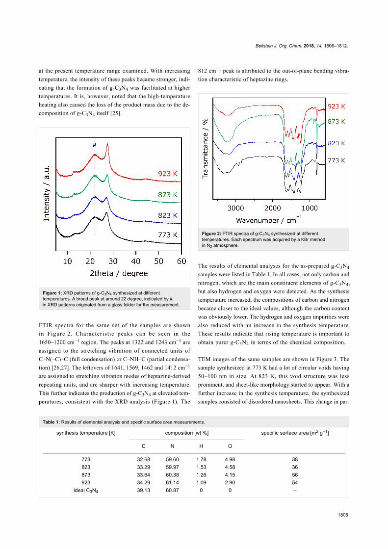
Results and DiscussionSynthesis of g-C3N4 by thermal heating ofurea at different temperaturesFigure 1 shows XRD patterns of g-C3N4 samples synthesized at
different temperatures. Two peaks are observed at 2theta = 13
and 27.4°, which are assigned to an in-planar repeating motif
and the stacking of the conjugated aromatic system, respective-
ly [25]. This result confirms the successful synthesis of g-C3N4
Beilstein J. Org. Chem. 2018, 14, 1806–1812.
1808
Table 1: Results of elemental analysis and specific surface area measurements.
synthesis temperature [K] composition [wt %] specific surface area [m2 g−1]
C N H O
773 32.68 59.60 1.78 4.98 38823 33.29 59.97 1.53 4.58 36873 33.64 60.38 1.26 4.15 56923 34.29 61.14 1.09 2.90 54
ideal C3N4 39.13 60.87 0 0 –
at the present temperature range examined. With increasing
temperature, the intensity of these peaks became stronger, indi-
cating that the formation of g-C3N4 was facilitated at higher
temperatures. It is, however, noted that the high-temperature
heating also caused the loss of the product mass due to the de-
composition of g-C3N4 itself [25].
Figure 1: XRD patterns of g-C3N4 synthesized at differenttemperatures. A broad peak at around 22 degree, indicated by #,in XRD patterns originated from a glass folder for the measurement.
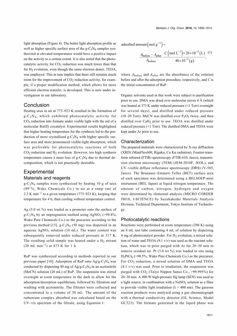
FTIR spectra for the same set of the samples are shown
in Figure 2. Characteristic peaks can be seen in the
1650–1200 cm−1 region. The peaks at 1322 and 1243 cm−1 are
assigned to the stretching vibration of connected units of
C–N(–C)–C (full condensation) or C–NH–C (partial condensa-
tion) [26,27]. The leftovers of 1641, 1569, 1462 and 1412 cm−1
are assigned to stretching vibration modes of heptazine-derived
repeating units, and are sharper with increasing temperature.
This further indicates the production of g-C3N4 at elevated tem-
peratures, consistent with the XRD analysis (Figure 1). The
812 cm−1 peak is attributed to the out-of-plane bending vibra-
tion characteristic of heptazine rings.
Figure 2: FTIR spectra of g-C3N4 synthesized at differenttemperatures. Each spectrum was acquired by a KBr methodin N2 atmosphere.
The results of elemental analyses for the as-prepared g-C3N4
samples were listed in Table 1. In all cases, not only carbon and
nitrogen, which are the main constituent elements of g-C3N4,
but also hydrogen and oxygen were detected. As the synthesis
temperature increased, the compositions of carbon and nitrogen
became closer to the ideal values, although the carbon content
was obviously lower. The hydrogen and oxygen impurities were
also reduced with an increase in the synthesis temperature.
These results indicate that rising temperature is important to
obtain purer g-C3N4 in terms of the chemical composition.
TEM images of the same samples are shown in Figure 3. The
sample synthesized at 773 K had a lot of circular voids having
50–100 nm in size. At 823 K, this void structure was less
prominent, and sheet-like morphology started to appear. With a
further increase in the synthesis temperature, the synthesized
samples consisted of disordered nanosheets. This change in par-

Beilstein J. Org. Chem. 2018, 14, 1806–1812.
1809
Figure 3: TEM images of g-C3N4 synthesized at different temperatures.
ticle morphology is in qualitative agreement with that in the
specific surface area (see Table 1).
Figure 4 shows the UV–visible diffuse reflectance spectra of
g-C3N4 synthesized at different temperatures. All of the sam-
ples exhibited an absorption edge at 420–450 nm, attributed to
electron transitions from the valence band formed by nitrogen
2p orbitals to the conduction band formed by carbon 2p orbitals
[25]. The band gaps of the synthesized g-C3N4 were estimated
to be ca. 2.8–3.0 eV, from the onset wavelength of the diffuse
reflectance spectra. This value is consistent with that reported
previously [24]. As the synthesis temperature increases, the
onset wavelength is shifted to longer wavelengths (i.e., band
gap is decreased), with more pronounced tailing absorption
extending to 550 nm that is assigned to n−π* transitions involv-
ing lone pairs on the edge nitrogen atoms of the heptazine rings
[28,29]. While the n−π* transitions are forbidden for perfectly
symmetric and planar heptazine units, they become partially
allowed with increasing the condensation of layers in g-C3N4,
which results from an increase in the synthesis temperature.
Figure 4: UV–visible diffuse reflectance spectra of g-C3N4synthesized at different temperatures.

Beilstein J. Org. Chem. 2018, 14, 1806–1812.
1810
Table 2: Photocatalytic activities of g-C3N4 synthesized at different temperatures for CO2 reduction and H2 evolution under visible light (λ > 400 nm)a.
synthesis temperature [K] CO2 reductionb [µmol] H2 evolutionc [µmol]
formate CO H2
773 2.8 0.2 0.1 7.0823 3.0 0.2 0.1 9.7873 5.2 0.1 0.1 17.9923 5.1 0.1 0.1 18.6
aReaction conditions: photocatalyst (4.0 mg); reactant solution (4.0 mL); light source, 400 W high-pressure Hg lamp with a NaNO2 aqueous solutionfilter. Reaction time: 5 h. bWith 2.0 µmol g–1 RuP and 5.0 wt % Ag. Performed in a DMA/TEOA mixed solution (4:1 v/v). cWith 3.0 wt % Pt. Performedin a water/TEOA mixed solution (9:1 v/v).
Photocatalytic activities for CO2 reductionand H2 evolutionUsing the as-prepared g-C3N4, CO2 reduction was conducted
with the aid of RuP cocatalyst and Ag promoter in a N,N-
dimethylacetamide (DMA)/TEOA mixed solution under visible
light (λ > 400 nm). Here TEOA works as an effective electron
donor that scavenges holes generated in the valence band of
g-C3N4 [25]. The use of DMA as the solvent for CO2 reduction
using RuP/mpg-C3N4 has previously been shown to be the best
choice of solvents to maximize the photocatalytic activity [10].
Because RuP does not absorb visible light efficiently, the
g-C3N4 component can be activated selectively by visible light
[10]. Our previous study also indicated that the amount of a mo-
lecular cocatalyst is very important in this kind of mononuclear-
complex/C3N4 hybrid photocatalyst for visible-light
CO2 reduction [8]. To eliminate any other effects other than
heating temperature of urea, we fixed the amount of RuP in this
study. Ag nanoparticles loaded on mpg-C3N4 serves as a
promoter of electron transfer from mpg-C3N4 to RuP, as dis-
cussed in our previous work [13]. TEM observation indicated
that the loaded Ag is in the form of nanoparticles of 5–10 nm in
size (Figure 5). Without Ag (i.e., RuP/g-C3N4), formate produc-
tion was clearly observable, but the activity was typically 20%
that of RuP/Ag/g-C3N4. Hence we employed Ag as an addition-
al promoter in all cases. It should be also noted that no reaction
took place using only g-C3N4.
As listed in Table 2, the main product during the reaction was
formate with 90–95% selectivity. Minor products were CO and
H2. With increasing the synthesis temperature, the formate
generation activity was improved to reach a maximum
at 873–923 K, while almost unchanging the CO and
H2 evolution. The catalytic turnover number of formate
generation calculated based on the mole amount of RuP at the
optimal conditions reached 650, which confirms the catalytic
cycle of the reaction.
Figure 5: A typical TEM image of Ag-loaded g-C3N4. The synthesistemperature of g-C3N4 was 873 K in this case.
H2 evolution was also conducted in a mixed solution of water
and TEOA. It is also noted that Pt was in situ loaded on g-C3N4
as a cocatalyst to facilitate H2 evolution. As listed in Table 2, all
of the synthesized samples produced H2. Similar to the trend in
CO2 reduction, the H2 evolution activity was enhanced as the
synthesis temperature was increased.
The results of the photocatalytic reactions thus indicated that
photocatalytic activities of g-C3N4 derived from urea were de-
pendent on the heating temperature of urea. The trend of activi-
ty observed in both CO2 reduction and H2 evolution could be
explained in terms of the formation of the g-C3N4 structure.
Physicochemical analyses indicated that increasing the synthe-
sis temperature of g-C3N4 promotes conversion of urea into
g-C3N4 (Figure 1 and Figure 2), which has even better visible-
Beilstein J. Org. Chem. 2018, 14, 1806–1812.
1811
light absorption (Figure 4). The better light absorption profile as
well as higher specific surface area of the g-C3N4 samples syn-
thesized at elevated temperatures would have a positive impact
on the activity to a certain extent. It is also noted that the photo-
catalytic activity for CO2 reduction was much lower than that
for H2 evolution, even though the same electron donor, TEOA,
was employed. This in turn implies that there still remains much
room for the improvement of CO2 reduction activity, for exam-
ple, if a proper modification method, which allows for more
efficient electron transfer, is developed. This is now under in-
vestigation in our laboratory.
ConclusionHeating urea in air at 773–923 K resulted in the formation of
g-C3N4 , which exhibited photocatalytic activity for
CO2 reduction into formate under visible light with the aid of a
molecular Ru(II) cocatalyst. Experimental results highlighted
that higher heating temperature for the synthesis led to the pro-
duction of more crystallized g-C3N4 with higher specific sur-
face area and more pronounced visible-light absorption, which
was preferable for photocatalytic reactions of both
CO2 reduction and H2 evolution. However, too high synthesis
temperature causes a mass loss of g-C3N4 due to thermal de-
composition, which is not practically desirable.
ExperimentalMaterials and reagentsg-C3N4 samples were synthesized by heating 10 g of urea
(99+%, Wako Chemicals Co.) in air at a ramp rate of
2.3 K min−1 to a given temperature (773–923 K), keeping that
temperature for 4 h, then cooling without temperature control.
Ag (5.0 wt %) was loaded as a promoter onto the surface of
g-C3N4 by an impregnation method using AgNO3 (>99.8%,
Wako Pure Chemicals Co.) as the precursor according to the
previous method [13]. g-C3N4 (50 mg) was dispersed in an
aqueous AgNO3 solution (10 mL). The water content was
subsequently removed under reduced pressure at 317 K.
The resulting solid sample was heated under a H2 stream
(20 mL min−1) at 473 K for 1 h.
RuP was synthesized according to methods reported in our
previous paper [10]. Adsorption of RuP onto Ag/g-C3N4 was
conducted by dispersing 40 mg of Ag/g-C3N4 in an acetonitrile
(MeCN) solution (20 mL) of RuP. The suspension was stirred
overnight at room temperature in the dark to allow for the
adsorption/desorption equilibrium, followed by filtration and
washing with acetonitrile. The filtrates were collected and
concentrated to a volume of 30 mL. The amount of the
ruthenium complex absorbed was calculated based on the
UV–vis spectrum of the filtrate, using Equation 1:
(1)
where Abefore and Aafter are the absorbance of the solution
before and after the adsorption procedure, respectively, and C is
the initial concentration of RuP.
Organic solvents used in this work were subject to purification
prior to use. DMA was dried over molecular sieves 4 Å (which
was heated at 373 K under reduced pressure (<1 Torr) overnight
for several days), and distilled under reduced pressure
(10–20 Torr). MeCN was distilled over P2O5 twice, and then
distilled over CaH2 prior to use. TEOA was distilled under
reduced pressure (<1 Torr). The distilled DMA and TEOA were
kept under Ar prior to use.
CharacterizationThe prepared materials were characterized by X-ray diffraction
(XRD) (MiniFlex600, Rigaku; Cu Kα radiation), Fourier-trans-
form infrared (FTIR) spectroscopy (FTIR-610, Jasco), transmis-
sion electron microscopy (TEM) (JEM-2010F, JEOL), and
UV–visible diffuse reflectance spectroscopy (DRS) (V-565,
Jasco). The Brunauer–Emmett–Teller (BET) surface area
of each specimen was determined using a BELSOEP-mini
instrument (BEL Japan) at liquid nitrogen temperature. The
amount of carbon, ni trogen, hydrogen and oxygen
were determined by elemental analysis (MICRO CORDER
JM10, J-SCIENCE) by Suzukakedai Materials Analysis
Division, Technical Department, Tokyo Institute of Technolo-
gy.
Photocatalytic reactionsReactions were performed at room temperature (298 K) using
an 8 mL test tube containing 4 mL of solution by dispersing
4 mg of photocatalyst powder. For H2 evolution, a mixed solu-
tion of water and TEOA (9:1 v/v) was used as the reactant solu-
tion, which was in prior purged with Ar for 20–30 min to
remove residual air. Pt (3.0 wt %) was loaded in situ using
H2PtCl6 (>98.5%, Wako Pure Chemicals Co.) as the precursor.
For CO2 reduction, a mixed solution of DMA and TEOA
(4:1 v/v) was used. Prior to irradiation, the suspension was
purged with CO2 (Taiyo Nippon Sanso Co., >99.995%) for
20–30 min. A 400 W high-pressure Hg lamp (SEN) was used as
a light source, in combination with a NaNO2 solution as a filter
to provide visible light irradiation (λ > 400 nm). The gaseous
reaction products were analyzed using a gas chromatograph
with a thermal conductivity detector (GL Science, Model
GC323). The formate generated in the liquid phase was

Beilstein J. Org. Chem. 2018, 14, 1806–1812.
1812
analyzed via a capillary electrophoresis system (Otsuka Elec-
tronics Co., Model CAPI-3300).
AcknowledgementsThis work was supported by a Grant-in-Aid for Young Scien-
tists (A) (Project JP16H06130) from JSPS. It was also partially
supported by a Grant-in-Aid for Scientific Research on Innova-
tive Area “Mixed Anion (Project JP16H06441)” and a CREST
program (Project JPMJCR13L1) (JST). K.M. acknowledges
The Noguchi Institute and Murata Research Foundation finan-
cial support. R.K. wishes to acknowledge support by a JSPS
Fellowship for Young Scientists (Project JP17J03705).
ORCID® iDsKazuhiko Maeda - https://orcid.org/0000-0001-7245-8318Ryo Kuriki - https://orcid.org/0000-0002-3843-2867Daling Lu - https://orcid.org/0000-0002-9084-480XOsamu Ishitani - https://orcid.org/0000-0001-9557-7854
References1. Liebig, J. Ann. Pharm. (Lemgo, Ger.) 1834, 10, 1–47.
doi:10.1002/jlac.183401001022. Wang, Y.; Wang, X.; Antonietti, M. Angew. Chem., Int. Ed. 2012, 51,
68–89. doi:10.1002/anie.2011011823. Zheng, Y.; Lin, L.; Wang, B.; Wang, X. Angew. Chem., Int. Ed. 2015,
54, 12868–12884. doi:10.1002/anie.2015017884. Zhang, J.; Chen, Y.; Wang, X. Energy Environ. Sci. 2015, 8,
3092–3108. doi:10.1039/C5EE01895A5. Kuriki, R.; Maeda, K. Phys. Chem. Chem. Phys. 2017, 19, 4938–4950.
doi:10.1039/C6CP07973C6. Yin, S.; Han, J.; Zhou, T.; Xu, R. Catal. Sci. Technol. 2015, 5,
5048–5061. doi:10.1039/C5CY00938C7. Fang, Y.; Wang, X. Chem. Commun. 2018, 54, 5674–5687.
doi:10.1039/C8CC02046A8. Maeda, K.; Sekizawa, K.; Ishitani, O. Chem. Commun. 2013, 49,
10127–10129. doi:10.1039/c3cc45532g9. Maeda, K.; Kuriki, R.; Zhang, X.; Wang, X.; Ishitani, O.
J. Mater. Chem. A 2014, 2, 15146–15151. doi:10.1039/C4TA03128H10. Kuriki, R.; Sekizawa, K.; Ishitani, O.; Maeda, K. Angew. Chem., Int. Ed.
2015, 54, 2406–2409. doi:10.1002/anie.20141117011. Kuriki, R.; Ishitani, O.; Maeda, K. ACS Appl. Mater. Interfaces 2016, 8,
6011–6018. doi:10.1021/acsami.5b1183612. Maeda, K.; Kuriki, R.; Ishitani, O. Chem. Lett. 2016, 45, 182–184.
doi:10.1246/cl.15106113. Kuriki, R.; Matsunaga, H.; Nakashima, T.; Wada, K.; Yamakata, A.;
Ishitani, O.; Maeda, K. J. Am. Chem. Soc. 2016, 138, 5159–5170.doi:10.1021/jacs.6b01997
14. Kuriki, R.; Yamamoto, M.; Higuchi, K.; Yamamoto, Y.; Akatsuka, M.;Lu, D.; Yagi, S.; Yoshida, T.; Ishitani, O.; Maeda, K.Angew. Chem., Int. Ed. 2017, 56, 4867–4871.doi:10.1002/anie.201701627
15. Wada, K.; Eguchi, M.; Ishitani, O.; Maeda, K. ChemSusChem 2017,10, 287–295. doi:10.1002/cssc.201600661
16. Wada, K.; Ranasinghe, C. S. K.; Kuriki, R.; Yamakata, A.; Ishitani, O.;Maeda, K. ACS Appl. Mater. Interfaces 2017, 9, 23869–23877.doi:10.1021/acsami.7b07484
17. Lin, J.; Pan, Z.; Wang, X. ACS Sustainable Chem. Eng. 2014, 2,353–358. doi:10.1021/sc4004295
18. Wang, S.; Lin, J.; Wang, X. Phys. Chem. Chem. Phys. 2014, 16,14656–14660. doi:10.1039/c4cp02173h
19. Walsh, J. J.; Jiang, C.; Tang, J.; Cowan, A. J.Phys. Chem. Chem. Phys. 2016, 18, 24825–24829.doi:10.1039/C6CP04525A
20. Zhao, G.; Pang, H.; Liu, G.; Li, P.; Liu, H.; Zhang, H.; Shi, L.; Ye, J.Appl. Catal., B: Environ. 2017, 200, 141–149.doi:10.1016/j.apcatb.2016.06.074
21. Maeda, K. J. Photochem. Photobiol., C 2011, 12, 237–268.doi:10.1016/j.jphotochemrev.2011.07.001
22. Maeda, K.; Domen, K. Bull. Chem. Soc. Jpn. 2016, 89, 627–648.doi:10.1246/bcsj.20150441
23. Goettmann, F.; Fischer, A.; Antonietti, M.; Thomas, A.Angew. Chem., Int. Ed. 2006, 45, 4467–4471.doi:10.1002/anie.200600412
24. Liu, J.; Zhang, T.; Wang, Z.; Dawson, G.; Chen, W. J. Mater. Chem.2011, 21, 14398–14401. doi:10.1039/c1jm12620b
25. Wang, X.; Maeda, K.; Thomas, A.; Takanabe, K.; Xin, G.;Carlsson, J. M.; Domen, K.; Antonietti, M. Nat. Mater. 2009, 8, 76–80.doi:10.1038/nmat2317
26. Lotsch, B. V.; Döblinger, M.; Sehnert, J.; Seyfarth, L.; Senker, J.;Oeckler, O.; Schnick, W. Chem. – Eur. J. 2007, 13, 4969–4980.doi:10.1002/chem.200601759
27. Bojdys, M. J.; Müller, J.-O.; Antonietti, M.; Thomas, A. Chem. – Eur. J.2008, 14, 8177–8182. doi:10.1002/chem.200800190
28. Jorge, A. B.; Martin, D. J.; Dhanoa, M. T. S.; Rahman, A. S.;Makwana, N.; Tang, J.; Sella, A.; Corà, F.; Firth, S.; Darr, J. A.;McMillan, P. F. J. Phys. Chem. C 2013, 117, 7178–7185.doi:10.1021/jp4009338
29. Zhang, H.; Yu, A. J. Phys. Chem. C 2014, 118, 11628–11635.doi:10.1021/jp503477x
License and TermsThis is an Open Access article under the terms of the
Creative Commons Attribution License
(http://creativecommons.org/licenses/by/4.0). Please note
that the reuse, redistribution and reproduction in particular
requires that the authors and source are credited.
The license is subject to the Beilstein Journal of Organic
Chemistry terms and conditions:
(https://www.beilstein-journals.org/bjoc)
The definitive version of this article is the electronic one
which can be found at:
doi:10.3762/bjoc.14.153