
GRAPEVINE FANLEAF VIRUS: BIOLOGY, BIOTECHNOLOGY AND ...
282
GRAPEVINE FANLEAF VIRUS: BIOLOGY, BIOTECHNOLOGY AND RESISTANCE A Dissertation Presented to the Faculty of the Graduate School of Cornell University In Partial Fulfillment of the Requirements for the Degree of Doctor of Philosophy by John Wesley Gottula May 2014
Transcript of GRAPEVINE FANLEAF VIRUS: BIOLOGY, BIOTECHNOLOGY AND ...
GRAPEVINE FANLEAF VIRUS: BIOLOGY, BIOTECHNOLOGY AND
RESISTANCE
A Dissertation
Presented to the Faculty of the Graduate School
of Cornell University
In Partial Fulfillment of the Requirements for the Degree of
Doctor of Philosophy
by
John Wesley Gottula
May 2014

© 2014 John Wesley Gottula

GRAPEVINE FANLEAF VIRUS: BIOLOGY, BIOTECHNOLOGY AND
RESISTANCE
John Wesley Gottula, Ph. D.
Cornell University 2014
Grapevine fanleaf virus (GFLV) causes fanleaf degeneration of grapevines. GFLV is
present in most grape growing regions and has a bipartite RNA genome. The three
goals of this research were to (1) advance our understanding of GFLV biology through
studies on its satellite RNA, (2) engineer GFLV into a viral vector for grapevine
functional genomics, and (3) discover a source of resistance to GFLV. This author
addressed GFLV biology by studying the least understood aspect of GFLV: its
satellite RNA. This author sequenced a new GFLV satellite RNA variant and
compared it with other satellite RNA sequences. Forensic tracking of the satellite
RNA revealed that it originated from an ancestral nepovirus and was likely introduced
from Europe into North America. Greenhouse experiments showed that the GFLV
satellite RNA has commensal relationship with its helper virus on a herbaceous host.
This author engineered GFLV into a biotechnology tool by cloning infectious GFLV
genomic cDNAs into binary vectors, with or without further modifications, and using
Agrobacterium tumefaciens delivery to infect Nicotiana benthamiana. Tagging GFLV
with fluorescent proteins allowed tracking of the virus within N. benthamiana and
Chenopodium quinoa tissues, and imbuing GFLV with partial plant gene sequences
proved the concept that endogenous plant genes can be knocked down. Infectivity of
the viral vector depended on the identity of the GFLV strains or reassortants, on co-
application of heterologous silencing suppressors and on lower ambient temperatures.
No natural sources of resistance to GFLV exist within Vitis spp., but certain
herbaceous hosts such as N. tabacum (tobacco) are resistant. This author used tobacco,
its wild relatives, and hybrids between tobacco and wild relatives to evaluate the
genomic and physiological basis of resistance. Resistance to GFLV in tobacco is
governed by systemic recovery from virus infection that is additively inherited and
likely multi-allelic. This research has opened new avenues to understand virus and
plant evolution, and furnishes geneticists with a new tool to functionally characterize
host genes. This dissertation also includes a history of pathogen-derived resistance
with specific reference to plant virus resistance.

v
BIOGRAPHICAL SKETCH
John was raised in Lubbock, Texas and attended Texas Tech University as an
undergraduate. At Texas Tech, he worked with Dr. Robert Wright on cotton breeding
and biotechnology. John’s research interests include transgenic technologies for plant
stress resistance, breeding for durable plant resistance and plant functional genomics
tools. John has been the first author/ co-author on seven publications and primary
investigator or project director on two grants focused on plant biology and
biotechnology. He has taken a variety of leadership roles in the Plant Pathology
Graduate Student Association at Cornell including treasurer (2009-2011), the Student
Association of the Geneva Experiment Station including garden coordinator (2010-
2012), has led a collaborative Cornell colloquium (2012) and has coordinated invited
lectures of multiple students and faculty from around the United States. John has
served as ad hoc reviewer for Crop Science and Transgenic Research, assisted
teaching the Cornell course Magical Mushrooms, Mischievous Molds, and has
participated in extension work for grapevine virus control in the Finger Lakes. When
John is not doing research, he enjoys pretending to fish with his friend Ben and
dancing a very serious two-step with his beautiful wife Kelly.

vi
Dedicated to Hussein Alzubi

vii
ACKNOWLEDGMENTS
First and foremost, I thank my major advisor Marc Fuchs for his support and
professionalism. I extend my appreciation to committee members Lisa Earle and
Sunny Power for their suggestions, advice and never-wavering support. I thank
Stewart Gray for hosting me in his laboratory and believing in me from day one. I
offer special thanks to Corinne Keichinger and family, Christophe Ritzenthaler,
Gérard Demangeat, Emmanuelle Vigne and family, Peggy Andret-Link, Veronique
Komar and Olivier Lemaire for entertaining me in beautiful Alsace, France. Luz
Marcela Yepes, Ramsey Lewis, Mei Cheung, David MacUmber, Pat Marsella-
Herrick, Mamta Srivastava, Dawn Smith, Jason Ingram and Tai Wei Guo each gave
critical assistance to parts of this research. Ben Bartlett, Dana Lapato, Halli Gutting,
Léa Ackerer, Rossella Labarile, Keiran Cantilina, Larissa Osterbaan, Libby
Cieniewicz and Melanie Isganitis also contributed significantly. Thanks to those who
contributed to my education in virology and molecular biology including John Hart,
Ben Orcheski, Jonathan Oliver, Jason Cavatorta, Jean-Michel Hily, Ho-Jong Ju, Balaji
Vasudevan, Stacy Singer, Kamal Hlebieh, Caroline Hemmer, Batiste Monsion and
Francois Berthold, Kerik Cox for statistics help, and Seiya Saito for his statistics help
and friendship. Thanks to the Cornell Plant Virology Group and students and faculty
at North Carolina State, Ohio State and Penn State universities for their stimulating
and helpful dialog. The research was funded in part by a USDA-AFRI-NIFA pre-
doctoral grant, a Grape Research Coordination Network Grant, assistantships from
Cornell Department of Plant Pathology and Plant-Microbe Biology, and the College of
Agriculture and Life Sciences through the New York State Agricultural Experiment
Station. Thanks to my wife Kelly Voll for her love and to Kemal Ozbek for being a
great roommate, too.

viii
PREFACE
Grapevine fanleaf virus (GFLV) is a small pathogen in size. It encodes only eight
individual proteins and is encapsidated in 30nm particles, but its interactions with
plant hosts are extraordinarily complex. This dissertation encompasses reviews and
primary research of GFLV biology including its genetic diversity, host range,
inoculation methods, evolutionary biology and uses in biotechnology. In Chapter 1,
this author reviews GFLV biology and discuss its relationship with other viruses of the
genus Nepovirus, family Secoviridae. In Chapter 2, this author discusses the natural
history, evolutionary biology, and host and helper virus interactions of the nepovirus
subgroup A satellite RNA. In Chapter 3, this author presents proofs-of-concept that
GFLV is engineered into a vector for plant functional genomics and other uses. In
Chapter 4, this author describes variables that are associated with reliable plant
systemic infection when GFLV is inoculated through Agrobacterium tumefaciens. In
Chapter 5, this author reviews the history of pathogen-derived resistance applied to
viruses through 2009. In Chapter 6, this author presents an assessment of the GFLV
host range within Nicotiana and a theory of how allopolyploids impact evolution of
basal virus resistance. Finally, in Chapter 7, this author suggests research projects to
better understand the GFLV satellite RNA, improve the GFLV vector, and an
overview of how plant resistance to viruses can be improved. Readers of this
dissertation will gain an appreciation of the complex yet elegant nature of GFLV
biology and insights into broader issues in plant virology, viral vectors for plant
functional genomics and plant resistance to viruses.
ix
TABLE OF CONTENTS
CHAPTER 1: GRAPEVINE FANLEAF VIRUS AND FANLEAF DEGENERATION
...................................................................................................................................... 17
THE DISEASE .................................................................................................... 17
PATHOGEN BIOLOGY ..................................................................................... 18
DISEASE MANAGEMENT ............................................................................... 25
BIOTECHNOLOGY ........................................................................................... 27
REFERENCES .................................................................................................... 29
CHAPTER 2: GENETIC VARIABILITY, EVOLUTION AND BIOLOGICAL
EFFECTS OF GRAPEVINE FANLEAF VIRUS SATELLITE RNAS ...................... 39
ABSTRACT ........................................................................................................ 39
INTRODUCTION ............................................................................................... 41
MATERIALS AND METHODS ........................................................................ 43
RESULTS ............................................................................................................ 50
REFERENCES .................................................................................................... 71
CHAPTER 3: A VIRAL VECTOR COMPOSED OF GRAPEVINE FANLEAF
VIRUS .......................................................................................................................... 77
ABSTRACT ........................................................................................................ 77
INTRODUCTION ............................................................................................... 78
MATERIALS AND METHODS ........................................................................ 82
RESULTS ............................................................................................................ 92
DISCUSSION .................................................................................................... 103

x
REFERENCES .................................................................................................. 109
CHAPTER 4: GENOMIC, ENVIRONMENTAL AND HOST VARIABLES
INFLUENCING GRAPEVINE FANLEAF VIRUS AGROINFECTION ................ 120
ABSTRACT ...................................................................................................... 120
INTRODUCTION ............................................................................................. 122
MATERIALS AND METHODS ...................................................................... 124
RESULTS .......................................................................................................... 136
DISCUSSION .................................................................................................... 148
REFERENCES .................................................................................................. 157
CHAPTER 5: TOWARDS A QUARTER CENTURY OF PATHOGEN-DERIVED
RESISTANCE AND PRACTICAL APPROACHES TO PLANT VIRUS DISEASE
CONTROL ................................................................................................................. 166
ABSTRACT ...................................................................................................... 166
INTRODUCTION ............................................................................................. 168
THE CONCEPT OF PDR ................................................................................. 169
CREATION OF VIRUS-RESISTANT TRANSGENIC CROPS BY APPLYING
THE CONCEPT OF PDR ................................................................................. 176
COMMERCIALIZATION OF VIRUS-RESISTANT TRANSGENIC CROPS
AND PRACTICAL CONTROL OF VIRUS DISEASES ................................. 180
DISCUSSION .................................................................................................... 187
REFERENCES .................................................................................................. 190

xi
CHAPTER 6: GENOMIC BASIS OF BASAL VIRUS RESISTANCE ................... 206
ABSTRACT ...................................................................................................... 206
INTRODUCTION ............................................................................................. 208
MATERIALS AND METHODS ...................................................................... 212
RESULTS .......................................................................................................... 219
DISCUSSION .................................................................................................... 242
CONCLUSION ................................................................................................. 249
REFERENCES .................................................................................................. 251
CHAPTER 7: FUTURE DIRECTIONS .................................................................... 261
NEPOVIRUS SUBGROUP A SATELLITE RNA ........................................... 261
IMPROVING VIRAL VECTORS .................................................................... 265
THE FUTURE OF PLANT VIRUS RESISTANCE ......................................... 269
REFERENCES .................................................................................................. 273

xii
LIST OF FIGURES
Figure 2-1 Phylogenetic relationships of type B satellite RNAs of Grapevine
fanleaf virus and Arabis mosaic virus.................................................53
Figure 2-2 Shared identity of nepovirus subgroup A type B satellite RNA with
nepovirus genomic RNAs…................................................................62
Figure 2-3 Gel showing RT-PCR products of Grapevine fanleaf virus strain R6-18
cured of its satRNA.............................................................................62
Figure 2-4 Accumulation of Grapevine fanleaf virus capsid in mechanically
inoculated Chenopodium quinoa.........................................................64
Figure 2-5 Symptom severity over time of Grapevine fanleaf virus strains
differing in genome and satRNAs.......................................................65
Figure 2-6 Physiological parameters of Chenopodium quinoa infected with
Grapevine fanleaf virus strains differing in satRNA presence or
absence.................................................................................................66
Figure 3-1 Schematic illustration of the Grapevine fanleaf virus genome and
vectors..................................................................................................80
Figure 3-2 Fluorescence expression in plants infected with the Grapevine fanleaf
virus vector..........................................................................................93

xiii
Figure 3-3 Comparative virus-induced gene silencing activity of the Grapevine
fanleaf virus and Tobacco rattle virus vectors for silencing phytoene
desaturase and enhanced Green fluorescent protein expression in
Nicotiana benthamiana.......................................................................96
Figure 3-4 Dual gene expression patterns of the Grapevine fanleaf virus
vector...................................................................................................98
Figure 3-5 Dosage of Grapevine fanleaf virus in leaves and berries of naturally
infected grapevines............................................................................101
Figure 4-1 Effect of Grapevine fanleaf virus genome composition on virus
accumulation in Nicotiana benthamiana at two, five and eight days
post-agroinfiltration...........................................................................139
Figure 4-2 Effect of silencing suppressor on Grapevine fanleaf virus accumulation
following coinfiltration with genomes FF, FG, GF and GG.............142
Figure 4-3 Comparative expression of Grapevine fanleaf virus and eGFP
expression among Nicotiana species.................................................149
Figure 5-1 Reaction of transgenic and nontransgenic summer squash and papaya to
virus infection....................................................................................182
Figure 6-1 Infection frequencies in inoculated and apical leaves of populations of
plants tested for resistance to Grapevine fanleaf virus strains GHu and
F13, and Tomato ringspot virus strain AP.........................................231
Figure 6-2 Reaction of synthetic Nicotiana allopolyploids to Grapevine fanleaf
xiv
virus strains F13 and GHu.................................................................232
Figure 6-3 Grapevine fanleaf virus strain GHu resistance categories superimposed
on a Nicotiana phylogenetic tree.......................................................234
Figure 6-4 Pictographic description of the modified matching allele model applied
to the additive resistance hypothesis.................................................245
Figure 6-5 Model of changes in quantitative innate virus resistance from a
moderately resistant progenitor exhibiting fixed heterozygosity for
resistance genes (e.g. a neoallopolyploid) ........................................247
xv
LIST OF TABLES
Table 2-1 Oligonucleotides used in the study for Grapevine fanleaf virus (GFLV)
RNA2 or satellite RNA detection, for 5' RACE and 3' amplification of
GFLV satRNAs by IC-RT-PCR.........................................................46
Table 2-2 Genetic parameters of genes 1EPol, 2AHP, 2BMP, 2CCP and the satellite
RNA within and between Grapevine fanleaf virus and Arabis mosaic
virus isolates........................................................................................57
Table 2-3 Estimates of selection pressures on select subgroup A nepovirus
proteins................................................................................................58
Table 3-1 Primers used in the cloning procedures to engineer Grapevine fanleaf
virus vectors.........................................................................................85
Table 3-2 Stability of Red fluorescent protein expression from the Grapevine
fanleaf virus vector in Nicotiana benthamiana and Chenopodium
quinoa..................................................................................................94
Table 3-3 Transmissibility of the Grapevine fanleaf virus vector by Xiphinema
index...................................................................................................104
Table 4-1 Primers used for the modification of Grapevine fanleaf virus cDNAs and
their placement in binary vectors.......................................................126
Table 4-2 Nomenclature of Grapevine fanleaf virus genomes used in Chapter
4.........................................................................................................127

xvi
Table 4-3 Odds Ratio and 95% confidence intervals of selected variables in
Agrobacterium tumefaciens-mediated Grapevine fanleaf virus infection
based on a stepwise categorical logistic regression...........................143
Table 4-4 Odds Ratio and 95% confidence intervals of Agrobacterium tumefaciens
versus transgenic expression of individual Grapevine fanleaf virus
genome parts based on direct categorical logistic
regression...........................................................................................147
Table 5-1 Successful application of pathogen-derived resistance in commercially
available virus-resistant crops...........................................................181
Table 6-1 Sources of Nicotiana species and synthetic allopolyploids used in
resistance experiments.......................................................................214
Table 6-2 Plant responses to Grapevine fanleaf virus strains F13 and GHu, and
Tomato ringspot virus strain AP........................................................222
Table 6-3 Grapevine fanleaf virus and Tomato ringspot virus resistance ratings of
Nicotiana species and synthetic allopolyploids.................................226
Table 6-4 Systemic recovery from Grapevine fanleaf virus strains F13 and GHu
...........................................................................................................237

17
CHAPTER 1
GRAPEVINE FANLEAF VIRUS AND FANLEAF DEGENERATION
THE DISEASE
Grapevine fanleaf virus (GFLV) is one of the most serious and widespread
grapevine virus diseases. GFLV causes grapevines to produce lower fruit yield and
reduced berry quality, misshapen leaves, shortened internodes, leaf yellowing,
mottling and vein clearing (Andret-Link et al., 2004). The economic impact on
grapevine production systems is severe with $1 billion annual losses to the French
grape and wine industries (Andret-Link and Fuchs, 2005).
Based on the natural distribution of its highly specific ectoparasitic nematode
vector, Xiphinema index, GFLV is thought to originate from the Caucasus region of
East Asia (Raski et al., 1983). Vitis vinifera (grapevine) was domesticated in its center
of origin in Anatolia or the Caucasus by 4,000 B.C.E. and was carried to Mesopotamia
and Egypt by 2,000 B.C.E., and France by 500 B.C.E. (Hancock, 2004). Although the
ancient use of cuttings and layering probably hindered the dissemination of the
nematode vector on a local level, long-distance grapevine transport would have
required rooted plants and thus facilitated the worldwide dispersal of the nematode
and associated virus. Today, GFLV is present in all major grape-growing regions
except the Finger Lakes Region of New York State and other central and northeastern
North American regions.
GFLV is one of several viruses that cause fanleaf degeneration. The other
18
viruses that are causal agents of fanleaf degeneration are related to GFLV and belong
to the genus Nepovirus in the family Secoviridae. Based on genome homology and
identity, nepoviruses are classified into subgroups A, B and C (Sanfaçon et al., 2009).
GFLV, Arabis mosaic virus (ArMV), Tobacco ringspot virus (TRSV) and Grapevine
deformation virus (GDefV) (Elbeaino et al., 2012) are subgroup A nepoviruses that
cause fanleaf degeneration. Subgroup B nepoviruses that cause fanleaf degeneration
are Tomato black ring virus (TBRV) and Grapevine chrome mosaic virus (GCMV). A
subgroup C nepovirus that causes fanleaf degeneration is Tomato ringspot virus
(ToRSV) (Sanfaçon et al., 2009).
The nepoviruses that cause fanleaf degeneration are systemically infective in
grapevines. Asexual propagules (cuttings) taken from infected tissue contain the
viruses, and thus the viral diseases will become established in vineyards where
infected clones are planted or used for grafting. The disease may be present but not
established in vineyards where infected clones were introduced in the absence of soil
infested by the nematode vectors. This has been observed for GFLV (Gottula et al.,
2013) and ArMV (Celebi-Toprak et al., 2013) in the U.S. Department of Agriculture
Cold Hardy Grape Genetics Germplasm Repository in Geneva, NY. Conversely,
aviruliferous X. index infesting vineyards will not spread fanleaf degeneration, and
nematode parasitism can be effectively controlled using resistant rootstock material
(Reisch et al., 2011).
PATHOGEN BIOLOGY
Nepoviruses share a common bipartite genome structure composed of single-

19
stranded positive-sense RNAs. The two nepovirus genomic RNAs are RNA1 and
RNA2, each of which includes a virus-encoded protein covalently attached to its 5’
end (VPg, viral protein, genome-linked) and a poly-A tail extending from its 3’ end.
Nepoviruses share common genome expression mechanisms with other members of
Picornavirales including monocistronic translation and proteolytic processing of
individual protein components (Sanfaçon et al., 2009). Nepovirus subgroup A RNA1
contains coding regions for the 1A, 1BHel (helicase), 1CVPg, 1DPro (proteinase) and
1ERdRp (RNA-dependent RNA polymerase) proteins. The RNA2 of subgroup A
nepovirus species contain coding regions for the 2AHP (homing protein), 2BMP
(movement protein) and 2CCP (coat protein) proteins. Additionally, about one third of
GFLV isolates from Europe and Asia contain a satellite RNA (Saldarelli et al., 1993).
The function of GFLV protein 1A is poorly characterized, but is thought to
form the structure of the nepovirus replication complex on ER membranes
(Ritzenthaler et al., 2002). The 1BHel protein contains a nucleoside triphosphate-
binding domain and is a putative helicase (Ritzenthaler et al., 1991). Certain
nepoviruses, including ArMV, show post-translational processing of the 1BHel into a
helicase and a hydrophobic protein (X2), which is a membrane anchor in the virus
replication complex (Sanfaçon et al., 2012). There is no evidence that GFLV produces
multiple 1BHel translation products (J. Gottula and C. Keichinger, unpublished data).
The identification of the GFLV silencing suppressor is pending (Vigne et al.,
2013), but may be the 1BHel, which contains a GW (glycine-tryptophan) motif. GW
motifs interact with Argonaute proteins to alter silencing suppression activity
(Burgyan and Hazevelda, 2011; Garcia et al., 2012) and can sometimes provide hints

20
as to the identity of viral suppressors of silencing, especially when multiple GW
motifs are present and are present with WG motifs (El-Shami et al., 2007). The GFLV
GW motif within 1BHel occurs without a WG motif and is fully conserved between
GFLV and ArMV, but so are 71.7% of 1BHel amino acids among the eight fully
sequenced GFLV and ArMV isolates (J. Gottula, unpublished data). Most other
sequenced nepoviruses contain one GW domain within 1BHel, though the positions are
not conserved among or within subgroups, and most 1BHel proteins do not contain a
WG motif (J. Gottula, unpublished data). The functional significance of the GW motif
in GFLV 1BHel is unknown.
Nepoviruses encode a VPg that is affixed to the 5’ ends of (+) and (-) strand of
GFLV RNAs including RNA1, RNA2 and the satellite RNA (Pinck et al., 1988). VPg
proteins interact with plant translation machinery to achieve either translation or
replication (Jiang and Laliberté, 2011). Nepovirus VPgs are much smaller than those
of other viral genera (Jiang and Laliberté, 2011). For ToRSV, the VPg exists in
proteolytically unprocessed forms with the neighboring helicase, proteinase and the
RdRp, and one of these unprocessed forms likely comprise the nepovirus primer for
replication (Chisholm et al., 2007). Like the VPg of potyviruses, the nepovirus VPg
interacts with eukaryotic Initiation factor 4E (Léonard et al., 2002), which could open
up the interesting possibility of achieving nepovirus resistance through mutation of
host eIF4E alleles (Charron et al., 2008).
The proteinase of GFLV is a cysteine-like proteinase structurally related to
chemotrypsin (Sanfaçon et al., 2009). It recognizes the following residue
combinations in GFLV: cysteine/arginine, arginine/glycine, glycine/serine and

21
glycine/glutamate (Ritzenthaler et al., 1991; Margis et al., 1994). Nine to 25 amino
acids surrounding each of these residues are conserved in GFLV and ArMV and are
probably necessary for proteinase recognition of these sites (J. Gottula, unpublished;
Wetzel et al., 2013). The proteinase functions on the RNA1 polyprotein in cis and
RNA2 polyprotein in trans and may require the 1A as a cofactor (Ritzenthaler et al.,
1991).
The amino acid sequence of GFLV RdRp is highly conserved (Oliver et al.,
2010) and shows high identity to the ArMV RdRp (Gottula et al., 2013). The RdRp
shares four conserved amino acid motifs with other members of Secoviridae including
a GDD (glycine-aspartate-aspartate) motif (Ritzenthaler et al., 1991). The RdRp was
recently found to be the GFLV symptom determinant in N. benthamiana and N.
clevelandii, and the region of the RdRp that determines symptoms was mapped to the
its 3’/C-terminal region upstream of the conserved GDD motif (Vigne et al., 2013).
Because this region is not post-translationally cleaved from the RdRp and does not
appear to relate to higher GFLV accumulation (Vigne et al., 2013), questions remain
about whether symptoms relate to protein or RNA factors encoded by the symptom-
producing GFLV strain GHu.
Little is known about the GFLV 2AHP protein other than it is necessary for
RNA2 replication (Gaire et al., 1999). The 2AHP protein shows relatively high amino
acid diversity among GFLV isolates (Oliver et al., 2010), though less interspecific
divergence than the other GFLV and ArMV RNA2-encoded proteins (Gottula et al.,
2013). The 2AHP-coding region encodes a higher proportion of non-synonymous to
synonymous mutations (Oliver et al., 2010) underlying positive selection that could

22
reflect virus-host coevolution at a virulence/immunity interface (Jones and Dangl,
2006).
The 2BMP movement protein of GFLV forms tubules and functions with the
2CCP protein (coat protein) for intercellular virus transport through plasmodesmata
(Ritzenthaler et al., 1995). It also interacts with plasmodesmata proteins that interface
in a general fashion with other RNA viruses showing similar transport mechanisms
(Amari et al., 2010). GFLV is encapsidated by its 2CCP protein formed into a 30nm
multimeric icosahedral particle with pseudo-T3 symmetry (Lai-Kee-Him et al., 2013).
GFLV RNA1, RNA2 and its RNA satellite are separately encapsidated (Quacquarelli
et al., 1976; Pinck et al., 1988).
GFLV is rarely seed transmitted (Martelli et al., 2003) and, like most other
plant viruses, is a vector specifist (Power, 2008). The longidorid ectoparasitic
nematode species X. index is primary agent of GFLV transmission (Andret-Link et al.,
2004). TRSV and ToRSV are transmitted by X. americanum sensu lato and ArMV is
transmitted by X. diversicaudatum. The nematode-specific basis of nepovirus
transmission specificity is uncertain. Different strains of X. index reproduce at
different rates but do not differ in GFLV transmission competencies (Demangeat et
al., 2010). The virus-encoded specificity of transmission has been mapped using
reverse genetics approaches. An 11 amino acid region of the coat protein (in the βB-
βC loop of the two-fold axis of the coat protein junction) determines transmission
specificities of X. index (Schellenberger et al, 2010) and X. americanum (Marmonier
et al., 2010).
The host range of nepoviruses varies from narrow or moderate to wide. The
23
host range of GFLV includes several species of Chenopodium and Nicotiana (Dias,
1963). Cucumis sativus and Phaseolus vulgaris were also reported to be experimental
hosts of GFLV (Dias, 1963), but these results could not be reproduced (J. Gottula and
J. P. Hart, unpublished). Although Cynodon dactylon (bermudagrass) was reported to
be a host (Izadpanah et al., 2003), the primary host of GFLV in the agroecosystem is
grapevine (Andret-Link et al., 2004). The host range of ArMV and ToRSV is wider
than GFLV (Ghotbi et al., 2009). Although tobacco is resistant to GFLV, it can
efficiently replicate GFLV in tobacco (BY-2) cell cultures (Laporte et al., 2003),
suggesting that the basis of resistance is not cell-autonomous.
Mutation rates for RNA viruses have been estimated to be 1 x 10⁻⁵ to 1 x 10⁻³
substitutions per site per round of replication. These high rates have been attributed to
the lack of proofreading capabilities of RNA-dependent RNA polymerases (Holmes,
2009), and positive selection for high mutation rates in RNA viruses (Hicks and
Duffy, 2011). Iteratively tested sequences in laboratory and field settings have
illustrated that GFLV mutations are fixed at a much lower-than-expected frequency
(Vigne et al., 2004; Vigne et al., 2013). This can be understood in light of selection,
where both protein-coding and non-protein-coding mutations can confer reduced viral
fitness (Holmes, 2009).
Haplotype surveys of different GFLV isolates have revealed considerable
diversity. Some surveys have focused primarily on GFLV RNA2 2BMP (Sokhandan-
Bashir and Melcher, 2012) and 2CCP sequences (Vigne et al., 2004), but others have
expanded the sequencing efforts to include 1ERdRp and 2AHP sequences (Mekuria et
al., 2009; Oliver et al., 2010). Currently there are five fully sequenced GFLV isolates:
24
F13 from France (Ritzenthaler et al., 1991; Serghini et al., 1990), WAPN172 and
WAPN6132 from Washington State (Mekuria et al., 2013), GHu from Hungary
(Vigne et al., 2013) and SAPCS3 from South Africa (Lamprecht et al., 2012).
Sequence analyses have revealed GFLV isolates are not unified geographically or by
grapevine scion genotype or rootstock. Instead, large swaths of the total scope of
GFLV diversity may be present in single fields where X. index transmission is
occurring (Oliver et al., 2010; Sokhandan-Bashir et al., 2012).
While most of the diversity in GFLV is due to divergence, recombination has
played an important role in shaping the population structure. The mechanism of action
is thought to be template switching during replication of distinct viral RNAs in a co-
infected cell. Numerous GFLV-GFLV recombinants exist (Mekuria et al., 2009;
Zarghani et al., 2013) and diverse GFLV-ArMV RNA2 recombinants containing
identical breakpoints suggest a hotspot of recombination at the 3’ extremities of the
2AHP-2BMP sequences in GFLV RNA2 (Oliver et al., 2010). For example, GFLV-GHu
RNA2 is a recombinant between GFLV and ArMV RNA2 in the 5’ UTR and 2AHP-
2BMP coding region (Vigne et al., 2008). No GFLV/ArMV reassortants in the 2CCP
have been reported, and this gene shows higher interspecific diversity than any other
gene surveyed (Gottula et al., 2013).
Multi-partite viral genomes occasionally reassort (Moury et al., 2006).
Comparisons of phylogenetic trees of GFLV RNA1 and RNA2 suggest natural
reassortants exist, as seen in the alternate phylogenetic groupings of the 1ERdRp and
2AHP sequences of variant CACSC3 (Oliver et al., 2010). An ancient reassortment
event may have played a role in the evolution of GDefV as well (Elbeaino et al.,

25
2012). Large nepovirus satellite RNAs may reassort between virus species or strains
(Lamprecht et al., 2013; Chapter 2). Nepovirus reassortment would require coinfection
of two nepovirus genotypes in the same cell and nematode uptake of virus particles
containing alternate genome parts or satellite RNAs. GFLV reassortants can also be
made in the laboratory (Vigne et al., 2013; Chapter 4).
Two types of nepovirus satellite RNAs have been reported including type A
and type B satellite RNAs (Fritsch and Mayo, 1993). Type A satellite RNAs are
around 200bp, viroid-like and non-protein coding, and type B satellite RNAs are
around 1kb, protein coding, and behave like genomic RNAs in terms of replication
and encapsidation, except they are dispensable to the helper virus (Mayo, 1991). Very
little is known about either type of nepovirus satellite RNA, though basic replication
mechanisms of type A satellite RNAs have been described (Roosinck and Sleat, 1992;
Etschied et al., 1995).
DISEASE MANAGEMENT
Resistance is the basis of integrated pest management but sources of resistance
are not always available. Vitis and Muscadinia species are incredibly diverse (Reisch
et al., 2011; Myles et al., 2012), but over sixty years of resistance screening have not
conclusively produced proven natural GFLV resistance in any grapevine genotype
(Oliver and Fuchs, 2011). No sources of resistance have been discovered at least in
part to the difficulty in inoculating grapevines (Valat et al., 2003). In the absence of
resistance, control measures are accomplished by preventing introduction of the virus
and control of nematode vectors.

26
There is no cure for nepovirus-infected grapevines in vineyard situations.
Infected plants can be cured of the virus through tissue culture procedures including
meristem tip culture and thermotherapy (Gambino et al., 2009). Although tissue
culture is expensive and labor-intensive, this process is a viable procedure to eliminate
viruses from infected otherwise valuable grapevine clones (M. Fuchs, personal
communication).
In the absence of resistance, the best way to manage a virus disease is to
prevent its introduction. This aphorism is especially true for perennial crops such as
grapevine. Foundation Plant Services, a unit of the University of California Davis,
provides clean, virus-tested certified scion and rootstock materials to US nurseries
(Rowhani et al., 2005). Similar grapevine virus testing and certification programs are
also underway in Europe and elsewhere (M. Fuchs, personal communication).
Managing nematode vectors can be difficult given the current ban on methyl
bromide and other nematicides. In the absence of reliable agrochemicals against
nematodes, alternative methods of X. index/GFLV control have been explored
including fallow periods (Villate et al., 2012), cover crops (Villate et al., 2012), cross
protection (Komar et al., 2008) and biocontrol agents (Daragó et al., 2013), each of
which is not fully effective and likely not economically attractive. One strategy that
has shown promise and is in use commercially is to plant grapevines grafted onto
rootstocks that are resistant to X. index (Hwang et al., 2010). Research is ongoing to
use biotechnology approaches to produce plants with nematode resistance (Li et al.,
2011; Yang et al., 2013), but experience with X. index resistant rootstocks have shown
that resistance to X. index is not sufficient for full control of GFLV (Oliver and Fuchs,

27
2011).
There is currently a critical need for more effective GFLV control measures.
Scientific and commercial perspectives agree that the most effective control strategy
will likely come from resistance at the rootstock level. Because no resistance to GFLV
is found in Vitis spp., pathogen-derived resistance or other forms of transgenic
resistance could provide a sound basis to impart resistance to GFLV.
BIOTECHNOLOGY
Pathogen-derived resistance to GFLV could provide a means to achieve
resistance in grapevine rootstocks. Challenged grapevines grafted onto transgenic
rootstocks expressing the GFLV strain F13 coat protein gene in naturally field
vineyards showed resistance in three of 16 lines (Vigne et al., 2004), though
subsequent tests of these lines in a different field environment challenged with
presumably different GFLV strains did not show resistance (M. Fuchs and O. Lemaire,
unpublished results). Similarly, transgenic expression of an ArMV CP gene in V.
rupestris showed no immunity following grafting onto ArMV-infected plants
(Spielmann et al., 2000). Transgenic GFLV resistance strategies that involve
plantibodies and hairpin RNAs potentially show promise (Andret-Link et al., 2004).
Because pathogen-derived resistance has shown efficacy in multiple crops and against
diverse viruses, it is plausible that a soundly designed construct could produce
effective nepovirus resistance in grapevines.
A GFLV vector (e.g. a virus-induced gene silencing vector) for grapevine
functional genomics would be incredibly beneficial for the grapevine research

28
community (Chapter 3). The ideal GFLV vector will produce reliable systemic
infection in grapevine, stably express proteins and silence endogenous genes, and
would not result in deleterious effects in inoculated plants or in vineyards in which the
vector is introduced.

29
REFERENCES
Amari, K., Boutant, E., Hofmann, C., Schmitt-Keichinger, C., Fernandez-Calvino, L.,
Didier, P., Lerich, A., Mutterer, J., Thomas, C. L., Heinlein, M., Mély, Y., Maule, A.
J., and Ritzenthaler, C. 2010. A family of plasmodesmal proteins with receptor-like
properties for plant viral movement proteins. PLoS Pathogens. 6:e1001119.
Andret-Link, P., and Fuchs, M. 2005. Transmission specificity of plant viruses by
vectors. Journal of Plant Pathology. 87:153–165.
Andret-Link, P., Laporte, C., Valat, L., Ritzenthaler, C., Demangeat, G., Vigne, E.,
Laval, V., Pfeiffer, P., Stussi-Garaud, C., and Fuchs, M. 2004. Grapevine fanleaf
virus: still a major threat to the grapevine industry. Journal of Plant Pathology.
86:183–195.
Burgyán, J., and Havelda, Z. 2011. Viral suppressors of RNA silencing. Trends in
Plant Science. 16:265–272.
Celebi-Toprak, F., Thompson, J. R., Perry, K. L., and Fuchs, M. 2013. Arabis mosaic
virus in grapevines in New York State. Plant Disease. 97:849–849.
Charron, C., Nicolaï, M., Gallois, J.-L., Robaglia, C., Moury, B., Palloix, A., and
Caranta, C. 2008. Natural variation and functional analyses provide evidence for co-
evolution between plant eIF4E and potyviral VPg. The Plant Journal. 54:56–68.
Chisholm, J., Zhang, G., Wang, A., and Sanfaçon, H. 2007. Peripheral association of a
polyprotein precursor form of the RNA-dependent RNA polymerase of Tomato
ringspot virus with the membrane-bound viral replication complex. Virology.
368:133–144.

30
Daragó, Á., Szabó, M., Hrács, K., Takács, A., and Nagy, P. I. 2013. In vitro
investigations on the biological control of Xiphinema index with Trichoderma species.
Helminthologia. 50:132–137.
Demangeat, G., Komar, V., and Van-Ghelder, C. 2010. Transmission competency of
single-female Xiphinema index lines for Grapevine fanleaf virus. Phytopathology.
4:384–389.
Dias, H. F. 1963. Host range and properties of grapevine fanleaf and grapevine yellow
mosaic viruses. Annals of Applied Biology. 51:85–95.
El-Shami, M., Pontier, D., Lahmy, S., Braun, L., Picart, C., Vega, D., Hakimi, M. A.,
Jacobsen, S. E., Cooke, R., and Lagrange, T. 2007. Reiterated WG/GW motifs form
functionally and evolutionarily conserved ARGONAUTE-binding platforms in RNAi-
related components. Genes & Development. 21:2539–2544.
Elbeaino, T., Digiaro, M., Ghebremeskel, S., and Martelli, G. P. 2012. Grapevine
deformation virus: completion of the sequence and evidence on its origin from
recombination events between Grapevine fanleaf virus and Arabis mosaic virus. Virus
Research. 166:136–140.
Etscheid, M., Tousignant, M. E., and Kaper, J. M. 1995. Small satellite of Arabis
mosaic virus: autolytic processing of in vitro transcripts of (+) and (-) polarity and
infectivity of (+) strand transcripts. Journal of General Virology. 76:271–282.
Fritsch, C., and Mayo, M. 1993. Properties of the satellite RNA of nepoviruses.
Biochimie. 75:561–567.
Gaire, F., Schmitt, C., Stussi-Garaud, C., Pinck, L., and Ritzenthaler, C. 1999. Protein
31
2A of Grapevine fanleaf nepovirus is implicated in RNA2 replication and colocalizes
to the replication site. Virology. 264:25–36.
Gambino, G., Di Matteo, D., and Gribaudo, I. 2009. Elimination of Grapevine fanleaf
virus from three Vitis vinifera cultivars by somatic embryogenesis. European Journal
of Plant Pathology. 123:57–60.
Garcia, D., Garcia, S., Pontier, D., Marchais, A., Renou, J. P., Lagrange, T., and
Voinnet, O. 2012. Ago hook and RNA helicase motifs underpin dual roles for SDE3
in antiviral defense and silencing of nonconserved intergenic regions. Molecular Cell.
48:109–120.
Ghotbi, T., and Shahraeen, N. 2009. Natural incidence and infectivity level of three
nepoviruses in ornamental crops in Iran. Journal of Plant Breeding and Crop Science.
1:39–44.
Gottula, J. W., Lapato, D., Cantilina, K. K., Saito, S., Bartlett, B., and Fuchs, M. 2013.
Genetic variability, evolution and biological effects of Grapevine fanleaf virus satellite
RNAs. Phytopathology. 103:1180–1187.
Hicks, A. L., and Duffy, S. 2011. Genus-specific substitution rate variability among
picornaviruses. Journal of Virology. 85:7942–7947.
Hancock, J. F. 2004. Fruits, vegetables, oils and fibres. Pages 226–245 in: Plant
Evolution and the Origin of Crop Species. CABI Publishing, Cambridge, MA.
Holmes, E. C. 2009. The mechanisms of RNA virus evolution. Pages 37–83 in: The
Evolution and Emergence of RNA Viruses. Oxford University Press, Oxford, United
Kingdom.
32
Hwang, C.-F., Xu, K., Hu, R., Zhou, R., Riaz, S., and Walker, M. A. 2010. Cloning
and characterization of XiR1, a locus responsible for dagger nematode resistance in
grape. Theoretical and Applied Genetics. 121:789–799.
Izadpanah, K., Zaki-Aghl, M., Zhang, Y., Daubert, S., and Rowhani, A. 2003.
Bermuda grass as a potential reservoir host for Grapevine fanleaf virus. Plant Disease.
87:1179–1182.
Jiang, J., and Laliberté, J.-F. 2011. The genome-linked protein VPg of plant viruses - a
protein with many partners. Current Opinion in Virology. 1:347–354.
Jones, J. D. G., and Dangl, J. L. 2006. The plant immune system. Nature. 444:323–
329.
Komar, V., Vigne, E., Demangeat, G., Lemaire, O., and Fuchs, M. 2008. Cross-
protection as control strategy against Grapevine fanleaf virus in naturally infected
vineyards. Plant Disease. 92:1689–1694.
Lai-Kee-Him, J., Schellenberger, P., Dumas, C., Richard, E., Trapani, S., Komar, V.,
Demangeat, G., Ritzenthaler, C., and Bron, P. 2013. The backbone model of the
Arabis mosaic virus reveals new insights into functional domains of nepovirus capsid.
Journal of Structural Biology. 182:1-9.
Lamprecht, R. L., Spaltman, M., Stephan, D., Wetzel, T., and Burger, J. T. 2013.
Complete nucleotide sequence of a South African isolate of Grapevine fanleaf virus
and its associated satellite RNA. Viruses. 5:1815–1823.
Lamprecht, R., Maree, H., Stephan, D., and Burger, J. T. 2012. Complete nucleotide
sequence of a South African isolate of Grapevine fanleaf virus. Virus Genes. 45:406–

33
410.
Laporte, C., Vetter, G., Loudes, A., Robinson, D. G., Hillmer, S., Stussi-Garaud, C.,
and Ritzenthaler, C. 2003. Involvement of the secretory pathway and the cytoskeleton
in intracellular targeting and tubule assembly of Grapevine fanleaf virus movement
protein in tobacco BY-2 cells. The Plant Cell. 15:2058–2075.
Léonard, S., Chisholm, J., Laliberté, J.-F., and Sanfaçon, H. 2002. Interaction in vitro
between the proteinase of Tomato ringspot virus (genus Nepovirus) and the eukaryotic
translation initiation factor iso4E from Arabidopsis thaliana. Journal of General
Virology. 83:2085–2089.
Li, J., Todd, T. C., Lee, J., and Trick, H. N. 2011. Biotechnological application of
functional genomics towards plant-parasitic nematode control. Plant Biotechnology
Journal. 9:936–944.
Margis, R., Viry, M., Pinck, M., Bardonnet, N., and Pinck, L. 1994. Differential
proteolytic activities of precursor and mature forms of the 24K proteinase of
Grapevine fanleaf nepovirus. Virology. 200:79–86.
Marmonier, A., Schellenberger, P., Esmenjaud, D., Schmitt-Keichinger, C.,
Ritzenthaler, C., Andret-Link, P., Lemaire, O., Fuchs, M., and Demangeat, G. 2010.
The coat protein determines the specificity of virus transmission by Xiphinema
diversicaudatum. Journal of Plant Pathology. 92:275–279.
Martelli, G. P. 1993. Grapevine degeneration - fanleaf. Pages 9–18 in: Graft-
Transmissible Diseases of Grapevines: Handbook for detection and diagnosis, G.P.
Martelli, ed. Food and Agriculture Organization of the United Nations, Rome, Italy.

34
Martínez-Zapater, J. M., Carmona, M. J., Diaz-Riquelme, J., Fernandez, L., and
Lijavetzky, D. 2010. Grapevine genetics after the genome sequence: challenges and
limitations. Australian Journal of Grape and Wine Research. 16:33–46.
Mayo, M. A. 1991. Satellites. Pages 400–402 in: Classification and nomenclature of
viruses. Fifth report of the International Committee on Taxonomy of Viruses. R.I.B.
Francki, C.M. Fauquet, D.L. Knudson, and F. Brown, eds. Elsevier, London, UK.
Mekuria, T. A., Gutha, L. R., Martin, R. R., and Naidu, R. A. 2009. Genome diversity
and intra- and interspecies recombination events in Grapevine fanleaf virus.
Phytopathology. 99:1394–1402.
Moury, B., Desbiez, C., Jacquemond, M., and Lecoq, H. 2006. Genetic diversity of
plant virus populations: Towards hypothesis testing in molecular epidemiology. Pages
49-87 in: Advances in Virus Research, Vol. 67. Thresh, J, ed. Elsevier, San Diego,
CA.
Myles, S., Boyko, A., Owens, C. L., Brown, P. J., Grassi, F., Aradhya, M. K., Prins,
B., Reynolds, A., Chia, J.-M., Ware, D., Bustamante, C. D., and Buckler, E. S. 2011.
Genetic structure and domestication history of the grape. Proceedings of the National
Academy of Sciences USA. 108:3530–3535.
Oliver, J. E., Vigne, E., and Fuchs, M. 2010. Genetic structure and molecular
variability of Grapevine fanleaf virus populations. Virus Research. 152:30–40.
Oliver, J., and Fuchs, M. 2011. Tolerance and resistance to viruses and their vectors in
Vitis sp.: a virologist's perspective of the literature. American Journal of Enology and
Viticulture. 62:438–451.

35
Pinck, L., Fuchs, M., Pinck, M., Ravelonandro, M., and Walter, B. 1988. A satellite
RNA in Grapevine fanleaf virus strain F13. Journal of General Virology. 69:233–239
Power, A. G. 2008. Community ecology of plant viruses. Pages 15–26 in: Plant Virus
Evolution. M.J. Roossinck, ed. Springer-Verlag, Heidelberg, Germany.
Quacquarelli, A., Gallitelli, D., Savino, V., and Martelli, G. P. 1976. Properties of
Grapevine fanleaf virus. Journal of General Virology. 32:349–360.
Raski, D. J., Goheen, A. C., Lider, L. A., and Meredith, C. P. 1983. Strategies against
Grapevine fanleaf virus and its nematode vector. Plant Disease. 67:335–339.
Reisch, B. I., Owens, C. L., and Cousins, P. S. 2011. Grape. Pages 225–262 in: Fruit
Breeding. M.L. Badenes and D.H. Byrne, eds. Springer US, Boston, MA.
Ritzenthaler, C., Laporte, C., Gaire, F., Dunoyer, P., Schmitt, C., Duval, S., Piequiet,
A., Loudes, A. M., Rohfritsch, C., Stussi-Garaud, C., and Pfeiffer, P. 2002. Grapevine
fanleaf virus replication occurs on endoplasmic reticulum-derived membranes. Journal
of Virology. 76:8808–8819.
Ritzenthaler, C., Schmit, A., Michler, P., Stussi-Garaud, C., and Pinck, L. 1995.
Grapevine fanleaf nepovirus P38 putative movement protein is located on tubules in
vivo. Molecular Plant-Microbe Interactions. 8:379–387.
Ritzenthaler, C., Viry, M., Pinck, M., Margis, R., Fuchs, M., and Pinck, L. 1991.
Complete nucleotide sequence and genetic organization of Grapevine fanleaf
nepovirus RNA1. Journal of General Virology. 72:2357–2365.
Roossinck, M., Sleat, D., and Palukaitis, P. 1992. Satellite RNAs of plant viruses:

36
structures and biological effects. Microbiological Reviews. 56:265–279.
Rowhani, A., Uyemoto, J. K., Golino, D. A., and Martelli, G. P. 2005. Pathogen
testing and certification of Vitis and Prunus species. Annual Review of
Phytopathology. 43:261–278.
Saldarelli, P., Minafra, A., and Walter, B. 1993. A survey of Grapevine fanleaf
nepovirus isolates for the presence of satellite RNA. Vitis. 32:99–102.
Sanfaçon, H. 2012. Investigating the role of viral integral membrane proteins in
promoting the assembly of nepovirus and comovirus replication factories. Frontiers in
Plant Science. 3:313.
Sanfaçon, H., Wellink, J., Gall, O., Karasev, A., Vlugt, R., and Wetzel, T. 2009.
Secoviridae: a proposed family of plant viruses within the order Picornavirales that
combines the families Sequiviridae and Comoviridae, the unassigned genera
Cheravirus and Sadwavirus, and the proposed genus Torradovirus. Archives of
Virology. 154:899–907.
Schellenberger, P., Andret-Link, P., Schmitt-Keichinger, C., Bergdoll, M., Marmonier,
A., Vigne, E., Lemaire, O., Fuchs, M., Demangeat, G., and Ritzenthaler, C. 2010. A
stretch of 11 amino acids in the βB-βC loop of the coat protein of Grapevine fanleaf
virus is essential for transmission by the nematode Xiphinema index. Journal of
Virology. 84:7924–7933.
Serghini, M. A., Fuchs, M., Pinck, M., Reinbolt, J., Walter, B., and Pinck, L. 1990.
RNA2 of Grapevine fanleaf virus: sequence analysis and coat protein cistron location.
Journal of General Virology. 71:1433–1441.

37
Sokhandan-Bashir, N., and Melcher, U. 2012. Population genetic analysis of
Grapevine fanleaf virus. Archives of Virology. 157:1919–1929.
Spielmann, A., Krastanova, S., Douet-Orhant, V., and Gugerli, P. 2000. Analysis of
transgenic grapevine (Vitis rupestris) and Nicotiana benthamiana plants expressing an
Arabis mosaic virus coat protein gene. Plant Science. 156:235–244.
Valat, L., Burrus, M., Fuchs, M., and Mauro, M.-C. 2003. Review of techniques to
inoculate grapevines with Grapevine fanleaf virus: lessons and perspectives. American
Journal of Enology and Viticulture. 54:279–285.
Vigne, E., Bergdoll, M., Guyader, S., and Fuchs, M. 2004a. Population structure and
genetic variability within isolates of Grapevine fanleaf virus from a naturally infected
vineyard in France: evidence for mixed infection and recombination. Journal of
General Virology. 85:2435–2445.
Vigne, E., Gottula, J., Schmitt-Keichinger, C., Komar, V., Ackerer, L., Belval, L.,
Rakotomalala, L., Lemaire, O., Ritzenthaler, C., and Fuchs, M. 2013. A strain-specific
segment of the RNA-dependent RNA polymerase of Grapevine fanleaf virus
determines symptoms in Nicotiana species. Journal of General Virology. 94:2803–
2813.
Vigne, E., Komar, V., and Fuchs, M. 2004. Field safety assessment of recombination
in transgenic grapevines expressing the coat protein of Grapevine fanleaf virus.
Transgenic Research. 13:165–179.
Vigne, E., Marmonier, A., and Fuchs, M. 2008. Multiple interspecies recombination
events within RNA2 of Grapevine fanleaf virus and Arabis mosaic virus. Archives of
Virology. 153:1771–1776.

38
Villate, L. L., Morin, E. E., Demangeat, G. G., Van Helden, M. M., and Esmenjaud,
D. D. 2012. Control of Xiphinema index under greenhouse and field conditions.
Phytopathology. 102:627–634.
Wetzel, T., Chisholm, J., Dupuis-Maguiraga, L., Bassler, A., and Sanfaçon, H. 2013.
In vitro and in vivo evidence for differences in the protease activity of two Arabis
mosaic nepovirus isolates and their impact on the infectivity of chimeric cDNA
clones. Virology. 446:102–111.
Yang, Y., Jittayasothorn, Y., Chronis, D., Wang, X., Cousins, P., and Zhong, G.-Y.
2013. Molecular characteristics and efficacy of 16D10 siRNAs in inhibiting root-knot
nematode infection in transgenic grape hairy roots. PLoS ONE. 8:e69463.
Zarghani, S. N., Shams-Bakhsh, M., Bashir, N. S., and Wetzel, T. 2013. Molecular
characterization of whole genomic RNA2 from Iranian isolates of Grapevine fanleaf
virus. Journal of Phytopathology. 161:419-425
39
CHAPTER 2
GENETIC VARIABILITY, EVOLUTION AND BIOLOGICAL EFFECTS OF
GRAPEVINE FANLEAF VIRUS SATELLITE RNAS∗
ABSTRACT
Large satellite RNAs (type B satRNAs) of Grapevine fanleaf virus (GFLV) from the
genus Nepovirus, family Secoviridae were identified in a naturally infected vineyard
and a grapevine germplasm collection. These GFLV satRNA variants had a higher
nucleotide sequence identity with satRNAs of Arabis mosaic virus (ArMV) strains
NW and J86 (93.8 to 94.6%) than with the satRNA of GFLV strain F13 and those of
other ArMV strains (68.3 to 75.0%). Phylogenetic analyses showed no distinction of
GFLV and ArMV satRNAs with respect to the identity of the helper virus. Seven
stretches of 8 to 15 conserved nucleotides (I-VII) were identified in the 5′ region of
subgroup A nepovirus genomic RNAs (GFLV, ArMV, and Grapevine deformation
virus) and nepovirus type B satRNAs, including previously reported motif I,
suggesting that large satRNAs might have originated from recombination between an
ancestral subgroup A nepovirus RNA and an unknown RNA sequence with the 5′
region acting as a putative cis-replication element. A comparative analysis of two
∗This chapter was published in: Gottula J.W., Lapato D., Cantilina K.K., Saito S.,
Bartlett B., and Fuchs M. 2013. Genetic variability, evolution and biological effects of
Grapevine fanleaf virus satellite RNAs. Phytopathology 103: 1180–1187. This
materials is copyrighted by American Phytopathological Society and is used with

40
GFLV strains carrying or absent of satRNAs showed no discernable effect on virus
accumulation and symptom expression in Chenopodium quinoa, a systemic
herbaceous host. This work sheds light on the origin and biological effects of large
satRNAs associated with subgroup A nepoviruses.

41
INTRODUCTION
Grapevine fanleaf virus (GFLV) is the primary causal agent of fanleaf
degeneration disease of grapevine. This virus causes severe economic losses
worldwide (Andret-Link et al., 2004). Arabis mosaic virus (ArMV) and Grapevine
deformation virus (GDefV) also cause fanleaf degeneration in central Europe (Martelli
et al., 2006). GFLV, ArMV and GDefV are subgroup A members of the genus
Nepovirus in the family Secoviridae and have similar bipartite single-stranded RNA
genomes (Sanfaçon et al., 2009). GFLV and ArMV are closely related (Andret-Link et
al., 2004; Sanfaçon et al., 2009) and GDefV may result from recombination between
GFLV and ArMV (Elbeaino et al., 2012, Ghanem-Sabanadzovic et al., 2005).
Nepoviruses, including some GFLV and ArMV isolates, contain large and
small satellite RNAs (satRNAs) (Fritsch and Mayo, 1993). Large satRNAs, which
tend to be greater than 1 kb and have an open reading frame (ORF), are referred to as
type B satRNAs (Mayo, 1991). They are absolutely dependent on a helper genome for
replication and encapsidation, and encode a nonstructural protein. Little is known
about the origin and function of nepovirus type B satRNAs or their encoded protein.
The satRNA associated with GFLV strain F13 is 1,114 nucleotides (nts) long and
encodes a 37-kDa protein called P3. This is the only large satRNA characterized so far
for GFLV (Fuchs et al., 1989; Hans et al., 1993; Moser et al., 1992; Pinck et al.,
1988) although a new GFLV satRNA was recently reported (Lamprecht et al., 2012).
SatRNAs of ArMV range from 1,092 to 1,139 nts in size and produce a protein of 39
kDa (Liu et al., 1990; Wetzel et al., 2006). SatRNAs associated with three ArMV
isolates from Neustadt an der Weinstrasse (NW) in Germany are 99% similar at the
42
nucleotide level, while satRNAs associated with other isolates differ greatly, showing
as low as 57% amino acid and 73% nucleotide identity to NW (Wetzel et al., 2006).
Interestingly, ArMV-NW satRNAs have slightly higher identity to the GFLV-F13
satRNA than to some other ArMV satRNAs (Wetzel et al., 2006). Replication of
nepovirus satRNAs by helper viruses is achieved with some degree of specificity. The
ArMV satRNA can be replicated by satRNA- deficient ArMV strains Ash and Ivy but
not by ArMV strains Hop or AB10 or the genome of other nepoviruses, including
GFLV (Liu et al., 1991a). The Tomato black ring virus (TBRV) satRNAs replicate
only with certain isolates of TBRV, owing specificity to either a helper virus-encoded
factor (Fritsch and Mayo, 1993) or the protein encoded by the satRNA (Hemmer et al.,
1993, Oncino et al., 1995). In contrast, the satRNA of GFLV-F13 replicates in
Chenopodium quinoa plants infected with satRNA-deficient GFLV strain TU (Pinck
et al., 1988) or ArMV (Fuchs et al., 1991; Hans et al., 1993). SatRNAs are not known
to have similarity to sequences available in GenBank, except a short sequence
conserved within the 5′ end of nepoviruses (Fuchs et al., 1989).
There is no clear association of nepovirus type B satRNAs and viral virulence
(Collmer et al., 1992; Roosinck et al., 1992). For example, symptoms of GFLV-
infected grapevines do not seem to be influenced by presence or absence of satRNAs
(Saldarelli et al., 1993). On model hosts, information on satRNA- induced symptoms
is contradictory, with some studies failing to detect an association (Fritsch and Mayo,
1993) and others showing an effect on symptoms and virus accumulation in a host
species-dependent manner. For example, the ArMV-lilac satRNA does not promote
significant differences in ArMV titer in C. quinoa, but prevents virus-induced tip

43
necrosis (Liu et al., 1991b). An experiment using GFLV strains devoid of satRNAs, to
which transcripts of the satRNA of GFLV-F13 were added, suggested a slight delay (1
to 2 days) in symptom development in C. quinoa (Fuchs et al., 1991), but this study
did not rely on GFLV strains with a homogenous genetic background.
The type B satRNA is fairly prevalent in GFLV or ArMV isolates. Surveys of
grapevine collections or virus cultures acquired from geographically diverse origins
detected a satRNA in 5 out of 34 GFLV-infected samples by RNA hybridization
(Saldarelli et al., 1993), and in 6 of 38 ArMV-infected samples using reverse
transcription- polymerase chain reaction (RT-PCR) (Wetzel et al., 2006). No
information is available on the occurrence and distribution of type B satRNA in
naturally infected commercial vineyards. This study addresses the GFLV satRNA
origin, epidemiology, genetic variability, and effect on helper virus multiplication and
symptomatology. This aim was to characterize satRNAs in a naturally GFLV-infected
vineyard, compare their genetic structure to those of known GFLV and ArMV
satRNAs and genomic RNAs, and determine their effect on GFLV virulence on the
model host C. quinoa.
MATERIALS AND METHODS
Plant material
Grapevine leaf samples were collected on 14 May 2010 and another set on 10
May 2012 in Lodi, CA. Fifty nine samples were taken from a vineyard that contained
a mixture of rootstock genotypes. These vines were established on a site where Vitis
vinifera ‘Zinfandel’ scions grafted onto Freedom (1613-59 × Dog Ridge) rootstocks
44
were previously grown (Oliver et al., 2010). Samples were also taken in 2010 from
two nearby vineyards, including 16 samples from a vineyard of V. vinifera ‘Zinfandel’
scions grafted onto Freedom rootstocks, and eight samples from a vineyard of V.
vinifera ‘Cabernet Sauvignon’ scions grafted onto Dog Ridge (V. champini)
rootstocks. Each vineyard was naturally infested with GFLV-viruliferous Xiphinema
index and ArMV was not present (Oliver et al., 2010). Leaf samples were also
collected on 15 June 2010 on GFLV-infected vines at the cold-hardy grape germplasm
collection, USDA-Plant Genetic Resource Unit (PGRU), Geneva, NY.
GFLV and satRNA detection by ELISA and IC-RT-PCR
Double-antibody sandwich (DAS) enzyme-linked immunosorbent assay
(ELISA) and immunocapture-reverse transcription (IC-RT) polymerase chain reaction
(PCR) was conducted with specific GFLV antibodies (Bioreba Inc., Reinach,
Switzerland). For ELISA, absorbance at OD405nm was read by a Synergy2
microplate reader and analyzed using Gen5 software (Biotek Corporation, Winooski,
VT). The mean absorbance of two in-plate replications for each ELISA sample value
was taken. Absorbance values of test samples were blanked by subtracting the
absorbance value of a GFLV-free grape leaf or C. quinoa leaf extract.
Reverse transcription was conducted following capture of GFLV virions
(Vigne et al., 2004) using AMV reverse transcriptase and an 18-mer poly-T primer
(New England Biolabs, Ipswich, MA) following manufacturer’s protocol. PCR was
conducted on cDNA with GoTaq PCR mixture (Promega, Southampton, UK). The
GFLV satRNA was detected using primers P1 and P2, and GFLV RNA2 was detected

45
using primers P3 and P4 (Table 2-1). All DNA amplicons were size fractioned by
electrophoresis on a 1.5% agarose gel in a TAE buffer. Positive controls included the
satRNA of GFLV strain F13 (Pinck et al., 1988) and isolate R3 from Lodi, CA (this
study). Negative controls for PCR included a water control in place of RNA template,
and GFLV-free grape or C. quinoa cDNA produced from the IC-RT step.
5′ Rapid amplification of cDNA ends (RACE) and 3′ amplification
A 5′ RACE procedure and 3′ amplification were carried out to determine the
nucleotide sequence of the termini of GFLV satRNA sequences. Immunocapture was
obtained from extracts of C. quinoa plants infected with satRNA-containing GFLV
strains R6-40 and R2-39, as well as satRNA negative GFLV strains R6-18 (this study)
and FF, the later being obtained from in vitro transcripts of GFLV-F13 RNA1 and
RNA2 cDNAs (Viry et al., 1993). Complementary DNA was synthesized as described
above with poly-T primers (for 3′ amplification) or P2 (for 5′ RACE). For 5′ RACE,
dATP was joined to the 3′ ends of cDNAs with terminal deoxynucleotide transferase
(New England Biolabs) and products were amplified by PCR first with P5 and P6,
and then with P7 (corresponding to the specific sequence in P5) and SP2. Initial 3′
amplification was conducted with P5 and P1, followed by a second round of PCR with
P1 and P7.
Cloning and sequencing PCR amplicons of GFLV genomic RNA and satRNAs
Size-fractioned PCR products were extracted from gels using an Omega Gel
Extraction Kit (Omega Biotek, Doraville, GA), T/A cloned into PCR4-TOPO

46
Table 2-1 Oligonucleotides used in the study for GFLV RNA2 or satRNA
detection, for 5’ RACE and 3’ amplification of GFLV satRNAs by IC-RT-PCR
Primer # Primer name Sequence 5’-3’a
1. NepSatF CGTGTAAGCACCGTGCACG
2. NepSatR GGCTAATGAGCAACCAAAATCCC
3. G34 CTWGATTTTAGGCTCAATGGTAT
4. G37 AAGAAACGAGAACCAATCTCAA
5. oligo dT-Target
GCTGTCAACGATACGCTACGTAACGGCATGACAGTGT(18)
6. SP1 ACTGCTGTTTGTGTCCAAGCGACACT
7. TargetF
GCTGTCAACGATACGCTACGTAACGGCATGACAGTG
8. SP2
GCGGGGCCACAGCAGAAGGACCCTGACCCATT
9. G38 CTTGCTGGTCAAAGTCAGAG
10. G39 ATAAATTTGCAAAACAGTAAAAAGA a Numeric subscript 18 indicates the presence of 18 T nucleotides

47
(Invitrogen, Carlsbad, CA) and transformed into chemically competent TOP10 E. coli.
Plasmids were extracted from single colony transformants with Plasmid Mini-Prep Kit
(Omega) and digested with EcoRI (New England Biolabs) to confirm expected size
fractions. Sequences were generated via Sanger sequencing with the M13 primer sets
at the Cornell University Life Sciences Core Laboratories Center and full bidirectional
coverage was obtained for each TOPO-cloned insert.
Phylogenetic trees and genetic analyses
Alignments for GFLV and ArMV genes 1EPol, 2AHP, 2BMP, 2CCP, and
satRNAs were created using all full-length or almost full-length sequences available in
GenBank. Other genes (1A, 1BHel, 1CVPg, and 1DPro) were not considered in this study
because of the present paucity of publically available sequence information. SatRNA
nucleotide sequences were aligned with genomic RNA 5′ UTRs with the Muscle
algorithm (Edgar, 2004) in Seaview (Gouy et al., 2010), and these alignments were
converted to FASTA files and uploaded to WebLogo3 to summarize sequence
conservation (Crooks et al., 2004) of the 5′ UTR of satRNAs, genomic GFLV RNAs,
genomic ArMV RNAs, and genomic GFLV, ArMV, and GDefV RNAs combined.
Base compositions of nepovirus subgroup A genomic RNAs and satRNAs (excluding
all gaps) were determined by Seaview and independence of base composition was
tested with χ2 tests.
Phylogenetic trees were made and sequences statistically analyzed as
previously described (Alabi et al., 2011). Briefly, sequences were aligned using
ClustalW (Larkin et al., 2007) and manually curated in Seaview to maintain expected
48
open reading frames (ORFs). The alignments were subjected to the ‘find best
nucleotide models’ program option of MEGA5 for maximum likelihood trees, and
phylogenetic trees were constructed using 5,000 bootstrap replications using the
maximum likelihood method (Tamura et al., 2011). Branches with less than 50%
bootstrap support were collapsed. Genetic distance was calculated for each gene
within and between virus species in MEGA5 using the maximum composite
likelihood model. DnaSP (Librado and Rozas, 2009) was used to conduct Tajima’s
neutrality test and to calculate Wright’s FST, Hudson’s statistics KST* and Snn for each
gene within and between virus species. DataMonkey software (Kosakovsky Pond and
Frost, 2005) set to SLAC default parameters was used to discover all nonsynonymous
(dN) and synonymous (dS) mutations and dN/dS ratio from GFLV and ArMV
alignments of each gene. Protein masses and isoelectric points were computed from
the open reading frame of satRNAs using the Protean software in the Lasergene 9
genetic analysis package (DNASTAR, Madison, WI).
Transfer of GFLV isolates carrying satRNAs from grapevine tissue to C. quinoa
Frozen GFLV-infected grapevine leaves (R1 through R11) from Lodi, CA,
were ground in inoculation buffer (15 mM Na2HPO4, 35 mM KH2PO4, pH 7.0, and
2% nicotine) and crude extract was pestle-inoculated onto four-leaved C. quinoa
plants dusted with corundum. Forty-five to fifty-five C. quinoa were inoculated per
GFLV isolate. Uninoculated apical leaves were tested for systemic infection by
ELISA 20 days after inoculation. Plants were characterized for the presence of a
satRNA by IC-RT-PCR as described above. The partial RNA2 of GFLV isolate R6

49
from grape and isolates R6-18 and R6-40 from C. quinoa was amplified by IC-RT-
PCR and sequenced using primers P3 and P4, and P9 and P10. Each strain used in this
study was passaged twice on C. quinoa before sequencing or use in experiments.
Effects of satRNAs on GFLV multiplication and symptoms in C. quinoa
GFLV strains F13 (Vuittenez et al., 1964), FF (Viry et al., 1993), and R6-40
and R6-18 (this study) were used to test biological effects of the satRNA. Strain F13
contains a satRNA (Fuchs et al., 1989; Hans et al., 1993; Pinck et al., 1988) while
strain FF does not (Viry et al., 1993). GFLV R6-18 and R6-40 were obtained from
passaging infected grapevine tissue of isolate R6 from Lodi, CA to C. quinoa.
Independent experiments were carried out to test the effect of satRNAs on either virus
multiplication or symptom effects. Each experiment was repeated once. All plants
were randomized on a greenhouse bench and the identities of each treatment
concealed through the course of the experiment. C. quinoa were grown to the four leaf
stage and mechanically inoculated with crude sap of infected C. quinoa leaves as
described above. The greenhouse was maintained at 28°C with a 16-h day length.
Groups of 20 (virus multiplication) or 10 (symptomatology) plants were inoculated
with each isolate. For the virus multiplication experiment, five plants were sampled at
four, seven, 13, and 20 days post-inoculation (dpi) and tested by DAS-ELISA. For the
symptom experiment, symptoms were rated twice daily for nine days followed by
once daily for 14 days, from four to 26 dpi. Six symptom categories were noted
including apical leaf curling (category 1), crumpling (category 2), vein clearing
(category 3), expanded-leaf flecking (category 4), yellowing/mottling (category 5),

50
and lateral leaf vein banding (category 6). At 27 dpi, after conclusion of symptom
analysis, each plant’s above ground fresh weight and height were recorded, and above
ground portions were dried in a cool greenhouse for two weeks at which time dry
weight was recorded.
Statistical analyses of symptom, physiological, and virus titer experiments
Statistical tests were conducted in SAS (SAS Institute, Cary, NC) for the virus
accumulation experiment and the experiment that tested the satRNA effect on
symptoms and physiology. Each plant inoculated with a given virus strain was
considered a replicate. Each data set was subjected to ANOVA followed by Tukey as
a post-hoc test using the GLM procedure in SAS. For the symptom study, data for the
six symptom categories was converted into a binary value (1 for presence of the
symptom, and 0 for absence of the symptom), and the values were summed for each
plant at each time point and this number was considered symptom severity. Area
under the disease progress curve (AUDPC) was calculated for symptom severity over
time (Jeger and Viljanen-Rollinson, 2001). Virus strains were compared for AUDPC,
plant height, plant fresh weight, and plant dry weight.
RESULTS
Grapevine leaf collection and GFLV satRNA detection and sequencing
Grapevine leaves were collected from 83 vines showing symptom
characteristics of GFLV, e.g., foliar mosaic, chlorosis deformation, and shortened
internodes, in three naturally infected vineyards in Lodi, CA in 2010 and 2012. The
51
presence of GFLV was confirmed in symptomatic samples by DAS-ELISA and a
satRNA was detected by IC-RT-PCR in GFLV-infected leaf samples from only one of
the three vineyards surveyed. The satRNA was scattered throughout this vineyard but,
among the 25 five-vine blocks where multiple GFLV-infected leaf samples were
taken, nine blocks contained vines where all samples tested positive for the satRNA,
seven contained only vines that tested negative for the satRNA, and nine contained
vines that tested positive or negative for the satRNA. An unusual angular mosaic
symptom was observed in one area of the vineyard containing satRNAs, although
typical GFLV symptoms were observed throughout the vineyard, but presence of the
satRNA did not correlate to this unusual GFLV symptom given its widespread
distribution throughout the field (data not shown).
DNA amplicons of GFLV satRNA obtained by IC-RT-PCR from 11 leaf
samples were gel extracted, cloned, and sequenced. SatRNA nucleotide sequences
from Lodi, CA showed at least 94% identity with each other, but only up to 78% with
the satRNA of GFLV-F13. Additionally, a GFLV isolate from the USDA-Plant
Genetic Resource Unit (PGRU) in Geneva, NY, “PGRU accession 106”, had a
satRNA with 94 to 98% identity at the nucleotide level with satRNAs from Lodi, CA
and 77.5% with the GFLV-F13 satRNA.
The full-length nucleotide sequence of the satRNA associated with GFLV
isolates R6 and R2 from Lodi, CA was determined. They are each 1,140 nts long,
compared with 1,114 nts of GFLV- F13 and 1,092 to 1,139 nts of ArMV satRNAs.
The GFLV satRNAs from Lodi, CA have a 24-nt 5′ UTR, 78-nt 3′ UTR, and a single
ORF of 1,038 nts. The full-length sequences of the satRNAs associated with GFLV
52
isolates R2 and R6 were deposited in GenBank as accessions KC162000 and
KC161999, respectively.
Phylogenetic and sequence analysis
A maximum likelihood tree of GFLV and ArMV large satRNAs was
constructed using TBRV large satRNAs C and E as outgroups (Figure 2-1). The
cladogram shows a clustering of the GFLV satRNAs from Lodi, CA (R2 and R6) with
satRNAs of ArMV strains J86 and NW. A second clade has satRNAs of GFLV-F13,
ArMV-Lilac, -P116, -P119, and -Hop (Figure 2-1). SatRNAs associated with GFLV-
R2 and -R6, and ArMV-J86 and -NW show less genetic distance overall or relative to
each other than members of the other clade. Interestingly, the GFLV-F13 satRNA
appears to share more recent ancestry with ArMV-Hop, -Lilac, -P116, and -P119
satRNAs than GFLV-R2 and -R6 satRNAs. Phylogenetic sequence relationships
reflect overall sequence similarities and suggest that large satRNAs of subgroup A
nepoviruses do not have a defined lineage based on identity of the helper virus.
In order to ascertain functional and evolutionary aspects of the GFLV satRNA,
full-length nucleotide sequence alignments of GFLV and ArMV satRNAs and genes
1EPol, 2AHP, 2BMP, or 2CCP were compared. SatRNAs from either GFLV or ArMV
show greater intraspecific genetic distance than genes 1EPol, 2AHP, 2BMP, or 2CCP
(Table 2-2). SatRNAs show comparable interspecific distance with respect to genes
1EPol, 2AHP, 2BMP, but less than 2CCP which is sensu stricto, the gene that defines
GFLV/ArMV speciation (Elbeaino et al., 2012). By using Tajima’s D as a measure of
the frequency and distribution of sequence polymorphisms in a gene to infer its
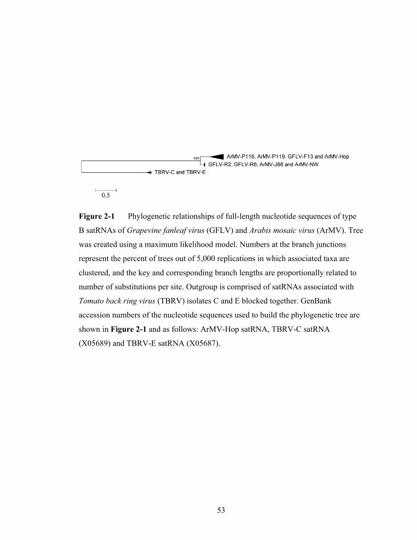
53
Figure 2-1 Phylogenetic relationships of full-length nucleotide sequences of type
B satRNAs of Grapevine fanleaf virus (GFLV) and Arabis mosaic virus (ArMV). Tree
was created using a maximum likelihood model. Numbers at the branch junctions
represent the percent of trees out of 5,000 replications in which associated taxa are
clustered, and the key and corresponding branch lengths are proportionally related to
number of substitutions per site. Outgroup is comprised of satRNAs associated with
Tomato back ring virus (TBRV) isolates C and E blocked together. GenBank
accession numbers of the nucleotide sequences used to build the phylogenetic tree are
shown in Figure 2-1 and as follows: ArMV-Hop satRNA, TBRV-C satRNA
(X05689) and TBRV-E satRNA (X05687).

54
microevolutionary history, no significant nonrandom distribution of sequence
polymorphisms was obtained for the satRNAs, indicating that the sequence
composition cannot be attributed to population expansion or decline. Tajima’s D was
significantly large for gene 2BMP, which could suggest population expansion or
balancing selection within ArMV and GFLV populations, although this result is likely
attributable to the prevalence of interspecies GFLV/ArMV recombinants within this
gene (Sokhandan-Bashir and Melcher, 2012).
In order to better understand the interspecific diversity of satRNAs, sequence
alignments were subjected to statistical tests for comparative relationships (Table 2-
3). KST*, which compares within-group diversity to total diversity (Hudson et al.,
1992), signaled genes 2BMP and 2CCP possess significant species subdivision, whereas
genes 1EPol and 2AHP and the satRNA do not. The nearest-neighbor sequence statistic
(Snn) (Hudson, 2000) showed that while genes 1EPol, 2AHP, 2BMP, and 2CCP are highly
likely to have their most closely related sequences from the same virus species, the
satRNAs nearest neighbors are less likely to be from the same virus species (Table 2-
3). The satRNA FST showed that only 4% of genetic variation is attributable to
whether it belongs to GFLV or ArMV, whereas other genes showed 36 to 63% of their
variation arising from their GFLV or ArMV identities. All together, three measures of
intra-versus interpopulation diversity showed the GFLV and ArMV satRNAs are not
differentiated based on the identity of their helper virus, which suggests no species-
specific identity for these satRNAs.
The ORFs of the GFLV satRNA from Lodi, CA correspond to a translation
product of 346 amino acids, compared with 341 amino acids for GFLV-F13 satRNA
55
(Hans et al., 1993), and 338 to 360 amino acids for ArMV satRNAs (Wetzel et al.,
2006). The GFLV-R2 and -R6 satRNA-encoded proteins have a predicted molecular
weight of 37kDa and an isoelectric point of 10.4, near the median of predictions for
other GFLV and ArMV satRNA-encoded peptides. The ratio of non-synonymous to
synonymous nucleotide substitutions is higher for the satRNA-encoded proteins than
for GFLV and ArMV proteins 1EPol, 2AHP, 2BMP, or 2CCP (Table 2-3), indicating the
satRNA undergoes less negative selection than gene counterparts in its helper viruses.
Twenty sites within the satRNA ORF were identified as having significant (P < 0.05)
evidence for negative selection and none of the sites in the satRNA ORF showed
significant (P < 0.05) evidence for positive selection. The fact that a large percentage
of satRNA protein failed to be identified as under selection, relative to genes present
on genomic RNAs, reflects the need for more satRNA sequences to gain a clearer
picture of which sites are under negative or positive selection.
Relationships of 5′-terminal nucleotide sequences of GFLV and ArMV satellite
and genomic RNAs
Close inspection of the 5′ UTR of the GFLV and ArMV satRNAs showed that they
possess greater identity to their helper viruses’ genomic RNAs than previously
realized. While it has been previously noted that the first nucleotides of the GFLV-F13
satRNA were nearly identical to those of nepovirus genomic RNAs (Fuchs et al.,
1991; Hans et al., 1993), it is apparent that the first 148 to 155 nts of the GFLV and
ArMV satRNAs show significant identity to subgroup A nepovirus genomic RNAs’ 5′
UTRs (Figure 2-2). The conservation is distributed across the first 137 to 191
56
nucleotides of GFLV, ArMV, or GDefV RNA1 or RNA2. The homologous area ends
approximately at the uracil-rich tract in the genomic RNAs (nt 225 in Figure 2-2).
Much of the homologous sequence is contained within the ORF of the satRNAs (nts
15 to 25 and beyond) and shows six highly conserved stretches (motifs II-VII) of 8 to
15 nts in addition to the first stretch of 12 conserved nucleotides (motif I) previously
described (9,13) (Figure 2-2). The base compositions of the satRNAs and genomic
RNAs are not significantly different before nt 226 in the alignment (Figure 2-2) (P =
0.471) but are different after this nucleotide position (P = 0.0014) according to χ2
analysis.
Passaging GFLV herbaceous hosts and curing the satRNA
Ten GFLV-infected grapevine leaf samples from Lodi, CA that contained a
satRNA were used to inoculate C. quinoa. Infected C. quinoa isolates were produced
from 6 of the 10 GFLV inocula, as shown by DAS-ELISA and characteristic vein
clearing in apical leaves. Multiplex RT-PCR for GFLV RNA2 and the satRNA was
performed using total RNA extracted from 10 infected C. quinoa. Inoculation with R6
resulted in two infected C. quinoa that differed in presence or absence of satRNA: R6-
40 contained a satRNA while R6-18 did not. After two additional passages through C.
quinoa, RT-PCR analysis confirmed presence or absence of the satRNA was
maintained (Figure 2-3). A partial GFLV RNA2 sequence was obtained for strains
R6-18 and R6-40 in infected C. quinoa tissue and isolate R6 in grape tissue (GenBank
accession number KC162001). The sequences, which include most of gene 2BMP, all
of gene 2CCP, and most of the 3′ UTR, are 95% identical to GFLV isolate CACSB3
(GenBank accession number GU972578), which is from the same vineyard in Lodi
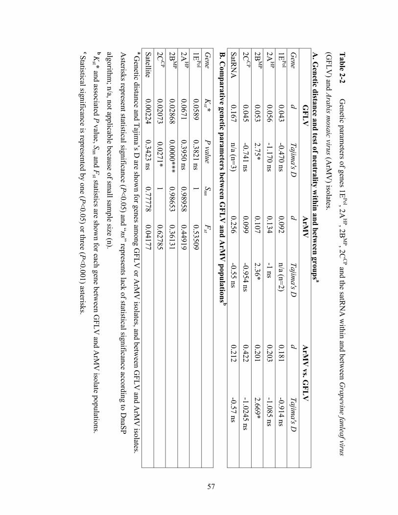
57
Table 2-2
Genetic param
eters of genes 1EPol, 2A
HP, 2B
MP, 2C
CP and the satR
NA
within and betw
een Grapevine fanleaf virus
(GFLV
) and Arabis mosaic virus (A
rMV
) isolates.
A. G
enetic distance and test of neutrality within and betw
een groups a
GFL
V
A
rMV
ArM
V vs. G
FLV
G
ene d
Tajima's D
d
Tajima's D
d
Tajima's D
1E
Pol 0.043
-0.470 ns 0.092
n/a (n=2) 0.181
-0.914 ns 2A
HP
0.056 -1.170 ns
0.134 -1 ns
0.203 -1.085 ns
2BM
P 0.053
2.75* 0.107
2.36*
0.201 2.669*
2CC
P 0.045
-0.741 ns 0.099
-0.954 ns 0.422
-1.0245 ns SatR
NA
0.167
n/a (n=3) 0.256
-0.55 ns 0.212
-0.57 ns
B. C
omparative genetic param
eters between G
FLV
and ArM
V populations b
Gene
Kst *
P value S
nn F
st
1E
Pol 0.0589
0.3821 ns 1
0.53509
2A
HP
0.0671 0.3950 ns
0.98958 0.44919
2BM
P 0.02868
0.0000*** 0.98653
0.36131
2C
CP
0.02073 0.0271*
1 0.62785
Satellite 0.00224
0.3423 ns 0.77778
0.04177 a G
enetic distance and Tajima’s D
are shown for genes am
ong GFLV
or ArM
V isolates, and betw
een GFLV
and ArM
V isolates.
Asterisks represent statistical significance (P<0.05) and “ns” represents lack of statistical significance according to D
naSP
algorithm; n/a, not applicable because of sm
all sample size (n).
b Kst * and associated P value, S
nn and Fst statistics are show
n for each gene between G
FLV and A
rMV
isolate populations. c Statistical significance is represented by one (P<0.05) or three (P<0.001) asterisks.

58
Table 2-3 Estim
ates of selection pressures on select subgroup A nepovirus proteins a
Gene
Log (L)
Mean
# Positive
% Positive
# Negative
% N
egative # N
eutral %
Neutral
1EPol
-12776.9
0.134241 0
0
228
27.67
596
72.33
2AH
P
-16684.9 0.291038
5
1.76
138
48.59
141
49.65
2BM
P
-16012.7 0.0430145
0
0
300
84.27
56
15.73
2CC
P
-39521.8 0.078302
2
0.396
437
86.53
66
13.07
satelliteP3
-5227.99 0.519967
0
0
20
6.15
305
93.85
a Norm
alized dN/dS ratios (m
ean) for each gene of GFLV
and ArM
V and log likelihood (L) scores for fitted m
odels. Num
ber and
percent of sites under positive, negative and neutral selection (P<0.05) are noted.
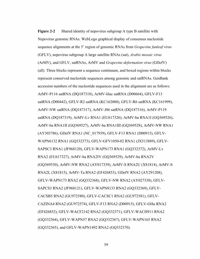
59
Figure 2-2 Shared identity of nepovirus subgroup A type B satellite with
Nepovirus genomic RNAs. WebLogo graphical display of consensus nucleotide
sequence alignments at the 5’ region of genomic RNAs from Grapevine fanleaf virus
(GFLV), nepovirus subgroup A large satellite RNAs (sat), Arabis mosaic virus
(ArMV), and GFLV, satRNAs, ArMV and Grapevine deformation virus (GDefV)
(all). Three blocks represent a sequence continuum, and boxed regions within blocks
represent conserved nucleotide sequences among genomic and satRNAs. GenBank
accession numbers of the nucleotide sequences used in the alignment are as follows:
ArMV-P116 satRNA (DQ187318), ArMV-lilac satRNA (D00664), GFLV-F13
satRNA (D00442), GFLV-R2 satRNA (KC162000), GFLV-R6 satRNA (KC161999),
ArMV-NW satRNA (DQ187317), ArMV-J86 satRNA (DQ187316), ArMV-P119
satRNA (DQ187319), ArMV-Lv RNA1 (EU617326), ArMV-ba RNA1I (GQ369526),
ArMV-ba RNA1II (GQ369527), ArMV-ba RNA1III (GQ369528), ArMV-NW RNA1
(AY303786), GDefV RNA1 (NC_017939), GFLV-F13 RNA1 (D00915), GFLV-
WAPN6132 RNA1 (GQ332373), GFLV-GFV1050-02 RNA1 (JX513889), GFLV-
SAPSC3 RNA1 (JF968120), GFLV-WAPN173 RNA1 (GQ332372), ArMV-Lv
RNA2 (EU617327), ArMV-ba RNA2IV (GQ369529), ArMV-ba RNA2V
(GQ369530), ArMV-NW RNA2 (AY017339), ArMV-S RNA2U (X81814), ArMV-S
RNA2L (X81815), ArMV-Ta RNA2 (EF426853), GDefV RNA2 (AY291208),
GFLV-WAPN173 RNA2 (GQ332368), GFLV-NW RNA2 (AY027338), GFLV-
SAPCS3 RNA2 (JF968121), GFLV-WAPN8133 RNA2 (GQ332369), GFLV-
CACSB5 RNA2 (GU972580), GFLV-CACSC1 RNA2 (GU972581), GFLV-
CAZINA4 RNA2 (GU972574), GFLV-F13 RNA2 (D00915), GFLV-GHu RNA2
(EF426852), GFLV-WACF2142 RNA2 (GQ332371), GFLV-WACH911 RNA2
(GQ332364), GFLV-WAPN57 RNA2 (GQ332367), GFLV-WAPN165 RNA2
(GQ332365), and GFLV-WAPN1492 RNA2 (GQ332370).
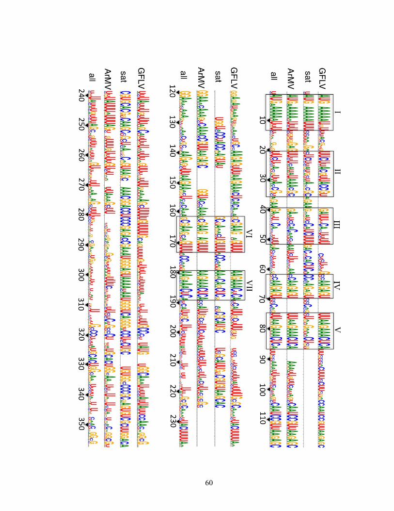
60

61
CA (Oliver et al., 2011). The GFLV-R6-18 and GFLV-R6-40 RNA2 sequences were
99.6% identical at the nucleotide level, which showed that the two strains established
on C. quinoa were nearly identical except for the presence of absence of the satRNA,
and these strains also showed similar identity to GFLV from the grapevine source
from which they originated. Similarly, cDNAs of viral RNA progeny derived from
F13 transcripts show 99.8% nucleotide identity to the wild-type GFLV-F13. Thus, two
sets of GFLV sister strains (R6-40 and R6-18, and F13 and FF) differing in presence
or absence of satRNAs were created and these were used to characterize the biological
effect of the satRNA.
Effect of the satRNA on GFLV accumulation and symptoms in C. quinoa
The GFLV-F13 and GFLV-R6 satRNAs were used to determine whether a
satRNA has a measurable impact on its helper virus or on the systemic host C. quinoa.
Viral protein accumulation was measured by DAS-ELISA at four, seven, 13, and 20
dpi (Figure 2-4). The effect of time point was significant (F = 105.03, P < 0.0001),
but effect of the virus strain was not (F = 1.30, P = 0.2825), nor was the interaction of
virus strain and time point (F = 0.76, P = 0.6559). GFLV capsid accumulation
increased successively at four, seven and 13 dpi, and these differences were
statistically significant (P < 0.05). Virus accumulation at 20 dpi was not significantly
different from 13 dpi but was significantly different from all other time points.
Presence or absence of the satRNA did not significantly impact viral protein
accumulation overall or at any time point. This indicates the GFLV satRNA did not
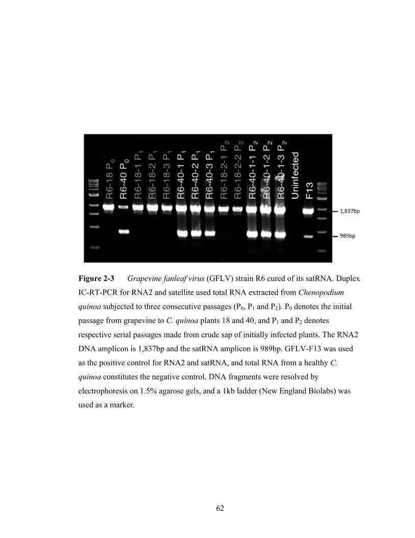
62
Figure 2-3 Grapevine fanleaf virus (GFLV) strain R6 cured of its satRNA. Duplex
IC-RT-PCR for RNA2 and satellite used total RNA extracted from Chenopodium
quinoa subjected to three consecutive passages (P0, P1 and P2). P0 denotes the initial
passage from grapevine to C. quinoa plants 18 and 40, and P1 and P2 denotes
respective serial passages made from crude sap of initially infected plants. The RNA2
DNA amplicon is 1,837bp and the satRNA amplicon is 989bp. GFLV-F13 was used
as the positive control for RNA2 and satRNA, and total RNA from a healthy C.
quinoa constitutes the negative control. DNA fragments were resolved by
electrophoresis on 1.5% agarose gels, and a 1kb ladder (New England Biolabs) was
used as a marker.
63
discernably change the competitive host–virus interaction.
The four GFLV strains were inoculated to C. quinoa and symptoms were
recorded at multiple time points after which plant physiological measurements were
taken. Strains produced significant differences for each measurement including
symptom severity over time (AUDPC) (F = 31.07, P < 0.0001), height (F = 4.68, P =
0.0073), above-ground fresh weight (F = 7.93, P=0.0003), and above-ground dry
weight (F = 3.88, P = 0.0168). The GFLV genomic RNAs had a striking impact on
symptoms (Figure 2-5), though not necessarily on whole-plant physiology (Figure 2-
6), and the satRNA had no striking impact on either. No significant differences in
AUDPC were detected between GFLV strains FF and F13, or between GFLV strains
R6-18 and R6-40, but each strain of the F13 background was significantly different
from each strain of the R6 background (Figure 2-5). This author detected significant
differences between GFLV strains for three plant physiological parameters including
height, dry weight and fresh weight. The only statistically significant physiological
difference observed between sister strains was that fresh weight of FF was higher than
that of F13, as well as R6-18 and R6-40 (Figure 2-6). Except that FF- infected plants
produced higher fresh weight than F13-infected plants, satRNAs did not have a
significant measurable impact on C. quinoa symptoms or plant physiology.
DISCUSSION
GFLV satRNAs that are genetically similar were characterized by IC-RT-PCR
and sequencing from vines in a naturally infected vineyard in Lodi, CA, as well as a
vine in a USDA-PGRU grape germplasm collection in Geneva, NY. Based on the
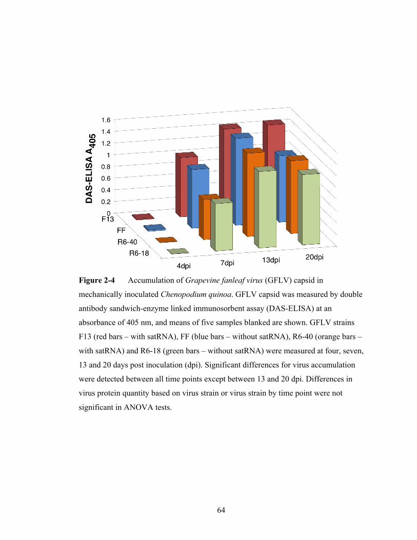
64
Figure 2-4 Accumulation of Grapevine fanleaf virus (GFLV) capsid in
mechanically inoculated Chenopodium quinoa. GFLV capsid was measured by double
antibody sandwich-enzyme linked immunosorbent assay (DAS-ELISA) at an
absorbance of 405 nm, and means of five samples blanked are shown. GFLV strains
F13 (red bars – with satRNA), FF (blue bars – without satRNA), R6-40 (orange bars –
with satRNA) and R6-18 (green bars – without satRNA) were measured at four, seven,
13 and 20 days post inoculation (dpi). Significant differences for virus accumulation
were detected between all time points except between 13 and 20 dpi. Differences in
virus protein quantity based on virus strain or virus strain by time point were not
significant in ANOVA tests.
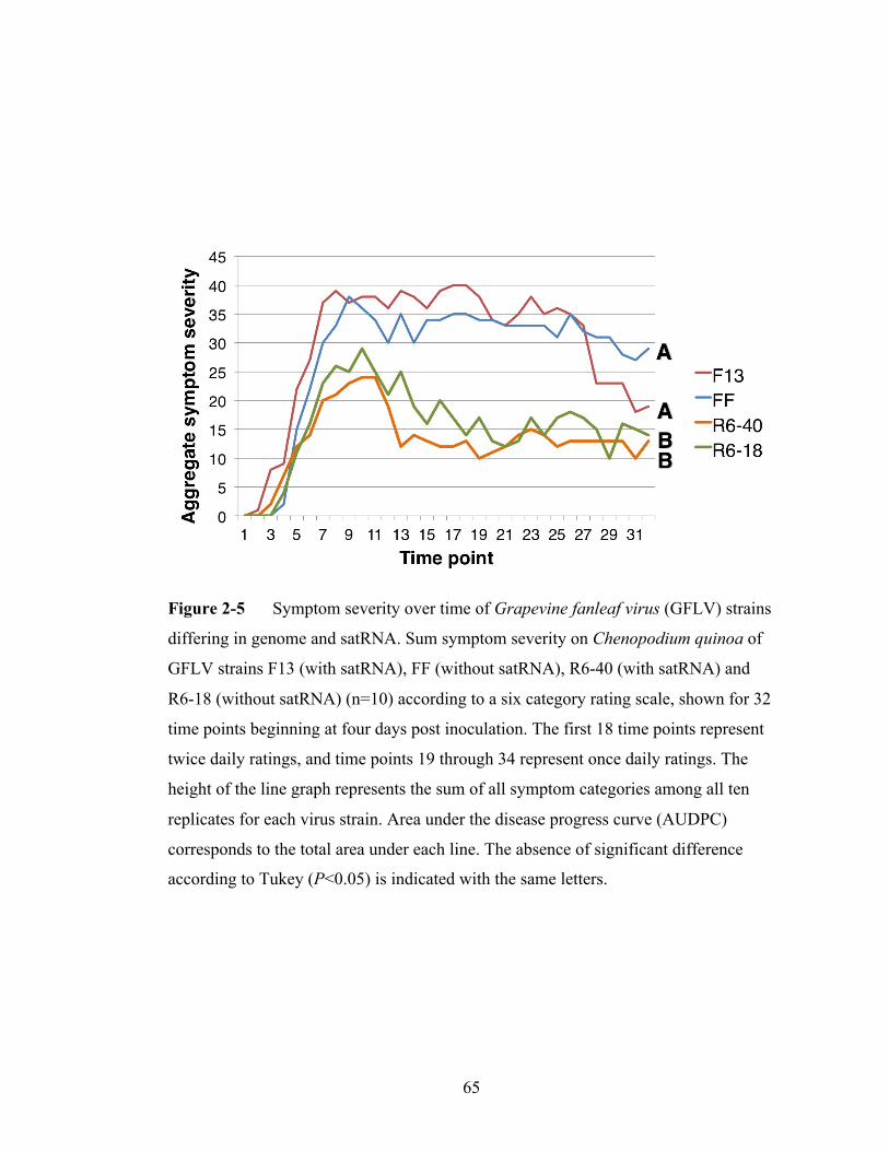
65
Figure 2-5 Symptom severity over time of Grapevine fanleaf virus (GFLV) strains
differing in genome and satRNA. Sum symptom severity on Chenopodium quinoa of
GFLV strains F13 (with satRNA), FF (without satRNA), R6-40 (with satRNA) and
R6-18 (without satRNA) (n=10) according to a six category rating scale, shown for 32
time points beginning at four days post inoculation. The first 18 time points represent
twice daily ratings, and time points 19 through 34 represent once daily ratings. The
height of the line graph represents the sum of all symptom categories among all ten
replicates for each virus strain. Area under the disease progress curve (AUDPC)
corresponds to the total area under each line. The absence of significant difference
according to Tukey (P<0.05) is indicated with the same letters.
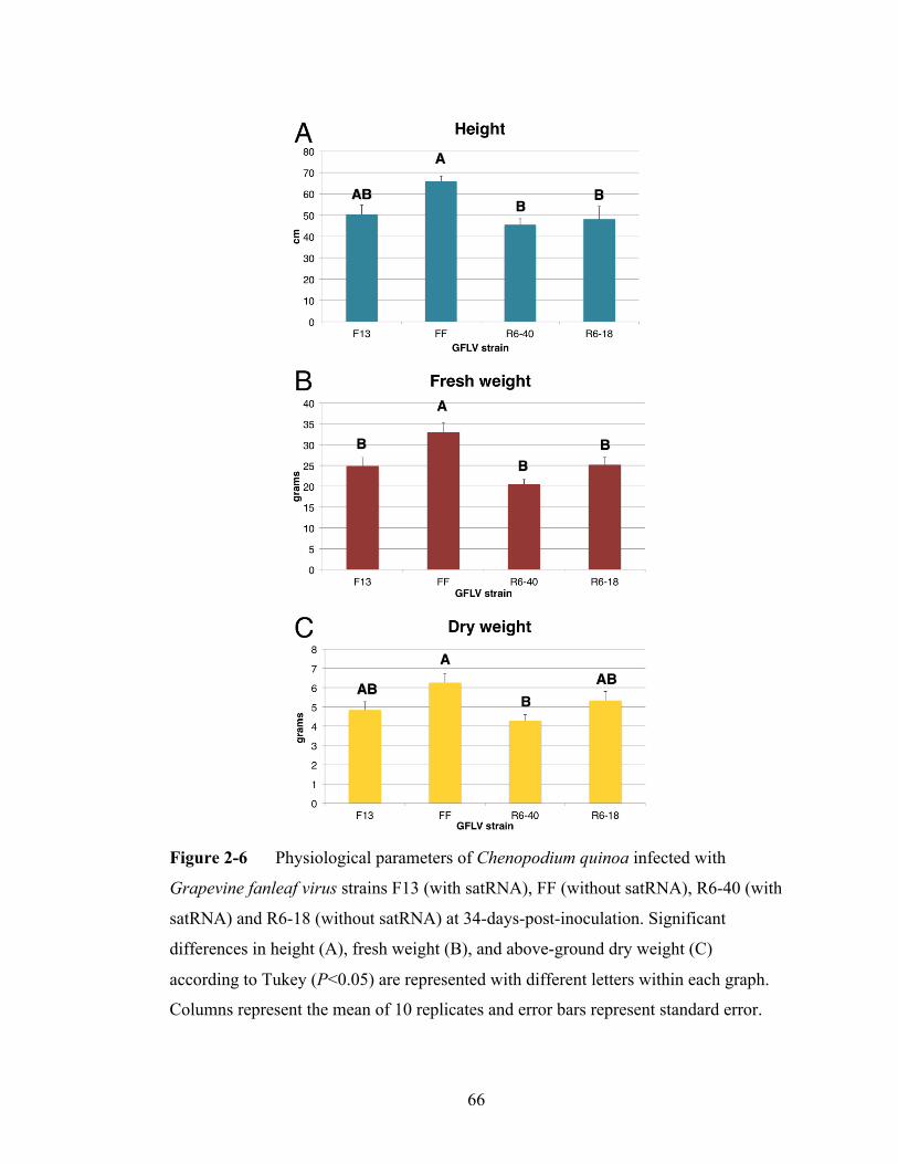
66
Figure 2-6 Physiological parameters of Chenopodium quinoa infected with
Grapevine fanleaf virus strains F13 (with satRNA), FF (without satRNA), R6-40 (with
satRNA) and R6-18 (without satRNA) at 34-days-post-inoculation. Significant
differences in height (A), fresh weight (B), and above-ground dry weight (C)
according to Tukey (P<0.05) are represented with different letters within each graph.
Columns represent the mean of 10 replicates and error bars represent standard error.
67
discovery of a very similar satRNA from disparate sources, records related to the
USDA-PGRU accession were searched to attempt to trace the natural history of this
satRNA. The GFLV- and satRNA-containing vine at USDA-PGRU (accession 106) is
a selection of ‘Grande Glabre’, which was introduced to the United States from the
Germplasm Repository in Bordeaux, France in 1962, kept at the USDA-Animal Plant
Health Inspection Service Plant Pest Quarantine station in Beltsville, MD, for 5 years
prior to introduction to the repository in Geneva, NY. Based on these records, PGRU
accession 106 was likely infected at the time of introduction because GFLV and
Xiphinema index, its ectoparasitic nematode vector (Andret-Link et al., 2004), do not
naturally occur in the eastern United States, including New York and Maryland, and
pollen-transmission of GFLV is not known. The nucleotide sequence similarities
suggest the satRNAs in Lodi, CA may share very recent ancestry with the satRNA that
can be traced back to Bordeaux, France in the early 1960s.
Field and laboratory observations support the hypothesis that the helper virus
may easily dispense of its satRNA during plant-to-plant transmission. The distribution
of the satRNA among GFLV-infected vines in the naturally infected vineyard in Lodi,
CA showed that the satRNA was absent from some GFLV-infected plants adjacent to
others that contain the satRNA. Similarly, mechanical inoculation of crude sap from
GFLV-infected grape leaves containing the satRNA resulted in a GFLV-infected plant
that did not contain a satRNA (R6-18). This information suggests that plant-to-plant
transmission in field and laboratory settings can result in loss of the satRNA.
Conversely, detection of a satRNA in PGRU-106, which could only have been
infected along with GFLV over 50 years prior, shows the satRNA can be stably
68
maintained like its helper virus.
This author did not detect an association between GFLV symptoms in a
vineyard with presence of the satRNA. Saldarelli et al. (1993) also reported no
association between the satRNA and GFLV symptoms in diverse grapevine cultivars.
Neither my study, nor Saldarelli et al. (1993) evaluated the effect of the satRNA on
symptoms and virus accumulation in a common grapevine cultivar. This author tested
for satRNA-induced symptom effects in a systemic host of GFLV, C. quinoa, but
could not discern much impact of two diverse GFLV satRNAs on virus accumulation
or symptoms. This was in contrast to a previous experiment showing the association of
the ArMV-Lilac satRNA with an amelioration of symptoms in C. quinoa (Liu et al.,
1991b). Symptom measurements on C. quinoa showed greater differences between the
two GFLV sister strains selected for this study than differences within GFLV sister
strains differing in presence or absence of diverse satRNAs. Together, my data
suggests the satRNA has a commensal relationship with GFLV and C. quinoa.
The origins of satRNAs are generally not known, but they may originate by
chance recombination of viral and/or host nucleic acids (Simon et al., 2004). The type
B satRNAs associated with subgroup A nepoviruses have substantial identity with
genomic RNAs of the helper virus in their first 150 nucleotides. Based on this
extended similarity, a recombination event likely occurred between the 5′ UTR of an
ancestral subgroup A nepovirus RNA and another unidentified RNA with a putative
break point at nucleotide 150. Given that previous analyses of in vitro and in vivo
translation products have demonstrated expression of the full-length satRNA-encoded
protein (Hans et al., 1992; Liu and Cooper, 1993; Moser et al., 1992), the

69
recombination event appears to have given rise to satRNAs encoding a protein
composed of an amino terminal region for which the corresponding nucleotide
sequence is of a helper virus’s 5′ UTR. Because of an extended sequence relationship
of this 5′ satRNA region specifically with ArMV, GFLV or GDefV, and because of
the promiscuity of these satRNA associations, it appears satRNAs might have arisen in
a common ancestor of these virus species and diversified in a non-helper virus specific
fashion. Currently, there is no nucleotide or amino acid sequence available in
GenBank that shows continuous identity to the rest of the satRNA sequence beyond
the first 150 nucleotides; therefore, the other parental RNA sequence remains
unknown.
The evidence that satRNAs have diversified in a helper virus independent
manner suggests certain conserved elements of the satRNA must be requisite for
functionality in both GFLV and ArMV. Although the protein encoded by the satRNA
is indispensable for satRNA replication by a GFLV or an ArMV helper (Hans et al.,
1993; Liu and Cooper, 1993), the satRNA protein apparently undergoes less negative
selection than other nepovirus genes. The conservation of nepovirus satRNAs and
genomic RNAs 5′ ends well within the satRNA ORF suggests RNA replication
determinants could be maintained within the satRNA 5′ UTR and 5′ end of its ORF.
Like nepovirus satRNAs, the satellite RNA of Bamboo mosaic virus (satBaMV)
possesses similarity to its helper virus in the 5′ untranslated region (Lin and Hsu,
1994). This similarity is biologically significant since the BaMV RdRp specifically
interacts with cis-acting elements in the satBaMV to initiate replication (Huang et al.,
2010), and mutagenic analyses suggest this could also be true for nepovirus satRNAs

70
(Hans et al., 1993; Hemmer et al., 1993; Liu et al., 1991a).
71
REFERENCES
Alabi, O. J., Rwahnih, Al, M., Karthikeyan, G., Poojari, S., Fuchs, M., Rowhani, A.,
and Naidu, R. A. 2011. Grapevine leafroll-associated virus 1 occurs as genetically
diverse populations. Phytopathology. 101:1446–1456.
Andret-Link, P., Laporte, C., Valat, L., Ritzenthaler, C., Demangeat, G., Vigne, E.,
Laval, V., Pfeiffer, P., Stussi-Garaud, C., and Fuchs, M. 2004. Grapevine fanleaf
virus: still a major threat to the grapevine industry. Journal of Plant Pathology.
86:183–195.
Collmer, C. W., and Howell, S. H. 1992. Role of satellite RNA in the expression of
symptoms caused by plant viruses. Annual Review of Phytopathology. 30:419–442.
Crooks, G. E., Hon, G., Chandonia, J. M., and Brenner, S.E. 2004. WebLogo: a
sequence logo generator. Genome Research. 14:1188-1190.
Edgar, R.C. 2004. MUSCLE: multiple sequence alignment with high accuracy and
high throughput. Nucleic Acids Research. 32:1792-1797.
Elbeaino, T., Digiaro, M., Ghebremeskel, S., and Martelli, G. P. 2012. Grapevine
deformation virus: completion of the sequence and evidence on its origin from
recombination events between Grapevine fanleaf virus and Arabis mosaic virus. Virus
Research. 166:136–140.
Fritsch, C., and Mayo, M. 1993. Properties of the satellite RNA of nepoviruses.
Biochimie. 75:561–567.
72
Fuchs, M., Pinck, M., Etienne, L., Pinck, L., and Walter, B. 1991. Characterization
and detection of Grapevine fanleaf virus by using cDNA probes. Phytopathology.
81:559–565.
Fuchs, M., Pinck, M., Serghini, M. A., Ravelonandro, M., Walter, B., and Pinck, L.
1989. The nucleotide sequence of satellite RNA in Grapevine fanleaf virus, strain F13.
Journal of General Virology. 70:955–962.
Ghanem-Sabanadzovic, N. A., Sabanadzovic, S., Digiaro, M., and Martelli, G. P.
2005. Complete nucleotide sequence of the RNA-2 of Grapevine deformation and
Grapevine Anatolian ringspot viruses. Virus Genes. 3:335-340.
Gouy, M., Guindon, S., and Gascuel., O. 2010. SeaView version 4: a multiplatform
graphical user interface for sequence alignment and phylogenetic tree building.
Molecular Biology and Evolution. 27:221-224.
Hans, F., Fuchs, M., and Pinck, L. 1992. Replication of Grapevine fanleaf virus
satellite RNA transcripts in Chenopodium quinoa protoplasts. Journal of General
Virology. 73:2517–2523.
Hans, F., Pinck, M., and Pinck, L. 1993. Location of the replication determinants of
the satellite RNA associated with Grapevine fanleaf nepovirus (strain-F13).
Biochimie. 75:597–603.
Hemmer, O., Oncino, C., and Fritsch, C. 1993. Efficient replication of the in vitro
transcripts from cloned cDNA of Tomato black ring virus satellite RNA requires the
48K satellite RNA-encoded protein. Virology. 194:800–806.

73
Huang, Y.-W., Hu, C.-C., Lin, N.-S., and Hsu, Y.-H. 2010. Mimicry of molecular
pretenders. RNA Biology. 7:1–10.
Hudson, R. R. 2000. A new statistic for detecting genetic differentiation. Genetics.
155:2011–2014.
Hudson, R. R., Boos, D. D., and Kaplan, N. L. 1992. A statistical test for detecting
geographic subdivision. Molecular Biology and Evolution. 9:138–151.
Jeger, M. J., and Viljanen-Rollinson, S. L. H. 2001. The use of the area under the
disease-progress curve (AUDPC) to assess quantitative disease resistance in crop
cultivars. Theoretical and Applied Genetics. 102:32–40.
Kosakovsky Pond, S. L., and Frost, S. D. W. 2005. DataMonkey: rapid detection of
selective pressure on individual sites of codon alignments. Bioinformatics. 21:2531–
2533.
Lamprecht, R. L., Maree, H., Stephan, D., Wetzel, T. and Burger, J. T. 2012.
Molecular characterization of South African isolates of Grapevine fanleaf virus and an
associated new satRNA. Pages 36-37 in: Proc. 17th Congress of the International
Council for the Study of Virus and Virus-like Diseases of the Grapevine. Davis, CA,
USA, Oct. 7-14.
Larkin, M. A., Blackshields, G., Brown, N. P., Chenna, R., McGettigan, P. A.,
McWilliam, H., Valentin, F., Wallace, I. M., Wilm, A., Lopez, R., Thompson, J. D.,
Gibson, T. J., and Higgins, D. G. 2007. Clustal W and Clustal X version 2.0.
Bioinformatics. 23:2947–2948.

74
Librado, P., and Rozas, J. 2009. DnaSP v5: a software for comprehensive analysis of
DNA polymorphism data. Bioinformatics. 25:1451–1452.
Lin, N. S., and Hsu, Y. H. 1994. A satRNA associated with Bamboo mosaic
potexvirus. Virology. 202:707-714.
Liu, Y. L., Cooper, J. I., Coates, D., and Bauer, G. 1991(a). Biologically active
transcripts of a large satellite RNA from Arabis mosaic nepovirus and the importance
of 5′ end sequences for its replication. The Journal of General Virology. 72:2867–
2874.
Liu, Y. L., Cooper, J. I., Coates, D., and Bauer, G. 1991(b). Biologically active
transcripts of a large satRNA from Arabis mosaic nepovirus and the importance of 5′
end sequences for its replication. Journal of General Virology. 72:2867-2874.
Liu, Y. Y., and Cooper, J. I. 1993. The multiplication in plants of Arabis mosaic virus
satellite RNA requires the encoded protein. Journal of General Virology. 74:1471–
1474.
Liu, Y. Y., Hellen, C. U. T., Cooper, J. I., Bertioli, D. J., Coates, D., and Bauer, G.
1990. The nucleotide sequence of a satellite RNA associated with Arabis mosaic
nepovirus. Journal of General Virology. 71:1259–1263.
Martelli, G.P., and E. Boudon-Padieu. 2006. Directory of infectious diseases of
grapevine and viruses and virus-like diseases of the grapevine. Option Médit. Série B,
Studies and Research 55:35-50.
75
Mayo, M. A. 1991. Satellites. Pages 400–402 in: Classification and nomenclature of
viruses. Fifth report of the International Committee on Taxonomy of Viruses. R.I.B.
Francki, C.M. Fauquet, D.L. Knudson, and F. Brown, eds. Archives of Virology.
S2:400-402.
Moser, O., Fuchs, M., Pinck, L., and Stussi-Garaud, C. 1992. Immunodetection of
grapevine fanleaf virus satellite RNA-encoded protein in infected Chenopodium
quinoa. Journal of General Virology. 73:3033–3038.
Oliver, J. E., Vigne, E., and Fuchs, M. 2010. Genetic structure and molecular
variability of Grapevine fanleaf virus populations. Virus Research. 152:30–40.
Oncino, C., Hemmer, O., and Fritsch, C. 1995. Specificity in the association of
Tomato black ring virus satellite RNA with helper virus. Virology. 213:87–96.
Pinck, L., Fuchs, M., Pinck, M., Ravelonandro, M., and Walter, B. 1988. A satellite
RNA in Grapevine fanleaf virus strain F13. Journal of General Virology. 69:233–239.
Roosinck, M.J., Sleat, D. and Palukaitis, P. 1992. Satellite RNAs of plant viruses:
structures and biological effects. Microbiology Review. 56: 265-279.
Saldarelli, P., Minafra, A., and Walter, B. 1993. A survey of Grapevine fanleaf
nepovirus isolates for the presence of satellite RNA. Vitis. 32:99–102.
Sanfaçon, H., Wellink, J., Gall, O., Karasev, A., Vlugt, R., and Wetzel, T. 2009.
Secoviridae: a proposed family of plant viruses within the order Picornavirales that
combines the families Sequiviridae and Comoviridae, the unassigned genera
Cheravirus and Sadwavirus, and the proposed genus Torradovirus. Archives of
Virology. 154:899–907.
76
Simon, A., Roossinck, M., and Havelda, Z. 2004. Plant virus satellite and defective
interfering RNAs: New paradigms for a new century. Annual Review of
Phytopathology. 42:415–437.
Sokhandan-Bashir, N., and Melcher, U. 2012. Population genetic analysis of
Grapevine fanleaf virus. Archives of Virology. 157:1919–1929.
Tamura, K., Peterson, D., Peterson, N., Stecher, G., Nei, M., and Kumar, S. 2011.
MEGA5: molecular evolutionary genetics analysis using maximum likelihood,
evolutionary distance, and maximum parsimony methods. Molecular Biology and
Evolution. 28:2731–2739.
Vigne, E., Komar, V., and Fuchs, M. 2004. Field safety assessment of recombination
in transgenic grapevines expressing the coat protein of Grapevine fanleaf virus.
Transgenic Research. 13:165–179.
Viry, M., Serghini, M. A., Hans, F., Ritzenthaler, C., Pinck, M., and Pinck, L. 1993.
Biologically active transcripts from cloned cDNA of genomic Grapevine fanleaf
nepovirus RNAs. Journal of General Virology. 74:169–174.
Wetzel, T., Bassler, A., Amren, M. A. W., and Krczal, G. 2006. A RT/PCR-partial
restriction enzymatic mapping (PREM) method for the molecular characterisation of
the large satellite RNAs of Arabis mosaic virus isolates. Journal of Virology Methods.
132:97–10.

77
CHAPTER 3
A VIRAL VECTOR COMPOSED OF GRAPEVINE FANLEAF VIRUS
ABSTRACT
Versatile and highly effective tools would be useful for Vitis spp. (grapevine)
functional genomics. To this aim, a viral vector capable of stable heterologous protein
expression and gene silencing was developed from Grapevine fanleaf virus (GFLV), a
member of the genus Nepovirus, family Secoviridae. This virus has a bipartite RNA
genome, accumulates to high levels in grape berries, and has no recognized tissue
tropism. The GFLV vector is from two attenuated, wild type strains or assortants
derived thereof and is deficient in transmission by the ectoparasitic nematode
Xiphinema index because of targeted coat protein mutations. The GFLV vector can be
modified by insertion of heterologous genetic material and delivered to plants via
agroinfiltration. The GFLV vector exhibits stable expression of Red fluorescent
protein following four serial passages and gene silencing capabilities equivalent to
Tobacco rattle virus, a standard viral vector in herbaceous hosts. The GFLV vector
can conduct spatially phased expression of enhanced Green fluorescent protein and
Red fluorescent protein when tagged to each of its genomic RNAs. The versatility,
effectiveness and biosafety of this grapevine viral vector make it a good candidate
platform for grapevine functional genomics research.

78
INTRODUCTION
Viral vectors have emerged as key tools for functional genomics in plant
biology. Application of viral vectors is a popular approach to validate activity of
candidate genes without resorting to cumbersome and time-consuming stable
transformation procedures (Vaghchhipawala et al., 2010; Senthil-Kumar and Mysore,
2011). Despite their promise, viral vectors sometimes have limitations such as the
quantity of foreign genetic material they can stably support (Arazi et al., 2001;
Avesani et al., 2007), a lack of systemic infectivity (Liu et al., 2009) or lack a
seamless one-step inoculation method (Satyanarayana et al., 2001, Yoon et al., 2011;
Agüero et al., 2012). Furthermore, only few viral vectors have been extensively
validated in woody crops (Dawson and Folimonova, 2013).
Vitis vinifera is one of the most ancient and most valuable horticultural crops
(Myles et al., 2011; Reisch, 2011). Coupling the availability of genome sequences
(Jaillon et al., 2007; Velasco et al., 2007; Adam-Blondon et al., 2011) with numerous
phenotypes such as berry flavor and stress resistance, there is growing interest in
grapevine functional genomics. A lack of rapid and scalable reverse genetics tools
hinders functional grapevine gene characterization. Existing platforms for grapevine
functional gene characterization encompass (i) stable transformation, which takes
eighteen months to three years and is difficult to master (Iocco et al., 2001), (ii) ‘hairy
root’ (Agrobacterium rhizogenes) transformation, which is in grapevine is prone to
intra-plant expression variability and restricted to roots (Yang et al., 2013), and (iii)
transient agroinfiltration assays, which can be cultivar specific and inappropriate for
some tissues such as roots or grape berries (e.g. Santos-Rosa et al., 2008; Bertazzon et
79
al., 2011; Visser et al., 2012). Given the limitations in grapevine functional genomics
platforms, very few grapevine genes have been functionally characterized in planta
(Martínez-Zapater et al., 2011; Romieu et al., 2011; Tillett et al., 2011). Instead,
researchers have generally relied on herbaceous substitutes and in vitro biochemical
approaches to elucidate the function of candidate genes (Battilana et al., 2011; Mejía
et al, 2013; Nicolas et al., 2013). A robust functional genomics platform for grapevine
would greatly enhance existing research tools and facilitate improvement of this high
value specialty crop.
A handful of viral vectors have been developed for economically important
woody plants, including stone fruits, citrus and grapevine (Dawson and Folimonova,
2013). For grapevine, Grapevine virus A (GVA) (Muruganantham et al. 2009),
Grapevine rupestris stem pitting-associated virus (GRSPaV) (Meng et al., 2013) and
Grapevine leafroll-associated virus 2 (GLRaV-2) (Kurth et al., 2012) have been
engineered to be viral vectors. GVA, GRSPaV and GLRaV-2 have monopartite RNA
genomes and are restricted to phloem tissue. In addition, GVA and GRSPaV vectors
lack stability or infectivity (Dawson and Folimonova, 2013; Meng et al., 2013).
Grapevine fanleaf virus (GFLV) is one of the 64 recognized viruses of
grapevine (Andret-Link et al., 2004, Martelli, 2014) In contrast to GVA, GRSPaV and
GLRaV-2, GFLV is not phloem restricted and has a bipartite RNA genome (Figure 3-
1A). RNA1 codes for proteins involved in replication and polyprotein maturation, and
RNA2 codes for proteins involved in RNA2 replication, virus movement and RNA
encapsidation (Figure 3-1A). Both genomic RNA species are necessary for systemic
infection (Andret-Link et al., 2004). Here, we report the engineering of GFLV into a

80
Figure 3-1 Schematic illustration of the Grapevine fanleaf virus (GFLV) genome
and vectors. A) GFLV is composed of RNA1 and RNA2 with a 5’ genome-linked
protein and a 3’ poly-A tail. Polyproteins (boxes) are cleaved by the GFLV proteinase
(1DPro) into individual protein components (annotated). The polyprotein intervenes 5’
and 3’ untranslated region (UTRs) represented as horizontal lines. B) GFLV cDNAs
were placed downstream of a Cauliflower mosaic virus (CaMV) 35S promoter and
into binary vectors. Terminal sequences to the GFLV cDNA include a CaMV 35S
terminator (RNA1, RNA2, RNA2-EGFP and RNA2-pds) or a CaMV polyadenylation
signal (RNA1-EGFP, RNA1-RFP and RNA2-RFP). Sequences of enhanced Green
fluorescent protein (EGFP), Red fluorescent protein (RFP) or a partial Phytoene

81
desaturase (pds) from Nicotiana benthamiana were inserted within the GFLV cDNAs
of RNA1 or RNA2 as shown. Right border (RB) and left border (LB) components of
the binary vector are shown. RNA2, RNA2-EGFP and RNA2-pds contain an
expression cassette for the 2b silencing suppressor of Cucumber mosaic virus. The
vector component derived from RNA2 was modified to contain a multiple cloning site
(MCS) composed of unique BsiWI and Mlu1 restriction sites, a synthetic polyprotein
cleavage site intervening the MCS and 2BMP and a mutation in the coat protein coding
region for abolition of transmissibility by the ectoparasitic nematode Xiphinema index,
except for RNA2-RFP. Heterologous sequences were inserted into RNA1 or RNA2 as
shown.
82
viral vector for grapevine functional genomics. Infectious cDNA clones of the
genomic RNAs (Viry et al., 1993; Vigne et al., 2013) from two mild GFLV strains
(Huss et al., 1989; Legin et al., 1993) were placed under the control of the Cauliflower
mosaic virus 35S promoter (35S promoter) and delivered to plants using
Agrobacterium tumefaciens (agroinoculation) to establish infection. The GFLV vector
can be used to stably express foreign genetic material, silence endogenous host genes
and co-express two heterologous genes. Additionally, the transmission of the GFLV
vector by the ectoparasitic nematode Xiphinema index was abolished by directed
mutagenesis of the coat protein-coding region. The GFLV vector technology furnishes
a reliable, versatile and biosafe tool to conduct functional genomics research in
grapevine.
MATERIALS AND METHODS
Plant material and inoculations
N. benthamiana was grown in 10cm x 10cm pots in a greenhouse with a 16/8
hr. photoperiod at 24°C ± 3°C. Greenhouse experiments were conducted during fall,
lighting was supplemented by high pressure sodium lamps and temperature was
automatically controlled by steam-heated radiators and fans, and by ventilator,
convection and evaporative cooling systems. Plants were grown in Cornell Mix
supplemented with 10-10-10 (N/P/K) fertilizer weekly. GFLV-free V. vinifera cv.
Cabernet Sauvignon were micropropagated in woody plant medium supplemented
with 37 mgL-1 cysteine contained in polyethylene Star*Pac® bags (Phytotechnology
laboratories, Shawnee Mission, KS) with a 16-hr photoperiod at 25 ± 2°C. Grapevines

83
were agroinoculated at four weeks post-rooting and acclimatized in the greenhouse
(Alzubi et al., 2012). GFLV-infected grapevines used for virus quantification were
accessed at the USDA Cold Hardy Grapevine Plant Genetics Resource Unit in
Geneva, NY. These were the interspecific hybrid Landot noir (one vine) and the
rootstock Vitis rupestris cv. Alphonse de Serres (two vines). Ten clusters were
sampled per vine and tissues from 10 to 20 berries from single clusters were pooled
prior to tissue isolation.
All A. tumefaciens cultures were grown and inoculated according to the
protocol of Vaghchhipawala et al. (2010). Individual cultures containing plasmids
with GFLV RNA1 or RNA2 cDNA components and silencing suppressors 2b of
Cucumber mosaic virus (Choi et al., 2008) or p24 of GLRaV-2 (Chiba et al., 2006;
Vigne et al., 2013) were mixed and inoculated in equal parts. Leaves of three week-
old N. benthamiana plants were syringe-infiltrated and grapevine plantlets sonicated
for one minute before vacuum infiltration for seven minutes at approximately 90kPa.
Mechanical inoculation of GFLV crude sap in N. benthamiana and C. quinoa was
conducted as described (Gottula et al., 2013).
Nucleic acid and microbiology procedures
RNA extraction was accomplished with RN-EZ plant RNA isolation kit from
Omega Biotek (Doraville, GA) and reverse transcription and amplification reactions
were accomplished with Qiagen One-Step RT-PCR kit according to manufacturer’s
protocols (Hilden, Germany). Other enzymes and protocols including Phusion DNA
polymerase, restriction enzymes and T4 ligase were provided by New England

84
Biolabs (Ipswich, MA). Primers (Table 3-1) were provided by Integrated DNA
Technologies (Coralville, IA). GFLV cDNAs from which infectious in vitro
transcripts were derived (Viry et al., 1993; Vigne et al., 2013) were cloned into CaMV
35S expression cassettes in binary vectors mobilized into A. tumefaciens strains
GV3101 or C58Z707 via electroporation. RFP was derived from pTagRFP-C
(Evrogen) and EGFP was amplified from pEGFP (Clontech). All clones were
sequenced (Sanger method) at the Cornell Biotechnology Resource Center (Ithaca,
NY) and were comprised of the expected sequence.
Cloning strategies
GFLV-F13 RNA1 cDNA was amplified by PCR from pMV13 (Viry et al.,
1993) using primers JG08ForSalRNA1 and JG08RevRNA1Sal and placed in pEPT8
(Vigne et al., 2013) via SalI digestion. The F13 RNA1 cDNA subclone was digested
by BglII, fractioned in a 1.5% agarose gel and was ligated into the BglII site of the
binary vector pGA482G (designated ‘RNA1’ in Figure 3-1B).
GFLV-F13 RNA1 cDNA was modified for insertion of RFP or EGFP between
the 5’UTR and the 1A coding region. This was accomplished by modifying pMV13
clone to contain an AvrII cloning site between the 5’ UTR and the 1A coding region
by a PCR fusion with primers LR1ST7 and CKAvrNt1AR, and primers CKAvrNt1A
and LRRI798, and a SalI and AgeI co-digestion.
Polyprotein P1 was fused to EGFP by PCR amplification with primers
CK1ANterAttB1 and CK1ECterAttB2 and successive Gateway recombination in
pDonRZeo and pK7GWF2. From this EGFP-1A adjoined construct sequence, EGFP,

85
Table 3-1 Primers used in the cloning procedure to engineer GFLV vectors
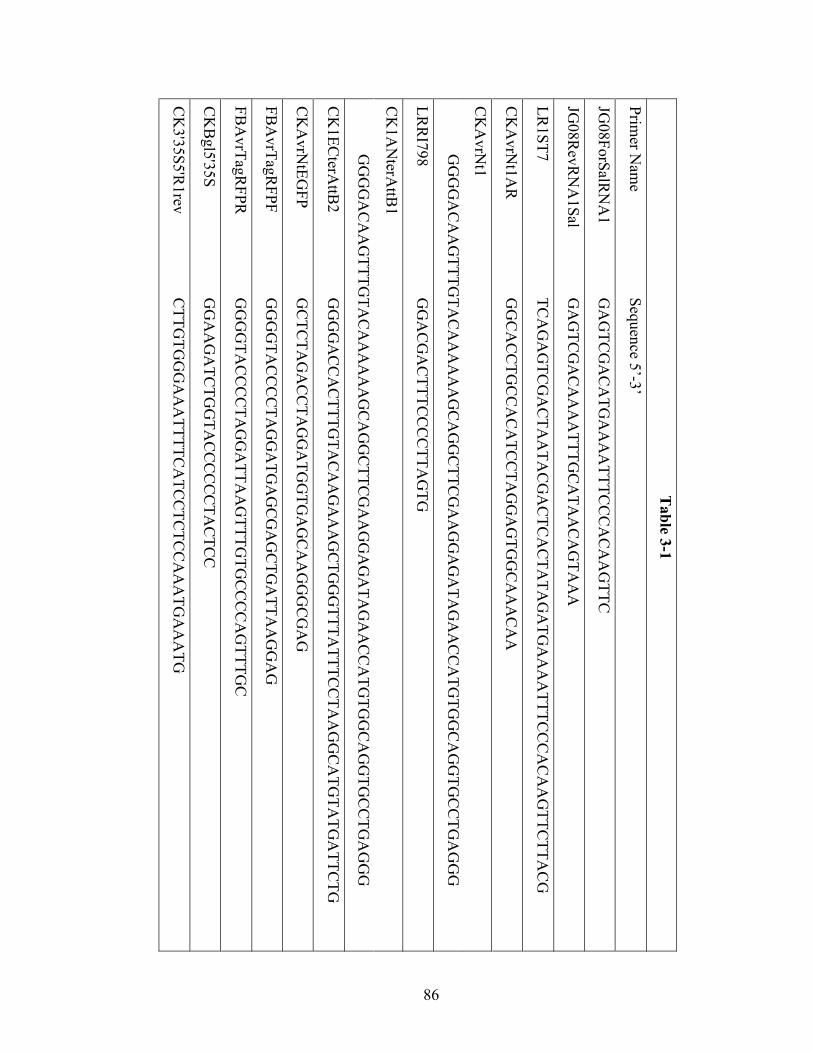
86
Table 3-1
Primer N
ame
Sequence 5’-3’
JG08ForSalR
NA
1
GA
GTC
GA
CA
TGA
AA
ATTTC
CC
AC
AA
GTTC
JG08R
evRN
A1Sal
G
AG
TCG
AC
AA
AA
TTTGC
ATA
AC
AG
TAA
A
LR1ST7
TCA
GA
GTC
GA
CTA
ATA
CG
AC
TCA
CTA
TAG
ATG
AA
AA
TTTCC
CA
CA
AG
TTCTTA
CG
CK
AvrN
t1AR
GG
CA
CC
TGC
CA
CA
TCC
TAG
GA
GTG
GC
AA
AC
AA
CK
AvrN
t1
G
GG
GA
CA
AG
TTTGTA
CA
AA
AA
AG
CA
GG
CTTC
GA
AG
GA
GA
TAG
AA
CC
ATG
TGG
CA
GG
TGC
CTG
AG
GG
LRR
I798
G
GA
CG
AC
TTTCC
CC
TTAG
TG
CK
1AN
terAttB
1
G
GG
GA
CA
AG
TTTGTA
CA
AA
AA
AG
CA
GG
CTTC
GA
AG
GA
GA
TAG
AA
CC
ATG
TGG
CA
GG
TGC
CTG
AG
GG
CK
1ECterA
ttB2
G
GG
GA
CC
AC
TTTGTA
CA
AG
AA
AG
CTG
GG
TTTATTTC
CTA
AG
GC
ATG
TATG
ATTC
TG
CK
AvrN
tEGFP
G
CTC
TAG
AC
CTA
GG
ATG
GTG
AG
CA
AG
GG
CG
AG
FBA
vrTagRFPF
G
GG
GTA
CC
CC
TAG
GA
TGA
GC
GA
GC
TGA
TTAA
GG
AG
FBA
vrTagRFPR
GG
GG
TAC
CC
CTA
GG
ATTA
AG
TTTGTG
CC
CC
AG
TTTGC
CK
Bgl5'35S
GG
AA
GA
TCTG
GTA
CC
CC
CC
TAC
TCC
CK
3'35S5'R1rev
C
TTGTG
GG
AA
ATTTTC
ATC
CTC
TCC
AA
ATG
AA
ATG

87
Table 3-1
Primer N
ame
Sequence 5’-3’
CK
3'35S5'R1sens
C
ATTTC
ATTTG
GA
GA
GG
ATG
AA
AA
TTTCC
CA
CA
AG
CK
3'R1B
glrev
GG
AA
GA
TCTTTA
TTTAG
AA
ATA
AA
AA
TTTGC
ATA
AC
AG
TAA
AA
AG
JG10SalG
HR
NA
2For G
GA
GTC
GA
CA
TGA
AA
AA
TTTTGG
TAG
GTTC
JG10G
HR
NA
2Bam
H1R
ev G
GA
GG
ATC
CA
TAA
ATTTG
CA
AA
AC
AG
RLFus1F
GA
AC
CTA
GG
CG
CG
AA
TCTTTG
ATTC
G
RLFus1R
C
TTGTA
GG
CA
CTG
GTTG
CA
CTTA
AG
TCA
TATTC
TATG
GG
AG
GG
CA
AG
TCA
AA
AA
TTGG
AA
TTTG
RLFus2F
GA
ATA
TGA
CTTA
AG
TGC
AA
CC
AG
TGC
CTA
CA
AG
AG
TGC
TGG
GC
TAA
CTTTG
GG
CC
RLFus2R
C
ATA
CA
CC
CC
GG
GA
TATTTG
AA
AA
GTTC
AT
RL11M
CS2F
A
GC
AC
TTCTG
TGTG
CTG
TCG
TAC
GG
AC
AC
GC
GTA
CTTA
TTCTTC
TGTTA
TTCC
TAA
TTATTC
TTCTTC
TTTTATT
AG
RL11M
CS2R
C
CA
CC
GG
TAG
TTCG
GC
CA
TCC
GC
TCC
ATC
TTCA
GG
AA
CA
TAA
GG
AA
CTTC
TCC
TCTA
ATA
AA
AG
AA
GA
AG
AA
TAA
TTAG
G
RL11R
NA
2F
ATG
CTC
GA
GC
GC
TTGA
ATTC
TGA
TCA
AG
ATC
TCC
GG

88
Table 3-1
Primer N
ame
Sequence 5’-3’
RL11R
NA
2R
G
CG
CTC
GA
GC
TGA
TCA
AG
ATC
TTAG
TAC
TGA
TTTTGG
TTTTAG
G
eGFPtopR
OX
F
GA
GA
CG
TAC
GA
TGG
TGA
GC
AA
GG
GC
GA
GG
AG
C
eGFPtopR
OX
R
G
AG
AA
CG
CG
TATC
CG
AG
TAC
TTGTA
CA
GC
TCG
TCC
NbPD
SBsiW
IF
AA
TCA
TCG
TAC
GA
TGC
CC
CA
AA
TCG
GA
CTTG
TATC
TGC
TG
NbPD
SMluIR
CTC
TTAA
CG
CG
TGA
ATA
TGTG
CA
AC
CC
AG
TCTC
GTA
CC
AA
TC
CK
Sal5'35S
A
TCG
ATG
TCG
AC
GG
TAC
CC
CC
CTA
CTC
C
CK
3'35S5'R2sens
G
TTCA
TTTCA
TTTGG
AG
AG
GA
TGA
AA
AA
TGTTTA
CG
CK
3'35S5'R2rev
C
GTA
AA
CA
TTTTTCA
TCC
TCTC
CA
AA
TGA
AA
TGA
AC
CK
3'R2PA
SSalrev
ATC
GA
TGTC
GA
CTTTA
TTTAG
AA
ATA
ATA
AA
TTTGC
AA
AA
CA
G
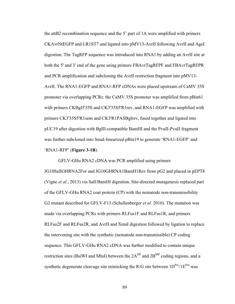
89
the attB2 recombination sequence and the 5’ part of 1A were amplified with primers
CKAvrNtEGFP and LR1ST7 and ligated into pMV13-AvrII following AvrII and AgeI
digestion. The TagRFP sequence was introduced into RNA1 by adding an AvrII site at
both the 5' and 3' end of the gene using primers FBAvrTagRFPF and FBAvrTagRFPR
and PCR amplification and subcloning the AvrII restriction fragment into pMV13-
AvrII. The RNA1-EGFP and RNA1-RFP cDNAs were placed upstream of CaMV 35S
promoter via overlapping PCRs: the CaMV 35S promoter was amplified from pBin61
with primers CKBgl5'35S and CK3'35S5'R1rev, and RNA1-EGFP was amplified with
primers CK3'35S5'R1sens and CK3'R1PASBglrev, fused together and ligated into
pUC19 after digestion with BglII-compatible BamHI and the PvuII-PvuII fragment
was further subcloned into SmaI-linearized pBin19 to generate ‘RNA1-EGFP’ and
‘RNA1-RFP’ (Figure 3-1B).
GFLV-GHu RNA2 cDNA was PCR amplified using primers
JG10SalIGHRNA2For and JG10GHRNA1BamH1Rev from pG2 and placed in pEPT8
(Vigne et al., 2013) via SalI/BamHI digestion. Site-directed mutagenesis replaced part
of the GFLV-GHu RNA2 coat protein (CP) with the nematode non-transmissibility
G2 mutant described for GFLV-F13 (Schellenberger et al. 2010). The mutation was
made via overlapping PCRs with primers RLFus1F and RLFus1R, and primers
RLFus2F and RLFus2R, and AvrII and XmaI digestion followed by ligation to replace
the intervening site with the synthetic (nematode non-transmissible) CP coding
sequence. This GFLV-GHu RNA2 cDNA was further modified to contain unique
restriction sites (BsiWI and MluI) between the 2AHP and 2BMP coding regions, and a
synthetic degenerate cleavage site mimicking the R/G site between 1DPro/1EPol was

90
synthesized upstream of the multiple cloning site (MCS) to allow for maturation of
protein 2BMP. The MCS and cleavage sequence was synthesized between the
2AHP/2BMP coding regions using megaprimers RL11MCS2F and RL11MCS2R
containing native restriction sites AleI and AgeI on the 5’ and 3’ ends, respectively.
The CP-modified GFLV-GHu RNA2 clone was digested with AleI and AgeI and the
intervening RNA2 with MCS was inserted. The 35S:RNA2-CP-MCS sequence was
then amplified with primers RL11RNA2F and RL11RNA2R, and ligated into binary
vector G2b (Choi et al., 2008) via PspXI digestion. This clone is designated ‘RNA2’
in Figure 3-1B. The BsiWI/MluI cloning site on GFLV RNA2 was used to clone
EGFP gene after amplification with primers EGFPtopROXF and EGFPtopROXR to
generate ‘RNA2-EGFP’ (Figure 3-1B). N. benthamiana pds gene fragment was
amplified from total RNA of N. benthamiana using primers NbPDSBsiWIF and
NbPDSMluIR and ligated into GFLV-RNA2 to generate ‘RNA2-pds’ (Figure 3-1B).
The insertion of TagRFP gene into GFLV RNA2 was previously reported (Amari et
al., 2010). This clone was placed under the control of a CaMV 35S promoter by
overlapping PCRs. The CaMV 35S promoter was amplified from pBin61 with primers
CKSal5'35S and CK3'35S5'R2rev, and RNA2-RFP was amplified with primers
CK3'35S5'R2sens and CK3'R2PASSalrev, fused together, ligated into SalI-digested
pUC19, further subcloned into pBin19 via SalI digestion and named ‘RNA2-RFP’.
Protein and fluorescence assays
GFLV ELISA, immunocapture (IC)-RT-PCR, quantitative fluorescence
measurements and t-tests were conducted as described (Gottula et al., 2013; Vigne et

91
al., 2013). Microscopy was accomplished with a Leica SP5 Confocal Microscope
(Leica Microsystems, Exton, PA USA) at the Plant Cell Imaging Center at Boyce
Thomson Institute for Plant Research at Cornell University (Ithaca, NY). Fresh virus-
infected or negative control leaves were fixed in water and illuminated with DIC or
Argon lasers. Predefined excitation and emission spectra were used for EGFP and RFP
and fluorescence emissions were simultaneously captured with HYD2 detectors using
10X, 20X or 40X objectives. Images were processed using the Leica Application
Software Advanced Fluorescence Suite (v 2.6.0) and viewed in ImageJ.
Nematode transmission assays
Wild-type GFLV and GFLV variants based on various RNA1 and RNA2
cDNA constructs were mechanically inoculated to N. benthamiana. Infected and
control N. benthamiana plants were planted into flats containing 300 aviruliferous X.
index previously reared on fig plants. Nematodes were allowed to feed for six weeks.
After acquisition of the virus and removal of the infected N. benthamiana plants,
nematodes were exposed to healthy N. benthamiana or grapevines for eight weeks.
Roots of bait N. benthamiana and grapevines were tested for GFLV by ELISA and IC-
RT-PCR, respectively (Schellenberger et al., 2010).
Grapevine agroinfection
This author undertook seven experiments to test and validate GFLV
agroinfection in grapevine. In these experiments, Vitis cultivars (‘Riesling’ F1
seedlings and ‘Cabernet franc’ cuttings) were agroinoculated with various GFLV
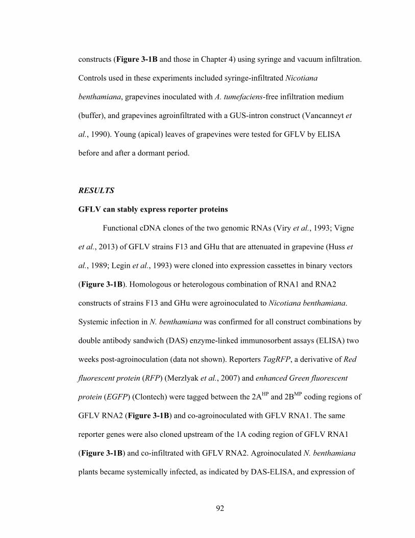
92
constructs (Figure 3-1B and those in Chapter 4) using syringe and vacuum infiltration.
Controls used in these experiments included syringe-infiltrated Nicotiana
benthamiana, grapevines inoculated with A. tumefaciens-free infiltration medium
(buffer), and grapevines agroinfiltrated with a GUS-intron construct (Vancanneyt et
al., 1990). Young (apical) leaves of grapevines were tested for GFLV by ELISA
before and after a dormant period.
RESULTS
GFLV can stably express reporter proteins
Functional cDNA clones of the two genomic RNAs (Viry et al., 1993; Vigne
et al., 2013) of GFLV strains F13 and GHu that are attenuated in grapevine (Huss et
al., 1989; Legin et al., 1993) were cloned into expression cassettes in binary vectors
(Figure 3-1B). Homologous or heterologous combination of RNA1 and RNA2
constructs of strains F13 and GHu were agroinoculated to Nicotiana benthamiana.
Systemic infection in N. benthamiana was confirmed for all construct combinations by
double antibody sandwich (DAS) enzyme-linked immunosorbent assays (ELISA) two
weeks post-agroinoculation (data not shown). Reporters TagRFP, a derivative of Red
fluorescent protein (RFP) (Merzlyak et al., 2007) and enhanced Green fluorescent
protein (EGFP) (Clontech) were tagged between the 2AHP and 2BMP coding regions of
GFLV RNA2 (Figure 3-1B) and co-agroinoculated with GFLV RNA1. The same
reporter genes were also cloned upstream of the 1A coding region of GFLV RNA1
(Figure 3-1B) and co-infiltrated with GFLV RNA2. Agroinoculated N. benthamiana
plants became systemically infected, as indicated by DAS-ELISA, and expression of
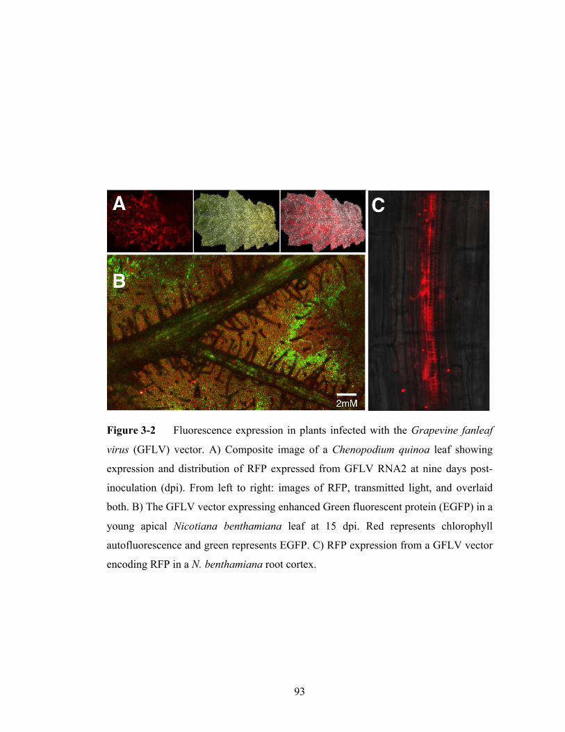
93
Figure 3-2 Fluorescence expression in plants infected with the Grapevine fanleaf
virus (GFLV) vector. A) Composite image of a Chenopodium quinoa leaf showing
expression and distribution of RFP expressed from GFLV RNA2 at nine days post-
inoculation (dpi). From left to right: images of RFP, transmitted light, and overlaid
both. B) The GFLV vector expressing enhanced Green fluorescent protein (EGFP) in a
young apical Nicotiana benthamiana leaf at 15 dpi. Red represents chlorophyll
autofluorescence and green represents EGFP. C) RFP expression from a GFLV vector
encoding RFP in a N. benthamiana root cortex.

94
Table 3-2 Stability of Red fluorescent protein (RFP) expression from the GFLV vector in Nicotiana benthamiana and Chenopodium quinoa.
a Total number of ELISA positive plants for GFLV over the total number of inoculated
plants. After initial infection in N. benthamiana and each passage, one RFP
(fluorescence microscopy) and GFLV positive N. benthamiana (ELISA) was used as
inoculum at two weeks post-inoculation for each subsequent passage. N/A: not
applicable. b Total number plants expressing red fluorescence assessed by confocal microscopy
and a microplate read over the total number of plants tested.
Nicotiana benthamiana
Chenopodium quinoa
Infection Infection ratea RFP expressionb Infection ratea RFP expressionb
1st passage 2/5 2/5 6/13 6/13
2nd passage 2/2 2/2 8/8 8/8
3rd passage 2/2 2/2 15/15 15/15
4th passage 2/2 2/2 15/15 15/15

95
EGFP was observed in leaves (Figure 3-2B). Expression of EGFP was also observed
in leaves and roots of Chenopodium quinoa following mechanical inoculation that
used sap of agroinfected N. benthamiana (Figure 3-2A and 4-2C). The stability of the
RFP-tagged GFLV vector was determined by four serial passages in N. benthamiana
and Chenopodium quinoa at two-week intervals following agroinfection of N.
benthamiana (Table 3-2). The presence of RFP in every infected plant following four
serial passages is promising, given that other viral vectors undergo insert deletions in
zero to four passages (Fernández-Fernández et al., 2001; Haviv et al., 2006; Touriño
et al., 2008). These results indicated GFLV vectors can stably express foreign genetic
material in plant leaves and roots.
GFLV is an effective vector for silencing endogenous genes
Because null mutants are not always available for gene complementation
assays, knockdown of host genes of known sequence is key for functional genomics
research. Therefore, we tested the GFLV vector for capability of virus-induced gene
silencing (VIGS) and compared it to the widely used VIGS vector Tobacco rattle virus
(TRV) (Ratcliff et al., 2001). To this aim, we cloned a partial fragment (501bp) of
phytoene desaturase (pds) sequence from N. benthamiana between the 2AHP and 2BMP
coding regions of GFLV RNA2 (Figure 3-1B) and agroinoculated N. benthamiana
with GFLV RNA1 in a comparative test to the TRV vector with a 402bp pds insert. At
nine days post-inoculation (dpi), the expected photobleaching phenotype was obtained
(Figure 3-3A and 3-3B) and maintained for up to 120dpi. In order to quantify VIGS
efficacy, we inoculated N. benthamiana constitutively expressing high levels of EGFP

96
Figure 3-3 Comparative virus-induced gene silencing activity of the Grapevine
fanleaf virus (GFLV) and Tobacco rattle virus (TRV) vectors for silencing phytoene
desaturase (pds) and enhanced Green fluorescent protein (EGFP) expression in
Nicotiana benthamiana. A) GFLV-pds-induced photobleaching versus B) TRV-pds-
induced photobleaching phenotype at 14 days post-inoculation (dpi). C) Quantitative
comparison of GFLV and TRV-induced silencing of EGFP in EGFP transgenic plants
at 14dpi. Error bars represent standard error of the mean (n=3).
97
with GFLV carrying EGFP or TRV carrying the same sequence (Vigne et al., 2013).
Fluorescence measurements confirmed the EGFP-tagged GFLV vector reduced EGFP
expression, as expected (Figure 3-3C). The fluorescence outputs from TRV and
GFLV VIGS vectors were not significantly different according to Student’s t test
(P=0.56). Fluorescent microscopic observations confirmed that both virus vectors
dramatically suppressed EGFP expression (data not shown). These experiments
indicated GFLV is a highly effective VIGS vector.
GFLV can concurrently express two genes
Following validation of GFLV for single gene expression, EGFP or RFP were
cloned in front of the 1A coding sequence of GFLV RNA1 (Figure 3-1B) and co-
agroinfiltrated plants with GFLV RNA2 carrying RFP or EGFP into the 2AHP-2BMP
junction (Figure 3-1B). The constructs were designed such that fluorescent proteins
(FP) tagged the 1A and 2AHP proteins. Expression of EGFP and RFP was observed in
agroinoculated and systemic leaves of N. benthamiana and a time course analysis was
undertaken to characterize the dynamics of FP expression from GFLV RNA1 and
RNA2. Cells of infiltrated N. benthamiana tissue expressed primarily the FP tagged to
RNA1 (RFP, red) at three dpi and only occasionally co-expressed RNA1- and RNA2-
encoded FPs (RFP and EGFP, overlayed yellow) (Figure 3-4A). In systemic leaves,
EGFP and RFP were co-expressed in most cells. A front of infection was observed
with veins and epidermal cells expressing only the FP tagged to RNA1 (Figure 3-2).
In particular, RFP expressed in RNA1 showed fluorescence in secondary C. quinoa
veins at the front of infection (Figure 3-4B), and EGFP expressed in RNA1 showed

98
Figure 3-4 Dual gene expression patterns of the Grapevine fanleaf virus vector.
Red fluorescent protein (RFP, red) and enhanced Green fluorescent protein (EGFP,
green) genes were inserted in either GLFV RNA1 or RNA2. Each subject is
represented for EGFP fluorescence (top segments), RFP fluorescence (middle
segments) and the EGFP-RFP overlay (lower segments). GFLV vectors are indicated
at the bottom of each panel and arrows show the direction of the infection front in
panels B and C. A) Infection sites are present at three days post-inoculation (dpi) in
agroinoculated Nicotiana benthamiana cells expressing RNA1-RFP and RNA2-EGFP.
B) Patterns of FP expression in upper non-inoculated leaves for RNA1-RFP and
RNA2-EGFP in C. quinoa. C) Patterns of FP expression at the front of infection for
RNA1-EGFP and RNA2-RFP in N. benthamiana at 6 dpi. D) Co-expression of
RNA1-EGFP and RNA2-RFP showing 1A-2AHP protein aggregates in a grapevine
leaf at nine dpi.
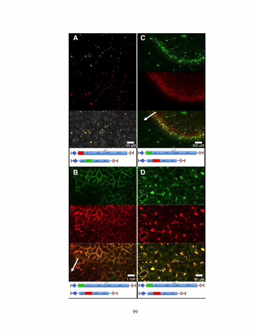
99

100
fluorescence at the front of infection in N. benthamiana epidermal cells (Figure 3-
4C). These observations indicated that RNA1 is able to express its heterologous insert
in advance of RNA2, which reflects the dependence of RNA2 polyprotein maturation
on the RNA1-encoded proteinase (Margis et al., 1994). These experiments showed
GFLV vector can be used to concurrently express two heterologous genes and can be
used for tandem or sequential gene expression experiments when multiple genes are to
be functionally characterized.
The EGFP and RFP-tagged GFLV vector was agroinoculated to ten plants of
Vitis vinifera cv. Cabernet Sauvignon. Agroinoculated leaves revealed expression of
EGFP and RFP by nine dpi in eight of ten plants. Confocal microscopy images
indicated EGFP and RFP co-expression in multiple cells in aggregates characteristic of
1A-2AHP co-localization (Figure 3-4A and 3-4D). Guard cells, epidermal cells and
mesophyll tissue contained EGFP and RFP overlaid, and a few guard cells contained
solely 1A-TagRFP aggregates. Expression of the EGFP and RFP-tagged GFLV
aggregates were confirmed through 30dpi in inoculated leaves.
GFLV expresses proteins at high levels in grape berries
Because grapevines generally require three years to fruit, we tested naturally
GFLV-infected grapevines for virus titer in berry tissues. Grape clusters from three
infected grapevines were harvested at maturity and the seeds, skin and flesh was
manually separated and tested by semi-quantitative DAS-ELISA. Results indicate that
GFLV accumulates in the different berry tissues tested (Figure 3-5). A comparative
analysis of relative GFLV titer shows an approximately 100-fold lower accumulation
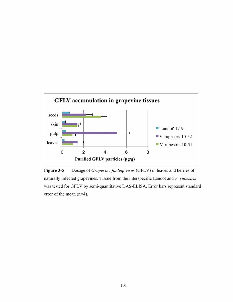
101
Figure 3-5 Dosage of Grapevine fanleaf virus (GFLV) in leaves and berries of
naturally infected grapevines. Tissue from the interspecific Landot and V. rupestris
was tested for GFLV by semi-quantitative DAS-ELISA. Error bars represent standard
error of the mean (n=4).
0! 2! 4! 6! 8!
leaves
pulp
skin
seeds
Purified GFLV particles (µg/g)
GFLV accumulation in grapevine tissues
'Landot' 17-9
V. rupestris 10-52
V. rupestris 10-51

102
in grapevine berry and leaf tissue (Figure 3-5) than in systemic herbaceous hosts
(Vigne et al., 2013), but a similar accumulation compared to other viral vectors in
herbaceous hosts (Fernández-Fernández et al., 2001; Li et al., 2004; Yoon et al., 2011;
Chen et al., 2012). This indicates the GFLV vector will be suitable for functional
genetics testing of grapevine berries and can be used to test genetic hypotheses in each
of the two genomic layers of grapevines (Reisch et al., 2011).
The GFLV vector is abolished for nematode vector transmission
A growing interest in using viral vectors as tools for vaccination of perennial
crops (Dawson and Folimonova, 2013) necessitates the addition of a biosafety
component. Since GFLV is vectored specifically by the ectoparasitic nematode
Xiphinema index and the viral determinant of nematode transmission is well
characterized (Schellenberger et al, 2010; 2011), we conducted site-directed
mutagenesis of 11 amino acids that are responsible for transmission and are located
within the GFLV vector-RNA2-encoded coat protein coding region (Protocol S1). The
vector and controls, including transmissible and non-transmissible GFLV strains
(Schellenberger et al., 2010), were deployed in X. index transmission assays following
agroinoculation of N. benthamiana. Transmission assays showed that the GFLV
vector is X. index non-transmissible (Table 3-3). This indicates the GFLV vector
could be deployed in vineyards or other low-containment settings without concern that
it will become naturalized through X. index-mediated transmission, unless
recombination with the RNA genome of wild-type strains would restore
transmissibility.
103
Lack of grapevine systemic infection following agroinfiltration
Grapevine plants were agroinfiltrated with GFLV or Gus-intron controls.
Excellent Gus-intron expression was verified at nine days post-inoculation in multiple
experiments (data not shown). ELISA tests for GFLV before and after dormant
periods revealed no instance of systemic GFLV infection following agroinfiltration in
seven experiments where 323 plants were used. Local GFLV infection was observed
using fluorophor-tagged RNA1 and RNA2 constructs derived from GFLV-F13
(Figure 3-4D), but likewise these plants did not become systemically infected
according to ELISA and fluorescence microscopy.
DISCUSSION
Functional genomics platforms based on viral vectors have numerous
advantages compared to stable transformation or transient expression systems
including reduced time from concept to phenotype, greater ease of use, grander
scalability potential and often greater consistency within treatments. However, viral
vectors have weaknesses compared to other functional genomics systems including a
frequent lack of stability, tissue specificity, relatively low carrying capacity of genetic
information, host-pathogenic effects and concerns about environmental release of the
infectious agent. Therefore, although dozens of viral vectors have been validated, few
have been widely used in economically important crops, and even fewer in perennial
woody crops (Dawson and Folimonova, 2013). A few research groups have developed
viral vectors for grapevine, but each of these vectors comes with some limitations
including tissue specificity (GLRaV-2, GVA and GRSPaV are phloem limited) and
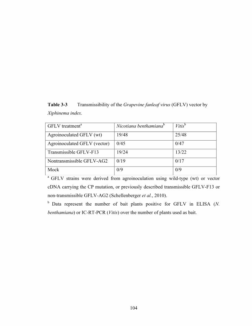
104
Table 3-3 Transmissibility of the Grapevine fanleaf virus (GFLV) vector by
Xiphinema index.
a GFLV strains were derived from agroinoculation using wild-type (wt) or vector
cDNA carrying the CP mutation, or previously described transmissible GFLV-F13 or
non-transmissible GFLV-AG2 (Schellenberger et al., 2010). b Data represent the number of bait plants positive for GFLV in ELISA (N.
benthamiana) or IC-RT-PCR (Vitis) over the number of plants used as bait.
GFLV treatmenta Nicotiana benthamianab Vitisb
Agroinoculated GFLV (wt) 19/48 25/48
Agroinoculated GFLV (vector) 0/45 0/47
Transmissible GFLV-F13 19/24 13/22
Nontransmissible GFLV-AG2 0/19 0/17
Mock 0/9 0/9
105
possibility of unintended release [very little information is known about GVA
transmission by mealybugs and no vector is known for GLRaV-2 and GRSPaV].
Additionally, GFLV is based on a bipartite RNA genome, which can facilitate
spatially phased expression of heterologous proteins, unlike vectors based on viruses
with monopartite RNA genomes such as GLRaV-2, GVA and GRSPaV.
The GFLV vectors capture all of the advantages and limits most or all
disadvantages of other grapevine viral vector technologies in the following ways: the
vector (1) is easy to use given that GFLV-RNA2 can be modified to contain inserts
based on restriction enzyme digestion (BsiWI and MluI) in the small modular binary
vector pGreen, (2) can stably carry inserts, (3) can efficiently silence endogenous
genes, (4) can concurrently or simultaneously express two foreign sequences
depending on the spatio-temporal position of virus infection and (5) cannot be
transmitted by its ectoparasitic nematode vector. In addition, GFLV is expressed to
high levels in all tissues of grapevine (Andret-Link et al., 2004), including all parts of
berries (Figure 3-5). For these reasons, the GFLV vector could be an ideal functional
genomics platform for grapevine.
The present generations of the GFLV vector have the critical limitation of not
producing systemic infection in grapevine following GFLV cDNA agroinfiltration.
The lack of grapevine systemic infection could relate to one of several issues with the
constructs. The first hypothesis is that the GFLV transcripts derived from cDNAs used
are not infectious in grapevine, as was the case with precursory versions of the
Grapevine leafroll associated virus-2 vector (Liu et al., 2009; Kurth et al., 2013). This
hypothesis is unlikely because the cDNAs derived from GFLV-F13 and GFLV-GHu

106
(i.e. FF and GG, Chapter 4) are infectious in grapevine following heterologous
grafting (E. Vigne, personal communication). Another possible explanation of the lack
of systemic infectivity in grapevine could relate to the tagging of many GFLV
transcript constructs (e.g. those used in Chapter 4) with the Alfalfa mosaic virus RNA4
translational enhancer at their 5’ ends. Because fidelity of 5’ ends of RNA viruses
including GFLV is critical to their infectivity (Chapter 2; Liu et al., 1991; Annamalai
et al., 2003), it is plausible that this structure comprises an impediment to infectivity
of GFLV transcripts following agroinfiltration.
Provided that future generations of the GFLV vector could be successfully
inoculated to grapevine, this vector could be utilized in ways similar to vectors
validated for several other crop species. In wheat, Barley stripe mosaic virus (BSMV)-
based vectors were used to knock down expression of candidate genes for powdery
mildew resistance and these functional analyses conclusively identified Triticum
aestivum powdery mildew resistance 5 and mildew resistance locus O as bases of
resistance (Várallyay et al., 2012; Yuan et al., 2012). It can be envisioned that the
GFLV vector could be used to individually down regulate expression of eight
candidate genes contained in the resistance to Uncinula necator 1 linkage group
associated with powdery mildew resistance (Dry et al., 2010). In another study, a
BSMV vector was used to identify (or disprove) involvement of several candidate
genes in drought resistance in wheat (Manmathan et al., 2013). Similarly, the GFLV
vector could be deployed to better understand the role of aldehyde dehydrogenases for
drought tolerance in grapevine (Zhang et al., 2012). The stability, ability to express
novel or silence endogenous genes, and lack of tissue tropism in the GFLV vector

107
makes it ideal to study genes responsible for biotic and abiotic stress tolerance in
grapevines.
Deployment of the GFLV vector could facilitate the selection of grapevines
with optimal flavor profiles among breeding populations. Numerous genes have been
identified as candidates for grape flavor (Battilana et al., 2011; Dunlevy et al., 2013)
however, conclusive proof by an effective complementation system remains elusive
and functional analysis in grapevine is necessary before surefire marker-assisted
selection tools can be developed. Some viral vectors have extraordinary versatility in
that they support the expression of two genes (Roy et al., 2011; Kurth et al., 2012) and
the two gene expression capability of the GFLV vector should be particularly handy
because it could be used to parse out interconnected and occasionally redundant
biochemical pathways that create grape flavor profiles (Dai et al., 2011). The facts that
GFLV can express high levels of protein in grape skin and pulp and express proteins
in a phased manner illustrate its potential utility for modifying these pathways.
Grape growers are increasingly dismayed by the growing prevalence of insect-
vectored bacterial diseases such as Pierce’s Disease for which no effective control
measures exist. Plant pathologists and entomologists have responded by positing the
use of viral vectors as tools for vaccination against the agents of disease or the insects
that carry them (Folimonova and Dawson, 2013; Karthikeyan et al., 2013; Gu and
Knipple, 2013). Recent proofs-of-concept have shown that viral vectors can
effectively silence insect genes and kill insects (Khan et al., 2013; Wuriyanghan and
Falk, 2013). Foundational work on plant-expressed diffusible signal factor and
antibacterial protein chimeras has demonstrated the feasibility of attenuating Xylella
108
fastidiosa infection in grapevines (Chatterjee et al., 2008; Dandekar et al., 2012). The
GFLV vector could theoretically be used to deliver proteins with insecticidal,
antibacterial or even antiviral activities to grapevines in vineyard situations, with
assurances that pathogenic effects of the vector is minimal (given the attenuation of
the strains used here) and that the vector would not be disseminated to neighboring
grapevines by X. index-mediated transmission.

109
REFERENCES
Adam-Blondon, A.-F., Jaillon, O., Vezzulli, S., Zharkikh, A., Troggio, M., and
Velasco, R. 2011. Genome sequence initiatives. Pages 211–234 in: Genomics, and
Breeding of Grapes. Adam-Blondon, A.-F., Martínez-Zapater J. M., and Kole, C.,
eds. Science Publishers, Enfield, NJ.
Agüero, J., Ruiz-Ruiz, S., Vives, M. C., Velázquez, K., Narvarro, L., Peña, L.,
Moreno, P., and Guerri, J. 2012. Development of viral vectors based on Citrus leaf
blotch virus to express foreign proteins or analyze gene function in citrus plants.
Molecular Plant-Microbe Interactions. 25:1236–1337.
Alzubi, H., Yepes, L. M., and Fuchs, M. F. 2012. Enhanced micropropagation and
establishment of grapevine rootstock genotypes. International Journal of Plant
Developmental Biology. 6:9-14.
Amari, K., Boutant, E., Hofmann, C., Schmitt-Keichinger, C., Fernandez-Calvino, L.,
Didier, P., Lerich, A., Mutterer, J., Thomas, C. L., Heinlein, M., Mély, Y., Maule, A.
J., and Ritzenthaler, C. 2010. A family of plasmodesmal proteins with receptor-like
properties for plant viral movement proteins. PLoS Pathogens. 6:e1001119.
Andret-Link, P., Laporte, C., Valat, L., Ritzenthaler, C., Demangeat, G., Vigne, E.,
Laval, V., Pfeiffer, P., Stussi-Garaud, C., and Fuchs, M. 2004. Grapevine fanleaf
virus: still a major threat to the grapevine industry. Journal of Plant Pathology.
86:183–195.
Annamalai, P., Hsu, Y. H., Liu, Y. P., Tsai, C. H., and Lin, N. S. 2003. Structural and
mutational analyses of cis-acting sequences in the 5′-untranslated region of satellite
RNA of bamboo mosaic potexvirus. Virology. 311:229–239.
110
Arazi, T., Slutsky, S. G., Shiboleth, Y. M., Wang, Y., Rubinstein, M., Barak, S., Yang,
J., and Gal-On, A. 2001. Engineering Zucchini yellow mosaic potyvirus as a non-
pathogenic vector for expression of heterologous proteins in cucurbits. Journal of
Biotechnology. 87:67-82.
Avesani, L., Marconi, G., Morandini, F., Albertini, E., Bruschetta, M., Bortesi, L.,
Pezzotti, M., and Porceddu, A. 2007. Stability of Potato virus X expression vectors is
related to insert size: implications for replication models and risk assessment.
Transgenic Research. 16:587–597.
Battilana, J., Emanuelli, F., Gambino, G., Gribaudo, I., Gasperi, F., Boss, P. K., and
Grando, M. S. 2011. Functional effect of grapevine 1-deoxy-D-xylulose 5-phosphate
synthase substitution K284N on Muscat flavour formation. Journal of Experimental
Botany. 62:5497–5508.
Bertazzon, N., Raiola, A., Castiglioni, C., Gardiman, M., Angelini, E., Borgo, M., and
Ferrari, S. 2011. Transient silencing of the grapevine gene VvPGIP1 by
agroinfiltration with a construct for RNA interference. Plant Cell Reports. 31:133–
143.
Chatterjee, S., Wistrom, C., and Lindow, S. E. 2008. A cell-cell signaling sensor is
required for virulence and insect transmission of Xylella fastidiosa. Proceedings of the
National Academy of Sciences USA. 105:2670–2675.
Chen, A. Y. S., Pavitrin, A., and Ng, J. C. K. 2012. Agroinoculation of the cloned
infectious cDNAs of Lettuce chlorosis virus results in systemic plant infection and
production of whitefly transmissible virions. Virus Research. 167:310–315.
Chiba, M., Reed, J., Prokhnevsky, A., Chapman, E., Mawassi, M., Koonin, E.,

111
Carrington, J., and Dolja, V. 2006. Diverse suppressors of RNA silencing enhance
agroinfection by a viral replicon. Virology. 346:7–14.
Choi, M. S., Yoon, I.-S., Rhee, Y., Choi, S. K., Lim, S.-H., Won, S.-Y., Lee, Y.-H.,
Choi, H.-S., Lee, S.-C., Kim, K.-H., Lomonossoff, G., and Sohn, S.-H. 2008. The
effect of Cucumber mosaic virus 2b protein to transient expression and transgene
silencing mediated by agro-infiltration. Plant Pathology Journal. 24:296–304.
Dai, Z. W., Ollat, N., Gomes, E., Decroocq, S., Tandonnet, J. P., Bordenave, L., Pieri,
P., Hilbert, G., Kappel, C., van Leeuwen, C., Vivin, P., and Delrot, S. 2011.
Ecophysiological, genetic, and molecular causes of variation in grape berry weight and
composition: A review. American Journal of Enology and Viticulture. 62:413–425.
Dandekar, A. M., Gouran, H., Ibáñez, A. M., Uratsu, S. L., Agüero, C. B., McFarland,
S., Borhani, Y., Feldstein, P. A., Bruening, G., Nascimento, R., Goulart, L. R.,
Pardington, P. E., Chaudhary, A., Norvell, M., Civerolo, E., and Gupta, G. 2012. An
engineered innate immune defense protects grapevines from Pierce disease.
Proceedings of the National Academy of Sciences USA. 109:3721–3725.
Dawson, W. O., and Folimonova, S. Y. 2013. Virus-based transient expression vectors
for woody crops: a new frontier for vector design and use. Annual Review of
Phytopathology. 51:321–337.
Dry, I. B., Feechan, A., Anderson, C., Jermakow, A. M., Bouquet, A., Adam-Blondon,
A. F., and Thomas, M. R. 2010. Molecular strategies to enhance the genetic resistance
of grapevines to powdery mildew. Australian Journal of Grape and Wine Research.
16:94–105.
Dunlevy, J. D., Dennis, E. G., Soole, K. L., Perkins, M. V., Davies, C., and Boss, P.
112
K. 2013. A methyltransferase essential for the methoxypyrazine-derived flavour of
wine. Plant Journal. 75:606–617.
Fernández-Fernández, M. R., Mouriño, M., Rivera, J., Rodríguez, F., Plana-Durán, J.,
and García, J. A. 2001. Protection of rabbits against rabbit hemorrhagic disease virus
by immunization with the VP60 protein expressed in plants with a potyvirus-based
vector. Virology. 280:283–291.
Gottula, J. W., Lapato, D., Cantilina, K. K., Saito, S., Bartlett, B., and Fuchs, M. 2013.
Genetic variability, evolution and biological effects of Grapevine fanleaf virus satellite
RNAs. Phytopathology. 103:1180–1187.
Gu, L., and Knipple, D. C. 2013. Recent advances in RNA interference research in
insects: implications for future insect pest management strategies. Crop Protection.
45:36–40.
Haviv, S., Galiakparov, N., Goszczynski, D. E., Batuman, O., Czosnek, H., and
Mawassi, M. 2006. Engineering the genome of Grapevine virus A into a vector for
expression of proteins in herbaceous plants. Journal of Virological Methods. 132:227–
231.
Huss, B., Walter, B., and Fuchs, M. 1989. Cross-protection between Arabis mosaic
virus and Grapevine fanleaf virus isolates in Chenopodium quinoa. Annals of Applied
Biology. 114:45–60.
Iocco, P., Franks, T., and Thomas, M. R. 2001. Genetic transformation of major wine
grape cultivars of Vitis vinifera L. Transgenic Research. 10:105–112.
Jaillon, O., Aury, J.-M., Noel, B., Policriti, A., Clepet, C., Casagrande, A., Choisne,
N., Aubourg, S., Vitulo, N., Jubin, C., Vezzi, A., Legeai, F., Hugueney, P., Dasilva,

113
C., Horner, D., Mica, E., Jublot, D., Poulain, J., Bruyère, C., Billault, A., Segurens, B.,
Gouyvenoux, M., Ugarte, E., Cattonaro, F., Anthouard, V., Vico, V., Del Fabbro, C.,
Alaux, M., Di Gaspero, G., Dumas, V., Felice, N., Paillard, S., Juman, I., Moroldo,
M., Scalabrin, S., Canaguier, A., Le Clainche, I., Malacrida, G., Durand, E., Pesole,
G., Laucou, V., Chatelet, P., Merdinoglu, D., Delledonne, M., Pezzotti, M., Lecharny,
A., Scarpelli, C., Artiguenave, F., Pè, M. E., Valle, G., Morgante, M., Caboche, M.,
Adam-Blondon, A.-F., Weissenbach, J., Quétier, F., and Wincker, P. 2007. The
grapevine genome sequence suggests ancestral hexaploidization in major angiosperm
phyla. Nature. 449:463–467.
Karthikeyan, A., Deivamani, M., Shobhana, V. G., Sudha, M., and Anandhan, T.
2013. RNA interference: evolutions and applications in plant disease management.
Archives of Phytopathology and Plant Protection. 43:1430–1441.
Khan, A. M., Ashfaq, M., Kiss, Z., Khan, A. A., Mansoor, S., and Falk, B. W. 2013.
Use of recombinant Tobacco mosaic virus to achieve RNA interference in plants
against the citrus mealybug, Planococcus citri (Hemiptera: Pseudococcidae). PLoS
ONE. 8:e73657.
Kurth, E., Peremyslov, V., and Prokhnevsky, A. 2012. Virus-derived gene expression
and RNAi vector for grapevine. Journal of Virology. 86:6002–6009.
Legin, R., Bass, P., Etienne, L., and Fuchs, M. 1993. Selection of mild virus strains of
fanleaf degeneration by comparative field performance of infected grapevines. Vitis.
32:103–110.
Li, C., Sasaki, N., Isogai, M., and Yoshikawa, N. 2004. Stable expression of foreign
proteins in herbaceous and apple plants using Apple latent spherical virus RNA2
vectors. Archives of Virology. 149:1541-1558.

114
Liu, Y. L., Cooper, J. I., Coates, D., and Bauer, G. 1991. Biologically active
transcripts of a large satellite RNA from Arabis mosaic nepovirus and the importance
of 5′ end sequences for its replication. The Journal of General Virology. 72:2867–
2874.
Liu, Y.-P., Peremyslov, V. V., Medina, V., and Dolja, V. V. 2009. Tandem leader
proteases of Grapevine leafroll-associated virus-2: Host-specific functions in the
infection cycle. Virology. 383:291–299.
Manmathan, H., Shaner, D., Snelling, J., Tisserat, N., and Lapitan, N. 2013. Virus-
induced gene silencing of Arabidopsis thaliana gene homologues in wheat identifies
genes conferring improved drought tolerance. Journal of Experimental Botany.
64:1381–1392.
Margis, R., Viry, M., Pinck, M., Bardonnet, N., and Pinck, L. 1994. Differential
proteolytic activities of precursor and mature forms of the 24K proteinase of
Grapevine fanleaf nepovirus. Virology. 200:79–86.
Martelli G. P. 2014. Directory of virus and virus-like diseases of the grapevine and
their agents. Journal of Plant Pathology. 96: in press.
Martínez-Zapater, J. M., Carmona, M. J., Diaz-Riquelme, J., Fernandez, L., and
Lijavetzky, D. 2010. Grapevine genetics after the genome sequence: challenges and
limitations. Australian Journal of Grape and Wine Research. 16:33–46.
Mejía, N., Soto, B., Guerrero, M., Casanueva, X., Huel, C., de los Angeles Miccono,
M., Ramos, R., Le Cunff, L., Boursiquot, J.-M., Hinrichsen, P., and Adam-Blondon,
A.-F. 2011. Molecular, genetic and transcriptional evidence for a role of VvAGL11 in
stenospermocarpic seedlessness in grapevine. BMC Plant Biology. 11:1–18.

115
Meng, B., Venkataraman, S., Li, C., Wang, W., Dayan-Glick, C., and Mawassi, M.
2013. Construction and biological activities of the first infectious cDNA clones of the
genus Foveavirus. Virology. 435:453–462.
Merzlyak, E. M., Goedhart, J., Shcherbo, D., Bulina, M. E., Shcheglov, A.S., Fradkov,
A.F, Gaintzeva, A, Lukyanov, K. A., Lukyanov, S., Gadella, T. W. J. and Chudakov,
D.M. 2007. Bright monomeric red fluorescent protein with an extended fluorescence
lifetime. Nature Methods. 4:555–557.
Muruganantham, M., Moskovitz, Y., Haviv, S., Horesh, T., Fenigstein, A., Preez, du,
J., Stephan, D., Burger, J. T., and Mawassi, M. 2009. Grapevine virus A-mediated
gene silencing in Nicotiana benthamiana and Vitis vinifera. Journal of Virology
Methods. 155:167–174.
Myles, S., Boyko, A., Owens, C. L., Brown, P. J., Grassi, F., Aradhya, M. K., Prins,
B., Reynolds, A., Chia, J.-M., Ware, D., Bustamante, C. D., and Buckler, E. S. 2011.
Genetic structure and domestication history of the grape. Proceedings of the National
Academy of Sciences USA. 108:3530–3535.
Nicolas, P., Lecourieux, D., Gomès, E., Delrot, S., and Lecourieux, F. 2013. The grape
berry-specific basic helix–loop–helix transcription factor VvCEB1 affects cell size.
Journal of Experimental Botany. 64:991-1003.
Ratcliff, F., Martin-Hernandez, A. M., and Baulcombe, D. C. 2001. Technical
Advance. Tobacco rattle virus as a vector for analysis of gene function by silencing.
Plant Journal. 25:237–245.
Reisch, B. I., Owens, C. L., and Cousins, P. S. 2011. Grape. Pages 225–262 in: Fruit
Breeding. M.L. Badenes and D.H. Byrne, eds. Springer US, Boston, MA.

116
Romieu, C., Kappel, C., Delrot, S. 2011. Functional genomics: Closed system
approaches for transcriptome analyses. Pages 270–298 in: Genomics, and Breeding of
Grapes. Adam-Blondon, A.-F., Martínez-Zapater J. M., and Kole, C., eds. Science
Publishers, Enfield, NJ.
Roy, G., Weisburg, S., Foy, K., Rabindran, S., Mett, V., and Yusibov, V. 2011. Co-
expression of multiple target proteins in plants from a Tobacco mosaic virus vector
using a combination of homologous and heterologous subgenomic promoters.
Archives of Virology. 156:2057–2061.
Santos-Rosa, M., Poutaraud, A., Merdinoglu, D., and Mestre, P. 2008. Development
of a transient expression system in grapevine via agro-infiltration. Plant Cell Reports.
27:1053–1063.
Satyanarayana, T., Bar-Joseph, M., Mawassi, M., Albiach-Martí, M. R., Ayllón, M.
A., Gowda, S., Hilf, M. E., Moreno, P., Garnsey, S. M., and Dawson, W. O. 2001.
Amplification of Citrus tristeza virus from a cDNA clone and infection of citrus trees.
Virology. 280:87–96.
Schellenberger, P., Andret-Link, P., Schmitt-Keichinger, C., Bergdoll, M., Marmonier,
A., Vigne, E., Lemaire, O., Fuchs, M., Demangeat, G., and Ritzenthaler, C. 2010. A
stretch of 11 amino acids in the beta B-beta C loop of the coat protein of Grapevine
fanleaf virus is essential for transmission by the nematode Xiphinema index. Journal of
Virology. 84:7924–7933.
Schellenberger, P., Sauter, C., Lorber, B., Bron, P., Trapani, S., Bergdoll, M.,
Marmonier, A., Schmitt-Keichinger, C., Lemaire, O., Demangeat, G., and
Ritzenthaler, C. 2011. Structural insights into viral determinants of nematode mediated
Grapevine fanleaf virus transmission. PLoS Pathogens. 7:e1002034.

117
Senthil-Kumar, M., and Mysore, K. S. 2011. New dimensions for VIGS in plant
functional genomics. Trends in Plant Science. 16:656–665.
Tillett, R., and Cushman, J. 2011. Vitis functional genomics: Open systems for
transcriptome analysis. Pages 235–269 in: Genomics and Breeding of Grapes. Adam-
Blondon, A.-F., Martínez-Zapater J. M., and Kole, C., eds. Science Publishers,
Enfield, NJ.
Touriño, A., Sánchez, F., Fereres, A., and Ponz, F. 2008. High expression of foreign
proteins from a biosafe viral vector derived from Turnip mosaic virus. Spanish Journal
of Agricultural Research. 6:48–58.
Vaghchhipawala, Z., Rojas, C. M., Senthil-Kumar, M., and Mysore, K. S. 2010.
Agroinoculation and agroinfiltration: Simple tools for complex gene function
analyses. Pages 65-76 in: Methods in Molecular Biology. A. Pereira, ed. Humana
Press, Totowa, NJ.
Vancanneyt, G., Schmidt, R., O'Connor-Sanchez, A., Willmitzer, L., and Rocha-Sosa,
M. 1990. Construction of an intron-containing marker gene - splicing of the intron in
transgenic plants and its use in monitoring early events in Agrobacterium-mediated
plant transformation. Molecular Genetics and Genomics. 220:245–250.
Várallyay, É., Giczey, G., and Burgyan, J. 2012. Virus-induced gene silencing of Mlo
genes induces powdery mildew resistance in Triticum aestivum. Archives of Virology.
157:1345–1350.
Velasco, R., Zharkikh, A., Troggio, M., Cartwright, D. A., Cestaro, A., Pruss, D.,
Pindo, M., FitzGerald, L. M., Vezzulli, S., Reid, J., Malacarne, G., Iliev, D., Coppola,
G., Wardell, B., Micheletti, D., Macalma, T., Facci, M., Mitchell, J. T., Perazzolli, M.,

118
Eldredge, G., Gatto, P., Oyzerski, R., Moretto, M., Gutin, N., Stefanini, M., Chen, Y.,
Segala, C., Davenport, C., Demattè, L., Mraz, A., Battilana, J., Stormo, K., Costa, F.,
Tao, Q., Si-Ammour, A., Harkins, T., Lackey, A., Perbost, C., Taillon, B., Stella, A.,
Solovyev, V., Fawcett, J. A., Sterck, L., Vandepoele, K., Grando, S. M., Toppo, S.,
Moser, C., Lanchbury, J., Bogden, R., Skolnick, M., Sgaramella, V., Bhatnagar, S. K.,
Fontana, P., Gutin, A., Van de Peer, Y., Salamini, F., and Viola, R. 2007. A high
quality draft consensus sequence of the genome of a heterozygous grapevine variety.
PLoS ONE. 2:e1326.
Vigne, E., Gottula, J., Schmitt-Keichinger, C., Komar, V., Ackerer, L., Belval, L.,
Rakotomalala, L., Lemaire, O., Ritzenthaler, C., and Fuchs, M. 2013. A strain-specific
segment of the RNA-dependent RNA polymerase of Grapevine fanleaf virus
determines symptoms in Nicotiana species. Journal of General Virology. 94:2803–
2813.
Viry, M., Serghini, M. A., Hans, F., Ritzenthaler, C., Pinck, M., and Pinck, L. 1993
Biologically active transcripts from cloned cDNA of genomic Grapevine fanleaf
nepovirus RNAs. Journal of General Virology. 74:169–174.
Visser, M., Stephan, D., Jaynes, J. M., and Burger, J. T. 2012. A transient expression
assay for the in planta efficacy screening of an antimicrobial peptide against grapevine
bacterial pathogens. Letters in Applied Microbiology. 54:543–551.
Wuriyanghan, H., and Falk, B. W. 2013. RNA interference towards the potato psyllid,
Bactericera cockerelli, is induced in plants infected with recombinant Tobacco mosaic
virus (TMV). PLoS ONE. 8:e66050.
Yang, Y., Jittayasothorn, Y., Chronis, D., Wang, X., Cousins, P., and Zhong, G.-Y.
2013. Molecular characteristics and efficacy of 16D10 siRNAs in inhibiting root-knot
nematode infection in transgenic grape hairy roots. PLoS ONE. 8:e69463.

119
Yoon, J.-Y., Choi, S. K., Palukaitis, P., and Gray, S. M. 2011. Agrobacterium-
mediated infection of whole plants by yellow dwarf viruses. Virus Research. 160:428–
434.
Yuan, C., Li, C., Yan, L., Jackson, A. O., Liu, Z., Han, C., Yu, J., and Li, D. 2011. A
high throughput Barley stripe mosaic virus vector for virus induced gene silencing in
monocots and dicots. PLoS ONE. 6:1–16.
Zhang, Y., Mao, L., Wang, H., Brocker, C., Yin, X., Vasiliou, V., Fei, Z., and Wang,
X. 2012. Genome-wide identification and analysis of grape aldehyde dehydrogenase
(ALDH) gene superfamily. PLoS ONE. 7:e32153.
120
CHAPTER 4
GENOMIC, ENVIRONMENTAL AND HOST VARIABLES INFLUENCING
GRAPEVINE FANLEAF VIRUS AGROINFECTION
ABSTRACT
Parameters that support local and systemic infection of Grapevine fanleaf virus
(GFLV), a bipartite RNA virus from the genus Nepovirus, family Secoviridae, in
Nicotiana benthamiana following Agrobacterium tumefaciens-mediated delivery were
tested. The cDNAs of GFLV strains F13 and GHu and recombinants derived thereof
were reassorted and tested alongside cDNAs of wild type strains for infectivity.
Measures of transient GFLV accumulation following agroinfiltration did not always
reflect differential systemic infectivity of the GFLV genomes, but a synthetic assortant
of F13 RNA1 and GHu RNA2 cDNAs consistently showed the highest rate of local
and systemic infection. Multiple experiments that iteratively tested individual
variables showed that systemic GFLV infection was correlated with lower ambient
greenhouse temperatures, reduced A. tumefaciens suspension density and co-
infiltration with silencing suppressors, but not with A. tumefaciens strain identity, co-
culture with acetosyringone or plant size. Agroinfiltrating transgenic plants expressing
either RNA1 or RNA2 cDNAs with a complementary genome part established
systemic infection but at a lower rate relative to transiently expressed counterparts.
Finally, agroinoculating a panel of Nicotiana species showed differential GFLV
accumulation in patch assays that could not be explained by A. tumefaciens transient
expression capabilities or status of resistance to mechanical inoculation, suggesting

121
complex virus-host interactions. This research shed light on factors governing the
success of A. tumefaciens-mediated delivery of GFLV for Nicotiana infection.
122
INTRODUCTION
Since Grimsley et al. (1987) reported the first use of Agrobacterium
tumefaciens as a tool to deliver viruses to plants, there has been an explosion of
publications on the creation and development of viral vectors (Senthil-Kumar and
Mysore, 2011; Dawson and Folimonova, 2013). A. tumefaciens-mediated virus
inoculation (agroinfection) has been developed for various crops including orchids
(Hsieh et al., 2013), monocot field crops (Yuan et al., 2011), tree fruit crops (Ambros
et al., 2011) and vegetable crops (Delbianco et al., 2013). Viral vectors have been
used for very diverse research endeavors including expressing metabolites (Zhang et
al., 2013) or MYB transcription factors (Bedoya et al., 2012), and silencing bacterial
resistance and susceptibility genes (Balaji et al., 2011). Viral vectors have been put to
use in allelism tests (Fitzgerald et al., 2012), to produce heterologous antigens for
vaccines (Gleba et al., 2007; Thuenemann et al., 2013), to deliver insect-disruption
molecules (Wuriyanghan and Falk, 2013) and to facilitate stable genome modification
(Kopertekh et al., 2012). In 28 years, viral vectors have moved from proof-of-concept
technologies to widely used research tools and may have a future in direct agronomic
applications (Dawson and Folimonova, 2013).
Compared to substitute plant biotechnologies, viral vectors have both
advantages and limitations. Viral vectors can produce large amounts of heterologous
protein or efficiently knock down mRNA expression 90% or more (Kagale, 2012; Ma
et al., 2012). Viral vectors can reveal functional phenotypes faster than stably
transformed plant lines (Pogue et al., 2002). On the other hand, viral vectors exhibit
limitations on the amount of genetic material they can stably replicate (Senthil-Kumar

123
and Mysore, 2011), they can be destructive in controlled greenhouse situations if the
live agent is unintentionally disseminated (Hayward et al., 2011), and may not be fully
reliable due to lack of infectivity (Liu et al., 2009). Improving viral vector infectivity
is often quite challenging because only a handful of mutations can render a virus
noninfective in a given host (Kurth et al., 2012).
Grapevine fanleaf virus (GFLV) from the genus Nepovirus, family Secoviridae
has a bipartite genome composed of single stranded positive-sense RNA molecules
(Sanfaçon et al., 2009). Each genome segment contains a viral genome-linked protein
(VPg) at the 5’ end and a poly-A tail at the 3’ terminus, and is translated into a
polyprotein. RNA1 (7.3kb) contains the materials necessary and sufficient for
replication and protein maturation, including a helicase, proteinase, VPg, RNA-
dependent RNA polymerase, and a protein (1A) of an unknown function. RNA2
(3.7kb) encodes the movement protein and coat protein, and the 2AHP protein
responsible for RNA2 replication in conjunction with RNA1 replicase proteins
(Andret-Link et al., 2004). Both GFLV genomic RNAs are required for systemic plant
infection (Andret-Link et al., 2004). The virus is specifically vectored by the dagger
nematode Xiphinema index, and is host-specialized to grapevine and a few herbaceous
hosts including Nicotiana benthamiana. Mechanically inoculation with infectious sap
or transcripts derived from full-length cDNA clones leads to GFLV infection in N.
benthamiana (Valat et al., 2003). Recently, infectious GFLV cDNA clones
corresponding to the genomic RNAs of strain F13 (Viry et al., 1993) and GHu (Vigne
et al., 2013) were placed downstream of the Cauliflower mosaic virus (CaMV) 35S
promoter into binary vectors suitable for A. tumefaciens-mediated infection in planta

124
(Gottula et al., 2014a). The GFLV vector can simultaneously express two
heterologous proteins, efficiently silence plant genes through VIGS, and is unable to
be transmitted by X. index as a result of a targeted mutation in the coat protein
(Gottula et al., 2014a). In the present manuscript, variables tested to optimize
conditions for agroinfection of GFLV in N. benthamiana are reported.
MATERIALS AND METHODS
Molecular cloning procedures
All enzymes, buffers and protocols were from New England Biolabs (NEB,
Ipswich, MA). This included PCR materials (Phusion DNA Polymerase), reverse
transcriptase (AMV), T4 DNA ligase, and several restriction enzymes. All plasmids
were transformed into chemically competent Escherichia coli strain DH5α, which
were grown in agar-containing or liquid Luria-Bertani (LB) medium at 37οC. The
pEPT8-derived plasmids (Ling et al., 1997; Gottula et al., 2014a) were selected with
ampicillin (100µg/mL), pGreenII- (Choi et al., 2008) and pROK2- (Gottula et al.,
2014a) based plasmids with kanamycin (50µg/mL), and pGA482G-based plasmids
(Chee et al., 1989) with gentamicin (100µg/mL). Primers were from Integrated DNA
Technologies (Iowa City, IA) (Table 4-1). Plasmids were extracted with Omega
Miniprep Kit (Norcross, GA) and Sanger sequencing was accomplished at Cornell
Biotechnology Resource Center (Ithaca, NY).
GFLV vector and silencing suppressors
Previously validated cDNAs of two GFLV strains were cloned downstream of

125
the CaMV 35S promoter in binary vectors. The cDNAs of RNA1 and RNA2 of strains
GFLV-F13 (Viry et al., 1993) and GFLV-GHu (Vigne et al., 2013) were subcloned
into pEPT8 and into the binary vector pGA482G (Chee et al., 1989) as described by
Gottula et al. (2014a) and in Supplementary Protocol 1. These clones were used to
generate four GFLV genomes, i.e. FF, GG, FG and GF, named on the basis of the
F13- or GHu- composition of the RNA1 and RNA2 constructs (Table 4-2).
Additionally, two GFLV RNA2 recombinants were generated: The FG2A construct had
the 2AHP protein coding sequence of GFLV-GHu placed in the GFLV-F13 RNA2
background; the other RNA2 construct GR is comprised of GFLV-GHu RNA2 with a
multiple cloning site composed of BsiWI and MluI restriction sites, and a site-directed
coat protein mutation for abolishment of X. index transmissibility (Gottula et al.,
2014a) (Table 4-2). All GFLV constructs were present in the CaMV 35S expression
cassette (35S promoter and terminator) from pEPT8. All GFLV constructs were
engineered into the CaMV 35S expression cassette (35S promoter and terminator)
from pEPT8. All GFLV constructs were contained in the binary vector pGA482G,
except the GR construct was contained in the modular binary pGreenII-0229 that also
contains a Cucumber mosaic virus (CMV) 2b expression cassette (Choi et al., 2008).
The different GFLV constructs in A. tumefaciens vectors, i.e. FF, GG, GF, FG, FFG2A,
GFG2A, FGR, GGR, are referred to herein as ‘genomes’ for simplicity. The cloning of
F13 RNA1 cDNA and GR was previously described, where Chapter 3 denoted F13
RNA1 cDNA as ‘RNA1 cDNA’ and GR was previously denoted ‘pROX’.
Silencing suppressor constructs CMV 2b (Choi et al., 2008) and Tomato bushy
stunt virus (TBSV) p19 (Canto et al., 2006) were used to test whether heterologous

126
Table 4-1
Primers used for the m
odification of Grapevine fanleaf virus cD
NA
s and their placement in binary vectors
Primer #
Primer nam
e
Sequence 5’-3’
1.
EVG
32
AG
AG
CTG
CG
GC
AC
TCTTTG
C
2.
EVG
33
AG
MA
GA
WC
CA
ATA
AA
GG
CG
CG
3.
JGnptII5F
G
GA
ATA
CTG
CC
TAG
CA
CC
GT
4.
JGnptII5R
GA
CA
TCC
TCC
AA
CG
AG
CA
TA
5.
JG08ForSalR
NA
2
G
AG
TCG
AC
ATA
AA
TTTGC
AA
AA
CA
GTA
AA
AA
GA
AA
GG
6.
JG08R
evRN
A2Sal
GA
GTC
GA
CG
ATG
AA
AA
ATTTTTA
CG
TTTTCTTA
CG
7.
JG10SalIG
HR
NA
1For
GG
AG
TCG
AC
ATG
AA
AA
ATTTC
TAC
AG
GTTC
8.
JG10G
HR
NA
1NotR
ev
GG
AG
CG
GC
CG
CTA
AA
TTTGC
AA
AA
CA
GT
9.
JG10SalIG
HR
NA
2For
GG
AG
TCG
AC
ATG
AA
AA
ATTTTG
GTA
GG
TTC
10.
JG10G
HR
NA
2Bam
H1R
ev
GG
AG
GA
TCC
ATA
AA
TTTGC
AA
AA
CA
G
11.
JG10ForB
glSaxXbapEPB
B2
GG
AA
GA
TCTG
AG
CTC
TCTA
GA
TAG
TAC
TGA
TTTTGG
TTTTAG
G
12.
JG10R
evpEPBB
XbaSacB
gl
GG
AA
GA
TCTG
AG
CTC
TCTA
GA
CC
GG
GC
AA
TTGA
GA
CTTTTC
13.
JG08ForEcoB
clBglpEPT8
G
AG
AA
TTCTG
ATC
AA
GA
TCTTA
GTA
CTG
ATTTTG
GTTTTA
GG
14.
JG08R
evpEPT8BglB
clEco
GA
GA
ATTC
TGA
TCA
AG
ATC
T
15.
JG10Sal1R
FPFor
G
GA
GTC
GA
CA
TGA
GC
GA
GC
TGA
TTAA
GG
AG
AA
CA
TGC
16.
JG10R
FPNotR
ev
G
GA
GC
GG
CC
GC
TCA
CTTG
TGC
CC
CA
GTTTG
CTA
GG
G
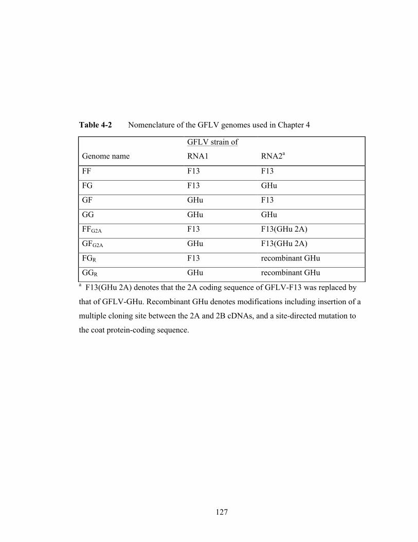
127
Table 4-2 Nomenclature of the GFLV genomes used in Chapter 4
a F13(GHu 2A) denotes that the 2A coding sequence of GFLV-F13 was replaced by
that of GFLV-GHu. Recombinant GHu denotes modifications including insertion of a
multiple cloning site between the 2A and 2B cDNAs, and a site-directed mutation to
the coat protein-coding sequence.
Genome name
GFLV strain of
RNA1 RNA2a
FF F13 F13
FG F13 GHu
GF GHu F13
GG GHu GHu
FFG2A F13 F13(GHu 2A)
GFG2A GHu F13(GHu 2A)
FGR F13 recombinant GHu
GGR GHu recombinant GHu

128
silencing suppressors affect GFLV protein accumulation and systemic infection. CMV
2b is contained in the pGreenII-0229 binary vector and TBSV p19 is in the pROK2
binary vector. The negative controls for silencing suppressor assays were fluorescent
proteins enhanced Green fluorescent protein (eGFP) and Red fluorescent protein
(RFP) expressed from CaMV 35S cassettes in pGA482G. The construction of the
eGFP clone was previously reported (Vigne et al., 2013).
Specific cloning strategies for the GFLV vectors used in this study
GFLV cDNAs in T7 expression cassettes that were previously verified to be
functional for plant inoculation (pVECP2, pG1 and pG2) (Viry et al., 1993; Vigne et
al., 2013) were amplified by PCR and ligated into cassettes containing a Cauliflower
mosaic virus (CaMV) 35S expression cassette containing a duplicated enhancer
(pEPT8) (Ling et al., 1997) or modified versions of pEPT8 (Vigne et al., 2013). The
cloning of F13 RNA1 cDNA and mutated GHu RNA2 cDNA (GR, alias pROX) was
reported in Chapter 3. The GFLV-F13 RNA2 cDNA was amplified by PCR using
primers P5 and P6 (Table 4-1) from pVECP2 and cloned into pEPT8BB with SalI to
generate RNA2-17. The F13 RNA2 with the GHu 2AHP replacement was created from
plasmid pGORF2 digested with AgeI and AlwNI followed by ligation of the
corresponding fragment into plasmid pF2 (Vigne et al., 2013). The RNA2 cDNA was
amplified from the modified pF2 with primers P5 and P6 and cloned into pEPT8BB as
described for RNA2-17 to generate 2ABB. The GHu RNA1 cDNA was cloned into
pEPT8MCS2 with SalI and NotI after amplification with primers P7 and P8 to generate
1M2. The GHu RNA2 cDNA was cloned into pEPT8MCS2 with SalI and BamHI after

129
amplification with primers P9 and P10 to generate 2MCS20.
The binary vector used to propagate and deliver GFLV cDNAs is a derivative
pGA482 modified to contain a gentamycin selectable marker, designated pGA482G
(Ling et al., 1997). The T-DNA of pGA482G contains unique restriction sites BglII
and XbaI, which were used to insert individual GFLV cDNAs in the CaMV 35S
cassettes. The RNA1 cDNA of GFLV strain GHu in the CaMV 35S cassette (1M2)
was amplified with primers P11 and P12 and ligated into pGA482G using XbaI to
generate GHu RNA1 cDNA. RNA2 cDNAs of GFLV F13-based clones (RNA2-17
and 2ABB) and GHu RNA2 (2MCS20) and their expression cassettes were amplified
via PCR with primers P13 and P14, digested with BclI, and cloned into the overhang
compatible BglII site in pGA482G to generate F13 RNA2 cDNA, FG2A and GHu
RNA2 cDNA.
RFP (Merzlyak et al., 2007) was amplified with primers P15 and P16 and
inserted into pEPT8MCS2 after restriction digestion of insert and vector with SalI and
NotI. The RFP and 35S expression cassette sequence was amplified with primers P9
and P10, digested with BglII and cloned into the BglII site of pGA482G. Confocal
microscopy revealed this clone produced fluorescence following agroinfiltration (data
not shown).
Integrity of selected recombinant clones was verified by sequencing, and in all
cases no modification of the GFLV cDNA was observed. Mutations in the TATA box
were observed in the promoters of each cassette with pEPT8 (TATATAA to
TATATAT), but high levels of expression have been verified from clones resulting
from this construct (Vigne et al., 2013). An unanticipated consequence of PCR

130
amplification of the expression cassettes was the truncation of the duplicated CaMV
35S enhancer motifs to a single promoter in GHu RNA1, GHu RNA2, and FG2A
clones.
Silencing suppressor constructs CMV 2b (Choi et al., 2008) and Tomato bushy
stunt virus (TBSV) p19 (Canto et al., 2006) were used to test whether heterologous
silencing suppressors affect GFLV protein accumulation and systemic infection. CMV
2b is contained in the pGreenII-0229 binary vector and TBSV p19 is in the pROK2
binary vector. The negative controls for silencing suppressor assays were fluorescent
protein sequences enhanced Green fluorescent protein (eGFP) and Red fluorescent
protein (RFP) cloned in pGA482G in CaMV 35S expression cassettes. The
construction of the eGFP clone was previously reported (Vigne et al., 2013). RFP
(Merzlyak et al., 2007) was subcloned into pEPT8 and the resulting 35S:RFP cloned
into pGA482G.
Agrobacterium tumefaciens strains and agroinfiltration
Electrocompetent cells of A. tumefaciens strains C58Z707 (C58), LBA4404 +
pSB1 (LBA4404) and GV3101 + pMP90 (GV3101) were transformed via
electroporation with individual binary vectors and all transformants were selected with
gentamicin (100µg/mL) plus kanamycin (50µg/mL). Strains C58 and LBA4404 were
used for pGA482G-based clones, and GV3101 was used for pGreenII- or pROK2-
based clones. The helper binary pClean-S161 was used in conjunction with the
pGreenII plasmids (GR and CMV 2b). The identity of the binary vectors in each A.
tumefaciens stock was confirmed by plasmid extraction and restriction digestion.

131
Agroinfiltration was done according to Vaghchhipawala et al. (2010) except that all A.
tumefaciens cultures were prepared to an initial density of OD600nm=1.0. GFLV
(RNA1 and RNA2) and heterologous silencing suppressor (p19 and 2b) cultures were
mixed together in equal ratios unless otherwise noted. N. benthamiana plants were
infiltrated with a needleless syringe in two leaves (Vaghchhipawala et al., 2010).
Several systemic GFLV agroinfection experiments employed treatments in which A.
tumefaciens suspensions were co-cultured with acetosyringone (Vaghchhipawala et
al., 2010).
GFLV accumulation and silencing suppressor efficacy experiments in infiltrated
zones of N. benthamiana
GFLV genomes FF, FG, GF and GG were tested for virus accumulation
against each other and an eGFP control at two, five and eight days post-inoculation
(dpi) in N. benthamiana. Each treatment and time point utilized four plants and two
leaves per plant (eight leaves per treatment). Within time points, plants (treatments)
were randomized on a greenhouse bench. In a second experiment, infiltrated zones of
FF, FG, GF, GG, FF2A and GF2A were tested for GFLV protein accumulation at nine
dpi. Each treatment used four plants and two leaves per plant (eight leaves per
treatment).
Silencing suppressors CMV 2b and TBSV p19 were tested in infiltrated zones
of N. benthamiana against eGFP and RFP for effects on GFLV accumulation.
Treatments were co-infiltrated with GFLV genomes FF, FG, GF or GG. Two leaves of
seven plants per treatment per genome were agroinfiltrated and 11 to 13 leaf samples
132
were collected and tested by ELISA at seven dpi. Plants were randomized on a
greenhouse bench and a third party researcher concealed all treatments until
conclusion of the collection and data analysis.
GFLV protein and fluorescence measurements
For each fluorescence or serological assay, two punches of a one-cm2 cork
borer (70 mg ± 8.2 mg) were collected and frozen until collections from each
experiment were complete. Frozen samples were ground in 0.7 mL phosphate buffer
pH 7.4 using a TissueLyser (Qiagen, Hilden, Germany) and 100 µL of crude leaf
extracts were applied to GFLV double antibody sandwich enzyme-linked
immunosorbent assay (DAS-ELISA) (Bioreba, Reinach, Switzerland) in each of two
wells (technical replicates) on 96 well plates containing positive and negative controls
(Gottula et al., 2013; Vigne et al., 2013). Plates were read after one hour of substrate
incubation. The Synergy2 96 well plate reader and Gen5 software package (Biotek
Corporation, Winooski, VT) were used to measure absorbance, average reads of
technical replicates and blank-subtract absorbance output of GFLV-negative N.
benthamiana tissue. Materials and methods for quantitative eGFP measurements were
described by Vigne et al. (2013).
Systemic GFLV infection experiments
Fifteen experiments that iteratively tested variables in systemic infection were
carried out over a one-year period. In 931 agroinfiltrated N. benthamiana plants,
systemic GFLV infection was tested by ELISA at four weeks post-inoculation on

133
apical, non-inoculated leaves. The variables included genome identity (Table 4-2),
silencing suppressor (TBSV p19 and CMV 2b) co-infiltration, acetosyringone co-
culture, A. tumefaciens density (OD600nm= 0.1 to 1.0), plant size (two versus six true
leaves), and cooling degree days were estimated post-hoc. Several systemic infection
experiments were not included in this analysis but were used to confirm the trends and
are reported individually in the results.
Plant growth conditions and cooling degree day estimation
N. benthamiana was grown in four-inch pots containing Cornell mix potting
medium (Balaji et al., 2011) in a greenhouse with a 16/8 hr. photoperiod maintained
with lighting supplemented by high pressure sodium lamps. Plants were watered daily
or every other day, as needed, and fertilized weekly. The greenhouses present at the
Cornell University New York State Agricultural Experiment Station were low frame
glasshouses with automatic temperature controls which were set to a constant
temperature of 22°C. Greenhouse heating consisted of radiator and convection
systems and cooling was achieved by evaporative coolers and automatic vents.
Since greenhouse temperatures were not directly recorded, estimates were
obtained from historical weather data from a local weather station. Sum cooling
degree days were calculated from a base temperature of 22°C for the four-week period
of each plant systemic infection experiment (from date of inoculation to tissue
collection). The degree day calculations were used as corollaries for ambient
greenhouse temperatures.

134
Transformation of N. benthamiana and transgene insertion
Constructs designed to express GFLV-F13 RNA1, GFLV-F13 RNA2, GFLV-
GHu RNA1 and GFLV-GHu RNA2 were used to stably transform N. benthamiana.
Clones in A. tumefaciens strain C58 were prepared as described for agroinfiltration
experiments and applied to N. benthamiana leaf discs, as previously reported (Yepes
et al., 1996). The regenerated plants (R0) were tested by ELISA for expression of
neomycin phosphotransferase II protein (nptII) (Agdia, Elkhart, IN) and by PCR for
GFLV cDNAs insertion using primers G13 and G16 for RNA1 (Vigne et al., 2013),
and primers P1 and P2 for RNA2 (Table 4-1). R0 plants were self-pollinated and T1
plants were tested for transgene insertion and selectable marker expression by a
kanamycin seed germination assay (seeds were germinated on MS medium with
300µg/mL kanamycin) and nptII ELISA. DNA extracted from T1 plants were
subjected to quantitative PCR using transgene primers P3 and P4 (Table 4-1) and
reference (β-actin) primers (Quiapim et al., 2009) with conditions and calculations
according to Bubner et al. (2004).
One or two transgenic lines per GFLV construct showing segregation ratios
consistent with nptII presence at one or two independent loci (qPCR) were used for
functional transgene expression analysis, which included agroinfiltration of the
complementary GFLV genome part and silencing suppressors followed by collection
of an apical leaf at four weeks post-inoculation for determining GFLV infection by
ELISA.

135
Agroinfiltration of Nicotiana spp.
Five Nicotiana species (N. benthamiana, N. clevelandii, N. glutinosa, N.
obtusifolia, and N. paniculata) were assayed for agroinfiltration expression capability
(eGFP) and infiltrated-zone GFLV protein accumulation. Three plants and two leaves
per plant were agroinfiltrated with the GFLV genome FG plus silencing suppressors
(left side of each leaves) or eGFP plus silencing suppressors (right side of each leaf).
The infiltrated zones were collected at five dpi and processed by ELISA for GFLV or
fluorescence (eGFP) expression.
Statistics
All statistics were conducted in SAS version 9.3 (SAS Institute, Cary, NC)
unless otherwise noted. Each data set was verified for homogeneity of variance via
Levene’s tests (P>0.05) prior to ANOVA. Where Levene’s test revealed significant
deviation from homogeneity (P<0.05), non-parametric tests were conducted.
For the infiltrated zone experiment that compared agroinfiltrated-zone GFLV
protein expression, absorbance outputs of individual GFLV genomes FF, FG, GF and
GG were evaluated among time points by ANOVA, followed by Tukey post-hoc tests.
A Kruskal-Wallis test, followed by Scheffe’s test, was used to compare absorbance
values of FF, FG, GF, GG, FFG2A and GFG2A using Excel Statistics Software 2010
(Social Survey Research Information Co., Ltd).
To compare relative GFLV ELISA OD405nm values and eGFP fluorescence
(508nm), absorbance and fluorescence data points were divided by the maximum
ELISA absorbance and eGFP fluorescence outputs, respectively, of each experiment
136
and multiplied by a factor of 100 to generate relative expression values. GFLV versus
eGFP relative expression values were evaluated at each time point by the Dunnett’s
multiple range test. ELISA absorbance values for each GFLV genome were examined
in the presence or absence of silencing suppressors and resulting data were subjected
to Mann-Whitney-U test following log10 transformation. An ANOVA followed by an
LSD post-hoc test was used to compare GFLV accumulation.
Two binary logit regression models were used to assign odds ratios (ORs),
95% confidence intervals and P values to variables affecting systemic GFLV infection
outcomes (Anderson et al., 2003). For the first model, a binary logit regression in a
stepwise method was employed to account for variables of GFLV infection including
A. tumefaciens strain, plant size, the coinfiltration of versus absence of heterologous
silencing suppressors, GFLV genome, and two continuous variables: density of A.
tumefaciens and cooling degree days in the greenhouse for the four-week duration of
the experiment. For the second analysis, a binary logit regression in direct method was
used to account for differences in systemic GFLV infection depending on transgene
versus agroinfiltration-delivery of a genome part. ORs were assigned based on
genome or RNA2 delivery method (transgenic or transient expression).
RESULTS
Creation and validation of GFLV constructs for agroinfiltration
Full-length RNA1 and RNA2 cDNAs of GFLV strains F13 and GHu from
previously validated transcription vectors (Viry et al., 1993; Vigne et al., 2013) were
subcloned into CaMV 35S expression cassettes of pEPT8 and GFLV cDNAs in

137
CaMV 35S expression cassettes were cloned via PCR and restriction digestion into
binary vector pGA482G for mobilization into A. tumefaciens. Complete sequencing of
each clone revealed no polymorphisms between the original GFLV cDNA sequences
and the corresponding cDNA sequences in binary vectors but GFLV GHu RNA1,
GFLV GHu RNA2, and FG2A contained one CaMV 35S enhancer while all other
clones contained a duplicated CaMV 35S enhancer. Each A. tumefaciens clone used in
this study was verified to contain the appropriate binary vectors following plasmid
extraction and restriction digestion.
Effect of genome composition on GFLV accumulation in the agroinfiltrated zone
of N. benthamiana
GFLV genomes FF, FG, GF and GG were agroinfiltrated into N. benthamiana
leaves and virus accumulation was quantified (Figure 4-1A). There was a significant
difference in ELISA absorbance output at two, five and eight dpi with P values of
0.0275, 0.0010 and 0.000, respectively according to ANOVA. FG produced higher
expression than FF at two dpi, and FF and GF at five dpi. At eight dpi, FG showed
greater expression than all other genomes, and GG expression exceeded that of FF and
GF.
The experiment was repeated using FF, FG, GF, GG, FFG2A and GFG2A and
samples were collected at nine dpi. The goal was to test whether protein 2AHP of strain
GHu could be used to rescue the low GFLV accumulation associated with F13 RNA2
since protein 2AHP is involved in RNA2 replication (Gaire et al. 1999). Significant
difference was detected in ELISA output among six GFLV genomes according to the
138
Kruskal-Wallis test (P=0.0001). Accumulation of FFG2A or GFG2A following
coinfiltration was not significantly different from FF or GF, and GG produced greater
accumulation than FF and GF (Figure 4-1B). FG produced greater infiltrated zone
accumulation than GG, and GG produced greater accumulation than FF and GF
(Figure 4-1B). These results suggested that the composition of GFLV protein 2AHP
does not impact GFLV expression levels in agroinfiltrated zones of N. benthamiana.
The ELISA outputs of each GFLV genome were compared to eGFP
fluorescence expression at two, five and eight dpi (Figure 4-1C). As expected, eGFP
showed a bell-curve expression pattern with a peak at five dpi. Although no individual
GFLV genome showed a significant deviation from eGFP expression at two dpi
according to Dunnett’s test (P<0.05), at five dpi, all genomes except FG were
significantly lower than eGFP. At eight dpi, FG showed significantly higher
accumulation than eGFP, and GF and FF showed significantly lower expression than
eGFP. These data suggested that FG is able to rise above the expected level of A.
tumefaciens-mediated protein expression and that protein expression of agroinfiltrated
GF and FF is suppressed in plants compared to eGFP
Effect of silencing suppressors on GFLV protein expression
GFLV genomes were tested for their response to coinfiltration of heterologous
silencing suppressors. Silencing suppressors (CMV 2b and TBSV p19) or null controls
(eGFP and RFP) were co-agroinfiltrated with the GFLV vectors and GFLV
accumulation was measured by ELISA at seven dpi. Coinfiltration of silencing
suppressors increased accumulation of each GFLV genome three to 23 fold, and to a
139
Figure 4-1 Effect of Grapevine fanleaf virus genome composition on virus
accumulation in agroinfiltrated zones of Nicotiana benthamiana. A, Accumulation of
different GFLV genomes (see Table 4-1 for details) was measured by ELISA at two,
five and eight days post agroinfiltration (dpi). Same letters represent no significant
difference for individual virus genomes among time points (n=8) at P<0.05 according
to Tukey. B, Accumulation of different GFLV genomes (See Table 4-1 for details)
measured by ELISA at nine dpi. Same letters represent no significant difference for
individual virus genomes (n=8) at P<0.05 according to Scheffe. C, Accumulation of
different GFLV genomes against enhanced Green Fluorescent Protein (eGFP) at two,
five and eight dpi. GFLV accumulation was measured by ELISA (A405nm) and eGFP
fluorescence (508nm) using a Synergy2 microplate reader. Relative expression was
calculated by converting measured values to percent maximum expression for each
treatment. An asterisk (*) indicates a treatment (n=8) was significantly less or greater
than the eGFP control (n=8) at P<0.05 at a given time point according to Dunnett’s
test.

140
141
statistically significant level for FF, FG and GG (Figure 4-2). No significant
difference was observed for GF, which showed the lowest virus accumulation in both
treatments. These local infection analyses indicated that silencing suppressors can
enhance GFLV accumulation in N. benthamiana following agroinoculation.
Systemic infection of the GFLV genomes
Following observation of GFLV expression in agroinfiltrated zones,
experiments were carried out to assess the frequency of systemic infection. The GFLV
genomes tested were those used in infiltrated zone experiments and also included an
additional GHu RNA2-based construct (GR), which was co-agroinfiltrated with F13
RNA1 (FGR) or GHu RNA1 (GGR) (Table 4-2).
In 15 independent experiments, 931 plants were agroinfiltrated with different
GFLV genomes and exposed to other variables. A total of 239 plants achieved
systemic infection. The systemic infection data were appropriate for stepwise logit
regression analysis according to Hosmer and Lemeshow Goodness-of-Fit test
(P=0.5782). A. tumefaciens strain identity (LBA4404 or C58), co-culture with
acetosyringone and plant size were not significant and these effects were excluded
from the model. Other variables and interactions explained 90.4% of the systemic
infection outcomes and included significant (P<0.05) effects for genome composition,
density of the A. tumefaciens suspension, cooling degree day accumulation and
presence of silencing suppressors (Table 4-3).
The coinfiltration of silencing suppressors showed the largest effect of any
variable considered. The binary logit model showed that silencing suppressors
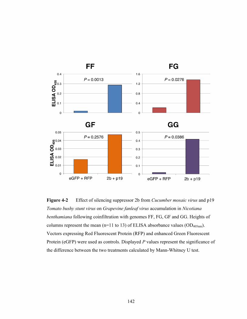
142
Figure 4-2 Effect of silencing suppressor 2b from Cucumber mosaic virus and p19
Tomato bushy stunt virus on Grapevine fanleaf virus accumulation in Nicotiana
benthamiana following coinfiltration with genomes FF, FG, GF and GG. Heights of
columns represent the mean (n=11 to 13) of ELISA absorbance values (OD405nm).
Vectors expressing Red Fluorescent Protein (RFP) and enhanced Green Fluorescent
Protein (eGFP) were used as controls. Displayed P values represent the significance of
the difference between the two treatments calculated by Mann-Whitney U test.

143
Table 4-3 Odds Ratio (OR) and 95% confidence intervals (CI) of variables in
Agrobacterium tumefaciens-mediated GFLV infection of Nicotiana benthamiana
based on a binary logit regression.
Variable OR (95% CI) P Value
A. tumefaciens density 0.251 (0.109-0.580) 0.0012
Cooling degree days 0.993 (0.991-0.995) <.0001
Silencing suppressors (versus none) 12.811 (7.678-21.375) <.0001
Genome FFG2A versus genome FF 6.807 (1.722-26.908) 0.0062
Genome FG versus genome FF 7.571 (3.589-15.975) <.0001
Genome FGR versus genome FF 29.356 (3.427-251.454) 0.0020
Genome GF versus genome FF 0.281 (0.087-0.904) 0.0332
Genome GG versus genome FF 1.116 (0.450-2.772) 0.8123
Genome GGR versus genome FF 0.345 (0.082-1.462) 0.1487

144
increase the likelihood of infection by 12.8 fold (Table 4-3). Silencing suppressors
positively affected frequencies of systemic infection in independent experiments as
well. In one experiment, systemic infection occurred with FG and silencing
suppressors in nine of 31 plants, whereas with FG without silencing suppressors (null
controls) only in five of 31 plants. The effect on GG was more dramatic, where GG
without silencing suppressors produced no infections in 31 plants and GG with
silencing suppressors infected nine of 31 plants. The A. tumefaciens density had a
negative effect on systemic infection where an OD600nm of 0.1 was more likely to
produce systemic infection than an OD of 1.0 (Table 4-3).
Greenhouse temperatures fluctuated between 22°C and 32°C depending on
ambient outdoor temperatures. Higher cooling degree days (hotter temperatures) had
negative effects on the frequency of agroinfection (Table 4-3). This corresponded to
observations that systemic infection was difficult to achieve in the low frame
greenhouse in summer when temperatures regularly approached daytime highs of
32°C. In contrast, when temperatures remained close to the set 22°C constantly,
systemic infection frequencies were improved.
The GFLV genome composition had a major effect on systemic infection.
Maximum likelihood estimates indicate the performance of genomes relative to FF
(Table 4-3). FFG2a, FG and FGR produced significantly more systemic infection than
FF. Although GFG2A was included in this analysis, the sample size was too small to
compute a reliable confidence interval and this genome did not produce a single
instance of systemic infection in 18 plants in two independent experiments (data not
shown). GG and GGR were not significantly different from FF (P<0.05) and GF

145
produced significantly less infection than FF (Table 4-3). The ORs and confidence
intervals generated in the logit regression analysis reflect broader trends observed
between genome composition and systemic infection. In one experiment that
compared systemic infectivity of FGR, GGR to FG and GG each mixed with
heterologous silencing suppressors at an A. tumefaciens suspension at OD600nm=1, FGR
produced infection in 18 of 20 plants, GGR in 3 of 20 plants, FG in 10 of 10 plants and
GG in 1 of 10 plants. These results showed that the composition of RNA1 and RNA2
affects systemic infection in N. benthamiana where F13 RNA1 outperforms GHu
RNA1, and GHu RNA2 including the GR construct outperforms F13 RNA2.
Stable versus transient expression of GFLV constructs
Transgenic T1 N. benthamiana plants expressing F13 RNA1, GHu RNA1, F13
RNA2 or GHu RNA2 were generated. According to seed germination assays on
selective medium, ELISA for nptII protein expression and qPCR-based transgene copy
number estimation, T1 lines segregated for one or two independent loci (data sets not
shown). These tests allowed identification of null segregants, which were excluded
from further analysis. Plants constitutively expressing RNA1 constructs were
agroinfiltrated with GFLV GHu RNA2 and plants expressing RNA2 constructs were
agroinfiltrated with GFLV GHu RNA1, and each A. tumefaciens culture containing a
GFLV genome part was mixed with heterologous silencing suppressors. ELISA was
used to test systemic GFLV infection at four weeks post-inoculation, respectively.
Each transgenic line became systemically infected at least once in samples of 20 to 40
plants, which indicates stable full-length T-DNA integration in each line and

146
functional expression following complementation through agroinoculation. There
results indicated that systemic infection can be achieved if a GFLV component is
stably expressed and the other is transiently expressed.
A direct logit regression analysis was run to compare transgenic genome part
expression versus agroinfiltrated genome expression on systemic infection outcomes.
Out of 200 plants analyzed among five experiments, 47 plants became systemically
infected. The data were appropriate for logit regression analysis as seen by a Hosmer-
Lemeshow Goodness-of-Fit test (P=0.2054). Transgenic expression of RNA1 and
RNA2 penalized systemic infection with transgenic expression of RNA2 having the
largest penalty. Systemic infection frequencies of genomes GG and FG were
compared to GF when both genome parts were delivered by agroinfiltration versus one
delivered by transgenic expression (Table 4-4). FG and GG performed better than GF
in terms of capability to cause systemically infection if one genome part was delivered
by transgene expression (Table 4-4). All constructs were penalized if one genome part
was transgenically expressed: systemic infection was nine times less likely if RNA2
was stably rather than transiently expressed (Table 4-4). These results suggested that
transgene expression of one genome part and agroinfiltration of the other genome part
diminishes GFLV infection frequency compared to when both genome parts are
agroinfiltrated.
Reaction of a Nicotiana panel to agroinfiltration with GFLV
N. benthamiana, N clevelandii, N. paniculata, N. glutinosa and N. obtusifolia

147
Table 4-4 Odds Ratio (OR) and 95% confidence intervals (CI) of Agrobacterium
tumefaciens versus transgenic expression of individual genome parts in Nicotiana
benthamiana based on direct logistic regression.
Variable OR (95% CI) P Value
Genome FG versus genome GF 5.546 (0.684-44.981) 0.1087
Genome GG versus genome GF 10.566 (1.318-84.692) 0.0264
A. tumefaciens versus transgene
delivery of RNA2
9.283 (2.540-33.931) 0.0008

148
were tested for GFLV accumulation following agroinfiltration with FG and
fluorescence following eGFP agroinfiltration. Prior experiments had revealed various
degrees of susceptibility to mechanical inoculation of GFLV strain GHu: N.
benthamiana and N. clevelandii are completely susceptible (Vigne et al., 2013), N.
obtusifolia and N. glutinosa are highly resistant, and N. paniculata is moderately
resistant, as shown by ELISA in non-inoculated apical leaves (Gottula et al., 2014b).
FG or eGFP were mixed with silencing suppressors 2b and p19 prior to infiltration.
ELISA absorbance values representing GFLV protein accumulation were compared
among plants species, and significant differences were obtained according to
ANOVA (P=0.0057). N. benthamiana, N. paniculata and N. glutinosa showed the
highest GFLV accumulation followed by N. obtusifolia which showed intermediate
GFLV expression, and N. clevelandii showed the lowest GFLV accumulation (Figure
4-3). Differences in eGFP expression could not explain the ELISA value differentials
(Figure 4-3). These results indicate that GFLV protein accumulation following
agroinfiltration does not reflect differences in resistance to mechanical inoculation in
this Nicotiana panel, which suggest Nicotiana resistance to GFLV are altered by
different inoculation methods, different GFLV genomes or co-application of
heterologous silencing suppressors.
DISCUSSION
In this study, parameters for an optimal expression of CaMV 35S-driven
RNA1 and RNA2 cDNAs from two distinct GFLV strains were determined following
agroinfiltration in N. benthamiana. The GFLV system studied here has several
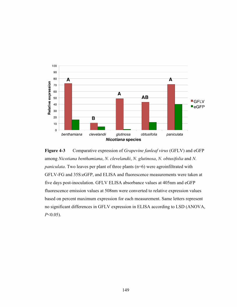
149
Figure 4-3 Comparative expression of Grapevine fanleaf virus (GFLV) and eGFP
among Nicotiana benthamiana, N. clevelandii, N. glutinosa, N. obtusifolia and N.
paniculata. Two leaves per plant of three plants (n=6) were agroinfiltrated with
GFLV-FG and 35S:eGFP, and ELISA and fluorescence measurements were taken at
five days post-inoculation. GFLV ELISA absorbance values at 405nm and eGFP
fluorescence emission values at 508nm were converted to relative expression values
based on percent maximum expression for each measurement. Same letters represent
no significant differences in GFLV expression in ELISA according to LSD (ANOVA,
P<0.05).

150
features that make it amenable to transient expression assays and evaluation of virus
accumulation detection by ELISA. First, the GFLV antibodies (Bioreba) detect
purified virus particles from strains F13 and GHu equally well (Vigne et al., 2013).
Second, the translation strategy of GFLV based on proteolytic processing of
polyproteins means that ELISA-measured virus accumulation could be representative
not only of the coat protein expression but also of the genome expression of the virus
(Scholthof et al., 1996). Third, GFLV cDNAs used in this study are identical to full-
length cDNAs that produce functional in vitro transcripts: functionalities of in vitro
transcripts FF, FG, GF, and GG were reported by Vigne et al. (2013), and
functionalities of in vitro transcripts FGG2A and GFG2A were also verified (E. Vigne,
unpublished results). These features allowed us to reliably test which factors impact
GFLV agroinfection on local and systemic levels.
Almost 1,000 plants were tested for factors involved in systemic infection
following agroinfection. A. tumefaciens strain identity [C58 (a nopaline strain) versus
LBA4404 (an octopine strain)], presence or absence of acetosyringone and plant sizes
were not identified as significant factors in systemic infection. Stepwise logit
regression procedures attempt to parse effects of individual variables (risk factors) on
a binary outcome (in this case, presence or absence systemic infection) but sometimes
cannot separate the effect of one variable from another (Anderson et al., 2003). Thus
whether some variables tested in this study truly did not impact systemic infection
remains unclear. However, the model did explain 90.4% of systemic infection
outcomes, which suggested that the variables identified as significant (greenhouse
temperatures, co-infiltration of silencing suppressors, genomes and A. tumefaciens
151
density) governed most of the successful systemic infection in these experiments.
Composition of the virus genome has major effects on successes and failures
of diverse plant virus agroinfection systems (German-Retana et al., 2003; Youssef et
al., 2011; Kurth et al., 2012). By comparing the levels of GFLV accumulation to
nonviral expression (35S:eGFP), autonomous viral expression levels could be
distinguished from protein expression due to agroinfiltration with FG showing a stable
or subtle increase in expression, GG a stable or slightly diminishing expression, and
FF and GF a rapidly diminishing expression (Figure 4-1C). This indicated that
GFLV-F13 RNA2 performed poorly and its weak performance could not be rescued
by the 2AHP coding sequence of GFLV-GHu (Figure 4-1B). Compared to eGFP
expression, FF and GF seem to be vulnerable to virus-specific suppression
mechanisms. On the other hand, FG expression showed greater expression than eGFP
at eight dpi, suggesting that the virus’s autonomous replication outpaced antiviral
defense mechanisms in N. benthamiana. The flat-line trajectory of GG between two,
five and eight dpi suggested this genome maintains equilibrium between defense and
replication.
The systemic infection frequencies of individual genomes were not always
related to their accumulation capabilities in infiltrated leaves. FFG2A outperformed FF
in terms of systemic infection but not local infection, and conversely, GFLV protein of
GG accumulated at higher levels than FF, but did not show significantly higher
systemic infection frequencies. These discrepancies suggest that factors governing
local infection are different from those that influence systemic infection. The
differences between local and systemic infection could relate to the transcript’s RNA

152
structure (Meshcheriakova et al., 2014) or to systemic GFLV movement, which is
determined by its movement and coat proteins (Andret-Link et al., 2004).
Silencing suppressors enhance agroinfection of diverse viruses (Chiba et al.,
2006; Wege and Siegmund, 2007; Yoon et al., 2011). Therefore, the effect of
silencing suppressor coinfiltrated with GFLV genomes on FF, FG, GF and GG
accumulation was tested in patch and systemic infection assays. The silencing
suppressors CMV 2b and TBSV p19 together increased FF, FG and GG accumulation
to statistically significant levels (Figure 4-2). Notably, silencing suppressors did not
equalize the genomes’ overall expression levels: with or without silencing suppressors
the pattern of infiltrated zone virus accumulation was superior for the FG genome.
This suggested that RNA silencing is an important factor that dampens viral
expression following agroinfection, but may not be the sole factor. Antiviral
mechanisms specific to A. tumefaciens-mediated delivery may operate particularly
strongly against F13 RNA2 constructs including the 2AHP recombinant construct
because the corresponding constructs resulting from in vitro transcription are perfectly
functional in planta (Amari et al., 2010; Vigne et al., 2013).
The GR construct produced similar systemic infection frequencies compared to
the analogous GHu RNA2 construct. GR is comprised of a GHu RNA2 expression
cassette with a separate CMV 2b expression cassette on the same T-DNA (Gottula et
al., 2014a). The association of high GHu RNA2 systemic infection when a silencing
suppressor is co-expressed from the same T-DNA corresponds well to the positive
correlation between coinfiltration of silencing suppressors and systemic infection. This
could suggest that the delivery of a silencing suppressor in trans is equally as effective

153
as its delivery in cis. The FGR genome produced nearly 100% infection in N.
benthamiana and formed the basis of GFLV heterologous gene delivery, reported
elsewhere (Gottula et al., 2014a).
The cloning procedure produced several constructs with a single rather than a
double CaMV 35S enhancer sequence (GHu RNA1, GHu RNA2 and the FG2A
constructs). The truncation of the CaMV 35S promoter sequence was likely due to
homology of the PCR primers with the duplicated enhancer sequences. The number of
CaMV 35S enhancers could affect the level of viral RNA expression. However, since
GHu RNA2 was the best performing RNA2 construct and GHu RNA1 was the worst
performing RNA1 construct in terms of local and systemic infection of N.
benthamiana, single versus double CaMV enhancers does not solely account for
discrepancies in agroinfection efficacies.
Transgenic N. benthamiana plants that express the full-length cDNAs of
RNA1 or RNA2 of GFLV strains F13 and GHu were used to test whether stable
integration and constitutive expression of the T-DNAs could enhance the infection
potential when the complement constructs were agroinfiltrated. Because the T-DNAs
containing RNA1 cDNAs exceed 10kb, it was hypothesized that A. tumefaciens does
not consistently integrate the full-length genome during transient expression and that
could explain the lower-than-expected infection frequency. On the contrary, stable
integration of the RNA1 or RNA2 T-DNAs decreased the infection potential
compared to transient expression experiments in which both T-DNAs were
agroinfiltrated. Calvo et al. (2010) found that stably integrated Plum pox virus cDNA
in N. benthamiana rarely converts into infective viral transcripts and that genome
154
expression is suppressed by gene silencing. Transgenically expressed RNA viral
genomes may fail to efficiently infect due to unfavorable timing or level of genome
expression relative to the onset of resistance mechanisms. .
Greenhouse temperature and density of A. tumefaciens suspension were
negatively correlated to systemic infection outcomes (Table 4-4). Higher temperatures
may negatively impact virus replication following agroinfiltration (Jiang et al., 2011)
and do negatively affect nepovirus systemic infection following mechanical
inoculation (Siddiqui et al., 2008). The negative relationship of A. tumefaciens density
on systemic infection was surprising given that higher concentrations of A.
tumefaciens can promote greater heterologous viral protein expression (Lindbo, 2007).
This could indicate that there is a fine balance between GFLV protein expression
following agroinfiltration, and activation of host defenses that target GFLV RNAs
One potential use of the GFLV constructs is to test plants for GFLV resistance
following agroinfiltration (Bhaskar et al., 2009). To determine if GFLV
agroinfiltration provide a reliable inoculation method to screen for resistance, five
Nicotiana species with varying degrees of resistance and susceptibility to mechanical
inoculation (Gottula et al., 2014b) were agroinfiltrated with the GFLV genome FG
and silencing suppressors. No relationship was obtained between pre-defined GFLV
resistance status and virus accumulation in agroinfiltrated leaves even when A.
tumefaciens transient expression for eGFP was taken into account. For example, the
susceptible N. clevelandii showed lower GFLV accumulation in agroinfiltrated zones
than the resistant N. glutinosa even though N. clevelandii produced more eGFP
fluorescence on average. These results suggested that host identity plays a major role

155
in infiltrated zone GFLV accumulation and is not related to GFLV susceptibility
following mechanical inoculation. This discrepancy could be explained by the fact that
silencing suppressors were coinfiltrated with GFLV in these experiments, which could
mask important differential innate resistance mechanisms in these hosts. Another
explanation could be the different GFLV genome composition (GFLV-GHu in
mechanical inoculation experiments and FG in transient expression experiments) with
regard to resistance/virulence interactions. It is known that host identity can play a role
in agroinfection (Satyanarayana et al., 2001; German-Retana et al., 2003), but the
mechanisms and significance of these differences observed with GFLV are unclear.
Unlike mechanical or insect vector-based inoculation strategies,
agroinoculation of RNA viruses can present artificial constraints including A.
tumefaciens pathogenesis, efficiency of faithful T-DNA transfer, nuclear transcription,
processing and export of viral RNAs, non-specific transcriptional start sites from plant
promoters, the presence of cryptic introns within viral cDNAs and altered host
resistance dynamics. Future GFLV vectors could potentially benefit from more
prudent vector designs including the use of promoters with specific transcriptional
start sites, which has shown to boost agroinoculation efficacy of Alteranthera mosaic
virus (Lim et al., 2010) and Tobacco mosaic virus (TMV) (Komarova et al., 2012a).
TMV vector cDNAs showed improved infectivity if cryptic introns were removed or
replaced by heterologous intron sequences (Marillonnet et al., 2005; Komarova et al.,
2012b). The co-dependence of the GFLV genome parts on systemic plant infection
could warrant the co-delivery of RNA1 and RNA2 components on a single T-DNA.
Single cell co-delivery of RNA1 and RNA2 could be achieved by using binary vectors
156
specialized in transformation of very large T-DNA inserts (BiBACs) (Hamilton et al.,
1999). Using a BiBAC vector was associated with markedly improved agroinfection
of the very large viral cDNA insert (about 19kb) comprising the Citrus tristeza virus
vector (Ambros et al., 2011). Nevertheless, this research has defined variables
necessary for success of GFLV agroinfection in N. benthamiana such as coinfiltration
of silencing suppressors, low temperature and GFLV genome composition, and has
raised new questions about GFLV resistance dynamics in Nicotiana species.

157
REFERENCES
Amari, K., Boutant, E., Hofmann, C., Schmitt-Keichinger, C., Fernandez-Calvino, L.,
Didier, P., Lerich, A., Mutterer, J., Thomas, C. L., Heinlein, M., Mély, Y., Maule, A.
J., and Ritzenthaler, C. 2010. A family of plasmodesmal proteins with receptor-like
properties for plant viral movement proteins. PLoS Pathogens. 6:e1001119.
Ambros, S., El-Mohtar, C., Ruiz-Ruiz, S., Leandro, P., Guerri, J., Dawson, W. O., and
Moreno, P. 2011. Agroinoculation of Citrus tristeza virus causes systemic infection
and symptoms in the presumed nonhost Nicotiana benthamiana. Molecular Plant-
Microbe Interactions. 24:1119–1131.
Anderson, R. P., Jin, R. Y., and Grunkemeier, G. L. 2003. Understanding logistic
regression analysis in clinical reports: An introduction. Annals of Thoracic Surgery.
75:753–757.
Andret-Link, P., Laporte, C., Valat, L., Ritzenthaler, C., Demangeat, G., Vigne, E.,
Laval, V., Pfeiffer, P., Stussi-Garaud, C., and Fuchs, M. 2004. Grapevine fanleaf
virus: still a major threat to the grapevine industry. Journal of Plant Pathology.
86:183–195.
Balaji, V., Sessa, G., and Smart, C. D. 2011. Silencing of host basal defense response-
related gene expression increases susceptibility of Nicotiana benthamiana to
Clavibacter michiganensis subsp. michiganensis. Phytopathology. 101:349–357.
Bedoya, L. C., Martínez, F., Orzáez, D., and Daros, J.-A. 2012. Visual tracking of
plant virus infection and movement using a reporter MYB transcription factor that
activates anthocyanin biosynthesis. Plant Physiology. 158:1130–1138.
158
Bhaskar, P. B., Venkateshwaran, M., Wu, L., Ane, J.-M., and Jiang, J. 2009.
Agrobacterium-mediated transient gene expression and silencing: A rapid tool for
functional gene assay in potato. PLoS ONE. 4:e5812.
Bubner, B., Gase, K., and Baldwin, I. T. 2004. Two-fold differences are the detection
limit for determining transgene copy numbers in plants by real-time PCR. BMC
Biotechnology. 4:14.
Calvo, M., Dujovny, G., Lucini, C., Ortuno, J., Alamillo, J. M., Simon-Mateo, C., Jose
Lopez-Moya, J., and Antonio Garcia, J. 2010. Constraints to virus infection in
Nicotiana benthamiana plants transformed with a potyvirus amplicon. BMC Plant
Biology. 10:139.
Canto, T., Uhrig, J., Swanson, M., Wright, K., and MacFarlane, S. A. 2006.
Translocation of Tomato bushy stunt virus P19 protein into the nucleus by ALY
proteins compromises its silencing suppressor activity. Journal of Virology. 80:9064–
9072.
Chee, P. P., Fober, K. A., and Slightom, J. L. 1989. Transformation of soybean
(Glycine max) by infecting germinating seeds with Agrobacterium tumefaciens. Plant
Physiology. 91:1212–1218.
Chiba, M., Reed, J., Prokhnevsky, A., Chapman, E., Mawassi, M., Koonin, E.,
Carrington, J., and Dolja, V. 2006. Diverse suppressors of RNA silencing enhance
agroinfection by a viral replicon. Virology. 346:7–14.
Choi, M. S., Yoon, I.-S., Rhee, Y., Choi, S. K., Lim, S.-H., Won, S.-Y., Lee, Y.-H.,
Choi, H.-S., Lee, S.-C., Kim, K.-H., Lomonossoff, G., and Sohn, S.-H. 2008. The
effect of Cucumber mosaic virus 2b protein to transient expression and transgene
159
silencing mediated by agro-infiltration. Plant Pathology Journal. 24:296–304.
Dawson, W. O., and Folimonova, S. Y. 2013. Virus-based transient expression vectors
for woody crops: a new frontier for vector design and use. Annual Review of
Phytopathology. 51:321–337.
Delbianco, A., Lanzoni, C., Klein, E., Rubies Autonell, C., Gilmer, D., and Ratti, C.
2013. Agroinoculation of Beet necrotic yellow vein virus cDNA clones results in plant
systemic infection and efficient Polymyxa betae transmission. Molecular Plant
Pathology. 14:422–428.
Fitzgerald, T. L., Kazan, K., and Manners, J. M. 2012. The application of reverse
genetics to polyploid plant species. Critical Reviews in Plant Sciences. 31:181–200.
Gaire, F., Schmitt, C., Stussi-Garaud, C., Pinck, L., and Ritzenthaler, C. 1999. Protein
2A of Grapevine fanleaf nepovirus is implicated in RNA2 replication and colocalizes
to the replication site. Virology. 264:25–36.
German-Retana, S., Redondo, E., Tavert-Roudet, G., Le Gall, O., and Candresse, T.
2003. Introduction of a NIa proteinase cleavage site between the reporter gene and
HC-Pro only partially restores the biological properties of GUS- or GFP-tagged LMV.
Virus Res. 98:151–162.
Gleba, Y., Klimyuk, V., and Marillonnet, S. 2007. Viral vectors for the expression of
proteins in plants. Current Opinion in Biotechnology. 18:134–141.
Gottula, J. W., Lapato, D., Cantilina, K. K., Saito, S., Bartlett, B., and Fuchs, M. 2013.
Genetic variability, evolution and biological effects of Grapevine fanleaf virus satellite
RNAs. Phytopathology. 103:1180–1187.

160
Gottula, J., Berthold, F., Labarile, R., Gutting, H., Demangeat, G., Cantilina, K.K.,
Vigne, E., Marmonier, A., Lemaire, O., Ritzenthaler, C., Schmitt-Keichinger, C., and
Fuchs, M. 2014a. Grapevine fanleaf virus and a virus-induced gene silencing and
protein expression vector for Vitis spp. Submitted.
Gottula, J., Lewis, R.S., and Fuchs, M. 2014b. Genomic basis of basal virus resistance.
Submitted.
Grimsley, N., Hohn, T., Davies, J. W., and Hohn, B. 1987. Agrobacterium-mediated
delivery of infectious Maize streak virus into maize plants. Nature. 325:177–179.
Hamilton, C. M., Frary, A., Xu, Y., Tanksley, S. D., and Zhang, H.-B. 1999.
Construction of tomato genomic DNA libraries in a binary-BAC (BIBAC) vector. The
Plant Journal. 18:223–229.
Hayward, A., Padmanabhan, M., and Dinesh-Kumar, S. P. 2011. Virus-induced gene
silencing in Nicotiana benthamiana and other plant species. Pages 55–63 in: Plant
Reverse Genetics: Methods and Protocols, Methods in Molecular Biology. A. Pereira,
ed. Springer Science+Business Media, San Diego, CA.
Hsieh, M.-H., Lu, H.-C., Pan, Z.-J., Yeh, H.-H., Wang, S.-S., Chen, W.-H., and Chen,
H.-H. 2013. Optimizing virus-induced gene silencing efficiency with Cymbidium
mosaic virus in Phalaenopsis flower. Plant Science. 202:25–41.
Jiang, C.-Z., Chen, J.-C., and Reid, M. 2011. Virus-induced gene silencing in
ornamental plants. Methods Molecular Biology. 744:81–96.
161
Kagale, S., Uzuhashi, S., Wigness, M., Bender, T., Yang, W., Borhan, M. H., and
Rozwadowski, K. 2012. TMV-Gate vectors: Gateway compatible Tobacco mosaic
virus based expression vectors for functional analysis of proteins. Scientific Reports.
2:874.
Komarova, T. V., Schwartz, A. M., Makarov, A. A., and Dorokhov, Y. L. 2012a. A
new viral vector exploiting RNA polymerase I-mediated transcription. Biochemistry
(Moscow). 77:532–538.
Komarova, T. V., Sheval, E. V., Pozdyshev, D. V., Kolesnikova, V. S., and Dorokhov,
Y. L. 2012b. Rapid and massive green fluorescent protein production leads to
formation of protein Y-bodies in plant cells. Biochemistry (Moscow). 77:603–608.
Kopertekh, L., Saint Paul, V. V., Krebs, E., and Schiemann, J. 2012. Utilization of
PVX-Cre expression vector in potato. Transgenic Research. 21:645–654.
Kurth, E., Peremyslov, V., and Prokhnevsky, A. 2012. Virus-derived gene expression
and RNAi vector for grapevine. Journal of Virology. 86:6002–6009.
Lim, H.-S., Vaira, A. M., Domier, L. L., Lee, S. C., Kim, H. G., and Hammond, J.
2010. Efficiency of VIGS and gene expression in a novel bipartite potexvirus vector
delivery system as a function of strength of TGB1 silencing suppression. Virology.
402:149–163.
Lindbo, J. A. 2007. TRBO: a high-efficiency Tobacco mosaic virus RNA-based
overexpression vector. Plant Physiology. 145:1232–1240.
Ling, K. S., Zhu, H. Y., Alvizo, H., Hu, J. S., Drong, R. F., Slightom, J. L., and
Gonsalves, D. 1997. The coat protein gene of Grapevine leafroll associated
162
closterovirus-3: cloning, nucleotide sequencing and expression in transgenic plants.
Archives of Virology. 142:1101–1116.
Liu, Y.-P., Peremyslov, V. V., Medina, V., and Dolja, V. V. 2009. Tandem leader
proteases of Grapevine leafroll-associated virus-2: Host-specific functions in the
infection cycle. Virology. 383:291–299.
Ma, M., Yan, Y., Huang, L., Chen, M., and Zhao, H. 2012. Virus-induced gene-
silencing in wheat spikes and grains and its application in functional analysis of
HMW-GS-encoding genes. BMC Plant Biology. 12:141.
Marillonnet, S., Thoeringer, C., Kandzia, R., Klimyuk, V., and Gleba, Y. 2005.
Systemic Agrobacterium tumefaciens–mediated transfection of viral replicons for
efficient transient expression in plants. Nature Biotechnology. 23:718–723.
Merzlyak, E. M., Goedhart, J., Shcherbo, D., Bulina, M. E., Shcheglov, A.S., Fradkov,
A.F, Gaintzeva, A, Lukyanov, K. A., Lukyanov, S., Gadella, T. W. J. and Chudakov,
D.M. 2007. Bright monomeric red fluorescent protein with an extended fluorescence
lifetime. Nature Methods. 4:555–557.
Meshcheriakova, Y. A., Saxena, P., and Lomonossoff, G. P. 2014. Fine-tuning levels
of heterologous gene expression in plants by orthogonal variation of the untranslated
regions of a nonreplicating transient expression system. Plant Biotechnology Journal.
doi: 10.1111/pbi.12175.
Quiapim, A. C., Brito, M. S., Bernardes, L. A. S., Dasilva, I., Malavazi, I., DePaoli, H.
C., Molfetta-Machado, J. B., Giuliatti, S., Goldman, G. H., and Goldman, M. H. S.
2009. Analysis of the Nicotiana tabacum stigma/style transcriptome reveals gene
expression differences between wet and dry stigma species. Plant Physiology.

163
149:1211–1230.
Pogue, G. P., Lindbo, J. A., Garger, S. J., and Fitzmaurice, W. P. 2002. Making an ally
from an enemy: plant virology and the new agriculture. Annual Review of
Phytopathology. 40:45–74.
Sanfaçon, H., Wellink, J., Gall, O., Karasev, A., Vlugt, R., and Wetzel, T. 2009.
Secoviridae: a proposed family of plant viruses within the order Picornavirales that
combines the families Sequiviridae and Comoviridae, the unassigned genera
Cheravirus and Sadwavirus, and the proposed genus Torradovirus. Archives of
Virology. 154:899–907.
Satyanarayana, T., Bar-Joseph, M., Mawassi, M., Albiach-Martí, M. R., Ayllón, M.
A., Gowda, S., Hilf, M. E., Moreno, P., Garnsey, S. M., and Dawson, W. O. 2001.
Amplification of Citrus tristeza virus from a cDNA clone and infection of citrus trees.
Virology. 280:87–96.
Senthil-Kumar, M., and Mysore, K. S. 2011. New dimensions for VIGS in plant
functional genomics. Trends in Plant Science. 16:656–665.
Scholthof, H. B., Scholthof, K., and Jackson, A. O. 1996. Plant virus gene vectors for
transient expression of foreign proteins in plants. Annual Review of Phytopathology.
34:299–323.
Senthil-Kumar, M., and Mysore, K. S. 2011. New dimensions for VIGS in plant
functional genomics. Trends in Plant Science. 16:656–665.
Siddiqui, S. A., Sarmiento, C., Kiisma, M., Koivumaki, S., Lemmetty, A., Truve, E.,
and Lehto, K. 2008. Effects of viral silencing suppressors on Tobacco ringspot virus

164
infection in two Nicotiana species. Journal of General Virology. 89:1502–1508.
Thuenemann, E. C., Lenzi, P., Love, A. J., Taliansky, M., Becares, M., Zuniga, S.,
Enjuanes, L., Zahmanova, G. G., Minkov, I. N., Matic, S., Noris, E., Meyers, A.,
Hattingh, A., Rybicki, E. P., Kiselev, O. I., Ravin, N. V., Eldarov, M. A., Skryabin, K.
G., and Lomonossoff, G. P. 2013. The use of transient expression systems for the
rapid production of virus-like particles in plants. Current Pharmaceutical Design.
19:5564–5573.
Vaghchhipawala, Z., Rojas, C. M., Senthil-Kumar, M., and Mysore, K. S. 2010. Pages
65-76 in: Agroinoculation and Agroinfiltration: Simple Tools for Complex Gene
Function Analyses. A. Pereira, ed. Methods in Molecular Biology, Totowa, NJ.
Valat, L., Burrus, M., Fuchs, M., and Mauro, M-C. 2003. Review of techniques to
inoculate grapevines with Grapevine fanleaf virus: lessons and perspectives. American
Journal of Enology and Viticulture. 54:279–285.
Vigne, E., Gottula, J., Schmitt-Keichinger, C., Komar, V., Ackerer, L., Belval, L.,
Rakotomalala, L., Lemaire, O., Ritzenthaler, C., and Fuchs, M. 2013. A strain-specific
segment of the RNA-dependent RNA polymerase of Grapevine fanleaf virus
determines symptoms in Nicotiana species. Journal of General Virology. 94:2803–
2813.
Viry, M., Serghini, M. A., Hans, F., Ritzenthaler, C., Pinck, M., and Pinck, L. 1993.
Biologically active transcripts from cloned cDNA of genomic Grapevine fanleaf
nepovirus RNAs. Journal of General Virology. 74:169–174.
Wege, C., and Siegmund, D. 2007. Synergism of a DNA and an RNA virus: enhanced
tissue infiltration of the begomovirus Abutilon mosaic virus (AbMV) mediated by
165
Cucumber mosaic virus (CMV). Virology. 357:10–28.
Wuriyanghan, H., and Falk, B. W. 2013. RNA interference towards the potato psyllid,
Bactericera cockerelli, is induced in plants infected with recombinant Tobacco mosaic
virus (TMV). PLoS ONE. 8:e66050.
Yepes, L. M., Fuchs, M., Slightom, J. L., and Gonsalves, D. 1996. Sense and antisense
coat protein gene constructs confer high levels of resistance to Tomato ringspot
nepovirus in transgenic Nicotiana species. Phytopathology. 86:417–424
Yoon, J.-Y., Choi, S. K., Palukaitis, P., and Gray, S. M. 2011. Agrobacterium-
mediated infection of whole plants by yellow dwarf viruses. Virus Research. 160:428–
434.
Youssef, F., Marais, A., Faure, C., Gentit, P., and Candresse, T. 2011. Strategies to
facilitate the development of uncloned or cloned infectious full-length viral cDNAs:
Apple chlorotic leaf spot virus as a case study. Virology Journal. 8:488.
Yuan, C., Li, C., Yan, L., Jackson, A. O., Liu, Z., Han, C., Yu, J., and Li, D. 2011. A
high throughput Barley stripe mosaic virus vector for virus induced gene silencing in
monocots and dicots. PLoS ONE. 6:1–16.
Zhang, H., Wang, L., Hunter, D., Voogd, C., Joyce, N., and Davies, K. 2013. A
Narcissus mosaic viral vector system for protein expression and flavonoid production.
Plant Methods. 9:28

166
CHAPTER 5
TOWARDS A QUARTER CENTURY OF PATHOGEN-DERIVED
RESISTANCE AND PRACTICAL APPROACHES TO PLANT VIRUS
DISEASE CONTROL∗
ABSTRACT
The concept of pathogen-derived resistance (PDR) describes the use of genetic
elements from a pathogen’s own genome to confer resistance in an otherwise
susceptible host via genetic engineering [J. Theor. Biol. 113 (1985) 395]. Illustrated
with the bacteriophage Qβ in Escherichia coli, this strategy was conceived as a
broadly applicable approach to engineer resistance against pathogens. For plant
viruses, the concept of PDR was validated with the creation of tobacco plants
expressing the coat protein gene of Tobacco mosaic virus (TMV) and exhibiting
resistance to infection by TMV [Science 232 (1986) 738]. Subsequently, virus-
resistant horticultural crops were developed through the expression of viral gene
constructs. Among the numerous transgenic crops produced and evaluated in the field,
papaya resistant to Papaya ringspot virus (PRSV) [Annu. Rev. Phytopathol. 36 (1998)
415] and summer squash resistant to Cucumber mosaic virus (CMV), Zucchini yellow
∗This chapter was published in: Gottula, J., and Fuchs, M. 2009. Toward a quarter
century of pathogen-derived resistance and practical approaches to plant virus
disease control. In: G. Loebstein, ed. Advances in Virus Research. 75:161-183.
Elsevier, Oxford. This materials is copyrighted by Elsevier, Inc. and is used with
permission.

167
mosaic virus, and/or Watermelon mosaic virus [Biotechnology 13 (1995) 1458] were
released for commercial use in the USA. Although cultivated on limited areas, the
adoption rate of cultivars derived from these two crops is increasing steadily. Tomato
and sweet pepper resistant to CMV and papaya resistant to PRSV were also released
in the People’s Republic of China. Applying the concept of PDR provides unique
opportunities for developing virus-resistant crops and implementing efficient and
environmentally sound management approaches to mitigate the impact of virus
diseases. Based on the tremendous progress made during the past quarter century, the
prospects of further advancing this innovative technology for practical control of virus
diseases are very promising.

168
INTRODUCTION
Plant viruses are responsible for severe economic crop losses worldwide (Hull,
2002). The development and use of resistant crop cultivars is the most efficient
strategy to mitigate the impact of virus diseases in agricultural settings. Traditionally,
host resistance is exploited by conventional breeding methods to create virus-resistant
cultivars. Protection from virus infection can be achieved by using dominant or
recessive genes. Examples of dominant resistance genes are Ry for Potato virus Y
(PVY) in potato and Sw5 for Tomato spotted wilt virus (TSWV) in tomato. The
eukaryotic translation initiation factor (eIF4E) is an example of a recessive resistance
gene for potyviruses (Kang et al., 2005; Lanfermeijer and Hille, 2007).
The concept of pathogen-derived resistance (PDR) offers a different approach
to develop virus-resistant crop plants. This concept was conceived a quarter century
ago (Sanford and Johnston, 1985). It describes the engineering of resistance in
otherwise susceptible hosts, including plants, by using genetic elements, for example
coding and noncoding sequence elements, from a pathogen’s own genome (Sanford
and Johnston, 1985). Cross-protection, a biological means for protecting plants from
virus infection, was considered as an example of the concept of PDR that is already
operational in nature. Cross-protection relies on the use of mild virus strains to protect
plants from economic damage caused by severe virus strains (Fuchs et al., 1997;
Muller and Rezende, 2004). It was argued that a mutated form of a viral replicase
similar enough to the one encoded by a challenge virus could bind to cell host
attachment sites and prevent virus replication (Sanford and Johnston, 1985).
For plant viruses, the concept of PDR was first validated with the development

169
of tobacco expressing the coat protein gene of Tobacco mosaic virus (TMV) and
exhibiting resistance to TMV infection (Powell Abel et al., 1986). This breakthrough
discovery paved the way for the creation of numerous virus-resistant transgenic plants,
including horticultural crops. Some crop plants expressing viral genetic elements have
been tested successfully in the field and a few have been commercialized. The
deployment of virus-resistant transgenic plants has become an important strategy for
effective and sustainable control of major virus diseases. This chapter provides a
historical perspective on the concept of PDR from its inception to the release of the
first virus- resistant transgenic crop resulting from its application. It also discusses
how this concept led to an explosion in the development of virus-resistant plants and
discusses advances made in terms of practical control of virus diseases during the past
25 years.
THE CONCEPT OF PDR
A description of the concept of PDR
The concept of PDR describes the use of a pathogen’s own genetic material as
resistance genes for engineering resistance in an otherwise susceptible host. Sanford
and Johnston, the two visionary scientists who articulated the concept, initially
proposed this strategy as a broadly applicable approach for genetically engineering
resistance to parasites. Resistance was hypothesized to be routinely achievable by
cloning appropriate parasite genes, modifying their expression, if necessary, and
transferring them into the host genome.
Sanford and Johnston (1985) reasoned that pathogens produce molecules that

170
are unique and critical for their pathogenic process. They proposed that dysfunctional
pathogen-derived gene products could inhibit the pathogen by disrupting pathogen–
host interactions if expressed by a host cell genome. To this extent, resistance could
theoretically be achieved from the pathogen’s own genetic material. The predicted
advantages of this approach to engineer resistance in an otherwise susceptible host
were that (i) genes from a pathogen would have a minimal effect on the host and likely
not produce substances harmful to humans, (ii) the resistance was anticipated to be
more stable than host resistance, (iii) cloning genes from a pathogen would be
relatively easy compared to host genes due to the small genome size, and (iv) genes
from a pathogen would be always present and available for cloning purposes
regardless of the diversity of the pathogen. For plant viruses, the concept of PDR and
successful transfer of foreign DNA into plant cells that regenerate into transgenic
plants opened new avenues for the development of virus-resistant plants.
A conceivable application of the concept of PDR
Sanford and Johnston (1985) used the bacteriophage Qβ as a model to illustrate
the concept of PDR. They hypothesized that four Qβ-encoded gene constructs, for
example the coat protein gene, a modified replicase gene, the RNA segment encoding
the replicase binding site, and the gene encoding the maturation protein, as well as an
antisense RNA complementary to the Qβ RNA could be used as resistance genes
against the bacteriophage Qβ in Escherichia coli. Sanford and Johnston (1985) further
suggested that the strategy outlined for the bacteriophage Qβ in Escherichia coli could
have a broader application for engineering resistance to other pathogens, opening an
171
unsuspected path for practical control of diseases. To this extent, the stage was set for
innovative ways to create virus-resistant plants by applying the concept of PDR and
developing efficient protocols for plant transformation.
HISTORICAL PERSPECTIVES
The first application of the concept of PDR for virus resistance in a model host
Powell Abel et al. (1986) were the first to apply the concept of PDR to a plant
virus. These authors produced Nicotiana tabacum cv. Xanthi and cv. Samsun
expressing the coat protein gene of TMV and showed that transgenic tobacco
exhibited resistance following infection by TMV via mechanical inoculation. Some
transgenic tobacco failed to express symptoms for the duration of the experiments
whereas others exhibited a substantial delay (two to 14 days) in disease development.
Resistance was related to the level of expression of the viral coat protein and could be
overcome by high doses of inoculum under which conditions plants developed typical
systemic symptoms and systemically infected leaves contained high TMV titer
(Powell Abel et al., 1986). Plants had only a slight enhanced resistance to TMV RNA
as inoculum (Nelson et al., 1987). The resistance was strong to tobamoviruses closely
related to TMV but weak or not detectable to distantly related tobamoviruses (Nejidat
and Beachy, 1990). Additional experiments suggested that increased levels of TMV
coat protein expression correlated with increased levels of resistance (Osbourn et al.,
1989; Powell Abel et al., 1989; Prins et al., 2008; Register and Beachy, 1988, 1989).
The initial intent of Beachy and colleagues for transferring and expressing the TMV
coat protein gene into tobacco was to gain a better understanding of the mechanisms
172
of cross-protection and provide new insights into virus–host interactions. Their
seminal work launched a new era for the production of virus-resistant plants.
Other early applications of the concept of PDR for virus resistance
As a consequence of the discovery by Powell Abel et al. (1986), resistance to
numerous plant viruses was engineered primarily by using coat protein genes (Beachy
et al., 1990; Prins et al., 2008; Tepfer, 2002). Other viral sequences, such as the RNA-
dependent RNA polymerase read-through domain of TMV were also shown to induce
resistance (Golembowski et al., 1990), as well as the movement protein (Malyshenko
et al., 1993), proteinase (Maiti et al., 1993; Vardi et al., 1993), satellite RNA (Gerlach
et al., 1987; Harrison et al., 1987), defective interfering RNA (Kollar et al., 1993), and
5’ (Nelson et al., 1993; Stanley et al., 1990) and 3’ (Zaccomer et al., 1993) noncoding
regions. It soon became apparent that almost any viral genetic element could be used
to confer resistance to virus infection in plants. These observations validated some of
the earlier predictions by Sanford and Johnston (1985) on the notion that several genes
from a pathogen could be used to engineer resistance.
The concept of PDR and the antiviral pathways of RNA silencing
The mechanism of engineered resistance through the application of the concept
of PDR was poorly understood 25 years ago. It was hypothesized that a dysfunctional
viral gene in a host could somehow interfere with virus multiplication. By analogy
with cross-protection, the mechanisms consisted conceivably of competition for host
factors, inhibition of the uncoating of challenge virus (Sherwood, 1987), disruption of
173
the replication of the challenge virus due to annealing of RNA species of the
protective and challenge viruses (Palukaitis and Zaitlin, 1984), among other plausible
explanations.
Expression of a viral coat protein in a transgenic plant was suggested initially
to interfere with the uncoating step during an early event of the virus multiplication
cycle (Osbourn et al., 1989; Register and Beachy, 1988). Interaction of the viral coat
protein with a host component or directly with the challenge viral RNA was
hypothesized to prevent replication, translation, or virion assembly (Asurmendi et al.,
2007; Beachy, 1997, 1999; Bendahmane and Beachy, 1999; Clark et al., 1995).
Subsequently, a breakthrough discovery showed that an untranslatable coat protein
gene of Tobacco etch virus (TEV) protected tobacco plants from TEV infection.
Resistant plants were immune to TEV infection (Lindbo and Dougherty, 1992a, b). A
recovery phenotype was also observed with plants infected and displaying symptoms
similar to those of nontransgenic plants but newly emerging leaves were asymptomatic
two weeks post-inoculation and transgene mRNA as well as viral RNA were rapidly
degraded (Lindbo et al., 1993). It became clear that the viral transgene protein product
was not needed for engineered resistance and that there was an inverse correlation
between transgene expression and resistance to virus infection (Dougherty et al.,
1994). In other words, the TEV coat protein RNA sequence was responsible for the
resistance phenotype rather than the coat protein itself. This was unexpected as it was
suggested that plants expressing high levels of viral coat protein would be likely
resistant to virus infection in comparison with plants expressing little or no viral coat
protein (Lindbo and Dougherty, 2005). Similar findings were published early on for

174
TSWV (de Haan et al., 1992) and Potato virus X (PVX) (Longstaff et al., 1993) in
tobacco plants. While coat protein-mediated resistance is effective against a number of
viruses (Asurmendi et al., 2007; Bendahmane and Beachy, 1999; Dinant et al., 1998;
Schubert et al., 2004; Wintermantel and Zaitlin, 2000), the majority of PDR
phenomena seem to work through RNA-mediated mechanisms (Baulcombe, 2007;
Eamens et al., 2008; Prins et al., 2008; Voinnet, 2008).
Plant RNA-dependent RNA polymerase and double-stranded (ds) RNAase
activities were proposed to be part of the mechanism of resistance by producing short
RNA of 10–20 nt in length complementary in sequence to the RNA to be degraded
from the transgene RNAs (Lindbo et al., 1993). These short RNAs would target
specific RNAs for degradation by a dsRNase activity (Dougherty and Parks, 1995).
The studies by Dougherty and colleagues advanced our understanding of the
mechanisms underlying engineered virus resistance in plants and highlighted the role
of a sequence-specific RNA degradation phenomenon through post-transcriptional
gene silencing (PTGS). Their findings paved the way to the discovery of RNA
silencing as a potent defense mechanism against plant viruses (Baulcombe, 2004,
2007; Eamens et al., 2008; Lin et al., 2007; Prins et al., 2008; Voinnet, 2001, 2005,
2008; Waterhouse et al., 1999, 2001). Later, it was shown that antiviral silencing
occurred during the recovery phase of virus infection in nontransgenic plants (Covey
et al., 1997; Ratcliff et al., 1997).
RNA silencing is initiated by double-stranded RNA (dsRNA) structures that
are identical to the RNA to be degraded (Waterhouse et al., 1998). Silencing is
associated with the production of 21–25 nt duplexes called small interfering RNAs
175
(siRNAs) (Hamilton and Baulcombe, 1999; Hamilton et al., 2002). The siRNAs are
produced from dsRNA precursors by an endonuclease known as Dicer and become
incorporated and converted to single-stranded RNAs (ssRNAs) in a Argonaute-
containing ribonuclease complex (RISC) that target RNA for cleavage (Deleris et al.,
2006; Hannon, 2002; Obbard et al., 2009; Voinnet, 2001, 2005, 2008). The pioneering
work by Baulcombe and Waterhouse and their respective colleagues showed that RNA
silencing is an innate and potent plant response to virus infection and a natural
example of the concept of PDR.
The first application of the concept of PDR to a horticultural crop
Soon after its first application for virus resistance in a model host plant (Powell
Abel et al., 1986), the concept of PDR was validated in a horticultural crop with the
aim of providing practical control of a viral disease. Tomato was the first horticultural
crop engineered for virus resistance through the application of the concept of PDR. In
the first field trial ever of transgenic plants engineered for virus resistance, tomato
plants expressing the coat protein gene of TMV were evaluated for resistance to
mechanical inoculation by TMV (Nelson et al., 1988). Only 5% of the transgenic
plants were symptomatic at the end of the trial compared with 99% of the
nontransformed control plants. Also, inoculated transgenic and uninoculated
nontransformed plants had identical fruit yield, indicating that the transformation
process and expression of the TMV coat protein gene did not alter the horticultural
performance of the transgenic tomato plants. Sanders et al. (1992) extended the field
characterization of transgenic tomato plants and showed resistance to distinct strains
176
of TMV. These studies confirmed Sanford and Johnston’s conception of PDR as a
practical solution for controlling virus diseases in plants.
CREATION OF VIRUS-RESISTANT TRANSGENIC CROPS BY APPLYING
THE CONCEPT OF PDR
Early applications
Effective resistance is desirable against virus inoculation via vectors to
manage, for instance, aphid-transmitted virus diseases. The efficiency of viral genes at
conferring resistance against vector-mediated virus transmission was shown first with
cucumber plants engineered for resistance to Cucumber mosaic virus (CMV).
Cucumber plants expressing the coat protein gene of CMV had a significantly reduced
incidence of CMV and a lower percentage of symptomatic plants than nontransformed
control plants following CMV inoculation via aphid vectors (Gonsalves et al., 1992).
In these studies, mechanically inoculated cucumber plants dispersed throughout the
field provided reliable sources of inoculum for natural aphid populations to vector
CMV. This approach coupled with the fact that field trials were established at a time
of abundant endemic aphid flights caused sufficient disease pressure to make
inferences about disease progress, resistance, and yield (Gonsalves et al., 1992).
Subsequently, many other studies have illustrated the usefulness of engineered
resistance at providing practical control of aphid-transmitted virus diseases (reviewed
by Fuchs and Gonsalves, 2007).
Resistance to more than one virus is useful for practical control of virus
diseases as mixed virus infections are common in agricultural settings. PDR offers
177
unique solutions to mixed virus infection, for example, by co-engineering and co-
transferring genes from several viruses into a single host plant. The usefulness of
multiple viral genes to control mixed virus infections was demonstrated early on with
potato plants expressing the coat protein genes of PVX and PVY (Kaniewski et al.,
1990; Lawson et al., 1990). Potato line 303 was highly resistant to infections by PVX
and PVY in the field (Kaniewski et al., 1990). Later, summer squash plants expressing
coat protein gene constructs of CMV, Zucchini yellow mosaic virus (ZYMV), and/or
Watermelon mosaic virus (WMV) were engineered for resistance to single viruses and
combinations of these three viruses (Tricoli et al., 1995). Among summer squash
engineered for multiple virus resistance, line ZW-20 expressing the coat protein genes
of ZYMV, and WMV was highly resistant whether infection occurred by mechanical
inoculation or was mediated by aphid vectors (Fuchs and Gonsalves, 1995; Tricoli et
al., 1995). In addition, line CZW-3 expressing the coat protein genes of CMV, ZYMV
and WMV was highly resistance to mixed infections by these three viruses (Fuchs et
al., 1998; Tricoli et al., 1995). The three coat protein genes used to engineer multiple
virus resistance in summer squash were transferred successfully in cantaloupes (Fuchs
et al., 1997). The concept of PDR has provided a platform for virus control that has
facilitated new approaches to develop resistant crop cultivars and expanded
opportunities to implement effective and sustainable management strategies of virus
diseases.
Other examples
Agronomic and horticultural plants, such as cereal, vegetable, legume, flower,

178
forage, turf, and fruit crops expressing virus-derived gene constructs have been created
(Fuchs and Gonsalves, 2007). While testing in the field is underway for at least one
dozen crop species expressing sequences derived from numerous viruses, very few of
these field trials have yet been published in scientific journals.
Part of the difficulty of field-testing for resistance evaluation is ensuring high
and consistent virus inoculation that can distinguish resistant and susceptible
phenotypes (Gilbert et al., 2009). Inoculation from external field sources can be
reliable when studies are with insect-transmitted viruses. Presence of naturally
viruliferous aphid populations allowed Lee et al. (2009) to discern resistant pepper
expressing a CMV coat protein gene in conditions relevant to commercial agriculture.
Natural infection by thrips vectors yielded statistically significant differences in
TSWV incidence in peanut expressing an antisense TSWV nucleoprotein sequence
(Magbanua et al., 2000). In another study, peanut expressing a TSWV nucleoprotein
had a strong tendency to be asymptomatic under field locations, although resistance
was moderate following mechanical inoculation in a growth chamber (Yang et al.,
2004). Transgenic peanut and pepper showed good yield and quality parameters,
respectively (Lee et al., 2009; Yang et al., 2004). For perennial crops, plum trees
expressing a coat protein gene construct of Plum pox virus (PPV) were highly resistant
to PPV infection during 6–8 years in varied orchard locations in Europe (Capote et al.,
2007; Hily et al., 2004; Malinowski et al., 2006; Ravelonandro, 2007; Zagrai et al.,
2008). The growth of knowledge about RNA silencing has provided a basis to
optimize constructs to make engineered resistance to viruses more reliable and broadly
applicable.
179
More recent applications
The trigger for RNA silencing is dsRNA or double-stranded regions within the
secondary structure of single-stranded RNA (Eamens et al., 2008; Prins et al., 2008;
Voinnet, 2008; Waterhouse et al., 1998). Several approaches have been used to
express dsRNA cognate to viral RNA for activation of RNA silencing. Expressing
sense and antisense viral genes or inverted repeat viral genes to express hairpin RNAs
(hpRNA) for the formation of duplex RNA are some of the most recent strategies to
engineer resistance (Missiou et al., 2004; Praveen et al., 2009; Prins et al., 2008;
Smith et al., 2000; Tougou et al., 2006; Wesley et al., 2001). For example, intron-
spliced hairpin RNA (ihpRNA), ihpRNA overhang, and ihpRNA spacer were
evaluated for resistance to PVY (Smith et al., 2000; Wesley et al., 2001). The ihpRNA
was found to be the most efficient constructs to conferring resistance to PVY with
90% of the plants exhibiting RNA silencing (Wesley et al., 2001). The same strategy
based on the use of highly conserved genetic segments of several viruses into a single
transgene construct achieved multiple virus resistance (Bucher et al., 1996).
Artificial plant micro RNA (amiRNAs) can also be used for virus resistance.
The Arabidopsis thaliana pre-miR159a precursor was used to generate two
amiRNAs159 (amiR-P69159 and amiR-HC-Pro159) with sequences complementary to
Turnip yellow mosaic virus (TYMV) and Turnip mosaic virus (TuMV), respectively
(Niu et al., 2006). The amiR- P69159 was designed to target the TYMV silencing
suppressor P69 while amiR-HC-Pro159 targeted the TuMV silencing suppressor HC-
Pro. Transgenic plants carrying both transgenes expressed the corresponding
amiRNAs and showed specific resistance to TYMV and TuMV. Low temperatures
180
had no substantial effect on miRNA accumulation (Niu et al., 2006). Similarly, the
miR171 of Nicotiana benthamiana was used to target the 2b gene of CMV and confer
resistance to CMV (Qu et al., 2008).
COMMERCIALIZATION OF VIRUS-RESISTANT TRANSGENIC CROPS AND
PRACTICAL CONTROL OF VIRUS DISEASES
Virus-resistant summer squash
Summer squash expressing the CP gene of ZYMV and WMV received
exemption status in the USA in 1994 and was released thereafter. This was the first
disease-resistant transgenic crop to be commercialized in the USA (Table 5-1). Plants
of line ZW-20 are vigorous following exposure to aphid-mediated transmission of
ZYMV and WMV (Figure 5-1A) and produce marketable fruits (Figure 5-1B) unlike
conventional squash. Summer squash expressing the CP gene of CMV, WMV, and
ZYMV was deregulated and commercialized in 1996. Subsequently, numerous squash
types and cultivars have been developed by crosses and backcrosses with the two
initially deregulated lines. Currently there are five zucchini and six straightneck or
crookneck yellow squash cultivars for which combinations of resistance to ZYMV and
WMV or resistance to CMV, ZYMV, and WMV are available.
The adoption of virus-resistant summer squash cultivars is steadily increasing
since 1996. In 2006, the adoption rate was estimated to 22% (3,250 hectares) across
the country with an average rate of 70% in New Jersey and 20% in Florida, Georgia,
and South Carolina. The benefit to growers was estimated to $24 million in 2006
(Johnson et al., 2007).
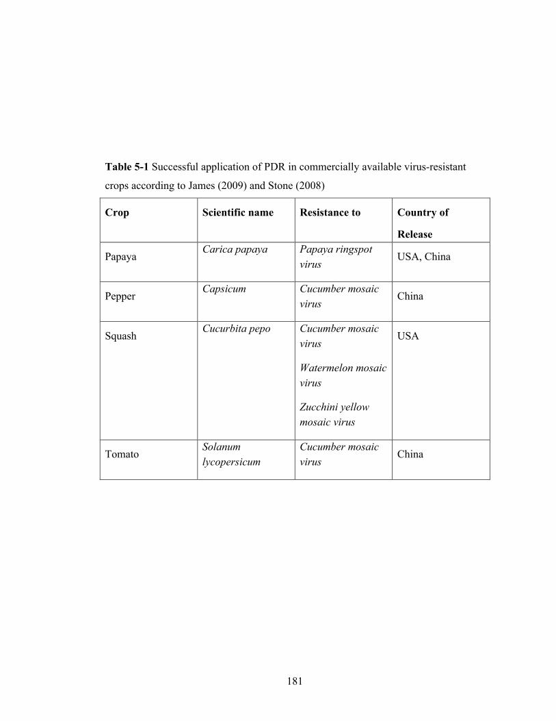
181
Table 5-1 Successful application of PDR in commercially available virus-resistant
crops according to James (2009) and Stone (2008)
Crop Scientific name Resistance to Country of
Release
Papaya Carica papaya Papaya ringspot
virus USA, China
Pepper Capsicum Cucumber mosaic
virus China
Squash Cucurbita pepo Cucumber mosaic
virus
Watermelon mosaic virus
Zucchini yellow mosaic virus
USA
Tomato Solanum lycopersicum
Cucumber mosaic virus
China
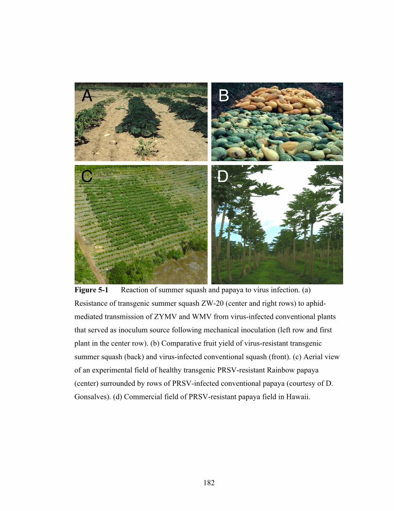
182
Figure 5-1 Reaction of summer squash and papaya to virus infection. (a)
Resistance of transgenic summer squash ZW-20 (center and right rows) to aphid-
mediated transmission of ZYMV and WMV from virus-infected conventional plants
that served as inoculum source following mechanical inoculation (left row and first
plant in the center row). (b) Comparative fruit yield of virus-resistant transgenic
summer squash (back) and virus-infected conventional squash (front). (c) Aerial view
of an experimental field of healthy transgenic PRSV-resistant Rainbow papaya
(center) surrounded by rows of PRSV-infected conventional papaya (courtesy of D.
Gonsalves). (d) Commercial field of PRSV-resistant papaya field in Hawaii.

183
Virus-resistant papaya
Papaya expressing the coat protein gene of Papaya ringspot virus (PRSV) was
deregulated in 1998 and commercialized in Hawaii (Table 5-1). PRSV is a major
limiting factor to papaya production in Hawaii and around the world. After extensive
experimental testing (Figure 5-1C), PRSV-resistant papaya was released in 1998 as
devastation caused by the virus reached record proportions in the archipelago’s main
production region (Gonsalves, 1998). The impact of PRSV-resistant papaya on the
papaya industry in Hawaii is evidenced by its rapid adoption rate (Figure 5-1D). In
2000, the first wave of transgenic papaya bore fruit on more than 42% of the total
acreage (Johnson et al., 2007). Resumption of fruitful harvests put papaya packing
houses back in business and provided a $4.3 million impact over a 6-year period
(Fuchs, 2008).
By 2006, transgenic papaya cultivars were planted on more than 90% of the
total papaya land in Hawaii (780 of 866 total hectares) (Johnson et al., 2007), with the
remaining conventional fruit shipped mainly to Japan, one of the major export
countries for the Hawaiian papaya industry along with Canada (Suzuki et al., 2007).
After a decade of segregating transgenic and nontransgenic papaya fruits, this practice
may be nearing end due to the recent deregulation of the transgenic fruit in Japan (D.
Gonsalves, personal communication) following deregulation in Canada (Suzuki et al.,
2007).
Other examples
Two virus-resistant potato lines were deregulated in 1998 and 2000 in the

184
USA. After failed attempts to create a potato line resistant to Potato leafroll virus
(PLRV) by coat protein gene expression, lines expressing a PLRV replicase gene were
created, field tested, deregulated, and commercialized (Kaniewski and Thomas, 2004).
Later, this resistance was stacked with a synthetic Cry gene that conferred resistance
to Colorado potato beetle. Another potato cultivar was developed by adding the coat
protein gene of PVY. Although many growers in the Pacific Northwest, Midwest US
and Canada were growing transgenic potato, and no resistance breakage was reported,
nor any detrimental impact on the environmental or human health, virus-resistant
potato were withdrawn from the market after the 2001 season due to the reluctance of
several large processors and exporters to adopt these products (Kaniewski and
Thomas, 2004).
In the People’s Republic of China, tomato and sweet pepper resistant to CMV
were released as well as papaya resistant to PRSV (James, 2009; Stone, 2008) (Table
4-1). Limited if any, information is available on their adoption rate.
Although not released yet, the plum cultivar ‘Honeysweet’ resistant to PPV is under
consideration for deregulation in the USA. The US Department of Agriculture
(USDA) Animal and Plant Health Inspection Service (APHIS) has granted this
cultivar deregulated status (Bech, 2007) and the Food and Drug Administration (FDA)
has deemed a pre-market review of the ‘Honeysweet’ unnecessary. Presently, the
Environmental Protection Agency (EPA) is examining deregulation petitions for
‘Honeysweet.’ Another PRSV-resistant papaya has been deregulated by two of the
three US biotechnology regulatory authorities. Line X17-2 differs from the previously
deregulated Hawaiian papaya in that it expresses the CP gene of a Florida isolate of

185
PRSV and is suitable for cultivation in Florida (Davis, 2004). APHIS and the FDA
have granted X17-2 deregulated status (Anonymous, 2009; Shea, 2009). The realized
economic benefits and minimal environmental hazards of the previously deregulated
virus-resistant Hawaiian papaya figured prominently into APHIS’ favorable
consideration (Gregoire and Abel, 2008). The EPA will consider the plant pest risk of
X17-2 after the developer submits a petition for deregulation.
Stability and durability of engineered virus resistance
Plant viruses can evade the antiviral defense response by encoding RNA
silencing-suppressor genes (Díaz-Pendón and Ding, 2008; Ding and Voinnet, 2007;
Eamens et al., 2008; Li and Ding, 2006; Voinnet, 2008). The HC-Pro protein of TEV
and the 2b protein of CMV were amongst the first viral suppressors of transgene
silencing identified (Anandalakshmi et al., 1998; Brigneti et al., 1998; Kasschau and
Carrington, 1998). Silencing suppressors from different plant viruses counteract
various steps in the RNA silencing process (Díaz-Pendón and Ding, 2008; Ding and
Voinnet, 2007; Li and Ding, 2006; Voinnet et al., 1999). As a consequence, silencing-
based resistance to one virus can be partially counteracted by infection with an
unrelated virus carrying a silencing suppressor gene (Mitter et al., 2003). Such an
effect was not observed with plum trees expressing the PPV CP gene (Ravelonandro,
2007; Zagrai et al., 2008) or summer squash expressing the CP genes of CMV,
ZYMV, and WMV (Fuchs et al., 1998; Tricoli et al., 1995) following infection with
heterologous viruses.
Transcriptional gene silencing and genetic background are two documented

186
variables that can cause transgenic plants expressing viral sequences to lack the
expected resistance phenotype (Febres et al., 2008). Wheat transformed with the coat
protein gene or replicase gene of Wheat streak mosaic virus tended to display more
severe symptoms and higher relative virus titers in the field compared to the
nontransformed parent cultivar (Sharp et al., 2002). These results suggested that
environmental conditions can affect the stability of engineered resistance since all
transgenic lines showed a recovery phenotype in greenhouse experiments. It is known
that RNA silencing is inactive at low temperatures (Szittya et al., 2003). This provides
good conceptual rationale to pyramid virus-derived transgenes with conventional
resistance genes, which can be inactivated at high temperature (Wang et al., 2009).
Resistance breakdown has not been reported in more than 10 years of
commercial deployment of transgenic summer squash and papaya. Similarly,
resistance is durable for PPV-resistant plum trees tested in experimental orchards over
13 years in Europe, despite constant exposure to viruliferous aphids vectoring diverse
PPV populations (Capote et al., 2007; Malinowski et al., 2006; Ravelonandro, 2007;
Zagrai et al., 2007). In contrast, resistance breakdown has been demonstrated in
laboratory and greenhouse settings with papaya. Early work revealed that resistance to
PRSV was narrow in cultivars expressing the CP gene from a Hawaiian isolate of
PRSV; plants were resistant to PRSV isolates from Hawaii but largely susceptible to
isolates outside of Hawaii, depending on the extent of sequence divergence (Suzuki et
al., 2007). Efforts to pyramid genes from highly conserved region of the PRSV
genome from various isolates for broad-spectrum resistance are underway.

187
DISCUSSION
The concept of PDR was described a quarter century ago (Sanford and
Johnston, 1985). This theory has provided a framework to engineer genetic constructs
from a viral genome and use them as resistance genes to protect plants from virus
infection. Application of this conceptual knowledge has introduced novel approaches
for virus control by providing new means to develop resistant crop cultivars and
increase opportunities to implement effective and sustainable management strategies
of virus diseases. After its validation with TMV in tobacco plants in 1986, the concept
of PDR has been applied successfully against a wide range of viruses in many plant
species so that the past 25 years have witnessed an explosion in the development of
virus-resistant transgenic plants.
Several virus-resistant transgenic crops resulting from the application of PDR
have been extensively evaluated under field conditions and many more have been
created and validated in laboratory or greenhouse conditions. The first resistant
horticultural crops resulting from the application of PDR were vegetable (summer
squash, sweet pepper, tomato, and potato) and fruit (papaya and plum) crops. Based on
their efficacy at controlling virus diseases (Eamens et al., 2008; Hily et al., 2004;
Prins et al., 2008; Suzuki et al., 2007; Tricoli et al., 1995), a history of ready adoption
by growers (Suzuki et al., 2007) and no documented detrimental environmental impact
(Fuchs and Gonsalves, 2007), more virus-resistant transgenic crops are likely to reach
the market in the future. While several crop plants show good resistance to virus
infection in the field, the dearth of commercialized examples beyond summer squash,
papaya, tomato, and sweet pepper suggests that steep legal or regulatory issues, among
188
other issues, have barred market entry.
The dedication and perseverance on the part of a handful of researchers in the
public and private sectors have extended PDR beyond an academic exercise to a
proven technology for commercial use and efficient management of virus diseases.
The creation and deployment of PRSV-resistant papaya have provided a safe and
effective way to save an entire fruit industry on the Hawaiian Islands. The same could
be true for Thailand but for negative intervention by an international nongovernmental
organization (Davidson, 2008). Virus-resistant summer squash and potato have been
deregulated in the USA but only summer squash remain commercially available to
date. A virus-resistant plum and another virus-resistant papaya await full deregulation
in the USA. The People’s Republic of China is likewise moving forward with virus-
resistant transgenic crops and has already commercialized virus-resistant sweet
pepper, tomato, and papaya (James, 2009; Stone, 2008).
The application of the concept of PDR also paved the way for tremendous
progress to be made at unraveling the biology of antiviral pathways of RNA silencing
in plants, a natural and potent defense mechanism against viruses that can be triggered
by the insertion and expression of viral gene constructs in susceptible hosts
(Baulcombe, 2007; Eamens et al., 2008; Lin et al., 2007; Obbard et al., 2009; Prins et
al., 2008; Voinnet, 2008). Knowledge of RNA silencing has provided new and
unprecedented insights into virus–host interactions. dsRNA was identified as trigger of
the antiviral defense mechanism, virus-encoded silencing suppressors as counterattack
factors and symptom inducers, and pathogen-homing siRNAs as guides for the
destruction of viral RNA by RISC (Baulcombe, 2007; Eamens et al., 2008; Lin et al.,
189
2007; Obbard et al., 2009; Voinnet, 2008). These developments stemming from the
theory of PDR (Sanford and Johnston, 1985) shed light on the molecular and cellular
mechanisms underlying engineered resistance in plants expressing virus-derived gene
constructs.
The concept of PDR (Sanford and Johnston, 1985) provided unique
opportunities for innovative solutions to control virus diseases by developing virus-
resistant crops expressing genetic elements derived from a virus’ own genome. A
quarter century later, lessons from field experiments with various transgenic crops
engineered for virus resistance and the commercial release of virus-resistant papaya,
summer squash, sweet pepper, and tomato have conclusively demonstrated that
applying the concept PDR is a practical strategy to mitigate the impact of virus
diseases on agriculture.

190
REFERENCES
Anandalakshmi, R., Pruss, G. J., Ge, X., Marathe, R., Mallory, A. C., Smith, T. H.,
and Vance, V. B. 1998. A viral suppressor of gene silencing in plants. Proceedings of
the National Academy of Sciences USA. 95:13079–13084.
Anonymous. 2009. Completed consultations on bioengineered foods. U.S. Food and
Drug Administration.
http://www.fda.gov/Food/Biotechnology/Submissions/default.htm. Accessed 1 July
2009.
Asurmendi, S., Berg, R. H., Smith, T. J., Bendahmane, M., and Beachy, R. N. 2007.
Aggregation of TMV CP plays a role in CP functions and in coat-protein-mediated
resistance. Virology. 366:98–106.
Bech, R. 2007. Finding of no significant impact and decision notice. Determination of
nonregulated status for C5 plum resistant to Plum pox virus. USDA Animal and Plant
Health Inspection Service. http://www.aphis.usda.gov/brs/aphis-
docs2/04_26401p_com.pdf. Accessed 1 July 2009.
Baulcombe, D. C. 2007. Amplified Silencing. Science. 315:199–200.
Baulcombe, D. C. 2004. RNA silencing in plants. Nature. 431:356–363.
Beachy, R. N. 1997. Mechanisms and application of pathogen-derived resistance in
transgenic plants. Current Opinion in Plant Biotechnology. 8:215–220.
191
Beachy, R. N. 1999. Coat-protein-mediated resistance to Tobacco mosaic virus:
discovery mechanisms and exploitation. Philosophical Transactions of the Royal
Society of Biological Sciences. 354:659–664.
Beachy, R. N., Loesch-Fries, S., and Tumer, N. E. 1990. Coat protein-mediated
resistance against virus infection. Annual Review of Phytopathology. 28:451–472.
Bendahmane, M., and Beachy, R. N. 1999. Control of tobamovirus infections via
pathogen-derived resistance. Adv. Virus Research. 53:369–386.
Brigneti, G. 1998. Viral pathogenicity determinants are suppressors of transgene
silencing in Nicotiana benthamiana. The EMBO Journal. 17:6739–6746.
Bucher, E., Lohuis, D., van Poppel, P. M. J. A., Geerts-Dimitriadou, C., Goldbach, R.,
and Prins, M. 2006. Multiple virus resistance at a high frequency using a single
transgene construct. Journal of General Virology. 87:3697–3701.
Capote, N., Pérez-Panadés, J., Monzó, C., Carbonell, E., Urbaneja, A., Scorza, R.,
Ravelonandro, M., and Cambra, M. 2008. Assessment of the diversity and dynamics
of Plum pox virus and aphid populations in transgenic European plums under
Mediterranean conditions. Transgenic Research. 17:367–377.
Clark, W. G., Fitchen, J. H., and Beachy, R. N. 1995. Studies of coat protein-mediated
resistance to TMV. I. The PM2 assembly defective mutant confers resistance to TMV.
Virology. 208:485–491.
Davidson, S. N. 2008. Forbidden fruit: transgenic papaya in Thailand. Plant
Physiology. 147:487–493.
192
Davis, M. J. 2004. Petition for determination of nonregulated status for the X17-2 line
of papaya: a Papaya ringspot virus-resistant papaya. USDA Animal and Plant Health
Inspection Service. http://www.aphis.usda.gov/brs/aphisdocs/ 04_33701p.pdf.
Accessed 1 July 2009.
de Haan, P., Gielen, J. J. L., Prins, M., Wijkamp, I. G., Schepen, A. V., Peters, D.,
Grinsven, M. Q. J. M. V., and Goldbach, R. 1992. Characterization of RNA–mediated
resistance to Tomato spotted wilt virus in transgenic tobacco plants. Nature
Biotechnology. 10:1133–1137.
Deleris, A., Gallego-Bartolome, J., Bao, J., Kasschau, K. D., Carrington, J. C., and
Voinnet, O. 2006. Hierarchical action and inhibition of plant dicer-like proteins in
antiviral defense. Science. 313:68–71.
Dinant, S., Kusiak, C., Cailleteau, B., Verrier, J. L., Chupeau, M. C., Chupeau, Y., Le,
T. A. H., Delon, R., and Albouy, J. 1998. Field resistance against Potato virus Y
infection using natural and genetically engineered resistance genes. European Journal
of Plant Pathology. 104:377–382.
Ding, S.-W., and Voinnet, O. 2007. Antiviral immunity directed by small RNAs. Cell.
130:413–426.
Díaz-Pendón, J. A., and Ding, S. W. 2008. Direct and indirect roles of viral
suppressors of RNA silencing in pathogenesis. Annual Review of Phytopathology.
46:303–326.
Dougherty, W. G., and Parks, T. D. 1995. Transgene and gene suppression: telling us
something new? Current Opinion in Cell Biology. 7:399–405.

193
Dougherty, W. G., Lindbo, J. A., Smith, H. A., Parks, T. D., Swaney, S., and
Proebsting, W. M. 1994. RNA-mediated virus resistance in transgenic plants:
exploitation of a cellular pathway possibly involved in RNA degradation. Molecular
Plant-Microbe Interactions. 7:544–552.
Eamens, A., Wang, M. B., Smith, N. A., and Waterhouse, P. M. 2008. RNA silencing
in plants: yesterday, today, and tomorrow. Plant Physiology. 147:456–468.
Febres, V. J., Lee, R. F., and Moore, G. A. 2008. Transgenic resistance to Citrus
tristeza virus in grapefruit. Plant Cell Reports. 27:93–104.
Fuchs, M. 2008. Plant resistance to viruses: Engineered resistance. Pages 156–164 in:
Encyclopedia of Virology. Mahy, B. W. J. and van Regenmortel, M., eds. Academic
Press, San Diego, CA.
Fuchs, M., and Gonsalves, D. 1995. Resistance of transgenic squash Pavo ZW-20
expressing the coat protein genes of zucchini yellow mosaic virus and watermelon
mosaic virus 2 to mixed infections by both potyviruses. Bio/ Biotechnol. 13:1466–
1473.
Fuchs, M., and Gonsalves, D. 2007. Safety of virus-resistant transgenic plants two
decades after their introduction: lessons from realistic field risk assessment studies.
Annual Review of Phytopathology. 45:173–202.
Fuchs, M., Ferreira, S., and Gonsalves, D. 1997. Management of virus diseases by
classical and engineered protection. Molecular Plant Pathology Online.
http://www.bspp.org.uk/mppol/download/0116fuch.zip.

194
Fuchs, M., Tricoli, D. M., Carney, K. J., Schesser, M., McFerson, J. R., and
Gonsalves, D. 1998. Comparative virus resistance and fruit yield of transgenic squash
with single and multiple coat protein genes. Plant Disease. 82:1350–1356
Gerlach, W. L., Llewellyn, D., and Haseloff, J. 1987. Construction of a disease
resistant gene using the satellite RNA to Tobacco ringspot virus. Nature. 328:802–
805.
Gilbert, R. A., Glynn, N. C., Comstock, J. C., and Davis, M. J. 2009. Agronomic
performance and genetic characterization of sugarcane transformed for resistance to
Sugarcane yellow leaf virus. Field Crops Research. 111:39–46.
Golemboski, D. B., Lomonossoff, G. P., and Zaitlin, M. 1990. Plants transformed with
a Tobacco mosaic virus nonstructural gene sequence are resistant to the virus.
Proceedings of the National Academy of Sciences USA. 87:6311–6315.
Gonsalves, D. 1998. Control of Papaya ringspot virus in papaya: A case study.
Annual Review of Phytopathology. 36:415–437.
Gregoire, M., and Abel, S. 2008. USDA/APHIS draft environmental assessment. In
response to University of Florida petition 04-337-01P seeking a determination of
nonregulated status for X17-2 papaya resistant to Papaya ringspot virus. USDA
Animal and Plant Health Inspection Service.
http://www.aphis.usda.gov/brs/aphisdocs/04_33701p_pea.pdf. Accessed 1 July 2009.
Hamilton, A. J., and Baulcombe, D. C. 1999. A species of small antisense RNA in
posttranscriptional gene silencing in plants. Science. 286:950–952.

195
Hamilton, A. J., Voinnet, O., Chappell, L., and Baulcombe, D. C. 2002. Two classes
of short interfering RNA in RNA silencing. The EMBO Journal. 21:4671–4679.
Hannon, G. J. 2002. RNA interference. Nature. 418:244–251.
Harrison, B. D., Mayo, M. A., and Baulcombe, D. C. 1987. Virus resistance in
transgenic plants that express Cucumber mosaic virus satellite RNA. Nature. 328:799–
802.
Hily, J. M., Scorza, R., Malinowski, T., Zawadzka, B., and Ravelonandro, M. 2004.
Stability of gene silencing-based resistance to Plum pox virus in transgenic plum
(Prunus domestica L.) under field conditions. Transgenic Research. 5:427–436.
Hull, R. 2002. Economic importance. Pages 591–596 in: Matthew’s Plant Virology,
4th ed. Hull, R., ed. Academic Press, New York, NY.
James, C. 2009. Global status of commercialized biotech/GM crops: 2008. The first
thirteen years, 1996 to 2008. International Service for the Acquisition of Agri-Biotech
Applications.
http://www.isaaa.org/resources/publications/briefs/39/executivesummary/default.html.
Accessed 1 July 2009.
Johnson, S. R., Strom, S., and Grillo, K. 2007. Quantification of the impacts on US
agriculture of biotechnology-derived crops planted in 2006. National Center for Food
and Agriculture policy. http://www.ncfap.org. Accessed 1 July 2009.
Kang, B.-C., Yeam, I., and Jahn, M. M. 2005. Genetics of plant virus resistance.
Annual Review of Phytopathology. 43:581–621.

196
Kaniewski, W. K., and Thomas, P. E. 2004. The potato story. AgBio Forum. 7:41–46.
Kaniewski, W., Lawson, C., Sammons, B., Haley, L., Hart, J., Delannay, X., and
Tumer, N. 1990. Field resistance of transgenic Russet Burbank potato to effects of
infection by Potato virus X and Potato virus Y. Biotechnology. 8:750–754.
Kasschau, K. D., and Carrington, J. C. 1998. A counterdefensive strategy of plant
viruses: Suppression of posttranscriptional gene silencing. Cell. 95:461–470.
Kollar, A., Dalmay, T., and Burgyan, J. 1993. Defective interfering RNA mediated
resistance against Cymbidium ringspot tombusvirus in transgenic plants. Virology.
193:313–318.
Lanfermeijer, F. C. and Hille, J. 2007. Molecular characterization of endogenous plant
virus resistance genes. Pages 395–415 in: Biotechnology and Plant Disease
Management. Punja, Z. K., De Boer, S. H. and Sanfaçon, H. eds. CABI, Cambridge,
MA.
Lawson, C., Kaniewski, W., Haley, L., Rozman, R., Newell, C., Sanders, P., and
Tumer, N. 1990. Engineering resistance to mixed virus infection in a commercial
potato cultivar: Resistance of Potato virus X and Potato virus Y in transgenic Russet
Burbank. Biotechnology. 8:127–134.
Lee, Y. H., Jung, M., Shin, S. H., Lee, J. H., Choi, S. H., Her, N. H., Lee, J. H., Ryu,
K. H., Paek, K. Y., and Harn, C. H. 2009. Transgenic peppers that are highly tolerant
to a new CMV pathotype. Plant Cell Reports. 28:223–232.
Li, F., and Ding, S-W. 2006. Virus counterdefense: Diverse strategies for evading the
RNA-silencing immunity. Annual Review of Microbiology. 60:503–531.

197
Lin, S.-S., Henriques, R., Wu, H.-W., Niu, Q.-W., Yeh, S.-D., and Chua, N.-H. 2007.
Strategies and mechanisms of plant virus resistance. Plant Biotechnology Reports.
1:125–134.
Lindbo, J. A., and Dougherty, W. G. 1992. Untranslatable transcripts of the Tobacco
etch virus coat protein gene sequence can interfere with Tobacco etch virus replication
in transgenic plants and protoplasts. Virology. 189:725–733.
Lindbo, J. A., and Dougherty, W. G. 1992. Pathogen-derived resistance to a potyvirus:
Immune and resistant phenotypes in transgenic tobacco expressing altered form of a
potyvirus coat protein nucleotide sequence. Molecular Plant-Microbe Interactions.
5:144–153.
Lindbo, J. A., and Dougherty, W. G. 2005. Plant pathology and RNAi: A brief history.
Annual Review Phytopathology. 43:191–204.
Lindbo, J. A., Silva-Rosales, L., Proebsting, W. M., and Dougherty, W. G. 1993.
Induction of a highly specific antiviral state in transgenic plants: Implications for
regulation of gene expression and virus resistance. Plant Cell. 5:1749–1759.
Longstaff, M., Brigneti, G., Bocard, F., Chapman, S., and Baulcombe, D. 1993.
Extreme resistance to Potato virus X infection in plants expressing a modified
component of the putative viral replicase. The EMBO Journal. 12:379–386.
Magbanua, Z. V., Wilde, H. D., Roberts, J. K., Chowdhury, K., Abad, J., Moyer, J.,
Wetzstein, H. Y., and Parrott, W. A. 2000. Field resistance to Tomato spotted wilt
virus in transgenic peanut (Arachis hypogaea L.) expressing an antisense nucleocapsid
gene sequence. Molecular Breeding. 6:227–236.
198
Maiti, I. B., Murphy, J. F., Shaw, J. G., and Hunt, A. G. 1993. Plants that express a
potyvirus proteinase gene are resistant to virus infection. Proceedings of the National
Academy of Sciences USA. 90:6110–6114.
Malinowski, T., Cambra, M., Capote, N., Zawadzka, B., Gorris, M. T., and
Ravelonandro, M. 2006. Field trials of plum clones transformed with the Plum pox
virus coat protein (PPV-CP) gene. Plant Disease. 90:1012–1018.
Malyshenko, S. I., Kondakova, O. A., Nazarova, J. V., Kaplan, I. B., Taliansky, M. E.,
and Atabekov, J. G. 1993. Reduction of Tobacco mosaic virus accumulation in
transgenic plants producing nondysfunctional viral transport proteins. Journal of
General Virology. 74:1149–1156.
Mitter, N., Sulistyowati, E., and Dietzgen, R. G. 2003. Cucumber mosaic virus
infection transiently breaks dsRNA-induced transgenic immunity to Potato virus Y in
tobacco. Molecular Plant-Microbe Interactions. 16:936–944.
Missiou, A., Kalantidis, K., Boutla, A., Tzortzakaki, S., Tabler, M., and Tsagris, M.
2004. Generation of transgenic potato plants highly resistant to Potato virus Y (PVY)
through RNA silencing. Molecular Breeding. 14:185–197.
Muller, G. W. and Rezende, J. A. M. 2004. Preimmunization: Application and
perspectives in viral disease control. Pages 361–396 in: Diseases of Fruits and
Vegetables, Diagnosis and Management. Naqvi, S. A. M. H., ed. Kluwer Academic
Publishers, Dordrecht, Netherlands.
Nelson, R. S., Powell Abel, P., and Beachy, R. N. 1987. Lesions and virus
accumulation in inoculated transgenic tobacco plants expressing the coat protein gene
of Tobacco mosaic virus. Virology. 158:126–132.

199
Nelson, R. S., McCormick, S. M., Delannay, X., Dube, P., Layton, J., Anderson, E. J.,
Kaniewskia, M., Proksch, R. K., Horsch, R. B., Rogers, S. G., Fraley, R. T., and
Beachy, R. N. 1988. Virus tolerance, plant growth and field performance of transgenic
tomato plants expressing coat protein from tobacco mosaic virus. Biotechnology.
6:403–409.
Nelson, R. S., Roth, D. A., and Johnson, J. D. 1993. Tobacco mosaic virus infection of
transgenic Nicotiana tabacum plants is inhibited by antisense constructs directed at the
5u region of viral RNA. Gene. 127:227–232.
Nejidat, A., and Beachy, R. N. 1990. Transgenic tobacco plants expressing a coat
protein gene of Tobacco mosaic virus are resistant to some other tobamoviruses.
Molecular Plant-Microbe Interactions. 3:247–251.
Niu, Q.-W., Lin, S.-S., Reyes, J. L., Chen, K.-C., Wu, H.-W., Yeh, S.-D., and Chua,
N- H. 2006. Expression of artificial microRNAs in transgenic Arabidopsis thaliana
confers virus resistance. Nature Biotechnol. 11:1420–1428.
Obbard, D. J., Gordon, K. H., Buck, A. H., and Jiggins, F. M. 2009. The evolution of
RNAi as a defence against viruses and transposable elements. Philosophical
Transactions of the Royal Society of Biological Sciences. 364:99–115.
Osbourn, J. K., Watts, J. W., Beachy, R. N., and Wilson, T. M. A. 1989. Evidence that
nucleocapsid disassembly and a later step in virus replication are inhibited in
transgenic tobacco protoplasts expressing TMV coat protein. Virology. 172:370–373.
Palukaitis, P. and Zaitlin, M. 1984. A model to explain the “cross protection”
phenomenon shown by plant viruses and viroids. Pages 420–429 in: Plant–Microbe
Interactions. T. Kosuge and E. W. Nester, eds. McMillan, New York, NY.
200
Powell Abel, P., Nelson, R. S., De, B., Hoffman, N., Rogers, S. G., Fraley, R. T., and
Beachy, R. N. 1986. Delay of disease development in transgenic plants that express
the Tobacco mosaic virus coat protein gene. Science 232:738–743.
Powell Abel, P., Stark, D. M., Sanders, P. R., and Beachy, R. N. 1989. Protection
against Tobacco mosaic virus in transgenic plants that express Tobacco mosaic virus
antisense RNA. Proceedings of the National Academy of Sciences USA. 86:6949–
6952.
Praveen S., Ramesh, S. V., Mishra, A. K., Koundal, V., and Palukaitis, P. 2010.
Silencing potential of viral derived RNAi constructs in Tomato leaf curl virus- AC4
gene suppression in tomato. Transgenic Research. 45:45–55.
Prins, M., Laimer, M., Noris, E., Schubert, J., Wassenegger, M., and Tepfer, M. 2008.
Strategies for antiviral resistance in transgenic plants. Molecular Plant Pathology.
88:2638–2647.
Qu, F., Ye, X., and Morris, T. J. 2008. Arabidopsis DRB4, AGO1, AGO7, and RDR6
participate in a DCL4-initiated antiviral RNA silencing pathway negatively regulated
by DCL1. Proceedings of the National Academy of Sciences USA. 105:14732–14737.
Ratcliff, F., Harrison, B. D., and Baulcombe, D. C. 1997. A similarity between viral
defense and gene silencing in plants. Science. 276:1558–1560.
Ravelonandro, M. 2007. Transgenic virus resistance using homology-dependent RNA
silencing and the impact of mixed virus infections. Pages 374–394 in: Biotechnology
and Plant Disease Management. Punja, Z. K., De Boer, S. H. and Sanfaçon, H. eds.
CABI, Cambridge, MA.
201
Register, J. C. III, and Beachy, R. N. 1988. Resistance to TMV in transgenic plants
results from interference with an early event in infection. Virology. 166:524–532.
Register, J. C., and Beachy, R. N. 1989. Effect of protein aggregation state on coat
protein-mediated protection against Tobacco mosaic virus using a transient protoplast
assay. Virology. 173:656–663.
Sanders, P. R., Sammons, B., Kaniewski, W., Haley, L., Layton, J., Lavallee, B. J.,
Delannay, X., and Tumer, N. E. 1992. Field resistance of transgenic tomatoes
expressing the Tobacco mosaic virus or Tomato mosaic virus coat protein genes.
Phytopathology. 82:683–690.
Sanford, J. C., and Johnston, S. A. 1985. The concept of parasite-derived resistance —
Deriving resistance genes from the parasite’s own genome. Journal of Theoretical
Biology. 113:395–405.
Schubert, J., Matousek, J., and Mattern, D. 2004. Pathogen-derived resistance in
potato to Potato virus Y—aspects of stability and biosafety under field conditions.
Virus Research. 100:41–50.
Sharp, G. L., Martin, J. M., Lanning, S. P., Blake, N. K., Brey, C. W., Sivamani, E.,
Qu, R., and Talbert, L. E. 2002. Field evaluation of transgenic and classical sources of
Wheat streak mosaic virus resistance. Crop Science. 42:105–110.
Shea, K. 2009. University of Florida; Determination of nonregulated status for papaya
genetically engineered for resistance to the papaya ringspot virus. USDA Animal and
Plant Health Inspection Service. Federal Register 74:45163–45164.
http://edocket.access.gpo.gov/2009/pdf/E9-21092.pdf. Accessed 1 July 2009.
202
Sherwood, J. L. 1987. Mechanisms of cross-protection between plant virus strains.
Pages 136–150 in “Plant resistance to viruses,” Ciba Foundation Symposium 133.
Evered, D. and Harnett, S., eds. Wiley, Hoboken, NJ.
Smith, N. A., Singh, S. P., Wang, M. B., Stoutjesdijk, P. A., Green, A. G., and
Waterhouse, P. M. 2000. Total silencing by intron-spliced hairpin RNAs. Nature.
407:319–320.
Stanley, J., Frischmuth, T., and Ellwood, S. 1990. Defective viral DNA ameliorates
symptoms of geminivirus infection in transgenic plants. Proceedings of the National
Academy of Sciences USA. 87:6291–6295.
Stone, R. 2008. China plans $3.5 billion GM crops initiative. Science. 321:1279.
Suzuki, J. Y., Tripathi, S., and Gonsalves, D. 2007. Virus-resistant transgenic papaya:
Commercial development and regulatory and environmental issues. Pages 436–461 in:
Biotechnology and Plant Disease Management. Punja, Z. K., De Boer, S. H. and
Sanfaçon, H. eds. CABI, Cambridge, MA.
Szittya, G., Silhavy, D., Molnar, A., Havelda, Z., Lowas, A., Lakatos, L., Banfalvi, Z.,
and Burgyan, J. 2003. Low temperature inhibits RNA silencing-mediated defence by
the control of siRNA generation. The EMBO Journal. 22:633–640.
Tepfer, M. 2002. Risk assessment of virus-resistant transgenic plants. Annual Review
of Phytopathology. 40:467–491.
Tougou, M., Furutani, N., Yamagishi, N., Shizukawa, Y., Takahata, Y., and Hidaka, S.
2006. Development of resistant transgenic soybeans with inverted repeat-coat protein
genes of Soybean dwarf virus. Plant Cell Reports. 25:1213–1218.
203
Tricoli, D. M., Carney, K. J., Russell, P. F., McMaster, J. R., Groff, D. W., Hadden, K.
C., Himmel, P. T., Hubbard, J. P., Boeshore, M. L., and Quemada, H. D. 1995. Field
evaluation of transgenic squash containing single or multiple virus coat protein gene
constructs for resistance to Cucumber mosaic virus, Watermelon mosaic virus 2, and
Zucchini yellow mosaic virus. Biotechnology. 13:1458–1465.
Vardi, E., Sela, I., Edelbaum, O., Livneh, O., Kuznetsova, L., and Stram, Y. 1993.
Plants transformed with a cistron of Potato virus Y protease (NIa) are resistant to virus
infection. Proceedings of the National Academy of Sciences USA. 90:7513–7517.
Voinnet, O. 2001. RNA silencing as a plant immune system against viruses. Trends
Genet. 17:449–459.
Voinnet, O. 2005. Induction and suppression of RNA silencing: Insights from viral
infections. Nature Rev. Genet. 6:206–221.
Voinnet, O. 2008. Post-transcriptional RNA silencing in plant–microbe interactions: A
touch of robustness and versatility. Current Opinion in Plant Biology. 11:464–470.
Voinnet, O., Pinto, Y. M., and Baulcombe, D. 1999. Suppression of gene silencing: a
general strategy used by diverse DNA and RNA viruses of plants. Proceedings of the
National Academy of Sciences USA. 96:14147–14152.
Wang, Y., Bao, Z., Zhu, Y., and Hua, J. 2009. Analysis of temperature modulation of
plant defense against biotrophic microbes. Molecular Plant-Microbe Interactions.
22:498–506.

204
Waterhouse, P. M., Graham, M. W., and Wang, M.-B. 1998. Virus resistance and gene
silencing in plants is induced by double-stranded RNA. Proceedings of the National
Academy of Sciences USA. 95:13959–13964.
Waterhouse, P. M., Smith, N. A., and Wang, M.-B. 1999. Virus resistance and gene
silencing: Killing the messenger. Trends in Plant Science. 4:452–457.
Waterhouse, P. M., Wang, M. B., and Lough, T. 2001. Gene silencing as an adaptative
defense against viruses. Nature. 411:834–842.
Wesley, V., Helliwell, C. A., Smith, N. A., Wang, M. B., Rouse, D. T., Liu, Q.,
Gooding, P. S., Singh, S. P., Abbott, D., Stoutjesdijk, P. A., Robinson, S. P., Gleave,
A. P., Green, A. G., and Waterhouse, P. M. 2001. Construct design for efficient,
effective and high-throughput gene silencing in plants. Plant J. 27:581–591.
Wintermantel, W. M., and Zaitlin, M. 2000. Transgene translatability increases
effectiveness of replicase-mediated resistance to Cucumber mosaic virus. Journal of
General Virology. 81:587–595.
Yang, H., Ozias-Akins, P., Culbreath, A. K., Gorbet, D. W., Weeks, J. R., Mandal, B.,
and Pappu, H. R. 2004. Field evaluation of Tomato spotted wilt virus resistance in
transgenic peanut (Arachis hypogaea). Plant Disease. 88:259–264.
Zaccomer, B., Cellier, F., Boyer, J. C., Haenni, A. L., and Tepfer, M. 1993.
Transgenic plants that express genes including the 3’ untranslatable region of the
Turnip yellow mosaic virus (TYMV) genome are partially protected against TYMV
infection. Gene. 136:87–94.

205
Zagrai, I., Capote, N., Ravelonandro, M., Ravelonandro, M., Cambra, M., Zagrai, L.,
and Scorza, R. 2008. Plum pox virus silencing of C5 transgenic plums is stable under
challenge inoculation with heterologous viruses. Journal of Plant Pathology.
90:S1.63–71.

206
CHAPTER 6
GENOMIC BASIS OF BASAL VIRUS RESISTANCE(
ABSTRACT
The relationship between allopolyploidy and plant virus resistance is poorly
understood. To determine the relationship of plant evolutionary history and basal virus
resistance, a panel of Nicotiana species from diverse geographic regions and ploidy
levels was assessed for resistance to non-coevolved viruses from the genus Nepovirus,
family Secoviridae. The heritability of resistance was tested in a panel of synthetic
allopolyploids. Leaves of different positions on each inoculated plant were tested for
virus presence and a subset of plants was re-inoculated and assessed for systemic
recovery. Depending on the host-virus combination, plants displayed immunity,
susceptibility or intermediate levels of resistance. Synthetic allopolyploids showed an
incompletely dominant resistance phenotype and manifested systemic recovery. Plant
ploidy was weakly negatively correlated with virus resistance in Nicotiana species, but
this trend did not hold when synthetic allopolyploids were taken into account.
Furthermore, a relationship between resistance and geographical origin was observed.
The gradients of resistance and virulence corresponded to a modified matching allele
( A manuscript comprised of substantially similar content (Allopolyploidy and the
evolution of plant virus resistance) by Gottula, J., Lewis, R., Saito, S. and Fuchs, M.)
has been submitted for publication in BMC Evolution and is used here in accordance
with this journal’s copyright policy.

207
model of resistance. Intermediate resistance responses of allopolyploids corresponded
with a model of multi-allelic additive resistance. The variable virus resistance of
extant allopolyploids suggested that selection-based mechanisms surpass ploidy with
respect to evolution of basal resistance to viruses.

208
INTRODUCTION
The ‘Red Queen Hypothesis’ suggests that coevolution between hosts and
pathogens or pests results in a ‘boom and bust’ cycle where neither host nor its invader
can gain lasting supremacy (Clay and Kover, 1996). Allopolyploidy could provide an
opportunity for host species to outpace Red Queen coevolution and achieve epochal
gains in resistance such as when two moderately-resistant diploids give rise to an
allotetraploid with a full complement of resistance genes. This allopolyploid resistance
hypothesis incorporates resistance into models explaining heterosis (Jackson and
Tinsley, 2003; Prentis et al., 2008), and has been tested experimentally in multiple
plant and animal systems (Moulia et al., 1999; Wolinska et al., 2008).
Allopolyploidization contributes to 2-4% of speciation events in Angiosperms (Otto
and Whitton, 2000).
Viruses have challenged plants for millennia (Mette et al., 2002; Nawaz-ul-
Rehman and Fauquet, 2009; Gibbs et al., 2010). The genus Nicotiana has been used as
a model system for studying plant-virus interactions and for investigating genotypic
and phenotypic changes that occur at and after polyploidization (Lewis, 2011). The
genus Nicotiana has 76 recognized species, 35 of which are allotetraploids arising
from at least five independent interspecific allopolyploidization events (Lewis, 2011).
The most likely diploid progenitors of most Nicotiana allopolyploids have been
determined using nuclear and plastid DNA sequence information (Chase et al., 2003;
Kovarik et al., 2004; Clarkson et al., 2010; Kelly et al., 2013). While the majority of
Nicotiana allopolyploids retained their original chromosome number, most species in
section Suaveolentes underwent a reduction in chromosome number. Genomic

209
changes can occur in the earliest generations following polyploidization (Patel et al.,
2011; Renny-Byfield et al., 2011, Renny-Byfield et al., 2012), and all well-studied
Nicotiana allotetraploids have undergone gene loss or conversion (Kovarik et al.,
2004; Clarkson et al., 2005). The main center of diversity for Nicotiana is Bolivia and
the natural range of this genus extends throughout South America, to the Western US,
Australia and Africa (Lewis, 2011). In particular, N. tabacum and N. rustica likely
originated in South America, N. clevelandii and N. quadrivalvis are endemic to the
Western US, and all but one species of section Suaveolentes are endemic to Australia
(Lewis, 2011).
Plant viruses are commonly characterized by their experimental host ranges,
sometimes incorporating reactions on Nicotiana species in their descriptions (Dawson
and Hilf, 1992). The susceptibility status of N. tabacum is known for 541 plant
viruses, and at least 29 Nicotiana species have been used in virus host range studies
(Brunt et al., 1996). Members of Nicotiana section Suaveolentes (such as N.
benthamiana) tend to have the widest experimental host ranges. (Christie and
Crawford, 1978; Dijk et al., 1987; Dijk and Cuperus, 1989), and N. benthamiana’s
multi-pathogen susceptibility makes it an important tool for phytopathology research
(Goodin et al., 2008; Lewis, 2011). Although the biological basis of Nicotiana
nonhost resistance to viruses is unknown, a mutated form of RNA-dependent RNA
polymerase 1 in N. benthamiana compromises its broad-spectrum antiviral resistance
response (Yang et al., 2004). Several dominant, strain-specific virus resistance
mechanisms have been described in Nicotiana (Holmes et al., 1938; Cole et al., 2001;
Taliansky et al., 1994), and closely related viruses exhibit differential capacities for
210
Nicotiana systemic infection (Cole et al., 2001, Taliansky et al., 1993)
Interspecific hybridization can be a useful tool for transferring resistance genes
to crops species and for investigating virus resistance (Fraser, 1992; Dawson and Hilf,
1992; Maule et al., 2007). Interspecific (euploid) hybrids of Solanum tuberosum and
S. brevidens showed quantitative resistance to three diverse potato viruses compared
to S. tuberosum, which exhibited high virus titers after inoculation (Pehu et al., 1990).
The broad-spectrum virus resistance was quantitatively enhanced if the hybrid
contained additional copies of the S. brevidens genome or if the plants were
aneuploids missing an S. tuberosum chromosome (Pehu et al., 1990). Introgression of
an alien chromosome from N. africana into N. tabacum produced tolerance (an
amelioration of symptoms) to Potato virus Y in N. tabacum, but did not confer the
immunity exhibited by N. africana per se (Lewis, 2005). These data support the
conclusion that virus resistance is quantitatively controlled by multiple genes.
Nepoviruses are nematode-transmitted polyhedral-shaped viruses of the family
Secoviridae (Sanfaçon et al., 2009). These viruses, including Grapevine fanleaf virus
(GFLV) and Tomato ringspot virus (ToRSV), have single-stranded, bipartite, RNA
genomes in the positive-sense orientation. GFLV and ToRSV are present in most
arable temperate regions and cause severe economic losses to grapevine and woody
crops (Andret-Link et al., 2004; Sanfaçon and Fuchs, 2011). Based on the distribution
of their highly specific nematode vectors, the likely origins of GFLV and ToRSV are
the Near East and Eastern North America, respectively (Raski et al., 1983; Lamberti
and Golden, 1984). N. tabacum exhibits a recovery reaction after infection of GFLV
and ToRSV, and salicylic acid (SA)-based resistance mechanisms appear to be critical
211
for recovery from ToRSV (Dias and Harrison, 1963; Jovel et al., 2011). RNA
silencing mediates N. tabacum resistance (Ratcliff et al., 1997; Siddiqui et al., 2008)
and tolerance (Yang et al., 2004; Schwach et al., 2005; Qu et al., 2005; Ying et al.,
2010) to the nepoviruses Tomato black ring virus and Tobacco ringspot virus.
Although RNA silencing- and SA-based mechanisms of nepovirus resistance have
been described, no nepovirus resistance genes have been identified in Vitis spp.
(Oliver et al., 2011) or other plants, and the diversity and heritability of nepovirus
resistance responses are unknown.
Although experimental work has shed light on the effect of allopolyploidy on
pest resistance (Moulia et al., 1999; Wolinska et al., 2008), very little is currently
known about how allopolyploidy could impact evolution of plant virus resistance. The
objective of this research was to investigate the relationship between allopolyploidy,
geographical origin and genomic bases of basal anti-nepoviral responses in Nicotiana.
The Nicotiana-nepovirus pathosystem is a logical choice to test basal (nonspecific)
antiviral responses because Nicotiana species are generally inbreeding (Lewis, 2011),
nepovirus strains are genetically stable (Gottula et al., 2013) and these plants and
viruses have not coevolved. In this study, nepovirus resistance status of Nicotiana was
tested and heritability of virus resistance using synthetic allopolyploids was
ascertained. A local or systemically acquired resistance was also tested. The central
hypothesis was that greater or lesser basal resistance could be explained by geography
and ancestry, and that allopolyploids exhibit greater levels of virus resistance than
diploids.
212
MATERIALS AND METHODS
Plant material
Seventeen Nicotiana species and seven synthetic allopolyploids (Table 6-1)
were assayed for nepovirus resistance. With the exception of 2x(N. tabacum x N.
benthamiana), an infertile amphihaploid, all genetic materials were self-fertile. The
synthetic allopolyploids exhibited no obvious phenotypic segregation. Seeds of 2x(N.
tabacum x N. benthamiana) (DeVerna et al., 1987) were a gift from Dr. G.B. Collins’s
research program (University of Kentucky, Lexington, KY). Seeds of N. benthamiana,
N. tabacum cv. Xanthi and N. clevelandii were from Drs. D. Gonsalves and R.
Provvidenti (Cornell University New York State Agricultural Experiment Station,
Geneva, NY). Seeds of N. rustica, N. glauca, N. glutinosa and N. sylvestris were
obtained from commercial sources. All of the other Nicotiana seeds were provided by
the United States Nicotiana Germplasm Collection maintained at North Carolina State
University (Raleigh, NC). Seedlings were grown in four-inch pots containing soilless
potting media. Plants were grown in a greenhouse maintained at 24-26°C
supplemented with high pressure sodium lamps for an 18 hour light/ 8 hour dark
photoperiod, and watered daily or every other day as needed, and fertilized weekly.
Virus strains and inoculation procedure
GFLV strain F13 from France (Ritzenthaler et al., 1991; Serghini et al., 1990)
and strain GHu from Hungary (Huss et al., 1989, Vigne et al., 2013) were isolated
from infected grapevines, and ToRSV strain AP was isolated from an infected apricot
tree in New York State (Bitterlin and Gonsalves, 1988). GFLV and ToRSV strain AP
213
Table 6-1 Sources of Nicotiana species and synthetic allopolyploids used in this
study
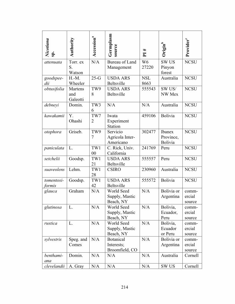
214
Nic
otia
na
sp.
Aut
hori
ty
Acc
essi
ona
Ger
mpl
asm
so
urce
PI #
Ori
ginb
Prov
ider
c
attenuata Torr. ex S. Watson
N/A Bureau of Land Management
W6 27220
SW US Pinyon forest
NCSU
goodspee-dii
H.-M. Wheeler
25-G USDA ARS Beltsville
NSL 8663
Australia NCSU
obtusifolia Martens and Galeotti
TW98
USDA ARS Beltsville
555543 SW US/ NW Mex
NCSU
debneyi Domin. TW36
N/A N/A Australia NCSU
kawakamii Y. Ohashi
TW72
Iwata Experiment Station
459106 Bolivia NCSU
otophora Griseb. TW97
Servicio Agricola Inter-Americano
302477 Ibanex Province, Bolivia
NCSU
paniculata L. TW100
C. Rick, Univ. California
241769 Peru NCSU
setchelii Goodsp. TW121
USDA ARS Beltsville
555557 Peru NCSU
suaveolens Lehm. TW128
CSIRO 230960 Australia NCSU
tomentosi-formis
Goodsp. TW142
USDA ARS Beltsville
555572 Bolivia NCSU
glauca Graham N/A World Seed Supply, Mastic Beach, NY
N/A Bolivia or Argentina
comm-ercial source
glutinosa L. N/A World Seed Supply, Mastic Beach, NY
N/A Bolivia, Ecuador, Peru
comm-ercial source
rustica L. N/A World Seed Supply, Mastic Beach, NY
N/A Bolivia, Ecuador or Peru
comm-ercial source
sylvestris Speg. and Comes
N/A Botanical Interests; Broomfield, CO
N/A Bolivia or Argentina
comm-ercial source
benthami-ana
Domin. N/A N/A N/A Australia Cornell
clevelandii A. Gray N/A N/A N/A SW US Cornell
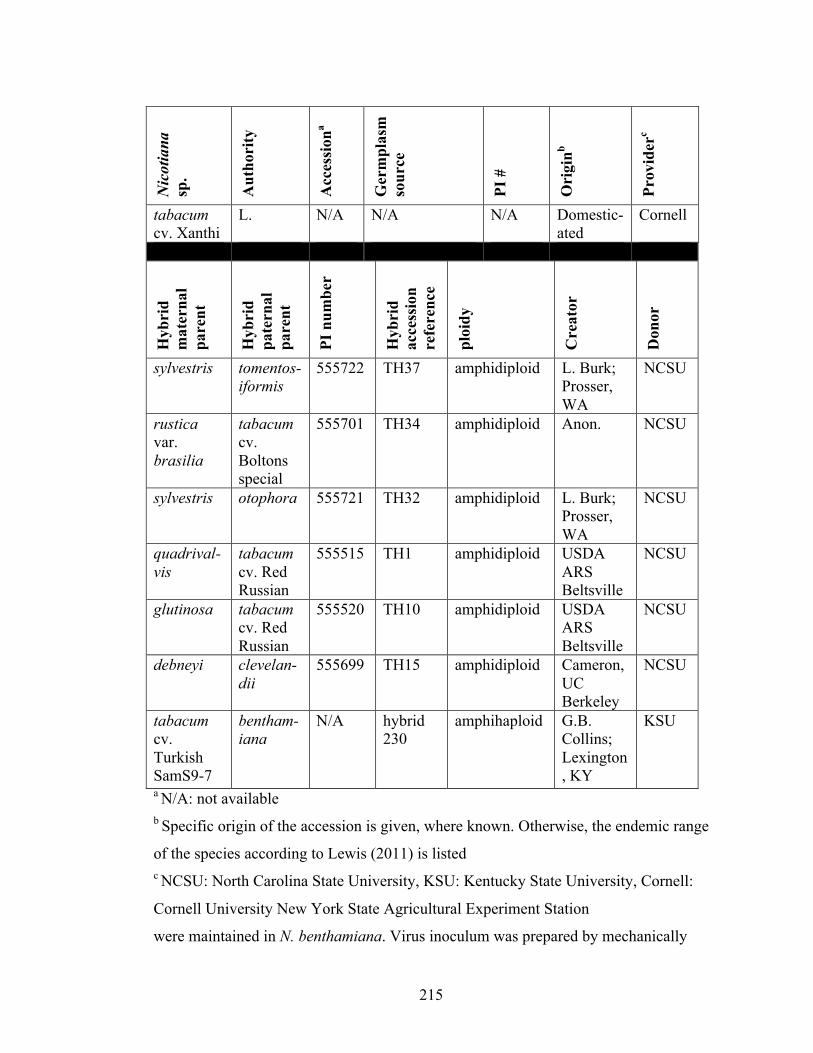
215
Nic
otia
na
sp.
Aut
hori
ty
Acc
essi
ona
Ger
mpl
asm
so
urce
PI #
Ori
ginb
Prov
ider
c
tabacum cv. Xanthi
L. N/A N/A N/A Domestic-ated
Cornell
Hyb
rid
mat
erna
l pa
rent
Hyb
rid
pate
rnal
pa
rent
PI n
umbe
r
Hyb
rid
acce
ssio
n re
fere
nce
ploi
dy
Cre
ator
Don
or
sylvestris tomentos-iformis
555722 TH37 amphidiploid L. Burk; Prosser, WA
NCSU
rustica var. brasilia
tabacum cv. Boltons special
555701 TH34 amphidiploid Anon. NCSU
sylvestris otophora 555721 TH32 amphidiploid L. Burk; Prosser, WA
NCSU
quadrival-vis
tabacum cv. Red Russian
555515 TH1 amphidiploid USDA ARS Beltsville
NCSU
glutinosa tabacum cv. Red Russian
555520 TH10 amphidiploid USDA ARS Beltsville
NCSU
debneyi clevelan-dii
555699 TH15 amphidiploid Cameron, UC Berkeley
NCSU
tabacum cv. Turkish SamS9-7
bentham-iana
N/A hybrid 230
amphihaploid G.B. Collins; Lexington, KY
KSU
a N/A: not available b Specific origin of the accession is given, where known. Otherwise, the endemic range
of the species according to Lewis (2011) is listed c NCSU: North Carolina State University, KSU: Kentucky State University, Cornell:
Cornell University New York State Agricultural Experiment Station
were maintained in N. benthamiana. Virus inoculum was prepared by mechanically

216
inoculating N. benthamiana and storing infected tissue at -80°C until inoculation of
the host panel. Infected N. benthamiana tissue was ground 1:10 (w:v) in inoculation
buffer (15mM Na2HPO4 and 35mM KH2PO4 pH 7.0) using a steel grinding set in a
tissue-lyser (Qiagen, Valencia, CA) and inoculated to three corundum-dusted leaves of
each test plant with a ceramic pestle. Panels of four to 32 (median 17) plants per virus-
host combination were selected for uniformity in size and mechanically inoculated
when they had 4-5 leaves and were approximately 3 cm in height. All plants were
rinsed with water five to ten minutes after inoculation.
Sampling and virus tests
Plant tissue was collected from inoculated plants and processed for virus
detection via double antibody-sandwich enzyme-linked immunosorbent assay (DAS-
ELISA). Apical leaf positions were defined by counting nodes on the whorl upwards
from the highest inoculated leaf. Apical leaves were collected at time points sufficient
to detect cumulative virus infection: nine to 18 dpi for position one, 17 to 28 dpi for
position two, and 26-60 dpi for position three. In plants where intermediate resistance
phenotypes were observed, additional collections were made at 41 to 57 dpi for
position four. Inoculated leaves were collected and processed between 21 and 54 dpi.
Fresh tissue was ground in 1:10 (w:v) in 25mM sodium phosphate buffer using a
semi-automated HOMEX 6 tissue homogenizer and mesh grinding bags (Bioreba,
Reinach, Switzerland). DAS-ELISAs for GFLV and ToRSV were carried out in Nunc
MaxiSorp® flat-bottom 96 well polystyrene microtiter plates (Fisher Scientific,
Pittsburgh, PA) according to the manufacturer’s protocol (Bioreba). Absorbance

217
(OD405nm) was measured after two hours of substrate incubation using a BioTek
Synergy2 plate reader and Gen5 software was used to calculate blank-subtracted
absorbance (Biotek, Winooski, VT). Each ELISA plate contained positive and
negative checks, and the validity of each test was ascertained before data was
processed. Samples were considered positive if their absorbance values were greater
than two times the mean absorbance values of negative controls.
Evaluation of infection phenotypes
Virus symptoms were monitored daily on inoculated and apical plant leaves.
Leaf samples that were positive or negative in DAS-ELISA for GFLV or ToRSV in
each inoculation group were counted and converted into percent infection at each leaf
position. Six resistance categories were assigned based on the infection outcome in
inoculated leaves and in successive apical leaves. Virus-host combinations that yielded
no detectable virus in the inoculated leaf (and apical leaves) were designated as
‘immune’ (category 1). ‘Early recovery’ (category 2) was defined as any level of
inoculated leaf infection (10% to 100%) but the virus was rarely or infrequently
(<10%) detected in the first apical leaf. ‘Late recovery’ (category 3) was defined at
10% to 100% infection in the first or second apical leaf position but a decline in virus
incidence at higher leaf axes. ‘Intermediate recovery’ (category 4) was defined as 20%
to 80% infection frequencies in all leaf axes, and no clear pattern of reduction or
expansion of virus incidence in successively higher axes. ‘Delayed susceptibility’
(category 5) was defined as a steady increase in virus incidence at successively higher
apical leaf axes until the highest tested position contained >75% frequency of virus
218
infection. ‘Full susceptibility’ (category 6) was defined as virus incidence in 100% of
apical leaves. The inoculated leaf was tested to discern immunity from early recovery.
Tests for systemic recovery
A subset of the host panel exhibiting recovery from inoculation with GFLV
strains F13 or GHu [4x(N. sylvestris x N. tomentosiformis), 4x(N. glutinosa x N.
tabacum) and 4x(N. sylvestris x N. otophora)] was re-inoculated with GFLV-GHu in
the fourth leaf position 34 days after the original inoculation. Re-inoculated leaves
were tested for GFLV incidence at five dpi by DAS-ELISA. Negative values were
interpreted as systemic recovery and positive values were interpreted as a lack of
systemic recovery.
Statistics
Statistics were computed on JMP version 10.0 (SAS Institute, Cary, NC). A
score of one was assigned for each leaf infected in the first three apical leaf positions,
and the sum of these scores are referred to as virus incidence, Each plant inoculated
with a given virus was considered a replicate. Contingency analyses were used to
compute Pearson’s correlations (r) and contingency tables. Correlation analyses were
made for species origin (South America, California, Australia or synthetic), ploidy (x=
12 to 48), and virus inoculum (GFLV-F13, GFLV-GHu or ToRSV-AP) with respect to
virus incidence at each leaf position. Origin and virus inoculum was considered as
categorical variables, ploidy as continuous and virus incidence as ordinal data.
Correlation analyses were conducted where synthetic allopolyploids were either
219
included or excluded in the data set.
RESULTS
Test for virus presence
Twenty-four Nicotiana species and synthetic allopolyploids of distinct
geographic origins were evaluated for their reaction to infection with GFLV strains
GHu and F13, and ToRSV strain AP (Table 6-1). Since GFLV-GHu displays levels of
virulence intermediate to that of GFLV-F13 and ToRSV-AP in most Nicotiana
species, plants were primarily assessed for resistance to GFLV-GHu. Each plant-virus
combination was sampled at three or more time points except when a definite
resistance or susceptibility determination could be made in the first or second apical
leaf i.e. for GFLV-F13-inoculated 4x(N. sylvestris x N. tomentosiformis), 4x(N.
glutinosa x N. tabacum), 4x(N. sylvestris x N. otophora) and 4x(N. rustica x N.
tabacum) (sampled once), and GFLV-GHu-inoculated 4x(N. glutinosa x N. tabacum)
and N. goodspeedii (sampled twice). All panels were surveyed for virus presence in
every plant [populations of four to 32 (median 17) plants], except for GFLV-F13-
inoculated 2x(N. tabacum x N. benthamiana), where 23 plants in an original
population of 70 plants was sampled for virus presence in apical leaves in a stratified
sampling approach.
DAS-ELISA was used to determine virus presence or absence for 2719 GFLV
samples and 536 ToRSV samples in 48 plant-virus combinations. DAS-ELISA
reactions produced a bimodal distribution of absorbance values, which allowed a clear

220
delineation of virus-positive from virus-negative samples. Infection frequencies at
each leaf position in each virus-host sample group were summed to calculate virus
incidence, and this was used as the dependent variable in correlation analyses.
Correlations between sum virus incidence and two variables, ploidy and
geographic origin, were computed for GFLV-GHu. Correlations between virus
composition and sum virus incidence were also computed. Correlations were not made
between ploidy or geographic origin and virus incidence levels for GFLV-F13 and
ToRSV-AP inoculations, because limited inoculations were made with these viruses.
Symptoms
Virus-inoculated plants were checked regularly for symptoms. The only
instances of visible symptoms were for GFLV-GHu on N. benthamiana, N.
clevelandii, N. goodspeedii and 2x(N. tabacum x N. benthamiana), and for ToRSV-AP
on N. benthamiana and 2x(N. tabacum x N. benthamiana). GFLV-GHu symptoms on
N. benthamiana and N. clevelandii were consistent with those previously described
(Vigne et al., 2013), and included vein clearing on N. benthamiana and amorphous
ring-like mottling on N. clevelandii. GFLV-GHu symptoms on N. goodspeedii
included vein clearing analogous to that observed for N. benthamiana. GFLV-GHu
symptoms on the 2x(N. tabacum x N. benthamiana) amphihaploid were composed of
non-necrotic ringspots on the first or second leaf position. ToRSV-AP symptoms on
N. benthamiana were similar to those previously described (Jovel et al., 2007), and
included stunting, severe mottling, and necrosis from which the plant ultimately
recovered. ToRSV-AP caused mild mottling and slight stunting on 2x(N. tabacum x N.
221
benthamiana) but necrotic ringspots were not observed on N. tabacum cv. ‘Xanthi’.
Inoculated leaf infection
DAS-ELISA revealed different frequencies of virus infection in inoculated
leaves (Figure 6-1). Some host-virus combinations consistently produced absorbance
values below the virus detection threshold, which reflects immunity or perhaps limited
subliminal (single cell) infections. 4x(N. sylvestris x N. tomentosiformis), 4x(N.
sylvestris x N. otophora), 4x(N. glutinosa x N. tabacum) and N. paniculata exhibited
immunity to GFLV-F13 in inoculated leaves. Some host-virus combinations resulted
in less than 50% inoculated leaf infection including GFLV-GHu-inoculated N.
obtusifolia (13%) and N. glauca (14%), and GFLV-F13-inoculated 4x(N. rustica x N.
tabacum) (43%) and 4x(N. glutinosa x N. tabacum) (44%) (Table 6-2). All other
tested host-virus combinations produced 50% or greater inoculated leaf infection
(Table 6-2). Since GFLV-GHu always produced infections in inoculated or apical
leaves, and ToRSV-AP inoculations always produced some frequency of infection in
the first apical leaf, there is no immunity within this Nicotiana panel to these two virus
strains (Table 6-3).
High resistance interactions
Virus-host combinations yielding no detectable virus in inoculated leaves (and
apical leaves) were designated as immune (category 1). Immunity was observed for
N. paniculata, 4x(N. sylvestris x N. tomentosiformis), 4x(N. sylvestris x N. otophora)
and 4x(N. glutinosa x N. tabacum) inoculated with GFLV-F13 (Figure 6-1; Table 6
222
Table 6-2 Plant responses to Grapevine fanleaf virus (GFLV) strains F13 and
GHu, and Tomato ringspot virus (ToRSV) strain AP.
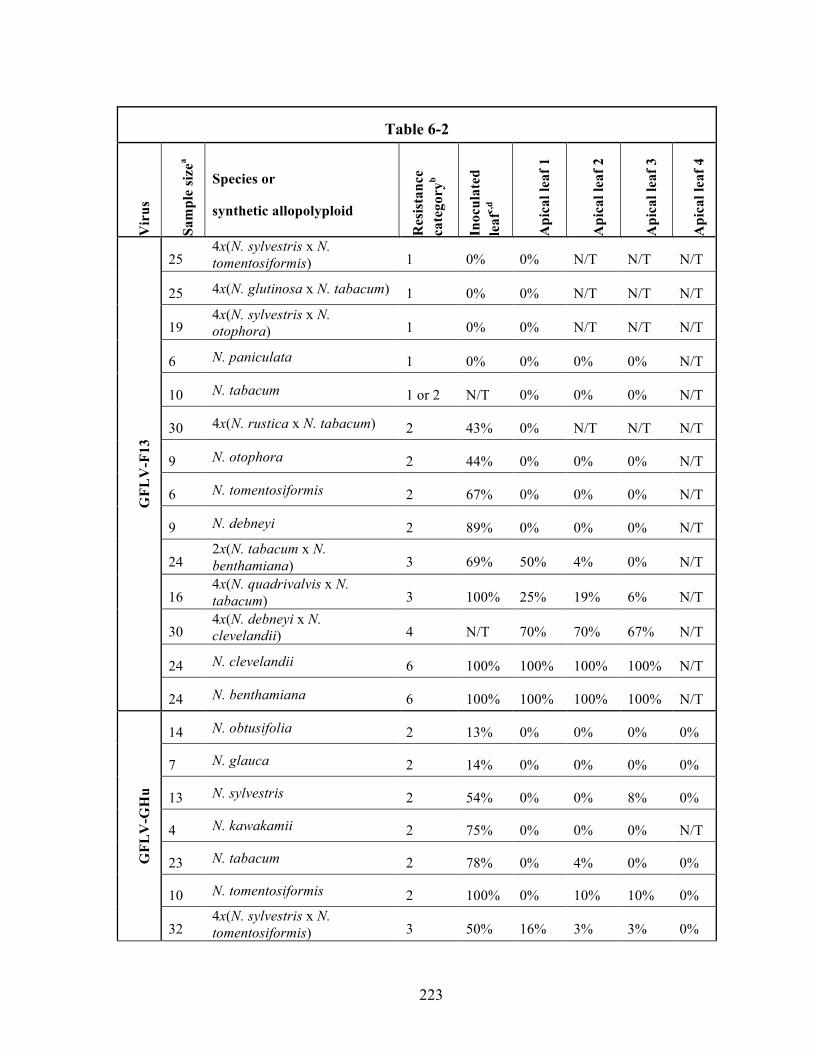
223
Table 6-2 V
irus
Sam
ple
size
a Species or
synthetic allopolyploid
Res
ista
nce
cate
gory
b
Inoc
ulat
ed
leaf
c,d
Api
cal l
eaf 1
Api
cal l
eaf 2
Api
cal l
eaf 3
Api
cal l
eaf 4
GFL
V-F
13
25 4x(N. sylvestris x N. tomentosiformis) 1 0% 0% N/T N/T N/T
25 4x(N. glutinosa x N. tabacum) 1 0% 0% N/T N/T N/T
19 4x(N. sylvestris x N. otophora) 1 0% 0% N/T N/T N/T
6 N. paniculata 1 0% 0% 0% 0% N/T
10 N. tabacum 1 or 2 N/T 0% 0% 0% N/T
30 4x(N. rustica x N. tabacum) 2 43% 0% N/T N/T N/T
9 N. otophora 2 44% 0% 0% 0% N/T
6 N. tomentosiformis 2 67% 0% 0% 0% N/T
9 N. debneyi 2 89% 0% 0% 0% N/T
24 2x(N. tabacum x N. benthamiana) 3 69% 50% 4% 0% N/T
16 4x(N. quadrivalvis x N. tabacum) 3 100% 25% 19% 6% N/T
30 4x(N. debneyi x N. clevelandii) 4 N/T 70% 70% 67% N/T
24 N. clevelandii 6 100% 100% 100% 100% N/T
24 N. benthamiana 6 100% 100% 100% 100% N/T
GFL
V-G
Hu
14 N. obtusifolia 2 13% 0% 0% 0% 0%
7 N. glauca 2 14% 0% 0% 0% 0%
13 N. sylvestris 2 54% 0% 0% 8% 0%
4 N. kawakamii 2 75% 0% 0% 0% N/T
23 N. tabacum 2 78% 0% 4% 0% 0%
10 N. tomentosiformis 2 100% 0% 10% 10% 0%
32 4x(N. sylvestris x N. tomentosiformis) 3 50% 16% 3% 3% 0%

224
Table 6-2 V
irus
Sam
ple
size
a Species or
synthetic allopolyploid
Res
ista
nce
cate
gory
b
Inoc
ulat
ed
leaf
c,d
Api
cal l
eaf 1
Api
cal l
eaf 2
Api
cal l
eaf 3
Api
cal l
eaf 4
30 4x(N. quadrivalvis x N. tabacum) 3 57% 10% 10% 3% N/T
10 N. paniculata 3 60% 10% 30% 0% 0%
4 N. setchelii 3 75% 25% 0% 0% N/T
21 4x(N. sylvestris x N. otophora) 3 81% 10% 5% 0% N/T
8 N. glutinosa 3 100% 13% 0% 0% N/T
G
FLV
-GH
u (c
ontin
ued)
24 4x(N. glutinosa x N. tabacum) 3 100% 12% 4% N/T N/T
30 2x(N. tabacum x N. benthamiana) 4 80% 50% 57% 60% 53%
14 4x(N. rustica x N. tabacum) 4 100% 33% 24% 36% N/T
5 N. rustica 4 100% 80% 40% 60% 20%
8 N. debneyi 4 100% 33% 89% 89% 88%
7 N. otophora 5 100% 0% 56% 44% 78%
24 N. suaveolens 5 N/T 4% 83% 79% N/T
19 4x(N. debneyi x N. clevelandii) 5 N/T 93% 93% 100% N/T
8 N. attenuata 6 100% 100% 100% 100% N/T
24 N. clevelandii 6 100% 100% 100% 100% N/T
24 N. benthamiana 6 100% 100% 100% 100% N/T
12 N. goodspeedii 6 N/T 100% 100% N/T N/T
ToR
SV-A
P
8 N. otophora 3 N/T 100% 0% 0% N/T
16 N. tabacum 3 N/T 88% 19% 6% N/T
16 N. setchelii 3 N/T 100% 13% 0% N/T
11 N. kawakamii 3 N/T 100% 27% 0% N/T

225
Table 6-2 V
irus
Sam
ple
size
a Species or
synthetic allopolyploid
Res
ista
nce
cate
gory
b
Inoc
ulat
ed
leaf
c,d
Api
cal l
eaf 1
Api
cal l
eaf 2
Api
cal l
eaf 3
Api
cal l
eaf 4
12 4x(N. rustica x N. tabacum) 3 N/T 100% 100% 0 N/T
ToR
SV-A
P (c
ontin
ued)
23 4x(N. sylvestris x N. tomentosiformis) 3 N/T 100% 83% 0% N/T
27 4x(N. sylvestris x N. otophora) 3 N/T 100% 89% 26% N/T
16 2x(N. tabacum x N. benthamiana) 5 N/T 75% 81% 100% N/T
12 4x(N. quadrivalvis x N. tabacum) 6 N/T 100% 100% 100% N/T
24 N. benthamiana 6 N/T 100% 100% 100% N/T a The sample size denotes the lowest number of samples tested at any given time point
to account for missing samples. b Categories of resistance (1, most resistant, through 6, most susceptible) are indicated
for each virus-host combination tested. c Values represent the percent of plants in the sample showing detectable virus at each
given leaf position. dN/T: Not tested
226
Table 6-3 Grapevine fanleaf virus and Tomato ringspot virus resistance ratings of
Nicotiana species and synthetic allopolyploids

227
Sectiona Origin x= Nicotiana species
GFL
V-
GH
ub G
FLV
-F1
3c, d
T
oRSV
-A
P
Tomentosae Bolivia 12 N. otophora 5 2 3
Bolivia 12 N. tomentosiformis 2 2
Bolivia 12 N. kawakamii 2 3
Peru 12 N. setchelii 3 3
Paniculatae Peru 12 N. paniculata 3 1
Noctiflorae Bolivia 12 N. glauca 2
Sylvestres Bolivia 12 N. sylvestris 2
Undulatae Peru 12 N. glutinosa 3
Trigonophyllae SW US 12 N. obtusifolia 2
Petunioides SW US 12 N. attenuata 6
Suaveolentes Australia 16 N. suaveolens 5
Australia 19 N. benthamiana 6 6 6
Australia 20 N. goodspeedii 6
Australia 24 N. debneyi 4 2
Polydicliae SW US 24 N. clevelandii 6 6
Rusticae Bolivia 24 N. rustica 4
Nicotiana Domesticated 24 N. tabacum 2 1-2 3
Wide crosses
(synthetic allopolyploids)
24 4x(N. sylvestris x
N. otophora)
3 1 3
24 4x(N. sylvestris x
N. tomentosiformis)
3 1 3
36 4x(N. glutinosa x
N. tabacum)
3 1

228
Sectiona Origin x= Nicotiana species
GFL
V-
GH
ub G
FLV
-F1
3c, d
T
oRSV
-A
P
Wide crosses
(synthetic allopolyploids)
continued
43 2x(N. tabacum x N.
benthamiana)
4 3 5
48 4x(N. quadrivalvis
x N. tabacum)
3 3 6
48 4x(N. rustica x N.
tabacum)
4 2 3
48 4x(N. debneyi x N.
clevelandii)
5 4
a Species and synthetic allopolyploids are referenced by their sections within the genus
Nicotiana, primary location of origin, and their haploid chromosome numbers
according to Lewis (2011) b Categories of resistance (1, most resistant, through 6, most susceptible) are indicated
for each virus-host combination tested. c Boxes without resistance ratings represent virus-host combination not tested d Category ‘1-2’ denotes category 1 or 2 resistance could be operational
229
3). GFLV-F13-inoculated N. tabacum did not produce apical leaf infection, but
whether this plant is immune (category 1) or possesses early recovery (category 2) to
GFLV-F13 is uncertain because inoculated leaves were not tested. All tested members
of section Tomentosae, N. debneyi and 4x(N. rustica x N. tabacum) exhibited early
recovery (category 2) to GFLV-F13 (Table 6-3). N. obtusifolia, N. glauca, N.
sylvestris, N. kawakamii, N. tabacum and N. tomentosiformis exhibited early recovery
after GFLV-GHu inoculation. Early recovery was not observed for these species in
response to inoculation with ToRSV-AP (Table 6-3).
Moderate resistance interactions
Late recovery (category 3) was the most frequent host-virus interaction
phenomenon observed in this test panel, and was seen for all virus isolates tested. All
tested members of section Tomentosae, 4x(N. rustica x N. tabacum), N. tabacum and
resynthesized allopolyploids involving possible N. tabacum progenitor species [4x(N.
sylvestris x N. tomentosiformis) and 4x(N. sylvestris x N. otophora)] showed late
recovery to ToRSV-AP (Table 6-3). 2x(N. tabacum x N. benthamiana) and 4x(N.
quadrivalvis x N. tabacum) showed late recovery to GFLV-F13, and 4x(N.
quadrivalvis x N. tabacum), 4x(N. sylvestris x N. otophora), 4x(N. sylvestris x N.
tomentosiformis), 4x(N. glutinosa x N. tabacum), N. glutinosa, N. paniculata and N.
setchelii showed late recovery to GFLV-GHu (Table 6-3). Intermediate recovery
(category 4), characterized by fluctuation of virus incidence over three or more leaf
axes (typically between 33% and 67%, Table 6-2), was observed in GFLV-GHu-
inoculated N. debneyi, N. rustica, 4x(N. rustica x N. tabacum) and 2x(N. tabacum x N.

230
benthamiana), and in GFLV-F13-inoculated 4x(N. debneyi x N. clevelandii) (Table 6-
3).
Low or no resistance interactions
Delayed susceptibility (category 5) was observed only in response to GFLV-
GHu inoculation of N. otophora, N. suaveolens, and 4x(N. debneyi x N. clevelandii)
(Figure 6-1; Table 6-3). Plants were designated as susceptible (category 6) when
100% of the plants became infected and virus was present in all tested leaves. N.
benthamiana and N. clevelandii were susceptible to GFLV-F13 and GFLV-GHu, as
expected (Vigne et al., 2013), N. goodspeedii and N. attenuata were susceptible to
GFLV-GHu, and N. benthamiana and 4x(N. quadrivalvis x N. tabacum) were
susceptible to ToRSV-AP (Figure 6-1; Table 6-3).
Additive resistance phenomena in synthetic polyploid plants
Incompletely dominant virus resistance was observed in synthetic Nicotiana
allopolyploids. Whereas N. tabacum showed high resistance to GFLV-GHu, ToRSV-
AP and GFLV-F13, and N. benthamiana was fully susceptible to all three virus
strains, 2x(N. tabacum x N. benthamiana) exhibited delayed susceptibility to GFLV-
GHu, intermediate recovery to ToRSV-AP, and late recovery to GFLV-F13 (Figure 6-
2; Table 6-3). N. debneyi exhibited early recovery to GFLV-F13 and intermediate
recovery to GFLV-GHu. These differential resistance responses also appeared in 4x(N.
debneyi x N. clevelandii), which exhibited intermediate recovery to GFLV-F13 and

231
Figure 6-1 Infection frequencies in inoculated and apical leaves of populations of plants tested for resistance to G
rapevine
fanleaf virus (GFLV
) strains GH
u and F13, and Tomato ringspot virus (ToR
SV) strain A
P. The percent virus incidence is indicated
for inoculated and apical leaves (positions 1-3). Asterisks (*) after plant nam
es indicate that the inoculated leaves in the plant-virus
combination w
ere not tested.
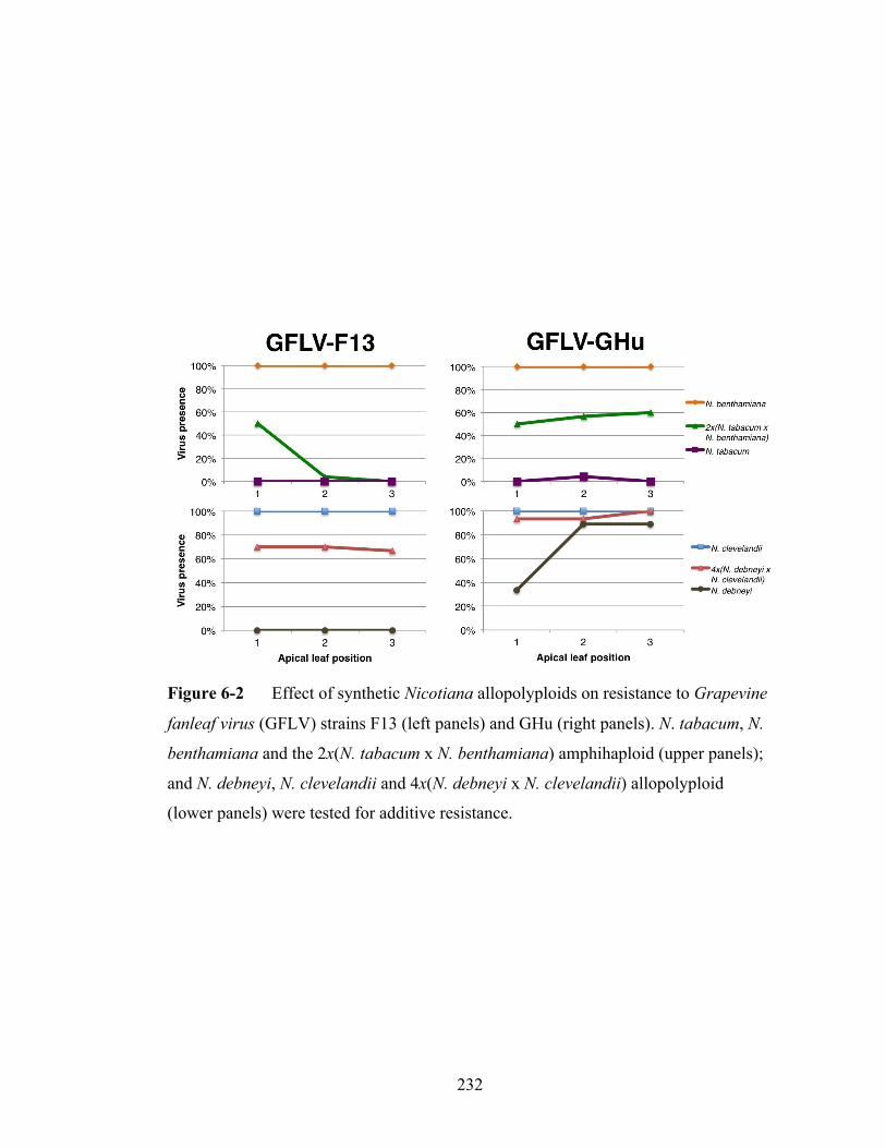
232
Figure 6-2 Effect of synthetic Nicotiana allopolyploids on resistance to Grapevine
fanleaf virus (GFLV) strains F13 (left panels) and GHu (right panels). N. tabacum, N.
benthamiana and the 2x(N. tabacum x N. benthamiana) amphihaploid (upper panels);
and N. debneyi, N. clevelandii and 4x(N. debneyi x N. clevelandii) allopolyploid
(lower panels) were tested for additive resistance.
233
delayed susceptibility to GFLV-GHu (Figure 6-2, Table 6-3). Whereas N. clevelandii
was fully susceptible to all viruses tested, and N. debneyi exhibited early recovery to
GFLV-F13 and intermediate recovery to GFLV-GHu, 4x(N. debneyi x N. clevelandii)
exhibited intermediate recovery to GFLV-F13 and delayed susceptibility to GFLV-
GHu (Figure 6-2, Table 6-3). The 4x(N. rustica x N. tabacum) response to GFLV-
GHu was not categorically different than the response of N. rustica (both category 4),
but the synthetic allopolyploid showed consistently lower incidence of infection in
apical leaves (23-40%) compared to N. rustica (40-80%), which could reflect the
contribution of N. tabacum (category 2) to resistance (Figure 6-1; Table 6-3). The
intermediate virus resistance observed across Nicotiana lineages (Figure 6-3) suggests
quantitative resistance is not due to a single gene with dosage effects, but due to
multiple genes with dosage effects.
Resistance profiles of allopolyploids and their progenitors
Two natural allopolyploids (N. clevelandii and N. tabacum) and the closest
relatives of their known progenitors were tested for GFLV-GHu resistance. The
closest extant diploid progenitors of N. clevelandii are N. obtusifolia (maternal
genome donor) and N. attenuata (paternal genome donor) (Clarkson et al., 2010).
While N. obtusifolia exhibited an early recovery phenotype (category 2), both N.
clevelandii and N. attenuata showed complete susceptibility (Figure 6-1; Table 6-3).
N. tabacum, its representative maternal genome donor (N. sylvestris), and one possible
representative paternal genome donor (N. tomentosiformis) each exhibited early
recovery (category 2). N. otophora, another representative of the possible N. tabacum

234
Figure 6-3 Grapevine fanleaf virus strain GHu (GFLV-GHu) resistance categories
superimposed on a Nicotiana phylogenetic tree modified from Clarkson et al. (2010)
(curved lines), containing sections (abbreviated in black lettering) with allopolyploid
ancestries as established by Clarkson et al. (2010) and Kelly et al. (2013) (solid
straight lines). Shading surrounding sections denote the resistance category of
representative species tested for GFLV-GHu resistance: blue (category 2, early
recovery), purple (categories 3 and 4, late or intermediate recovery), or red (categories
5 and 6, delayed or full susceptibility). Representative Nicotiana species (sections)
used in this study are N. paniculata (Paniculatae, ‘Pan’), N. rustica (Rusticae, ‘Rus’),
N. obtusifolia (Trigonophyllae, ‘Tri’), N. benthamiana, N. debneyi, N. suaveolens and
N. goodspeedii (Suaveolentes, ‘Sua’), N. clevelandii (Polydicliae, ‘Pol’), N. glauca
(Noctiflorae, ‘Noc’), N. sylvestris (Sylvestres ‘Syl’), N. tabacum (Nicotiana ‘Nic’), N.
glutinosa (Undulatae, ‘Und’) N. attenuata (Petunioides, ‘Pet’), (Tomentosae, ‘Tom’)
235
including N. kawakamii, N. otophora, N. setchelii and N. tomentosiformis. Members of
Tomentosae and Suaveolentes exhibited different GFLV-GHu resistance profiles and
are accordingly dually or triply colored. Descent of synthetic allopolyploids used in
this study (white letters) is indicated by dashed lines: 4x(N. sylvestris x N.
tomentosiformis) (‘sxt’), 4x(N. rustica x N. tabacum) (‘rxt’), 4x(N. glutinosa x N.
tabacum) (‘gxt’), 2x(N. tabacum x N. benthamiana) (‘txb’), 4x(N. quadrivalvis x N.
tabacum) (‘qxt’) and 4x(N. debneyi x N. clevelandii) (‘dxc’).

236
paternal genome donors exhibited delayed susceptibility (category 5) to GFLV-GHu.
Resynthesized allopolyploids corresponding to either N. tabacum ancestry scenario
exhibited late recovery phenotypes (category 3) GFLV-GHu, with low virus incidence
levels (Figure 6-1; Table 6-2). Thus, N. tabacum exhibits an early recovery
phenotype similar to that of its maternal genome donor and of N. tomentosiformis, but
less than that of N. otophora or representative resynthesized allopolyploids.
Additionally, species of section Suaveolentes showed low or occasionally moderate
resistance to GFLV-GHu, while its most closely related proposed paternal genome
donor (N. sylvestris) (Kelly et al., 2013) showed high resistance (early recovery)
(Table 6-3). While neoallopolyploids showed intermediate GFLV-GHu resistance
characteristics, extant allopolyploids did not show intermediate GFLV-GHu resistance
characteristics (Figure 6-3).
Systemic recovery
Systemic recovery was tested in apical leaves of GFLV-resistant (categories 1
or 3) synthetic allopolyploids 4x(N. sylvestris x N. tomentosiformis), 4x(N. glutinosa x
N. tabacum), and 4x(N. sylvestris x N. otophora) (Table 6-4). Resistance was induced
with GFLV-GHu or GFLV-F13, and one upper, apical leaf of each recovered plant
was re-inoculated with GFLV-GHu and tested for virus presence. Notably, plants that
showed inoculated leaf susceptibility to GFLV-GHu lost this susceptibility in the
apical leaf of the recovered plant, no matter whether the resistance was induced with
GFLV-F13 or GFLV-GHu (Table 6-4). GFLV-GHu was occasionally detected in the
apical inoculated leaf of GFLV-GHu-recovered plants encompassing two of 21 plants

237
Table 6-4 Systemic recovery from Grapevine fanleaf virus (GFLV) strains F13
and GHu.
1st
inoculationa
Resistance
response
Systemic
recoveryb
4x(N. sylvestris x N.
tomentosiformis)
GFLV-GHu Late recovery 90%
GFLV-F13 Immunity 100%
4x(N. glutinosa x N. tabacum) GFLV-GHu Late Recovery 100%
GFLV-F13 Immunity 100%
4x(N. sylvestris x N. otophora) GFLV-GHu Late Recovery 89% a GFLV-GHu was inoculated to the fourth apical leaf following induction of resistance
(resistance response against the virus in the 1st inoculation is indicated) b Plants (n = 9 to 29) were characterized as having systemic recovery if GFLV was
undetectable at five days post-inoculation.
238
in 4x(N. sylvestris x N. tomentosiformis) and one of nine plants in 4x(N. sylvestris x N.
otophora). Of the plants that did not acquire systemic recovery, we cannot exclude the
possibility of the originally-inoculated GFLV-GHu infecting these apical leaves, given
late recovery does not bar the virus from infecting the fourth leaf position, albeit at a
low incidence.
Relationship between host geographic origin and virus resistance
Australian and North American accessions generally displayed greater levels
of susceptibility than South American accessions to all virus strains tested (Table 6-3).
The Australian species N. benthamiana and the North American species N. clevelandii
were fully susceptible to all viruses tested, and Australian species N. debneyi, N.
suaveolens and N. goodspeedii, and North American species N. attenuata and N.
quadrivalvis displayed lower levels of resistance than South American species to
GFLV-GHu individually or in hybrid backgrounds (Table 6-3). Exceptions to these
geography-based resistance trends included the N. debneyi (Australia) early recovery
response to GFLV-F13, the N. obtusifolia (North America) early recovery response to
GFLV-GHu, and the N. otophora (South America) delayed susceptibility response to
GFLV-GHu. Overall, origin had a significant (P<0.0001) and moderate correlation for
GFLV-GHu virus incidence when hybrids were excluded from the analysis (r=0.683)
and a weaker correlation (r=0.5422, P<0.0001) when hybrids were included, with
South American species showing greater resistance than Australian species, which
showed greater resistance than species from the Southwest US.
239
Limited relationship between host ploidy level and virus resistance
There was a weak association between ploidy level and virus susceptibility.
For example, n=12 diploids from section Tomentosae generally displayed greater
levels of resistance than n=16-24 allopolyploids of section Suaveolentes, and similar
levels of resistance to N. tabacum and N. rustica (n=24) (Table 6-3). The correlation
between GFLV-GHu virus incidence and chromosome number was low (r=-0.036)
and nonsignificant (P=0.2597) when hybrids were included in the analysis, and low
(r=-0.286) but significant (P<0.0001) when hybrids were excluded, indicating that
increasing ploidy is weakly negatively related to GFLV-GHu virus incidence among
extant Nicotiana species. These results indicate that increasing ploidy is correlated
with slightly greater virus susceptibility, but that the trend is abolished when synthetic
allopolyploids are taken into account.
Other trends in virus resistance
Members of section Tomentosae produced higher inoculated leaf infection
rates (75-100%) for GFLV-GHu than for GFLV-F13 (44-67%) (Table 6-2). Every
tested member of section Tomentosae produced an early recovery phenotype for
GFLV-F13 and a late recovery phenotype for ToRSV-AP (Table 6-3). Members of
section Tomentosae showed variability in response to GFLV-GHu, where N.
kawakamii and N. tomentosiformis exhibited early recovery, N. setchelii displayed late
recovery, and N. otophora showed delayed susceptibility (Table 6-3). The delayed
susceptibility of N. otophora to GFLV-GHu was masked in the 4x(N. sylvestris x N.
otophora) synthetic allopolyploid, which reflected the early recovery of N. sylvestris

240
to GFLV-GHu (Table 6-3). Early recovery was also observed for N. tabacum
inoculated with GFLV-GHu, a species believed to have evolved from a N. sylvestris x
N. otophora or N. sylvestris x N. tomentosiformis hybridization event (Lewis, 2011).
Members of section Suaveolentes exhibited intermediate or low resistance to the
nepovirus strains tested, except for N. debneyi, which displayed early recovery after
inoculation with GFLV-F13 (category 2) (Table 6-3).
N. tabacum and its corresponding resynthesized allopolyploids [4x(N.
sylvestris x N. otophora) and 4x(N. sylvestris x N. tomentosiformis)] exhibited high or
moderate virus resistance phenotypes for each virus tested (Table 6-3). Both
resynthesized allopolyploids are immune to GFLV-F13, and N. tabacum also displays
high resistance to this virus. N. tabacum and its resynthesized allopolyploids showed
late recovery to ToRSV-AP, though N. tabacum frequently had lower frequencies of
infection at any given leaf position than its corresponding neoallopolyploids (Figure
6-1; Table 6-2). The response of N. tabacum and the synthetic allopolyploids 4x(N.
sylvestris x N. otophora) and 4x(N. sylvestris x N. tomentosiformis) to GFLV-GHu
were similar in terms of inoculated leaf infection, but N. tabacum showed early
recovery whereas the neoallopolyploids showed late recovery, though the overall
apical virus incidence levels were similar (Table 6-2 and 6-3). The recovery
responses of N. tabacum to GFLV and ToRSV inoculation confirm previous reports
(Dias and Harrison, 1963; Jovel et al., 2011).
Synthetic polyploids formed from resistant and susceptible species frequently
displayed resistance in the moderate categories (Figure 6-3). 2x(N. tabacum x N.
benthamiana) and 4x(N. debneyi x N. clevelandii) exhibited intermediate resistance

241
phenotypes after inoculation with GFLV-GHu and GFLV-F13 as compared to their
parents (Figure 6-2; Table 6-3). The same was true for the 2x(N. benthamiana x N.
tabacum) response to ToRSV-AP (Table 6-3). An intermediate level of apical leaf
infection was also seen in the 4x(N. rustica x N. tabacum) response to GFLV-GHu
(Figure 6-1). ToRSV-AP typically produced equal or greater categorical ratings than
GFLV-GHu, and GFLV-GHu always produced equal or higher category ratings than
GFLV-F13 (Table 6-3). An exception to this virulence trend was that N. otophora and
4x(N. rustica x N. tabacum) showed lower resistance (higher category ratings) to
GFLV-GHu than to ToRSV-AP (Table 6-3). Virulence differences between GFLV-
F13 and GFLV-GHu were highly apparent in synthetic allopolyploid plants with
resistant and susceptible parents, including 2x(N. tabacum x N. benthamiana), 4x(N.
rustica x N. tabacum), and 4x(N. debneyi x N. clevelandii) (Table 6-3; Figure 6-2).
There was a significant (P<0.0001) but weak (r=0.406) correlation between virus
composition and infection frequencies across plant genotypes (species or synthetic
allopolyploids).
According to individual components of χ2 in the contingency table that
compared observed and expected virus incidence frequencies for each virus at each
leaf position, there is a higher virus incidence in the first apical leaf than expected for
ToRSV-AP; conversely, there is less virus incidence in the first apical leaf than
expected for GFLV-F13 (data not shown). Expected and observed apical virus
incidence values are similar for GFLV-GHu. These results suggest that ToRSV-AP
displays higher virulence and GFLV-F13 displays lower virulence than GFLV-GHu in
this panel of Nicotiana species.
242
DISCUSSION
A spectrum of plant resistance and viral virulence was observed in the
Nicotiana-nepovirus present panel. While all host-virus combinations exhibiting low
leaf inoculation frequencies (<50%) exhibited early recovery, this phenotype was
frequently associated with a high infection frequency (>50%) in inoculated leaves
(Table 6-2). Moderate or high leaf inoculation frequencies (≥50%) were associated
with an entire range of resistance and susceptibility phenotypes (category 2 through
category 6) (Figure 6-1). Within individual plant genotypes, ToRSV-AP generally
produced higher susceptibility ratings than GFLV-GHu, and GFLV-GHu always
produced an equal or greater susceptibility rating than GFLV-F13 (Table 6-3), and the
correlation between virus identity and virus incidence ratings were significant. The
spectra of quantitative resistance displayed by Nicotiana accessions and virulence
among nepoviruses suggest the role of multiple interacting alleles from Nicotiana
accessions and nepoviruses in the determination of the ultimate infection outcomes.
Similar plant genotype by virus genotype interactions were observed in a panel of 21
Arabidopsis accessions challenged with three Cucumber mosaic virus isolates (Pagán
et al., 2007).
The full susceptibility seen for 4x(N. quadrivalvis x N. tabacum) and delayed
susceptibility of the 2x(N. tabacum x N. benthamiana) responses to ToRSV raises the
interesting possibility that N. quadrivalvis and N. benthamiana may possess a
dominant ToRSV susceptibility factor in N. tabacum backgrounds. The observation of
ringspot symptoms on the GFLV-GHu-inoculated 2x(N. tabacum x N. benthamiana)
amphihaploid suggests that the vein clearing symptomology typical of N. benthamiana

243
infection (Vigne et al., 2013) is a recessive trait. Similarly, while ToRSV-AP
produced necrosis on N. benthamiana, necrosis was not observed on the 2x(N.
tabacum x N. benthamiana) amphihaploid or on N. tabacum. The absence of N.
tabacum-ToRSV necrotic ringspot symptoms was unexpected given previous reports
(Ross, 1961; Jovel et al., 2011). The lack of hypersensitive responses observed in this
host panel is consistent with the lack of involvement of a specific gene-for-gene
recognition system in Nicotiana-GFLV and Nicotiana-ToRSV interactions. This lack
of hypersensitive response and the absence of coevolutionary history between
Nicotiana and GFLV or ToRSV supports the idea that resistance or susceptibility is
due to the interaction of broad-spectrum immune responses and virulence factors
(Schulze-Lefert and Panstruga, 2011).
Most plants in the host panel used in this study recovered from virus infection
after infection was initially established in inoculated leaves. Recovery from virus
infection is common, can be controlled by simple or complex host plant genetics, and
can be countered by effective pathogen virulence factors (Gunduz et al., 2004;
Bruening, 2006; Maule et al., 2007). Host plant and pathogen genotype determined the
level of plant recovery to GFLV (Figure 6-2). Compatibility between host and viral
components is a prerequisite for infection in the matching allele model (Lambrechts et
al., 2006; Fraile and García-Arenal, 2010). The partial resistance phenotypes observed
in this study do not fit with the strict bimodality of the matching allele concept.
However, a modified matching allele model that allows for partial compatibility and
limited infection (Figure 6-4) (Clay and Kover, 1996; Fraile and García-Arenal, 2010)
could explain the range of resistance and virulence observed in the Nicotiana-
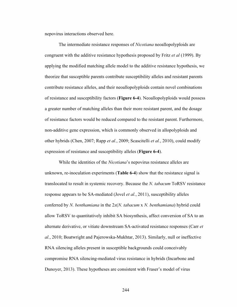
244
nepovirus interactions observed here.
The intermediate resistance responses of Nicotiana neoallopolyploids are
congruent with the additive resistance hypothesis proposed by Fritz et al (1999). By
applying the modified matching allele model to the additive resistance hypothesis, we
theorize that susceptible parents contribute susceptibility alleles and resistant parents
contribute resistance alleles, and their neoallopolyploids contain novel combinations
of resistance and susceptibility factors (Figure 6-4). Neoallopolyploids would possess
a greater number of matching alleles than their more resistant parent, and the dosage
of resistance factors would be reduced compared to the resistant parent. Furthermore,
non-additive gene expression, which is commonly observed in allopolyploids and
other hybrids (Chen, 2007; Rapp et al., 2009; Scascitelli et al., 2010), could modify
expression of resistance and susceptibility alleles (Figure 6-4).
While the identities of the Nicotiana’s nepovirus resistance alleles are
unknown, re-inoculation experiments (Table 6-4) show that the resistance signal is
translocated to result in systemic recovery. Because the N. tabacum ToRSV resistance
response appears to be SA-mediated (Jovel et al., 2011), susceptibility alleles
conferred by N. benthamiana in the 2x(N. tabacum x N. benthamiana) hybrid could
allow ToRSV to quantitatively inhibit SA biosynthesis, affect conversion of SA to an
alternate derivative, or vitiate downstream SA-activated resistance responses (Carr et
al., 2010; Boatwright and Pajerowska-Mukhtar, 2013). Similarly, null or ineffective
RNA silencing alleles present in susceptible backgrounds could conceivably
compromise RNA silencing-mediated virus resistance in hybrids (Incarbone and
Dunoyer, 2013). These hypotheses are consistent with Fraser’s model of virus
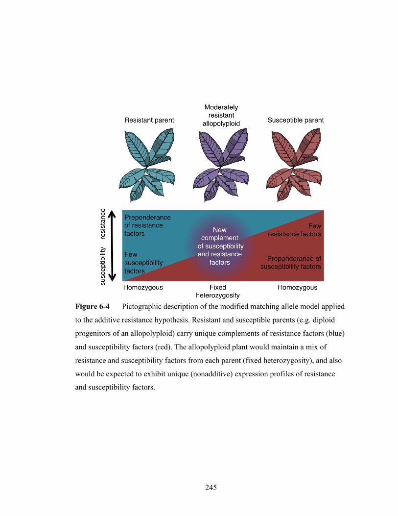
245
Figure 6-4 Pictographic description of the modified matching allele model applied
to the additive resistance hypothesis. Resistant and susceptible parents (e.g. diploid
progenitors of an allopolyploid) carry unique complements of resistance factors (blue)
and susceptibility factors (red). The allopolyploid plant would maintain a mix of
resistance and susceptibility factors from each parent (fixed heterozygosity), and also
would be expected to exhibit unique (nonadditive) expression profiles of resistance
and susceptibility factors.

246
resistance (1992), which postulates that the effects of resistance alleles are
proportional to their dosage and levels of influence on resistance pathways.
Although interspecific animal allopolyploids frequently show dominant
parasite susceptibility (Moulia, 1999; Wolinska et al., 2008; King et al., 2012),
Nicotiana neoallopolyploids exhibit virus resistance that is greater than one but not
both of their parents (Figure 6-3). In cases where both parents were either resistant or
susceptible, the neoallopolyploid displayed a resistance response similar to their
parents, and thus there was no inherent penalty or benefit from hybridization or
genome duplication (Figure 6-3). Contrary to the model that neoallopolyploid plants
could face a depression of innate immunity (Jackson and Chen, 2010), our findings
suggest that allopolyploidization itself did not penalize Nicotiana for virus resistance.
‘Revolutionary changes’ that accompany polyploidy can be distinguished from
‘evolutionary changes,’ which follow allopolyploidization (Feldman et al., 2012;
Soltis, 2013). The maintenance of virus resistance in N. tabacum contrasts with the
apparent loss of virus resistance in section Polydicliae, which did not maintain partial
virus resistance imparted by its likely maternal genome donor (N. obtusifolia) (Figure
6-3). Similarly, members of Suaveolentes exhibited high degrees of virus
susceptibility despite the resistance of their paternal genome donor’s closest relative
(N. sylvestris). Low virus resistance in sections Polydicliae and Suaveolentes suggests
genetic drift and/or selection conferred a loss of virus resistance inherited by
neoallopolyploids. Nicotiana neoallopolyploids show gene loss and
neofunctionalization (Kovarik et al., 2004; Clarkson et al., 2005; Renny-Byfield et al.,
2011). Since favorable alleles have a lower chance of becoming fixed in
247
Figure 6-5 Model of changes in quantitative innate virus resistance from a
moderately resistant progenitor exhibiting fixed heterozygosity for resistance genes
(e.g. a neoallopolyploid). Random divergence of the allopolyploid progeny leads to
several possible lineages containing different resistance or susceptibility phenotypes
whose existence depends on drift and pathogen pressure. High pathogen pressure
would select for the loss of susceptibility factors and maintenance and gain of
resistance factors (trajectory 1, top row). Moderate or irregular pathogen pressure
would maintain an equilibrium of resistance and susceptibility factors within the plant
population (trajectory 2, middle row). Low pathogen pressure would remove the
selective advantage of maintaining resistance factors, and could result in the loss of
resistance factors and the maintenance of susceptibility factors (trajectory 3, bottom
row).

248
allopolyploids than diploids (Otto and Whitton, 2000), drift could result in losses of
innate immunity alleles in the Polydicliae and Suaveolentes lineages (Figure 6-5).
The correlation of higher levels of virus resistance in South American
Nicotiana species than North American and Australian species suggest that geographic
influences had a major effect on the efficacy of antiviral resistance responses. Because
natural Nicotiana neoallopolyploids exhibit an additive basal antiviral resistance
(Figure 6-3), long-term biota-specific interactions would be critical factors to select
for improved virus resistance. Existing virus resistance alleles could be maintained or
enhanced if virus challengers perennially recur (trajectories 1 or 2), or virus resistance
alleles could be lost if virus challenges diminish (trajectory 3) (Figure 6-5).
Comparison of several allopolyploids used in these virus resistance
experiments suggest that N. tabacum, endemic to the relatively large and competitive
biome of the Eastern Andes has followed trajectory 1, while members of sections
Suaveolentes and Polydicliae, endemic to the relatively isolated biota of Australia and
Southwest US, respectively, have followed trajectories 2 or 3 (Figure 6-5). Because
N. debneyi and N. benthamiana are monophyletic (Lewis, 2011), but N. debneyi shows
greater antiviral resistance than N. benthamiana (Figure 6-3), random or selection-
based processes may have driven divergence of innate immune functions within this
allopolyploid lineage. The sister allopolyploids N. clevelandii and N. quadrivalvis
have similarly diverged for herbivory resistance responses (Lou and Baldwin, 2003).
Changes in immune function due to allopolyploidy could precipitate changes
in challenging pathogens, and prompt a Red Queen-type evolutionary response
between the plant and pathogen (Wolinska et al., 2008). Ineffective innate immune

249
systems could allow otherwise ill adapted viruses to acquire more effective virulence
factors and erode quantitative resistance (Vallad and Goodman, 2004; Palloix et al.,
2009). An allopolyploid that can endure colonization by a pathogen or pest and that
permits pathogen adaptation to an otherwise resistant host progenitor is referred to as a
hybrid bridge (Floate and Witham, 1993). Rather than escaping virus infection,
allopolyploids could furnish another niche for viruses to expand their host ranges.
CONCLUSION
The Red Queen Hypothesis explains how resistance and virulence temporally
change in parasitic relationships. Allopolyploidy might represent an opportunity for
plant hosts to break the Red Queen cycle of coevolution by gaining a new complement
of dominant resistance factors, but the potential for allopolyploids to experience an
epochal gain in innate immune function may be compromised by the inheritance of
susceptibility alleles or genetic dysfunctionality caused by hybridization. The
Nicotiana-nepovirus interaction sheds light on potential dynamics of how
allopolyploidy may affect innate immunity. Based on a detailed survey of the
interaction of non-coevolved plant and virus species, it appears that host and pathogen
genotypes contain multiple alleles that interact in a quantitative fashion to determine
the level of resistance or susceptibility. Synthetic allopolyploids faithfully display
additive virus resistance characteristics that correspond to modified matching allele
interactions (Figure 6-4). Virus resistance/susceptibility factors change in
allopolyploid progeny due to classical drift and selection (Figure 6-5). These changes
raise the interesting possibility that moderately resistant allopolyploids could provide a

250
hybrid bridge, which could result in a new Red Queen cycle of coevolution.
251
REFERENCES
Andret-Link, P., Laporte, C., Valat, L., Ritzenthaler, C., Demangeat, G., Vigne, E.,
Laval, V., Pfeiffer, P., Stussi-Garaud, C., and Fuchs, M. 2004. Grapevine fanleaf
virus: still a major threat to the grapevine industry. Journal of Plant Pathology.
86:183–195.
Bitterlin, M.W., and Gonsalves, D. 1988. Serological grouping of Tomato ringspot
virus isolates: implications for diagnosis and cross-protection. Phytopathology.
78:278–285.
Boatwright, J. L., and Pajerowska-Mukhtar, K. 2013. Salicylic acid: an old hormone
up to new tricks. Molecular Plant Pathology. 14:623–634.
Bruening, G. 2006. Resistance to infection. Pages 211–240 in: Natural Resistance
Mechanisms of Plants to Viruses. Loebstein, G, and Carr, J.P., eds. Springer
Netherlands, Dordrecht, Netherlands.
Brunt, A., Crabtree, K., Dallwitz, M., Gibbs, A. J., Watson L, and Zurcher, E., eds.
1996. Plant viruses online: descriptions and lists from the VIDE database. Version 20.
http://biology.anu.edu.au/Groups/MES/vide. Accessed January 24, 2014. Accessed 31
January 2014.
Carr, J. P., Lewsey, M. G., and Palukaitis, P. 2010. Signaling in induced resistance.
Pages 57–121 in: Advances in Virus Research, Vol. 76. Maramorosch, K., Shatkon, A,
and Murphy, F., eds. Elsevier, San Diego, CA.
Chase, M. W. 2003. Molecular Systematics, GISH and the Origin of Hybrid Taxa in
Nicotiana (Solanaceae). Annals of Botany. 92:107–127.
252
Chen, Z. J. 2007. Genetic and epigenetic mechanisms for gene expression and
phenotypic variation in plant polyploids. Annual Review of Plant Biology. 58:377.
Christie, S. R., and Crawford, W. E. 1978. Plant virus range of Nicotiana
benthamiana. Plant Disease Reporter. 62:20–22.
Clarkson, J. J., Kelly, L. J., Leitch, A. R., Knapp, S., and Chase, M. W. 2010. Nuclear
glutamine synthetase evolution in Nicotiana: Phylogenetics and the origins of
allotetraploid and homoploid (diploid) hybrids. Molecular Phylogenetics and
Evolution. 55:99–112.
Clarkson, J. J., Lim, K. Y., Kovarik, A., Chase, M. W., Knapp, S., and Leitch, A. R.
2005. Long-term genome diploidization in allopolyploid Nicotiana section Repandae
(Solanaceae). New Phytologist. 168:241–252.
Clay, K., and Kover, P. X. 1996. The Red Queen hypothesis and plant/pathogen
interactions. Annual Review of Phytopathology. 34:29–50.
Cole, A. B., Király, L., Ross, K., and Schoelz, J. E. 2001. Uncoupling resistance from
cell death in the hypersensitive response of Nicotiana species to Cauliflower mosaic
virus infection. Molecular Plant-Microbe Interactions. 14:31–41.
Dawson, W. O., and Hilf, M. E. 1992. Host-range determinants of plant viruses.
Annual Review of Plant Biology. 43:527–555.
DeVerna, J. W., Myers, J. R., and Collins, G. B. 1987. Bypassing prefertilization
barriers to hybridization using in vitro pollination and fertilization. Theoretical and
Applied Genetics. 73:665–671.
253
Dias, H. F., and Harrison, B. D. 1963. The relationship between Grapevine fanleaf,
Grapevine yellow mosaic and Arabis mosaic viruses. Annals of Applied Biology.
51:97–105.
Dijk, P., and Cuperus, C. 1989. Reactions on Nicotiana species to potato viruses A, X
and Y and Tobacco mosaic virus in relation to their taxonomy and geographical
origin. European Journal of Plant Pathology. 95:343–356.
Dijk, P., Meer, F. A., and Piron, P. G. M. 1987. Accessions of Australian Nicotiana
species suitable as indicator hosts in the diagnosis of plant virus diseases. European
Journal of Plant Pathology. 93:73–85.
Feldman, M., Levy, A. A., Fahima, T., and Korol, A. 2012. Genomic asymmetry in
allopolyploid plants: wheat as a model. Journal of Experimental Botany. 63:5045–
5059.
Floate, K. D., and Whitham, T. G. 1993. The hybrid bridge hypothesis: host shifting
via plant hybrid swarms. The American Naturalist. 141:651–662.
Fraile, A., and García-Arenal, F. 2010. The coevolution of plants and viruses. Pages
1–32 in: Advances in Virus Research, Vol. 76. Maramorosch, K., Shatkon, A, and
Murphy, F., eds. Elsevier, San Diego, CA.
Fraser, R. 1992. The genetics of plant-virus interactions: implications for plant
breeding. Euphytica. 63:175–185.
Fritz, R. S. 1999. Resistance of hybrid plants to herbivores: genes, environment, or
both? Ecology. 80:382–391.
254
Gibbs, A. J., Fargette, D., García-Arenal, F., and Gibbs, M. J. 2010. Time--the
emerging dimension of plant virus studies. Journal of General Virology. 91:13–22.
Goodin, M. M., Zaitlin, D., Naidu, R. A., and Lommel, S. A. 2008. Nicotiana
benthamiana: its history and future as a model for plant–pathogen interactions.
Molecular Plant-Microbe Interactions. 21:1015–1026.
Gottula, J. W., Lapato, D., Cantilina, K. K., Saito, S., Bartlett, B., and Fuchs, M. 2013.
Genetic variability, evolution and biological effects of Grapevine fanleaf virus satellite
RNAs. Phytopathology. 103:1180–1187.
Gunduz, I., Buss, G. R., Chen, P., and Tolin, S. A. 2004. Genetic and phenotypic
analysis of Soybean mosaic virus resistance in PI 88788 soybean. Phytopathology.
94:687–692.
Holmes, F.O. 1938. Inheritance of resistance to tobacco-mosaic disease in tobacco.
Phytopathology. 28: 553–561.
Huss, B., Walter, B., and Fuchs, M. 1989. Cross-protection between Arabis mosaic
virus and Grapevine fanleaf virus isolates in Chenopodium quinoa. Annals of Applied
Biology. 114:45–60.
Incarbone, M., and Dunoyer, P. 2013. RNA silencing and its suppression: novel
insights from in planta analyses. Trends in Plant Science. 18:382–392
Jackson, J. A., and Tinsley, R. C. 2003. Parasite infectivity to hybridising host species:
a link between hybrid resistance and allopolyploid speciation? International Journal
for Parasitology. 33:137–144.
255
Jackson, S., and Chen, Z. J. 2010. Genomic and expression plasticity of polyploidy.
Current Opinion in Plant Biology. 13:153–159.
Jovel, J., Walker, M., and Sanfaçon, H. 2007. Recovery of Nicotiana benthamiana
plants from a necrotic response induced by a nepovirus is associated with RNA
silencing but not with reduced virus titer. Journal of Virology. 81:12285–12297.
Jovel, J., Walker, M., and Sanfaçon, H. 2011. Salicylic acid-dependent restriction of
Tomato ringspot virus spread in tobacco is accompanied by a hypersensitive response,
local RNA silencing, and moderate systemic resistance. Molecular Plant-Microbe
Interactions. 24:706–718
Kang, B.-C., Yeam, I., and Jahn, M. M. 2005. Genetics of plant virus resistance.
Annual Review of Phytopathology. 43:581–621.
Kelly, L. J., Leitch, A. R., Clarkson, J. J., Knapp, S., and Chase, M. W. 2013.
Reconstructing the complex evolutionary origin of wild allopolyploid tobaccos
(Nicotiana section Suaveolentes). Evolution. 67:80–94.
Kovarik, A., Matyášek, R., Lim, K. Y., Skalická, K., Koukalová, B., Knapp, S.,
Chase, M., and Leitch, A. R. 2004. Concerted evolution of 18-5.8-26S rDNA repeats
in Nicotiana allotetraploids. Biological Journal of the Linnean Society. 82:615–625.
Lamberti, F., and Golden, A. M. 1984. Redescription of Xiphinema americanum
Cobb, 1913 with comments on its morphometric variations. Journal of Nematology.
16:204–206.
Lambrechts, L., Fellous, S., and Koella, J. C. 2006. Coevolutionary interactions
between host and parasite genotypes. Trends in Parasitology. 22:12–16.

256
Leitch, I. J., Hanson, L., Lim, K. Y., Kovarik, A., Chase, M. W., Clarkson, J. J., and
Leitch, A. R. 2008. The ups and downs of genome size evolution in polyploid species
of Nicotiana (Solanaceae). Annals of Botany. 101:805–814.
Lewis, R. S. 2011. Nicotiana. Pages 185–208 in: Wild Crop Relatives: Genomic and
Breeding Resources. C. Kole, ed. Springer Berlin Heidelberg, Berlin.
Lewis, R. S. 2005. Transfer of resistance to Potato virus Y (PVY) from Nicotiana
africana to Nicotiana tabacum: possible influence of tissue culture on the rate of
introgression. Theoretical and Applied Genetics. 110:678–687.
Lou, Y., and Baldwin, I. T. 2003. Manduca sexta recognition and resistance among
allopolyploid Nicotiana host plants. Proceedings of the National Academy of Sciences
USA. 100:14581–14586.
Maule, A. J., Caranta, C., and Boulton, M. I. 2007. Sources of natural resistance to
plant viruses: status and prospects. Molecular Plant Pathology. 8:223–231.
Mette, M. F., Kanno, T., Aufsatz, W., Jakowitsch, J., van der Winden, J., Matzke, M.
A., and Matzke, A. J. M. 2002. Endogenous viral sequences and their potential
contribution to heritable virus resistance in plants. The EMBO Journal. 21:461–469.
Moulia, C. 1999. Parasitism of plant and animal hybrids: are facts and fates the same?
Ecology. 80:392–406.
Nawaz-ul-Rehman, M. S., and Fauquet, C. M. 2009. Evolution of geminiviruses and
their satellites. FEBS Letters. 583:1825–1832.
257
Oliver, J., and Fuchs, M. 2011. Tolerance and resistance to viruses and their vectors in
Vitis sp.: a virologist's perspective of the literature. American Journal of Enology and
Viticulture. 62:438–451
Otto, S. P. 2007. The evolutionary consequences of polyploidy. Cell. 131:452–462.
Pagán, I., Alonso-Blanco, C., and García-Arenal, F. 2007. The relationship of within-
host multiplication and virulence in a plant-virus system. PLoS ONE. 2:e786–e786.
Palloix, A., Ayme, V., and Moury, B. 2009. Durability of plant major resistance genes
to pathogens depends on the genetic background, experimental evidence and
consequences for breeding strategies. New Phytologist. 183:190–199
Patel, D., Power, J. B., Anthony, P., Badakshi, F., Heslop-Harrison, J. S. P., and
Davey, M. R. 2011. Somatic hybrid plants of Nicotiana × sanderae (+) N. debneyi
with fungal resistance to Peronospora tabacina. Annals of Botany. 108:809–819.
Pehu, E., Gibson, R. W., Jones, M., and Karp, A. 1990. Studies on the genetic basis of
resistance to Potato leaf roll virus, Potato virus Y and Potato virus X in Solanum
brevidens using somatic hybrids of Solanum brevidens and Solanum tuberosum. Plant
Science. 69:95–101.
Prentis, P. J., Wilson, J. R. U., Dormontt, E. E., Richardson, D. M., and Lowe, A. J.
2008. Adaptive evolution in invasive species. Trends in Plant Science. 13:288–294.
Qu, F., Ye, X., Hou, G., Sato, S., Clemente, T. E., and Morris, T. J. 2005. RDR6 has a
broad-spectrum but temperature-dependent antiviral defense role in Nicotiana
benthamiana. Journal of Virology. 79:15209–15217.
258
Rapp, R. A., Udall, J. A., and Wendel, J. F. 2009. Genomic expression dominance in
allopolyploids. BMC Biology. 7:18.
Raski, D. J., Goheen, A. C., Lider, L. A., and Meredith, C. P. 1983. Strategies against
Grapevine fanleaf virus and its nematode vector. Plant Disease. 67:335–339.
Ratcliff, F., Harrison, B. D., and Baulcombe, D. C. 1997. A similarity between viral
defense and gene silencing in plants. Science. 276:1558–1560
Renny-Byfield, S., Chester, M., Kovarik, A., Le Comber, S. C., Grandbastien, M. A.,
Deloger, M., Nichols, R. A., Macas, J., Novak, P., Chase, M. W., and Leitch, A. R.
2011. Next generation sequencing reveals genome downsizing in allotetraploid
Nicotiana tabacum, predominantly through the elimination of paternally derived
repetitive DNAs. Molecular Biology and Evolution. 28:2843–2854.
Renny-Byfield, S., Kovarik, A., Chester, M., Nichols, R. A., Macas, J., Novák, P., and
Leitch, A. R. 2012. Independent, rapid and targeted loss of highly repetitive DNA in
natural and synthetic allopolyploids of Nicotiana tabacum. PLoS ONE. 7:e36963.
Ritzenthaler, C., Viry, M., Pinck, M., Margis, R., Fuchs, M., and Pinck, L. 1991.
Complete nucleotide sequence and genetic organization of Grapevine fanleaf
nepovirus RNA1. Journal of General Virology. 72:2357–2365
Ross, A. F. 1961. Localized acquired resistance to plant virus infection in
hypersensitive hosts. Virology. 14:329–339.
Sanfaçon H, Fuchs M. 2011. Tomato ringspot virus. Pages 41-48 in: Virus and Virus-
Like Diseases of Pome and Stone Fruits. Hadidi, A., Barba, M., Candresse, T. and
Jelkmann, W. eds. APS Press, St. Paul, MN.

259
Sanfaçon, H., Wellink, J., Gall, O., Karasev, A., Vlugt, R., and Wetzel, T. 2009.
Secoviridae: a proposed family of plant viruses within the order Picornavirales that
combines the families Sequiviridae and Comoviridae, the unassigned genera
Cheravirus and Sadwavirus, and the proposed genus Torradovirus. Archives of
Virology. 154:899–907.
Scascitelli, M., Cognet, M., and Adams, K. L. 2010. An interspecific plant hybrid
shows novel changes in parental splice forms of genes for splicing factors. Genetics.
184:975–983.
Schulze-Lefert, P., and Panstruga, R. 2011. A molecular evolutionary concept
connecting nonhost resistance, pathogen host range, and pathogen speciation. Trends
in Plant Science. 16:1360–1386.
Schwach, F., Vaistij, F. E., Jones, L., and Baulcombe, D. C. 2005. An RNA-
Dependent RNA Polymerase prevents meristem invasion by Potato virus X and is
required for the activity but not the production of a systemic silencing signal. Plant
Physiology. 138:1842–1852.
Serghini, M. A., Fuchs, M., Pinck, M., Reinbolt, J., Walter, B., and Pinck, L. 1990.
RNA2 of Grapevine fanleaf virus: sequence analysis and coat protein cistron location.
Journal of General Virology. 71:1433–1441.
Siddiqui, S. A., Sarmiento, C., Kiisma, M., Koivumaki, S., Lemmetty, A., Truve, E.,
and Lehto, K. 2008. Effects of viral silencing suppressors on Tobacco ringspot virus
infection in two Nicotiana species. Journal of General Virology. 89:1502–1508.
Soltis, P. S. 2013. Hybridization, speciation and novelty. Journal of Evolutionary
Biology. 26:291–293.

260
Taliansky, M., Aranda, M. A., and García-Arenal, F. 1994. Differential invasion by
tobamoviruses of Nicotiana megalosiphon following the hypersensitive response.
Phytopathology. 84:812–815.
Vallad, G. E., and Goodman, R. M. 2004. Systemic acquired resistance and induced
systemic resistance in conventional agriculture. Crop Science. 44:1920–1934.
Vigne, E., Gottula, J., Schmitt-Keichinger, C., Komar, V., Ackerer, L., Belval, L.,
Rakotomalala, L., Lemaire, O., Ritzenthaler, C., and Fuchs, M. 2013. A strain-specific
segment of the RNA-dependent RNA polymerase of Grapevine fanleaf virus
determines symptoms in Nicotiana species. Journal of General Virology. 94:2803–
2813.
Wolinska, J., Lively, C. M., and Spaak, P. 2008. Parasites in hybridizing communities:
the Red Queen again? Trends in Parasitology. 24:121–126
Yang, S., Carter, S., Cole, A., Cheng, N. H., and Nelson, R. S. 2004. A natural variant
of a host RNA-dependent RNA polymerase is associated with increased susceptibility
to viruses by Nicotiana benthamiana. Proceedings of the National Academy of
Sciences USA. 101:6297–6302.
Ying, X., Dong, L., Zhu, H., Duan, C., Du, Q. S., Lv, D. Q., Fang, Y., García, J. A.,
Fang, R.-X., and Guo, H. S. 2010. RNA-Dependent RNA Polymerase 1 from
Nicotiana tabacum suppresses RNA silencing and enhances viral infection in
Nicotiana benthamiana. Plant Cell. 22:1358–1372.
261
CHAPTER 7
FUTURE DIRECTIONS
NEPOVIRUS SUBGROUP A SATELLITE RNA
Future studies on the GFLV satRNA could shed light on how viral RNAs are
recognized and replicated specifically by RNA dependent RNA polymerases (RdRps).
The GFLV satellite RNA encodes a highly basic protein (P3, comprised of isoelectric
points ranging from 10.2 to 11.8), which does not have any known ontology or
homology to other proteins. High pH proteins are known to be involved in interactions
with nucleic acids, and the satellite RNA of Bamboo mosaic virus (satBaMV), a
potexvirus, encodes a highly basic protein that specifically brings the Bamboo mosaic
virus (BaMV) RdRp in contact with satBaMV (Tsai et al., 1999; Palani et al., 2009).
At the bare minimum, protein P3 of the GFLV satellite RNA is expected to
effect its replication. Quite possibly the GFLV satellite RNA may confer specific
selective advantages that could result in its acquisition and maintenance by GFLV and
ArMV strains. The interactors of the GFLV satellite RNA could be identified with
immunoprecipitation assays, yeast two-hybrid assays or yeast three-hybrid assays
(with the satellite RNA used as a link between bait and prey). Additionally, Moser et
al. (1992) tentatively identified protein P3 in the nuclei of infected cells, which could
suggest the satellite RNA interacts with DNA as a transcriptional regulator. To test the
transcriptional regulator theory, a chromatin immunoprecipitation (ChiP) assay could
be designed to target the P3 protein using nanobodies followed by DNA purification

262
and sequencing or microarray analysis (Nguyen-Duc et al., 2012). Identifying the
host- and virus-targets of P3 should shed light on how the satellite RNA is able to be
specifically replicated by its helper virus, and could reveal its functional significance.
Viral satellites, including satellite RNAs, can alter virus host range (Zhou et
al., 2013), modulate symptoms (Shimura et al., 2011), or regulate viral RNA silencing
suppression (Thomas et al., 2003). According to this author’s research (Chapter 2) and
the research of others (Saldarelli et al., 1993; Lamprecht et al., 2013), the satellite
RNA of GFLV does not affect infected grapevines or C. quinoa. Because the Lilac
satellite of Arabis mosaic virus (ArMV) ameliorates symptoms in C. quinoa (Liu et
al., 1991), it could be interesting to test whether this is due to an interaction with the
helper virus (i.e. GFLV versus ArMV) or a satellite genotype-specific response. The
ultimate test of satellite RNA pathological impact would be an experiment in
grapevine, the primary natural host of GFLV. Experiments are underway at INRA
Colmar, France to test the pathological impact of various GFLV strains, including
some with a satellite RNA, in several cultivars (E. Vigne, personal communication).
This information could yield clues about virulence mechanisms of GFLV and could
facilitate the design of disease control strategies (Collmer and Howell, 1992).
The incomplete discovery of the origin of the nepovirus subgroup A satellite
RNA (Chapter 2) opens new questions in virus evolution. The nepovirus subgroup A
satellite RNA appears to have originated as a recombination event between an
ancestral nepovirus genomic RNA and another unknown RNA. This most likely
would have occurred during positive-strand synthesis of the genomic RNA, where a
template switch by the viral RdRp resulted in recombination between the 5’ end of the
263
viral genomic RNA and the 3’ end of a pathogen or host RNA. Although the
mechanics of nepovirus replication are currently poorly understood, it is possible that
nepoviruses behave like other viruses with genome-linked proteins (VPgs) and poly-A
tails (e.g. potyviruses) in that they use the translation initiation complex to anchor for
replication (Lellis et al., 2002). Proximity of heterologous templates bound to the
translation initiation complex could provide the opportunity for recombination. The
identities of host proteins and membranes interacting to form nepovirus replication
complexes could shed light on mechanistic features of nepovirus recombination (Jiang
and Laliberté, 2011; Sanfaçon et al., 2012), including that of the subgroup A satellite
RNA.
The recombination event that gave rise to the satellite RNA resulted in a
truncation of its 5’ untranslated region (UTR) and an early start codon. The start codon
lies after a 14 to 18 nucleotide UTR, which is sufficient for eukaryotic translation
initiation (Watson et al., 2007). The satellite RNA has seven conserved blocks of
nucleotides that extend 140 nucleotides from its 5’ end. This feature suggests it has
maintained the RNA structural elements necessary for VPg, RdRp and capsid
recognition. This overlap of protein coding- and RNA structural regions suggests dual
selective mechanisms are at work on the 5’ 140 nt of the satellite RNA, and indeed the
N-terminus of P3 possesses biochemical properties atypical of the rest of the protein,
including high hydrophobicity and an overabundance of positively charged amino
acids (Fuchs et al., 1989). The nepovirus subgroup A satellite RNA’s conservation of
seven groups of 5’ nucleic acid stretches point to the elements that are needed for viral
RNA recognition by viral proteins. Comparative and mutagenic studies on the 5’

264
nucleotides of the satBaMV have yielded an abundance of information about its cis-
and trans-replication elements (Annamalai et al., 2003; Chen et al., 2012) and similar
research projects could be envisioned for the GFLV satellite RNA.
The identity and source of the gene that comprises the 3’ end of the GFLV
satellite RNA is unknown. There is currently no parameter on NCBI BLAST that
shows molecules with significant protein or nucleic acid homology to the protein of
the satellite RNA. This could be because the satellite RNA has diverged significantly
from its progenitor, or because the gene has not been sequenced or deposited in NCBI.
More publically available sequences from multiple organisms will increase the
likelihood that genes with significant homology will be found. Because viruses are
estimated to contain 50% of protein diversity on earth (Holmes, 2009), there is a high
probability that the recombinant satellite RNA gene came from a virus.
Sequencing more GFLV isolates including targeted amplification of the
satellite RNA ought to shed light on the natural history of GFLV with respect to
anthropogenic interactions. Humans have distributed GFLV worldwide, and high
levels of diversity are often found inside single vineyards (Mekuria et al., 2009; Oliver
et al., 2010). High intra-population diversity coupled with numerous instances of
recombination makes it difficult to trace the origin of particular strains to a site of
origin. The satellite RNA, on the other hand, is less efficiently transmitted from plant-
to-plant by X. index than its helper virus (Chapter 2). This means that the satellite
RNA could function as a ‘stable biomarker’ that can be used to trace the origin of
particular strains. For example, the presence of a common satellite RNA in a
grapevine in Bordeaux and a vineyard in California suggests a very recent common

265
origin of that satellite RNA variant (Chapter 2).
Using the satellite RNA as a viral vector for plant functional genomics could
be an interesting proposition because it could provide higher levels of expression than
the genomic RNAs. Although this author attempted to use the GFLV satellite RNA as
a vector, it was non-infectious. Lamprecht et al. (2013) designed a construct based on
the GFLV satellite RNA SACH44 using a pBluescript vector where a CaMV 35S
promoter drove expression of the satellite RNA and the vector was rub-inoculated to a
C. quinoa mechanically inoculated with sap containing GFLV, and the satellite RNA
was replicated. It could be interesting to see if this infectious satellite RNA could
stably replicate and express heterologous genes such as fluorophors, or if any
modification will abolish its ability to be replicated. Agroinfection is the unattained
ideal for a GFLV vector for grapevine, so the next logical step would be to attempt
agroinfection of the GFLV genomic RNAs with the satellite RNA.
IMPROVING VIRAL VECTORS
Plant virus vectors share few commonalities, so there are few generalizable
improvements that can be prescribed to all plant virus vectors. The main aspect that
needs attention is the fidelity of the heterologous virus sequence. Infectious transcripts
are key to developing RNA viral vectors, but frequently cDNA copies of viral RNA
transcripts are not infectious (Liu et al., 2009; Youssef et al., 2011, Kurth et al., 2013).
The cDNAs could be noninfectious because of the error-prone nature of reverse
transcriptases (RTs) or that non-infective variants of RNAs are amplified in
subsequent PCR (Chapman, 2008). Strategies to generate infectious clones of RNA

266
viruses are generally more successful when multiple clones are tested and high fidelity
RTs and DNA polymerases are used (Youssef et al., 2011).
DNA viruses and RNA viruses have been engineered into viral vectors and the
route of delivery varies based on the nucleic acid identity of the virus. DNA viruses
may be amplified by PCR and circularized by DNA ligation and directly inoculated to
plants or agroinoculated if the virus is imparted with an A. tumefaciens origin of
replication (Huang et al., 2009). Positive-sense RNA viral vectors can be delivered to
plants through inoculation of in vitro transcripts or transcription driven by the CaMV
35S promoter which include biolistic inoculation, direct plasmid rub-inoculation or
agroinfection (Robertson, 2004). Negative sense RNA viruses can be agroinoculated
to plants as long as the necessary replicase proteins are co-inoculated (Ganesan et al.,
2013). The optimal method of virus vector inoculation depends on the genomic
structure of individual viruses and is best determined empirically.
RNA-based viral vectors can be designed for in planta transcription by the
CaMV 35S promoter or other promoters. Lim et al. (2010) developed a novel in
planta promoter based on the T7 phage RNA polymerase (T7RNAP) to express
Alteranthera mosaic virus. This strategy placed the viral cDNA upstream of the T7
promoter and co-expressed the T7RNAP protein via the CaMV 35S promoter. These
researchers showed that the T7 system shortens the time to systemic infection from
two weeks to seven days (Lim et al., 2010). Similar benefits were achieved when
Tobacco mosaic virus (TMV), another RNA virus, was expressed by an RNA
polymerase I promoter (Komarova et al., 2012a). It is notable that because the
capping, splicing and polyadenylation machinery is transcriptionally interconnected

267
with RNA polymerase II function (Watson et al., 2007), transcripts corresponding to
viral RNAs would not be modified in the same way as messenger RNAs if expressed
via T7RNAP or RNA polymerase I.
Another way to avoid splicing is to mutate ‘cryptic introns’ encoded by the
viral cDNA, i.e. to change putative acceptor and donor sites that could form the
extremities of an intron (Marillonet et al., 2005; Komarova et al., 2012b). Ideally, the
mutation of cryptic introns can be accomplished through silent mutations so that
functionality of the viral proteins will not be affected. Alternatively, placing
heterologous plant introns within the viral sequence can diminish splicing of cryptic
introns within the viral cDNA (Marillonet et al., 2005). Because changing the
predicted splicing characteristics of viral vector RNAs has only been accomplished
with TMV, it remains to be seen how generalizable the observed expression
enhancements to RNA viruses can be.
Because RNA viruses vary in types of 5’ and 3’ genomic modifications (e.g. 5’
caps, genome-linked proteins, poly-A tails or cloverleaf structures), optimal viral RNA
expression strategies should depend on the nature of the viral RNA. In general,
researchers should avoid using certain promoters just because they are readily
available, but instead give sober consideration to expression of a transcript as close as
possible to authentic viral RNA.
Viral RNAs corresponding to about 60 virus species have been transiently
expressed in planta using diverse methods of inoculation (Senthil-Kumar and Mysore,
2012). Most have relied on the CaMV 35S promoter following rub inoculation or
agroinoculation of the plasmid and/or it’s T-DNA, respectively. Particle bombardment

268
or transfection of protoplasts are also commonly used to express a CaMV 35S-driven
RNA transcript. Several binary vectors have been used to express viral RNAs
following agroinoculation including pBIN (Ratcliff et al., 2001; Agüero et al., 2012;
Delfosse et al., 2013), pGreen (Ratcliff et al., 2001; Larsen and Curtis, 2012; Bedoya
et al., 2012), and pCAMBIA (Muruganantham et al., 2009; Lim et al., 2010). Though
pGA482 has been used to express individual proteins of Turnip vein clearing virus
(Harries et al., 2009) and Potato virus X (Larsen and Curtis, 2012) upstream of a
CaMV 35S promoter with a heterologous translational enhancer sequence, this author
is not aware of any published viral vectors that used pGA482-derived binary vectors.
The GFLV vector (Chapters 3 and 4) uses pGA482 for agroinoculation with the
CaMV 35S expression cassette from pEPT8. The plasmid EPT8 contains a
translational enhancer sequence derived from the 5’ end of Alfalfa mosaic virus
(AlMV) RNA4, which forms a hairpin (Ling et al., 1997). Because the 5’ ends of viral
genomic RNAs are essential for RNA-RdRp interactions (Annamalai et al., 2003;
Chen et al., 2012), the heterologous hairpin tagged to the 5’ end of the GFLV genomic
RNA could possibly interfere with its RNA-RdRp interactions. Notably, GFLV
vectors that use the same viral cDNAs as those described in Chapters 3 and 4 (F13
RNA1 plus Green fluorescent protein or Red fluorescent protein-tagged F13 RNA2)
but expressed in vectors that do not tag the 5’ ends of transcripts with the AlMV
hairpin (Chapter 4) show 100% systemic infection in N. benthamiana, whereas the
GFLV-eGFP and the GFLV-RFP chimeras containing AlMV 5’ hairpin result in 0%
and 1.6% plant infection in populations of 60 to 100 plants, respectively (Gottula and
Keichinger, unpublished results). These results suggest that using a protein expression

269
vector (i.e. a vector that imparts a heterologous virus hairpin molecule for translation
enhancement) is not the ideal vector to deliver infectious RNA transcripts in planta. It
will be interesting to see if functional GFLV cDNAs without heterologous 5’ hairpins
and/or fused protein reporters will be systemically infectious in grapevine.
THE FUTURE OF PLANT VIRUS RESISTANCE
Plant viruses were first described by Beijerinck (1892) who discovered a
subcellular agent (TMV) is responsible for a mosaic on tobacco. Since that time, TMV
has been a model for plant-virus interactions. Resistance to TMV was discovered in N.
glutinosa and the single-gene source of resistance was introgressed into N. tabacum
(Lewis, 2011). The N gene has since been characterized on a cell and molecular basis
and typifies single gene sources of resistance in that is encodes for an NB-LRR protein
which induces hypersensitive responses in a temperature-dependent manner (Kang et
al., 2005). Single gene sources of resistance can be durable, especially against viral
pathogens (Fraile and García-Arenal, 2010), but are generally less durable than
quantitative, multigenic types of resistance (McDonald, 2009). Plant virus resistance
genetics ranging from single gene dominant, to quantitative and single gene recessive
have been reported (Palukaitis and Carr, 2008). Single gene sources of resistance are
typically qualitative and race/strain-specific, while quantitative types of resistance
form non-race-specific defenses against whole classes of invading pathogens. The
latter may be termed basal resistance (Jones and Dangl, 2006)
Quantitative resistance to viruses is difficult to introgress into breeding
populations and can be complicated to study. It usually does not confer immunity but

270
instead partial resistance that is not race dependent (Palloix et al., 2009). One
manifestation of quantitative virus resistance is recovery from infection, where plant
viruses can infect part of the plant but resistance is acquired in other parts of the plant
system (Cooper and Jones, 1983; Bruening, 2006). Although recovery from virus
infection is frequently associated with hypersensitive reaction (HR), reverse and
forward genetic evidence has decoupled the HR response and recovery from virus
infection (Kim and Palukaitis, 1997; Bendahmane et al., 1999; Cole et al., 2001). Cole
et al. (2001) demonstrated that HR and recovery from CaMV strain W260 infection
were separate phenomena governed by separate genes in segregating interspecific
hybrids of N. clevelandii and N. glutinosa.
Host genotypic effects, viral and environmental factors govern recovery from
infections. Recovery from plant virus infections may be due to dominant genes such as
DSTM1 in Arabidopsis and Wmv in bean (Serrano et al., 2008; Kyle and Provvidenti,
1987), incompletely dominant genes such as three RTM genes in Arabidopsis and Tm-
1 in tomato (Chisholm et al., 2001; Cosson et al., 2010; Ishibashi et al., 2007), or
recessive genes such as tomato tm-1 transgenically expressed in tobacco, ra in potato
and vsm-1 in Arabidopsis (Ishibashi et al., 2009; Hämäläinen et al., 2000; Lartey et
al., 1998). Quantitative virus resistance genes can also confer recovery from virus
infection (Chandra-Shekara et al., 2004; Kang et al., 2005; Maule et al., 2007). Viral
virulence factors can compromise Rsv4-dependent recovery from Soybean mosaic
virus in soybean (Gunduz et al., 2004), and CaMV recovery in N. edwardsonii (Cole
et al., 2001). Temperature and gene dosage positively regulates I gene-mediated Bean
common mosaic virus recovery in bean (Collmer et al., 2000), and temperature
271
negatively regulates recovery from Tobacco ringspot virus (TRSV) in N. tabacum
(Siddiqui et al., 2008). Recovery from virus infection appears to be due to a
quantitative interaction influenced by host genetics, virus virulence and environment.
Quantitative resistance is generally more durable, less pathogen race/strain-
specific, and more stable at a wider range of environmental conditions than NB-LRR
types of resistance (Boiteux et al., 2012; Robinson, 2007). In the age of whole genome
sequencing, minichromosomes, and high-throughput phenotypic resources, studying
and selecting for quantitative resistance has become more feasible than ever.
Pathogen-derived resistance (PDR) shares features with both single-gene
qualitative resistance and quantitative/basal resistance. PDR is like single gene
resistance in that it is usually conferred by a single transgene, and thus may be easily
selected during breeding. PDR can also confer qualitative resistance that has been
observed, for example in breakdowns of resistance in the SunUp papaya to Papaya
ringspot virus isolates that are highly diverged from the Hawaiian strain from which
the transgene was sourced (Chapter 5). PDR can behave more like qualitative
resistance in that it can confer stable resistance to multiple virus strains if the
transgene is designed in conserved regions of virus genomes (Chapter 5). Thus PDR
can capture many of the advantages of single gene resistance (for breeding using
single gene introgression) while also maintaining the durability of resistance that
accompanies quantitative resistance.
The future of plant virus resistance will be determined through advanced
understanding of plant-virus interactions. Breeders should select varieties that are
tested over long periods of time in multiple locations with high and uniform levels of
272
pathogen pressure (Robinson, 2007). Interspecific hybridization will play a role in
introgressing quantitative sources of virus resistance into crops provided barriers to
recombination and reduced hybrid vigor can be overcome (Thurston, 1961). PDR will
be of greatest use when designed to target conserved regions of viral genomes and
tested over long periods of time in multiple locations. The ideal strategy to create
durable virus resistance will involve stacking quantitative and pathogen-derived
resistance genes to form a multi-layered barrier to virus infection. Quantitative
resistance and PDR together is the best way to achieve long-lasting and stable virus
resistance in crops.
273
REFERENCES
Agüero, J., Ruiz-Ruiz, S., Vives, M. C., Velázquez, K., Narvarro, L., Peña, L.,
Moreno, P., and Guerri, J. 2012. Development of viral vectors based on Citrus leaf
blotch virus to express foreign proteins or analyze gene function in citrus plants.
Molecular Plant-Microbe Interactions. 25:1236–1337.
Annamalai, P., Hsu, Y. H., Liu, Y. P., Tsai, C. H., and Lin, N. S. 2003. Structural and
mutational analyses of cis-acting sequences in the 5′-untranslated region of satellite
RNA of bamboo mosaic potexvirus. Virology. 311:229–239.
Bedoya, L. C., Martínez, F., Orzáez, D., and Daros, J.-A. 2012. Visual tracking of
plant virus infection and movement using a reporter MYB transcription factor that
activates anthocyanin biosynthesis. Plant Physiology. 158:1130–1138.
Beijerinck, M. W. 1892. Über ein Contagium vivum fluidum als Ursache der
Fleckenkrankheit der Tabaksblätter. Pages 33–52 in: Phytopathological Classics. J.
Johnson, ed. The American Phytopathological Society Press, St. Paul, MN.
Bendahmane, A., Kanyuka, K., and Baulcombe, D. C. 1999. The Rx gene from potato
controls separate virus resistance and cell death responses. Plant Cell. 11:781–792.
Boiteux, L. S., Noronha Fonseca, M. E., Vieira, J. V., and Cássia Pereira-Carvalho, R.
2012. Breeding for resistance to viral diseases. Pages 57–79 in: Breeding for Biotic
Stress Resistance. Fritsche-Neto, R. and Borém, A, eds. Springer Berlin Heidelberg,
Heidelberg, Germany.
274
Bruening, G. 2006. Resistance to infection. Pages 211–240 in: Natural Resistance
Mechanisms of Plants to Viruses. Loebstein, G, and Carr, J.P., eds. Springer
Netherlands, Dordrecht, Netherlands.
Chandra-Shekara, A. C., Navarre, D., Kachroo, A., Kang, H.-G., Klessig, D., and
Kachroo, P. 2004. Signaling requirements and role of salicylic acid in HRT- and rrt-
mediated resistance to Turnip crinkle virus in Arabidopsis. The Plant Journal. 40:647–
659.
Chapman, S. N. 2008. Construction of infectious clones for RNA viruses: TMV.
Pages 477–490 in: Methods in Molecular Biology, Methods in Molecular Biology.
G.D. Foster, E. Johansen, Y. Hong, and P.D. Nagy, eds. Humana Press, Totowa, NJ.
Chen, H. C., Kong, L. R., Yeh, T. Y., Cheng, C. P., Hsu, Y. H., and Lin, N. S. 2012.
The conserved 5′ apical hairpin stem loops of Bamboo mosaic virus and its satellite
RNA contribute to replication competence. Nucleic Acids Research. 40:4641–4652.
Chisholm, S. T., Parra, M. A., Anderberg, R. J., and Carrington, J. C. 2001.
Arabidopsis RTM1 and RTM2 genes function in phloem to restrict long-distance
movement of Tobacco etch virus. Plant Physiology. 127:1667–1675.
Cole, A. B., Király, L., Ross, K., and Schoelz, J. E. 2001. Uncoupling resistance from
cell death in the hypersensitive response of Nicotiana species to Cauliflower mosaic
virus infection. Molecular Plant-Microbe Interactions. 14:31–41.
Collmer, C. W., and Howell, S. H. 1992. Role of satellite RNA in the expression of
symptoms caused by plant viruses. Annual Review of Phytopathology. 30:419–442.
275
Collmer, C. W., Marston, M. F., Taylor, J. C., and Jahn, M. 2000. The I gene of bean:
a dosage-dependent allele conferring extreme resistance, hypersensitive resistance, or
spreading vascular necrosis in response to the potyvirus Bean common mosaic virus.
Molecular Plant-Microbe Interactions. 13:1266–1270.
Cooper, J. I., and Jones, A. T. 1983. Responses of plants to viruses: proposals for the
use of terms. Phytopathology. 73:127–128.
Cosson, P., Sofer, L., Le, Q. H., Léger, V., Schurdi-Levraud, V., Whitham, S. A.,
Yamamoto, M. L., Gopalan, S., Le Gall, O., Candresse, T., Carrington, J. C., and
Revers, F. 2010. RTM3, which controls long-distance movement of potyviruses, is a
member of a new plant gene family encoding a meprin and TRAF homology domain-
containing protein. Plant Physiology. 154:222–232.
Fraile, A., and García-Arenal, F. 2010. The coevolution of plants and viruses. Pages
1–32 in: Advances in Virus Research. Vol. 76. Maramorosch, K., Shatkon, A, and
Murphy, F., eds. Elsevier, San Diego, CA.
Fuchs, M., Pinck, M., Serghini, M. A., Ravelonandro, M., Walter, B., and Pinck, L.
1989. The nucleotide sequence of satellite RNA in Grapevine fanleaf virus, strain F13.
Journal of General Virology. 70:955–962.
Ganesan, U., Bragg, J. N., Deng, M., Marr, S., Lee, M. Y., Qian, S., Shi, M., Kappel,
J., Peters, C., Lee, Y., Goodin, M. M., Dietzgen, R. G., Li, Z., and Jackson, A. O.
2013. Construction of a Sonchus yellow net virus minireplicon: a step toward reverse
genetic analysis of plant negative-strand RNA viruses. Journal of Virology. 78:10598–
10611.
276
Gunduz, I., Buss, G. R., Chen, P., and Tolin, S. A. 2004. Genetic and phenotypic
analysis of Soybean mosaic virus resistance in PI 88788 soybean. Phytopathology.
94:687–692.
Harries, P. A., Park, J. W., Sasaki, N., Ballard, K. D., Maule, A. J., and Nelson, R. S.
2009. Differing requirements for actin and myosin by plant viruses for sustained
intercellular movement. Proceedings of the National Academy of Sciences USA.
106:17594–17599.
Hämäläinen, J. H., Kekarainen, T., Gebhardt, C., Watanabe, K. N., and Valkonen, J. P.
T. 2000. Recessive and dominant genes interfere with the vascular transport of Potato
virus A in diploid potatoes. Molecular Plant-Microbe Interactions. 13:402–412.
Holmes, E. C. 2009. The mechanisms of RNA virus evolution. Pages 37–83 in: The
Evolution and Emergence of RNA Viruses. Oxford University Press, Oxford, United
Kingdom.
Huang, C., Xie, Y., and Zhou, X. 2009. Efficient virus-induced gene silencing in
plants using a modified geminivirus DNA1 component. Plant Biotechnology Journal.
7:254–265.
Ishibashi, K., Masuda, K., Naito, S., Meshi, T., and Ishikawa, M. 2007. An inhibitor
of viral RNA replication is encoded by a plant resistance gene. Proceedings of the
National Academy of Sciences USA. 104:13833–13838.
Ishibashi, K., Naito, S., Meshi, T., and Ishikawa, M. 2009. An inhibitory interaction
between viral and cellular proteins underlies the resistance of tomato to nonadapted
tobamoviruses. Proceedings of the National Academy of Sciences USA. 106:8778–
8783.
277
Jiang, J., and Laliberté, J.-F. 2011. The genome-linked protein VPg of plant viruses - a
protein with many partners. Current Opinion in Virology. 1:347–354.
Jones, J. D. G., and Dangl, J. L. 2006. The plant immune system. Nature. 444:323–
329.
Kang, B.-C., Yeam, I., and Jahn, M. M. 2005. Genetics of plant virus resistance.
Annual Review of Phytopathology. 43:581–621.
Kim, C.-H., and Palukaitis, P. 1997. The plant defense response to cucumber mosaic
virus in cowpea is elicited by the viral polymerase gene and affects virus accumulation
in single cells. The EMBO Journal. 16:4060–4068.
Komarova, T. V., Schwartz, A. M., Makarov, A. A., and Dorokhov, Y. L. 2012a. A
new viral vector exploiting RNA polymerase I-mediated transcription. Biochemistry
(Moscow). 77:532–538.
Komarova, T. V., Sheval, E. V., Pozdyshev, D. V., Kolesnikova, V. S., and Dorokhov,
Y. L. 2012b. Rapid and massive green fluorescent protein production leads to
formation of protein Y-bodies in plant cells. Biochemistry (Moscow). 77:603–608.
Kurth, E., Peremyslov, V., and Prokhnevsky, A. 2012. Virus-derived gene expression
and RNAi vector for grapevine. Journal of Virology. 86:6002–6009.
Kyle, M. M., and Provvidenti, R. 1987. Inheritance of resistance to potato y viruses in
Phaseolus vulgaris L. Theoretical and Applied Genetics. 74:595–600.
278
Lamprecht, R. L., Spaltman, M., Stephan, D., Wetzel, T., and Burger, J. T. 2013.
Complete nucleotide sequence of a South African isolate of Grapevine fanleaf virus
and its associated satellite RNA. Viruses. 5:1815–1823.
Larsen, J. S., and Curtis, W. R. 2012. RNA viral vectors for improved Agrobacterium-
mediated transient expression of heterologous proteins in Nicotiana benthamiana cell
suspensions and hairy roots. BMC Biotechnology. 12:21–21.
Lartey, R. T., Ghoshroy, S., and Citovsky, V. 1998. Identification of an Arabidopsis
thaliana mutation (vsm1) that restricts systemic movement of tobamoviruses.
Molecular Plant-Microbe Interactions. 11:706–709.
Lellis, A. D., Kasschau, K. D., Whitham, S. A., and Carrington, J. C. 2002. Loss-of-
susceptibility mutants of Arabidopsis thaliana reveal an essential role for eIF(iso)4E
during potyvirus infection. Current Biology. 12:1046–1051.
Lewis, R. S. 2011. Nicotiana. Pages 185–208 in: Wild Crop Relatives: Genomic and
Breeding Resources. C. Kole, ed. Springer Berlin Heidelberg, Berlin, Heidelberg.
Lim, H.-S., Vaira, A. M., Domier, L. L., Lee, S. C., Kim, H. G., and Hammond, J.
2010. Efficiency of VIGS and gene expression in a novel bipartite potexvirus vector
delivery system as a function of strength of TGB1 silencing suppression. Virology.
402:149–163
Ling, K. S., Zhu, H. Y., Alvizo, H., Hu, J. S., Drong, R. F., Slightom, J. L., and
Gonsalves, D. 1997. The coat protein gene of Grapevine leafroll associated
closterovirus-3: cloning, nucleotide sequencing and expression in transgenic plants.
Archives of Virology. 142:1101–1116.

279
Liu, Y. L., Cooper, J. I., Edwards, M. L., and Hellen, C. U. 1991. A satellite RNA of
Arabis mosaic nepovirus and its pathological impact. Annals of Applied Biology.
118:577–587.
Liu, Y.-P., Peremyslov, V. V., Medina, V., and Dolja, V. V. 2009. Tandem leader
proteases of Grapevine leafroll-associated virus-2: Host-specific functions in the
infection cycle. Virology. 383:291–299.
Marillonnet, S., Thoeringer, C., Kandzia, R., Klimyuk, V., and Gleba, Y. 2005.
Systemic Agrobacterium tumefaciens–mediated transfection of viral replicons for
efficient transient expression in plants. Nature Biotechnology. 23:718–723.
Maule, A. J., Caranta, C., and Boulton, M. I. 2007. Sources of natural resistance to
plant viruses: status and prospects. Molecular Plant Pathology. 8:223–231.
McDonald, B. 2009. How can we achieve durable disease resistance in agricultural
ecosystems? New Phytologist. 185:3–5.
Mekuria, T. A., Gutha, L. R., Martin, R. R., and Naidu, R. A. 2009. Genome diversity
and intra- and interspecies recombination events in Grapevine fanleaf virus.
Phytopathology. 99:1394–1402.
Moser, O., Fuchs, M., Pinck, L., and Stussi-Garaud, C. 1992. Immunodetection of
grapevine fanleaf virus satellite RNA-encoded protein in infected Chenopodium
quinoa. Journal of General Virology. 73:3033–3038.
Muruganantham, M., Moskovitz, Y., Haviv, S., Horesh, T., Fenigstein, A., Preez, du,
J., Stephan, D., Burger, J. T., and Mawassi, M. 2009. Grapevine virus A-mediated
gene silencing in Nicotiana benthamiana and Vitis vinifera. Journal of Virological
Methods. 155:167–174.

280
Nguyen-Duc, T., Peeters, E., Muyldermans, S., Charlier, D., and Hassanzadeh-
Ghassabeh, G. 2012. Nanobody-based chromatin immunoprecipitation/micro-array
analysis for genome-wide identification of transcription factor DNA binding sites.
Nucleic Acids Research. 41:e59.
Oliver, J. E., Vigne, E., and Fuchs, M. 2010. Genetic structure and molecular
variability of Grapevine fanleaf virus populations. Virus Research. 152:30–40.
Palani, P. V., Chiu, M., Chen, W., Wang, C. C., Lin, C. C., Hsu, C. C., Cheng, C. P.,
Chen, C. M., Hsu, Y. H., and Lin, N. S. 2009. Subcellular localization and expression
of Bamboo mosaic virus satellite RNA-encoded protein. Journal of General Virology.
90:507–518.
Palloix, A., Ayme, V., and Moury, B. 2009. Durability of plant major resistance genes
to pathogens depends on the genetic background, experimental evidence and
consequences for breeding strategies. New Phytologist. 183:190–199.
Palukaitis, P., and Carr, J. P. 2008. Plant resistance responses to viruses. Journal of
Plant Pathology. 90:153–171.
Ratcliff, F., Martin-Hernandez, A. M., and Baulcombe, D. C. 2001. Tobacco rattle
virus as a vector for analysis of gene function by silencing. Plant Journal. 25:237–245.
Robertson, D. 2004. VIGS Vectors for gene silencing: Many targets, many tools.
Annual Review of Plant Biology. 55:495–519.
Robinson, R. 2007. Return to Resistance. 3rd ed. Sharebooks Publishing, Online.
281
Saldarelli, P., Minafra, A., and Walter, B. 1993. A survey of Grapevine fanleaf
nepovirus isolates for the presence of satellite RNA. Vitis. 32:99–102.
Sanfaçon, H. 2012. Investigating the role of viral integral membrane proteins in
promoting the assembly of nepovirus and comovirus replication factories. Frontiers in
Plant Science. 3:313.
Senthil-Kumar, M., and Mysore, K. S. 2011. New dimensions for VIGS in plant
functional genomics. Trends in Plant Science. 16:656–665.
Serrano, C. C., González-Cruz, J. J., Jauregui, F. F., Medina, C. C., Mancilla, P. P.,
Matus, J. T. J., and Arce-Johnson, P. P. 2008. Genetic and histological studies on the
delayed systemic movement of Tobacco mosaic virus in Arabidopsis thaliana. BMC
Genetics. 9:59–59.
Shimura, H., Pantaleo, V., Ishihara, T., Myojo, N., Inaba, J.-I., Sueda, K., Burgyan, J.,
and Masuta, C. 2011. A viral satellite RNA induces yellow symptoms on tobacco by
targeting a gene involved in chlorophyll biosynthesis using the RNA silencing
machinery. PLoS Pathogens. 7:e1002021.
Siddiqui, S. A., Sarmiento, C., Kiisma, M., Koivumaki, S., Lemmetty, A., Truve, E.,
and Lehto, K. 2008. Effects of viral silencing suppressors on Tobacco ringspot virus
infection in two Nicotiana species. Journal of General Virology. 89:1502–1508.
Thomas, C. L., Leh, V., Lederer, C., and Maule, A. J. 2003. Turnip crinkle virus coat
protein mediates suppression of RNA silencing in Nicotiana benthamiana. Virology.
306:33–41.

282
Thurston, R. 1961. Resistance in Nicotiana to the Green Peach Aphid and some other
tobacco insect pests. Journal of Economic Entomology. 54:946–494.
Tsai, M.-S., Hsu, Y.-H., and Lin, N.-S. 1999. Bamboo mosaic potexvirus satellite
RNA (satBaMV RNA)-encoded P20 protein preferentially binds to satBaMV RNA.
Journal of Virology. 73:3032–3039.
Watson, J. D., Baker, T. A., Bell, S. P., Gann, A., Levine, M., and Losick, R. 2007.
Molecular Biology of the Gene. Cold Spring Harbor Laboratory Press, Cold Spring
Harbor, NY.
Youssef, F., Marais, A., Faure, C., Gentit, P., and Candresse, T. 2011. Strategies to
facilitate the development of uncloned or cloned infectious full-length viral cDNAs:
Apple chlorotic leaf spot virus as a case study. Virology Journal. 8:488.
Zhou, X. 2013. Advances in understanding begomovirus satellites. Annual Review of
Phytopathology. 51:357–381.


