Granulation tissue formation
35
Granulation tissue formation Dr Mohammad Manzoor Mashwani Healing: 1. Regeneration 2. Repair Repair: 1. Granulation tissue formation 2. Contraction of wounds Granulation tissue is highly vascularized connective tissue composed of 1. newly formed capillaries , 2. proliferating fibroblasts and residual 3. inflammatory cells . Cell injury is defined as a variety of stresses a cell encounters as a result of changes in its internal and external environment.
-
Upload
mohammad-manzoor -
Category
Health & Medicine
-
view
254 -
download
3
Transcript of Granulation tissue formation
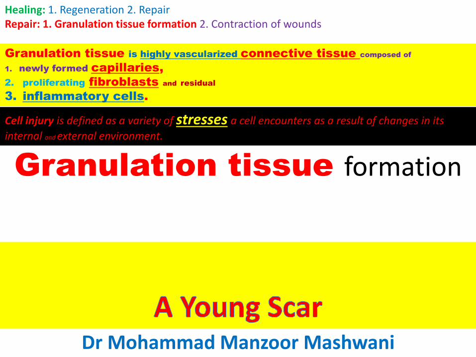
Granulation tissue formation
Dr Mohammad Manzoor Mashwani
Healing: 1. Regeneration 2. RepairRepair: 1. Granulation tissue formation 2. Contraction of wounds
Granulation tissue is highly vascularized connective tissue composed of
1. newly formed capillaries,
2. proliferating fibroblasts and residual
3. inflammatory cells.
Cell injury is defined as a variety of stresses a cell encounters as a result of changes in its
internal and external environment.
Healing
Healing is the body response to injury in an attempt to restorenormal structure and function.
Healing involves 2 distinct processes:
1. Regeneration
2. Repair
Healing by Regeneration• Regeneration when healing takes place by
proliferation of parenchymal cells and usually results in complete restoration of the original tissues.
OR
• The replacement of the destroyed tissue by the parenchymal cells of the same type is called regeneration. OR
• The replacement of destroyed cells by proliferation of surrounding undamaged cells of the same type is called regeneration.
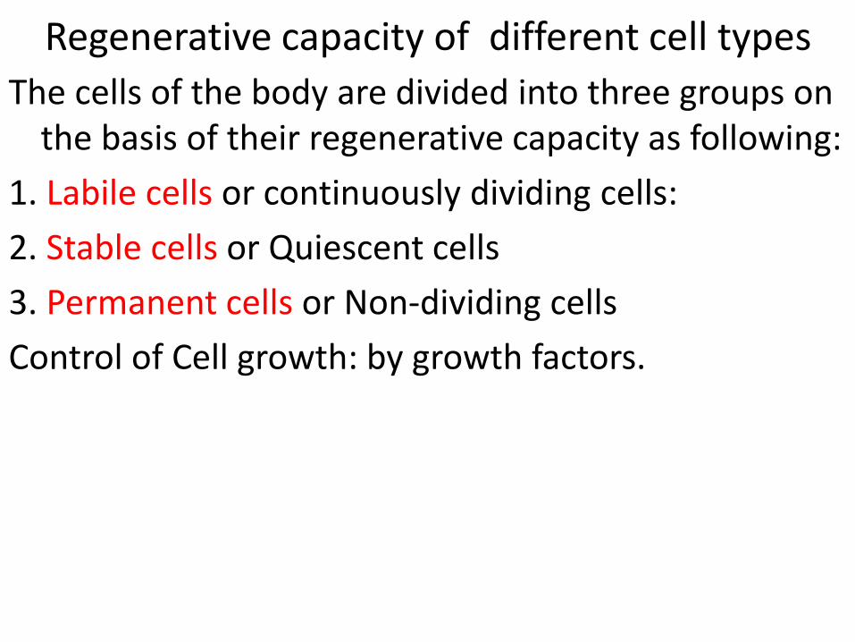
Regenerative capacity of different cell types
The cells of the body are divided into three groups on the basis of their regenerative capacity as following:
1. Labile cells or continuously dividing cells:
2. Stable cells or Quiescent cells
3. Permanent cells or Non-dividing cells
Control of Cell growth: by growth factors.
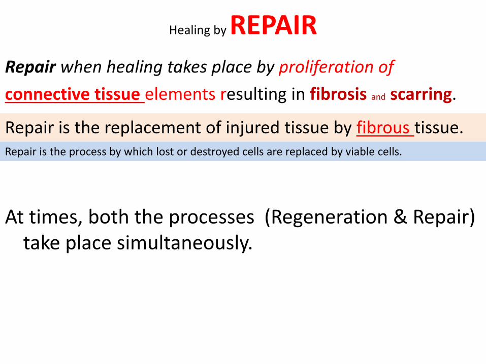
Healing by REPAIR
Repair when healing takes place by proliferation of
connective tissue elements resulting in fibrosis and scarring.
At times, both the processes (Regeneration & Repair) take place simultaneously.
Repair is the replacement of injured tissue by fibrous tissue.Repair is the process by which lost or destroyed cells are replaced by viable cells.
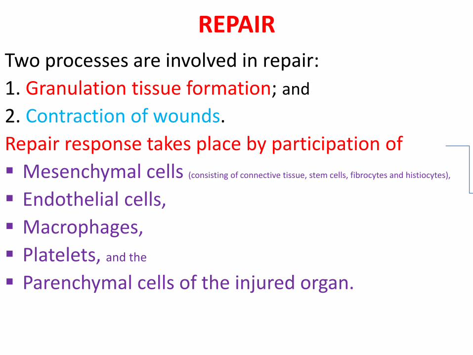
REPAIR
Two processes are involved in repair:
1. Granulation tissue formation; and
2. Contraction of wounds.
Repair response takes place by participation of
Mesenchymal cells (consisting of connective tissue, stem cells, fibrocytes and histiocytes),
Endothelial cells,
Macrophages,
Platelets, and the
Parenchymal cells of the injured organ.
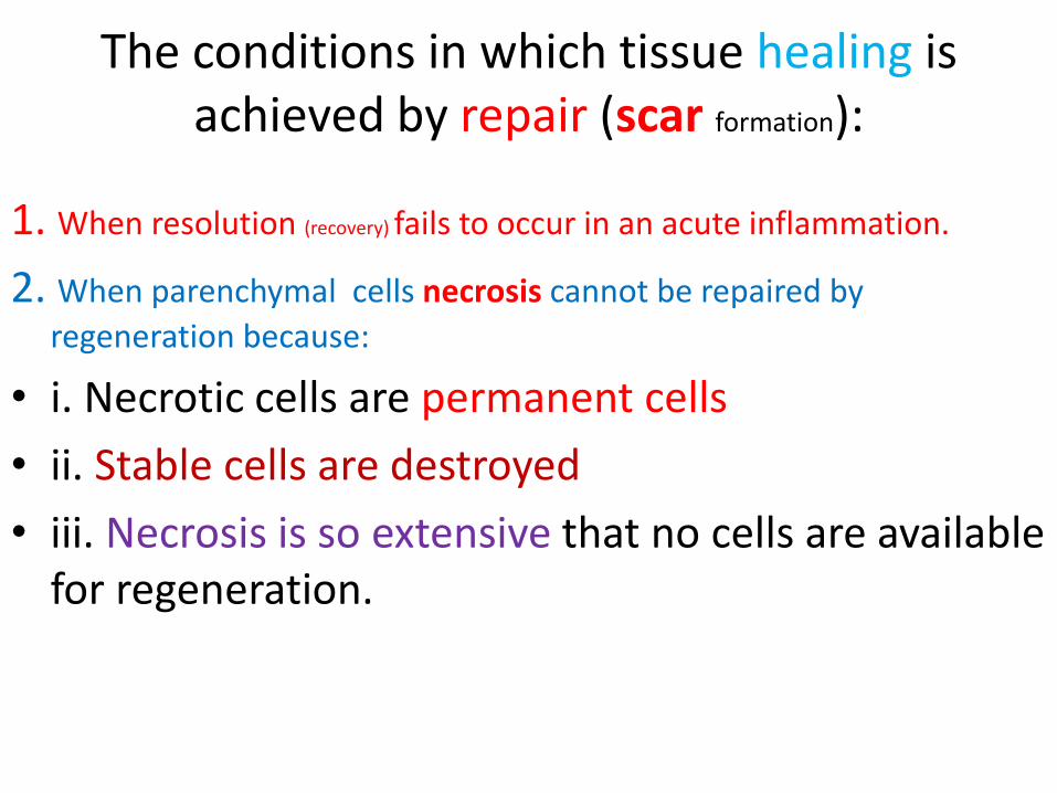
The conditions in which tissue healing is achieved by repair (scar formation):
1. When resolution (recovery) fails to occur in an acute inflammation.
2. When parenchymal cells necrosis cannot be repaired by
regeneration because:
• i. Necrotic cells are permanent cells
• ii. Stable cells are destroyed
• iii. Necrosis is so extensive that no cells are available for regeneration.
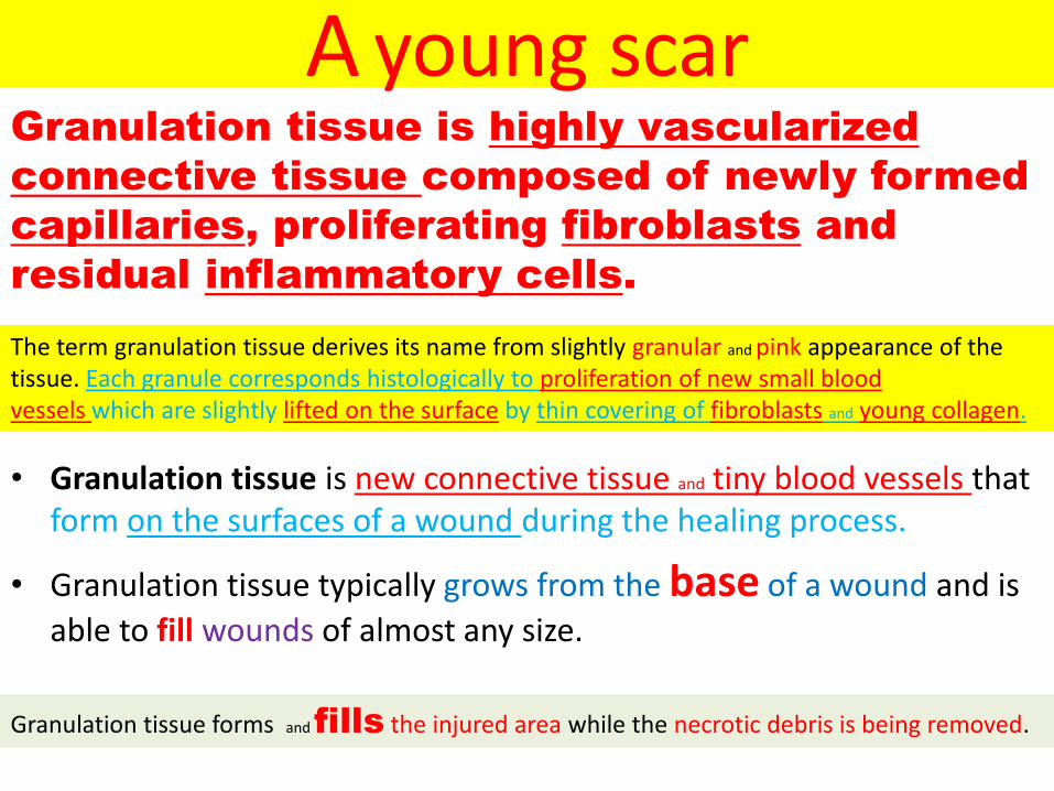
A young scar
• Granulation tissue is new connective tissue and tiny blood vessels that form on the surfaces of a wound during the healing process.
• Granulation tissue typically grows from the base of a wound and is
able to fill wounds of almost any size.
The term granulation tissue derives its name from slightly granular and pink appearance of the tissue. Each granule corresponds histologically to proliferation of new small bloodvessels which are slightly lifted on the surface by thin covering of fibroblasts and young collagen.
Granulation tissue forms and fills the injured area while the necrotic debris is being removed.
Granulation tissue is highly vascularized
connective tissue composed of newly formed
capillaries, proliferating fibroblasts and
residual inflammatory cells.
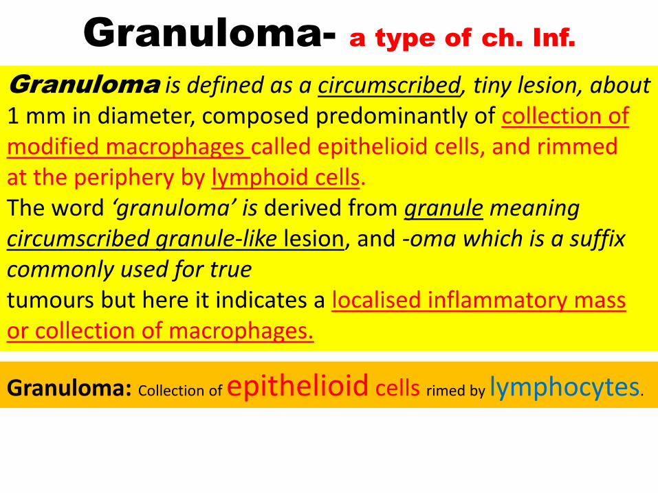
Granuloma- a type of ch. Inf.
Granuloma is defined as a circumscribed, tiny lesion, about 1 mm in diameter, composed predominantly of collection of modified macrophages called epithelioid cells, and rimmedat the periphery by lymphoid cells.The word ‘granuloma’ is derived from granule meaning circumscribed granule-like lesion, and -oma which is a suffix commonly used for truetumours but here it indicates a localised inflammatory mass or collection of macrophages.
Granuloma: Collection of epithelioid cells rimed by lymphocytes.


Primary & secondary intensions
• Wound healing• 1 intention• Edges lined up•
• 2 intention• Edges not lined up• Ergo….• More granulation• More epithelialization• More fibrosis

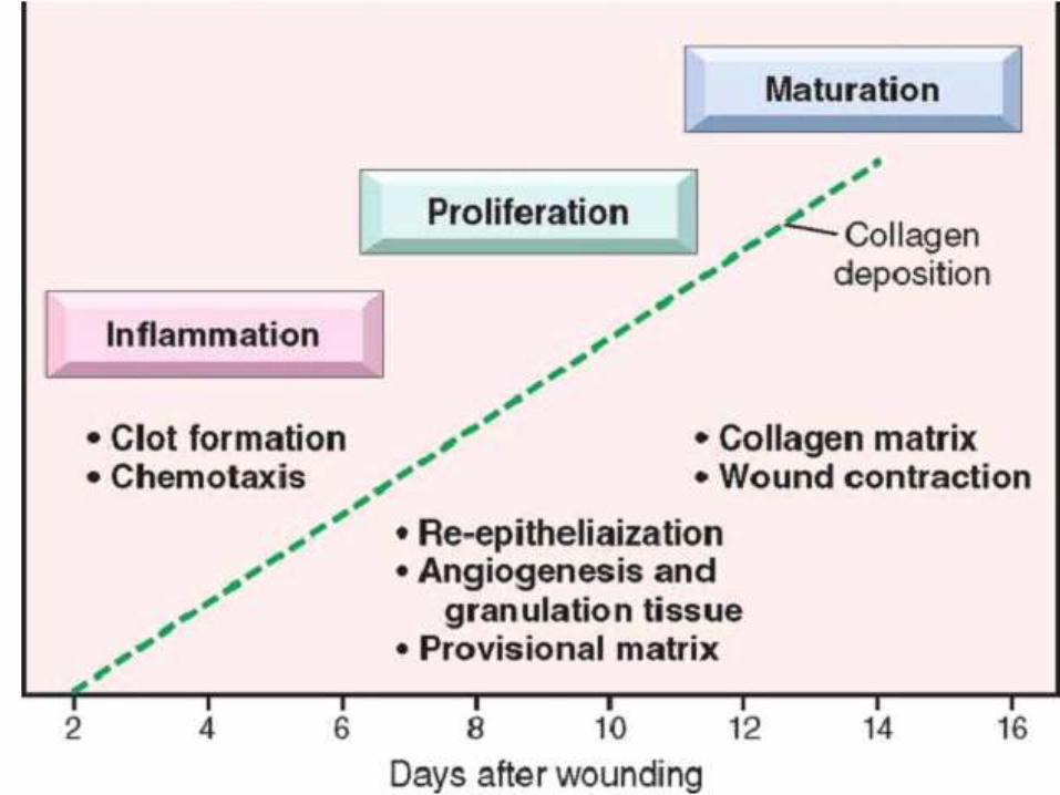
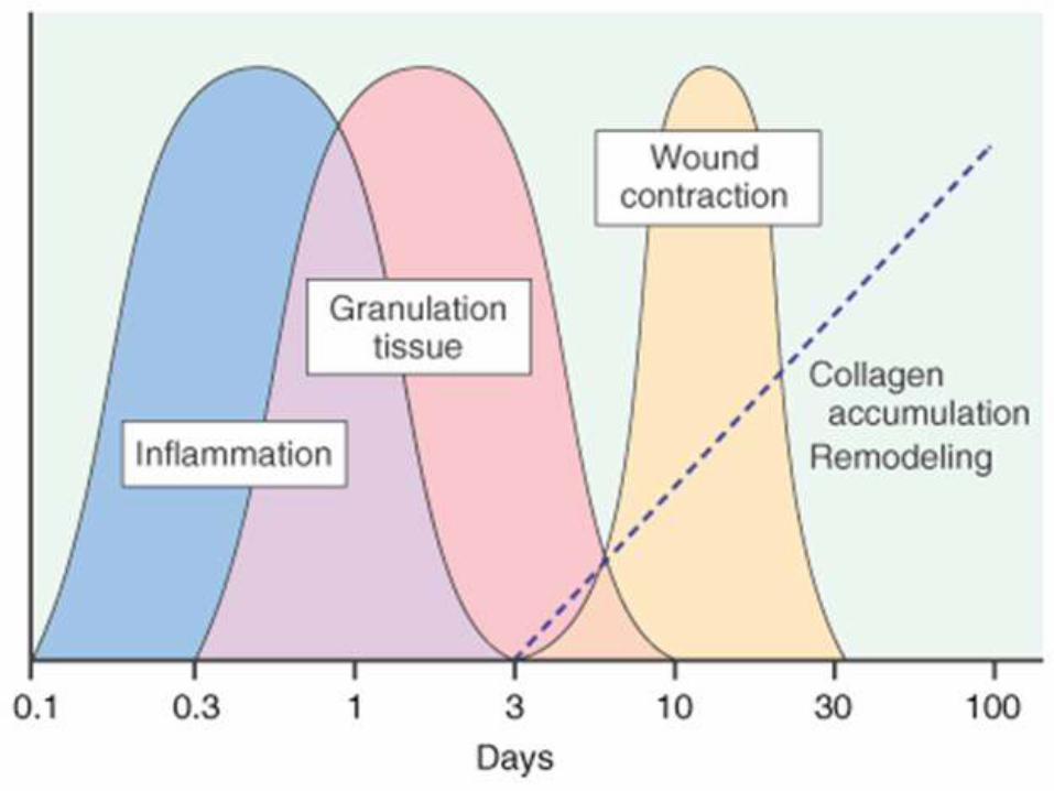
`The following 3 phases are observed in the formation ofgranulation tissue:1. PHASE OF INFLAMMATION. Following trauma, bloodclots at the site of injury. There is acute inflammatoryresponse with exudation of plasma, neutrophils and some
monocytes within 24 hours.2. PHASE OF CLEARANCE. Combination of
Proteolytic enzymes liberated from neutrophils, autolytic enzymes from dead tissues cells, and
phagocytic activity of macrophages clear off the necrotic tissue, debris and red blood cells.

3. PHASE OF INGROWTH OF GRANULATION TISSUE
This phase consists of 2 main processes:
i. angiogenesis or neovascularisation, and
ii. fibrogenesis.i) Angiogenesis (neovascularisation).
Formation of new blood vessels at the site of injury takes place by proliferation of endothelial cells from the margins of severed blood vessels.
Initially, the proliferated endothelial cells are solid buds but
within a few hours develop a lumen and start carrying blood.The newly formed blood vessels are more leaky, accounting
for the oedematous appearance of new granulation tissue.
Soon, these blood vessels differentiate into musculararterioles, thin-walled venules and true capillaries.

The process of angiogenesis is stimulated with proteolytic
destruction of basement membrane. Angiogenesis takes place under the influence of following factors:
a) Vascular endothelial growth factor (VEGF) elaborated by mesenchymal cells
while its receptors are present in endothelial cells only.
b) Platelet-derived growth factor (PDGF),
Transforming growth factor-β (TGF-β),
basic fibroblast growth factor (bFGF) and
surface integrins are all associated with cellular proliferation.
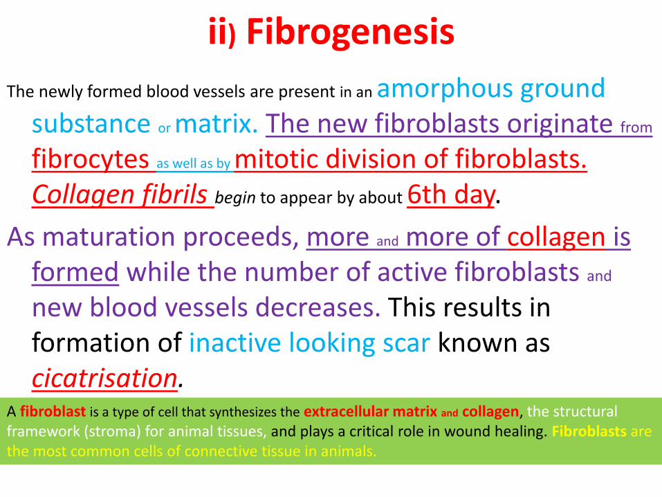
ii) Fibrogenesis
The newly formed blood vessels are present in an amorphous ground substance or matrix. The new fibroblasts originate from
fibrocytes as well as by mitotic division of fibroblasts.Collagen fibrils begin to appear by about 6th day.
As maturation proceeds, more and more of collagen is formed while the number of active fibroblasts and
new blood vessels decreases. This results in formation of inactive looking scar known as cicatrisation.
A fibroblast is a type of cell that synthesizes the extracellular matrix and collagen, the structural framework (stroma) for animal tissues, and plays a critical role in wound healing. Fibroblasts are the most common cells of connective tissue in animals.
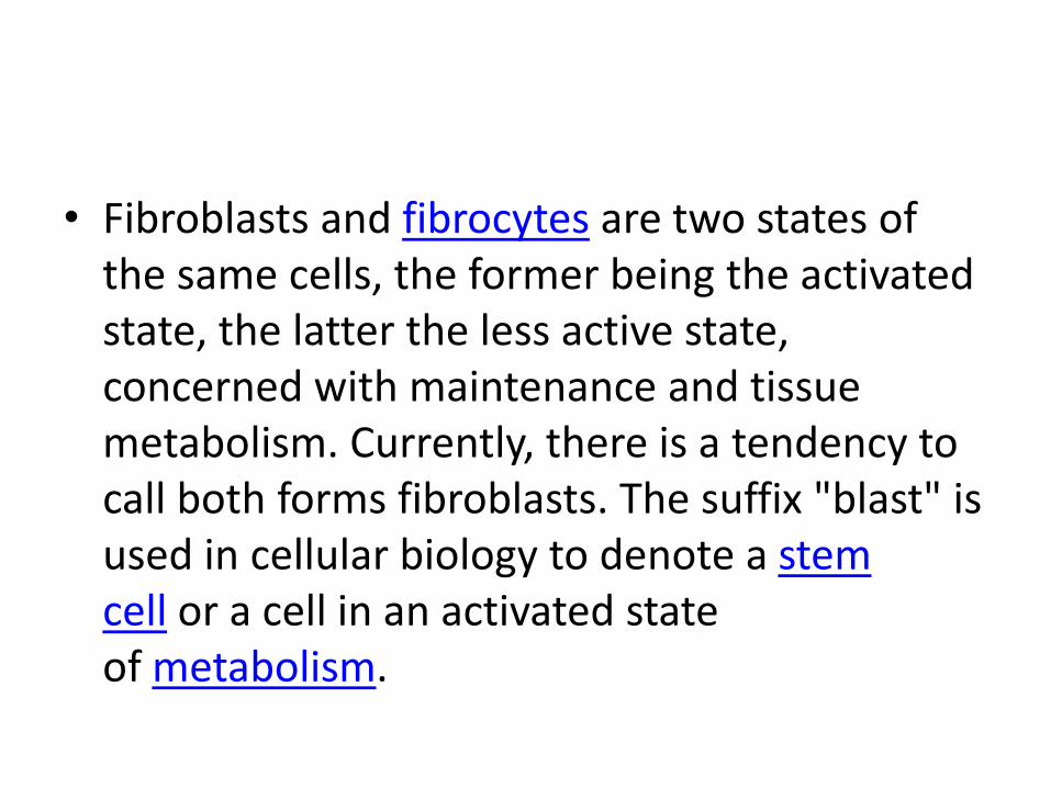
• Fibroblasts and fibrocytes are two states of the same cells, the former being the activated state, the latter the less active state, concerned with maintenance and tissue metabolism. Currently, there is a tendency to call both forms fibroblasts. The suffix "blast" is used in cellular biology to denote a stem cell or a cell in an activated state of metabolism.

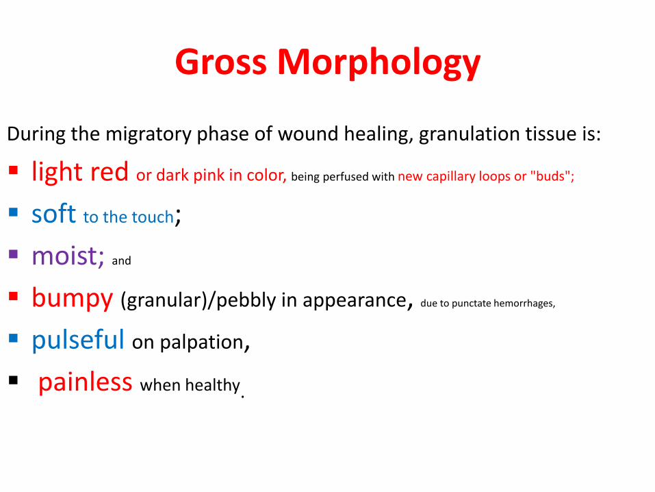
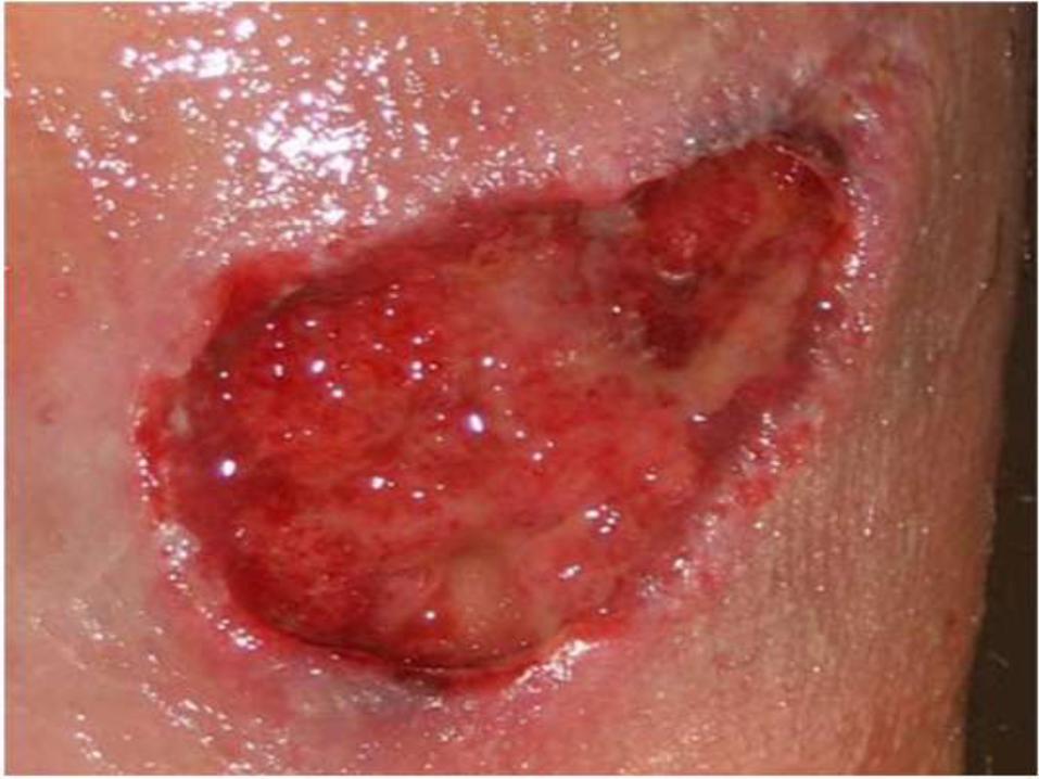
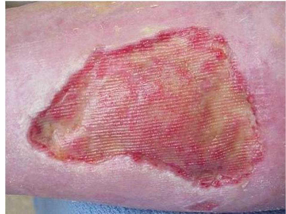
Gross Morphology
During the migratory phase of wound healing, granulation tissue is:
light red or dark pink in color, being perfused with new capillary loops or "buds";
soft to the touch;
moist; and
bumpy (granular)/pebbly in appearance, due to punctate hemorrhages,
pulseful on palpation,
painless when healthy.

Microscopy
• Microscopic examination shows thin-walled capillaries lined by endothelium
and surrounded by fibroblasts.
Residual inflammatory cells: Neutrophils, lymphocytes, plasma cells & macrophages

Active granulation tissue has inflammatory cell infiltrate, newly formed blood vessels and young fibrous tissue in loose matrix.


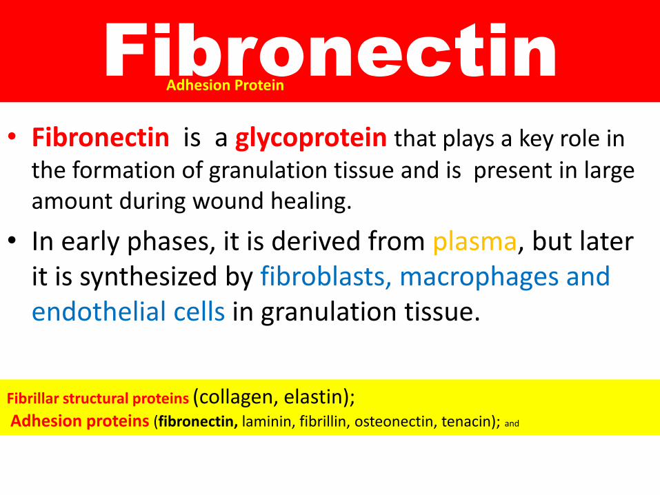
Fibronectin
• Fibronectin is a glycoprotein that plays a key role in the formation of granulation tissue and is present in large amount during wound healing.
• In early phases, it is derived from plasma, but later it is synthesized by fibroblasts, macrophages and endothelial cells in granulation tissue.
Adhesion Protein
Fibrillar structural proteins (collagen, elastin);Adhesion proteins (fibronectin, laminin, fibrillin, osteonectin, tenacin); and
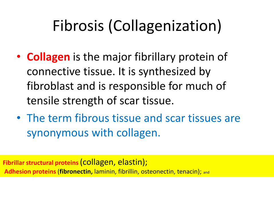
Fibrosis (Collagenization)
• Collagen is the major fibrillary protein of connective tissue. It is synthesized by fibroblast and is responsible for much of tensile strength of scar tissue.
• The term fibrous tissue and scar tissues are synonymous with collagen.
Fibrillar structural proteins (collagen, elastin);Adhesion proteins (fibronectin, laminin, fibrillin, osteonectin, tenacin); and
Methionin
• Proteins are needed for the process of healing. Methionin (an amino acid) is essential for building up of granulation tissue.
• In protein deficiency granulation tissue and collagen production is delayed, resulting in weak scar.
Vitamin C
• Vitamin C is essential for the synthesis of collagen fibers.
• In vitamin C deficiency fibroblasts produce little collagen, therefore, healing is poor and delayed.

Maturation of Scar
• h
• As the scar matures the amount of collagen increases and the scar becomes less cellular and
less vascular.
• The mature scar is composed of an avascular, poorly cellular mass of collagen and is white on gross examination.
Proud flesh
• Formation of excessive amount of granulation tissue which protrudes above the level of the surrounding skin, is called proud flesh.

Example of granulation tissue from a cut on a finger with "proud flesh".
Contraction of Wound
• Contraction decreases the size of scar and
enables the surviving cells of the organ to function with maximum effectiveness.

THANKS
• THANKS FOR YOUR ATTENSION