
Grading definitions for referable diseases - gov.uk Definitions For Referable Disease 2 About Public...
16
NHS Diabetic Eye Screening Programme Grading definitions for referable disease Public Health England leads the NHS Screening Programmes
Transcript of Grading definitions for referable diseases - gov.uk Definitions For Referable Disease 2 About Public...
NHS Diabetic Eye Screening Programme Grading definitions for referable diseasePublic Health England leads the NHS Screening Programmes

Grading Definitions For Referable Disease
2
About Public Health England
Public Health England exists to protect and improve the nation’s health and wellbeing,
and reduce health inequalities. We do this through world-class science, knowledge
and intelligence, advocacy, partnerships and the delivery of specialist public health
services. We are an executive agency of the Department of Health, and are a distinct
delivery organisation with operational autonomy to advise and support government,
local authorities and the NHS in a professionally independent manner.
Public Health England, Wellington House, 133-155 Waterloo Road, London SE1 8UG
Tel: 020 7654 8000 www.gov.uk/phe
Twitter: @PHE_uk Facebook: www.facebook.com/PublicHealthEngland
About PHE screening
Screening identifies apparently healthy people who may be at increased risk of a disease
or condition, enabling earlier treatment or better informed decisions. National population
screening programmes are implemented in the NHS on the advice of the UK National
Screening Committee (UK NSC), which makes independent, evidence-based
recommendations to ministers in the four UK countries. The Screening Quality Assurance
Service ensures programmes are safe and effective by checking that national standards
are met. PHE leads the NHS Screening Programmes and hosts the UK NSC secretariat.
PHE Screening, Floor 2, Zone B, Skipton House, 80 London Road, London SE1 6LH
www.gov.uk/topic/population-screening-programmes
Twitter: @PHE_Screening Blog: phescreening.blog.gov.uk
For queries relating to this document, please contact: [email protected]
© Crown copyright 2016
You may re-use this information (excluding logos) free of charge in any format or
medium, under the terms of the Open Government Licence v3.0. To view this licence,
visit OGL or email [email protected]. Where we have identified any third
party copyright information you will need to obtain permission from the copyright
holders concerned.
Published January 2017
PHE publications gateway number: 2016527
Grading Definitions For Referable Disease
3
Contents
About Public Health England 2
About PHE screening 2
Classifying referable retinopathy 4
Grading classification for pre‐proliferative DR (R2) 5
Grading classification for proliferative DR (R3) 11
Grading classification for maculopathy – groups of exudates (M1) 12
Classifying the macula where amblyopia and age-related macular degeneration
(AMD) are known. 15

Grading Definitions For Referable Disease
4
Classifying referable retinopathy
Feature based grading (FBG)
Graders identify individual features of diabetic retinopathy (DR) by selecting a given
feature from the feature-based grading (FBG) form. This produces a grade which is
determined by rules applied in the grading software. FBG forms for routine digital
screening, digital surveillance and SLB surveillance are found in feature-based grading
forms v1.4 on GOV.UK.
Questionable features
Grading should be conducted in line with national guidance. Equipment should meet
national specifications and should be used in line with recognised procedures for
grading. This includes avoiding excessive enhancement and enlargement of images
beyond 1:1. A lesion should only be recorded if it is definitely present.
Microaneurysms should be differentiated from pigment spots by viewing in colour and
red free and from artefacts by viewing on overlapping images where possible.
IRMA should not be recorded unless visible on colour images, without enlarging the
image area, in addition to red free images.
Cotton wool spots
Isolated cotton wool spots (one or more) in the absence of any microaneurysm or
haemorrhage should be counted as no DR (R0).
Any number of cotton wool spots (CWS) in the presence of other non‐referable features
of DR should be graded as background DR (R1).
Where CWS are detected, graders should ensure they have checked for features of
referable DR – in particular IRMA and early venous beading.
Venous loops
A venous loop should no longer be referred and should be regarded as a feature of R1.

Grading Definitions For Referable Disease
5
Photocoagulation scars
If there is no evidence of previous photocoagulation, P0 grade is assigned. If there is
evidence of previous photocoagulation (focal/grid to macula or peripheral scatter) a P1
grade is assigned.
Definition of the macula
The macula is defined as that part of the retina which lies within a circle centred on the
centre of the fovea whose radius is the distance between the centre of the fovea and
the temporal margin of the disc.
Grading classification for pre‐proliferative DR (R2)
The grading classification for R2 is:
venous beading (venous beading from ischaemia in diabetic retinopathy does
not occur in isolation)
venous reduplication
multiple blot haemorrhages (if uncertain, refer only in the presence of IRMA that
are definitely seen)
intraretinal microvascular abnormality (IRMA) (check that they can still be seen
on the colour image, that has not been enlarged)
Venous beading
Patients with venous beading should be referred. Venous beading from ischaemia in
diabetic retinopathy does not occur in isolation from multiple blot haemorrhages or
IRMA.
Venous reduplication
Patients with venous reduplication should be referred.
Multiple blot haemorrhages (MBH)
Patients with multiple blot haemorrhages should be referred. If uncertain, refer only in
the presence of IRMA that are definitely seen.
The image set MBH (previously known as MBH 3) in the macula and nasal photographs
below shows the amount of haemorrhage present in the retina to warrant an R2
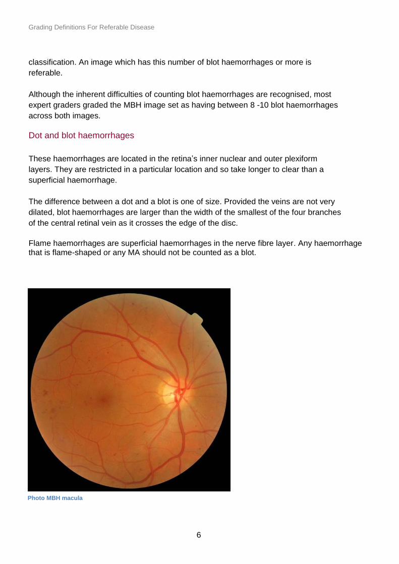
Grading Definitions For Referable Disease
6
classification. An image which has this number of blot haemorrhages or more is
referable.
Although the inherent difficulties of counting blot haemorrhages are recognised, most
expert graders graded the MBH image set as having between 8 -10 blot haemorrhages
across both images.
Dot and blot haemorrhages
These haemorrhages are located in the retina’s inner nuclear and outer plexiform
layers. They are restricted in a particular location and so take longer to clear than a
superficial haemorrhage.
The difference between a dot and a blot is one of size. Provided the veins are not very
dilated, blot haemorrhages are larger than the width of the smallest of the four branches
of the central retinal vein as it crosses the edge of the disc.
Flame haemorrhages are superficial haemorrhages in the nerve fibre layer. Any haemorrhage that is flame-shaped or any MA should not be counted as a blot.
Photo MBH macula

Grading Definitions For Referable Disease
7
Intraretinal microvascular abnormality (IRMA)
Patients with IRMA should be referred. Only IRMA that are definitely seen should be
classified as R2.
If an IRMA is found, the grader should return to the colour image. IRMA is considered
present if the IRMA can still be seen on the colour image, that has not been enlarged,
as well as on the red free.
If an IRMA can only be seen on a red free image and not on the colour image a referral
should not be made (returned to annual screening).
The above assumes screen settings, colour balance, monitor, software and camera
settings are optimal according to the recommendations of the NHS Diabetic Eye
Screening Programme.
Sometimes collaterals from vein occlusions can look like IRMA. In cases where there is
a localised patch of possible IRMA, the likelihood of a vein occlusion should be
considered. If it is judged that small collaterals are present from an old vein occlusion
rather than IRMA, this should not be given an R2 grade.
Photo MBH nasal
Grading Definitions For Referable Disease
8
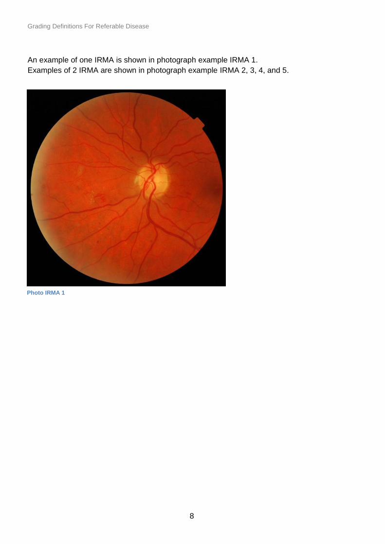
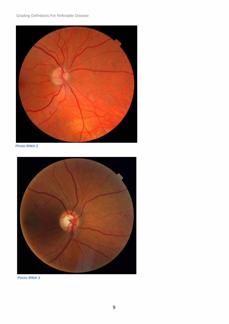
An example of one IRMA is shown in photograph example IRMA 1.
Examples of 2 IRMA are shown in photograph example IRMA 2, 3, 4, and 5.
Photo IRMA 1
Grading Definitions For Referable Disease
9
Photo IRMA 2
Photo IRMA 3

Grading Definitions For Referable Disease
10
Photo IRMA 4
Photo IRMA 5
Grading Definitions For Referable Disease
11
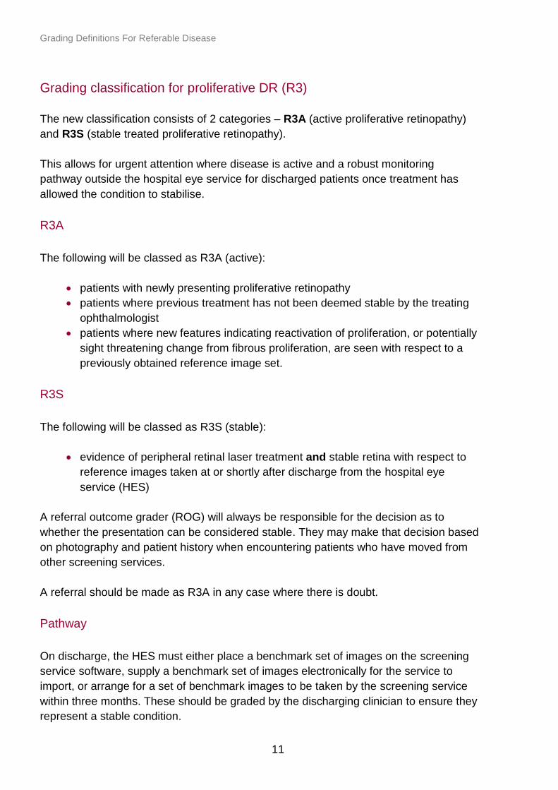
Grading classification for proliferative DR (R3)
The new classification consists of 2 categories – R3A (active proliferative retinopathy)
and R3S (stable treated proliferative retinopathy).
This allows for urgent attention where disease is active and a robust monitoring
pathway outside the hospital eye service for discharged patients once treatment has
allowed the condition to stabilise.
R3A
The following will be classed as R3A (active):
patients with newly presenting proliferative retinopathy
patients where previous treatment has not been deemed stable by the treating
ophthalmologist
patients where new features indicating reactivation of proliferation, or potentially
sight threatening change from fibrous proliferation, are seen with respect to a
previously obtained reference image set.
R3S
The following will be classed as R3S (stable):
evidence of peripheral retinal laser treatment and stable retina with respect to
reference images taken at or shortly after discharge from the hospital eye
service (HES)
A referral outcome grader (ROG) will always be responsible for the decision as to
whether the presentation can be considered stable. They may make that decision based
on photography and patient history when encountering patients who have moved from
other screening services.
A referral should be made as R3A in any case where there is doubt.
Pathway
On discharge, the HES must either place a benchmark set of images on the screening
service software, supply a benchmark set of images electronically for the service to
import, or arrange for a set of benchmark images to be taken by the screening service
within three months. These should be graded by the discharging clinician to ensure they
represent a stable condition.

Grading Definitions For Referable Disease
12
When such patients are screened subsequently, their images must be compared with
the benchmark images taken on discharge before deciding the grade. Patients who are
graded as R3S following discharge from the HES should be managed in the digital
surveillance pathway. Patients with stable treated retinopathy currently in routine annual
screening should be graded as R3S at their next routine annual screen, have
benchmark images taken and transferred to digital surveillance pathway for their next
and subsequent routine appointments.
The only R grades that will be allowed for such patients are R3A and R3S
The grading would be R3S if there are no significant changes from the baseline
discharge images.
If there are significant changes then the patient would revert to R3A and be urgently
referred back to the HES. Not all changes will be clinically urgent but the grading
committee decided it is better to keep things simple and not introduce the concept of
routine referral of R3.
‘Significant changes’ requiring urgent re-referral would include signs of active
neovascularisation, including active new vessels, pre-retinal or vitreous haemorrhage.
Grading classification for maculopathy – groups of exudates (M1)
A group of exudates is an area of exudates that is greater than or equal to half the disc
area and this area is all within the macular area.
How to work out the area
The outer points of the exudates are joined and compared to half the area of the optic
disc.
Examples of referable groups of exudates are given below as well as example
photographic images that are not referable.

Grading Definitions For Referable Disease
13
Example of an area of
exudates that is less than half
a disc area is given in photo
GE 1 and would not be
referred.
Example of an area of
exudates that is less than half
a disc area which is borderline
in size but there is less than
half a disc area within the
macular area is given in photo
GE 2 and would not be
referred.
Photo GE 1
Photo GE 2

Grading Definitions For Referable Disease
14
Example of an area of
exudates that is greater than
half a disc area is given in
photo GE 3 and this would be
referred.
Example of an area of
exudates that is greater than
half a disc area is given in
photo GE 4 and this would be
referred.
Photo GE 3
Photo GE 4
Grading Definitions For Referable Disease
15
Classifying the macula where amblyopia and age-related macular degeneration
(AMD) are known.
There will be cases when the VA is less than or equal to 6/12 and microaneurysms or
haemorrhages present within one disc diameter of the centre of the fovea. If screener
has documented known amblyopia, or there is AMD (which may also show exudates
within the macula) to account for the poor VA:
these images should be graded by the ROG grader and a decision made from
the available information whether it is considered that the reduced vision is due
to the amblyopia, the AMD or diabetic maculopathy
if the ROG decides the reduced vision is due to the amblyopia or AMD, the
maculopathy should be graded as M0 – local protocols should be followed for
referral of non-DR lesions
if the ROG decides that the reduced VA could be caused by diabetic
maculopathy, the maculopathy should be graded as M1 and the patient should
follow the nationally recommended pathway
Grading Definitions For Referable Disease
16
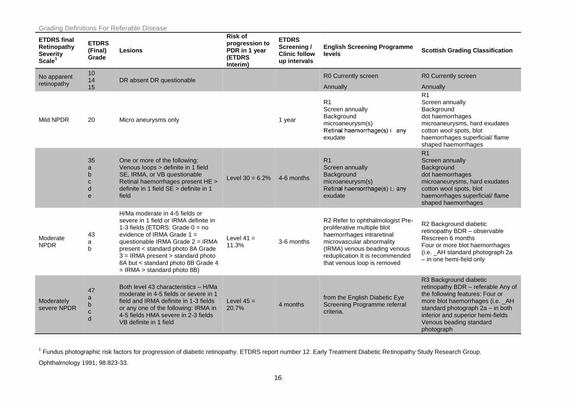
ETDRS final Retinopathy Severity Scale
1
ETDRS (Final) Grade
Lesions
Risk of progression to PDR in 1 year (ETDRS Interim)
ETDRS Screening / Clinic follow up intervals
English Screening Programme levels
Scottish Grading Classification
No apparent retinopathy
10 14 15
DR absent DR questionable R0 Currently screen
Annually
R0 Currently screen
Annually
Mild NPDR 20 Micro aneurysms only 1 year
R1 Screen annually Background microaneurysm(s)
exudate
R1 Screen annually Background dot haemorrhages microaneurysms, hard exudates cotton wool spots, blot haemorrhages superficial/ flame shaped haemorrhages
35 a b c d e
One or more of the following: Venous loops > definite in 1 field SE, IRMA, or VB questionable Retinal haemorrhages present HE > definite in 1 field SE > definite in 1 field
Level 30 = 6.2% 4-6 months
R1 Screen annually Background microaneurysm(s) Rexudate
R1 Screen annually Background dot haemorrhages microaneurysms, hard exudates cotton wool spots, blot haemorrhages superficial/ flame shaped haemorrhages
Moderate NPDR
43 a b
H/Ma moderate in 4-5 fields or severe in 1 field or IRMA definite in 1-3 fields (ETDRS: Grade 0 = no evidence of IRMA Grade 1 = questionable IRMA Grade 2 = IRMA present < standard photo 8A Grade 3 = IRMA present > standard photo 8A but < standard photo 8B Grade 4 = IRMA > standard photo 8B)
Level 41 = 11.3%
3-6 months
R2 Refer to ophthalmologist Pre-proliferative multiple blot haemorrhages intraretinal microvascular abnormality (IRMA) venous beading venous reduplication It is recommended that venous loop is removed
R2 Background diabetic retinopathy BDR – observable Rescreen 6 months Four or more blot haemorrhages (i.e. _AH standard photograph 2a – in one hemi-field only
Moderately severe NPDR
47 a b c d
Both level 43 characteristics – H/Ma moderate in 4-5 fields or severe in 1 field and IRMA definite in 1-3 fields or any one of the following: IRMA in 4-5 fields HMA severe in 2-3 fields VB definite in 1 field
Level 45 = 20.7%
4 months from the English Diabetic Eye Screening Programme referral criteria.
R3 Background diabetic retinopathy BDR – referable Any of the following features: Four or more blot haemorrhages (i.e. _AH standard photograph 2a – in both inferior and superior hemi-fields Venous beading standard photograph
1 Fundus photographic risk factors for progression of diabetic retinopathy. ETDRS report number 12. Early Treatment Diabetic Retinopathy Study Research Group.
Ophthalmology 1991; 98:823-33.


