(GRADES 5–8) GUIDE - OMSI
36
(GRADES 5–8) GUIDE EDUCATOR’S Resource Package for Junior/Intermediate School Groups
Transcript of (GRADES 5–8) GUIDE - OMSI

1
(GRADES 5–8) GUIDEEDUCATOR’S
Resource Package for Junior/Intermediate School Groups

GUIDE (GRADES 5–8)
EDUCATOR’S

This material is protected under copyright laws and may not be reproduced in any manner without the express written permission of the Institute for Plastination.
Contents
Planning your Visit
Note to Educators
Frequently Asked Questions
Classroom Activities
Exhibition Overview including Human Facts
Additional Resources
5
6
8
11
25
34

4

5
Planning Your Visit
Before• Read the Letter to Educators and Frequently Asked Questions in this guide.• Visit the BODY WORLDS website (www.bodyworlds.com) for a preview.• It is recommended for teachers complete pre-visit activities with students such as those included in this Educator’s Guide. This will help prepare your students for what they will encounter within the exhibition.• Discuss the visit with students and explain what they are going to see and why.• Administer permission forms to parents and guardians.
During• Bring the Educator’s Guide with you to the BODY WORLDS exhibition.• Seek out the museum staff for answers to your questions about the exhibition.
After• Complete the suggested activities with your students.• Consider sharing your students’ reaction to BODY WORLDS.• Visit some of the websites listed in the additional resources section.
6
Note to Educators
Dear Educator:
Our mission is to delight, inform and challenge visitors through engaging and thought-provokingexperiences in science and technology. BODY WORLDS provides students and visitors with theopportunity to learn about anatomy and health by viewing real, preserved human bodies. More than 50 million people have seen BODY WORLDS worldwide.
BODY WORLDS uses modern plastination technology as a tool to enhance the study of anatomy. Students will understand how the body works when it’s healthy and what happens when it breaks down, as well as the effects of lifestyle choices on the body. For instance, they can see the effects of smoking on the lungs, and how muscles work together during exercise.
Important information to know about BODY WORLDS:
• The BODY WORLDS exhibitions rely on the generosity of body donors, individuals who bequeathed that, upon their death, their bodies could be used for educational purposes in the exhibition. All of the fullbody plastinates and the majority of the specimens are fromthesebodydonors;somespecificspecimensthatshowunusualconditionscomefrom old anatomical collections.
• All body specimens are without skin so you can see the bones, muscles, tendons, nerves, blood vessels and organs. Eyes and genitals of the bodies remain.
• Plastinationisaprocessthatreplacesthenaturalfluidsinthebodywithatypeofflexible plastic. The use of plastics for preservation means that the specimens are odorless and completelydry.Plastinationallowsthebodiestobefixedintolife-likeposes,illustrating how our bodies are structured and how they function when performing everyday activities.
• It is recommended that teachers prepare students for their visit by completing some of the Classroom Activities in this guide.
Teachers are responsible for distributing and collecting the enclosed parental permission form.By entering the exhibition with your students, you are acknowledging that all of your studentshave parental permission to view the exhibition. We will not review permission forms;it is not necessary to bring the completed forms on your visit.
Please note that we have ensured that any of our visitors or school groups who do not wishto see BODY WORLDS will still be able to visit the museum without viewing any part of the exhibition.

7
Permission Form
Tolearnmoreabouttheexhibitionandtofindtheanswerstosomefrequentlyaskedquestions,please visit our website at www.bodyworlds.com. After viewing the exhibition,your student may want to discuss what they have seen with you. On the website you willalsofindaFamilyGuidetoassistyouinansweringquestionsthatyourchildrenmayhave.
We require that all students in school groups visiting the BODY WORLDS exhibitionreceive permission from their parents/guardians prior to coming to the exhibition.
By signing this form, you are acknowledging that your child has permission to view the exhibitionwith a teacher, school supervisor or other school representative. If you do not wish for your childto see this exhibition, please ask the teacher to make other arrangements.
Yes, I give (student‘s name)permission to view the BODY WORLDS exhibition.
Parent/Guardian’s name (please print)
Parent/Guardian Signature
Date

8
Frequently Asked Questions
What is BODY WORLDS?TheBODYWORLDSexhibitionsarefirst-of-their-kindexhibitionsthroughwhichvisitorslearnaboutanatomy, physiology, and health by viewing real human bodies, preserved using an extraordinary process called plastination: a groundbreaking method for specimen preservation invented by Dr. von Hagens in 1977.
Each exhibition features more than 100 real human specimens, including whole-body plastinates, individualorgans,bloodvesselconfigurationsandtransparentbodyslices.Thespecimensondisplaystem from a unique body donation program established in Heidelberg, Germany and managed by the Institute for Plastination. The exhibitions also allow visitors to see and better understand the long-term impact of diseases, the effects of tobacco consumption and the mechanics of artificial supports. To date,more than 50million people around the world have viewed the BODY WORLDS exhibits.
What is the purpose of the exhibition?The BODY WORLDS exhibitions aim to educate the public about the inner workings of the human body and show the effects of poor health, good health and lifestyle choices. They are also meant to create interest in and increase knowledge of anatomy and physiology among the public.
Couldn’t I learn just as much from books or models of human anatomy?Real human bodies show the details of disease and anatomy that cannot be shown with models. They also allow us to understand how each body has its own unique features, even on the inside. Visitors are drawn to real specimens in a way that they are not to plastic models. One of the special features of museums and science centers is that they offer people a chance to see the real thing in a safe and informative environment.
9
What is plastination?Invented by scientist and anatomist Dr. Gunther von Hagens in 1977, plastination is the groundbreakingmethodofhaltingdecompositionandpreservinganatomicalspecimensforscientificandmedicaleducation.Plastinationistheprocessofextractingallbodilyfluidsandsolublefatfrom specimens, replacing them through vacuum forced impregnation with reactive resins and elastomers, and then curing them with light, heat, or certain gases, which give the specimens rigidity and permanence.
Where did the specimens on display come from? Will we know who the plastinates are or how they died?The BODY WORLDS exhibitions rely on the generosity of body donors; individuals who bequeathed that, upon their death, their bodies could be used for educational purposes in the exhibitions. Currently, the Institute for Plastination has a donor roster of more than 18,000 individuals, 2,200 of which are already deceased. All of the whole body plastinates and the majority of the specimens are from thesebodydonors; somespecific specimens that showunusual conditions come fromold anatomical collections and morphological institutes. As agreed upon by the body donors, their identities and causes of death are not provided. The exhibition is focused on the nature of our bodies, not on providing personal information.
Why are the plastinates posed the way they are?The poses of the plastinates have been carefully thought out and serve educational aims. Each plastinate is posed to illustrate different anatomical features. For instance, the athletic poses illustrate the use of muscle systems while playing sports. The poses allow the visitor to relate the plastinate to his or her own body.

10
Will I be able to touch any of the plastinates?While you will be able to get very close to the plastinates, as a rule, visitors are not allowed to touch them.
Are these exhibitions appropriate for children?More than 50 million people, including young children, have viewed the BODY WORLDS exhibitions around the world. It is important to note that the exhibition includes full-body plastinates with exposed genitals.
Why is it important for the public to see these exhibits?We believe that when people understand more about how the body works and how it can break down, they are more likely to choose healthy and sustainable lifestyles. We also hope it will inspire visitors to learn more about the life sciences. Knowledge about what the human body looks like and how it functions is basic life science information that should be available to everyone.
How long can I stay inside the exhibits?You can stay as long as you like, but we recommend allowing yourself about one to two hours. The length of time will vary on how long each visitor wishes to examine each specimen and read the information provided.
Are food and drink permitted in BODY WORLDS?Food and drink are not permitted in the exhibit galleries. This policy helps to protect the BODY WORLDS plastinates.

11
Classroom Activities
These classroom activities can be used as either pre-visit activities to prepare students for BODY WORLDS or as post-visit activities to help debrief them. As you know your students best, please modify these activities to suit your class’ needs.
Make a Breathing Model
Materials:
• 1 clear plastic cup
• 1 plastic drinking straw
• 1 small round balloon
• 1 large round balloon
• 2 rubber bands
• 1 small ball of plasticine
• 1 pair of scissors
Plastic cup
Small round balloon
Large round balloon
Drinking straw
Plasticine
Rubber band
Ribcage
Lung
Diaphragm
Wind-pipe

12
Preparation and Method:
1. Insert the straw into the neck of the small balloon. Wrap the rubber band around the balloon neck and the end of the straw to make an airtight seal. (You should still be able to blow up the balloon through the straw).
2. Punch a small hole in the bottom of the cup. Enlarge it so that the free end of the straw can just be pushed through from inside the cup.
3. Seal around the straw and the hole using the plasticine.
4. Cut off the bottom half of the large balloon and stretch it over the opening of the cup. Use the other rubber band to hold it in place.
Observation:
The straw represents the trachea, the cup represents the chest, the small balloon represents the lungs and the stretched balloon represents the diaphragm. When you pull on the middle of the diaphragm (simulates breathing in), the lungs will increase in size; when you release the diaphragm, the lungs will decrease in size.
Explanation:
When you pull on the middle of the diaphragm, this increases the volume of the “chest cavity” and lowers the air pressure inside. The higher pressure of air outside then pushes air into the “lungs” through the “trachea”. To exhale air, the diaphragm is allowed to return to its resting point, reversing the process.

13
Make a Stethoscope to Listen to your Heart and Guts
You can listen to your own heart and intestines with a homemade stethoscope.
Materials:
• scissors
• small plastic funnel
• plastic tubing
• 3-way hose connector (from hardware store)
• clear tape
• stopwatch
• notepad
• pen
Plastic funnel
Rubber band
3-way hose connector
Plastic tubing
14
Preparation and Method:
1. Carefully cut three lengths of tubing, each about 12 inches long. Tape the funnel to the end of one of the tubes. Push the other end of this tube and one end of each of the other tubes into the hose connector. Usetapetomakesurethattheyfitsnugly.
2. Ask a classmate to do something active such as running for one minute. Since the sounds that you are listening for are very faint, you must do these activities in a quiet room. Now ask your classmate to hold the funnel over his or her chest and hold the tube ends to your ears, do not push them in. Count the number of heartbeats in one minute—this is their heart rate .
Additional ExperimentSometimes you can hear sounds from your stomach and intestines. While you are digesting your food, faint gurgling sounds may be heard as the organs squeeze food along. Listen over different parts of the abdomen to try to hear some of these sounds. The stomach makes much louder sounds when it is empty compared to when it is full. These result from the squeezing around of air bubbles and stomach juices.
15
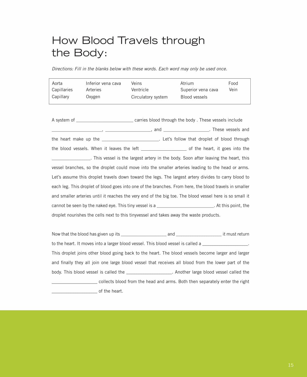
How Blood Travels through the Body:Directions: Fill in the blanks below with these words. Each word may only be used once.
A system of _________________________ carries blood through the body . These vessels include
______________________, ____________________, and ____________________. These vessels and
the heart make up the _________________________. Let’s follow that droplet of blood through
the blood vessels. When it leaves the left ____________________ of the heart, it goes into the
_________________. This vessel is the largest artery in the body. Soon after leaving the heart, this
vessel branches, so the droplet could move into the smaller arteries leading to the head or arms.
Let’s assume this droplet travels down toward the legs. The largest artery divides to carry blood to
each leg. This droplet of blood goes into one of the branches. From here, the blood travels in smaller
and smaller arteries until it reaches the very end of the big toe. The blood vessel here is so small it
cannot be seen by the naked eye. This tiny vessel is a _________________________. At this point, the
droplet nourishes the cells next to this tinyvessel and takes away the waste products.
Now that the blood has given up its ____________________ and ____________________ it must return
to the heart. It moves into a larger blood vessel. This blood vessel is called a ____________________.
This droplet joins other blood going back to the heart. The blood vessels become larger and larger
andfinally theyall joinone largebloodvessel that receivesallblood fromthe lowerpartof the
body. This blood vessel is called the ____________________. Another large blood vessel called the
____________________ collects blood from the head and arms. Both then separately enter the right
____________________ of the heart.
AortaCapillaries
Inferior vena cava VeinsArteries
Capillary Oxygen
VentricleAtrium
Circulatory system
Superior vena cava
Blood vessels
FoodVein

16
A system of blood vessels carries blood through the body. These vessels include arteries, veins, and
capillaries. These vessels and the heart make up the circulatory system. Let’s follow that droplet of
blood through the blood vessels. When it leaves the left ventricle of the heart, it goes into the aorta.
This vessel is the largest artery in the body. Soon after leaving the heart, this vessel branches, so the
droplet could move into the smaller arteries leading to the head or arms. Let’s assume this droplet
travels down toward the legs. The largest artery divides to carry blood to each leg. This droplet of
blood goes into one of the branches. From here, the blood travels in smaller and smaller arteries
until it reaches the very end of the big toe. The blood vessel here is so small it cannot be seen by the
naked eye. This tiny vessel is a capillary. At this point, the droplet nourishes the cells next to this
tiny vessel and takes away the waste products.
Now that the blood has given up its oxygen and food it must return to the heart. It moves into a
larger blood vessel. This blood vessel is called a vein. This droplet joins other blood going back to the
heart.Thebloodvesselsbecomelargerandlargerandfinallytheyalljoinonelargebloodvesselthat
receives all blood from the lower part of the body. This blood vessel is called the inferior vena cava.
Another large blood vessel called the superior vena cava collects blood from the head and arms. Both
then separately enter the right atrium of the heart.
ANSWER KEY
How Blood Travels Through the Body:
17
Myself in Numbers
Here, the students discover how diversely and, at the same time, how similarly people are built.
With this aim, the students take various measurements of themselves or their classmates.
Exercises:
1. Use a soft tape measure and take the following measurements:
• Height
• Length of foot
• Outstretched arm length
• Forearm length
• Circumference of the head
• Width of chest
• Width of waist
•Heightofhighestpointyourfingerscanreach
2. Produce a large poster with a table into which all students can enter their results.
3. Compare the results.
4. Lie on a large piece of cardboard. Ask a classmate to draw your silhouette on the cardboard. Now turn yourself through 90° on the same piece of cardboard and get someone to draw your silhouette once again. What do you notice?
5. In the exhibition, look for a similar plastinate and compare it with your picture on the piece of cardboard!

18
The Mind of the Inventor
Gunther von Hagens is the creator of plastination. For more than 40 years he has worked in this field, inventing various methods to make possible something completely new. He describes what he does as follows:
During the forty years I have worked on plastination, I have produced a whole host of individualinventions. I am always being asked how I think up such ideas and how I take each of them forward. How I come up with inventions relating to the development of plastination corresponds to the four usualstagesofinvention:identifyingtheproblem,analyzingit,workingoutsolutionsand,finally,putting them into practice.
1. Identifying the problem:
I basically question everything. Even good things can be improved upon, as “good” can always be made “better.” So, in inventing plastination, I realized there was one key problem. Saturating specimens in synthetic substances had to be an improvement on the usual practice up to that point of laying them in blocks of synthetic material.
2. Analyzing the problem:
I try not only to identify the problem at hand but also to imagine what additional questions it couldprovoke. Part of that process involves studying manuals, textbooks, literature on patents and company brochures, as well as regularly visiting trade shows.
3. Solving the problem:
Iamnevercompletelysatisfiedwithanysolution.Instead,Ialwaysfollowanumberoftrainsofthought;youshouldallowthreetofivepossiblesolutionstocompeteforawhile.Itisalsoimportantnottofixononeparticularsolutiontooearly.WhenIampursuingthebeginningsofasolution,Ihave complete faith in the fact that the solution I am working on will succeed, even if, from a purely factual point of view, that is rubbish. You have to keep returning to the problem and factoring in your own mistakes. When discussing possible solutions with experts, I often evaluate them in the following way: the more emotional it would be to reject a given solution, the more likely it is to be revolutionary and, in principle, possible.

19
Putting the solution into practice:
At this stage, studying company brochures and visiting trade shows is again important. You cannotafford not to be constantly improving your technical knowledge and repeatedly thinking throughpossible ways of putting ideas into practice. So I spend almost all my time thinking about plastination: even before getting up, when thinking about my plans for the day in the shower, while driving, when doing the shopping. Only through this can the blancmange mould become the skull, the meat slicer a machine to slice up the brain, the machine that turns chips a machine that turns brain sections, thepricetagholderintheshopwindowtheclipsfortheplatesthatflattenplastinatedsections,andthe aquarium pump a sprayer for gas hardening techniques. This process of adapting and exploiting established technology is the lifeblood of invention. I often try the impossible or the downright ludicrous. Often it is in trying out nonsensical ideas that I have crucial thoughts. So I permit myself mistakes or even make them on purpose. The strangest experiments, mistakes and accidents lead to inventions.
Exercises:
1. Read the above text. Pick out the individual steps of an invention. Present them graphically on a sheet of paper or on a poster. Use the following symbols:
Of course you can also use, or invent, your own symbols!
2. List what Gunther von Hagens uses for his inventions:
a. Logicb. Imaginationc. ...d.e.f.g.h.
3. Put the areas you named in number 2 onto a small ‘Thought Map’. You can decide how big to make it.Thenbrieflydescribewhatviewofthehumanthoughtprocessunderpinsaninventor’swork.

20
Stomach:
1. Write the biography of a stomach (from beginning to end)2. While doing so, think about its consequences for the body.3. Draw pictures to accompany your biography to make a poster.
Esophagus
Diaphragm
Longitudinal Musculature
Circular Musculature
Duodenum
Gall Bladder
Pancreas
Pylorus
Gastric Mucous Membranes
Oblique Musculature
21
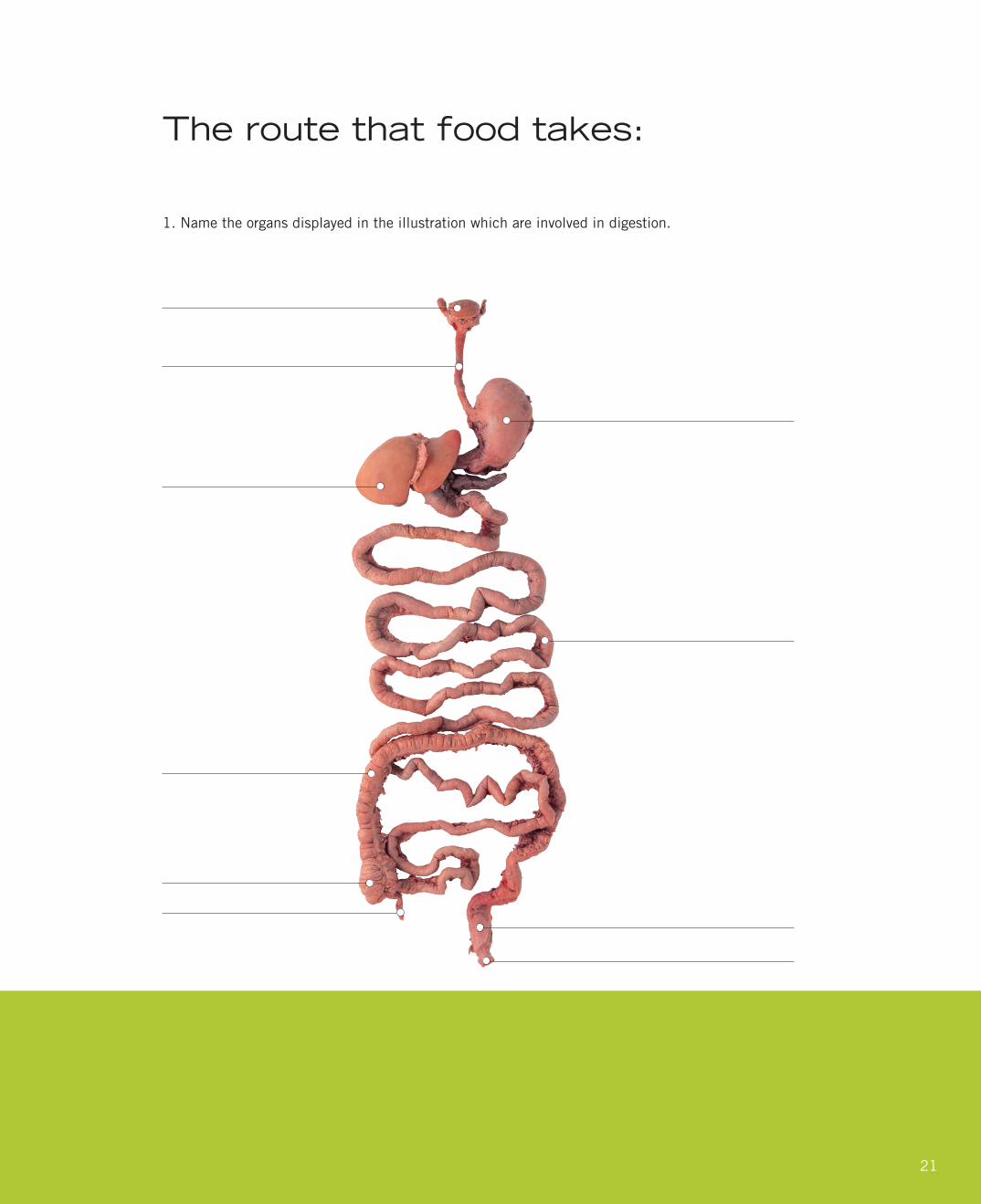
The route that food takes:
1. Name the organs displayed in the illustration which are involved in digestion.

22
2. Summarize the functions of individual sections of the gastro-intestinal tract. Fill in the table:
ORGAN FUNCTIONS

23
ANSWER KEY
The route that food takes
ORGAN FUNCTIONS
Mouth
Stomach
Small intestine
Colon
Reducing the food pieces in size,insalivation, starch digestion, transportation of the food pulp through the esophagus
Collection, mixing, gastric juice is added here, killing of bacteria, digestion of proteins
Juice from the liver and the stomach’s salivary glands are added, digestion of the three basic materials (carbohydrates, proteins and fat), absorbing into the blood
Removal of water, transportation of the indigestible remnants, expulsion through the anus

24
Dear Students,
Haveyoueverwatchedaprofessionalbasketballplayerseemtofloatinairasheorsheleapsuptodunktheballinthe
basket? Or maybe you watched skiers and skaters competing at the Olympics, and wondered “How did they do that?”
Well, our bodies are pretty amazing. And the more we learn about ourselves and how our bodies work, the better we can
take care of ourselves and others. And, the healthier we will be—making us better on the ice rink, basketball or tennis
court, jumping hurdles, or just walking down the street.
“Gunther von Hagens’ BODY WORLDS: The Original Exhibition of Real Human Bodies” was developed by a German
doctor and anatomist to help people understand how their bodies work by letting them look inside real human bodies.
When you visit with your school or family, you will see exactly how
your brain and your heart look and what happens to them when
certain diseases take over. You will see how smoking destroys
your lungs, and how your bones, muscles and ligaments all work
togethersoyoucanshootbaskets,dance,orfigureskate.
The activities inside this guide will help you learn more
about the human body.
Then come visit us to see BODY WORLDS.
You’ll really get to know yourself!
WelcomeA letter from BODY WORLDS
Dr. Angelina Whalley
Conceptual Designer for BODY WORLDS and
President and CEO of the Institute for Plastination

25
Exhibition Overview
Gunther von Hagens’ BODY WORLDS
exhibits use the science of plastination
to let visitors see how human bodies are
put together. The exhibit also teaches
how different anatomical systems
work in the human body. This special
student supplement also explores
several of the systems featured in the
exhibit, including the locomotive system,
the respiratory system, the digestive
system, the nervous system and the
cardiovascular system.
26
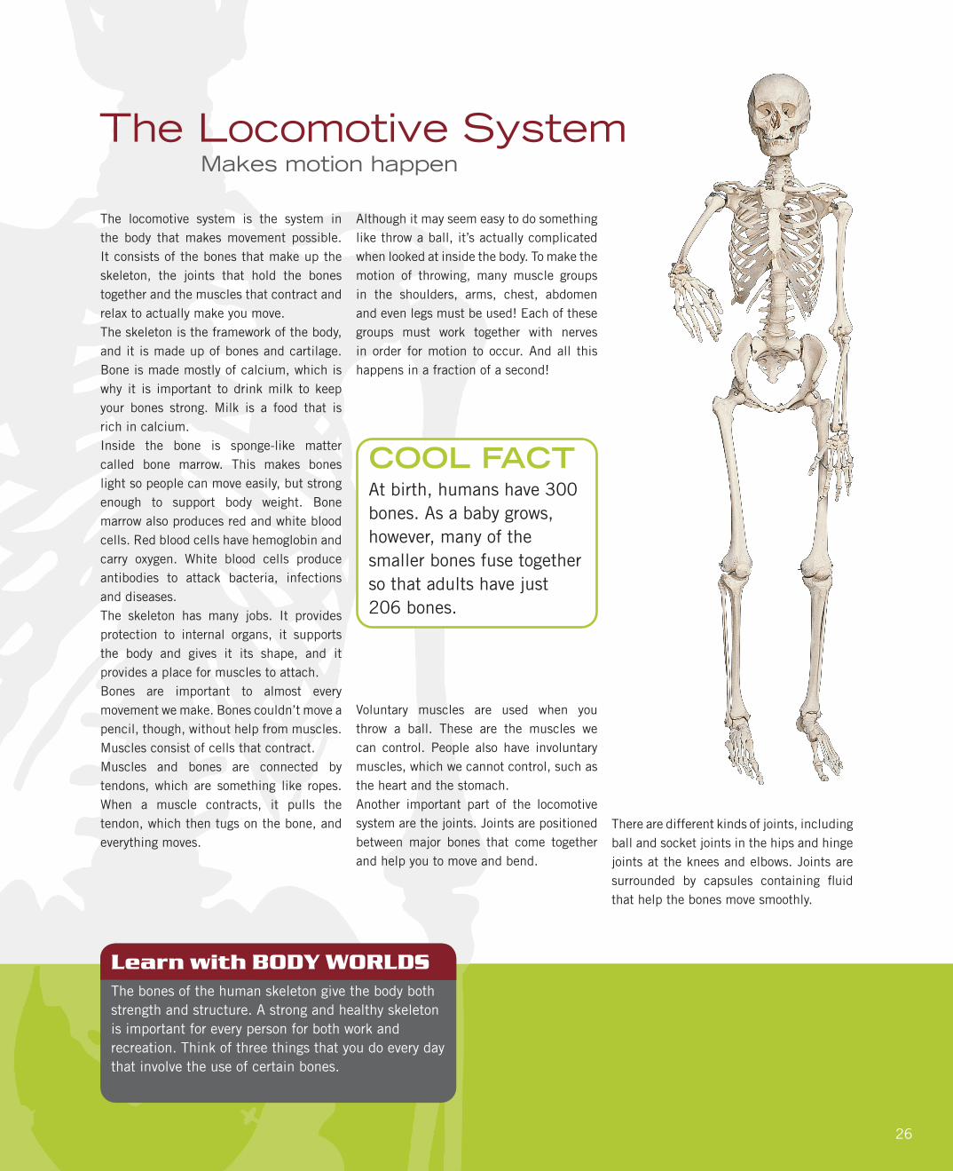
The locomotive system is the system in the body that makes movement possible. It consists of the bones that make up the skeleton, the joints that hold the bones together and the muscles that contract and relax to actually make you move.The skeleton is the framework of the body, and it is made up of bones and cartilage. Bone is made mostly of calcium, which is why it is important to drink milk to keep your bones strong. Milk is a food that is rich in calcium.Inside the bone is sponge-like matter called bone marrow. This makes bones light so people can move easily, but strong enough to support body weight. Bone marrow also produces red and white blood cells. Red blood cells have hemoglobin and carry oxygen. White blood cells produce antibodies to attack bacteria, infections and diseases.The skeleton has many jobs. It provides protection to internal organs, it supports the body and gives it its shape, and it provides a place for muscles to attach.Bones are important to almost every movement we make. Bones couldn’t move a pencil, though, without help from muscles. Muscles consist of cells that contract. Muscles and bones are connected by tendons, which are something like ropes. When a muscle contracts, it pulls the tendon, which then tugs on the bone, and everything moves.
The Locomotive SystemMakes motion happen
At birth, humans have 300 bones. As a baby grows, however, many of the smaller bones fuse together so that adults have just 206 bones.
COOL FACT
Learn with BODY WORLDSThe bones of the human skeleton give the body both strength and structure. A strong and healthy skeleton is important for every person for both work and recreation. Think of three things that you do every day that involve the use of certain bones.
Although it may seem easy to do something like throw a ball, it’s actually complicated when looked at inside the body. To make the motion of throwing, many muscle groups in the shoulders, arms, chest, abdomen and even legs must be used! Each of these groups must work together with nerves in order for motion to occur. And all this happens in a fraction of a second!
Voluntary muscles are used when you throw a ball. These are the muscles we can control. People also have involuntary muscles, which we cannot control, such as the heart and the stomach.Another important part of the locomotive system are the joints. Joints are positioned between major bones that come together and help you to move and bend.
There are different kinds of joints, including ball and socket joints in the hips and hinge joints at the knees and elbows. Joints are surrounded by capsules containing fluidthat help the bones move smoothly.

27
The Nervous SystemThe messenger and the boss
The nervous system is the system of the body that controls movements, thoughts and emotions throughout the body. Without it, you wouldn’t be able to function!There are two parts to the nervous system: the central nervous system and the peripheral nervous system.The central nervous system includes the brain and the spinal cord. They work together with nerves to send messages back and forth between the brain and the rest of the body.Thebrainistheboss.Ithasfiveparts:thecerebrum, the cerebellum, the brain stem, the pituitary gland and the hypothalamus.The cerebrum is the biggest part of the brain and controls thoughts, language and voluntary muscles, which are the muscles you can control. You also use the cerebrum when you think hard in school and when you need to remember things.
The nervous system carries messages from the brain to other parts of the body at more than 100 miles per hour.
COOL FACT
Learn with BODY WORLDSThe nervous system carries messages to the brain that makeitpossibleforthebody’sfivesensestowork.Thefivesensesaretouch,taste,hearing,sightandsmell.Explorethefivesensesbywritingaboutone of your favorite things for each sense. For example you may say that you enjoy listening to classical music, because it helps you concentrate. This relates to your sense of hearing.
The cerebellum is a lot smaller than the cerebrum, but still very important. It controls balance, movement and coordination. If it weren’t for the cerebellum, you wouldn’t be able to stand without falling!The brain stem connects the rest of the brain to the spinal cord. It’s the part in charge of major things that keep you alive like breathing, blood pressure and
digesting food. Unlike the cerebrum, the brain stem controls the involuntary
muscles—the ones that work without you thinking about it, such as the heart and stomach.
The tiny pituitary gland produces and releases hormones into the body—hormones like those that help you grow and change.
Finally, the hypothalamus regulates your body temperature, your emotions and hunger and thirst.The brain has many jobs, but it needs help from nerves and the spinal cord, too. Every action you do happens because your brain, your nerves and your spinal cord work together.The nervous system includes millions and millions of neurons, which are microscopic cells. When you do something, messages travel from the neurons to your brain.The peripheral nervous system is composed of the nerves and neurons that go outside the central nervous system to operate the body’s limbs and organs. It is here that everything gets connected.
Next time you take a test, drink a glass of water, laugh or do anything at all, thank your nervous system. Actually, you can thank it right now since it just helped you read this!
28
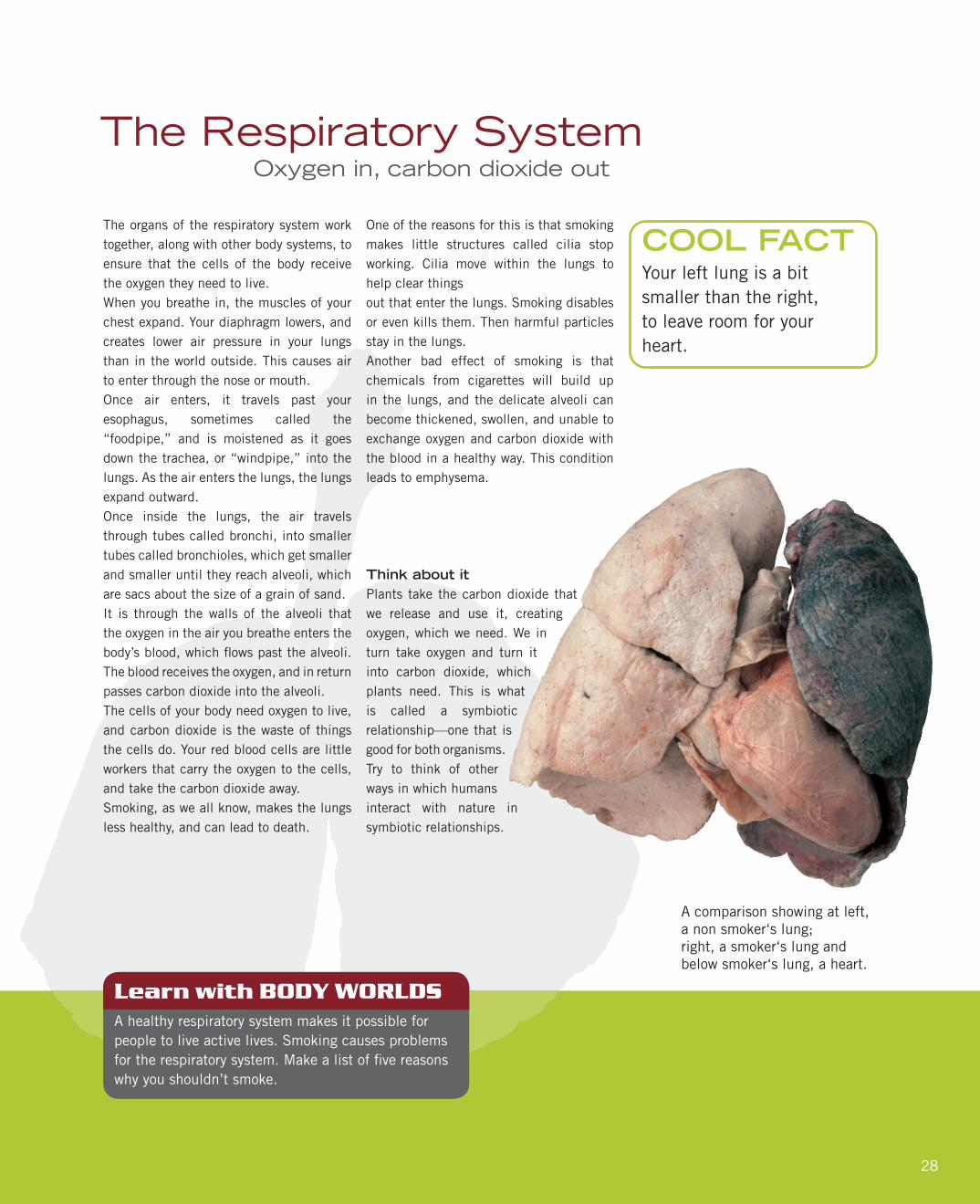
The organs of the respiratory system work together, along with other body systems, to ensure that the cells of the body receive the oxygen they need to live. When you breathe in, the muscles of your chest expand. Your diaphragm lowers, and creates lower air pressure in your lungs than in the world outside. This causes air to enter through the nose or mouth. Once air enters, it travels past your esophagus, sometimes called the “foodpipe,” and is moistened as it goes down the trachea, or “windpipe,” into the lungs. As the air enters the lungs, the lungs expand outward. Once inside the lungs, the air travels through tubes called bronchi, into smaller tubes called bronchioles, which get smaller and smaller until they reach alveoli, which are sacs about the size of a grain of sand.It is through the walls of the alveoli that the oxygen in the air you breathe enters the body’sblood,whichflowspastthealveoli.The blood receives the oxygen, and in return passes carbon dioxide into the alveoli. The cells of your body need oxygen to live, and carbon dioxide is the waste of things the cells do. Your red blood cells are little workers that carry the oxygen to the cells, and take the carbon dioxide away.Smoking, as we all know, makes the lungs less healthy, and can lead to death.
Learn with BODY WORLDSA healthy respiratory system makes it possible for people to live active lives. Smoking causes problems fortherespiratorysystem.Makealistoffivereasonswhy you shouldn’t smoke.
One of the reasons for this is that smoking makes little structures called cilia stop working. Cilia move within the lungs to help clear things out that enter the lungs. Smoking disables or even kills them. Then harmful particles stay in the lungs. Another bad effect of smoking is that chemicals from cigarettes will build up in the lungs, and the delicate alveoli can become thickened, swollen, and unable to exchange oxygen and carbon dioxide with the blood in a healthy way. This condition leads to emphysema.
Think about itPlants take the carbon dioxide that we release and use it, creating oxygen, which we need. We in turn take oxygen and turn it into carbon dioxide, which plants need. This is what is called a symbiotic relationship—one that is good for both organisms. Try to think of other ways in which humans interact with nature in symbiotic relationships.
The Respiratory SystemOxygen in, carbon dioxide out
Your left lung is a bit smaller than the right, to leave room for your heart.
COOL FACT
A comparison showing at left, a non smoker‘s lung; right, a smoker‘s lung and below smoker‘s lung, a heart.

29
The Cardiovascular SystemThe body’s great pump
Learn with BODY WORLDSThe cardiovascular system is a delicate system and can be affected by many things. Fats and cholesterol, forexample,cansloworevenblocktheflowofbloodin the body. Fats and cholesterol enter the body through foods people eat, and that is one reason people are encouraged to limit the amount of fatty or oily foods they eat. Think of ten fatty foods and ten healthier options. For example, you may think of a doughnut as a fatty food and toast as an alternative.
Images of hearts are often used to symbolize romance or love.But actually—and more importantly—the heart is the central organ of the cardiovascular system, and it doesn’t look much like the drawings found on Valentines.Cardio means heart, and the cardiovascular system is essential to our survival.The cardiovascular system is sometimes referred to as the circulatory system because it’s responsible for the circulation of blood through the body.
At every stage of life, your heart is about the size of thefistyoumakewhenyouclose your hand.
COOL FACT
It consists of the heart, which is a muscular pumping device, and a closed system of vessels called arteries, veins and capillaries.The cardiovascular system’s vital role is to provide a continuous and controlled movement of blood through the thousands of miles of microscopic capillaries that reach every tissue and cell in the body. Human survival depends on the circulation of blood to the organs, tissues and cells of your body. Arteries carry blood enriched with oxygen away from the heart, and veins carry blood
that has used up its oxygen back to the heart. Through the heart and lungs,
the blood gets a fresh supply of oxygen and delivers it to the rest of the body.
Twenty major arteries make a path through the tissues of the body. Then they branch out into smaller vessels called arterioles. These branch further into the capillaries, most of which are thinner than a hair—some so tiny, in fact, that only one blood cell can move through at a time. Once the blood in capillaries delivers oxygen and nutrients, it picks up carbon dioxide and other waste. Then blood moves back through wider vessels, called venules. These eventually join to form veins, which deliver the blood back to your heart to pick up oxygen. If all the vessels of this network were laid end to end, they would extend about 60,000 miles, far enough to circle Earth more than twice!Because all the tissues in the body rely on it, the cardiovascular system appears early in developing embryos—in the fourth week after fertilization—and reaches a functioning state long before any other major organ system.

30
The Digestive SystemConverting food into energy
The body’s digestive system converts the food you eat into the energy you need to live. The journey through your digestive system is a long one for food. It starts in the mouth, where teeth grind and tear the food into small pieces. Saliva then wets and softens the food, and begins to dissolve carbohydrates. Once the food is properly mashed and wet, it is pushed by muscle action into the pharynx, or throat, and down the esophagus, which leads to the stomach. When food reaches the stomach it is mixed and broken down further by acids the
stomach produces. The stomach protects itself from these acids by secreting a layer of mucus that lines the inside of the stomach.Some things, such as water and sugars, can be absorbed right out of the stomach and into the bloodstream. The things that need more digestion have further steps ahead of them. When the stomach has made the food a liquid, the food passes through a valve into the small intestine.
Learn with BODY WORLDSThe digestive system breaks down the food that supplies the human body with energy. What foods would you eat if you needed energy for sports or active recreation? Pickfivefoodsyouthinkwouldbegoodsourcesofenergy. Then pair off and research your foods. Were they all healthy choices for getting the energy you needed?
Your mouth makes about half a quart of saliva each day, and you produce a total of about seven quarts of digestive juices.
COOL FACT
The small intestine has a large surface area because it contains villi. Villi are tiny little structures like very short hairs that stick out into the small intestine. Through the walls of the villi nutrients from food pass into the bloodstream. The bloodstream carries the nutrients to your cells so they can live. Once all the useful nutrients have been taken from food in the small intestine, the unusable parts pass into the large intestine, or colon. In the large intestine, water is extracted from the waste and the material hardens into feces. The feces are passed out of the body when you go to the bathroom.
Digestive helpersThe pancreas, liver and gallbladder are all organs that do things important to the digestive system. The pancreas makes enzymes that help digest proteins, fats and carbohydrates. The liver makes bile, which helps the body absorb fat. Bile is stored in the gallbladder until it is needed. Enzymes and bile travel into the small intestine through ducts. Interestingly, people don’t really need the gallbladder. If it is removed, the bile just flows right into the smallintestine and does its job.

31
Art in ScienceThe beauty of the body
The BODY WORLDS exhibits teach a great deal about the science and anatomy of the human body. They also teach about the form and art of the human body. Studies of anatomy have always been a key part of art education. Artists who know how the human body is put together, and how its muscles work, are better able to portray people in painting, sculpture and other art forms.
Learn with BODY WORLDSUnderstanding how the body works is important in many professions. Think about what you may want to be when you grow up, and write a short sentence or paragraph explaining what about anatomy is important in the job, and why.
This knowledge is important, even if artists choose to represent the human form in abstract ways rather than realistic representation. In the BODY WORLDS exhibits, Gunther von Hagens has positionedhumanfigurestorevealhowthebody is put together and how it performs different tasks. He also has presented human figures in ways that highlightdifferent body systems. A group display called “The Blood Vessel Family,” for example, reveals the human form through its network of blood vessels. The scientific choices he has made givevisitors a new way to understand how human bodies work. At the same time, he has revealed how beautiful the form and systems of the human body are.As visitors go through the exhibits, they learn the science and biology of anatomy. They also get to experience the artistic qualities of anatomy.This gives the exhibits appeal to all students, not just those in science classes.
Think like an artistLike Gunther von Hagens, artists sometimes liketofocusononeaspectofafigure.Inart, this may be done by emphasizing one feature of a person, or showing the subject from an unusual angle or perspective. Explore this idea by thinking about someone inyourfamily.Reflectonwhatthispersonis like, or what you admire about him or her. Then think about what you would focus on if you were to portray this person in an artwork. Draw a sketch of your artwork and explain your ideas to the class.
Photos as artNewspaper photographers often are asked to take photo portraits of people in the news. These portraits often could be considered photographic artworks. Look through the news and features sections of the newspaper for several days and clip photos portraying people. Pick the one you like the most and explain to the class what makes the portrayal effective or artistic in your eyes. Finish by giving the photo a title, and explain it to classmates.
Sports anatomyCoaches need to know how to evaluate the physical skills and talents of players. These talents often are based on anatomy. Pick an athlete you admire. Then think about the different body systems explored in this guide. Write out which systems contribute most to the success of this athlete.

32
Would you do it?Thoughts about plastination and your body
All specimens in Gunther von Hagens’ BODY WORLDS exhibits are authentic. They belonged to people who declared during their lifetime that their bodies should be made available after their deaths for the instruction of doctors and the education of the public.“BODY WORLDS is most of all a collaboration between the donors and myself, and all those who view the exhibit,” von Hagens says. “All of humanity owes the donors a great deal, for without them, there would be no BODY WORLDS.”To ensure that donors make the decision willingly, von Hagens’ Institute for Plastination requires that all donors sign anofficialconsentform.In the form, the donors must declare that they have made the decision “freely and voluntarily” to donate their body “for the purpose of anatomical research and education … for students and especially for the general public.”In addition, they must check off answers to specificquestionsthathavebeenraisedbyplastination so there is no doubt they fully understand their decision. “I agree for my body to be used for any purposes, provided it is to do with medical research or training” reads one example.Or “I agree that my plastinated body can be used for the medical enlightenment of laypeople and, to this end, exhibited in public (e.g. in a museum).”Or “I agree that my body can be used for an anatomical work of art.”
Plastination takes a very long time. A whole body can take up to 1,500 hours to prepare.
COOL FACT
Or “I agree that lay people be allowed to touch my plastinated body” in some exhibits. Donors to the Institute for Plastination have the option to donate all useable organs to save lives before their bodies are plastinated.
Talk about itAs a class, discuss whether you would want to have your body, or the body of a relative, plastinated for education or display. Then discuss whether you think it is a good idea to exhibit plastinates for the general public. To ease discussion, you can set up a “For Chair” and an “Against Chair” to sit in at the front of the room when offering your opinion.
In your discussion: • Consider what motivates a donor to allow his/her body to be plastinated for education or an exhibit.
• Consider how the friends and relatives of a donor might feel.
• Imagine that a member of your immediate family wanted to be plastinated.
• Consider what you might learn— or did learn—about your own body from viewing the BODY WORLDS exhibits.
Learn with BODY WORLDSAfter holding the class discussion, summarize the general feelings of the class in a news story of the style found on the front page of a newspaper. Talk about how newspaper reporters must weigh all information before making a general conclusion. Then compare summaries written by different members of the class. How similar were they? What were some differences? What was the source of those differences?
33
What is Plastination?The method of plastination explained
Learn with BODY WORLDSThe BODY WORLDS exhibits reveal how human bodies work when people take part in activities like sports, dance, chess or teaching. Different displays focus on differentsystemsinthebody.Intoday’spaper,findaphoto of a person involved in an activity that interests you. Think about what the body has to do for that activity. Then write a paragraph describing what part or system of the body you would like to show if you could create a plastinate in action.
Plastination is a process designed to preserve the body for educational and instructional purposes. Plastination, like many revolutionary inventions, is simple in concept:
Embalming and Anatomical Dissection The first step of the process involveshalting decay by pumping formalin into the body through the arteries. Formalin kills all bacteria and chemically stops the decay of tissue. Using dissection tools, the skin, fatty and connective tissues are removed in order to prepare the individual anatomical structures.
The plastination process itself is based on two exchange processes:
Removal of Body Fat and WaterInthefirststep,thebodywaterandsolublefats are dissolved from the body by placing it into a solvent bath (e.g., an acetone bath).
Forced ImpregnationThis second exchange process is the central step in plastination. During forced impregnation, a reactive polymer, e.g., silicone rubber, replaces the acetone. To achieve this, the specimen is immersed in a polymer solution and placed in a vacuum chamber. The vacuum removes the acetone from the specimen and helps the polymer to penetrate every last cell.
PositioningAfter vacuum impregnation, the body is positioned as desired. Every single anatomical structure is properly aligned andfixedwiththehelpofwires,needles,clamps, and foam blocks.
Curing (Hardening)Inthefinalstep,thespecimenishardened.Depending on the polymer used, this is done with gas, light, or heat.
Dissection and plastination of an entire body requires about 1,500 working hours and normally takes about one year to complete.
Sheet Plastination Sheet plastination is a special form of plastination. For this process, the body is deep frozen and cut into slices of 2 to 8 mm in thickness (1/12 to 1/3 inch). Instead of silicone, polyester resin or epoxy resin are used for impregnation.
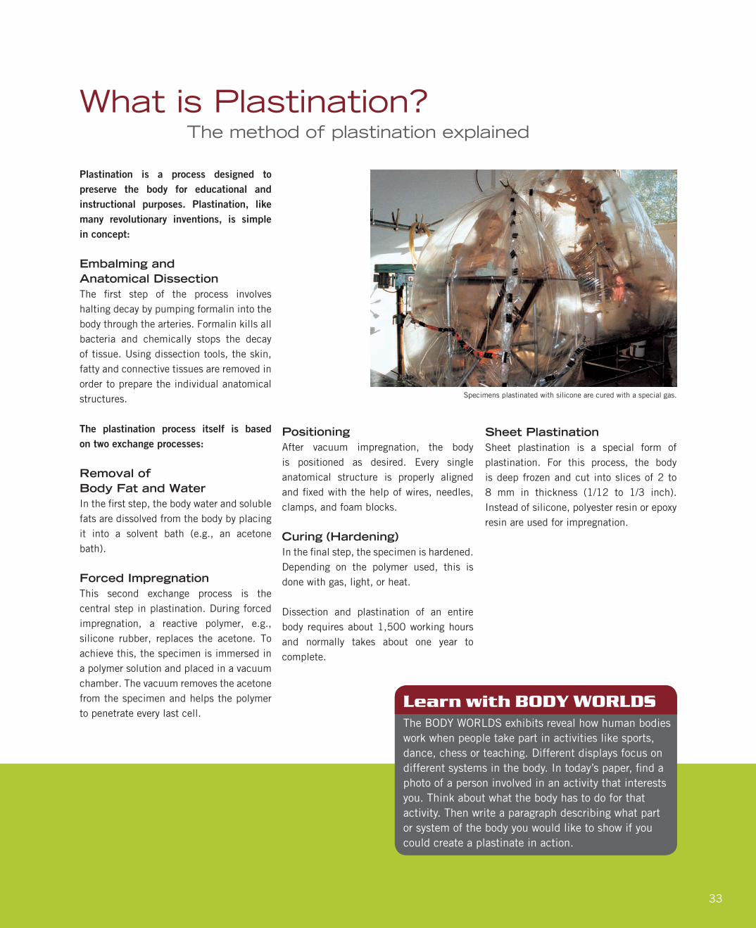
Specimens plastinated with silicone are cured with a special gas.

34
Additional Resources
National Institutes of Health—The Visible Human Project
+ http://www.nlm.nih.gov/research/visible/visible_human.html
Centers for Disease Control and Prevention
+ https://www.cdc.gov/
US Department of Agriculture Choose My Plate.gov Games:
+ https://www.choosemyplate.gov/games
Kids Health Organization
+ http://kidshealth.org/

35

Institute for Plastination
www.bodyworlds.com