Graded requirement for the zygotic terminal gene, tailless ... · and tail region of the Drosophila...
14
Development 102. 721-734 (19S8) Printed in Great Britain © The Company of Biologists Limited I9S8 721 Graded requirement for the zygotic terminal gene, tailless, in the brain and tail region of the Drosophila embryo TERESA R. STRECKER 1 *, JOHN R. MERRIAM 1 and JUDITH A. LENGYEL 12 ' Department of Biology and 2 Molecular Biology Inslilule, University of California, Los Angeles, CA 90024-1606. USA 'Present address: Division of Biology. California Institute of Technology. Pasadena. CA 91125. USA Summary We have used hypomorphic and null tailless (til) alleles to carry out a detailed analysis of the effects of the lack of til gene activity on anterior and posterior regions of the embryo. The arrangement of til alleles into a continuous series clarifies the relationship between the anterior and posterior functions of the til gene and indicates that there is a graded sensitivity of anterior and posterior structures to a decrease in til gene activity. With the deletion of both anterior and pos- terior pattern domains in til null embryos, there is a poleward expansion of the remaining pattern. Using anti-horseradish peroxidase staining, we show that the formation of the embryonic brain requires til. A phenotypic and genetic study of other pattern mutants places the til gene within the hierarchy of maternal and zygotic genes required for the formation of the normal body pattern. Analysis of mutants doubly deficient in til and maternal terminal genes is consist- ent with the idea that these genes act together in a common pathway to establish the domains at opposite ends of the embryo. We propose that til establishes anterior and posterior subdomains (acron and tail regions, respectively) within the larger pattern regions affected by the maternal terminal genes. Key words: pattern formation, segmentation, acron. Drosophila, tailless, terminal gene, brain, gene activity, mutant. Introduction The establishment of the segmentation pattern in the Drosophila embryo requires both maternal and zygotic gene products during early embryogenesis. Maternally active genes establish the overall anterior-posterior axis of the embryo and large regions of the pattern along this axis (Niisslein- Volhard, 1977, 1979; Schupbach & Wieschaus, 1986; Nusslein-Volhard et al. 1987). Zygotic genes are required for the formation of more localized regions of the segmentation pattern. The zygotic tailless {til) gene (Jiirgens et al. 1984; Strecker et al. 1986) is required for the formation of the anterior and pos- terior ectodermal regions that border the aggregate domains affected by the zygotic gap mutations (Wie- schaus et al. 1984; Lehmann & Nussslein-Volhard, 1987; Bender et al. 1987; Petschek et al. 1987). Defects observed in the mature embryonic cuticle of /// embryos can be traced to defects in the 9h embryo (Strecker et al. 1986). Anteriorly, an abnor- mal dorsal cephalopharyngeal skeleton can be traced to a reduced clypeolabrum and procephalic lobe. Posteriorly, the absence of the tail region [term used by Jiirgens (1987) to refer to the larval epidermis behind the posterior boundary of the seventh ab- dominal segment] can be traced to an absence of the most posterior abdominal segments. Despite the absence of the tail region, tailless embryos are of normal body length due to an expansion of the remaining abdominal segments (Strecker et al. 1986). This alteration in the remaining positional values has been shown to occur by the cellular blastoderm stage (Mahoney et al. 1986; Mahoney & Lengyel, 1987), consistent with the hypothesis that the tailless gene has a fundamental role in establishing and/or main- taining positional information along the anterior- posterior axis at the time of cell determination. We present here a detailed genetic and phenotypic investigation of the til gene and its relationship to the
Transcript of Graded requirement for the zygotic terminal gene, tailless ... · and tail region of the Drosophila...
Development 102. 721-734 (19S8)Printed in Great Britain © The Company of Biologists Limited I9S8
721
Graded requirement for the zygotic terminal gene, tailless, in the brain
and tail region of the Drosophila embryo
TERESA R. STRECKER1 *, JOHN R. MERRIAM1 and JUDITH A. LENGYEL12
' Department of Biology and 2Molecular Biology Inslilule, University of California, Los Angeles, CA 90024-1606. USA
'Present address: Division of Biology. California Institute of Technology. Pasadena. CA 91125. USA
Summary
We have used hypomorphic and null tailless (til) allelesto carry out a detailed analysis of the effects of the lackof til gene activity on anterior and posterior regions ofthe embryo. The arrangement of til alleles into acontinuous series clarifies the relationship between theanterior and posterior functions of the til gene andindicates that there is a graded sensitivity of anteriorand posterior structures to a decrease in til geneactivity. With the deletion of both anterior and pos-terior pattern domains in til null embryos, there is apoleward expansion of the remaining pattern. Usinganti-horseradish peroxidase staining, we show that theformation of the embryonic brain requires til. Aphenotypic and genetic study of other pattern mutants
places the til gene within the hierarchy of maternaland zygotic genes required for the formation of thenormal body pattern. Analysis of mutants doublydeficient in til and maternal terminal genes is consist-ent with the idea that these genes act together in acommon pathway to establish the domains at oppositeends of the embryo. We propose that til establishesanterior and posterior subdomains (acron and tailregions, respectively) within the larger pattern regionsaffected by the maternal terminal genes.
Key words: pattern formation, segmentation, acron.Drosophila, tailless, terminal gene, brain, gene activity,mutant.
Introduction
The establishment of the segmentation pattern in theDrosophila embryo requires both maternal andzygotic gene products during early embryogenesis.Maternally active genes establish the overallanterior-posterior axis of the embryo and largeregions of the pattern along this axis (Niisslein-Volhard, 1977, 1979; Schupbach & Wieschaus, 1986;Nusslein-Volhard et al. 1987). Zygotic genes arerequired for the formation of more localized regionsof the segmentation pattern. The zygotic tailless {til)gene (Jiirgens et al. 1984; Strecker et al. 1986) isrequired for the formation of the anterior and pos-terior ectodermal regions that border the aggregatedomains affected by the zygotic gap mutations (Wie-schaus et al. 1984; Lehmann & Nussslein-Volhard,1987; Bender et al. 1987; Petschek et al. 1987).
Defects observed in the mature embryonic cuticleof /// embryos can be traced to defects in the 9h
embryo (Strecker et al. 1986). Anteriorly, an abnor-mal dorsal cephalopharyngeal skeleton can be tracedto a reduced clypeolabrum and procephalic lobe.Posteriorly, the absence of the tail region [term usedby Jiirgens (1987) to refer to the larval epidermisbehind the posterior boundary of the seventh ab-dominal segment] can be traced to an absence of themost posterior abdominal segments. Despite theabsence of the tail region, tailless embryos are ofnormal body length due to an expansion of theremaining abdominal segments (Strecker et al. 1986).This alteration in the remaining positional values hasbeen shown to occur by the cellular blastoderm stage(Mahoney et al. 1986; Mahoney & Lengyel, 1987),consistent with the hypothesis that the tailless genehas a fundamental role in establishing and/or main-taining positional information along the anterior-posterior axis at the time of cell determination.
We present here a detailed genetic and phenotypicinvestigation of the til gene and its relationship to the

722 T. R. Strecker, J. R. Merrlam and J. A. Lengyel
maternal terminal gene family (defined by Niisslein-Volhard et al. 1987). The isolation of simple, overlap-ping deficiencies of til facilitated the characterizationof null and hypomorphic genotypes. We have deter-mined the elements of the embryo most sensitive to ///gene activity and have correlated the degree ofanterior and posterior phenotypic severity in the tilallelic series. Using anti-horseradish peroxidase stain-ing, we tested the effect of a lack of til gene activity onthe brain. Characterization of double mutants estab-lishes the relationship between til, maternal terminaland zygotic gap genes.
Materials and methods
StocksThe original til allele (til1 = L10.22) was isolated on a st emarked chromosome in an EMS mutagenesis screen(Jurgens et al. 1984). Gene and chromosome symbols aredescribed by Lindsley & Grell (1968) and Lindsley & Zimm(1985, 1986, 1987). All stocks were maintained on standardDrosophila media (Ashburner & Thompson, 1978).
The deficiency and duplication chromosomes used todetermine the location of til are shown in Fig. 1. Theterminal 3R duplications, Dp(3;l)s 34,152P, 93,150P, 79,1A and 48, were provided by Dr R. Maclntyre and havebeen previously described (Frisardi & Maclntyre, 1984;Kongsuwan et al. 1986; Strecker, 1987). The terminal 3Rdeficiencies, Df(3R)s J55, R97, A113 and LI29, have alsobeen previously described (Lindsley et al. 1972; Kongsuwanetal. 1986; Strecker, 1987).
Screen for X-ray-induced til alleles2- to 5-day-old males homozygous for the recessive markerca (Lindsley & Grell, 1968) were irradiated with 3600radand crossed to untreated HDl cd ca/ln(3R)C, Tb Sb cd cavirgin females at 25°C (50 males:200 females per bottle).After 2-3 days, males were transferred to fresh bottles andgiven additional untreated HDl cd ca/In(3R)C, Tb Sb cd cavirgin females. Males were discarded 5 days after ir-radiation. The heterozygous (ca)*/ln(3R)C Fi progenywere then screened for induced til mutations as follows.Single (ca)*/ln(3R)C F] males were crossed to three to fivetll'/In(3R)C, Tb Sb cd ca virgin females. Each test matingwas kept in a vial for 12 to 15 days at 25°C after which it wasscreened for the absence of the (ca)*/tlll F2 progeny class.Since only the latter will have normally shaped pupal cases,while the remaining progeny carry the Tb (Tubby) pupalmarker on the ln(3R)C balancer chromosome, the absenceof wildtype pupae was used as a selection criterion for linescontaining putative /// alleles. In addition, virgin(ca)*/In(3R)C F, females were mated en masse totl!1/In(3R)C males for two days, after which fertilizedfemales were placed individually in vials and kept at 25°Cfor 12 to 15 days. Each vial was then screened as describedabove. From matings that exhibited an absence of wildtypepupae, (ca)*/In(3R)C males and virgin females werecrossed inter se to establish a balanced stock.
Analysis of structures in larval cuticleThe cuticle of unhatched mature embryos was prepared forexamination by the procedure of van der Meer (1977),modified as described previously by Strecker et al. (1986).Anteriorly, structures of the cephalopharyngeal skeleton(described by Jurgens et al. 1986) were examined in detail,namely the dorsal arms, dorsal bridge, vertical plates,ventral arms, median tooth (labrum) and dorsal sac(Fig. 3A). (Note that the region termed the pharyngealridge in our original description of the til mutant phenotype[Strecker et al. 1986] is what we now refer to as the dorsalcephalopharyngeal skeleton.) Posteriorly, ///embryos wereexamined for the presence of cuticular structures of the tailregion as defined by Jurgens (1987). In addition to theventral denticle belts and dorsal hairs of the seventh andeighth abdominal segments, the presence or absence of thedorsal straight spinules (or Fell), Filzkorper, posteriorspiracles, anal tuft and anal pads was noted (Fig. 31; Lohs-Schardin et al. 1979; Sato & Denell, 1986; Whittle et al.1986; Jurgens, 1987).
Analysis of early embryos using scanning electronmicroscopy (SEM)Staged embryos were collected and prepared for SEMaccording to the protocol of Mahowald & Turner (1978)modified as described by Strecker et al. (1986). Anteriorly,9 h til embryos were examined for the degree of reductionof the clypeolabrum, procephalic lobe and optic plaques(Fig. 4A; Turner & Mahowald, 1979; Schoeller, 1964).(Note that in our original description of the /// mutantphenotype [Strecker et al. 1986] the optic plaques wereincorrectly termed optic lobes.)
Horseradish peroxidase staining of the centralnervous systemThe supra- and suboesophageal ganglia were visualized bystaining with anti-horseradish peroxidase antibody (Jan &Jan, 1982; Hartenstein & Campos-Ortega, 1986) usingmodifications suggested by J. Campos-Ortega (personalcommunication). Embryos were collected and aged to16-18 h at 22°C at which time they were dechorionated with50% bleach and transferred to heptane:4% paraformalde-hyde/PBS (phosphate-buffered saline: 130 mM-NaCl, 7 mM-Na2HPO4, 3mM-NaH2PO4) pH7-2 (1:1) and rotated for20min. The fixative was removed and embryos weredevitellinized by shaking vigorously in 100% methanol for1 min. Then embryos were washed in PBS and postfixed for40min in PBS containing 4% paraformaldehyde. Embryoswere washed 20 min each in PBS, PBS containing 0-2%BSA (PBS/BSA) and PBS containing 0-2% BSA and0-2 % Triton X-100 (PBT) and then incubated for 20 min in10% goat serum (Sigma) in PBT. Following addition ofrabbit anti-horseradish peroxidase (HRP; Cappel) to a finaldilution of 1:4000, the embryos were incubated overnight atroom temperature with gentle agitation. Embryos werethen washed in PBT, preblocked by a 30 min incubation in10 % goat serum in a volume appropriate for diluting thesecond antibody, then incubated for 3 h at room tempera-ture with goat anti-rabbit IgG conjugated to HRP (Sigma)added to a final dilution of 1:50. Embryos were washed

tailless, a zygotic terminal gene 723
20min each in PBT, PBS/BSA and PBS, and werethen stained for HRP activity using diaminobenzidine(lrngml"1, 001 % H2O2; Polysciences). The reaction wasmonitored under a dissecting microscope and stopped bydiluting with PBS. Stained embryos were stored in PBScontaining 002 % NaN3 and were mounted in glycerol formicroscopic examination and photography using differen-tial interference contrast optics. Embryos were scored forthe presence of the following brain structures; supra- andsuboesophageal ganglia, anterior and posterior trunks ofthe supraoesophageal neuropile and the frontal commissure(Campos-Ortega & Hartenstein, 1985; Fig. 3E).
Double-mutant constructionsMutant alleles of various genes were placed in combinationwith til. The alleles used and their sources were trunk[trkRMl, Schiipbach & Wieschaus, 1986] from T. Schup-bach and E. Wieschaus; fs(l)Nasrat \fs(l)N2n, Degelmannetal. 1986] from A. P. Mahowald; Kruppel [Krl, Wieschauset al. 1984] and hunchback [hbl4F, Lehmann & Nusslein-Volhard, 1987; Bender et al. 1987] from Bowling GreenStock Center; extra sex combs [esc2 and esc5, Struhl, 1981]from G. Struhl. Double-mutant embryos were obtainedutilizing standard genetic crosses; the mature cuticle ofthese embryos was examined as described above.
Results
Generation of til alleles and cytological mapping oftil locusIn the EMS mutagenesis screen in which it wasidentified (Jiirgens et al. 1984), only one til allele wasrecovered. We wished to obtain additional til allelesto determine the null phenotype, the regions of thepattern most sensitive to loss of til gene function andwhether anterior and posterior regions of the embryoare affected to a similar degree by different til alleles.From 10100 X-ray mutagenized chromosomes, weidentified six additional /// alleles. Two of the alleles,tlP and tlF, are cytologically normal. The cytology ofthe three recovered deletions, tlF, tlf and HP, placesthe til locus in the chromosomal region 1OOA1,2-100B4,5 (Fig. 1). The localization of til was furtherrefined with the recovery of the til2 allele, an inver-sion with a distal breakpoint between bands 100A5,6and 1OOB1,2. Additional confirmation of this localiz-ation was obtained from the observation that thecombination of the proximal part of T(Y;3)A113 withDp(3;l)150P, which creates a synthetic deletion ofthe region between 100A5,6 and 100Bl,2 (Kong-suwan et al. 1986), uncovers the til mutant phenotype(Fig. 1). Embryos carrying this synthetic deficiency,and til1, til2, til3 or Df(3R)tlF, die and exhibit theembryonic /// phenotype. All the evidence is thusconsistent with the localization of the til gene within100A5,6-100B1,2.
100A ia7 1
)B
9
10OC
7
Deficiencies '• '• Covers tilDf(3R)J55 =====D4(3R)R97 =====Df(3R)A113Df(3R)tir . .Df(3R)tlfDf(3R)tlP =====
DuplicationsDp (3; 1)34Dp(3;l)152PDp (3; 1)93Dp(3,l)150PDp(3;l)79Dp(3;l)lADp(3:l)48
Fig. 1. Mapping of the tailless gene to the 100A5,6 to100Bl,2 interval. Bars represent the cytological extents ofthe deficiencies (open) and the duplications (solid). Thedotted boxes represent the uncertainty in the limits of thecytological breakpoints. The results of mapping thetailless mutation are summarized in the right column:' - ' represents a deficiency that uncovers or a duplicationthat fails to cover the til mutation, while '+ ' represents adeficiency that does not uncover or a duplication thatcovers the til mutation. Df(3R)L129 (not shown) isdeficient for the 100D,E-telomere region and does notuncover the /// locus. Although, due to the limits of thecytological analysis, the uncertainties in the breakpointsof Df(3R)A113 and Dp(3;l)150P overlap in the figure,genetic analysis shows that the combination ofDf(3R)A113 and Dp(3;l)150P generates a smalldeficiency which uncovers til (see text).
tailless phenotypic seriesThe til alleles were ordered into a graded phenotypicseries on the basis of the following description of thedevelopment of the anterior and posterior regions ofthe embryo. Anteriorly, the major elements of thedorsal cephalopharyngeal skeleton are the dorsalbridge, dorsal arms and vertical plates (Fig. 3A); fatemapping traces the origin of these structures to theprocephalic lobe of the early segmented embryo, andearlier, to the acronal anlage of the cellular blasto-derm (Jiirgens etal. 1986). Posteriorly, fate mappingindicates that the cuticular elements of the tail regionarise from the prospective abdominal segments in thecellular blastoderm as follows: anal pads (All andA10), anal tuft (telson, which is dorsal to A9/A10),dorsal straight spinules or Fell (anterodorsal A8),spiracles and Filzkorper (posterodorsal A8) and the

724 T. R. Strecker, J. R, Merriam and J. A. Lengyel
eighth abdominal denticle belt (anteroventral A8)(Fig. 31; Jurgens, 1987; Sato & Denell, 1986; Whittleetal. 1986).
Recovery of the Df(3R)tlF and Df(3R)tl[e overlap-ping deficiencies (Fig. 1) permitted the constructionof trans-heterozygotes which contain a small de-ficiency of the region including the /// gene and whichgive rise to the til null phenotype (Fig. 2E). An-teriorly, tlP/tlP (=tll null) embryos have normalderivatives of the gnathal (mandibular, maxillary andlabial) segments, i.e. antennomaxillary sense organs,lateralgraten, ventral arms, H-piece, mouthhooks,cirri and labial sense organs. The most obviousanterior cuticle defect is an abnormal dorsal cephalo-pharyngeal apparatus; the contralateral homologuesof the dorsal bridge fail to fuse and the dorsal armsare reduced in length (Fig. 3C). Although the mediantooth (labrum) is formed, it is located in a moreposterior position along the dorsal sac; additionally,scleritized material lies within the dorsal sac justposterior to the labrum ('dorsal pouch' syndromedescribed by Jurgens et al. 1986). Posteriorly, theseembryos lack all derivatives of the tail region, as wellas the posteroventral cuticle of A7. In the mostextreme embryos (a small fraction of the total), thedenticle belt and dorsal hairs of A7 are also deleted(Fig. 3K). Although all cuticular structures of the tailregion are missing in til null embryos, the analopening and proctodeum are always present.
We describe below the phenotypes of several tilalleles, which exhibit weaker phenotypes than that oftil null embryos. Each of these alleles, when in transto Df(3R)tir, give rise to embryos which exhibit aslightly more extreme distribution of mutantphenotypes than that observed when the allele ishomozygous; this was determined by scoring thephenotypic range of 50 embryos of each genotype.Based on these observations, ///', tlP and til2 areconsidered hypomorphs.
Embryos that are tlll/tlll or til1/Df(3R)tlF(Fig. 2D) are indistinguishable from /// null embryosin the anterior, but show a weaker phenotype in theposterior: while the ventral denticle belt of A8, dorsalstraight spinules, Filzkorper, posterior spiracles, analtuft and anal pads are missing, a small tuft of A8dorsal hairs and all cuticular structures of A7 arepresent (Fig. 3J). In a few less extreme cases, tlP/tll1
embryos display a cluster of denticles, probably aremnant of the A8 denticle belt, in addition to dorsalhairs at the posterior end of the embryo.
Homozygous tlP or tlP/Df(3R)tlF embryos show aweaker phenotype (Fig. 2C) than til null and til1
embryos. Anteriorly, tlP/tlP embryos have a con-tinuous dorsal bridge but reduced dorsal arms(Fig. 3B); the median tooth is positioned just pos-terior to the mouthhooks as in wildtype embryos.
There is little, if any, scleritized material in the dorsalsac. Posteriorly, tlP/tlF' embryos are indistinguish-able from til1 embryos (Fig. 3J).
Homozygous til2 embryos exhibit the weakest mu-tant phenotype. Approximately half of these embryoshatch from the chorion and die as first instar larvae,showing the same phenotype as the lethal embryos(Fig. 2B). Anteriorly, the cephalopharyngeal skel-eton is normal in til2 homozygous or til2 / Df(3R)tlFembryos. Posteriorly, the majority of tlP/tll2 andtll2/Df(3R)tlF embryos and larvae have a slightlyreduced eighth abdominal denticle belt and partialFilzkorper. These embryos frequently have only oneposterior spiracle and an anal tuft in an abnormalposition, adjacent to the dorsal straight spinules. Themost extreme til2j'Df(3R)tW embryos have a reducedA8 ventral denticle belt and lack the dorsal straightspinules in addition to the remaining elements of thetail region, while the least extreme til2/til2 embryosexhibit all tail derivatives except the anal pads(Fig. 2B).
Effect of tailless on the supraoesophageal ganglionStructures of the cephalopharyngeal skeleton that arereduced or missing in mature til embryos (dorsalbridge and arms) have been fate mapped to a positionin the blastoderm embryo adjacent to the presump-tive brain (supraoesophageal ganglion; Jurgens et al.1986). Furthermore, during germband extension, el-ements of the cephalopharyngeal skeleton map to thesurface of the procephalic lobe while the supra-oesophageal ganglion is formed from the procephalicand optic lobes (Fig. 4B; Jurgens et al. 1986; Campos-Ortega & Hartenstein, 1985).
To test the hypothesis that /// also results in thereduction of the adjacent supraoesophageal ganglionanlage, we used antibody to horseradish peroxidase,which stains the central nervous system (Jan & Jan,1982). In /// null embryos, the central nervous systemis truncated abruptly at the anterior end of thesuboesophageal ganglion (Fig. 3G). Both the anteriortrunk of the supraoesophageal neuropile (at) and theposterior trunk of the supraoesophageal commissure(pt) are missing. Consistent with this observation, theposterior half of the procephalic lobe (p) and theadjacent optic plaque (o) [described in Calliphora bySchoeller (1964)] are deleted in til null embryos at thecompletion of germ band retraction (Fig. 4C, arrow).The optic plaque borders the optic lobe invaginationand has been proposed to be the primordium ofBolwig's organ, the larval visual organ (Bolwig, 1946;Schoeller, 1964). The hypomorphic tlP/tlP embryosexhibit a consistently less extreme reduction of thebrain region (Fig. 3F), lacking only the portions

tailless, a zygotic terminal gene 725
Fig. 2. Cuticular phenotypes of null and hypomorphic tailless alleles. Dark-field photomicrographs of lateral views ofunhatched mature embryos are shown and are ordered from weakest to most extreme mutant phenotypes. The seventhabdominal segment is marked with an asterisk. (A) Wildtype, note the long dorsal arms and tail region.(B) tll2/Df(3R)tlF; anteriorly the cephalopharyngeal skeleton is normal; posteriorly, only the anal pads are missing.(C) tlP/tlP; anteriorly, the dorsal arms are shortened; posteriorly, the eighth abdominal denticle belt and tail region areabsent. (D) tll1/Df(3R)tllc; anteriorly, the dorsal arms are shortened and the dorsal bridge is missing; posteriorly, theeighth abdominal denticle belt and tail region are absent. (E) Df(3R)tir/Df(3R)tlle; anteriorly, the dorsal arms anddorsal bridge are missing; posteriorly, the seventh and eighth abdominal denticle belts and tail region are lacking.T1-T3, thoracic segments; A1-A8, abdominal segments; TL, tail region.

726 T. R. Strecker, J. R. Merriam and J. A. Lengyel
posterior to the posterior trunk of the supraoeso- skeleton (see above). Furthermore, the more hypo-phageal ganglion {pi), consistent with the hypomor- morphic tll2/Df(3R)tir embryos have a normallyphic effect of the tlP allele on the cephalopharyngeal formed supraoesophageal ganglion, consistent with

tailless, a zygotic terminal gene 727
the normal cephalopharyngeal skeleton in til2 em-bryos.
Lack of til gene activity results in the expansion ofsubterminal positional valuesDue to the internalization of regions adjacent to theanterior domain (acron) affected by the til mutation,an alteration in the anterior segmentation pattern ofthe mature mutant embryo is difficult to assess.However, with the complete deletion of acronalderivatives in til null embryos there is a substantialincrease in segment length, not only posteriorly insegments A2 through A6, but also anteriorly in Tl(Fig. 5). Consistent with the increase of Tl length in
Fig. 3. Effect of til and trk mutations on the head andtail regions.
(A-D) Phase-contrast photomicrographs of thecephalopharyngeal skeleton in cuticle preparations.(A) Wildtype, note the long dorsal arms, dorsal bridge,vertical plates, median tooth and posterior wall of thepharynx. (B) tlP/tlP, note the shortened dorsal arms andintact dorsal bridge (arrow). (C) Df(3R)tl!e/Df(3R)tlP,note the shortened dorsal arms and absent dorsal bridge(arrow). (D) trk1 / trk1, note that, in addition to a reduceddorsal bridge and dorsal arms, this embryo lacks amedian tooth and has a reduced posterior wall of thepharynx [da, dorsal arms; db, dorsal bridge; vp, verticalplates; va, ventral arms; ds, dorsal sac; mt, median tooth;pwp, posterior wall of pharynx].
(E-H) Phase-contrast photomicrographs of thesupraoesophageal ganglion in fixed embryos stained withanti-horseradish peroxidase. (E) Wildtype, note the largesupraoesophageal ganglion and the anterior and posteriortrunks of the supraeosophageal neuropile. (F) tlP/tlP,note the absence of the posterior region of thesupraoesophageal ganglion. (G) Df(3R)tlle/Df(3R)tlli,note the central nervous system ends abruptly with theformation of the suboesophageal ganglion and the dorsalshift of the proventriculus. (H) trk1/trk1, note thereduced size of the supraoesophageal region and dorsalshift of the proventriculus [sbg, suboesophageal ganglion;spg, supraoesophageal ganglion; at, anterior trunk ofsupraoesophageal neuropile; fc, frontal commissure;pt, posterior trunk of supraoesophageal commissure;pv, proventriculus].
(I-L) Phase-contrast photomicrographs of the tailregion in cuticular preparations. (I) Wildtype, note theanal pads, anal tuft, Filzkorper with spiracles, dorsalstraight spinules, eighth abdominal denticle belt andseventh abdominal segment with denticle belt. (J) tlP/tlP,note that this embryo ends with a few dorsal straightspinules at the posterior end (arrow; til1 embryos exhibitthis same posterior mutant phenotype).(K) Df(3R)tllc/Df(3R)tlfi; this embryo ends at the sixthabdominal segment. (L) trk1/trk1, note that this embryoends in the denticle belt of the seventh abdominalsegment, ap, anal pads; at, anal tuft; F, Filzkorper;5/7, posterior spiracles; ds, dorsal straight spinules;A6-A8, abdominal segments.
these embryos, there is an increase in the size of thedorsal ridge, the dorsal component of the labialsegment (Fig. 4C; Campos-Ortega & Hartenstein,1985). Thus, concomitant with the deletion of theacron and tail regions in til embryos, there is anexpansion of the remaining segmentation patterntoward the opposite ends of the embryo, consistentwith previous observations (Strecker et al. 1986;Mahoney & Lengyel, 1987). The increase in segmentlength is correlated with the severity of the mutantphenotypes, such that til null embryos exhibit thegreatest increase in length of their anterior andposterior body segments, while hypomorphic tilembryos (ill1 /Df(3R)tlF, tlP/Df(3R)tlF and til2/Df(3R)tlF) exhibit proportionately smaller increasesin segment length (data not shown; Strecker, 1987).
Double-mutant combinations of til with other patterngenesWhat is the position of til in the hierarchy of maternaland zygotic loci that determine the body pattern inthe Drosophila embryo? The phenotypes of twoclasses of pattern mutants, i.e. the maternal terminal{torso, trunk, fs(l)Nasrat; originally referred to as'torso-like' genes; Schiipbach & Wieschaus, 1987;Degelmann et al. 1987; Nusslein-Volhard et al. 1987)and the zygotic gap genes (Nusslein-Volhard & Wie-schaus, 1980; Kriippel, Wieschaus et al. 1984; hunch-back, Lehmann & Nusslein-Volhard, 1987 andBender et al. 1987) suggest that they may be involvedin functions related to that of the til gene. Therelationship between til and these pattern mutantswas addressed through double mutant and pheno-typic analysis. To determine if the /// gene actsindependently of the segmental identity of the sub-region it affects, til was combined with the homeoticmutant, extra sex combs (esc).
til and the maternal terminal mutationsThe segmental domains deleted by the til mutationoverlap with those regions deleted by the maternaleffect terminal mutations (Fig. 7). A phenotypiccomparison of the anterior and posterior cuticledefects in trk and til embryos reveals the commonpattern elements deleted by these mutations. In theanterior, til embryos lack only acronal derivatives(dorsal arms, dorsal bridge), while trk embryos lackboth these and the labral derivative: the median tooth(Fig. 3D; Schupbach & Wieschaus, 1986; Jurgens etal. 1986). In addition, like til embryos, trk embryosexhibit a significant reduction in the size of thesupraoesophageal ganglion (Fig. 3H). In the pos-terior, til affects only the ectodermal tail region andA7, while trk embryos lack both this ectodermalregion and endodermal structures as well, i.e. the

728 T. R. Strecker, J. R. Merriam and J. A. Lengyel
B
Fig. 4. Comparison between wildtype and tailless head regionsat the completion of germ-band shortening. (A) Scanningelectron micrograph of wildtype; note the large procephaliclobe and the optic plaque. (B) Differential interferencecontrast photomicrograph of anti-HRP-stained wildtypeembryo; note that the supraeosophageal ganglion is located inthe posterior/dorsal region of the procephalon. (C) Scanningelectron micrograph of Df(3R)tlF/Df(3R)tlP embryo; note thatthe optic plaque and the posterior half of the procephalic lobeare absent (arrow) and the dorsal ridge is larger[c, clypeolabrum; /, labial lobe; p, procephalic lobe; o, opticplaque; d, dorsal ridge; spg, supraoesophageal ganglion;at, anterior trunk of supraoesophageal neuropile; pt, posteriortrunk of supraoesophageal commissure; sbg, suboesophagealganglion; pv, proventriculus].
posterior midgut and proctodeum (Fig. 3L; Schiip-bach & Wieschaus, 1986). Finally, as we have de-scribed above for til, the remaining positional valuesin maternal terminal embryos are expanded to re-place those which are deleted (Schupbach & Wie-schaus, 1986; Degelmann et al. 1986).
Using double-mutant constructs, we investigatedhow the embryo responds to an alteration inanterior-posterior positional values caused by boththe maternal effect terminal and zygotic tailless mu-tations. The larval cuticle of 50 embryos from a crossof trki/trki;tlP/ln(3R)C females to +/+;Df(3R)tlF/In(3R)C males was scored (Fig. 6A). Although 25 %(12 embryos) of these should have been tlP/Df andthus lacked tll+ gene activity, all 50 embryos ap-peared identical to trk embryos and exhibited themost extreme trk phenotype (Fig. 6A). [In contrast,18 % (9/50) of the tll+ embryos from trk)trk mothersexhibited weaker posterior phenotypes, namely acomplete seventh abdominal segment and reducedeighth abdominal denticle belt.] Similar results were
obtained in a double-mutant combination of fs(l)N2u
and tlP (Fig. 6B). Thus the loss of both til and trk (orfs(l)N) does not result in a more extreme mutantphenotype than for trk (or fs(l)N) alone, but doesresult more frequently in embryos exhibiting the mostextreme trk (or fs(l)N) phenotype. These results areconsistent with the idea that trk, fs(l)N and til areinvolved in a common process.
til and the gap mutations
Like til, the gap mutations also affect large, contigu-ous, aperiodic regions of the segmentation pattern.We placed til in combination with the gap mutants Krand hb to determine whether these zygotic genesinteract. The tU1,hb14F double mutant produced acuticular phenotype which is the sum of the til and hbmutant phenotypes (data not shown; Strecker, 1987).As expected, the til1 ,Krl embryo lacks Tl through A5as well as the telson and most of A8; A6 and A7 are

tailless, a zygotic terminal gene 729
0-20
~ 018
g 016
2 0-14
2 012
| 0-10
| 0-08
006
I--]/
-Hhi
T] T2 T3 AI A2 A3A4A5A6A7Body segments
Fig. 5. Expansion of anterior and posterior pattern in tilembryos. The proportion that each body segmentcontributed to the total length of the mature embryo wasdetermined by measuring the mature cuticle as previouslydescribed (Strecker et al. 1986). The average proportioneach body segment contributes to the total body length ofDf(3R)tlF/Df(3R)tlP (n = 19, dashed line) and wildtype{n = 30, solid line) embryos is shown; verticalbar = standard error.
present. The tll,Kr double-mutant phenotype is con-sistently more extreme, however, than the predictedsum of the two phenotypes: there are additionaldenticles in the region between A6 and A7, whichlack normal polarity and point toward the ventralmidline of the embryo. Furthermore, there is a partialfusion between all the ventral denticle belts (Fig.6D). This is similar to the 'lawn' phenotype observedin hb,kni and hjtz double-mutant embryos, wherethere is a fusion and loss in polarity of the ventraldenticle belts, which is interpreted as a failure of theembryo to form segments (Niisslein-Volhard et al.1985). A common characteristic among these threedouble-mutant combinations is that the mutant locithat were combined have complementary phenotypeswith respect to one another (Nusslein-Volhard et al.1985; Strecker & Merriam, 1986). Presumably thedeletion of large, complementary regions of thesegmentation pattern, found for the tll,Kr, hb,kn andftz,h double mutants, results in the remaining seg-ment boundaries becoming fused or failing to form.
til combined with a maternal homeotic mutationThe maternal effect gene, esc, is required duringdevelopment for the correct specification of segmentidentity; the body segments in embryos from esc/escfemales mated to esc/esc males develop like A8(Struhl, 1981). To determine whether til acts indepen-dently of the segment identity of the region it isrequired to specify, embryos from the cross ofesc2/esc5;tlli/+ females to esc2/esc5;tlll/+ maleswere examined (Fig. 6F). Approximately one quarter
of these embryos, while exhibiting the characteristicA8 transformation of the gnathal, thoracic and ab-dominal segments, also lacked the telson and had onefewer A8 denticle belt. These embryos were pre-sumed to be doubly mutant for esc and /// genefunctions. Although these embryos lacked derivativesof the three most-posterior abdominal segments, theywere not shorter in length, due to an increase in thewidth of the remaining body segments. The trans-formation of most of the body segments to A8 by theesc mutation did not alter the effect of the tilmutation. This is perhaps not surprising, given thatesc appears to act toward the end, and /// toward thebeginning, of nuclear cycle 14 (Struhl & Brower,1982; Mahoney & Lengyel, 1987). We conclude thatthe til gene acts independently of segment identity.
Test of maternal til activityAs the zygotic til and maternal terminal genes appearto be involved in the same process (see above), wetested for maternal tll+ expression by asking whetherextra maternal copies of the tll+ gene result in a lessextreme til mutant phenotype. This approach hasbeen shown to be consistent with results obtainedusing germline clones (Wieschaus & Noell, 1986).
Flies that are Dp(3;l)93/X;Df(3R)A113/TM6have two functional copies of the ///+ gene, whilethose that are tlll/TM3 have only one. If these twogenotypes are reciprocally crossed to each other, aproportion of the Fj progeny that aretlll/Df(3R)A113 will not inherit Dp(3;I)93 and hencewill have no functional /// gene (Fig. 1). If the ///+
gene is expressed maternally as well as zygotically, adifference in phenotypic severity should appearamong the resultant tlll/Df(3R)A113 progeny fromthese reciprocal crosses. 50 embryos from each re-ciprocal cross were scored with respect to the matureanterior and posterior cuticular phenotypes and werefound to be indistinguishable from one another.Furthermore, embryos from reciprocal crosses usingDp(3;l)34 and Dp(3;l)150P (Fig. 1) were also ob-served to be phenotypically indistinguishable. Theseresults indicate that wildtype maternal levels of thetll+ gene do not lessen the til mutant phenotype.Although gene dosage experiments do not provide asconclusive evidence as pole cell transplantation, theresults reported above are consistent with the propo-sal that tll+ is not maternally expressed.
Discussion
We have shown that in til embryos the supraoeso-phageal ganglion (brain) is deleted, the procephaliclobe is significantly reduced and, in the cephalophar-yngeal skeleton of the mature cuticle, the dorsal armsand dorsal bridge are missing. The anlagen for these

730 T. R. Strecker, J. R. Merriam and J. A. Lengyel
Fig. 6. Double-mutant constructs of /// with maternal and zygotic pattern mutants. Ventral views of the larval cuticle ofrepresentative embryos from the crosses of (A) trkl/trk';tlP/In(3R)C females to +/+ ;Df(3R)tlF/ln(3R)C males, and(B)fs(l)Nzu/fs(l)N2U;tlP/In(3R)C females to +/+;Df(3R)tllc/In(3R)C males. Lateral views of (C) Krl/Krl and(D) til1 /tllx;Krx/Krx embryos. Ventral views of (E) embryo from esc^/esc5 females and (F) til1/ill1 embryo from anesc^/esc5 female.

tailless, a zygotic terminal gene 731
structures have been fate mapped to the acron in thecellular blastoderm (see fig. 9 in Jurgens et al. 1986).In addition, til embryos lack derivatives of the sev-enth abdominal segment and tail region, which hasbeen defined as comprised of segments A8—All andan unsegmented telson (see fig. 7 in Jurgens, 1987).Based on these observations, we conclude that the ///gene is required for the formation of the acron andtail region in the cellular blastoderm. The anlagen forthe acron and tail occupy approximately the samearea at opposite ends of the blastoderm fate map andare equidistant from the midpoint (50 % egg length)along the anterior-posterior axis (Hartenstein et al.1985; Jurgens et al. 1986; Jurgens, 1987). The require-ment for the til gene by these two distant regions ofthe early embryo suggests that there is an underlyingdevelopmental relationship between the acron andtail regions in Drosophila.
There is a graded requirement for til in anterior-posterior ectodermal domainsThe til alleles were ordered in a continuous gradedseries ranging from the most extreme to the weakestallele: DfWtlF^fWtlP > til1 > tlP > til2 (datasummarized in Table 1). There is a reasonably goodcorrelation between the severity of the anterior andposterior mutant phenotypes, i.e. the weakest pos-terior phenotype is observed in til2 embryos whichhave a normal cephalopharyngeal skeleton, while themost severe posterior reduction in til null embryos iscorrelated with the most extreme deletions of struc-tures in the dorsal cephalopharyngeal apparatus. This
correlation between anterior and posterior pheno-typic severities suggests that the anterior and pos-terior functions of the til gene are not separable.
The allelic series further revealed the differentialrequirements of elements of the segmentation patternfor til gene activity (Table 1). In the posterior, themost hypomorphic til embryos lack only the analpads, which have been fate mapped to the presump-tive A10 and All region in the cellular blastodermand which constitute the most posterior structure ofthe tail region (Jurgens, 1987). With increasingphenotypic severity, we observed deletion of the analtuft, which arises from a more anterior blastodermposition, the telson region, dorsal to the presumptiveA9/A10 boundary. Less sensitive structures are theposterior spiracles (posterodorsal A8), followed bythe Filzkorper (posterodorsal A8) and the eighthabdominal denticle belt (anteroventral A8). Finally,in the most extreme til phenotypes, the anterior halfof A8 and posterior half of A7 are deleted. Theseobservations suggest that there is a graded, posterior-to-anterior, requirement for the til gene product inthe posterior ectodermal region of the embryo; thepeak of this requirement is in the presumptive Allregion (~12% egg length).
The effect of different til alleles on the formation ofthe supraoesophageal ganglion suggests that there is asimilar graded requirement for the til gene product inthe anterior (Table 1). Hypomorphic tlP embryoslack the most posterior region of the supraoeso-phageal ganglion; this region includes, but may not be
Table
Structure
1. Structures affected
Origin*
in til
m
phenotypic seriesAlleles
nn in' IIP HI2
(A) Anterior
(B) Posterior
BrainOptic plaqueProcephalic lobeDorsal armsDorsal bridge
Anal padsAnal tuft
Filzkorper/spiraclesDenticle beltDorsal hairsDenticle beltDorsal hairs
AcronAcronAcronAcronAcron
A10/A11Telson
(adjacent to A9/A10)A8A8A8A7A7
- A - A
--A
r
-
-Ar
-Ar/ +r/ +
r/
—, missing; r, reduced in size and/or abnormal in morphology; +. present and normal.* Based on recent fate mapping of head and tail regions (Jurgens el al. 1986; Jurgens, 1987).t Df(3R)tir/Df(3R)ilI* embryos.
732 T. R. Strecker, J. R. Merriam and J. A. Lengyel
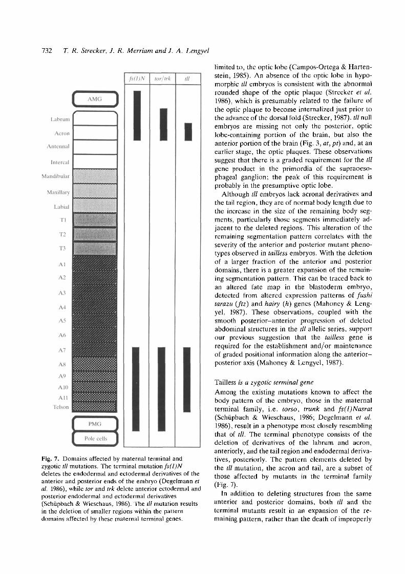
AMC'i
tor/Irk
Iill
I
Fig. 7. Domains affected by maternal terminal andzygotic /// mutations. The terminal mutation fs(l)Ndeletes the endodermal and ectodermal derivatives of theanterior and posterior ends of the embryo (Degelmann etal. 1986), while tor and trk delete anterior ectodermal andposterior endodermal and ectodermal derivatives(Schupbach & Wieschaus, 1986). The til mutation resultsin the deletion of smaller regions within the patterndomains affected by these maternal terminal genes.
limited to, the optic lobe (Campos-Ortega & Harten-stein, 1985). An absence of the optic lobe in hypo-morphic /// embryos is consistent with the abnormalrounded shape of the optic plaque (Strecker et al.1986), which is presumably related to the failure ofthe optic plaque to become internalized just prior tothe advance of the dorsal fold (Strecker, 1987). til nullembryos are missing not only the posterior, opticlobe-containing portion of the brain, but also theanterior portion of the brain (Fig. 3, at, pt) and, at anearlier stage, the optic plaques. These observationssuggest that there is a graded requirement for the tilgene product in the primordia of the supraoeso-phageal ganglion; the peak of this requirement isprobably in the presumptive optic lobe.
Although til embryos lack acronal derivatives andthe tail region, they are of normal body length due tothe increase in the size of the remaining body seg-ments, particularly those segments immediately ad-jacent to the deleted regions. This alteration of theremaining segmentation pattern correlates with theseverity of the anterior and posterior mutant pheno-types observed in tailless embryos. With the deletionof a larger fraction of the anterior and posteriordomains, there is a greater expansion of the remain-ing segmentation pattern. This can be traced back toan altered fate map in the blastoderm embryo,detected from altered expression patterns of fushitarazu (ftz) and hairy (h) genes (Mahoney & Leng-yel, 1987). These observations, coupled with thesmooth posterior-anterior progression of deletedabdominal structures in the til allelic series, supportour previous suggestion that the tailless gene isrequired for the establishment and/or maintenanceof graded positional information along the anterior-posterior axis (Mahoney & Lengyel, 1987).
Tailless is a zygotic terminal geneAmong the existing mutations known to affect thebody pattern of the embryo, those in the maternalterminal family, i.e. torso, trunk and fs(l)Nasrat(Schupbach & Wieschaus, 1986; Degelmann et al.1986), result in a phenotype most closely resemblingthat of til. The terminal phenotype consists of thedeletion of derivatives of the labrum and acron,anteriorly, and the tail region and endodermal deriva-tives, posteriorly. The pattern elements deleted bythe til mutation, the acron and tail, are a subset ofthose affected by mutants in the terminal family(Fig. 7).
In addition to deleting structures from the sameanterior and posterior domains, both til and theterminal mutants result in an expansion of the re-maining pattern, rather than the death of improperly

tailless, a zygotic terminal gene 733
patterned cells. This constitutes a fundamental dis-tinction between til and the gap gene mutants. An-teriorly, the pattern expansion is observed as anincrease in the segment width of Tl and the dorsalridge in til embryos (Fig. 4C), and an anterior shift ofthe cephalic furrow and anterior midgut invaginationin torso and trunk embryos (Schiipbach & Wieschaus,1986). Posteriorly, the pattern expansion is observedas an increase in abdominal segment width (A5, A6)in til embryos, and a posteriorward shift of subtermi-nal embryonic structures in torso and trunk embryos(Schiipbach & Wieschaus, 1986). The posteriorwardshift in pattern can be traced to changes in theblastoderm fate map. As assessed by h and ftz geneexpression, similar shifts are seen in fs(l)N, til, torand trk embryos (Degelmann et al. 1986; Mahoney etal. 1986; Mahoney & Lengyel, 1987; Mlodzik et al.1987). These results suggest that the zygotic taillessgene is required to maintain the positional infor-mation initially established by the action of thematernal terminal genes.
This idea is supported by the results of double-mutant analysis. The same structures are missingfrom embryos deficient for til and trk (or fs(l)N) asfrom embryos deficient only for trk (or fs(l)N);furthermore, trk,til double-mutant embryos morefrequently exhibit a more extreme trk phenotype thando trk,tll+ embryos. These results are consistent withthose obtained with tor,trk double mutants (Schiip-bach & Wieschaus, 1986). [In contrast to til, the gapmutant giant does not recognize the alteration inpositional values resulting from the fs(l)N2n
mutation (Petschek et al. 1987).] We interpret ourdouble-mutant results as suggesting that trk, fs(l)N,tor and til are involved in a common pathwayrequired to establish and maintain positional infor-mation at the anterior and posterior ends of theembryo. Specifically, we propose that til mediatesthe establishment of the acron and tail regions withinthe larger domains affected by the maternal terminalgenes.
We wish to thank Jose Campos-Ortega and VolkerHartenstein for helpful discussions regarding the centralnervous system, and Kathryn Anderson and Howard Lip-shitz for critically reading the manuscript. We also thankTrudi Schiipbach, Eric Wieschaus, and Gary Struhl forsending us fly strains. This research was supported by NSFGrant DCB 84-03539 to J.A.L. and J.R.M., and NIH GrantGM 38432 to J.R.M. T.R.S. was supported by USPHSNational Service Award GM7104 and a predoctoral fellow-ship from the American Association of University Women.
References
ASHBURNER, M. & THOMPSON, J. N. (1978). Thelaboratory culture of Drosophila. In The Genetics and
Biology of Drosophila, vol. 2a (ed. M. Ashburner & E.Novitski), pp. 2-109. New York: Academic Press.
BENDER, M., TURNER, R. R. & KAUFMAN, T. C. (1987).A developmental genetic analysis of the gene Regulatorof postbithorax in Drosophila melanogaster. Devi Biol.119, 418^32.
BOLWIG, N. (1946). Senses and sense organs of theanterior end of the house fly larva. Vidensk. Medd. fra.Dansk. Naturk. Foren. 109, 80-212.
CAMPOS-ORTEGA, J. A. & HARTENSTEIN, V. (1985). TheEmbryonic Development of Drosophila melanogaster.Berlin/Heidelberg: Springer-Verlag.
DEGELMANN, A., HARDY, P. A., PERRIMON, N. &MAHOWALD, A. P. (1986). Developmental analysis ofthe torso-like phenotype in Drosophila produced by amaternal-effect locus. Devi Biol. 115, 479-489.
FRISARDI, M. C. & MACINTYRE, R. J. (1984). Positioneffect variegation of an acid phosphatase gene inDrosophila. Mol. gen. Genet. 197, 403—413.
HARTENSTEIN, V., TECHNAU, G. M. & CAMPOS-ORTEGA, J.A. (1985). Fate-mapping in wildtype Drosophilamelanogaster. III. A fate map of the blastoderm.Wilhelm Roux Arch, devl Biol. 194, 213-216.
HARTENSTEIN, V. & CAMPOS-ORTEGA, J. A. (1986). Theperipheral nervous system of mutants of earlyneurogenesis in Drosophila melanogaster. WilhelmRoux Arch, devl Biol. 195, 210-221.
JAN, L. Y. & JAN, Y. N. (1982). Antibodies tohorseradish peroxidase as specific neuronal markers inDrosophila and in grasshopper embryos. Proc. natn.Acad. Sci. U.S.A. 72, 2700-2704.
JURGENS, G. (1987). Segmental organisation of the tailregion in the embryo of Drosophila melanogaster. Ablastoderm fate map of the cuticle structures of thelarval tail region. Wilhelm Roux Arch, devl Biol. 196,141-157.
JURGENS, G., KLUDING, H., NUSSLEIN-VOLHARD, C. &WIESCHAUS, E. (1984). Mutations affecting the patternof the larval cuticle in Drosophila melanogaster.II. Zygotic loci on the third chromosome. WilhelmRoux Arch, devl Biol. 193, 283-295.
JURGENS, G., LEHMANN, R., SCHARDIN, M. & NUSSLEIN-VOLHARD, C. (1986). Segmental organisation of thehead in the embryo of Drosophila melanogaster. Ablastoderm fate map of the cuticle structures of thelarval head. Wilhelm Roux Arch, devl Biol. 195,359-377.
KONGSUWAN, K., DELLAVALLE, R. P. & MERRIAM, J. R.(1986). Deficiency analysis of the tip of chromosome3R in Drosophila melanogaster. Genetics 112, 539-550.
LEHMANN, R. & NUSSLEIN-VOLHARD, C. (1987).hunchback, a gene required for segmentation of ananterior and posterior region of the Drosophilaembryo. Devl Biol. 119, 402-417.
LINDSLEY, D. L. & GRELL, E. H. (1968). GeneticVariations of Drosophila melanogaster. Carnegie Inst.Wash. Publ. 627.
LINDSLEY, D. L., SANDLER, L., BAKER, B. S., CARPENTER,A. T. C , DENELL, R. E., HALL, J. C , JACOBS, P. A.,MIKLOS, G. L. G., DAVIS, B. K., GETHMANN, R. C ,HARDY, R. W., HESSLER, A., MILLER, S. W., NOZAWA,

734 T. R. Strecker, J. R. Merriam and J. A. Lengyel
H., PARRY, D. M. & GOULD-SOMERO, M. (1972).
Segmental aneuploidy and the genetic gross structureof the Drosophila genome. Genetics 71, 157-184.
LINDSLEY, D. & ZIMM, G. (1985). The genome of
Drosophila melanogaster. Part 1: genes A-K. Dros.Info. Serv. 62.
LINDSLEY, D. & ZIMM, G. (1986). The genome of
Drosophila melanogaster. Part 2: lethals, maps. Dros.Info. Serv. 64.
LINDSLEY, D. & ZIMM, G. (1987). The genome of
Drosophila melanogaster. Part 3: rearrangements.Dros. Info. Serv. 65.
LOHS-SCHARDIN, M . , C R E M E R , C. & NUSSLEIN-VOLHARD,
C. (1979). A fate map for the larval epidermis ofDrosophila melanogaster: Localized cuticle effectsfollowing irradiation of the blastoderm with anultraviolet laser microbeam. Devi Biol. 73, 239-255.
MAHONEY, P. A. & LENGYEL, J. A. (1987). The zygotic
segmentation mutant tailless alters the blastoderm fatemap of the Drosophila embryo. Devi Biol. 122,464-470.
MAHONEY, P. A., STRECKER, T. R., MERRIAM, J. R. &
LENGYEL, J. A. (1986). The Drosophila taillessmutation alters blastoderm cell fate. In MolecularApproaches to Developmental Biology, UCLASymposia on Molecular and Cellular Biology, NewSeries, Vol. 51 (ed. R. A. Firtel & E. H. Davidson),pp. 167-176. New York: Alan R. Liss, Inc.
MAHOWALD, A. P. & TURNER, F. R. (1978). Scanning
electron microscopy of Drosophila embryos. ScanningElect. Microsc. 11, 11 -19 .
MLODZIK, M., DE MONTRION, C , HIROMI, Y., KRAUSE,
M. & GEHRING, W. (1987). The influence on the
blastoderm fate map of maternal-effect genes thataffect the antero-posterior pattern in Drosophila.Genes & Devel. 1, 603-614.
NUSSLEIN-VOLHARD, C. (1977). Genetic analysis ofpattern formation in the embryo of Drosophilamelanogaster. Wilhelm Roux Arch, devl Biol. 183,249-268.
NUSSLEIN-VOLHARD, C. (1979). Maternal effect mutationsthat alter the spatial coordinates of the embryo. InDeterminants of Spatial Organization (ed. S. Subtelny& I. R. Konigsberg), pp. 185-211. New York:Academic Press.
NOSSLEIN-VOLHARD, C , FROHNHOFER, H. G. & LEHMANN,
R. (1987). Determination of anterioposterior polarity inDrosophila. Science 238, 1675-1681.
NUSSLEIN-VOLHARD, C , KLUDING, H. & JURGENS, G.
(1985). Genes affecting the segmental subdivisions ofthe Drosophila embryo. In Molecular Biology ofDevelopment. Cold Spring Harbor Symposium onQuantitative Biology, vol. 50, pp. 145-154.
NOSSLEIN-VOLHARD, C. & WlESCHAUS, E . (1980).
Mutations affecting segment number and polarity inDrosophila. Nature, Lond. 287, 795-801.
PETSCHEK, J. P., PERRIMON, N. & MAHOWALD, A. P.
(1987). Region-specific defects in l(l)giant embryos ofDrosophila melanogaster. Devi Biol. 119, 175-189.
SATO, T. & DENELL, R. E. (1986). Segmental identity of
caudal cuticular features of Drosophila melanogasterlarvae and its control by the bithorax complex. DeviBiol. 116, 78-91.
SCHOELLER, J. (1964). Recherches descriptives etexperimentales sur la cephalogenese de Calliphoraerythrocephala (Meigen), au cours des developpementsembryonnaire et postembryonnaire. Arch. Zool. exp.gen. 103, 1-216.
SCHUPBACH, T. & WIESCHAUS, E. (1986). Maternal-effect
mutations altering the anterior-posterior pattern of theDrosophila embryo. Wilhelm Roux Arch, devl Biol.195, 302-317.
STRECKER, T. (1987). The phenogenetic characterizationof tailless: a mutation altering segmentation in theDrosophila embryo. Ph.D. Dissertation, Univ. ofCalif., Los Angeles.
STRECKER, T., KONGSUWAN, K., LENGYEL, J. & MERRIAM,
J. (1986). The zygotic mutant tailless affects theanterior and posterior of the Drosophila embryo. DevlBiol. 113, 64-76.
STRECKER, T. & MERRIAM, J. (1986). Kriippel and tailless
affect complementary regions of the segmentedDrosophila embryo. In Progress in DevelopmentalBiology, Part A (ed. H. C. Slavkin), pp. 289-292. NewYork: Alan R. Liss, Inc.
STRUHL, G. (1981). A gene product required for correctinitiation of segmental determination in Drosophila.Nature, Lond. 293, 36-41.
STRUHL, G. & BROWER, D. (1982). Early role of the esc+
gene product in the determination of segments inDrosophila. Cell 31, 285-292.
TURNER, F. R. & MAHOWALD, A. R. (1979). Scanning
electron microscopy of Drosophila melanogasterembryogenesis. III. Formation of the head and caudalsegments. Devl Biol. 68, 96-109.
VAN DER MEER, J. (1977). Optical clean and permanentwhole mount preparations for phase contrastmicroscopy of cuticular structures of insect larvae.Dros. Info. Serv. 52, 160.
WHITTLE, J. R. S., TIONG, S. Y. K. & SUNKEL, C. E.
(1986). The effect of lethal mutations and deletionswithin the bithorax complex upon the identity of caudalmetameres in the Drosophila embryo. J. Embryol. exp.Morph. 93, 153-166.
WIESCHAUS, E. & NOELL, E. (1986). Specificity of
embryonic lethal mutations in Drosophila analyzed ingerm line clones. Wilhelm Roux Arch, devl Biol. 195,63-73.
WIESCHAUS, E., NUSSLEIN-VOLHARD, C. & KLUDING, H.
(1984). Kriippel, a gene whose activity is required earlyin the zygotic genome for normal embryonicsegmentation. Devl Biol. 104, 172-186.
{Accepted 29 December 1987)