
Golgi-associated cPLA2 Regulates Endothelial...
10
Molecular Biology of the Cell Vol. 20, 4225– 4234, October 1, 2009 Golgi-associated cPLA2 Regulates Endothelial Cell–Cell Junction Integrity by Controlling the Trafficking of Transmembrane Junction Proteins Elsa Regan-Klapisz,* † Vincent Krouwer,* Miriam Langelaar-Makkinje,* Laxman Nallan, ‡ Michael Gelb, ‡ Hans Gerritsen, † Arie J. Verkleij,* and Jan Andries Post* *Cellular Architecture and Dynamics, Institute of Biomembranes, Utrecht University, 3584 CH Utrecht, The Netherlands; † Molecular Biophysics, Debye Institute for Nanomaterials Science, 3584 CC Utrecht, The Netherlands; and ‡ Departments of Chemistry and Biochemistry, University of Washington, Seattle, WA 98195 Submitted February 26, 2008; Revised July 6, 2009; Accepted July 30, 2009 Monitoring Editor: Vivek Malhotra In endothelial cells specifically, cPLA2 translocates from the cytoplasm to the Golgi complex in response to cell confluence. Considering the link between confluence and cell– cell junction formation, and the emerging role of cPLA2 in intracellular trafficking, we tested whether Golgi-associated cPLA2 is involved in the trafficking of junction proteins. Here, we show that the redistribution of cPLA2 from the cytoplasm to the Golgi correlates with adherens junction maturation and occurs before tight junction formation. Disruption of adherens junctions using a blocking anti-VE- cadherin antibody reverses the association of cPLA2 with the Golgi. Silencing of cPLA2 and inhibition of cPLA2 enzymatic activity using various inhibitors result in the diminished presence of the transmembrane junction proteins VE-cadherin, occludin, and claudin-5 at cell– cell contacts, and in their accumulation at the Golgi. Altogether, our data support the idea that VE-cadherin triggers the relocation of cPLA2 to the Golgi and that in turn, Golgi-associated cPLA2 regulates the transport of transmembrane junction proteins through or from the Golgi, thereby controlling the integrity of endothelial cell– cell junctions. INTRODUCTION Endothelial cells form a monolayer lining the luminal sur- face of the entire vascular system. One of their main func- tions is to provide a semipermeable barrier between the blood and the underlying tissues. This barrier function is regulated to a great extent by endothelial adherens and tight junctions. The formation and the dynamic maintenance of these endothelial cell– cell junctions are crucial processes for the regulation of vascular homeostasis, and loss of junctional integrity is associated with many pathological disorders (van Nieuw Amerongen and van Hinsbergh, 2002). Endothelial adherens junctions comprise the endothelial- specific transmembrane protein vascular endothelial (VE)- cadherin, whereas the transmembrane proteins occludin and endothelial-specific claudin-5 are part of the tight junctions (Bazzoni and Dejana, 2004). Like other transmembrane pro- teins, newly synthesized VE-cadherin, occludin, and clau- dins are transported through the secretory pathway to reach their final destination at the plasma membrane. One of the central organelles of the secretory pathway is the Golgi apparatus. In mammalian cells, it is composed of stacked cisternae linked to one another to form the so-called Golgi ribbon (Mogelsvang and Howell, 2006). To date, very little is known about the trafficking of VE-cadherin, occludin, and claudin-5 from the Golgi to the junctions. Furthermore, it is unclear how the synthesis and the targeted transport of these junction proteins are regulated to sustain the forma- tion, maturation, and dynamic maintenance of endothelial adherens and tight junctions in a timely manner. Growing evidence indicates that VE-cadherin and other adherens junction proteins are able to transduce long-lasting intracel- lular signals (Dejana, 2004). It is therefore possible that after their initial formation, adherens junctions transmit signals that regulate the synthesis and targeted transport of VE- cadherin and subsequently of tight junction components to their appropriate junctional location. In line with this idea, a recent elegant study demonstrated that VE-cadherin–medi- ated signaling directly controls the expression of claudin-5 and thereby the formation of tight junctions (Taddei et al., 2008). Phospholipases A2 (PLA2s) constitute a large family of enzymes that hydrolyze membrane phospholipids at the sn-2 position to generate free fatty acids and lysophospho- lipids (Schaloske and Dennis, 2006). On PLA2 enzymatic action, lysophospholipids locally accumulate in the mem- brane, thereby generating membrane curvature which con- tributes to the formation of transport carriers (Brown et al., 2003; Zimmerberg and Kozlov, 2006). In this way, cytoplas- mic PLA2s play a role in intracellular trafficking events as documented in several recent reports (Brown et al., 2003). However, the identity of cytoplasmic PLA2s that participate This article was published online ahead of print in MBC in Press (http://www.molbiolcell.org/cgi/doi/10.1091/mbc.E08 – 02– 0210) on August 12, 2009. Address correspondence to: Elsa Regan-Klapisz ([email protected]). Abbreviations used: cPLA2, cytosolic PLA2 alpha; HUVEC, hu- man umbilical vein endothelial cell; MAFP, methylarachidonyl flu- orophosphonate; PLA2, phospholipase A2; VE-cadherin, vascular endothelial cadherin. © 2009 by The American Society for Cell Biology 4225
Transcript of Golgi-associated cPLA2 Regulates Endothelial...

Molecular Biology of the CellVol. 20, 4225–4234, October 1, 2009
Golgi-associated cPLA2� Regulates Endothelial Cell–CellJunction Integrity by Controlling the Trafficking ofTransmembrane Junction ProteinsElsa Regan-Klapisz,*† Vincent Krouwer,* Miriam Langelaar-Makkinje,*Laxman Nallan,‡ Michael Gelb,‡ Hans Gerritsen,† Arie J. Verkleij,*and Jan Andries Post*
*Cellular Architecture and Dynamics, Institute of Biomembranes, Utrecht University, 3584 CH Utrecht, TheNetherlands; †Molecular Biophysics, Debye Institute for Nanomaterials Science, 3584 CC Utrecht, TheNetherlands; and ‡Departments of Chemistry and Biochemistry, University of Washington, Seattle, WA 98195
Submitted February 26, 2008; Revised July 6, 2009; Accepted July 30, 2009Monitoring Editor: Vivek Malhotra
In endothelial cells specifically, cPLA2� translocates from the cytoplasm to the Golgi complex in response to cellconfluence. Considering the link between confluence and cell–cell junction formation, and the emerging role of cPLA2�in intracellular trafficking, we tested whether Golgi-associated cPLA2� is involved in the trafficking of junction proteins.Here, we show that the redistribution of cPLA2� from the cytoplasm to the Golgi correlates with adherens junctionmaturation and occurs before tight junction formation. Disruption of adherens junctions using a blocking anti-VE-cadherin antibody reverses the association of cPLA2� with the Golgi. Silencing of cPLA2� and inhibition of cPLA2�enzymatic activity using various inhibitors result in the diminished presence of the transmembrane junction proteinsVE-cadherin, occludin, and claudin-5 at cell–cell contacts, and in their accumulation at the Golgi. Altogether, our datasupport the idea that VE-cadherin triggers the relocation of cPLA2� to the Golgi and that in turn, Golgi-associated cPLA2�regulates the transport of transmembrane junction proteins through or from the Golgi, thereby controlling the integrityof endothelial cell–cell junctions.
INTRODUCTION
Endothelial cells form a monolayer lining the luminal sur-face of the entire vascular system. One of their main func-tions is to provide a semipermeable barrier between theblood and the underlying tissues. This barrier function isregulated to a great extent by endothelial adherens and tightjunctions. The formation and the dynamic maintenance ofthese endothelial cell–cell junctions are crucial processes forthe regulation of vascular homeostasis, and loss of junctionalintegrity is associated with many pathological disorders(van Nieuw Amerongen and van Hinsbergh, 2002).
Endothelial adherens junctions comprise the endothelial-specific transmembrane protein vascular endothelial (VE)-cadherin, whereas the transmembrane proteins occludin andendothelial-specific claudin-5 are part of the tight junctions(Bazzoni and Dejana, 2004). Like other transmembrane pro-teins, newly synthesized VE-cadherin, occludin, and clau-dins are transported through the secretory pathway to reachtheir final destination at the plasma membrane. One of thecentral organelles of the secretory pathway is the Golgi
apparatus. In mammalian cells, it is composed of stackedcisternae linked to one another to form the so-called Golgiribbon (Mogelsvang and Howell, 2006). To date, very little isknown about the trafficking of VE-cadherin, occludin, andclaudin-5 from the Golgi to the junctions. Furthermore, it isunclear how the synthesis and the targeted transport ofthese junction proteins are regulated to sustain the forma-tion, maturation, and dynamic maintenance of endothelialadherens and tight junctions in a timely manner. Growingevidence indicates that VE-cadherin and other adherensjunction proteins are able to transduce long-lasting intracel-lular signals (Dejana, 2004). It is therefore possible that aftertheir initial formation, adherens junctions transmit signalsthat regulate the synthesis and targeted transport of VE-cadherin and subsequently of tight junction components totheir appropriate junctional location. In line with this idea, arecent elegant study demonstrated that VE-cadherin–medi-ated signaling directly controls the expression of claudin-5and thereby the formation of tight junctions (Taddei et al.,2008).
Phospholipases A2 (PLA2s) constitute a large family ofenzymes that hydrolyze membrane phospholipids at thesn-2 position to generate free fatty acids and lysophospho-lipids (Schaloske and Dennis, 2006). On PLA2 enzymaticaction, lysophospholipids locally accumulate in the mem-brane, thereby generating membrane curvature which con-tributes to the formation of transport carriers (Brown et al.,2003; Zimmerberg and Kozlov, 2006). In this way, cytoplas-mic PLA2s play a role in intracellular trafficking events asdocumented in several recent reports (Brown et al., 2003).However, the identity of cytoplasmic PLA2s that participate
This article was published online ahead of print in MBC in Press(http://www.molbiolcell.org/cgi/doi/10.1091/mbc.E08–02–0210)on August 12, 2009.
Address correspondence to: Elsa Regan-Klapisz ([email protected]).
Abbreviations used: cPLA2�, cytosolic PLA2 alpha; HUVEC, hu-man umbilical vein endothelial cell; MAFP, methylarachidonyl flu-orophosphonate; PLA2, phospholipase A2; VE-cadherin, vascularendothelial cadherin.
© 2009 by The American Society for Cell Biology 4225

in intracellular trafficking and the mechanism by which theyregulate this process are still far from being understood.
Although best studied for its role in arachidonic acidgeneration (Leslie, 2004), the group IVA cytosolic PLA2(cPLA2�) has recently been implicated in the trafficking of asubset of proteins from the Golgi to the cell surface inepithelial cells (Choukroun et al., 2000; Downey et al., 2001).To access its phospholipid substrate, cPLA2� translocatesfrom the cytosplasm to membranes in a calcium-dependentmanner (Clark et al., 1991; Schievella et al., 1995). In most celltypes, increased intracellular calcium levels induce thetranslocation of cPLA2� to the endoplasmic reticulum mem-brane, to the nuclear envelope, and to Golgi membranes (forreview see Ghosh et al., 2006 and references therein). Theassociation of cPLA2� to Golgi membranes occurs rapidlyand transiently in response to cell stimulation by calcium-mobilizing agents such as ATP, thapsigargin, or the calciumionophore A23187 (Evans et al., 2001; Grewal et al., 2003;Evans and Leslie, 2004).
In several endothelial cell types including human umbil-ical vein endothelial cells (HUVECs), cPLA2� also translo-cates to the Golgi complex but with the unique property thatthis translocation occurs in response to cell confluence and ispermanent, unless the cell monolayer is disrupted (Herbertet al., 2005, 2007). Considering 1) the link between endothe-lial cell confluence and the formation of cell–cell junctions(Dejana, 2004), 2) the relationship between endothelial cellconfluence and the Golgi association of cPLA2�, and 3) theemerging role of cPLA2� in intracellular trafficking, we hy-pothesize that Golgi-associated cPLA2� plays a role in thetransport of transmembrane junction proteins from theGolgi to the junctions, thereby regulating the formation andmaintenance of endothelial cell–cell junctions.
After showing that the translocation of cPLA2� to theGolgi occurs upon adherens junction maturation and beforetight junction formation, we tested the contribution ofcPLA2� to the transport of transmembrane junction proteinsfrom the Golgi to the junctions. To do so, we monitored thesubcellular distribution of the transmembrane junction pro-teins VE-cadherin, occludin, and claudin-5 upon cPLA2�depletion and upon inhibition of its enzymatic activity usingvarious specific inhibitors.
MATERIALS AND METHODS
Antibodies and InhibitorsMouse anti-VE-cadherin clone 75 (Cat. No. 610252) and mouse anti-GM130(Cat. No. 610823) were purchased from BD Transduction Laboratories (Lex-ington, KY). Rabbit anti-occludin (Cat. No. 71-1500) and mouse anti-claudin-5(clone 4C3C2, Cat. No. 35-2500) were obtained from Zymed Laboratories(South San Francisco, CA). Mouse anti-VE-cadherin clone TEA 1/31 wasobtained from Immunotech (Marseille, France; Cat. No. 1597). Mouse anti-tubulin (Ab-1) was obtained from Oncogene Research Products (Boston, MA;Cat. No. CP06). Mouse anti-cPLA2� (Cat. No. sc-454) and goat anti-cPLA2�(Cat. No. sc-1724) were purchased from Santa Cruz Biotechnology (SantaCruz, CA). We confirmed the specificity of the goat anti-cPLA2� by Westernblotting (data not shown) as described by others (Grewal et al., 2003; Herbertet al., 2005, 2007). Affinity-purified rabbit anti-GM130 (ML07) was a kind giftfrom Martin Lowe (University of Manchester, United Kingdom) and has beencharacterized previously (Nakamura et al., 1997). Alexa Fluor488–labeleddonkey anti-goat, goat anti-mouse, goat anti-rabbit and Alexa Fluor555–labeled goat anti-mouse antibodies were obtained from Molecular Probes(Eugene, OR). Cy3-conjugated goat anti-rabbit antibody was from JacksonImmunoResearch (West Grove, PA). Wyeth-1 and Pyrrolidine-2 (Pyr-2) weresynthesized as described previously (Seno et al., 2001; Ni et al., 2006; Ghosh etal., 2007). The commercial cPLA2� inhibitor referred to as Pyrrolidine-CB(Pyr-CB) in this work was obtained from Calbiochem (La Jolla, CA; Cat. No.525153). It is a pyrrolidine derivative characterized previously (compound 4ddescribed in Seno et al., 2000). Methylarachidonyl fluorophosphonate (MAFP)and bromoenol lactone (BEL) were obtained from Alexis Biochemicals (SanDiego, CA). Indomethacin was obtained from Sigma (St. Louis, MO).
HUVEC Isolation and CultureUmbilical cords were obtained from the Department of Obstetrics and Gyne-cology, Diakonessen Hospital, Utrecht, The Netherlands, with the informedconsent of the parents. HUVECs were isolated from umbilical veins accordingto the method of Jaffe (Jaffe et al., 1973). Cells were cultured on fibronectin-coated surfaces, in endothelial basal medium (EBM-2) supplemented with 2%fetal bovine serum, endothelial cell growth supplements EGM-2 (Cambrex,NJ), penicillin, streptomycin, and l-glutamine (Invitrogen, Carlsbad, CA).Cells were grown at 37°C in a 5% CO2 humidified atmosphere and usedbetween passages 1 and 4. To obtain sparse, subconfluent, and confluentcultures, HUVECs were seeded at 10,000 cells/cm2 and used, respectively, 2,5, or 7 d later. Medium was refreshed every 48 h. Postconfluent cells wereincubated 3 more days after cells had reached confluence. Different seedingdensities were used for RNA interference (RNAi) as indicated below. Allexperiments were reproduced at least three times using each time a differentHUVEC isolation.
Treatment of HUVEC MonolayersThe VE-cadherin blocking antibody cl75 was directly added (20 �g/ml) intothe culture medium of newly confluent HUVEC monolayers, and cells wereincubated for 5 h at 37°C as described previously (Corada et al., 2001) in a 5%CO2 humidified atmosphere before being processed for immunofluorescenceanalysis. F-actin staining was used to control that cl75 treatment was effective,namely that it induced stress fiber formation (Hordijk et al., 1999). For treat-ments with the cPLA2� inhibitors Wyeth-1, Pyr-2, Pyr-CB, and confluent orpostconfluent cells were washed once with complete medium before inhibi-tors were applied in complete EBM-2 medium. Cells were incubated for 17 hwith 5 �M Wyeth-1, 2.5 �M Pyr-2, 2.5 �M Pyr-CB, or 0.05% DMSO (control).For treatment with MAFP, postconfluent cells were washed twice with serum-free medium and 10 �M MAFP or 0.1% DMSO (control) was applied inserum-free EBM-2 containing all supplements for 17 h. Total lysates of treatedcells were prepared in parallel with samples for immunofluorescent stainingsin order to control the effect of the drugs on the expression levels of junctionproteins (see Cell Lysates Preparation). For treatment with the cyclooxygenaseinhibitor indomethacin, subconfluent or confluent cells were used. Cells wereincubated for 17 h with 10 �M indomethacin or 0.04% EtOH (control). Forimmunofluorescent experiments using postconfluent cells, stainings of junc-tion proteins were also done at the start of the treatment to confirm that cellsdisplayed adherens and tight junctions when the inhibitors were applied.
RNAiHUVEC were seeded at 30,000 cells/cm2 on fibronectin-coated 12-mm glasscoverslips in 24-well plates (for immunofluorescence) or on fibronectin-coated24-well plates (for lysates preparation). Cells were grown for 36–48 h incomplete EBM-2 growth medium with no antibiotics until they were 90–100%confluent. Cells were washed twice with OptiMEM (Invitrogen) and trans-fected with nontargeting small interfering RNA (siRNA; siCONT, D-001810-01, Dharmacon), cPLA2� siRNA duplex 01 (sicPLA2�#1, D-009886-01, Dhar-macon) or cPLA2� siRNA duplex 04 (sicPLA2�#4, D-009886-01, DharmaconResearch, Boulder, CO). Transfection was performed with Oligofectamine(Invitrogen) using the manufacturer’s instructions (300 nM oligonucleotides,2 �l Oligofectamine). After 4 h, EBM-2 with growth supplements but noantibiotics and containing 6% FBS was added to the cells. Cells were pro-cessed for immunofluorescence or lysates preparation 72 h after transfection.In these conditions, cells were not dedifferentiated because they expressed theendothelial-specific markers von Willebrand Factor and Endoglin. Silencedcells were still viable as confirmed by MTT viability assay. Note that at thetime of transfection, adherens junctions were already formed, whereas tightjunctions were not formed, as indicated by immunofluorescent staining ofVE-cadherin and Claudin-5 (not shown). Under these experimental condi-tions, we therefore examined the effect of cPLA2� silencing (over a period of72 h) on the maturation/maintenance of adherens junctions and on theformation of tight junctions.
Immunofluorescence MicroscopyHUVECs grown on fibronectin-coated glass coverslips were fixed with 1%paraformaldehyde in HBSS for 15 min. Cells were permeabilized for 10 minwith either 0.1% Triton X-100 (TX100) or 0.1% saponin, depending on theprimary antibodies to be used subsequently (see Results). All steps subsequentto permeabilization with saponin were performed using PBS containing 0.1%saponin (PBS-sapo). Free aldehyde groups were quenched with 50 mM gly-cine in PBS or PBS-sapo. Cells were incubated sequentially for 1 h withprimary antibodies diluted in PBS or PBS-sapo containing 1% BSA, except forthe goat anti-cPLA2� that was incubated overnight. Between antibody incu-bations, cells were washed three times for 10 min with PBS or PBS-sapo. Theywere incubated sequentially for 1 h with appropriate Alexa Fluor488–, AlexaFluor555–, or Cy3-conjugated secondary antibodies diluted in PBS or PBS-sapo containing 1% BSA. The precise combination and working dilution ofprimary and secondary antibodies used for double labelings are availableupon request. F-actin was stained with TRITC-conjugated phalloidin (Sigma).Nuclear staining was done with 4,6-diaminidino-2-phenylindole (DAPI) di-
E. Regan-Klapisz et al.
Molecular Biology of the Cell4226

luted in PBS or PBS-sapo. Cells were mounted with ProLong Gold antifadeReagent (Molecular Probes). Images were acquired with an Olympus AX70fluorescence microscope (Melville, NY) coupled to a CCD camera (NikonDXM1200) using Nikon ACT1 software (Melville, NY). Acquisition settingswere the same for different conditions within each experiment.
Cell Lysates PreparationCells plated in 24-well plates were treated with inhibitors or siRNA, inparallel to cells processed for immunofluorescence experiments. Cells werewashed once with HBSS (PAA Laboratories, Linz, Austria) and incubated onice for 5 min with lysis buffer (25 mM Tris HCl, pH 8, 1% TX100, 100 mMNaCl, 10 mM EDTA, 1� Complete EDTA-free protease inhibitor cocktail, and1 mM Na3VO4). Cells were scraped, pooled from three wells, and centrifugedfor 10 min at 13,0000 rpm at 4°C. Supernatants were collected and proteindetermination was performed using a BCA protein assay kit (Pierce, Rock-ford, IL).
Western BlottingWestern blotting, immunoblot analysis, and membrane stripping were per-formed as described previously (Klapisz et al., 2002). We loaded 5 �g proteinon 12% SDS-PAGE for the detection of claudin-5, and 15 �g protein on 8%SDS-PAGE for the detection of other proteins. For immunodetection ofcPLA2�, the mAb (sc-454) was used. For immunodetection of VE-cadherin,we used clone TEA 1/31. Working dilutions and incubation times used foreach primary antibody are available upon request.
Quantification and Statistical AnalysisFor experiments with the VE-cadherin blocking antibody cl75 (see Figure 2),the percentage of cells displaying Golgi-localized cPLA2� in control andcl75-treated HUVECs was quantified in three independent experiments byexamining 250 cells chosen randomly. We also scored hundred isolated cellsdisplaying no cell–cell contact with neighboring cells based on the F-actinstaining. Results are expressed as mean � SD (n � 3). One way analysis ofvariance (ANOVA) followed by Student-Newman-Keuls Multiple Compari-sons test was performed using GraphPad Prism version 5.00 for Windows(GraphPad Software, San Diego, CA). To quantify the relative amount ofGolgi-localized cPLA2� (Supplementary Figure S1), entire cells and Golgiregions from the corresponding cells were selected as regions of interest (ROI)using ImageJ software (http://rsb.info.nih.gov/ij/). Background was elimi-nated from images by subtracting 10% of the maximal pixel intensity from allpixel values (Herbert et al., 2007). Total fluorescence in ROI was determinedby multiplying mean pixel intensity by the surface area of the ROI. Therelative amount of Golgi-associated signal was determined by dividing totalGolgi fluorescence by total cell fluorescence. No pixel-saturated images wereused for analysis. Results are expressed as mean � SEM (�30 cells percondition). Unpaired t test was performed using GraphPad Prism.
RESULTS
cPLA2� Is Recruited to the Golgi Apparatus uponAdherens Junction Maturation and before Tight JunctionFormationThe confluence-dependent relocation of cPLA2� from thecytoplasm to the Golgi complex (Supplementary Figure S1)occurs specifically in endothelial cells (Herbert et al., 2005,2007). The cell type specificity of this phenomenon raises thepossibility that cPLA2� relocation to the Golgi is related toan endothelial-specific cellular process associated with cellconfluence, such as the formation of adherens and tightjunctions.
To test this, we first examined whether the confluence-dependent recruitment of cPLA2� to the Golgi correlatedwith the formation of adherens junctions, which was mon-itored by immunofluorescent staining of VE-cadherin. Insparse cultures, cells had not yet established cell–cell con-tacts, and VE-cadherin was not detected. In these cells,cPLA2� was present in cytoplasmic punctate structures (Fig-ure 1A), as described in other cell types (Bunt et al., 1997). Insubconfluent cultures, a subpopulation of cells displayed astrong continuous VE-cadherin labeling at sites of cell–cellcontacts indicating that adherens junctions were beingformed. In these particular cells, cPLA2� was located at theGolgi (Figure 1A, arrowheads). However, in cells with onlyinitial cell–cell contacts (as evidenced by a faint discontinu-
ous VE-cadherin labeling), cPLA2� was not associated withthe Golgi (Figure 1A, asterisks). In confluent cultures, allcells exhibited a strong continuous VE-cadherin staining atsites of cell–cell contacts, indicating that adherens junctionswere formed, and all cells displayed cPLA2� at the Golgi(Figure 1A). Western blotting showed that the expressionlevel of cPLA2� had not changed when cells reached con-fluence, whereas VE-cadherin expression level increased(Figure 1B).
We then investigated the recruitment of cPLA2� to theGolgi in relation to tight junction formation, monitored byimmunofluorescent staining of endogenous claudin-5. Inconfluent cells, cPLA2� was located at the Golgi as shownabove, whereas claudin-5 was hardly detectable at sites ofcell–cell contacts, indicating that tight junctions were not yetformed. In 10–15% of the confluent cells, claudin-5 wasdetected in intracellular vesicular structures (Figure 1C, ar-rowheads), possibly representing newly synthesized clau-din-5 en route to the junctional complex. In postconfluentcells (3 d after confluence; see Materials and Methods),cPLA2� was still located at the Golgi, and claudin-5 wasdetected at sites of cell–cell contacts in nearly all cells, indi-cating that tight junctions were formed (Figure 1C).
Figure 1. The confluence-dependent recruitment of cPLA2� at theGolgi coincides with the maturation of adherens junctions andprecedes the formation of tight junctions. (A) Sparse, subconfluent,and confluent HUVECs were processed for immunofluorescentstaining of cPLA2� and VE-cadherin (VE-Cad) using TX100 aspermeabilizing agent. In the panel showing subconfluent cells, ar-rowheads indicate Golgi-associated cPLA2�, and asterisks indicatecells where cPLA2� is not associated with the Golgi. Note thedifference in VE-cadherin staining intensity in these two cell popu-lations, which indicates a difference in adherens junction maturity.Bar, 20 �m. (B) Western blot analysis of cPLA2� and VE-cadherinexpression levels in cell lysates of sparse and confluent HUVECs.The membrane was reprobed with an anti-tubulin antibody to con-firm equal protein loading. (C) Confluent and postconfluentHUVECs were processed for immunofluorescent staining ofcPLA2� (green) and claudin-5 (red) using TX100. Arrowheads in-dicate intracellular vesicular claudin-5 staining. Bar, 20 �m.
cPLA2� and Endothelial Junction Integrity
Vol. 20, October 1, 2009 4227

The Golgi localization of cPLA2� was not affected by arefreshment of culture medium, nor was it by overnightserum deprivation (data not shown). Moreover, cPLA2�was still localized at the Golgi up to 5 d after confluence wasreached (data not shown).
Altogether, these data show that upon initial cell–cellcontacts, cPLA2� is not yet relocated to the Golgi and thatsome degree of maturity of adherens junctions is required toobserve the relocation of cPLA2� to the Golgi. Subsequently,tight junctions are formed, whereas cPLA2� remains asso-ciated with the Golgi.
Disruption of Adherens Junctions with the Blocking anti-VE-Cadherin Antibody cl75 Reverses the Association ofcPLA2� with the GolgiThe previous data suggest that adherens junctions mighttransduce signals that sustain the long-term association ofcPLA2� with the Golgi. In turn, Golgi-associated cPLA2�might contribute to the transport of junction proteins.
To test the first part of our hypothesis, we examinedwhether VE-cadherin is involved in regulating the associa-tion of cPLA2� with the Golgi. We disrupted adherensjunctions by treating newly confluent cells with the blockinganti-VE-cadherin antibody clone 75 (cl75; see Materials andMethods) that interferes with VE-cadherin homotypic adhe-sion (Corada et al., 2001) and induces the formation of stressfibers (Hordijk et al., 1999). As expected, cl75 efficientlydisrupted adherens junctions, leading to the formation ofintercellular gaps and even to the complete isolation of asmall number of cells, as evidenced by F-actin staining (Fig-ure 2A). In a majority of untreated cells (67.3 � 2.3%, n � 3),cPLA2� was localized at the Golgi, whereas only 29.6 �2.1% (n � 3) of cl75-treated cells displayed cPLA2� at theGolgi. More strikingly, when isolated cells were specificallyscored (identified by the absence of contact with neighbor-ing cells), only 12.6 � 6.8% (n � 3) of these cells displayedcPLA2� at the Golgi (Figure 2, A and B). These data showthat disruption of adherens junctions using the blockinganti-VE-cadherin antibody cl75 resulted in a dissociation ofcPLA2� from the Golgi. To rule out that this could primarilybe a consequence of Golgi dispersion, GM130 was localizedin cl75-treated cells. Figure 2C shows that cl75-treated cellsin which cPLA2� was not associated with the Golgi exhib-ited a normal distribution pattern of GM130, indicating thatthe Golgi was not dispersed. Altogether, these data suggestthat cPLA2� is maintained at the Golgi complex via a VE-cadherin–dependent mechanism.
Interfering with cPLA2� Expression Alters the Integrity ofCell–Cell Junctions and Induces the Accumulation ofJunction Proteins at the GolgiWe then tested the second part of our hypothesis, namelythat Golgi-associated cPLA2� facilitates the trafficking oftransmembrane junction proteins from the Golgi to the junc-tions, thereby regulating the integrity of endothelial adher-ens and tight junctions.
We first examined the effect of cPLA2� depletion on theintegrity of adherens junctions by monitoring the subcellulardistribution of the transmembrane protein VE-cadherin. Be-cause of different antibody sensitivities to detergents, it wasnot possible to score the efficiency of cPLA2� depletion atthe single-cell level when double labeling of cPLA2� andjunction proteins was performed (Supplementary FigureS2). However, Western blot analysis showed that the twoindependent siRNA oligonucleotides (sicPLA2�#4 and sic-PLA2�#1; see Materials and Methods) efficiently silencedcPLA2� (Figure 3C). In cPLA2�-depleted cells, the distribu-
tion of VE-cadherin was greatly disorganized at sites ofcell–cell contacts (Figure 3A, arrowheads) compared withmock-depleted cells (transfected with a nonrelevant controloligonucleotide, siCONT). A strong intracellular vesicularVE-cadherin staining was observed in cPLA2�-depletedcells but not in mock-depleted cells. Furthermore, VE-cad-herin staining was observed in the Golgi area of mostcPLA2�-depleted cells as demonstrated by colabeling with
Figure 2. The blocking VE-cadherin antibody cl75 induces a relo-cation of cPLA2�. (A) Newly confluent HUVECs were treated withanti-VE-cadherin–blocking antibody clone 75 (cl75, 20 �g/ml, 5 h)or left untreated (control), fixed, and processed to detect cPLA2�and F-actin. Bar, 20 �m. (B) The percentage of cells displayingGolgi-localized cPLA2� in control and cl75-treated HUVECs (leftand middle column, respectively) was quantified by examining 250cells chosen randomly. Hundred isolated cells displaying no cell–cell contact with neighboring cells were also scored (right column).Data are expressed as means � SD (n � 3). Values are: 67.3 � 2.3%(control), 29.6 � 2.1% (cl75, random), and 12.6 � 6.8% (cl75, isolatedcells). * p � 0.01 versus cl75 (random). ** p � 0.001 versus control.(C) Cells were treated for 5 h with 20 �g/ml cl75, followed byimmunofluorescent staining of cPLA2� and GM130 using TX100.Note that in cl75-treated cells, the Golgi is not dispersed, whereas inmost cells, cPLA2� is dissociated from the Golgi. Microscope set-tings for the cPLA2� staining are as in A. Untreated cells stained inparallel displayed colocalization of cPLA2� and GM130, and thedistribution of GM130 was identical to that in cl75-treated cells (notshown). Bar, 20 �m.
E. Regan-Klapisz et al.
Molecular Biology of the Cell4228

the cis-Golgi protein GM130 (Figure 3A, arrows). Westernblot analysis of cellular extracts revealed a small decrease inVE-cadherin expression levels in sicPLA2�#4-transfectedcells compared with mock-depleted cells and to sicPLA2�#1-transfected cells (Figure 3C), confirming that cPLA2� depletionaffected mostly VE-cadherin subcellular distribution ratherthan its expression level.
We next examined whether tight junctions were also af-fected by cPLA2� depletion. Because tight junctions werenot formed at the time of transfection (see Materials andMethods), we therefore evaluated the effect of cPLA2� deple-
tion on their biogenesis. This was tested by immunofluores-cent labeling of claudin-5 in cPLA2�-depleted cells. At thesingle-cell level, cPLA2� depletion led to a reduction ofclaudin-5 expression compared with mock-depleted cells,though the effect was more pronounced with sicPLA2�#4 thanwith sicPLA2�#1 (Figure 3B). Noteworthy, in sicPLA2�#1-transfected cells, neighboring cells that expressed claudin-5displayed little amount of claudin-5 at sites of cell–cell contacts(Figure 3B, arrowheads). Furthermore, double labeling of clau-din-5 and GM130 revealed a strong claudin-5 staining in theGolgi area of these cPLA2�-depleted cells (Figure 3B, arrows).Western blot analysis of cellular extracts confirmed that clau-din-5 expression levels were lower in cPLA2�-depleted cellscompared with mock-depleted cells, though the effect wasmore pronounced with sicPLA2�#4 than with sicPLA2�#1(Figure 3C) as seen with immunofluorescence. Because clau-din-5 was not expressed at the time of transfection (data notshown), it is likely that the lower expression levels of claudin-5in cPLA2�-depleted cells corresponds to a delay/block in clau-din-5 biosynthesis. However, the accumulation of clau-din-5 in the Golgi area of most cPLA2�-depleted cells(using sicPLA2�#1) indicates a block in claudin-5 transport.Importantly, in the same conditions, the subcellular distribu-tion of the nonjunctional transmembrane proteins ICAM-1 andendoglin was not affected by cPLA2� depletion (Supplemen-tary Figure S3).
Noticeably, the GM130 labeling pattern appeared differentin cPLA2�-depleted cells compared with mock-depletedcells (Figure 3, A and B). This was confirmed at the single-cell level by colabeling of GM130 and cPLA2� (Supplemen-tary Figure S4). This result indicates a change in Golgiorganization upon cPLA2� depletion.
Altogether, these data support the idea that cPLA2� reg-ulates specifically the transport of junction proteins in en-dothelial cells through or from the Golgi, thereby controllingjunction integrity. However, the effect of cPLA2� depletionon Golgi organization might account for the observed effecton junction integrity.
Inhibition of cPLA2� Enzymatic Activity Affects theIntegrity of Cell–Cell Junctions and Induces theAccumulation of Junction Proteins at the GolgiTo further investigate the role of Golgi-associated cPLA2� inthe regulation of adherens junction integrity and in tightjunction formation, we next tested whether the enzymaticactivity of cPLA2� is required in these processes. We treatedconfluent cells with various specific cPLA2� inhibitors andexamined the subcellular localization of VE-cadherin, occlu-din, and claudin-5 by immunofluorescence. We used a com-mercially available specific cPLA2� inhibitor (referred to asPyr-CB) that is a pyrrolidine derivative (compound 4d de-scribed in Seno et al., 2000) as well as two noncommercialbut well-characterized specific cPLA2� inhibitors, Pyr-2 andWyeth-1, which do not inhibit iPLA2 nor secreted PLA2s ascharacterized previously (Seno et al., 2001; Ono et al., 2002;Ghosh et al., 2007). Pyr-2 (also called pyrrophenone) andPyr-CB are pyrrolidine derivatives with slightly differentside chains. They are reversible, fast, and tight-binding in-hibitors of cPLA2�. They are active-site directed inhibitorsand can penetrate intact cell membranes. Wyeth-1, which isstructurally different from the pyrrolidine derivatives, is alsoa reversible, fast and tight binding inhibitor of cPLA2�.
Treatment of confluent cells with Wyeth-1, Pyr-2, andPyr-CB resulted in an increased intracellular VE-cadherinstaining in the Golgi area (Figure 4A, top panels, arrows) asconfirmed by double labeling with GM130 (not shown). Thetypical honeycomb-like pattern of VE-cadherin distribution
Figure 3. siRNA-mediated depletion of cPLA2� alters adherensand tight junctions. (A) Nearly confluent HUVECs were transfectedwith two independent cPLA2�-specific siRNA (sicPLA2�#4 or #1)or with a nonrelevant siRNA (siCONT). Note that adherens junc-tions were formed at the time of transfection (see Materials andMethods). After 72 h, immunofluorescent staining of GM130 andVE-cadherin (VE-Cad) was performed using saponin as permeabi-lizing agent. Arrowheads indicate highly disorganized areas ofcell–cell contacts typically found in cPLA2�-silenced cells. Arrowsindicate the Golgi area of some sicPLA2�-silenced cells where VE-cadherin accumulates. Bar, 20 �m. (B) Cells were processed as in A.Immunofluorescent staining of GM130 and claudin-5 was per-formed using saponin. Note that in sicPLA2�#1-transfected cells,cell–cell contacts display little claudin-5 (arrowheads). Arrows in-dicate the Golgi area of cPLA2�-silenced cells where claudin-5accumulates. Bar, 20 �m. (C) Cells were treated as in A and B. Celllysates were prepared and equal amounts of proteins were sepa-rated by 8 or 12% SDS-PAGE (see Material and Methods). Immuno-blot analysis was performed to detect VE-cadherin, cPLA2�, clau-din-5, and tubulin as a loading control.
cPLA2� and Endothelial Junction Integrity
Vol. 20, October 1, 2009 4229
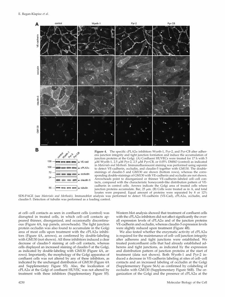
at cell–cell contacts as seen in confluent cells (control) wasdisrupted in treated cells, in which cell–cell contacts ap-peared thinner, disorganized, and occasionally discontinu-ous (Figure 4A, top panels, arrowheads). The tight junctionprotein occludin was also found to accumulate in the Golgiarea of most cells upon treatment with the cPLA2� inhibi-tors (Figure 4A, arrows), as confirmed by double-labelingwith GM130 (not shown). All three inhibitors induced a cleardecrease of claudin-5 staining at cell–cell contacts, whereascells displayed an increased staining of claudin-5 at the Golgi,as indicated by double-labeling with GM130 (Figure 4A, ar-rows). Importantly, the morphology of the Golgi apparatus ofconfluent cells was not altered by any of these inhibitors, asindicated by the unchanged distribution of GM130 (Figure 4Aand Supplementary Figure S5). Also, the localization ofcPLA2� at the Golgi of confluent HUVEC was not altered bytreatment with these inhibitors (Supplementary Figure S5).
Western blot analysis showed that treatment of confluent cellswith the cPLA2� inhibitors did not affect significantly the over-all expression levels of cPLA2� and of the junction proteinsVE-cadherin and occludin, whereas claudin-5 expression levelswere slightly reduced upon treatment (Figure 4B).
We also tested whether the enzymatic activity of cPLA2�is required for the maintenance of cell–cell junction integrityafter adherens and tight junctions were established. Wetreated postconfluent cells that had already established ad-herens and tight junctions, as indicated by the expressionand distribution pattern of junction proteins at the start oftreatment (data not shown). Both Wyeth-1 and Pyr-2 in-duced a decrease in VE-cadherin labeling at sites of cell–cellcontacts and an increased labeling of occludin at the Golgi(Supplementary Figure S6A) as confirmed by colabeling ofoccludin with GM130 (Supplementary Figure S6B). The or-ganization of the Golgi and the presence of cPLA2� at the
Figure 4. The specific cPLA2� inhibitors Wyeth-1, Pyr-2, and Pyr-CB alter adher-ens junction integrity and tight junction formation and induce the accumulation ofjunction proteins at the Golgi. (A) Confluent HUVECs were treated for 17 h with 5�M Wyeth-1, 2.5 �M Pyr-2, 2.5 �M Pyr-CB, or 0.05% DMSO (control) as indicatedin Materials and Methods. Immunofluorescent staining was performed using saponinto detect VE-cadherin, occludin, and claudin-5 together with GM130. The double-stainings of claudin-5 and GM130 are shown (bottom rows), whereas the corre-sponding double-stainings of GM130 with VE-cadherin and occludin are not shown.Arrowheads point to disorganized or thinner VE-cadherin–labeled cell–cell con-tacts, compared with the characteristic honeycomb-like distribution pattern of VE-cadherin in control cells. Arrows indicate the Golgi area of treated cells wherejunction proteins accumulate. Bar, 25 �m. (B) Cells were treated as in A, and totallysates were prepared. Equal amount of proteins were separated by 8 or 12%
SDS-PAGE (see Materials and Methods). Immunoblot analysis was performed to detect VE-cadherin (VE-Cad), cPLA2�, occludin, andclaudin-5. Detection of tubulin was performed as a loading control.
E. Regan-Klapisz et al.
Molecular Biology of the Cell4230

Golgi were not affected by these inhibitors (SupplementaryFigure S6C). Similar to the effects of Pyr-2 and Wyeth-1 onpostconfluent cells, the less specific cPLA2 inhibitor MAFPalso induced a decrease in VE-cadherin membrane staining,a decrease in the plasma membrane staining of occludinwith a concomitant increase of occludin staining at the Golgi(Supplementary Figure S7A). The overall claudin-5 stainingwas decreased upon MAFP treatment, and a significantamount of cells displayed a strong staining of claudin-5 atthe Golgi as indicated by double-labeling with GM130 (Sup-plementary Figure S7A, arrows). Western blot analysis con-firmed the immunofluorescence data (Supplementary Fig-ure S7B). Of note, when postconfluent cells were treatedwith BEL, the well-known inhibitor of the calcium-indepen-dent PLA2 group VI (iPLA2), the localization of the junctionproteins studied remained unchanged (data not shown).
DISCUSSION
In this work we provide evidence that once recruited at theGolgi via a VE-cadherin–mediated process, cPLA2� partic-ipates in the trafficking of junction proteins through or fromthe Golgi, thereby controlling endothelial cell–cell junctionintegrity. On cPLA2� depletion, VE-cadherin– and claudin-5–targeted transport to cell–cell contacts was greatly af-fected, and both proteins were accumulated in the Golgiarea. The inhibition of cPLA2� enzymatic activity with spe-cific inhibitors resulted in the decreased presence of adher-ens and tight junction proteins at cell–cell contacts and intheir accumulation at the Golgi. This supports the notionthat cPLA2� activity is involved in the trafficking of junctionproteins through or from the Golgi. Importantly, the effectsof these inhibitors on junction protein distribution were notaccompanied by a change in Golgi organization excludingthe possibility that junction integrity was compromised as aresult of Golgi disorganization. Also, the effects of the inhib-itors cannot be attributed to a loss of Golgi localization ofcPLA2�. Altogether, these data show that the presence of anactive cPLA2� at the Golgi is critical for the transport ofjunction proteins.
cPLA2� and the Regulation of Cell–Cell Junction IntegrityThe confluence-dependent translocation of cPLA2� to theGolgi is to our knowledge strictly endothelial-specific(Herbert et al., 2007), suggesting that it is mediated by VE-cadherin, an endothelial-specific molecule of which expres-sion and signaling function is closely linked to cell conflu-ence (Dejana, 2004; Liebner et al., 2006). This is supported byour data showing that the relocation of cPLA2� to the Golgicorrelates with a certain degree of maturity of adherensjunction and that cPLA2� dissociates from the Golgi whenadherens junctions are disrupted by an anti-VE-cadherinantibody.
It will be interesting to determine whether the initialrecruitment of cPLA2� to the Golgi and/or its long-termassociation with the Golgi is regulated by VE-cadherin–mediated signaling. VE-cadherin clustering activates severalsignaling responses such as the activation of phosphatidyl-inositol-3-kinase (PI3K) and Akt (Taddei et al., 2008). Inter-estingly, several studies have shown that PI3K activation isrequired for cPLA2� activation, although this was demon-strated in nonendothelial cells (Silfani and Freeman, 2002;Myou et al., 2003). Others have shown that the long-termassociation of cPLA2� with the Golgi in confluent endothe-lial cells is calcium-independent and annexin A1-dependent(Herbert et al., 2007). Annexin A1 becomes enriched at theGolgi in a confluence-dependent manner (Herbert et al.,
2007). A potential role of VE-cadherin–mediated signaling inthis process has not been investigated.
Others have suggested that the association of cPLA2�with Golgi membranes in confluent endothelial cells inacti-vates the enzyme, based on the fact that when cPLA2�becomes associated with the Golgi, calcium-induced arachi-donic acid release was greatly inhibited compared withnonconfluent cultures (Herbert et al., 2005). We argue thatthis does not demonstrate that Golgi-associated cPLA2� isenzymatically inactive, but rather shows that Golgi-associ-ated cPLA2� is less susceptible to calcium-induced acti-vation. Using the same experimental system (confluentHUVECs), we show a clear effect of various specific cPLA2�inhibitors on the trafficking of junction proteins through/from the Golgi. If Golgi-associated cPLA2� was inactive,these inhibitors would not induce any effect. An importantissue that will require further investigation is whethercPLA2� is constitutively active on Golgi membranes orwhether its activity is regulated, either from the cell surfacevia VE-cadherin–mediated signaling events or locally via atraffic-activated Golgi-based signaling pathway (Pulvirentiet al., 2008).
Our findings relate to two important recent concepts. Thefirst one emphasizes the importance of intracellular traffick-ing of junction components in the regulation of cell–celljunction integrity. However, to date, very little is knownabout the delivery of newly synthesized junction proteins tothe membrane because most studies have focused so faron the role of endocytosis in the stabilization of junctions(Bryant and Stow, 2004; Xiao et al., 2007; Yap et al., 2007). Thesecond concept is that VE-cadherin can transduce signalsthat regulate in a timely manner the formation and mainte-nance of adherens and tight junctions (Dejana, 2004; Liebneret al., 2006). Experimental evidences for such cross-talk be-tween VE-cadherin signaling and junction biogenesis/main-tenance are mostly extrapolated from studies on epithelialE-cadherin (Braga and Yap, 2005; Mege et al., 2006). How-ever, one recent study has demonstrated a direct role ofVE-cadherin–mediated adhesion/signaling in the control ofclaudin-5 expression (Taddei et al., 2008), placing VE-cad-herin as a master regulator of tight junction biogenesis andmaintenance.
Our data raise the question as to whether cPLA2� regu-lates claudin-5 expression as well as its trafficking. Indeed,cPLA2� depletion resulted in a strong reduction in claudin-5expression (Figure 3). This reduction in claudin-5 expressionlikely corresponds to a block in synthesis because at the timeof transfection with siRNA, cells do not express claudin-5. Incontrast the specific cPLA2� inhibitors only mildly de-creased claudin-5 expression when applied on confluentcells (Figure 4). The difference in these two approaches isthat VE-cadherin distribution at cell–cell contacts wasclearly more disrupted in cPLA2�-depleted cells than incPLA2�-inhibited cells, possibly because of different lengthsof treatment (72 vs. 17 h, respectively). Because impairedVE-cadherin adhesion/signaling induces the repression ofclaudin-5 expression (Taddei et al., 2008), it is likely that thereduction in claudin-5 expression in our experimental set-tings is not directly regulated by cPLA2� itself, but resultsfrom modifications in VE-cadherin junctional distribution.
On the basis of our findings, we propose a model (Figure5) whereby VE-cadherin mediates a signaling pathway thattriggers the association of cPLA2� with the Golgi complex.In turn, Golgi-localized cPLA2� participates in adherensjunction maturation/maintenance by regulating the traffick-ing of VE-cadherin through/from the Golgi to the junctions.We also propose that at a later stage, once cells express the
cPLA2� and Endothelial Junction Integrity
Vol. 20, October 1, 2009 4231
tight junction proteins occludin and claudin-5, Golgi-associ-ated cPLA2� regulates their trafficking through/from theGolgi, supporting the formation and the maintenance oftight junctions.
cPLA2� and Protein Trafficking through/from the GolgiHow does Golgi-localized cPLA2� participate in the traffick-ing of proteins through/from the Golgi? As describedabove, the enzymatic activity of cPLA2� is important for itsrole in trafficking. Two nonmutually exclusive mechanismsinitiated by cPLA2� enzymatic action could potentially reg-ulate trafficking events. First, cPLA2� could directly affectmembrane curvature by producing inverted-cone–shapedlysophospholipids in one leaflet of the lipid bilayer (Brownet al., 2003), thereby facilitating the formation of transportcarriers. Second, through the release of arachidonic acid,cPLA2� could influence transport processes by arachidonicacid–mediated regulation of the SNARE fusion machinery(Darios and Davletov, 2006; Davletov et al., 2007). Further-more, the arachidonic acid metabolites prostaglandin E2 andprostaglandin I2 (prostacyclin) are known to enhance theformation of endothelial adherens junctions via a cAMP-Epac-Rap1–signaling pathway on relatively a short timescale (minutes to 1 h; Fukuhara et al., 2005). It is thereforepossible that cPLA2� regulates junction formation via aprostaglandin-dependent pathway. Although prostaglandinproduction is very low in confluent endothelial cells (Evanset al., 1984; Whatley et al., 1994; Herbert et al., 2007), it mightstill be sufficient to elicit an autocrine cAMP-mediated re-sponse. We found that overnight treatment of subconfluentand confluent HUVECs with the cyclooxygenases inhibitorindomethacin (up to 10 �M) did not influence the subcellu-lar distribution of VE-cadherin and claudin-5 (data not
shown) as opposed to the observed effects with cPLA2�inhibitors over the same period of time. This indicates thatoxygenated metabolites of arachidonic acid do not play arole in the transport of junction proteins from/through theGolgi.
In line with our findings, previous work had establishedthat cPLA2� is involved in the delivery of a subset of trans-membrane proteins to the cell surface of kidney epithelialcells (Choukroun et al., 2000). Also, recent work demon-strates that cPLA2� is involved in the intra-Golgi traffickingof two secretory proteins in HeLa cells (San Pietro et al.,2009). These observations raise the questions as to whetherthe role of cPLA2� in trafficking is cell type– and cargo-specific. Although the function of cPLA2� has been exten-sively studied in many cell types (Ghosh et al., 2006), its rolein trafficking has so far only been evidenced in confluent en-dothelial cells (our work) and in epithelial cells (Choukroun etal., 2000; Downey et al., 2001; San Pietro et al., 2009). Becauseboth endothelial and epithelial cells form adherens and tightjunctions and become polarized, cPLA2� might be specifi-cally involved in trafficking processes related to junctionformation and/or to cell polarity. This idea also applies tothe role of cPLA2� in intra-Golgi trafficking in HeLa cells(San Pietro et al., 2009). Although HeLa cells cultured inmonolayer are nonpolarized and do not display tight junc-tions, these epithelial carcinoma cells do form tight junctionsand polarize under specific growth conditions (Shimojo etal., 1995; Dessus-Babus et al., 2000). Because these cells haveretained the capacity to polarize under specific circum-stances, it is possible that they possess polarity-relatedmodes of transport—such as cPLA2�-driven trafficking—that are effective even in a nonpolarized cellular context.
Several findings support the idea that cPLA2� regulatesthe trafficking of specific cargos. First, aged cPLA2��/�mice show a trafficking defect of aquaporin-1 and not ofother aquaporins (Downey et al., 2001). Second, in kidneyepithelial cells, cPLA2� overexpression abolished the baso-lateral delivery of the Na�-K�-ATPase � subunit, whereas itdid not affect the basolateral localization of the Cl�/HCO3�
anion exchanger (Choukroun et al., 2000). Our findings thatthe nonjunctional transmembrane proteins ICAM-1 and en-doglin are normally trafficked in cPLA2�-depleted cells,whereas the trafficking of VE-cadherin and claudin-5 is af-fected, also indicate a level of cargo specificity for cPLA2�-regulated transport.
There is to date no experimental evidence showing thatcPLA2� knockout mice have altered endothelial junctions. Itis possible that in these mice, another PLA2 compensates forthe lack of cPLA2�. It is also possible that the mouse and thehuman cPLA2� play different roles. One argument in favorof this idea is that a patient displaying an inherited cPLA2�functional deficiency (triple mutation S111P/R485H/K651R)suffers from small intestinal ulcerations that are much moresevere than those observed in cPLA2� knockout mice (Adleret al., 2008). Thus functional inactivation of the cPLA2�protein in human leads to more severe defects than knock-ing out the cPLA2� gene in mice, pointing to species-specificfunctional roles of cPLA2�.
cPLA2� and the Control of Golgi OrganizationWe are the first, together with the recent work of San Pietroet al., (2009), to address the role of cPLA2� in the control ofGolgi organization using specific cPLA2� inhibitors of dif-ferent structural classes, in combination with specific knock-down of the enzyme by siRNA. Previous work reportingthat PLA2 inhibition induces the fragmentation of the Golgiused several PLA2 inhibitors that are not specific for cPLA2�
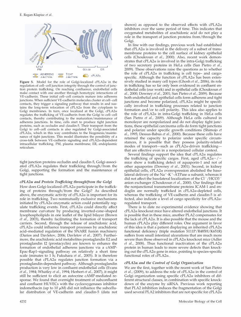
Figure 5. Model for the role of Golgi-localized cPLA2� in theregulation of cell–cell junction integrity through the control of junc-tion protein trafficking. On reaching confluence, endothelial cellsmake contact with one another through homotypic interactions ofVE-cadherin. These initial cell–cell contacts mature into adherensjunctions. When sufficient VE-cadherin molecules cluster at cell–cellcontacts, they trigger a signaling pathway that results in and sus-tains the long-term relocation of cPLA2� from the cytoplasm toGolgi membranes. In turn, once localized at the Golgi, cPLA2�regulates the trafficking of VE-cadherin from the Golgi to cell–cellcontacts, thereby contributing to the maturation/maintenance ofadherens junctions. In time, cells start to produce tight junctionproteins, such as occludin and claudin-5. Their transport from theGolgi to cell–cell contacts is also regulated by Golgi-associatedcPLA2�, which in this way contributes to the biogenesis/mainte-nance of tight junctions. This model illustrates the possibility of across-talk between VE-cadherin signaling and cPLA2�-dependentintracellular trafficking. PM, plasma membrane; ER, endoplasmicreticulum.
E. Regan-Klapisz et al.
Molecular Biology of the Cell4232
(de Figueiredo et al., 1999; Kuroiwa et al., 2001). We foundthat the depletion of cPLA2� induces a change in the mor-phology of the Golgi ribbon. This is in line with other studiessuggesting the involvement of cPLA2� in the maintenanceof the Golgi structure (Choukroun et al., 2000; Grimmer et al.,2005; San Pietro et al., 2009). In the present study we showthat the Golgi morphology was affected when cPLA2� wasdepleted (Supplemental Figure S4) but not when confluentor postconfluent cells were treated with specific cPLA2�inhibitors (Figure 4; Supplemental Figures S5 and S6). Thereason why the Golgi integrity was disrupted upon cPLA2�depletion and not when cPLA2� activity was inhibited in thecontext of confluent monolayers is still unclear. However,we consistently observed that in cPLA2�-silenced cells, theGolgi structure was more disorganized when cells weresubconfluent (unpublished findings). We also found thatwhen nonconfluent sparse HUVEC cultures were treatedovernight with cPLA2� inhibitors, the Golgi organizationwas disrupted (unpublished findings). Because inhibitionof cPLA2� did not affect Golgi integrity in the context ofnonproliferating cells (confluent and postconfluent cells),whereas the Golgi morphology was disrupted when cPLA2�inhibitors were applied on proliferating cells (sparse cells, un-published findings), it is possible that cPLA2� is involved inthe maintenance of the Golgi integrity in a cell cycle–depen-dent manner.
The fact that in confluent and postconfluent cells cPLA2�inhibitors affect the trafficking of transmembrane junctionproteins without affecting the Golgi organization suggeststhat the role of cPLA2� in the maintenance of Golgi organi-zation and its role in regulating the transport of proteinsthrough/from the Golgi are independent processes and areprobably regulated by different mechanisms.
In conclusion, our study illustrates a novel role forcPLA2� in the control of endothelial cell–cell junction integ-rity, through the regulation of junction proteins transportthrough/from the Golgi. Further investigation is needed toelucidate the mechanism by which cPLA2� regulates thistransport and to clarify at which level of the Golgi thisenzyme is acting.
ACKNOWLEDGMENTS
We thank the staff at the Department of Obstetrics and Gynecology, Diakon-essen Hospital, Utrecht, The Netherlands, for providing umbilical cords. Wethank Bart de Haan (Utrecht University) for his help in isolating HUVECs. Wethank Frits Kindt, Ronald Leito, and Misjael N. Lebbink for their help inpreparing figures. We thank Martin Lowe (University of Manchester) for thegift of rabbit anti-GM130 polyclonal antibody. We thank Roman Polishchukfor helpful discussions and Johannes Boonstra, Alexandre Benmerah, andCatherine Rabouille for critically reading the manuscript. This project wassupported by a grant from Senter Novem, The Netherlands, in the frame of anIOP Genomics project (IGE-03012).
REFERENCES
Adler, D. H., et al. (2008). Inherited human cPLA2� deficiency is associatedwith impaired eicosanoid biosynthesis, small intestinal ulceration, and plate-let dysfunction. J. Clin. Invest. 118, 2121–2131.
Bazzoni, G., and Dejana, E. (2004). Endothelial cell-to-cell junctions: molecularorganization and role in vascular homeostasis. Physiol. Rev. 84, 869–901.
Braga, V. M., and Yap, A. S. (2005). The challenges of abundance: epithelialjunctions and small GTPase signalling. Curr. Opin. Cell Biol. 17, 466–474.
Brown, W. J., Chambers, K., and Doody, A. (2003). Phospholipase A2 (PLA2)enzymes in membrane trafficking: mediators of membrane shape and func-tion. Traffic 4, 214–221.
Bryant, D. M., and Stow, J. L. (2004). The ins and outs of E-cadherin traffick-ing. Trends Cell Biol. 14, 427–434.
Bunt, G., de Wit, J., van den Bosch, H., Verkleij, A. J., and Boonstra, J. (1997).Ultrastructural localization of cPLA2 in unstimulated and EGF/A23187-stim-ulated fibroblasts. J. Cell Sci. 110(Pt 19), 2449–2459.
Choukroun, G. J., Marshansky, V., Gustafson, C. E., McKee, M., Hajjar, R. J.,Rosenzweig, A., Brown, D., and Bonventre, J. V. (2000). Cytosolic phospho-lipase A(2) regulates golgi structure and modulates intracellular trafficking ofmembrane proteins. J. Clin. Invest. 106, 983–993.
Clark, J. D., Lin, L. L., Kriz, R. W., Ramesha, C. S., Sultzman, L. A., Lin, A. Y.,Milona, N., and Knopf, J. L. (1991). A novel arachidonic acid-selective cyto-solic PLA2 contains a Ca(2�)-dependent translocation domain with homol-ogy to PKC and GAP. Cell 65, 1043–1051.
Corada, M., Liao, F., Lindgren, M., Lampugnani, M. G., Breviario, F., Frank,R., Muller, W. A., Hicklin, D. J., Bohlen, P., and Dejana, E. (2001). Monoclonalantibodies directed to different regions of vascular endothelial cadherin ex-tracellular domain affect adhesion and clustering of the protein and modulateendothelial permeability. Blood 97, 1679–1684.
Darios, F., and Davletov, B. (2006). Omega-3 and omega-6 fatty acids stimu-late cell membrane expansion by acting on syntaxin 3. Nature 440, 813–817.
Davletov, B., Connell, E., and Darios, F. (2007). Regulation of SNARE fusionmachinery by fatty acids. Cell Mol. Life Sci. 64, 1597–1608.
de Figueiredo, P., Polizotto, R. S., Drecktrah, D., and Brown, W. J. (1999).Membrane tubule-mediated reassembly and maintenance of the Golgi com-plex is disrupted by phospholipase A2 antagonists. Mol. Biol. Cell 10, 1763–1782.
Dejana, E. (2004). Endothelial cell-cell junctions: happy together. Nat. Rev.Mol. Cell Biol. 5, 261–270.
Dessus-Babus, S., Knight, S. T., and Wyrick, P. B. (2000). Chlamydial infectionof polarized HeLa cells induces PMN chemotaxis but the cytokine profilevaries between disseminating and non-disseminating strains. Cell Microbiol.2, 317–327.
Downey, P., Sapirstein, A., O’Leary, E., Sun, T. X., Brown, D., and Bonventre,J. V. (2001). Renal concentrating defect in mice lacking group IV cytosolicphospholipase A(2). Am. J. Physiol. Renal Physiol. 280, F607–F618.
Evans, C. E., Billington, D., and McEvoy, F. A. (1984). Prostacyclin productionby confluent and non-confluent human endothelial cells in culture. Prosta-glandins Leukot. Med. 14, 255–266.
Evans, J. H., and Leslie, C. C. (2004). The cytosolic phospholipase A2 catalyticdomain modulates association and residence time at Golgi membranes. J. Biol.Chem. 279, 6005–6016.
Evans, J. H., Spencer, D. M., Zweifach, A., and Leslie, C. C. (2001). Intracel-lular calcium signals regulating cytosolic phospholipase A2 translocation tointernal membranes. J. Biol. Chem. 276, 30150–30160.
Fukuhara, S., Sakurai, A., Sano, H., Yamagishi, A., Somekawa, S., Takakura,N., Saito, Y., Kangawa, K., and Mochizuki, N. (2005). Cyclic AMP potentiatesvascular endothelial cadherin-mediated cell-cell contact to enhance endothe-lial barrier function through an Epac-Rap1 signaling pathway. Mol. Cell. Biol.25, 136–146.
Ghosh, M., Loper, R., Ghomashchi, F., Tucker, D. E., Bonventre, J. V., Gelb,M. H., and Leslie, C. C. (2007). Function, activity, and membrane targeting ofcytosolic phospholipase A2� in mouse lung fibroblasts. J. Biol. Chem. 282,11676–11686.
Ghosh, M., Tucker, D. E., Burchett, S. A., and Leslie, C. C. (2006). Propertiesof the Group IV phospholipase A2 family. Prog. Lipid Res. 45, 487–510.
Grewal, S., Ponnambalam, S., and Walker, J. H. (2003). Association of cPLA2�and COX-1 with the Golgi apparatus of A549 human lung epithelial cells.J. Cell Sci. 116, 2303–2310.
Grimmer, S., Ying, M., Walchli, S., van Deurs, B., and Sandvig, K. (2005). Golgivesiculation induced by cholesterol occurs by a dynamin- and cPLA2-depen-dent mechanism. Traffic 6, 144–156.
Herbert, S. P., Odell, A. F., Ponnambalam, S., and Walker, J. H. (2007). Theconfluence-dependent interaction of cytosolic phospholipase A2� with an-nexin A1 regulates endothelial cell prostaglandin E2 generation. J. Biol. Chem.282, 34468–34478.
Herbert, S. P., Ponnambalam, S., and Walker, J. H. (2005). Cytosolic phospho-lipase A2� mediates endothelial cell proliferation and is inactivated by asso-ciation with the Golgi apparatus. Mol. Biol. Cell 16, 3800–3809.
Hordijk, P. L., Anthony, E., Mul, F. P., Rientsma, R., Oomen, L. C., and Roos,D. (1999). Vascular-endothelial-cadherin modulates endothelial monolayerpermeability. J. Cell Sci. 112(Pt 12), 1915–1923.
Jaffe, E. A., Nachman, R. L., Becker, C. G., and Minick, C. R. (1973). Culture ofhuman endothelial cells derived from umbilical veins. Identification by mor-phologic and immunologic criteria. J. Clin. Invest. 52, 2745–2756.
cPLA2� and Endothelial Junction Integrity
Vol. 20, October 1, 2009 4233

Klapisz, E., Sorokina, I., Lemeer, S., Pijnenburg, M., Verkleij, A. J., and vanBergen en Henegouwen, P.M. (2002). A ubiquitin-interacting motif (UIM) isessential for Eps15 and Eps15R ubiquitination. J. Biol. Chem. 277, 30746–30753.
Kuroiwa, N., Nakamura, M., Tagaya, M., and Takatsuki, A. (2001). Arachi-donyltrifluoromethy ketone, a phospholipase A(2) antagonist, induces dis-persal of both Golgi stack- and trans Golgi network-resident proteinsthroughout the cytoplasm. Biochem. Biophys. Res. Commun. 281, 582–588.
Leslie, C. C. (2004). Regulation of the specific release of arachidonic acid bycytosolic phospholipase A2. Prostaglandins Leukot. Essent. Fatty Acids 70,373–376.
Liebner, S., Cavallaro, U., and Dejana, E. (2006). The multiple languages ofendothelial cell-to-cell communication. Arterioscler. Thromb. Vasc. Biol. 26,1431–1438.
Mege, R. M., Gavard, J., and Lambert, M. (2006). Regulation of cell-celljunctions by the cytoskeleton. Curr. Opin. Cell Biol. 18, 541–548.
Mogelsvang, S., and Howell, K. E. (2006). Global approaches to study Golgifunction. Curr. Opin. Cell Biol. 18, 438–443.
Myou, S., Leff, A. R., Myo, S., Boetticher, E., Meliton, A. Y., Lambertino, A. T.,Liu, J., Xu, C., Munoz, N. M., and Zhu, X. (2003). Activation of group IVcytosolic phospholipase A2 in human eosinophils by phosphoinositide 3-ki-nase through a mitogen-activated protein kinase-independent pathway. J. Im-munol. 171, 4399–4405.
Nakamura, N., Lowe, M., Levine, T. P., Rabouille, C., and Warren, G. (1997).The vesicle docking protein p115 binds GM130, a cis-Golgi matrix protein, ina mitotically regulated manner. Cell 89, 445–455.
Ni, Z., Okeley, N. M., Smart, B. P., and Gelb, M. H. (2006). Intracellular actionsof group IIA secreted phospholipase A2 and group IVA cytosolic phospho-lipase A2 contribute to arachidonic acid release and prostaglandin productionin rat gastric mucosal cells and transfected human embryonic kidney cells.J. Biol. Chem. 281, 16245–16255.
Ono, T., Yamada, K., Chikazawa, Y., Ueno, M., Nakamoto, S., Okuno, T., andSeno, K. (2002). Characterization of a novel inhibitor of cytosolic phospho-lipase A2alpha, pyrrophenone. Biochem. J. 363, 727–735.
Pulvirenti, T., et al. (2008). A traffic-activated Golgi-based signalling circuitcoordinates the secretory pathway. Nat. Cell Biol 10, 912–922.
San Pietro, E., et al. (2009). Group IV phospholipase A2� controls the forma-tion of inter-cisternal continuities involved in intra-Golgi transport. PLoS Biol.7, e1000194.
Schaloske, R. H., and Dennis, E. A. (2006). The phospholipase A2 superfamilyand its group numbering system. Biochim. Biophys. Acta 1761, 1246–1259.
Schievella, A. R., Regier, M. K., Smith, W. L., and Lin, L. L. (1995). Calcium-mediated translocation of cytosolic phospholipase A2 to the nuclear envelopeand endoplasmic reticulum. J. Biol. Chem. 270, 30749–30754.
Seno, K., et al. (2000). Pyrrolidine inhibitors of human cytosolic phospholipaseA(2). J. Med. Chem. 43, 1041–1044.
Seno, K., Okuno, T., Nishi, K., Murakami, Y., Yamada, K., Nakamoto, S., andOno, T. (2001). Pyrrolidine inhibitors of human cytosolic phospholipase A2.Part 2, synthesis of potent and crystallized 4-triphenylmethylthio derivative‘pyrrophenone.’ Bioorg. Med. Chem. Lett. 11, 587–590.
Shimojo, Y., Hosaka, S., Usuda, N., and Nagata, T. (1995). The induction ofjunctional complexes of HeLa cells by Agar culture. Med. Electron Microsc.28, 57–62.
Silfani, T. N., and Freeman, E. J. (2002). Phosphatidylinositide 3-kinase regu-lates angiotensin II-induced cytosolic phospholipase A2 activity and growthin vascular smooth muscle cells. Arch Biochem. Biophys. 402, 84–93.
Taddei, A., Giampietro, C., Conti, A., Orsenigo, F., Breviario, F., Pirazzoli, V.,Potente, M., Daly, C., Dimmeler, S., and Dejana, E. (2008). Endothelial adhe-rens junctions control tight junctions by VE-cadherin-mediated upregulationof claudin-5. Nat. Cell Biol 10, 923–934.
van Nieuw Amerongen, G. P., and van Hinsbergh, V. W. (2002). Targets forpharmacological intervention of endothelial hyperpermeability and barrierfunction. Vascul. Pharmacol. 39, 257–272.
Whatley, R. E., Satoh, K., Zimmerman, G. A., McIntyre, T. M., and Prescott,S. M. (1994). Proliferation-dependent changes in release of arachidonic acidfrom endothelial cells. J. Clin. Invest. 94, 1889–1900.
Xiao, K., Oas, R. G., Chiasson, C. M., and Kowalczyk, A. P. (2007). Role ofp120-catenin in cadherin trafficking. Biochim. Biophys. Acta 1773, 8–16.
Yap, A. S., Crampton, M. S., and Hardin, J. (2007). Making and breakingcontacts: the cellular biology of cadherin regulation. Curr. Opin. Cell Biol. 19,508–514.
Zimmerberg, J., and Kozlov, M. M. (2006). How proteins produce cellularmembrane curvature. Nat. Rev. Mol. Cell Biol. 7, 9–19.
E. Regan-Klapisz et al.
Molecular Biology of the Cell4234


![A Golgi-Released Subpopulation of the Trans-Golgi · A Golgi-Released Subpopulation of the Trans-Golgi Network Mediates Protein Secretion in Arabidopsis1[OPEN] Tomohiro Uemura,a,b,2,3,4](https://static.fdocuments.net/doc/165x107/5eda9f5a09f66a09130ba5a1/a-golgi-released-subpopulation-of-the-trans-golgi-a-golgi-released-subpopulation.jpg)


