GM1-mediated neurodifferentiation is promoted by OligoGM1 ...
131
Dottorato di Ricerca in Scienze Biochimiche XXX ciclo Dipartimento di Biotecnologie Mediche e Medicina Traslazionale GM1-mediated neurodifferentiation is promoted by OligoGM1-TrkA interaction Margherita Maggioni Matricola n. R10940 Docente guida: Prof. Sandro Sonnino Tutor: Dott.ssa Elena Chiricozzi Coordinatore del Dottorato: Prof. Sandro Sonnino Anno Accademico 2016/2017
Transcript of GM1-mediated neurodifferentiation is promoted by OligoGM1 ...

Dottorato di Ricerca in Scienze Biochimiche
XXX ciclo
Dipartimento di Biotecnologie Mediche
e Medicina Traslazionale
GM1-mediated neurodifferentiation is promoted by
OligoGM1-TrkA interaction
Margherita Maggioni Matricola n. R10940
Docente guida: Prof. Sandro Sonnino Tutor: Dott.ssa Elena Chiricozzi Coordinatore del Dottorato: Prof. Sandro Sonnino
Anno Accademico 2016/2017
I
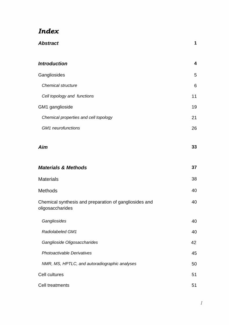
Index
Abstract
1
Introduction 4
Gangliosides 5
Chemical structure 6
Cell topology and functions 11
GM1 ganglioside 19
Chemical properties and cell topology 21
GM1 neurofunctions
26
Aim
33
Materials & Methods 37
Materials 38
Methods 40
Chemical synthesis and preparation of gangliosides and
oligosaccharides
40
Gangliosides 40
Radiolabeled GM1 40
Ganglioside Oligosaccharides 42
Photoactivable Derivatives 45
NMR, MS, HPTLC, and autoradiographic analyses 50
Cell cultures 51
Cell treatments 51
II
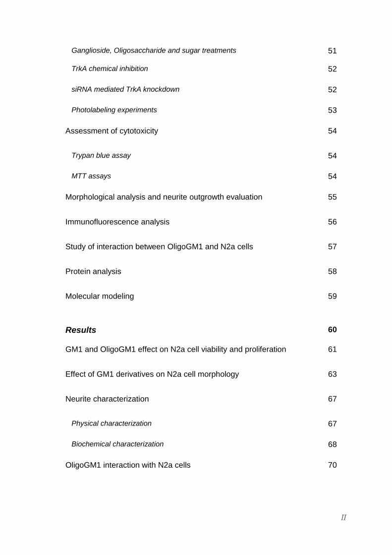
Ganglioside, Oligosaccharide and sugar treatments 51
TrkA chemical inhibition 52
siRNA mediated TrkA knockdown 52
Photolabeling experiments 53
Assessment of cytotoxicity 54
Trypan blue assay 54
MTT assays 54
Morphological analysis and neurite outgrowth evaluation 55
Immunofluorescence analysis 56
Study of interaction between OligoGM1 and N2a cells 57
Protein analysis 58
Molecular modeling
59
Results 60
GM1 and OligoGM1 effect on N2a cell viability and proliferation 61
Effect of GM1 derivatives on N2a cell morphology 63
Neurite characterization 67
Physical characterization 67
Biochemical characterization 68
OligoGM1 interaction with N2a cells 70

III
OligoGM1 effect on TrkA receptor pathway 72
TrkA receptor activation 72
Time course changing in TrkA-Erk signaling pathway 75
TrkA receptor inhibition 78
TrkA receptor expression silencing 81
Interaction between OligoGM1 and TrkA receptor 84
Covalent cross-linking interaction by photolabeling 84
Dynamic calculations for the TrkA-OligoGM1 complex
88
Discussion
91
References
100
Appendix I 113

Abstract

Abstract
- 2 -
The present study proposes a clarification on the molecular mechanism by
which ganglioside GM1 promotes neurodifferentiation, demonstrating in vitro
that neurotrophic functions are exerted by an interaction between the
oligosaccharide portion (OligoGM1) and an extracellular domain of TrkA
receptor.
Similarly to the entire molecule, the oligosaccharide portion of ganglioside GM1,
rather than ceramide, is responsible for neurodifferentiation by augmenting
neurite elongation and by increasing the expression of neurofilament proteins in
mouse neuroblastoma cell line Neuro2a (N2a).
Conversely, the single components of OligoGM1 (asialo-OligoGM1, OligoGM2,
OligoGM3, sialic acid or galactose) are not able to induce a neuro-like
morphology. The neurodifferentiative effect is exerted instead by fucosyl-
OligoGM1.
Contrarily to GM1, exogenous OligoGM1 never integrates in the plasma
membrane composition and does not belong to the intracellular metabolism: the
unique interaction with N2a is characterized by a weak non-covalent association
to the plasma membrane that suggests the existence of an OligoGM1-
stimulated target on the cell surface.
In fact, the neurotrophic properties of GM1 oligosaccharide are exerted by
activating TrkA receptor and the following cascade leading to
neurodifferentiation event.
The second part of this study elucidates the interaction between GM1 and TrkA,
revealing a direct association of OligoGM1 to an extracellular domain of the
receptor.
Photolabeling experiments, performed employing nitrogen azide radiolabeled
GM1 derivatives, show a direct association of the oligosaccharide chain to TrkA.
Moreover, a bioinformatics study reveals that OligoGM1 fits perfectly in a pocket
of the TrkA-NGF complex, stabilizing and favoring their intermolecular
interactions as revealed by the increase in energy associated to the new
complex TrkA-NGF-OligoGM1. A precise molecular recognition process
between OligoGM1 and a specific extracellular domain of the TrkA receptor is
Abstract
- 3 -
supposed. According to the weak association of OligoGM1 to the cell surface,
no covalent bounds between OligoGM1 and TrkA-NGF complex were found.
For the first time the molecular mechanism by which GM1 exerts its
neurodifferentiative potential was identified, finding out a direct interaction
between the oligosaccharide portion and an extracellular domain of TrkA
receptor responsible for enhancing the signal transduction related to the
neurodifferentiation pathway.

Introduction

Introduction
- 5 -
Gangliosides
Gangliosides are a large family of complex glycosphingolipids (GSLs) isolated
for the first time from beef brain in 1942 by Klenk, who among the hydrolysis
products identified their essential components: sphingosine, fatty acid, hexoses
and sialic acid (Klenk, 1942; Svennerholm, 1964).
Ganglioside components are shown in figure I1 reporting chemical structure of
the simplest ganglioside, GM3.
Figure I1: Chemical structure of the simplest ganglioside GM3. GM3 is reported to show typical components of gangliosides: sphingosine, fatty acid, sialic acid and hexoses. GM3 contains glucose and galactose.

Introduction
- 6 -
Chemical structure
Gangliosides are amphiphilic components typical of all deuterostomia cell
plasma membranes that assume structural and functional outstanding roles
internally to the lipid raft plasma membrane regions (Ghidoni et al. 1989; Senn et al.
1989; Valaperta et al. 2007; Merril, 2011).
According to the general glycolipid features, gangliosides are inserted only in
the outer leaflet of the plasma membrane integrating in the lipid core layer
through a hydrophobic moiety and simultaneously protruding in the extracellular
environment with a hydrophilic portion (Bertoli et al. 1981; Robert et al. 2011; Kolter,
2012; Shengrund, 2015).
Gangliosides are spread all over the tissues, but in mammals they are prevalent
in plasma membranes of nervous cells, in particular in the gray matter,
especially localized at the pre- and post-synaptic areas. Outside of the nervous
system they are relevant in serum, vehiculated by lipoproteins, in spleen, and in
erythrocytes (Svennerholm, 1964; Rösner et al. 1973; Hansson et al. 1977; Senn et al. 1989;
Kolter, 2012).
As a matter of fact, the gangliosides are composed by ceramide, the
hydrophobic core, resulting from a long chain fatty acid incorporation on the 2
position of a sphinganine basis promoted by the ceramide synthase acylation.
Dihydroceramide can be finally desaturated introducing a double carbon bond
between positions 4 and 5 of the acyl residue of sphingosine, derived generally
by palmitate (Sribney, 1966; Merril, 1991; Rother et al. 1992). The ceramide de novo
synthesis, shown in figure I2, takes place in the endoplasmic reticulum (Miller
Podrasa & Fishman, 1982; Schwarzmann & Sandhoff, 1990; Kolter, 2012).
According to the beginning discovery (Klenk, 1942), the hydrophilic head of
gangliosides, the oligosaccharide chain, is always characterized by the
presence of one or more residues of sialic acid (figure I3). The ceramide
glycosylation arises from the progressive addition by specific
glycosyltransferases of β-anomeric neutral monosaccharides from the
nucleotide-activated forms, usually by UDP (Van Den Eijnden, 1973; Basu & Basu,
1982; Kolter, 2012). The transfer of sialic acid by a sialyltransferase stands out for
the CMP-activated donor complex and for the formation of α-glycosidic bond via

Introduction
- 7 -
hydroxyl group at position 2 to a neutral monosaccharide unit or to another
sialic acid residue (Schauer, 1982; Tettamanti & Riboni, 1993; Schnaar et al. 2014), as
shown in figure I4.
These reactions occur in the luminal side of the Golgi apparatus explaining the
topological asymmetric insertion of the gangliosides in the external layer of the
plasma membrane (Ghidoni et al. 1989; Sandhoff & Kolter, 2003; Kolter et al. 2002).
Figure I2: De novo ceramide biosynthesis.

Introduction
- 8 -
Figure I3: Structure of Sialic Acid.
a.
b.
Figure I4: Sialic Acid linkages in gangliosides. Examples of an α-(2-3) glycosidic linkage between sialic acid and a neutral monosaccharide typically represented by a galactose residue (a) and of an α-(2-8) glycosidic linkage between sialic acid residues (b), characteristic of polysialylated brain gangliosides.

Introduction
- 9 -
Beyond the conserved structure, gangliosides present an interesting
heterogeneity given firstly by ceramide. It varies in relation to the sphingoid
basis, a sphingosine or a sphinganine, presenting or omitting the desaturation
respectively, and containing from 16 to 22 carbon atoms. Also the fatty acid
differs among ceramide types commonly represented by the palmitic acid
(16:0), stearic acid (18:0), oleic acid (18:1), and arachic acid (20:0) in the
nervous system and by behenic acid (22:0), docosanoic acid (22:1), lignoceric
acid (24:0) and nervonic acid (24:1) in extra nervous tissues (Schenground &
Garrigan, 1969; Karlsson 1970; Kolter, 2012).
Furthermore, the remarkable diversification in the oligosaccharide chains
among the gangliosides have provided an instrument for their classification
(Svennerholm & Fredman, 1980; Svennerholm, 1994) combined to the nomenclature
formulation by IUPAC IUB Commission (IUPAC-IUBMB JCoBN, 1998).
According to the oligosaccharide core sequences, the gangliosides can be
divided in six series: ganglio, gala, latto, neolatto, globo, isoglobo (Svennerholm,
1994; Kolter, 2012). The first letter is dependent on these categories: G stands for
ganglio, that presents oligosaccharide β-D-Glucose-β-D-Galactose-N-acetyl-β-
D-Galactosamine-β-D-Galactose.
The ganglio sub classification (figure I5), based on the sialic acid residues
number, is expressed by the second letters M, D, T, or Q, designing mono-, di-,
tri- or tetra-sialyl groups respectively. The following number 1, 2 or 3 agrees
with the order of migration of the ganglioside in thin layer chromatography (GM1
< GM2 < GM3), reflecting the presence of the entire oligosaccharide sequence,
the lack of the external galactose or the disaccharide galactosyl-N-
acetylgalactosamine respectively. The final letters evoke the role of the
sialytransferases in the ganglioside metabolism: letter “a” indicates the only
presence of linkages between sialic acid and galactose, letters “b” and “c” the
existence of linkages between two or three sialic acid residues respectively
(Svennerholm, 1994; Kolter et al. 2002; Kolter, 2012).

Introduction
- 10 -
Figure I5: Classification of Ganglio series gangliosides (IUPAC-IUB JCoBN, 1998).
Currently, IUPAC-IUB commission on the biochemical nomenclature purposes a
graphic symbology for glycans representation (IUPAC-IUBMB JCoBN, 1998).
Monosaccharides typical of Ganglio series gangliosides are identified by
symbols shown in figure I6.
Figure I6: Symbols used for monosaccharides of Ganglio serie gangliosides (IUPAC-IUBMB JCoBN, 1998).

Introduction
- 11 -
Cell topology and functions
The substantial amphiphilic feature of gangliosides represents the basis beyond
the structural ones, of their structural and physiological functions (Robert et al.
2011; Russo et al. 2016).
The ceramide portion, inserted in the plasma membrane outer layer, exerts an
influence on the establishment of arrangement with other complex lipid and
proteins, affecting the modulation of plasma membrane properties. On the other
side, the carbohydrate moiety, projected in extracellular environment, offers
many recognition and interaction sites for cell surrounding molecules playing a
crucial role in the cell response to an external stimuli, in signal transduction and
in mediation of cell activities (Bertoli et al. 1981; Hakomori, 1983; Arita et al. 1984; Merrill
& Sandhoff, 2002; Kolter, 2004; Shengrund, 2015).
Starting from the ceramide contribute in ganglioside functions, a remarkable
impact on plasma membrane organization is due to the presence of the trans
double bond between sphingosine C4 and C5, combined with the presence of a
saturated fatty acyl residue. These features allow the ganglioside hydrocarbon
chains to pack in the plasma membrane lipid layer more tightly than other
hydrophobic components, such as the phospholipids containing cis double bond
related to unsaturated fatty acids, enhancing the association to cholesterol as
well as to transmembrane protein domains (Shengrund & Garrigan, 1969; Tettamanti &
Riboni, 1993; Shengrund, 2015). The following interactions originate the particularly
enriched and specialized plasma membrane regions named lipid rafts (figure I7),
that, according to their experimental isolation are defined as detergent-resistant
assemblages (DRM) resulting as a low-density fractions from density-gradient
ultracentrifugation (Simons & Ikonen, 1997; Simons & Sampaio, 2011; Ohmi et al. 2012;
Kraft, 2013; Ledeen & Wu, 2015).

Introduction
- 12 -
The Figure I7 purposes an overall representation of a plasma membrane lipid
raft, presenting gangliosides among the characteristic elements (Malchiodi-Albedi
et al. 2011).
Figure I7: Schematic representation of a LR in the cell membrane. Enrichment in gangliosides, structurally and functionally correlated to cholesterol, proteoglycans, others glycolipids, transmembrane proteins (glycosylated and non-) and GPI-anchored proteins is shown.
The co-localization of gangliosides and cholesterol is important in maintaining
the plasma membrane adequate structural equilibrium and physical
characteristics such as the fluidity, the rigidity or the fusion temperature
(Lingwood, 2000; Shengrund, 2015). Plasma membrane areas particularly enriched in
gangliosides increase in rigidity (Bertoli et al. 1981).
Moreover, because the lipid tail region is critically smaller than polar moieties, in
aqueous solution gangliosides, as well as for all the sphingolipids, aggregate in
high molecular weight micelles, presenting a hydrophobic ceramide core and
isolated from polar environment by the enclosing sugar chains as shown in figure
I8 (Maggio et al. 1981; Sonnino et al. 1994; Sonnino & Prinetti, 2010).

Introduction
- 13 -
Figure I8: Ganglioside micella. The elevated volumetric ratio between hydrophilic head and hydrophobic tail of gangliosides originate high molecular weight micelles in aqueous solutions over a critical concentration.
In cell plasma membrane, the reported aggregative behavior produces physic
modification of plasma membrane in particularly ganglioside-enriched clusters,
introducing an increase in the layer curvature radius. In relation to the
implication of gangliosides in plasma membrane invagination, an increase in
endocytosis phenomena has been reported in correspondence to the leaf
curvatures (Thompson & Tillack, 1985; Tettamenti et al. 1985; Fra et al. 1995; Ewers et al.
2010; Sonnino & Prinetti, 2010).
In addition to ceramide, the oligosaccharide portions confer to gangliosides as
much structural properties finely correlated to biological functions. The
monosaccharide units of a ganglioside establish cooperative contacts through
hydrogen type binding that specifically offer an interactive potential to other
molecules in extracellular environment (Sharom & Grant, 1978; Kiarash et al. 1994;
Shengrund, 2015). Thanks to the oligosaccharide head, gangliosides can act as
specific receptors for many molecular species such as viruses, bacteria, toxins
(Yamakawa & Nagai, 1978; Borges et al. 2010; Ladisch & Liu, 2014), growth factors,
peptides and hormones (Vengris et al. 1976; Shengrund, 2015; Russo et al. 2016).

Introduction
- 14 -
Oligosaccharide chains of gangliosides, together with glycoproteins and
proteoglycans, contribute to sialylated sugar-coat construction of glycan-rich
glycocalix associated to the plasma membrane (Schnaar et al. 2014; Linnartz-Gerlach
et al. 2014; Shengrund, 2015; Ledeen & Wu, 2015). This structure supports different
functions such as cell differentiation, cell to cell interactions and certainly signal
transduction. In fact, the glycocalyx carbohydrate moiety, surrounding the outer
leaflet of the plasma membrane surface is responsible for the recognition of
other cells and for the attachment to extracellular components (Barrat et al. 1978;
Kolter, 2012; Zeng & Tarbell, 2014; Shengrund, 2015). Furthermore, connecting directly
or with mediator to internal elements (figure I9), the glycocalyx can initiate the
communication and the realization of a specific response (Linnartz & Neumann,
2013; Shengrund, 2015).
The biochemical composition of glycocalyx sugar portion shows a strictly
correlation to the functional meaning, presenting as a consequence, a
considerably high level of specificity depending on cytological types, but also on
the cycle phase or development stage of cells (Schnaar et al. 2014; Linnartz &
Neumann, 2013). This evidences the fundamental role of oligosaccharide chains in
determination of cell activities (Shengrund, 2015; Ledeen & Wu, 2015).
Figure I9: Glycocalyx. Oligosaccharide chains of gangliosides participate to the adequate composition of the cell glycocalyx that represents a crucial element for cell communication and response to external stimuli (Atukorale et al. 2015).

Introduction
- 15 -
A well known phenomena that underscored the fundamental role of
oligosaccharide chains in explicating cell ganglioside-depending functions
concerns their implication in cell differentiation and development (Schaal et al.
1985; Levine & Flynn, 1986; Yu et al. 1988; Shengrund, 1990; Kwak et al. 2006; Sonnino et al.
2010; Shengrund, 2015).
For what concerns the nervous system, where represent the main
glycosphingolipids, the ganglioside content and pattern change during the
neuron differentiation, aging and in neurodegenerative diseases. As shown in
the figure I10, in undifferentiated neurons, GM3 and GD3 are the main
gangliosides and they are characterized by a small hydrophilic head.
Conversely, during the axons and synapses formation, GM3 and GD3 are
displaced by the more complex gangliosides, such as GM1, GD1a, GT1b, and
GQ1b (Shengrund, 1990; Shengrund, 2015). The modification of ganglioside
composition reflects the differences in expression or in activities of specific
glycosyltransferases (Yu et al. 1988; Shengrund, 1990; Sonnino et al. 2010; Aureli et al.
2011).
Figure I10: Ganglioside changing in neurodifferentiation. Modification in neuron ganglioside pattern during neurodifferentiation reflects the differences in glycosyltransferases and suggests an important role of oligosaccharide chains in the process.

Introduction
- 16 -
The functional potential of oligosaccharide chains, peculiarly correlated to their
specific composition, together with the described biophysical influence of the
ceramide portion on plasma membrane regulation through the lateral portioning,
make the gangliosides exceptional players in recruiting membrane receptors
and in promote initiation of signaling cascades (Tettamanti et al. 1985; Nagai, 1985;
Ghidoni et al. 1989; Robert et al. 2011).
One of the results of the ceramide-enhanced ganglioside aggregation in plasma
membrane specialized clusters is represented by the instauration of specific
interaction between oligosaccharide chains and membrane receptors or
glycoproteins, promoting the regulation of many pathways (Fueshko & Schengrund,
1992; Nagai, 1995; Aureli et al. 2011; Russo et al. 2016).
At this purpose, the changes in ganglioside oligosaccharide composition in
specific sensing domains can act as an activator event of a co-localized tyrosine
kinase receptor, as schematized in figure I11. One emblematic example concerns
the activation of Epidermal Growth Factor (EGFR) receptor by modification of
neighboring gangliosides (Russo et al. 2016).
Figure I11: Ganglioside changing in signaling regulation. Modification in oligosaccharide chains of gangliosides, co-localized in sensing domains with tyrosine kinase receptors, can stimulates signaling activation promoting dimerization of receptor monomers and their autophosphorylation. PM, plasma membrane; GLSs, glycosphingolipids; GSD, glycosphingolipid sensing
domain; RTKs, receptor tyrosine kinases (Russo et al. 2016).

Introduction
- 17 -
This property can evolve also in regulation of membrane pores and channels,
modulating molecule transit across the plasma membrane (Tettamanti et al. 1985;
Ghidoni et al. 1989).
Moreover, another outcome imputable to the ganglioside specific features and
organization, as well as for the others sphingolipids, is revealed by the
sphingosine metabolites that can act themselves as a second messengers. In
fact, an extracellular stimuli, such as the interleukin 1 or the interferon, receipted
among a ganglioside clustering zone, can be translated in ceramide
detachment, degradation or following modifications and metabolites can
regulate intracellular enzymes (Svennerholm et al. 1994).
At this point, the requirement of ganglioside in signal transduction appears
extremely important and actually implicated in cell functionality. As a
consequence, pathologies and discordance from cell physiological state can
arise from alteration in ganglioside metabolism. A concerning description is
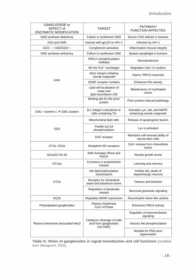
summarized and schematize in table I1 (Shengrund, 2015).
Introduction
- 18 -
GANGLIOSIDE or EFFECT of
ENZYMATIC MODIFICATION TARGET
PATHWAY/ FUNCTION AFFECTED
GM3 synthase deficiency Failure to synthesize GM3 Severe CNS deficits in humans
GD3 and GM3 Interact with gp120 on HIV-1 Infection by HIV-1
GD3_/_ + GM2/GD2_/_ Complement activation Inflammation /neural integrity
GM2 synthase deficiency Failure to synthesize GM2 Spastic paraplegia in humans
GM1
ERK1/2 phosphorylation inhibited
Neuroprotective
NE Na+/Ca2+ exchanger Regulates Ca2+ in nucleus
α5α1 Integrin initiating neurite outgrowth
Opens TRPC5 channels
GDNF receptor complex Enhances Ret activity
Lipid raft localization of caspr and
glial neurofascin-155
Maintenance of myelinated axons
Binding site for the prion
protein Prion protein-induced pathology
GM1 + laminin-1 GM1 clusters β-1 Integrin colocalizes to
rafts containing Trk Activates Lyn, Akt, and MAPK enhancing neurite outgrowth
GD3
Mitochondrial lipid rafts Release of apoptogenic factors
Paxillin tyr118 phosphorylation
Lyn is activated
EGF receptor Maintains self-renewal ability of
neural stem cells
GT1b, GD1b Bradykinin B2 receptors Ca2+ release from intracellular
stores
GD1a/GT1b-2b MAb Activates RhoA and
ROCK Neurite growth arrest
GT1aα Functions in acetylcholine
release Learning and memory
GT1b
Akt dephosphorylation (inactivation)
Inhibits Akt, death of dopaminergic neurons
Receptor for Clostridium tetani and botulinum toxins
Tetanus and botulism
Regulation of glutamate release
Neuronal glutamate signaling
GQ1b Regulates BDNF expression Neurotrophic factor-like actions
Polysialylated gangliosides Plasma membrane
Ca2+-ATPase Enhances PMCA activity
Plasma membrane associated Neu3 Catalyzes cleavage of sialic
acid from gangliosides (not GM1)
Regulator of transmembrane signaling
Induces Akt phosphorylation
Needed for PNS axon regeneration
Table I1: Roles of gangliosides in signal transduction and cell functions (modified
form Shengrund, 2015).

Introduction
- 19 -
GM1 ganglioside
Ganglioside GM1 has continuously stirred up an exceptional outstanding
attraction because its relevant implication and its key role in many signaling
systems and cell regulatory pathways (Fang et al. 2000; Ledeen & Wu, 2015; Aureli et
al. 2016). Abounding in the nervous system, GM1 appears involved in the
essential mechanisms of neurodifferentiation and neurodevelopment on which
its fame is continually depending such as the growing investigations about the
dire pathological consequences of its alteration and lacking (Shengrund & Ringler,
1989; Shengrund & Mummert, 1998; Ledeen & Wu, 2015; Zhai et al. 2015).
GM1 structure was depicted and elucidated for the first time by Kuhn and
Wiegandt in 1963 in correspondence to its TLC separation
(Kuhn & Wiegandt, 1963). According to classification and nomenclature (Svennerholm,
1980). GM1 is a monosialo- tetrahexoxylated glycosphinglolipid and belongs to
Ganglio series. Referring to IUPAC, GM1 formula is β-Gal-(1-3)-β-GalNAc-(1-
4)-[α-Neu5-(2-3)-]β-Gal-(1-4)-Glc-(1-1)-Cer.
Conversely from the others major GM1-interacting gangliosides in the brain,
GD1a, GD1b and GT1b, among which the typical sialic acid linkages are very
susceptible to hydrolytic removal by endogenous sialidase, GM1 sialic acid is
resistant to most forms of the mammalian enzyme (Sonnino et al. 2011, Mijagi &
Yamaguchi, 2012).
GM1 derived from sequential addiction of N-acetylgalactosamine and galactose
units from UDP-activated forms to GM3 by specific glycosyltransferase (figure I5).
The enzymatic complex for production of GM1 from GM3, occurring in the Golgi
membranes, is called GM1 synthase (Robert et al. 2011; Kolter, 2012).
The GM1 chemical structure and the IUPAC symbolic representation are shown
in figure I12.

Introduction
- 20 -
a.
b.
Figure I12: GM1.
a. Chemical structure of β-Gal-(1-3)-β-GalNAc-(1-4)-[α-Neu5-(2-3)-]β-Gal-(1-4)-Glc-(1-1)-Cer.
b. IUPAC symbolic representation.

Introduction
- 21 -
Chemical properties and cell topology
GM1 shows a characteristic amphiphilic equilibrium, essential for the
organization of membrane bilayer, given by the extended hydrophilic sugar
domain combined with the hydrophobic moiety. The amphiphilic balance is
easily perturbed by minimal modifications of ceramide or oligosaccharide chain
(Sonnino et al. 2006; Sonnino et al. 2007; Aureli et al. 2016).
The presence of sialic acid confers to ganglioside GM1 a negative charge in
biological environment, resulting however only the 16% of the expected value in
aggregative status (Cantù et al. 1986). The particular aggregative behavior of GM1
is due once again both to the flexible and packable hydrophilic pentasaccharide
chain, soluble in water and to the attached remarkably smaller hydrophobic
ceramide moiety. In aqueous solutions these physicochemical properties
determine the formation of micelles, according to the usual ganglioside
penchant (Urlich-Bott & Wiegandt, 1984; Cantù et al. 1986; Sonnino et al. 1994; Sonnino &
Prinetti, 2010).
Among the GM1 micelles, the negative charge power of sialic acid residues is
lowered by the polyelectrolyte effect and masked by the positive charges,
explaining the cited phenomena (Sonnino et al. 1994; Aureli et al. 2016).
GM1 micelles present small ellipsoidal form caused by the original geometry of
the monomer presenting a large hydrophilic head. For GM1, the critical micelles
concentration (c.m.c.), determined by experimental approaches, ranging from
10-8 to 10-9 M and the free monomers never exceed the c.m.c. value,
maintaining in equilibrium with aggregates at any GM1 concentration (Urlich-Bott
& Wiegandt, 1984; Cantù et al. 1986; Sonnino et al. 1994).
The aggregative properties of GM1 relevantly impact the interaction with cells
and the modulation of cell membrane organization (Facci et al. 1984; Sonnino et al.
2006; Prinetti et al. 2007; Cantù et al. 2011; Khatun et al. 2014).
GM1 interaction with cell components and the effect on cell membranes has
been studied in vitro, feeding cell cultures with the exogenous ganglioside.
Experimental evidences derived from GM1 administration to cultured neurons
report that at the c.m.c. concentration, the GM1 monomers insert into the
external layer of plasma membrane through lipid-lipid interactions, while the

Introduction
- 22 -
micelles bind to the cell surface proteins and can be endocytosed reaching then
lysosomes (Saqr et al. 1993). A prolonged treatment was proved to advance the
insertion of GM1 monomers into the plasma membrane. On the other hand, the
gradually augmenting in GM1 concentration, over the c.m.c. increase the
aggregative process (Tomasi et al. 1980; Venerando et al. 1982; Facci et al. 1984).
A study performed on fibroblasts agrees with time and concentration
dependence of exogenous GM1 behavior, underlining its modality of
association to cells. In fact, increasing the GM1 concentration over the c.m.c. up
to 10-4 M, the weakly association of GM1 to plasma membrane, typical of the
micelle forms, appears progressively more favorable. Alternatively, at the c.m.c.
the monomeric GM1, stably associated to cells and introduced in the cell
metabolism, prevails after 120 minutes treatment (Chigorno et al. 1985).
These outcomes are reported and detailed in figure I13.
Moreover, if c.m.c. value is increased, meaning that an unusual higher GM1
molarities are required to initiate aggregation process, the half-life of micelles is
reduced and the process is perturbed by the facilitated release of monomers
(Cantù et al. 1991).
GM1 monomers are stabilized into the outer layer of plasma membrane
establishing hydrogen bonds with surrounding glycerophospholipids and can
interact with proteins and membrane receptors. According to the typical
ganglioside properties, GM1 monomers integrated into plasma membrane tends
to segregate in specialized domains characterized by peculiar interaction with
others components and between themselves, that physically reflects its
amphiphilic balance (Prioni et al. 2004; Sonnino et al. 2006; Sonnino et al. 2007; Aureli et
al. 2016). In particular, at the aqueous/lipid interface the dynamic is reduced by a
network of hydrogen bonds created by the amide group of the ceramide that
acts both as a donor and as an acceptor of protons (Brocca et al. 1993). The
oligosaccharide heads interact reciprocally by hydrogen bonds too, allowed by
water molecules insinuated as linking bridges between two chains (Acquotti et al.
1990; Brocca et al. 1998; Brocca et al. 2000).

Introduction
- 23 -
Figure I13: Exogenous Ganglioside GM1 association to cultured fibroblasts. GM1 interaction with cells is based on its aggregative properties depending on time and concentration. Over the c.m.c. (10-9 M), the promoted state is progressively represented by the micelles that weakly bind the external cell surface and are easily removed by exchange with serum component (SERUM LABILE). This tendency however decrease in time and at fixed c.m.c. after 120 minutes, most GM1 administrated stays in monomer forms. Monomeric GM1, integrated into the plasma membrane, participating to the cell metabolism, can be isolated after cell treatment with trypsin (TRYPSIN STABLE). A third dynamic, intermediate and vulnerable state corresponds to simple monomers or micelles of GM1 interacting with some proteins protruding from the external layer of the cell membrane (TRYPSIN LABILE).
Introduction
- 24 -
As previously mentioned in relation to overall ganglioside properties, also GM1
chemical features, effectively decisive for its structural relations and
arrangement, play a determinant role in peculiar tendency to the lateral
segregation in the plasma membrane lipid rafts (Simons & Sampaio, 2011; Aureli et al.
2016).
Firstly, the above mentioned coexistence of opposite forces in the hydrogen
bonds proton exchanging and in the ceramide influence on the hydrophobic
packing, drive the physical phase separation of ganglioside from the fluidity of
the glycerophospholipid bilayer (Sonnino & Prinetti, 2010; Sonnino & Prinetti, 2013).
Moreover, an extremely important factor to determine the GM1 segregation is
represented by the bulkiness of the large surface area occupied by its
oligosaccharide chain. This characteristic has an impact not only on the packing
of the ganglioside, considering the volume ratio to the ceramide moiety (Acquotti
et al. 1990; Sonnino & Prinetti, 2010), but also on the acquisition of a spontaneous
positive curvature of the plasma membrane that affects the local lateral
organization favoring the phase separation of GM1 enriched microdomains
(Sonnino et al. 1994; Fra et al. 1995; Ewers et al. 2010; Patel et al. 2016).
GM1 in lipid rafts have been detected and localized thanks to its capacity to
bind subunit B of cholera toxin (Holmgren et al. 1973). The receptor activity was
proved to be imputable to GM1 oligosaccharide chain: the B pentameric subunit
binds to the five monomers independently from both the A subunit and the
ceramide with a low binding constant ranging from 10-9 to 10-12 M (Masserini et al.
1992; Kuziemko et al. 1996).
Alternatively, GM1 can be recognized also by using antibodies for which
employing techniques and the improvement in their specificity have been
implementing for many years (Wu & Ledeen, 1991; Watarai et al. 1994; Taylor et al. 1996;
Kaji & Kimura, 1999; Iglesias-Bartolomé et al. 2009; Ledeen & Wu, 2015).
GM1 is one of the physiologically essential ganglioside in neuronal plasma
membrane lipid rafts, concentrating at the presynaptic and postsynaptic
membranes of nerve endings and segregating especially with sphingomyelin,
phosphatidylinositols, glycosylphosphatidylinositols, cholesterol and others
gangliosides (Simons & Sampaio, 2011; Sonnino et al. 2007).

Introduction
- 25 -
From the analysis of lipid rafts in cerebellar granule cells, gangliosides have
found to represent only the 6-7% of the total lipid content, constituted over 50%
by glycerophospholipids (Sonnino et al. 2007). The most representative
gangliosides in these differentiate neurons are GD1a and GT1b and, regardless
the minor quantity, their co-localization with GM1 justifies from a functional point
of view their physiological essentiality (Sonnino et al. 2011; Shengrund, 2015).
In fact, mice genetically deficient in gangliosides show a distortion in lipid
microdomains, indicating their necessity in formation, stabilization and
dynamicity of lipid rafts. The overexpression of gangliosides is correlated to an
abnormal functioning of the process too (Sonnino et al. 2007; Pavlov et al. 2009;
Furukawa et al. 2011; Ohmi et al. 2012).
Neurological disorders that further accompanies this situation is explained by
the alteration in physiological GM1 concomitance with proteins among the lipid
rafts (Fang et al. 2000; Sonnino et al. 2007; Ledeen & Wu, 2015; Russo et al. 2016).
As a matter of fact, different proteins crucial for neural functions have been
identified to present glycolipid-binding domains, being interacting partners of
GM1, or to belong to a related pathway. Just to make some examples: β-
amyloid peptide (Fantini et al. 2013), α-synuclein (Fantini et al. 2011), Na+/Ca2+
exchanger, integrins (Ledeen & Wu, 2015; Xie et al. 2002), transient receptor potential
channel 5 (Wu et al. 2007), opioid receptors (Shen & Crain, 1990; Wu et al. 1997) and
Trk receptor (Mutoh et al. 2002; Nishio et al. 2004).
Moreover, an association of GM1 and GD1a to the plasma-membrane-bound
salidase has been reported too, suggesting a reserve-role of GD1a for GM1 and
as a consequence, the existence of a tight regulated cell content and
requirement of GM1, supporting its functional relevance (Sonnino et al. 2010;
Sonnino et al. 2011; Miyagi & Yamaguchi, 2012; Ledeen & Wu, 2015).

Introduction
- 26 -
GM1 neurofunctions
Thence, the impact of GM1 on plasma membrane microdomain assessment
and specialization, leading to its involvement in the regulation of the cell signal
transduction and in the modulation of several pathways, appears unavoidable
element to consider and to clearly realize the GM1 biological potential (Fueshko &
Shengrund, 1992; Nagai, 1995; Pavlov et al. 2009; Aureli et al. 2016; Ledeen & Wu, 2015).
Because of the GM1 relevant presence in the nervous system, most of the GM1
biological properties have been studied in neuronal models, following biological
or pharmacological approaches by manipulating the endogenous GM1 or by
treating cell cultures and animals with the exogenous one respectively (Ledeen &
Wu, 2015). Nevertheless, because of the evaluation of the GM1 potential in extra
nervous areas such as immune system, liver, kidney or lungs (Ozkök et al. 1999;
Saito & Sugiyama, 2000; Ledeen & Wu, 2015), it has been recently defined a ʺfactotum
of natureʺ (Ledeen & Wu, 2015).
A lot of studies have been performed in vitro in order to identify GM1 targets in
initiation of neurodifferentiation and neurotrophic effects. Even if the most
experiences were conducted following a pharmacological approach, by
administering exogenous GM1 to cells in culture or to animal models, some
results offer a physiological meaningful (Ledeen & Wu, 2015). It happened because
they appeared reproducible also manipulating endogenous GM1 enhancing
sialidase (Wu et al. 1998; Monti et al. 2000) or GM1 synthase activities (Mutoh et al.
2002; Dong et al. 2002), ascertaining the insertion of GM1 into specialized domains
of the cell membrane (Ledeen & Wu, 2015) or proving the natural increase in the
GM1 content occurring during spontaneous neurodeveloping processes (Fang et
al. 2000; Hasegawa et al. 2000).
According to above described physicochemical parameters and characteristics,
the effects exerted by GM1 occurs when its content increases in plasma
membrane lipid rafts (Hakomori et al. 1998; Dietrich et al. 2001; Mitsuda et al. 2002; Mutoh
et al. 2002; Dong et al. 2002; Pavlov et al. 2009; Sonnino et al. 2010; Coskun & Simons 2011;
Sonnno et al. 2011; Ledeen & Wu, 2015), as represented in figure I14.

Introduction
- 27 -
Effectively, the membrane local GM1 enrichment can support biological
functions in two main ways:
i. indirectly, by the GM1 content-dependent membrane reorganization, followed
by membrane properties modifications that ensure physical parameters required
for protein activities;
ii. by the GM1-proteins direct interactions allowing the modification in the protein
conformation and the signal onset.
The simultaneously occurrence of both phenomena is also contemplated
(Coskun & Simons 2011).
Figure I14: GM1 content augmenting in lipid rafts is the basis for its functions. GM1 concentration in lipid rafts causes modifications in protein activity i. indirectly, through a rearrangement of membrane parameters; ii. directly by interactions between GM1 and proteins.
Different authors describe the involvement of GM1 in the neuronal development
and in the maturation of mammalian brain (Ledeen et al. 1998; Ledeen & Wu, 2009;
Schnaar et al. 2014; Aureli et al. 2016; Schengrund, 2015). As mentioned above, GM1
appears with other complex gangliosides during neuronal differentiation- and
specialization-correlated processes, reflecting its participation in neurite
sprouting, elongation and in synaptogenesis (Arita et al. 1984; Facci et al. 1984;
Ledeen, 1984; Ledeen & Wu, 2015).
Among the biological roles of GM1, its contribution to the regulation of the
intracellular neuronal calcium homeostasis has emerged from many studies.
GM1 has been proved to induced Ca2+ influx in different neuroblastoma cell
lines (Wu & Ledeen, 1991; Wu et al. 1998; Fang et al. 2000; Hasegawa et al. 2000; Monti et al.
Introduction
- 28 -
2000) and in primary cultures of hippocampal neurons (Abad-Rodriguez et al. 2001),
interacting directly with T type channels (Wu & Ledeen, 1991; Fang et al. 2000). The
effect is evidenced both by enhancing of sialidase activity (Wu & Ledeen, 1991)
and by administering the exogenous ganglioside (Wu et al. 1998).
Alternatively, Ca2+ influx has been recorded as a consequence of GM1
crosslinking with B subunit of cholera toxin that allows the co-crosslinking of
GM1-associated proteins such as the integrins (Fang et al. 2000; Milani et al. 1992).
The following autophosphorylation of associated kinases induce the signaling
that trigger the activation of TRPC5 channels (Montell, 2004; Wu et al. 2007).
In both reported examples, the downstream result of the GM1-promoted Ca2+
influx is represented by the genesis and the outgrowth of neurite processes (Wu
& Ledeen, 1991; Fang et al. 2000; Hasegawa et al. 2000; Abad-Rodriguez et al. 2001; Wu et al.
2007).
Another related effect concerns the regeneration of damaged peripheral nerve
appearing concomitant to the Ca2+ influx and to the enhancement in sialidase
activity (Kappagantula et al. 2014).
GM1 has been further proved to modulate the Ca2+ efflux by Na+/Ca2+
exchanger interaction, also in nucleus membrane (Xie et al. 2002), and by Ca2+-
ATPase association in sarco/endoplasmic reticulum (Leon et al. 1981; Nowycky et al.
2014).
An overall scheme of GM1 contribution in Ca2+ cell flux regulation is reported in
figure I15 (Ledeen & Wu, 2015).

Introduction
- 29 -
Figure I15: Influence of GM1 on the regulation of calcium flux. GM1 can interact directly or indirectly with different proteins responsible to mediate influx o efflux of Ca2+ across cell membranes. NCX, Na+/Ca2+ exchanger; TRPC5, transient receptor potential channel 5; PMCA, plasma membrane Ca2+-ATPase pump; SERCA, sarco/endoplasmic Ca2+-ATPase pump; PTP, IP3-R, inositol-3-phosphate receptor; PTP, permeability transition pore (Ledeen & Wu, 2015).
In addition to the regulation of calcium flux, to explain GM1-promoted
neuritogenic effects (Abad-Rodriguez et al. 2001; Wu et al. 2007), another elucidated
pathway claimed for its involvement in GM1-mediated neurodifferentiation is the
TrkA receptor/MAP Kinases one (Ferrari et al. 1995; Mutoh et al. 1995; Farooqui et
al.1997; Bachis et al. 2002; Duchemin et al. 2002; Rabin et al. 2002).
GM1 has been reported to promote neurite outgrowth in rat pheochromocytoma
PC12 cells and in dorsal root ganglion binding directly laminin-1 and promoting
the constitution of a focal microdomain in the membrane. In this way, laminin-1
induces large clustering of GM1 in lipid rafts that causes translocation and
enrichment in β1 integrin. This aggregation allows the co-localization and
autophosphorylation of TrkA enhancing signal transduction by activation of Lyn,
Akt and MAPK promoting neurite outgrowth (Ichikawa et al. 2009).
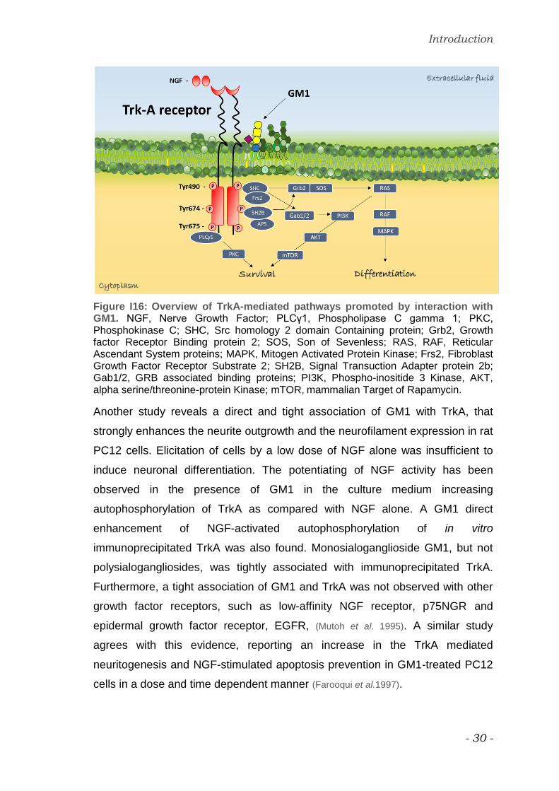
An overall representation of TrkA-mediated pathways and of the downstream
signaling are represented in figure I16. GM1 is supposed to colocalized with TrkA
receptor and to influence its activation (Ferrari et al. 1995; Mutoh et al. 1995; Farooqui
et al.1997; Bachis et al. 2002; Duchemin et al. 2002; Rabin et al. 2002).
Introduction
- 30 -
Figure I16: Overview of TrkA-mediated pathways promoted by interaction with GM1. NGF, Nerve Growth Factor; PLCγ1, Phospholipase C gamma 1; PKC, Phosphokinase C; SHC, Src homology 2 domain Containing protein; Grb2, Growth factor Receptor Binding protein 2; SOS, Son of Sevenless; RAS, RAF, Reticular Ascendant System proteins; MAPK, Mitogen Activated Protein Kinase; Frs2, Fibroblast Growth Factor Receptor Substrate 2; SH2B, Signal Transuction Adapter protein 2b; Gab1/2, GRB associated binding proteins; PI3K, Phospho-inositide 3 Kinase, AKT, alpha serine/threonine-protein Kinase; mTOR, mammalian Target of Rapamycin.
Another study reveals a direct and tight association of GM1 with TrkA, that
strongly enhances the neurite outgrowth and the neurofilament expression in rat
PC12 cells. Elicitation of cells by a low dose of NGF alone was insufficient to
induce neuronal differentiation. The potentiating of NGF activity has been
observed in the presence of GM1 in the culture medium increasing
autophosphorylation of TrkA as compared with NGF alone. A GM1 direct
enhancement of NGF-activated autophosphorylation of in vitro
immunoprecipitated TrkA was also found. Monosialoganglioside GM1, but not
polysialogangliosides, was tightly associated with immunoprecipitated TrkA.
Furthermore, a tight association of GM1 and TrkA was not observed with other
growth factor receptors, such as low-affinity NGF receptor, p75NGR and
epidermal growth factor receptor, EGFR, (Mutoh et al. 1995). A similar study
agrees with this evidence, reporting an increase in the TrkA mediated
neuritogenesis and NGF-stimulated apoptosis prevention in GM1-treated PC12
cells in a dose and time dependent manner (Farooqui et al.1997).

Introduction
- 31 -
Another example is reported to prove GM1 induction of Trk and Erk
phosphorylation in brain slice of striatum, hippocampus and frontal cortex,
underlining a higher level of specificity for TrkA with respect to TrkB and C and
an elicited activation of Erk2 superior than of Erk1. The time and dose
dependency was set also in this study (Duchemin et al. 2002).
A study revealed that GM1 deficient NG108 cells don’t locate TrkA receptor in
plasma membrane and doesn’t show any autophosphorylation. The stable
transfection of GM1 synthase into these cells restores the expression of TrkA in
plasma membrane and its activation, suggesting that GM1 is required for
normal maintaining and functionality of TrkA (Mutoh et al. 2002).
Focusing on neurotrophic and neuroprotective properties of GM1, Trk pathway
seems to be once again implicated.
GM1 was found to rescue cells from apoptotic death using serum-deprived
cultures of wild type and TrKA overexpressing PC12 cells. GM1-promoted
survival was demonstrated to be mediated by both TrkA and TrkB, and
potentially by tyrosine kinase receptors for additional neurotrophic growth
factors. To support these findings, K-252a, an inhibitor of Trk kinases, was
employed in PC12 cells overexpressing a dominant inhibitory form of Trk,
revealing that a portion of the survival-promoting activity of GM1 is due to
receptor dimerization and autophosphorylation (Ferrari et al. 1995).
Moreover, GM1 was proved to increase the survival of PC12 cells to hydrogen
peroxide and others reactive oxygen species. The TrkA receptor implication and
the downstream activation of Erk and Akt were considered target of GM1
because the ganglioside-promoted cell viability was abolished by TrkA inhibitor
(Zakharova et al. 2014).
Concerning the neurotrophic role shown by GM1 after exogenous
administration, the gathered pharmacological potential has open a window onto
development of semisynthetic derivatives of GM1, the LIGA series (Kharlamov et
al. 1993). Their remarkable difference from the parent compound reside in the
incremented membrane permeability and, therefore, enhanced capability to
penetrate the blood–brain barrier (BBB) and neuronal plasma membrane.
These property is imputable to the replacement the long-chain fatty acid of

Introduction
- 32 -
ceramide by shorter groups, such as acetyl (LIGA4) or dichloroacetyl (LIGA20),
as shown in figure I17.
a.
b.
Figure I17: Semisynthetic GM1 derivatives chemical structures.
a. LIGA 4
b. LIGA 20
LIGA20 provided examples of others neuroprotection activities, including
attenuation of ethanol-induced apoptosis in rat cerebellar granule neurons (Saito
et al. 1999). LIGA20 exhibit in vivo a protective behavior against enhanced
kainate-induced seizures (Wu et al. 2005). It also succeeded in ameliorating
Parkinsonian symptoms in a rodent model of Parkinson disease (Schneider & Di
Stefano, 1995)
LIGA20, as well as GM1 ganglioside, demonstrates different effects on
neurotrophic processes, even though the only structural modification realized on
fatty acid and the sphingosine moieties while the oligosaccharide chain stayed
unchanged.

Aim

Aim
- 34 -
Among all features of GM1 biological potential, neurodifferentiation,
neuroprotection and neurodevelopment are certainly the impacted processes
predominantly studied and debated.
Many cell pathways have been considered and investigated in order to
accurately clarify and completely dissect the biological role of endogenous GM1
in the nervous system.
Even if different GM1 targets have been successfully identified by several
studies, the precise dynamic interaction between GM1 and candidate proteins
such as the related molecular mechanisms at the bases of the observed effects
still remain elusive.
Moreover, the findings obtained following the pharmacological approaches don’t
explain utterly the physiological reasoning, opening and implementing however
the GM1 therapeutic employment as alternative research field.
The involvement of GM1 in neuronal differentiation has been demonstrated
using different experimental approaches in vitro:
i. in murine neuroblastoma cell line, N2a, a micromolar GM1 administration
have proved its capability to influence neurodifferentiation process by inducting
neuritogenesis (Facci et al. 1984)
ii. in hippocampal neurons, the overexpression of sialidase Neu3 allowed the
formation of GM1-enriched platforms inducing membrane structure
modifications. The resulting membrane configuration induces the Trk
dimerization and activation. This turn on a specific signaling cascade resulting
in the actin depolymerization generating axon protrusion and elongation (Abad-
Rodriguez et al. 2001; Da Silva et al. 2005).
iii. in murine neuroblastoma cell line, N2a, Neu3 silencing and the following
decrease in enzymatic activity has been demonstrated to induce neurite
sprouting too (Valaperta et al. 2007).
The evidence of the neurodifferentiation promotion obtained manipulating the
endogenous GM1 by two opposed mechanisms, augmenting or silencing the
plasma membrane-associated sialidase Neu3, suggests an important influence

Aim
- 35 -
of the oligosaccharide chain in the fine-tuning of the processes beginning at the
level of plasma membranes, figure A1.
The value of glyco-mediated signaling has been further strongly described for
gangliosides.
a. b.
Figure A1: Neurodifferentiative effect promoted by the modification of the Neu3 sialidase activity.
a. Increasing Neu3 activity. In hippocampal neurons GM1 augmenting, due to the Neu3 activity enhancement, leads to the plasma membrane reorganization in GM1-enrched lipid rafts where dimerization and autophosphorylation of Trk receptor can occur. Trk pathway activation provokes the downstream actin depolimerization promoting axon protrusion.
b. Silencing Neu3. In mouse neuroblastoma cells, silencing Neu3 activity, resulting in about 50% GM1 reduction, causes neurite sprouting and elongation.

Aim
- 36 -
In addition, the studies carried on using LIGA 20 GM1 derivative revealed a
responses both in differentiative and therapeutic employments. The only
structural difference concerns the fatty acid portion, unchanging the
oligosaccharide chain constitution and their feasible association with glycol-
sensitive protein domains but affecting only the GM1 availability to biological
membrane systems.
The conditions and premises above mentioned encourage to suppose that the
crucial point for GM1 to exert its tasks depend on the ‘’quality” of the
oligosaccharide chain into the lipid rafts, which promotes selective interaction
with plasma membrane specific proteins.
The aim of the proposed research is the clarification of the molecular
mechanism and the recognizing of the protein targets by which GM1
accompany and enhance neurodifferentiation process in cells, hypothesizing
the implication of its oligosaccharide chain.
On the bases of previous studies the attention is focused on TrkA signaling,
employing the murine neuroblastoma cell line, Neuro2a (N2a) as an in vitro
model. N2a have been in fact demonstrate to differentiate in neurons-like
phenotype under treatment with GM1 and by increasing its endogenous content
(Facci et al. 1984).
In the present study, the exogenous administration of OligoGM1 is really
employed with the purpose to demonstrate the essential implication of the
hydrophilic head in the natural GM1-promoted neuritogenesis, aspiring to confer
to achieving results a physiological significance.

Materials &
Methods

Materials & Methods
- 38 -
Materials
The murine neuroblastoma cell line, Neuro2a (N2a, RRID: CVCL_0470),
Phosphate-buffered saline (PBS), Trizma Base (Tris),
Ethylenediaminetetraacetic acid (EDTA), Sodium orthovanadate (Na3VO4),
Bovine Serum Albumin (BSA), 1,4-Dithiothreitol (DTT), 3-(4,5,-dimethylthiazole-
2yl)-2,5-diphenyltetrazolium bromide (MTT), Triethylamine (TEA), Sodium
dodecil sulfate (SDS), Tween, Glycerol, Paraformaldehyde, Triton X-100,
Methanol, 2-propanol, Formic acid, Blue bromophenol, Trypan blue, Donkey
serum, powder milk, anti-rabbit FITC conjugate, and mouse α-tubulin antibodies
galactose, sialic acid were purchased from Sigma-Aldrich (St. Louis, MO, USA).
Corning® cell culture flasks, dishes, and Corning® Costar® cell culture plates,
and Corning® Transfectagro™ reduced serum medium were purchased from
Corning (Corning, NY, USA).
Dulbecco’s modified Eagle’s high glucose medium (DMEM HG), fetal bovine
serum (FBS), L-glutamine (L-Glut), Penicillin (10000 U/mL), Streptomycin (10
mg/mL), and acrylamide, were purchased from EuroClone (Paignton, UK).
Gibco™ Opti-MEM™ I reduced serum medium, Lipofectamine® 2000 reagent,
ammonium persulfate (APS), goat anti-mouse IgG (H+L) antibody (RRID:
AB_228307) were from Thermo Fischer Scientific (Waltham, MA, USA).
TrkA inhibitor (CAS 388626-12-8) and high performance thin-layer
chromatography (HPTLC) were from Merk Millipore Merk Millipore (Billerica,
MA, USA).
The short interfering RNAs (siRNAs) were from Quiagen (Velno, Netherlands).
Rabbit anti-TrkA (RRID: AB_10695253), rabbit antiphospho-TrkA (tyrosine 490,
Tyr490) (RRID: AB_10235585), rabbit anti-p44/42 MAPK (Erk1/2) (RRID:
AB_390779), rabbit antiphospho-p44/42 MAPK (pErk1/2) (Thr202/Tyr204)
(RRID:AB_2315112) and anti-rabbit IgG (RRID: AB_2099233) antibodies were
from Cell Signaling Technology (Danvers, MA, USA). Rabbit anti-pan
Neurofilament (NF) antibody (RRID: AB_10539699) was from Biomol
International (Plymouth Meeting, PA, USA).
Protein assay kit and TEMED were from BioRad (Hercules, CA, USA).

Materials & Methods
- 39 -
Chemiluminescent kit for western blot was purchased from Cyanagen (Bologna,
Italy). Ultima gold was purchased from Perkin Elmer (Waltham, MA, USA).
Dako Fluorescent mounting medium was purchased from Agilent (Santa Clara,
CA, USA). Polyvinylidene difluoride (PVDF) membrane was from GE
Healthcare Life Sciences (Chigago, IL, USA).
Commercial chemicals were of the highest purity available, common solvents
were distilled before use and water was doubly distilled in a glass apparatus.

Materials & Methods
- 40 -
Methods
Chemical synthesis and preparation of gangliosides and
oligosaccharides
Gangliosides
Fucosyl-GM1, GM1, GM2, and GM3 gangliosides were obtained from total
ganglioside mixture extracted from pig brains (Tettamanti et al. 1973), by sialidase
hydrolysis and chromatographic purification (Acquotti et al. 1994). To obtain
desialylated GM1 (asialoGM1), GM1 underwent acid hydrolysis and
chromatographic purification (Ghidoni et al. 1976).
Gangliosides were solved in methanol and stored at -20 °C.
Radiolabeled GM1
GM1 containing tritium at position 6 of external galactose ([Gal-6-3H]GM1,
figure M1a) was obtained by an enzymatic oxidation reaction using galactose
oxidase followed by reduction with sodium boro[3H]hydride (Sonnino et al. 1992).
On the other hand, GM1 containing tritium at 3-position of sphingosine
([Sph-3-3H]GM1, figure M1b) resulted from a chemical approach by oxidation of
ganglioside at the 3-position sphingosine with 2,3-dichloro-5,6-
dicyanobenzoquinone (DDQ), a reagent that is specific for allylic hydroxyl
groups. Once again, the oxidation was followed by reduction with
boro[3H]hydride (Ghidoni et al. 1981).
Radiolabeled GM1 were solved in methanol and stored at 4 °C.

Materials & Methods
- 41 -
a.
b.
Figure M1: Radiolabeled GM1 derivative structures:
a. [Gal-6-3H]GM1. An enzymatic oxidation was employed to obtain tritium labeling at the 6-position of the external galactose.
b. [Sph-3-3H]GM1. tritium labeling at position 3 of the sphingosine is obtained by a chemical oxidation with DDQ.

Materials & Methods
- 42 -
Ganglioside oligosaccharides
The oligosaccharides Fuc-OligoGM1, OligoGM1, [Gal-6-3H]OligoGM1,
asialoOligoGM1, OligoGM2, and OligoGM3 were prepared by ozonolysis
followed by alkaline degradation with tryethilamine (Wiegandt & Bucking, 1970) of
Fuc-GM1, GM1, (3H)GM1, asialoGM1, GM2, and GM3 respectively.
Oligosaccharides were solved in methanol and stored at -20 °C. Radiolabeled
derivatives are stored at 4 °C.
OligoGM1 chemical synthesis and MS analysis are shown in figure M2 and M3.
.

Materials & Methods
- 43 -
a.
b.
Figure M2: Chemical synthesis of OligoGM1 (a) and [Gal-6-3H]OligoGM1 (b). Ozone-alkali fragmentation procedure was used to obtain desphingosino-gangliosides. Ozonolysis organic reaction cleaved unsaturated bond between sphingosine 4 and 5 carbons. Subsequently, the alkaline degradation with tryethilamine released the oligosaccharide chain form the residue.

Materials & Methods
- 44 -
Figure M3: OligoGM1 MS analysis.

Materials & Methods
- 45 -
Photoactivable derivatives
Photoactivable OligoGM1, [Gal-6-3H]OligoGM1(Glc-N3), bearing the
photosensitive group on the glucose, as well as GM1 photoactivable on the fatty
acid moiety, [Gal-6-3H]GM1(Cer-N3) were prepared from galactose-tritiated
GM1, shown in figure M1a.
On the other side, GM1 tritiated on sphingosine (figure M1b), was the precursor
for the GM1 with the photoactivable group on the last galactose residue, [Sph-3-
3H]GM1(Gal-N3).
To prepare photoactivable OligoGM1 on glucose (figure M5), an amount of 52
μmol of radiolabeled OligoGM1 (obtained by ozonolysis and alkaline
degradation of tritiated GM1) were dissolved in 33% ammonia and treated with
1 mg of ammonium hydrogen carbonate. The reaction was maintained under
stirring for 48 hours at 40 °C. The solution was then immediately freeze-dried
(Lubineau et al. 1995). The same approach was followed for the preparation of
photosensitive GM1 on the external galactose (figure M4). In this case, however,
tritiated GM1 was firstly submitted to enzymatic oxidation at position 6 of the
last galactose by galactose oxidation.
To insert the photoactivable group on fatty acid residue, galactose-tritiated GM1
was submitted to alkaline hydrolysis to remove the stearic acid residue, followed
by acid coupling with 12-aminododecanoic acid (figure M6). The reaction
occurred adding 350 μmol of 12-aminododecanoic acid, dissolved in 2.5 mL of
dry tetrahydrofuran to 1.5 mL of dimethylformamide containing 80 μmol of
deAcyl-GM1, Triton X-100 (1 mL), and dry triethylamine (15 mL) under
continuous magnetic stirring for 24 hours at 23 °C. The mixture was evaporated
under vacuum to 1 mL, and 25 mL of ethyl acetate was added (Sonnino et al.
1989).
Finally, all amino-derivatives were properly treated to insert the chosen
photoactivable group. In particular, the azide labeling procedure (figure M4-6)
started with the dissolution of the crude amino-derivatives obtained by previous
reactions in 0.5 mL of dry dimethylformamide. Subsequently, 1 mg of 2-nitro-4-
fluorophenylazide and 1 μL of tributylamine were added under dark conditions
Materials & Methods
- 46 -
in 25 μL of dry DMSO. Maintaining dark conditions for all the process, the
reaction mixture was stirred over night at 80°C. After solvent evaporation,
desired compounds were purified by flash chromatography using
chloroform/methanol/2-propanol/water 60:35:5:5 v/v/v/v as eluent for OligoGM1
(Mauri et al. 2003) and chloroform/methanol/water 60:35:8 v/v/v for GM1 series
(Sonnino et al. 1989).
All derivatives were dissolved in methanol and stored at 4 °C.

Materials & Methods
- 47 -
Figure M4: Synthesis of [Gal-6-3H]OligoGM1(Glc-N3).

Materials & Methods
- 48 -
Figure M5: Synthesis of [Sph-3-3H]GM1(Gal-N3).

Materials & Methods
- 49 -
Figure M6: Synthesis of [Gal-6-3H]GM1(Cer-N3).
Materials & Methods
- 50 -
NMR, MS, HPTLC, and autoradiographic analyses
Altogether, NMR, MS, HPTLC, and autoradiographic analyses showed a
homogeneity over 99% for all the prepared gangliosides and oligosaccharides.
NMR spectra were recorded with a Bruker AVANCE-500 spectrometer at a
sample temperature of 298 K. NMR spectra were recorded in CDCl3 or CD3OD
and calibrate using the TMS signals internal reference.
Mass spectrometric analysis were performed in positive or negative ESI-MS.
MS spectra were recorded on a Thermo Quest Finningan LCQTM DECA ion
trap mass spectrometer, equipped with a Finnigan ESI interface.
All reactions were monitored by HPTLC on silica gel 60 plates.

Materials & Methods
- 51 -
Cell cultures
Neuro2a (N2a) cells were cultured and propagated on 75 cm2 flasks in high
glucose Dulbecco’s modified Eagle’s medium (DMEM HG) supplemented with
10% fetal bovine serum (FBS), 1% L-glutamine and 1% penicillin/streptomycin
(P/S, v/v), at 37 °C in a humidify atmosphere of 95% air / 5% CO2. Cells were
sub-cultured to a fresh culture twice a week at reaching of 90% confluence (i.e.
every 3-4 days). In sub-cultures passages cells were washed twice with PBS
and detached by 0.02% EDTA - 0.6% glucose in PBS (w/v).
N2a cells were employed in experiments between the 5th and the 30th
passages.
For all cell culture procedures sterilized condition were maintained by using
sterile solutions and by working under the laminar flow cabinet.
Cell treatments
For all experiments N2a cells were plated at 5 x 10³ / cm2, on 6-well plates if not
specified, and incubated for 24 hours in complete DMEM HG medium to allow
cells attachment. Cells were counted by Bürker chamber system (Denham et al.
1971).
Control cells were always plated and incubated under identical conditions but
omitting any addition.
Ganglioside, Oligosaccharide and sugar treatments.
Growth medium was removed and cells were conditioned for at least 30
minutes at 37 °C in a humidify atmosphere of 95% air / 5% CO2 in pre-warmed
Transfectagro medium supplemented with 2% FBS, 1% L-glutamine, 1% P/S
(v/v). Cell treatments were performed in 2% FBS medium to minimize
interactions between serum and added components (Facci et al. 1984).
Subsequently, cells were incubated at 37 °C up to 48 hours in the presence of
50 μM gangliosides, oligosaccharides, galactose, or sialic acid.
Gangliosides, oligosaccharides and sugars were solved in methanol. To
perform cell treatment, they were dried by nitrogen gas, and dissolved in

Materials & Methods
- 52 -
Transfectagro complete medium by vortex agitation and by sonication in water
bath 3 times each for 30 seconds.
TrkA chemical inhibition
In order to block TrkA activity in N2a cells, 120 nM TrkA inhibitor (Wood et al.
2004) was added to the conditioning Transfectagro medium 1 hours before the
addition of GM1 or OligoGM1.
siRNA mediated TrkA knockdown
TrkA expression silencing was achieved by RNA interference experiments
applying siRNA.
Three different siRNAs were employed to silence TrkA:
i. Mm_Ntrk1_1 (sense 50-CCAUCAUAAUAGCAAUUAUTT-30,
antisense 50-AUAAUUGCUAUUAUGGAT-30);
ii. Mm_Ntrk1_5 (sense 50-GGUGGCUGCUGGUAUGGUATT-30,
antisense 50-UACCAUACCAGCAGCCACCTG-30 );
iii. Mm_Ntrk1_6 (sense 50-CCUUCUUGUGCUCAACAAATT-30,
antisense 50-UUUGUUGAGCACAAGAAGGAG-30 ).
Non-silencing siRNA with no homology to any known mammalian gene was
used (sense 50-UUCUUCGAACGUGUCACGUdTdT-30, antisense 50-
ACGUGACACGUUCGGAGAAdTdT-30) and the cells transfected by scramble
were considered the control condition for the experiment.
Transfection was performed 24 hours after cell plating in antibiotic and serum
free OptiMEM culture media containing 0.25% Lipofectamine 2000 (v/v) and 50
nM siRNA, previously solved in sterilized, deionized and nuclease free water
(16.7 nM of each siRNA). After 6 hours, the transfection medium was changed
to complete DMEM HG culture medium. The day after the silencing, cells were
treated with 50 μM GM1 or OligoGM1 as described above.
Materials & Methods
- 53 -
Photolabeling experiments
Cells were incubated with 50 μM [Sph-3-3H]GM1(Gal-N3) (figure M4), [Gal-6-
3H]OligoGM1(Glc-N3) (figure M5), and [Gal-6-3H]GM1(Cer-N3) (figure M6), for 3
hours at 37 °C in a humidify atmosphere of 95% air / 5% CO2 in obscure room.
After incubation, medium was removed and cells were illuminated for 40
minutes under UV light (λ= 360 nm) maintaining the plates on ice to induce
photo-activation.
All the procedures before exposure to UV light were performed in dark room,
under red safelight.
The cells were lysed by sample buffer containing 0.15 M DTT, 94 mM Tris-HCl,
15% glycerol (v/v), 3% SDS (w/v), 0.015% blue bromophenol (v/v), sonicated by
probe (50 W, 30 kHz) and boiled for 5 minutes at 99 °C. Denatured proteins
underwent to 4–20% SDS-polyacrylamide gel electrophoresis (SDS–PAGE)
and blotted on PVDF membrane by trans-blot turbo system. Digital
autoradiography of the PVDF membrane was performed with Beta-Imager
2000. PVDF membranes were then blocked, incubated with anti-TrkA antibody
and processed as follow described in the paragraph “Protein analysis” (Sonnino
et al. 1989; Sonnino et al. 1992; Chigorno et al. 1990; Loberto et al. 2003; Chiricozzi et al.
2015).

Materials & Methods
- 54 -
Assessment of cytotoxicity
Trypan blue assay
Cell viability was determined by Trypan blue exclusion assay after 24 and 48
hour treatments with 50 μM GM1 or OligoGM1. Cells were detached by 25 cm2
flasks. The numbers of living and death cells were discriminated according to
Trypan blue staining that selectively distinguished necrosis and apoptotic cells
from living ones (Mehlen et al. 1988; Aureli et al. 2011).
MTT assays
Cell proliferation was monitored after 12, 24 and 48 hour treatment with 50 μM
GM1 or OligoGM1 according to MTT method firstly described by Mosmann in
1983.
Briefly, cells were seeded on 24-well plates for MTT test.
At the end of incubation period, treatment medium was replaced with 2.4 mM
MTT (dissolved 4 mg/mL in PBS) diluted in Transfectagro complete medium.
Plates were re-incubated for 4 hours at 37 °C. Subsequently, MTT containing
medium was carefully removed and the cells were lysed with 2-propanol : formic
acid, 95 : 5 (v/v) in order to solve resulting formazan crystals. Plates were gently
agitated for 5 minutes to homogenate cell purple solution prior to read the
absorbance at 570 nm with microplate spectrophotometer.

Materials & Methods
- 55 -
Morphological analysis and neurite outgrowth evaluation
Cultured cells, treated with 50 μM GM1, oligosaccharides or sugars up to 48
hours, were observed by phase contrast microscopy.
The neurite-like processes length was measured after treatment with GM1 or
OligoGM1 on bidimensional images and expressed as the ratio between neurite
length and cell body diameter (Schengrund & Prouty, 1988; Sato et al. 2002).
Five random fields were examined from each well, giving a total cell count of at
least 200 cells per well.

Materials & Methods
- 56 -
Immunofluorescence analysis
After 24 hour treatment with 50 μM GM1 or OligoGM1, cells, attached to the
glass inserts, were washed with cold PBS and fixed in 4% paraformaldehyde for
20 minutes at 23 °C. Cells were washed, got permeable by 0.1% Triton X-100
for 30 minutes and then treated for 1 hour at 23 °C with the blocking solution
(5% donkey serum and 0.2% Triton X-100 in PBS, v/v). Cells were incubated
with rabbit polyclonal antibody anti-Neurofilament (NF) for 2 hours at 23 °C.
After three washing with PBS, cells were incubated 1 hour with secondary anti-
rabbit antibody FITC-conjugated.
Fluorescence signal was detected by fluorescence microscope and the images
were processed by ImageJ software.
Materials & Methods
- 57 -
Study of interaction between OligoGM1 and N2a cells
The fate of OligoGM1 administered to cells was determined using tritium-
labeled derivative shown in figure M2b.
After the cell loading with [Gal-6-3H]OligoGM1 for 0.5, 1, 6, and 24 hours the
medium was removed and the following treatments were performed
sequentially:
i. cells were washed 5 times/10 minutes each with 10% FBS-DMEM HG
medium to remove the amount of [Gal-6-3H]OligoGM1 weakly associated to the
cell was collected removable from the cell plasma membrane by the affinity to
the serum components. The resulting solution was called ʺserum labile fraction
(SL)ʺ;
ii. cells were treated with 0.1% trypsin-EDTA solution in PBS (v/v) for 1 minute
to obtain the [Gal-6-3H]OligoGM1 covalently linked to extracellular domain of
plasma membrane proteins. Trypsin removable-derived solution fraction was
called ʺtrypsin labile fractionʺ (TL);
iii. cells were lysed by trypsin-EDTA solution (0.05%-0.02%, w/v in PBS) in
order to evaluate the quantity of [Gal-6-3H]OligoGM1 internalized by the cells.
This fraction underwent to probe sonication (50 W, 30 kHz) and the relative
homogenate is called ʺtrypsin labile fractionʺ (TS).
Radioactivity associated with collected solutions was determined by liquid
scintillation counting.
The procedure was previously established to determine at the cell culture level
the fate of exogenously administered gangliosides (Chigorno et al. 1985).

Materials & Methods
- 58 -
Protein analysis
At the end of treatments, cells were washed with cold PBS containing 0.5% of 2
mM Na3VO4 (v/v), lysed by hot lysis Buffer (0.15 M DTT, 94 mM Tris-HCl, 15%
glycerol, v/v, 3% SDS w/v, 0.015% blue bromophenol, v/v) and detached using
scrapers. DC protein assay was performed in order to quantify sample proteins.
After the probe sonication (50 W, 30 kHz) and the boiling of the lysed samples
for 5 minutes at 99 °C, equal amounts of denatured proteins derived from
treated and untreated cells were separated on 7.5% polyacrylamide gels, and
transferred to PVDF membranes.
Electrophoresis was performed at 23 °C, applying 100 V constant voltage in the
stacking gel, augmenting at 170 V in the running. Blot transferring was
performed at 4 °C, maintaining for 3 hours constant current at 200 mA. PVDF
membranes were blocked with 5% milk (w/v) in TBS-0.1% tween (v/v) at 23 °C
for 2 hours under gently shaking.
The presence of Neurofilament (NF), TrkA, p-TrkA, extracellular signal-
regulated protein kinases 1 and 2 (ERK1/2) and p-ERK1/2 was determined by
specific rabbit primary antibodies, diluted 1:1000 in 5% BSA (w/v) in TBS-0.1%
tween. α-tubulin, used as loading, was detected by the specific mouse primary
antibody diluted 1:40000 in 5% milk (w/v) in TBS-0.1% tween (v/v). The
incubation was performed over night at 4 °C under gently shaking.
PVDF membrane were washed three times with TBS-0.1% tween. The reaction
with secondary horseradish peroxidase (HPR)-conjugated antibodies was
following performed at 23 °C in agitation, after 1:2000 dilution of anti-rabbit
antibody in 5% BSA (w/v) in TBS-0.1% tween and 1:30000 dilution of anti-
mouse antibody in 5% milk (w/v) in 0.1% TBS-tween.
After three washes with TBS-0.1% tween, PVDF signal, originated from luminol
chemiluminescence reaction, was acquired and analyzed by Uvitec.
Quantitative estimation were performed using ImageJ software.

Materials & Methods
- 59 -
Molecular modeling
Crystallographic structure of the extracellular segment of human TrkA in
complex with nerve growth factor (NGF) (RCSB PDB ID: 2IFG) was used for
molecular docking calculations. Protein complex was submitted to the Molecular
Operating Environment 2016.0802 (MOE) Structure Preparation application, in
order to fix all issues and to prepare structures for subsequent computational
analyses.
The OligoGM1 structure was built with the MOE Carbohydrate Builder and a
geometry optimization was carried out with MOPAC7 and the PM6 basis set.
Molecular docking was carried out through the MOE Dock program, setting as
receptor the complex between TrkA and NGF, as ligand the optimized
OligoGM1 structure. The binding site was identified at the interface between the
two proteins. Before placement procedure, 20 000 rotamers of the ligand was
generated, exploring all the molecule rotatable bonds. Alpha PMI placement
algorithm, specifically developed for tight binding pocket, was selected. The
London dG empirical scoring function was used for sorting the poses. The 30
top-scoring poses was refined through molecular mechanics, considering the
receptor as a rigid body, and the refined complexes were scored through the
GBVI/WSA dG empirical scoring function, keeping the five top-scoring poses.
The top-scoring pose from the docking procedure was refined by using the
MOE QuickPrep procedure aimed at relaxing and refining the complex before
calculating the approx. binding free energy via the GBVI/WSA dG empirical
scoring function (Naim et al. 2007).

Results

Results
- 61 -
GM1 and OligoGM1 effect on N2a cell viability and proliferation
GM1 and OligoGM1 concentration for cell treatments was fixed at 50 μM (Rabin
et al. 2002; Chiricozzi et al. 2017).
Trypan blue assay was preliminary performed after 24 and 48 hour treatment in
order to evaluate a possible toxic effect of 50 μM GM1 or OligoGM1 on N2a
cells (Mehlen et al. 1988; Strober, 2001; Aureli et al. 2011).
As shown in figure R1a, no significant difference in cell viability was observed
with respect to untreated cells (Chiricozzi et al. 2017).
Figure R1a: Effect of GM1 and OligoGM1 on cell viability. N2a cells were treated for 24 and 48 hours with 50 μM GM1 or OligoGM1. The numbers of living and dead cells were determined according to the Trypan blue exclusion assay and expressed in percentage of the total cell number. Bars show the mean values ± SEM of 5 different cell culture preparations (n = 5).
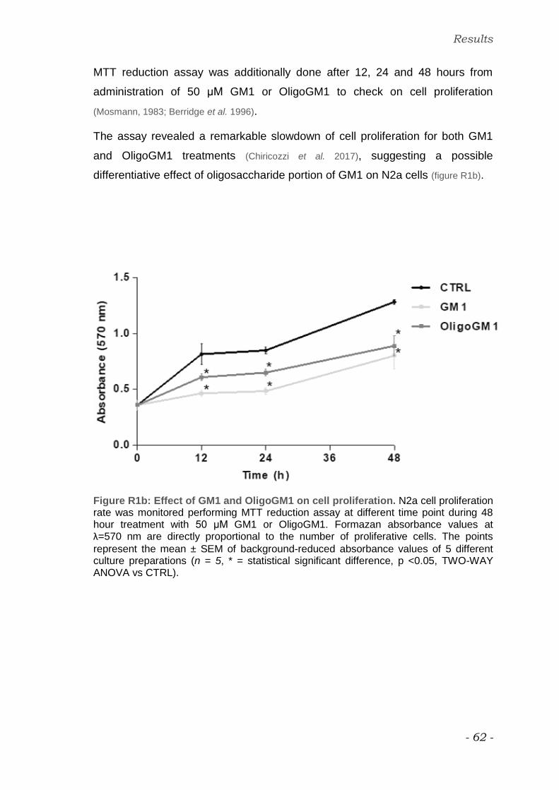
Results
- 62 -
MTT reduction assay was additionally done after 12, 24 and 48 hours from
administration of 50 μM GM1 or OligoGM1 to check on cell proliferation
(Mosmann, 1983; Berridge et al. 1996).
The assay revealed a remarkable slowdown of cell proliferation for both GM1
and OligoGM1 treatments (Chiricozzi et al. 2017), suggesting a possible
differentiative effect of oligosaccharide portion of GM1 on N2a cells (figure R1b).
Figure R1b: Effect of GM1 and OligoGM1 on cell proliferation. N2a cell proliferation rate was monitored performing MTT reduction assay at different time point during 48 hour treatment with 50 μM GM1 or OligoGM1. Formazan absorbance values at λ=570 nm are directly proportional to the number of proliferative cells. The points
represent the mean ± SEM of background-reduced absorbance values of 5 different culture preparations (n = 5, * = statistical significant difference, p <0.05, TWO-WAY ANOVA vs CTRL).
Results
- 63 -
Effect of GM1 derivatives on N2a cell morphology
In order to investigate a possible differentiative role of GM1 derivatives, treated
N2a cells were subjected to morphological analysis by phase contrast
microscopy 24 and 48 hours after treatments.
GM1 was employed as a positive control for the proved capacity to induce
neurite formation in N2a cells (Roisen et al. 1981; Leon et al. 1982; Facci et al. 1984;
Rabin et al. 2002).
OligoGM1 administration to N2a cells induced a neuron-like morphology in 24
hours, accentuated after 48 hours (figure R2). The effect was evidenced by the
enhanced sprouting and progressive elongation of cell extension, conversely
inappreciable in round-shaped control cells (Chiricozzi et al. 2017).
In order to clearly rationalize on the minimal GM1 oligosaccharide-included
portion essentially required to initiate neurodifferentiation process, N2a cells
were treated with 50 μM galactose, sialic acid, OligoGM3, OligoGM2 or asialo-
OligoGM1. OligoGM1 components were not able to induce any relevant
morphological changes in N2a cells after 48 hour incubation (figure R3).
The morphological effect was only obtained by using the entire OligoGM1
molecule suggesting that all included sugars are necessary to limit cell
proliferation and to induce neurite sprouting (Chiricozzi et al. 2017).
In addition, N2a cells were treated also with the fucosylated OligoGM1, since
Fuc-GM1 was able to induce neurodifferentiation (Masserini et al. 1992). As shown
in figure R4, Fuc-OligoGM1 was still able to induce neurite sprouting and
reduction in cell proliferation, similarly to OligoGM1 (Chiricozzi et al. 2017).
The obtained results suggest that the GM1 enhanced neurite sprouting in N2a
cells requires specifically β-Gal-(1-3)- β-GalNAc-(1-4)-[α-Neu5Ac-(2-3)]- β-Gal-
(1-4)-β-Glc structure and that the α-fucose addition at the 2 position of the
external galactose is irrelevant for impacting differentiative process.

Results
- 64 -
Figure R2: Morphological outcomes of N2a cells following 24 and 48 hours incubation with 50 μM GM1 or OligoGM1. Cells were examined with phase contrast
microscopy with 200X magnification. Images are representative of 10 independent experiments (n = 10).

Results
- 65 -
48 h
Figure R3: Morphological outcomes of N2a cells grown with 50 μM galactose,
sialic acid, OligoGM3, OligoGM2 and asialo-OligoGM1 for 48 hours. Cells were observed by phase contrast microscopy with 200X magnification. Images are representative of 3 independent experiments (n = 3).

Results
- 66 -
48 h
Figure R4: Morphological outcomes of N2a cells grown with 50 μM GM1,
OligoGM1 and fuco-OligoGM1 for 48 hours. Cells were observed by phase contrast microscopy with 200X magnification. Images are representative of 3 independent experiments (n = 3).

Results
- 67 -
Neurite characterization
Neurite outgrowth was analyzed to characterize from a physical and
biochemical points of view the morphological evidence of neuron-like cell
differentiation induced by OligoGM1.
Physical characterization
Neurite extensions were measured and expressed by the ratio between the
length of processes and the diameter of cell body (Schengrund & Prouty 1988; Sato
et al. 2002).
The values related to treated cells resulted significantly higher than control cells
after 24 hours, increasing two fold more up to 48 hours (Chiricozzi et al. 2017).
Values obtained with OligoGM1 were comparable to GM1 (figure R5).
Figure R5: Evaluation of neurite sprouting and elongation in N2a cells. Neurite extensions were evaluated as the ratio between process length and cell body diameter after 24 and 48 hours treatment with 50 μM GM1 or OligoGM1. The bars show the mean values ± SEM from 5 different experiments (n = 5, * = statistical significant difference, p < 0.01, Student’s t-test vs CTRL).

Results
- 68 -
Biochemical characterization
The level of intracellular neurofilament protein expression was evaluated as a
biochemical marker of neurodifferentiation. Neurofilament is the major
component of cytoskeleton supporting the axonal construction (Mao et al. 2000;
Wang et al. 2004; Fukuda et al. 2014).
Immunoblotting analysis highlighted a significant increase in the expression of
heavy, medium and light NF subunits (NF-h, NF-m, and NF-l) after 24 hours
treatment with both GM1 and OligoGM1 (Chiricozzi et al. 2017), (figure R6a).
The result was qualitatively supported by the immunofluorescence assays
(Chiricozzi et al. 2017) (figure R6b).
Since the presence of cell extensions, characterized by NF proteins expression,
is considered marker of neurodifferentiation, the oligosaccharide portion of GM1
is allowable to promote neuritogenesis in N2a cells, analogously to GM1.

Results
- 69 -
a.
b.
Figure R6: Neurofilament protein expression in N2a:
a. Western Blotting analysis. Immunoblotting for NF-h, NF-m, NF-l was performed after 24 hour treatment with 50 μM GM1 or OligoGM1. The proteins are revealed by the specific antibody and visualized by chemiluminescence. Left: immunoblotting images. Blots are representative of 3 independent experiments. Right: semi-quantitative analysis of NF proteins amount. α-tubulin was used for normalization. The bars represent the mean ± SEM values of 3 different experiments expressed as the fold increase over CTRL (n = 3, * = statistical significant difference, p < 0.01, Student’s t-test vs CTRL).
b. Immunofluorescence staining. Neurofilament was detected using the specific antibody after 24 hours treatment with 50 μM GM1 or OligoGM1. Cells were visualized by fluorescent microscopy at 400X magnification. Images are representative of 3 independent experiments (n = 3).

Results
- 70 -
OligoGM1 interaction with N2a cells
Discovering the interaction between OligoGM1 and N2a cells represented the
starting point to investigate about OligoGM1 targets in promoting
neurodiffererentiation. The fate of OligoGM1 was established using a procedure
described to search for gangliosides administered to cells in culture (Chigorno et
al. 1985). The experimental approach allows to search for three different
modalities of OligoGM1 association to cells: i. weakly to the cell surface, ii.
covalently to the cell surface, iii. internalized by cells.
Isotopic tritium labeled OligoGM1, [Gal-6-3H]OligoGM1, (figure M2b), was
administered to N2a cells. Radioactivity allowed the recognition and the
quantification of the molecule. After 0.5, 1, 6 and 24 hour treatments, cells were
subjected to 10% serum-containing medium cell wash in order to remove and
check the quantity of oligosaccharide weakly associated to the cells surface
(serum labile fraction, SL). Subsequently, cells were treated with low-
concentrated trypsin solution, searching for the oligosaccharide covalently
bonded on PM protein extracellular domains (trypsin labile fraction, TL). Finally,
cells were lysed in order to trace internalized OligoGM1 (trypsin stabile fraction,
TS). About 99% of collected OligoGM1 was found in the serum fraction at each
time point as shown in figure R7 (Chiricozzi et al. 2017).
The finding suggested that the oligosaccharide portion of GM1 does not enter
into the cells neither in the PM, but it exerts its function interacting in a no-
covalent manner with the cell surface.
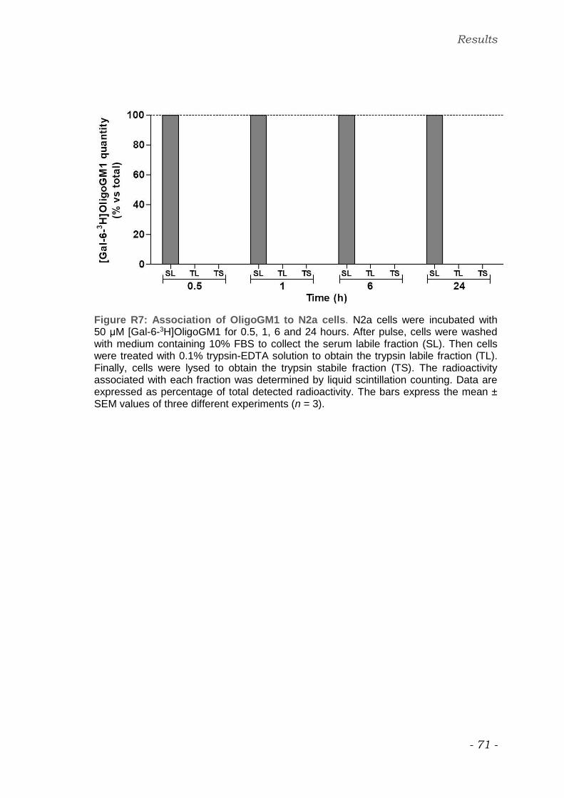
Results
- 71 -
Figure R7: Association of OligoGM1 to N2a cells. N2a cells were incubated with 50 μM [Gal-6-3H]OligoGM1 for 0.5, 1, 6 and 24 hours. After pulse, cells were washed with medium containing 10% FBS to collect the serum labile fraction (SL). Then cells were treated with 0.1% trypsin-EDTA solution to obtain the trypsin labile fraction (TL). Finally, cells were lysed to obtain the trypsin stabile fraction (TS). The radioactivity associated with each fraction was determined by liquid scintillation counting. Data are expressed as percentage of total detected radioactivity. The bars express the mean ± SEM values of three different experiments (n = 3).

Results
- 72 -
OligoGM1 effect on TrkA receptor pathway
On the basis of TrkA-mediated neurodifferentiation promoted by GM1
amplifying NGF activity (Farooqui et al.1997; Singleton et al. 2000; Duchemin et al. 2002;
Da Silva et al. 2005; Mocchetti 2005; Zakharova et al. 2014), TrkA pathway was chosen
as the subject of the study in order to investigate the molecular mechanism
responsible for OligoGM1-inducted neuritogenesis.
Particular attention was focused on the phosphorylation of the tyrosine 490
(Tyr490) that is relevant for differentiation pathway activation (Cordon-Cardo et al.
1991; Lavenius et al. 1995; Eggert et al. 2000; Damani et al. 2003; Biarc et al. 2012; Biarc et al.
2013). Furthermore, the activation of MAP Kinases Erk 1 and 2 (Erk1/2) was
checked as downstream actor in the signaling cascade (Chao. 1992; Cowley et al.
1994; Lavenius et al. 1995; Fukuda et al. 1995; Sweatt, 2001; Vaudry et al. 2002).
TrkA receptor activation
A significant activation of TrkA receptor detected as the increase in Tyr490
phosphorylation level was highlighted both in GM1-treated cells and in
OligoGM1-treated cells by immunoblotting analysis after 24 hours from the
beginning of the treatment (Chiricozzi et al. 2017), (figure R8).
Checking the activation of MAP kinases, an important augmenting in ERK1/2
phosphorylation was also found in treated cells at the same time point (Chiricozzi
et al. 2017), (figure R9).

Results
- 73 -
Figure R8: GM1 and OligoGM1 effect on TrkA activation. N2a cells were treated with 50 μM GM1 or OligoGM1 for 24 hours. Immunoblotting for pTrkA (Tyr490), TrkA and α-tub expression is revealed by specific antibodies and visualized by chemiluminescence. Top: immunoblotting images. Blots are representative of 8 independent experiments. Bottom: semi-quantitative analysis of pTrKA (Tyr490) related to the total TrkA level. α-tub was used to normalizing. The bars represent the mean ± SEM values of 8 different experiments, expressed as the fold increase over CTRL (n = 8, * = statistical significant difference, p < 0.05, Student’s t-test vs CTRL).

Results
- 74 -
Figure R9: GM1 and OligoGM1 effect on MAPK activation. N2a cells were treated with 50 μM GM1 or OligoGM1 for 24 hours. Immunoblotting for pErk1/2, Erk1/2 and α-tub expression is revealed by specific antibodies and visualized by chemiluminescence. Top: immunoblotting images. Blots are representative of 8 independent experiments. Bottom: semi-quantitative analysis of pErK1/2 related to the total Erk1/2 level. α-tub was used to normalizing. The bars represent the mean ± SEM values of 8 different experiments, expressed as the fold increase over CTRL (n = 8, * = statistical significant difference, p < 0.05, Student’s t-test vs CTRL).

Results
- 75 -
Time course changing in TrkA-Erk signaling pathway
In order to examine time course of TrkA-Erk pathway induction caused by GM1
and OligoGM1, the phosphorylation levels of TrkA (Tyr490) and Erk1/2 were
evaluated over the duration of the treatments by western blot analysis.
As shown in figure R10, GM1 and OligoGM1 caused a rapid elevation of TrkA
phosphorylation on Tyr490 that was significantly maintained over the 12 hours
time course.
The enhancement in MAPK phosphorylation appeared successively, getting
significant after 6 hours from GM1 and OligoGM1 administration (figure R11).
The results revealed that, following GM1 and OligoGM1 treatment, Tyr490-TrkA
phosphorylation was increased within 30 minutes and Erk1/2 after 3 hours and
both remained elevated throughout 24 hours, in parallel to morphological
differentiation outcomes.

Results
- 76 -
Figure R10: Time course of Tyr 490-TrkA phosphorylation. N2a cells were treated with 50 μM GM1 or OligoGM1 up to 12 hours. The figure shows analysis for pTrkA (Tyr490) and TrkA expression performed after 0.5, 1, 3, 6 and 12 hours treatment. Immunoblotting was revealed by specific antibodies and visualized by chemiluminescence. Top: immunoblotting images. Blots are representative of 3 independent experiments. Bottom: semi-quantitative analysis of pTrKA (Tyr490) related to the total TrkA level. The bars represent the mean ± SEM values of 3 different experiments, expressed as the fold increase over CTRL (n = 3, * = statistical significant difference, p < 0.05, Student’s t-test vs CTRL).

Results
- 77 -
Figure R11: GM1 Time course of Erk1/2 phosphorylation. N2a cells were treated with 50 μM GM1 or OligoGM1 up to 12 hours. The figure shows analysis for pErk1/2
and Erk1/2 expression performed after 3, 6 and 12 hours treatment. Immunoblotting was revealed by specific antibodies and visualized by chemiluminescence. Top: immunoblotting images. Blots are representative of 3 independent experiments. Bottom: semi-quantitative analysis of pErK1/2 related to the total Erk1/2 level. The bars represent the mean ± SEM values of 3 different experiments, expressed as the fold increase over CTRL (n = 3, * = statistical significant difference, p < 0.05, Student’s t-test vs CTRL).

Results
- 78 -
TrkA receptor inhibition
To prove that GM1 or OligoGM1 effect in N2a neurodifferentiation induction is
properly mediated by TrkA-Erk pathway, TrkA activation was blocked using a
specific inhibitor, able to fit the ATP pocket, preventing its consumption (Segal et
al. 1996; D’Ambrosi et al. 2001; Wood et al. 2004; Lemmon & Schlessinger, 2010).
A specific inhibitor for TrkA receptor was employed in combination to GM1 and
OligoGM1 treatments in order to prove that neuritogenic effect is properly
mediated by TrkA-ERK pathway. The addition of TrkA inhibitor together with
GM1 or OligoGM1 impeded phosphorylation processes in the signaling cascade
of both Tyr490 and Erk p44/42 (Chiricozzi et al. 2017), (figure R12 a and b).
Blocking TrkA activity, the neurite elongation is arrested even in presence of
GM1 and OligoGM1 for 24 hours (Chiricozzi et al. 2017), (figure R13).
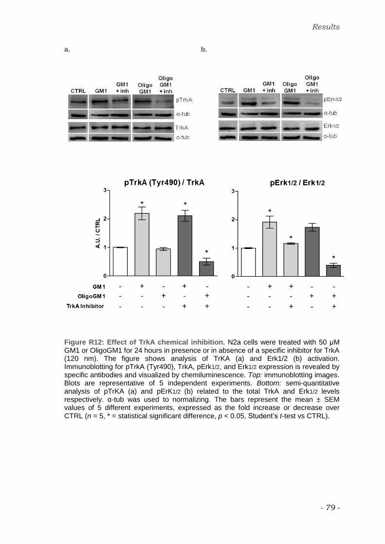
Results
- 79 -
a. b.
Figure R12: Effect of TrkA chemical inhibition. N2a cells were treated with 50 μM GM1 or OligoGM1 for 24 hours in presence or in absence of a specific inhibitor for TrkA (120 nm). The figure shows analysis of TrKA (a) and Erk1/2 (b) activation. Immunoblotting for pTrkA (Tyr490), TrkA, pErk1/2, and Erk1/2 expression is revealed by specific antibodies and visualized by chemiluminescence. Top: immunoblotting images. Blots are representative of 5 independent experiments. Bottom: semi-quantitative analysis of pTrKA (a) and pErK1/2 (b) related to the total TrkA and Erk1/2 levels respectively. α-tub was used to normalizing. The bars represent the mean ± SEM values of 5 different experiments, expressed as the fold increase or decrease over CTRL (n = 5, * = statistical significant difference, p < 0.05, Student’s t-test vs CTRL).

Results
- 80 -
Figure R13: Morphological outcomes of N2a cells after TrkA inhibition. After 24 hours incubation with TrkA inhibitor in combination with 50 μM GM1 or OligoGM1 cells were analyzed with phase contrast microscopy with 200X magnification. Images are representative of 5 independent experiments (n = 5).

Results
- 81 -
TrkA receptor expression silencing
In addition to the chemical inhibition, TrkA receptor expression was also
knocked down by siRNA transfection. The approach offered the possibility to
demonstrate the unavoidable implication of TrkA receptor in GM1 and
OligoGM1-promoted neuritogenesis. Control cells were transfected by scramble
siRNA. Silenced cells, resulting in a 70% reduction of TrkA expression, as
evidenced in figure R14 were incubated with GM1 or OligoGM1 for 24 hours
(Chiricozzi et al. 2017).
As shown by immunoblotting analysis, TrkA silencing prevented Erk1/2
phosphorylation due to GM1 and OligoGM1 in N2a cells (figure R14), that at the
same time didn’t undergo to differentiation (Chiricozzi et al. 2017), (figure R15).

Results
- 82 -
a. b.
Figure R14: Effect of silencing TrkA expression. N2a cells were transfected with siRNA against TrkA (50nM). Control cells were transfected with scramble siRNA. 24 hours after transfection cells were exposed to 50 μM GM1 or OligoGM1 for 24 hours. The figure shows the evaluation of TrKA (a) and Erk1/2 (b) phosphorylation. Immunoblotting for pTrkA (Tyr490), TrkA, pErk1/2, and Erk1/2 expression is revealed by specific antibodies and visualized by chemiluminescence. Top: immunoblotting images. Blots are representative of 3 independent experiments. Bottom: semi-quantitative analysis of pTrKA (a) and pErK1/2 (b) related to the total TrkA and Erk1/2 levels respectively. α-tub was used to normalizing. The bars represent the mean ± SEM values of 3 different experiments, expressed as the fold increase or decrease over untreated CTRL (n = 3, * = statistical significant difference, p < 0.05, Student’s t-test vs untreated CTRL).

Results
- 83 -
Figure R15: Morphological outcomes of N2a cells after TrkA expression silencing. After transfection with TrkA or scramble siRNA, N2a cells were treated for 24 h with 50 μM GM1 or OligoGM1 and analyzed with phase contrast microscopy with 200X magnification. Images are representative of 3 independent experiments (n = 3).

Results
- 84 -
Interaction between OligoGM1 and TrkA receptor
Covalent cross-linking interaction by photolabeling
In order to study the interaction between the oligosaccharide chain of GM1 and
TrkA receptor, radioactive and photoactivable GM1 derivatives,
[Gal-6-3H]GM1(Cer-N3), [Sph-3-3H]GM1(Gal-N3) and [Gal-6-3H]OligoGM1(Glc-
N3) were prepared according to the procedure shown in M section.
The use of photoactivable radiolabeled derivatives allows to identify the
interaction between the exogenous molecules and cell proteins. According to
this approach, the azide, contained in the photoactivable group, is activated
after UV illumination and becomes a nitrene. The nitrene is a very unstable
intermediate that immediately generates a covalent bond with the adjacent
molecules. So, in this way, proteins that interact with the azide-modified
molecules were linked to the photoactivable group and at the same time
recognizable by the radiolabeling (Sonnino et al. 1989).
N2a cells were incubated for 3 hours at 37 °C with the photosensitive
derivatives under dark conditions. After pulse, cells were illuminated by UV-light
for 40 minutes to induce covalent cross-linking between cell proteins and the
radioactive derivatives. Target proteins were separated by SDS-PAGE, blotted
on a PVDF membrane and visualized by digital autoradiography. Among the
entire N2a protein pattern (figure R16, line 2), only few radioactive bands were
detected on PVDF of every sample (figure R16, line 1). The radioactive tracks
corresponded to specific proteins that, after the illumination, are covalently bond
interacting with the derivatives, suggesting a specific association between GM1-
derivatives and N2a proteins. After the radioimaging the same PVDF
membranes were immunostained for TrkA: a specific radiolabeled band at
140 kDa overlapping TrkA signaling was found in [Sph-3-3H]GM1(Gal-N3) and
[Gal-6-3H]OligoGM1(Glc-N3) treated samples (figure R18 and R19).
No correspondence of TrkA signal with radioactive track was found out from the
PVDF related to [Gal-6-3H]GM1(Cer-N3) treated sample (figure R17).
The results suggested that TrkA is directly connected to the oligosaccharide
portion of GM1 ganglioside.

Results
- 85 -
Figure R16: N2a protein patterns. Line 1: radioactive track on PVDF membrane of OligoGM1-interacting N2a proteins obtained using [Gal-6-3H]OligoGM1(Glc-N3) and acquired by digital autoradiography. Line 2: entire N2a protein pattern marked by Red Ponceau PVDF staining.

Results
- 86 -
Figure R17: Interaction between TrkA and GM1 in N2a cells. [Gal-6-3H]GM1(Cer-N3) was added to N2a cells and cells were then illuminated. Cell lysate was submitted to 4–20% SDS–polyacrylamide gel electrophoresis, blotted on a PVDF membrane and visualized by digital autoradiography for 96 hours. TrkA receptor signal was visualized on the same PVDF by western blotting using specific antibody. The images are representative of three different experiments.
Figure R18: Interaction between TrkA and GM1 in N2a cells. [Sph-3-3H]GM1(Gal-N3) was added to N2a cells and cells were then illuminated. Cell lysate was submitted to 4–20% SDS–polyacrylamide gel electrophoresis, blotted on a PVDF membrane and visualized by digital autoradiography for 96 hours. TrkA receptor signal was visualized on the same PVDF by western blotting using specific antibody. The are images representative of three different experiments.

Results
- 87 -
Figure R19: Interaction between TrkA and OligoGM1 in N2a cells. [Gal-6-3H]OligoGM1(Glc-N3) was added to N2a cells and cells were then illuminated. Cell lysate was submitted to 4–20% SDS–polyacrylamide gel electrophoresis, blotted on a PVDF membrane and visualized by digital autoradiography for 96 hours. TrkA receptor signal was visualized on the same PVDF by western blotting using specific antibody. The images are representative of three different experiments (Chiricozzi et al.
2017).

Results
- 88 -
Dynamic calculations for the TrkA-OligoGM1 complex
The availability of the crystallographic structure of human TrkA extracellular
segment in complex with NGF allowed bioinformatics analysis to support
biochemical data.
The molecular docking of OligoGM1, purposely performed by exploring the
interaction interface between TrkA and NGF, revealed that OligoGM1 is able to
fit a space present between NGF and TrkA, tightly binding both simultaneously
(figure R20).
a. b.
Figure R20: prediction of OligoGM1-TrkA interaction.
a. Cartoon of TrkA-NGF complex. TrkA receptor structure is resolved as a dimer in the presence of NGF, its natural ligand. A space occupied by water is circled in red.
b. Crystallized structure of TrkA-NGF complex. TrkA-NGF interaction is characterized by the presence of a space occupied by water (red circles). In silico analysis revealed that OligoGM1 fits perfectly within this space.

Results
- 89 -
The presence of the oligosaccharide portion of GM1 into the TrkA-NGF complex
changes the binding free energy from -7 kcal/mol (TrkA-NGF) to -11.3 kcal/mol
(TrkA-OligoGM1-NGF) (Chiricozzi et al. 2017), (figure R21).
Obtained data suggested that the presence of OligoGM1 into the complex could
stabilize the interaction among TrkA and NGF.
Figure R21: Molecular dynamic calculations for the complex TrkA–nerve growth factor (NGF)–OligoGM1. Top-scoring docking pose of OligoGM1 in the TrkA-NGF crystallographic complex. TrkA in orange ribbons; two NGF molecules: one in cyan ribbons and one in magenta ribbons. OligoGM1 is represented in sticks, with blue color for carbon atoms and red color for oxygen atoms. Van der Waals interaction surface between OligoGM1 and proteins is represented as a white mesh map.

Results
- 90 -
The figure R22 shows the points of contact between NGF/TrkA and OligoGM1 as
weakly bonds.
This findings further supported the results previously obtained regarding the
not-covalent association of OligoGM1 to the cell surface.
Figure R22: Points of contact between TrkA-NGF and OligoGM1. The weak bonds between OligoGM1 and TrkA-NGF complex.

Discussion

Discussion
- 92 -
Gangliosides are a large family of glycosphingolipids prevailing in nervous
system cells representing averagely about one-sixth of the total lipid content of
plasma membranes. They are components only of the outer membrane leaflet
inserting through the hydrophobic ceramide within the lipid layer and protruding
with the sialic acid-containing hydrophilic glycosidic chain in cell surrounding
environment.
Gangliosides amphiphilicity and distinctiveness in elevated ratio between bulky
sugar head and packed lipid moiety give them important constitutional features
that allow their remarkable participation in lipid rafts separation and
organization. The quantitative and qualitative specificity of lateral segregation of
plasma membrane elements in lipid rafts is peculiarly related to the complexity
of participating gangliosides affected in turn by the geometric properties of
oligosaccharide chain.
Ganglioside structural influence on the assessment of plasma membrane
microdomains, and in particular on protein recruitment, makes them also
unavoidable players and mediators in cell signaling and regulatory pathways.
The ceramide portion maintains and stabilizes ganglioside insertion in the core
of lipid rafts, establishing hydrophobic interactions and providing a lateral
functional dynamicity. The oligosaccharide facing toward the extracellular
environment allowing specific side-to-side and head-to head recognition sites,
favoring ganglioside-protein interactions, which lead to cell function modulation.
GM1 ganglioside has been abundantly studied and continually estimated
because of its biological potential and attitude in modulation of neuronal
activities. It is proved and described to be implicated in processes of cell
differentiation, adhesion, migration, axon guidance and to take part in
neurotrophic factor signaling, synaptic transmission, myelin genesis, and
neuron–glia interactions (Schengrund 2015; Ledeen & Wu 2015; Aureli et al. 2016).
GM1-mediated neurodifferentiation has been investigated by in vitro and in vivo
experiments (Facci et al. 1984; Lipartiti et al. 1991; Abad-Rodriguez et al. 2001; Da Silva et
al. 2005; Mocchetti, 2005), but its proper molecular mechanism of action is still
unclear.
Discussion
- 93 -
The implication of oligosaccharide chain in physiological GM1-promted
neurodifferentiation have been hypothesized observing the neuritogenesis
onset originated by two opposed experimental situations, enhancing or silencing
Neu3 activity (Abad-Rodriguez et al. 2001; Da Silva et al. 2005; Valaperta et al. 2007).
Moreover, the amplification of GM1 effects obtained with LIGA20, optimizing its
availability to biological system by modifying only the fatty acid, but without
altering the hydrophilic group (Schneider & Di Stefano, 1995; Saito et al. 1999; Wu et al.
2005), let believe in the oligosaccharide chain essential potential. Together these
evidences support the idea of a qualitative GM1 impact on TrkA signaling,
supposing a glycol-sensitive response carried by the receptor depending on the
characteristic feature of the oligosaccharide chain.
The present thesis offers a perspective on the specific role of GM1
oligosaccharide in the process of GM1-mediated neurite elongation in murine
neuroblastoma N2a cells. According to a biological meaning, ceramide would
act as an aglycone, dynamically moving within the membrane layer and
allowing different carbohydrate–protein interactions that are dependent on
ganglioside content and membrane organization.
To realize the study, the preliminary step consisted in the preparation of GM1,
Fuc-GM1, asialoGM1, GM2, and OligoGM3 oligosaccharides, GM1 tritiated
derivatives, and GM1 tritiated-and-photoactivable derivatives following the
chemical procedures reported in Methods section. Chromatography and MS
analyses showed that the reaction products were pure (data not show).
Tritiated OligoGM1, [Gal-6-3H]OligoGM1, was added to culture medium in order
to recognize the modality of association of GM1 oligosaccharide chain to the
cells, according to the experimental techniques previously employed to
investigate on GM1 behavior (Chigorno et al. 1985).
At 50 μM concentration, OligoGM1 interacting to the cell could be completely
detached by a greater affinity to serum proteins (figure R7). No detectable
amount of OligoGM1 was taken up by the cells and no time influence could be
underlined, conversely to the time-dependent GM1 attitude (Chigorno et al. 1985).
The result suggests that the OligoGM1 fraction associated with the cell surface

Discussion
- 94 -
remains in equilibrium with the free soluble form and certainly does not
establish covalent linkage with cell protein domains.
The labile association of OligoGM1 to N2a cells, however, is able to reduced
cell proliferation rate, without provoking any toxic reactions or alteration in cell
viability (figure R1). The parallelism with GM1 impact on cell proliferation and
knowing its properties on cell differentiation, a comparable effect appeared
attributable to the oligosaccharide fraction.
Morphological analysis confirms differentiative properties exerted by GM1
oligosaccharide chains on N2a cells. In fact, OligoGM1 produced neurite
sprouting as well as GM1 (figure R2). The neurite elongation is accompanied by
the increase in neurofilament protein expression (figure R5 and R6).
Accounting that neuritogenesis, neurite outgrowth and neurofilament protein
expression are considered markers of neuronal differentiation (Fukuda et al. 2014),
the effect exerted by OligoGM1 on N2a cells can be overlapped to the
previously reported for ganglioside GM1 (Facci et al. 1984).
Moreover, the structure-specificity of OligoGM1 effect was demonstrated.
As a matter of fact, residues of the total structure, such as sialic acid, galactose,
OligoGM3, OligoGM2 or asialo-OligoGM1 did not show neuritegenic property
(figure R3), at least at the used concentration. On the other hand, fucosylated
OligoGM1 induces neurite sprouting (figure R4), suggesting that the terminal α-
Fuc-(1-2)-β-Gal linkage does not alter the glycosil conformation of
oligosaccharide chain required by the cells to activate neurite outgrowth
signaling. This evidence agrees with a previous data describing similar binding
constant showed by GM1 and Fuc-GM1 for cholera toxin (Masserini et al. 1992).
The positive correlation between the presence of GM1-enriched membrane
domains and nerve growth factor receptor TrkA activity and, inversely, the
loosing in TrkA pathway-related functions due to the absence of ganglioside
GM1 (Ferrari et al. 1995; Mutoh et al. 1995; Farooqui et al. 1997; Mutoh et al. 1998; Abad-
Rodriguez et al. 2001; Bachis et al. 2002; Da Silva et al. 2005) have been above reported.
TrkA autophosphorylation at the level of tyrosine 490 residue induces the
activation of downstream cascade effective in cell differentiation promotion
(Huang & Reichardt, 2003; Brodeur et al. 2009). Previous finding revealed the GM1

Discussion
- 95 -
influence in the activation of the TrkA-Erk1/2 pathway (Farooqui et al. 1997; Singleton
et al. 2000; Duchemin et al. 2002; Zakharova et al. 2014).
Thus, the present study investigate the involvement of TrkA-Erk1/2 pathway in
OligoGM1-mediated neurodifferentiation.
The TrkA-Erk1/2 pathway activation was determined evaluating phosphorylation
levels following the addition of OligoGM1 to N2a cells. For the first time,
OligoGM1 was proved to enhance the phosphorylation of Tyr490 of TrkA and of
Erk1/2 leading to neurodifferentiation (figure R8 and R9). The supposed
mechanism of action is shown in figure D1.
The examination of time course pathway phosphorylation revealed a
correspondence in the signaling stimulation between OligoGM1 and GM1,
evidencing the oligosaccharide portion as a necessary and sufficient condition
to promote neurodifferentiation by TrkA activation (figure R10 and R11).
TrkA receptor activity is inhibited (figure R12) or its expression was silenced (figure
R14) in order to verify the OligoGM1–TrkA interaction requirement for initiate
differentiation process. In both experiments, the N2a feeding with OligoGM1 did
not promote neurite sprouting nor reduced the cell proliferation (figure R13 and
R15).
These result support that the debated GM1 neurodifferentiative impact is really
mediated by an interaction with TrkA receptor, that, in particular, appears at the
expense of the oligosaccharide portion.

Discussion
- 96 -
Figure D1: Purposed mechanism for GM1-mediated neurodifferentiation in Neuro2a cells. TrkA autophosphorylation is regulated by GM1-enriched receptor environment. GM1 modulates TrkA activity by stabilizing the TrkA-nerve growth factor (NGF) complex with its oligosaccharide portion. The TrkA-GM1 interaction is represented as a side-by-side interaction. GM1 triggers the phosphorylation of Tyr490 promoting the differentiation signaling.

Discussion
- 97 -
In order to obtain a clarification about a possible direct interaction between the
oligosaccharide portion of GM1 and TrkA receptor, different radiolabeled and
photoactivable GM1 derivatives were employed to treat N2a cells. In particular,
in addition to the azide-conjugated OligoGM1, [Gal-6-3H]OligoGM1(Glc-N3), two
different GM1 derivatives, [Gal-6-3H]GM1(Cer-N3), photoactivable on
sphingosine and [Sph-3-3H]GM1(Gal-N3), photoactivable on the last galactose
residue, were employed to discriminate the N2a proteins interacting with the
ceramide and with oligosaccharide chain respectively. In fact the photosensitive
azide can be activated by UV radiation in nitrene, an unstable compound that
covalently linked neighboring molecules. The used approach allows to identified
proteins linked to these photoactivate compounds thanks to the simultaneously
radiolabeling.
Photolabeling experiments with photoactivable glycosphingolipids were
introduced several years ago (Loberto et al. 2003; Mauri et al. 2003) and permit to
identify the interaction of glycosphingolipids with different proteins such as the
insulin receptor (Kabayama et al. 2007), the receptor CD9 (Ono et al. 2001), the
cytoskeleton tubulin (Palestini et al. 2000), the membrane protein caveolin 1 (Fra et
al. 1995), the kinase Lyn (Chiricozzi et al. 2015), and the neuronal protein TAG1
(Loberto et al. 2003).
After incubation with the tritium-labeled photoactivable GM1 derivatives, cells
were washed once with PBS to remove the non-cell-associated compound and
following exposed to UV-light. According to the mechanism described above
and recalling that the exogenous OligoGM1 does not become component of the
cell membranes, any interaction of the [Gal-6-3H]OligoGM1(Glc-N3) with
proteins yields to tritium-labeled OligoGM1-protein stable complexes that should
involve the extracellular domain of transmembrane proteins or of GPI-anchored
proteins. Regarding the interaction with GM1 derivatives and proteins, the
difference in tritium-labeled GM1-protein stable complexes reflect the
discriminate interaction of the ceramide and oligosaccharide with proteins. By
SDS–PAGE separation, followed by radioimaging of the blotted material, few
bands were recognized. For [Gal-6-3H]OligoGM1(Glc-N3) and [Sph-3-
3H]GM1(Gal-N3) samples one of these bands showed a molecular mass of 140
KDa. A specific signal for TrkA was found overlapping the140 KDa radiolabeled

Discussion
- 98 -
band, in relation to the proteins derived from [Gal-6-3H]OligoGM1(Glc-N3) and
[Sph-3-3H]GM1(Gal-N3) treated samples, suggesting a direct interaction
between the oligosaccharide portion of GM1 and TrkA receptor (figure R18 and
R19). On the other hand, no correspondence between a radiolabeled band and
TrkA signal is revealed by the proteins of [Gal-6-3H]GM1(Cer-N3) treated
sample (figure R17), supporting the idea that the portion of GM1 responsible of
structural and functional association to TrkA is the oligosaccharide chain.
Over these experimental data, a molecular modeling tools was used to predict
whether OligoGM1 can increase the TrkA-NGF complex stability, favoring their
intermolecular interactions. Even if NGF is not expressed by N2a cells, it takes
place in our experimental conditions since the culture medium contains serum
(Leon et al. 1994).
The first step is the resolution of TrkA receptor crystallized structure, a dimer in
the presence of NGF. The crystal structure of the TrkA-NGF complex is
characterized by a pocket, which can be occupied by water molecules, as
showed in crystallographic structure of the same complex (PDB ID: 1WWW), in
figure R20a. In silico analysis revealed that OligoGM1 perfectly fits within this
space (figure R21). Moreover, the energy associated to the TrkA–NGF complex,
approx. 6.6 kcal/mol, becomes approx. 11.5 kcal/mol when OligoGM1 belongs
to the complex.
The supposition expecting a OligoGM1 stabilization of the TrkA–NGF
interaction, and a specific molecular recognition process between OligoGM1
and a specific extracellular domain of the TrkA receptor. According to weakly
association of OligoGM1 to the cell surface, no covalent bounds between
OligoGM1 and TrkA-NGF complex were found (figure R21).
Literature information clearly report that any experimental model capable to
modify the local plasma membrane GM1 content, promotes N2a differentiation.
GM1 is a natural amphiphilic compound with a structure that combines the
ceramide lipid moiety with the soluble oligosaccharide chain. Altogether, the
results obtained in this study, underlined GM1 oligosaccharide structural
responsibility in enhancing neurodifferentiative properties exerted by GM1 (figure
D1).

Discussion
- 99 -
The GM1 oligosaccharide directly interacts with TrkA receptor resulting in TrkA-
mediated neuritogenesis (Chiricozzi et al. 2017). Neuronal differentiation is
characterized by an enhancement of several glycosyltransferases activity
resulting in augmenting of PM ganglioside content (Aureli et al. 2011). According to
reported findings, ceramide act physiologically as an aglycone essential for
protrusion of glycan residues into the extracellular environment where
interaction with functional proteins occurs. In this way, GM1 exhibits its
oligosaccharide chain to the TrkA receptor. Molecular dynamic calculations
confirm that the GM1 oligosaccharide perfectly fits with a TrkA domain
stabilizing the TrkA-NGF interaction, allowing a rapid auto phosphorylation of
the receptor cytosolic portion.
Finally, rather than to a broad plasma membrane increase of GM1, OligoGM1-
TrkA interaction is attributable to a plasma membrane reorganization, that
originate a local GM1-enriched microdomains, among which TrkA is segregated
and environment is suitable for the recognition.

References

References
- 101 -
Abad-Rodriguez J, Piddini E, Hasegawa H, Miyagi T, and Dotti CG (2001) Plasma membrane ganglioside sialidase regulates axonal growth and regeneration in hippocampal neurons in culture. J Neurosci. 21:8387–8395.
Acquotti D, Poppe L, Dabrowski J,. von der Lieth CW, Sonnino S, and Tettamanti G (1990) Three-dimensional structure of the oligosaccharide chain of ganglioside revealed by distance mapping procedure: a rotating and laboratory frame nuclear Overhauser enhancement investigation of native glycolipid in dimethyl sulfoxide and in water-dodecylphosphocholine solution. J Am Chem Soc. 112:7772–7778.
Acquotti D, Cantù L, Ragg E, and Sonnino S (1994) Geometrical and conformational properties of ganglioside GalNAc-GD1a, IV4GalNAcIV3Neu5AcII3Neu5AcGgOse4Cer. Eur J Biochem. 225:271–288.
Arita M, Tsuji S, Omatsu M, and Nagai Y (1984) Studies on Bioactive Gangliosides: II. Requirement of Ganglioside GD1a for Prolonged GQ1b-Drive Nerve Growth Promotion in Neuroblastoma Cell Lines. J Neur Res. 12:289–297.
Atukorale PU, Yang YS, Bekdemir A, Carney RP, Silva PJ, Watson N, Stellacci F, and Irvine DJ (2015) Influence of the glycocalyx and plasma membrane composition on amphiphilic gold nanoparticle association with erythrocyte. Nanoscale. 7:11420–11432.
Aureli M, Loberto N, Lanteri P, Chigorno V, Prinetti A, and Sonnino S (2011) Cell surface sphingolipid glycohydrolase in neuronal differentiation and aging in culture. J Neurochem. 116(5):891–899.
Aureli M, Mauri L, Ciampa MG, Prinetti A, Toffano G, Secchieri C, and Sonnino S (2016) GM1 Ganglioside: Past Studies and Future Potential. Mol Neurobiol. 53:1824–1842.
Bachis A, Rabin SJ, Del Fiacco M, and Mocchetti I (2002) Ganglioside prevent excitotoxicity trought activation of TrkB receptor. Neurotox Res. 4:225–234.
Barrat DG, Rogers JD, Sharom FJ, and Gran CWM (1978) Direct modification of the glycocalyx of a cultured muscle cell line by incorporation of foreign gangliosides and an integral membrane glycoprotein. J Cell Biochem. 8(1):119–128.
Basu S & Basu M (1982) in The Glycoconjugates, ed. Horowitz M. (Academic, New York) 3:265–285.
Berridge MV, Tan AS, McCoy KD, Kansara M, and Rudert F (1996) CD95 (Fas/Apo-1)-induced apoptosis results in loss of glucose transporter function. J Immunol. 156(11): 4092–4099.
Bertoli E, Masserini M, Sonnino S, Ghidoni R, Riccardo, Cestaro B, and G Tettamanti (1981) Electron paramagnetic resonance studies on the fluidity and surface dynamics of egg phosphatidylcholine vesicles containing gangliosides. Biochem Biophys Acta. 2(6):196–202.
Biarc J, Chalkley RJ, Burlingame AL, and Bradshaw RA (2012) The Induction of Serine/Threonine Protein Phosphorylations by a PDGFR/TrkA Chimera in Stably Transfected PC12 Cells. Mol Cell Proteomics. 11(5):15–30.
Biarc J, Chalkley RJ, Burlingame AL, and Bradshaw RA (2013) Dissecting the Roles of Tyrosines 490 and 785 of TrkA Protein in the Induction of Downstream Protein Phosphorylation Using Chimeric Receptors. J Biol Chem. 288(23):16606–16618.
Borges AR, Wieczorek L, Johnson B, Benesi AJ, Brown BK, Kensinger RD, Krebs FC, Wigdahl B, Blumenthal R, Puri A, FE McCutchan, DL Birx,VR Polonis, and CL Schengrund (2010) Multivalent dendrimeric compounds containing carbohydrates

References
- 102 -
expressed on immune cells inhibit infection by primary isolates of HIV-1. Virol. 408(1):80–88.
Brocca P, Acquotti D and Sonnino S (1993) 1H-NMR study on ganglioside amide protons: evidence that the deuterium exchange kinetics are affected by the preparation of samples. Glycoconj J. 10(6):441–446.
Brocca P, Berthault P, and Sonnino S (1998) Conformation of the Oligosaccharide Chain of GM1 Ganglioside in a Carbohydrate-Enriched Surface. Biophys J. 74(1):309–318.
Brocca P, Bernardi A, Raimondi L, and Sonnino S (2000) Modeling ganglioside headgroups by conformational analysis and molecular dynamics. Glycoconj J. 17(5):283–299.
Brodeur GM, Minturn JE, Ho R, Simpson AM, Iyer R, Varela CR, Light JE, Kolla V, and Evans AE Trk receptor expression and inhibition in neuroblastomas (2009) Clin Cancer Res. 15(10):3244-50. Cantù L, Corti M, Sonnino S, and Tettamanti G (1986) Light scattering measurements on gangliosides: dependence of micellar properties on molecular structure and temperature. Chem Phys Lipids. 41:315–328.
Cantù L, Corti M, and Salina P (1991) Direct measurement of the formation time of mixed micelles. J Phys Chem. 95:5981–5983.
Cantù L, Del Favero E, Sonnino S, and Prinetti A (2011) Gangliosides and the multiscale modulation of membrane structure. Chem Phys Lipids. 164:796–810.
Chao MV (1992) Growth factor signaling: Where is the specificity? Cell. 68(6):995–997.
Chigorno V, Pitto M, Cardace G, Acquotti D, Kirschner G, Sonnino S, Ghidoni R, and Tettamanti G (1985) Association of Gangliosides to Fibroblasts in Culture: A Study Performed with I14Cl-labelled at the Sialic Acid Acetyl Group. Glyconj J. 2:279–291.
Chigorno V, Valsecchi M, Acquotti D, Sonnino S, and Tettamanti G.(1990) Formation of a cytosolic ganglioside-protein complex fol-lowing administration of photoreactive ganglioside GM1 to humanfibroblasts in culture. FEBS Lett. 263:329–331.
Chiricozzi E, Ciampa MG, Brasile G, Compostella F, Prinetti A, Nakayama H, Ekyalongo RC, Iwabuchi K, Sonnino S, and Mauri L (2015) Direct interaction, instrumental for signaling processes, between LacCer and Lyn in the lipid rafts of neutrophil-like cells. J Lipid Res. 56(1):129–141.
Chiricozzi E, Pomè YD, Maggioni M, Di Biase E, Parravicini C, Palazzolo L, Loberto N, Eberini I, and Sonnino S (2017) Role of the GM1 Ganglioside Oligosaccharide portion in the TrkA-dependent neurite sprouting in neuroblastoma cells. J Neurochem. 143(6):645–659.
Cordon-Cardo C, Tapley P, Jing S, Nanduri V, O’Rourke E, Lamballe F, Kovary K, Klein R, Jones KR, Reichardt LF, and Barbacid M (1991) The trk Tyrosine Protein Kinase Mediates the Mitogenic Properties of Nerve Growth Factor and Neurotrophin-3. Cell. 66(1):173–183.
Coskun U & Simons K (2011) Cell membranes: the lipid perspective. Structure. 19(11):1543–1548.
Cowley S, Paterson H, Kemp P, and Marshall CJ. (1994) Activation of MAP kinase kinase is necessary and sufficient for PC12 differentiation and for transformation of NIH 3T3 cells. Cell. 77(6):841–852.
D’Ambrosi N, Murra B, Cavaliere F, Amadio S, Bernardi G, Burnstock G, and Volonté C. (2001) Interaction between ATP and nerve growth factor signalling in the survival and neuritic outgrowth from PC12 cells. Neurosci. 108(3):527–534.

References
- 103 -
Da Silva JS, Hasegawa T, Miyagi T, Dotti CG, and Abad-Rodriguez (2005) Asymmetric membrane ganglioside sialidase activity specifies axonal fate. Nat Neurosci. 8(5):606–615.
Damani KP, Barone S, Mundy WR (2003) Methylmercury decreases NGF-induced TrkA autophosphorylation and neurite outgrowth in PC12 cells. Brain Res Dev Brain Res. 141:71–81.
Denham DA, Dennis DT, Ponnudurai T, Nelson GS, and Guy F (1971) Comparison of a counting chamber and thick smear methods of counting microfilariae. Trans R Soc Trop Med Hyg. 65(4):521–526.
Dietrich C, Bagatolli LA, Volovyk ZN, Thompson NL, Levi M, Jacobson K, and Gratton E (2001) Lipid rafts reconstituted in model membranes. Biophys J. 80(3):1417–1428.
Dong L, Liu Y, Colberg-Poley AM, Kaucic K, and Ladisch S (2002) Induction of GM1a/GD1b synthase triggers complex ganglioside expression and alters neuroblastoma cell behavior; a new tumor cell model of ganglioside function. Glyconj J. 28:137–147.
Duchemin AM, Ren Q, Mo L, Neff NH, and Hadjiconstantinou M (2002) GM1 ganglioside induces phosphorylation and activation of Trk and Erk in brain. J Neurochem. 81(4):696–707.
Eggert A, Ikegaki N, Liu X, Chou TT, Lee VM, Trojanowski JQ, and Brodeur GM (2000) Molecular dissection of TrkA signal transduction pathways mediating differentiation in human neuroblastoma cells. Oncogene. 19(16):2043–2051.
Ewers H, Römer W, Smith AE, Bacia K, Dmitrieff S, Chai W, Mancini R, Kartenbeck J, Chambon V, Berland L, Oppenheim A, Schwarzmann G, Feizi T, Schwille P, Sens P, Helenius A, and Johannes L. (2010) GM1 structure determines SV40-induced membrane invagination and infection. Nat Cell Biol. 12(1):11–18.
Facci L, Leon A, Toffano G, Sonnino S, Ghidoni R, and Tettamanti G (1984) Promotion of Neuritogenesis in Mouse Neuroblastoma Cells by Exogenous Gangliosides. Relationship Between the Effect and the Cell Association of Ganglioside GM1. J Neurochem. 42(2):299–305.
Fang Y, Wu G, Xie X, Lu ZH, and Ledeen RW (2000) Endogenous GM1 Ganglioside of the Plasma Membrane Promotes Neuritogenesis by Two Mechanisms. Neurochem Res. 25(7):931–940.
Fantini J, Carlus D, Yahi N. (2011). The fusogenic tilted peptide (67–78) of α-synuclein is a cholesterol binding domain. Biochim Biophys Acta. 1808:2343–2351.
Fantini J, Yahi N, and Garmy N (2013) Cholesterol accelerates the binding of Alzheimer's β-amyloid peptide to ganglioside GM1 through a universal hydrogen-bond-dependent sterol tuning of glycolipid conformation. Front Physiol. 4:120.
Farooqui T, Franklin T, Pearl DK, and Yates AJ (1997) Ganglioside GM1 enhances induction by nerve growth factor of a putative dimer of TrkA. J Neurochem. 68:2348–2355.
Ferrari G, Anderson BL, Stephens RM, Kaplan DR, and Greene LA (1995) Prevention of apoptotic neuronal death by GM1ganglioside. Involvement of Trk neurotrophin receptors. J Biol Chem. 270:3074–3080.
Fra AM, Masserini M, Palestini P, Sonnino S, and Simons K (1995) A photo-reactive derivative of ganglioside GM1 specifically cross-links VIP21-caveolin on the cell surface. FEBS Lett. 375:11–14.

References
- 104 -
Fueshko SM & Schengrund CL (1992) Identification of a GM1-binding protein on the surface of murine neuroblastoma cells. J Neurochem. 59(2):527–535.
Fukuda M, Gotoh Y, Tachibana T, Dell K, Hattori S, Yoneda Y, and Nishida E (1995) Induction of neurite outgrowth by MAP kinase in PC12 cells. Oncogene. 11(2):239–244.
Fukuda Y, Fukui T, Hikichi, C, Ishikawa T, Murate K, Adachi T, Imai H, Fukuhara K, Ueda A, Kaplan AP, and Mutoh T (2014) Neurotropin promotes NGF signaling through interaction of GM1 ganglioside with Trk neurotrophin receptor in PC12 cells. Brain Res. 1596:13–21.
Furukawa K, Ohmi Y, Ohkawa Y, Tokuda N, Kondo Y, Tajima O, and Furukawa K (2011) Regulatory mechanisms of nervous systems with glycosphingolipids. Neurochem Res. 36:1578–1586.
Ghidoni R, Sonnino S, Tettamanti G, Wiegandt H, and Zambotti V (1976) On the structure of two new gangliosides from beef brain. J Neurochem. 27:511–515.
Ghidoni R, Sonnino S, Masserini M, Orlando P, and Tettamanti G (1981) Specific tritium labeling of gangliosides at the3-position of sphingosines. J Lipid Res. 22(8):1286–1295.
Ghidoni R, Fiorilli A, Trinchera M, Venerando B, Chigorno V, and Tettamanti G (1989) Uptake, cell penetration and metabolic processing of exogenously administered GM1 ganglioside in rat brain. Neurochem Int. 4:455–465.
Hakomori S (1983) Glycosphingolipids as receptors in Handbook of Lipid Research, ed. Kanfer JN & Hakomori S. (Plenum, New York) 3: 437–471.
Hakomori S, Handa K, Iwabuchi K, Yamamura S, and Prinetti A (1998) New insights in glycosphingolipid function: ‘glycosignaling domain’, a cell surface assembly of glycosphingolipids with signal transducer molecules, involved in cell adhesion coupled with signaling. Glycobiol. 8(10) xi–xix.
Hansson HA, Holmgren J, and Svennerholm L (1977) Ultrastructural localization of cell membrane GM1 ganglioside by cholera toxin. PNAS. 74(9):3782–3786.
Hasegawa T, Yamaguchi K, Wada T, Takeda A, Itoyama Y, and Miyag T (2000) Molecular Cloning of Mouse Ganglioside Sialidase and Its Increased Expression in Neuro2a Cell Differentiation. J Biol Chem. 275:8007–8015.
Holmgren J, Lonnoroth I, and Svennerholm L (1973) Tissue receptor for cholera exotoxin: postulated structure from studies with GM1 ganglioside and related glycolipds. Infect Immun. 8:208–214.
Huang EJ & Reichardt LF (2003) Trk receptors: roles in neuronal signal transduction. Annu Rev Biochem. 72:609–42.
Ichikawa N, Iwabuchi K, Kurihara H, Ishii K, Kobayashi T, Sasaki T, Hattori, Mizuno Y, Hozumi K, Yamada Y, and Arikawa-Hirasawa E (2009) Binding of laminin-1 to monosialoganglioside GM1 in lipid rafts is crucial for neurite outgrowth. J Cell Sci. 8:208–214.
Iglesias-Bartolomé R, Trenchi A, Comín R, Moyano AL, Nores GA, and Daniotti JL (2009) Differential endocytic trafficking of neuropathy-associated antibodies to GM1 ganglioside and cholera toxin in epithelial and neural cells. Biochim Biophys Acta. 1788(12):2526–2540.
IUPAC-IUB Joint Commission on Biochemical Nomenclature (JCoBN, 1998). Nomenclature of glycolipids. Recommendations 1997. Eur. J. Biochem. 257:293–298.
References
- 105 -
IUPAC-IUBMB JCoBN (1998) Nomenclature of glycolipids. Carbohydr Res. 312: 167–175.
Kabayama K, Sato T, Saito K, Loberto N, Prinetti A, Sonnino S, Kinjo M, Igarashi Y, Inokuchi J (2007) Dissociation of the insulin receptor and caveolin-1 complex by ganglioside GM3 in the state of insulin resistance. Proc Natl Acad Sci USA. 104(34):13678–13683.
Kaji R & Kimura J (1999) Facts and fallacies on anti-GM1 antibodies: physiology of motor neuropathies. Brain. 122(5):797–798.
Kappagantula S, Andrews MR, Cheah M, Abad-Rodriguez J, Dotti CG, and Fawcett JW (2014) Neu3 sialidase-mediated ganglioside conversion is necessary for axon regeneration and is blocked in CNS axons. J Neurosci. 34(7):2477–2492.
Karlsson KA (1970) On the chemistry and occurrence of sphingolipid long-chain bases. Chem Phys Lipids. 5(1):6–43.
Kharlamov A, Guidotti A, Costa E, Hayes R, and Armstrong D (1993) Semisynthetic sphingolipids prevent protein kinase C translocation and neuronal damage in the perifocal area following a photochemically induced thrombotic brain cortical lesion. J Neurosci. 13:2483–2494.
Khatun UL, Gayen A, and Mukhopadhyay C (2014). Capability of ganglioside GM1 in modulating interactions, structure, location and dynamics of peptides/proteins: biophysical approaches. Glycoconj J. 31:435–467.
Kiarash A, Boyd B, Lingwood CA (1994). Glycosphingolipid receptor function is modified by fatty acid content. Verotoxin 1 and verotoxin 2c preferentially recognize different globotriaosyl ceramide fatty acid homologues. J Biol Chem. 269:11138–11146.
Klenk E (1942) Über die Ganglioside, eine neue Gruppe von zuckerhaltigen Gehirnlipoiden. Z Physiol Chem. 273:76–86.
Kolter T, Proia RL, Sandhoff K (2002) Combinatorial ganglioside biosynthesis. J Biol Chem. 277:25859–25862.
Kolter T (2004) Glycosphingolipids in Bioactive Lipids, ed. Nicolaou A & Kokotos G. (The Oily, Bridgwater) 169–196.
Kolter T (2012) Ganglioside Biochemistry. ISRN Biochem.506160.
Kraft ML (2013). Plasma membrane organization and function: moving past lipid rafts. Mol Biol Cell. 24:2765–2768.
Kuhn R & Wiegandt H (1963) Die Konstitution der Ganglio-N-tetraose und des Gangliosids GI. Chem Ber. 96(3):866–880.
Kuziemko GM, Stroh M, and Stevens RC (1996) Cholera toxin binding affinity and specificity for gangliosides determined by surface plasmon resonance. Biochem. 35(20):6375–6384.
Kwak DH, Yu K, Kim SM, Lee DH, Kim SM, Jung JU, Seo JW, Kim N, Lee S, Jung KY, You HK, Kim HA, and Choo YK (2006) Dynamic changes of gangliosides expression during the differentiation of embryonic and mesenchymal stem cells into neural cells. Exp Mol Med. 38:668–676.
Ladisch S & Liu Y, (2014) Dynamic Aspects of Neural Tumour Gangliosides in Glycobiology of the Nervous System, ed. Schengrund CL & Yu R. (Springer, New York) 501–516.

References
- 106 -
Lavenius E, Gestblom C, Johansson I, Nanberg E, and Pahlman S (1995) Transfection of TRK-A into human neuroblastoma cells restores their ability to differentiate in response to nerve growth factor. Cell Growth Differ. 6(6):727–736.
Ledeen RW (1984) Biology of gangliosides: Neuritogenic and neuronotrophic properties. J Neurosci Res.
Ledeen RW, Wu GS, Lu ZH, Kozireski-Chubak D, and Fang Y (1998) The role of GM1 and other gangliosides in neuronal differentiation ‐ overview and new findings. Ann NY Acad Sci. 845:341–348.
Ledeen RW & Wu G. (2009) Neurobiology meets glycosciences in The Sugar Code. Fundamentals of glycosciences, ed Gabius HJ. (Wiley‐VCH, Weinheim) 495–516.
Ledeen RW & Wu G (2015) The multi‐tasked life of GM1 ganglioside, a true factotum of nature. Trends Biochem Sci. 40:407–418.
Lemmon MA & Schlessinger J (2010) Cell Signaling by Receptor Tyrosine Kinases. Cell. 141(7):1117–1134.
Leon A, Facci L, Toffano G, Sonnino S, Tettamanti G (1981) Activation of (Na+, K+)-ATPase by nanomolar concentrations of GM1 ganglioside. J Neurochem. 37(2):350–357.
Leon A, Facci L, Benvegnù D, and Toffano G. (1982) Morphological and biochemical effects of gangliosides in neuroblastoma cells. Dev Neurosci. 5(1):108–114.
Leon A, Buriani A, Dal Toso R, Fabris M, Romanello S, Aloe L, and Levi-Montalcini R (1994) Mast cells synthesize, store, and release nerve growth factor. Proc Natl Acad Sci USA. 91(9):3739–43.
Levine JM & Flynn P (1986) Cell surface changes accompanying the neural differentiation of an embryonal carcinoma cell line. J Neurosci. 6(11):3374–3384.
Linnartz B & Neumann H (2013) Microglial activatory (immunoreceptor tyrosine-based activation motif)- and inhibitory (immunoreceptor tyrosine-based inhibition motif) signaling receptors for recognition of the neuronal glycocalyx. Glia. 61(1):37–46.
Linnarts-Gerlach B, Kopatz J, and Neumann H (2014) Siglec functions of microglia. Glycobiol. 24:794–799.
Lingwood, CA (2000) A holistic approach to glycolipid function; is the lipid moiety important? Trends Glycosci Glycotechnol.12:7–16.
Lipartiti M, Lazzaro A, Zanoni R, Mazzari S, Toffano G, and Leon A (1991) Monosialoganglioside GM1 reduces NMDA neurotoxicity in neonatal rat brain. Exp Neurol. 113:301−305.
Loberto N, Prioni S, Prinetti A, Ottico E, Chigorno V, Karagogeos D, and Sonnino S (2003) The adhesion protein TAG-1 has a ganglioside environment in the sphingolipid-enriched membrane domains of neuronal cells in culture. J Neurochem. 85(1):224–233.
Lubineau A, Bouchain G, and Y Queneau (1995) Reactivity of the carbonyl group in water. Generation of azomethine ylides from aqueous formaldehyde: Michael addition versus dipolar trapping. J Chem Soc. 24:2433–2437.
Maggio B, Cumar FA, Caputto R (1981) Molecular behaviour of glycosphingolipids in interfaces Possible participation in some properties of nerve membranes. Biochim Biophys Acta. 650:69–87.
Malchiodi-Albedi F, Paradisi S, Matteucci A, Frank C, and Diociaiuti M (2011) Amyloid Oligomer Neurotoxicity, Calcium Dysregulation, and Lipid Rafts. Int J Alzheimers Dis. 2011:906964.

References
- 107 -
Mao AJ, Bechberger J, Lidington D, Galipeau J, Laird DW, and Naus CG (2000) Neuronal Differentiation and Growth Control of Neuro-2a Cells After Retroviral Gene Delivery of Connexin43. J Biol Chem. 275:34407–34414.
Masserini M, Freire E, Palestini P, Calappi E, and Tettamanti G (1992) Fuc-GM1 ganglioside mimics the receptor function of GM1 for cholera toxin. Biochem. 31:2422–2426.
Mauri L, Prioni S, Loberto N, Chigorno V, Prinetti A, and Sonnino S (2003) Synthesis of radioactive and photoactivable ganglioside derivatives for the study of ganglioside-protein interactions. Glycoconj J. 20(1)11–23.
Mehlen P, Mehlen A, Godet J, and Arrigo AP (1988) Hsp27 as a Switch between Differentiation and Apoptosis in Murine Embryonic Stem Cell. J Biol Chem. 272: 31657–31665.
Merrill AH, (1991) Cell regulation by sphingosine and more complex sphingolipids. J Bioenerg Biomembr. 23:83–104.
Merrill AH (2011) Sphingolipid and glycosphingolipid metabolic pathways in the era of sphingolipidomics. Chem Rev. 111:6387–6422.
Merrill AH & Sandhoff K (2002) Sphingolipids: metabolism and cell signalling in Biochemistry of Lipids, Lipoproteins and Membranes, ed Vance DE & Vance J. (Elsevier, Amsterdam) 373–407.
Milani D, Minozzi MC, Petrelli L, Guidolin D, Skaper SD, and Spoerri PE (1992) Interaction of ganglioside GM1 with the B subunit of cholera toxin modulates intracellular free calcium in sensory neurons. J Neurosci Res. 33:466–475.
Miller-Podraza H & Fishman PH (1982) Translocation of newly synthesized gangliosides to the cell surface. Biochem. 21(14):3265–3270.
Mitsuda T, Furukawa K, Fukumoto S, Miyazaki H, Urano T, Furukawa K (2002) Overexpression of ganglioside GM1 results in the dispersion of platelet-derived growth factor receptor from glycolipid-enriched microdomains and in the suppression of cell growth signals. J Biol Chem. 277(13):11239–11246.
Miyagi T & Yamaguchi K (2012) Mammalian sialidases: physiological and pathological roles in cellular functions. Glycobiol. 22(7):880–896.
Mocchetti I (2005) Exogenous gangliosides, neuronal plasticity and repair, and the neurotrophins. Cell Mol Life Sci. 62:2283–2294.
Montell C (2004) Exciting trips for TRPs. Nat Cell Biol. 6:690–692.
Monti E, Bassi MT, Papini N, Riboni M, Manzoni M,Venerando B, Croci G, Preti A, Ballabio A, Tettamanti G, and Borsani G (2000) Identification and expression of NEU3, a novel human sialidase associated to the plasma membrane. Biochem J. 349:343–351.
Mosmann T (1983) Rapid colorimetric assay for cellular growth and survival: application to proliferation and cytotoxicity assays. J Immunol Methods. 65:55–63.
Mutoh T, Tokuda A, Miyadai T, Hamaguchi M, and Fujiki N (1995) Ganglioside GM1 binds to the Trk protein and regulates receptor function. PNAS. 74(9):3782–3786.
Mutoh T, Tokuda, A., Inokuchi, J. and Kuriyama, M. (1998). Glucosylceramide synthase inhibitor inhibits the action of nerve growth factor in PC12 cells. J Biol Chem. 273:26001–26007.

References
- 108 -
Mutoh T, Hamano T, Yano S, Koga H, Yamamoto H, Furukawa K, and Ledeen RW. (2002) Stable Transfection of GM1 Synthase Gene into GM1-Deficient NG108-15 Cells, CR-72 Cells, Rescues the Responsiveness of TRK-Neurotrophin Receptor to Its Ligand, NGF. Neurochem Res. 27:801–806.
Nagai Y (1985) Bioactive gangliosides: a proposal of the ganglioside-mediated bimodal modulation of cellular activity through cell membrane microdomain and the ganglioside cascade in Glycoconjugates, ed. Davidson EA et al (Prager Scientific, New York) 525–526.
Nagai Y (1995) Functional roles of gangliosides in bio-signalling. Behav Brain Res. 66:99–104.
Naïm M, Bhat S, Rankin KN, Dennis S, Chowdhury SF, Siddiqi I, Drabik P, Sulea T, Bayly CI, Jakalian A, Purisima EO (2007) Solvated interaction energy (SIE) for scoring protein-ligand binding affinities. 1. Exploring the parameter space. J Chem Inf Model. 47(1):122–33.
Nishio M, Fukumoto S, Furukawa K, Ichimura A, Miyazaki H, Kusunoki S, Urano T, and Furukawa K (2004) Overexpressed GM1 Suppresses Nerve Growth Factor (NGF) Signals by Modulating the Intracellular Localization of NGF Receptors and Membrane Fluidity in PC12 Cells. J Biol Chem. 279:33368–33378.
Nowycky M, Wu G, and Ledeen RW (2014). Glycobiology of Ion Transport in the Nervous System. Adv Neurobiol. 9:321–342.
Ohmi Y, Ohkawa Y, Yamauchi Y, Tajima O, Furukawa K, and Furukawa K, (2012) Essential Roles of Gangliosides in the Formation and Maintenance of Membrane Microdomains in Brain Tissues. Neurochem Res. 6:1185–1191.
Ono M, Handa K, Sonnino S, Withers DA, Nagai H, and Hakomori S (2001) GM3 ganglioside inhibits CD9-facilitated haptotactic cell motility: coexpression of GM3 and CD9 is essential in the downregulation of tumor cell motility and malignancy. Biochem. 40:6414–6421.
Ozkök E, Cengiz S, and Güvener B (1999) Age-dependent changes in liver ganglioside levels. J Basic Clin Physiol Pharmacol. 10(4):337–344.
Palestini P, Pitto M, Tedeschi G, Ferraretto A, Parenti M, Brunner‖ J, and Massimo Masserini (2000) Tubulin Anchoring to Glycolipid-enriched, Detergent-resistant Domains of the Neuronal Plasma Membrane. J Biol Chem. 277:49466–49472.
Patel DS, Park S, Wu EL, Yeom MS, Widmalm G, Klauda JB, and Im W (2016) Influence of Ganglioside GM1 Concentration on Lipid Clustering and Membrane Properties and Curvature. Biophys J. 111(9):1987–1999.
Pavlov KV, Akimov SA, Bashkirov PV, Boldyrev IA, Tellford WG, and Molotkovskaya IM (2009) Influence Of Ganglioside GM1 On Formation And Properties Of Rafts In Lipid Membranes. Biophys J. 96(3):448a.
Prinetti A, Chigorno V, Mauri L, Loberto N, and Sonnino S (2007) Modulation of cell functions by glycosphingolipid metabolic remodeling in the plasma membrane. J Neurochem. 103(1):113–125.
Prioni S, Mauri L, Loberto N, Casellato R, Chigorno V, Karagogeos D, Prinetti A, and Sonnino S (2004) Interactions between gangliosides and proteins in the exoplasmic leaflet of neuronal plasma membranes: A study performed with a tritium-labeled GM1 derivative containing a photoactivable group linked to the oligosaccharide chain. Glycoconj J. 21:461–470.
Rabin SJ, Bachis A, and Mocchetti I (2002) Gangliosides Activate Trk Receptors by Inducing the Release of Neurotrophins. J Biol Chem. 277:49466–49472.

References
- 109 -
Robert KY, Yi-Tzang T, Toshio A, and Makoto Y (2011) Structures, Biosynthesis, and Functions of Gangliosides-an Overview. J Oleo Sci 60.(10):537–544.
Roisen FJ, Bartfeld H, Nagele R, Yorke G (1981) Ganglioside stimulation of axonal sprouting in vitro. Science. 214(4520):577–578.
Rösner H, Wiegandt H, Rahmann H (1973) Sialic acid incorporation into gangliosides and glycoproteins of the fish brain. J Neurochem. 21(3):655–665.
Rother J, van Echten G, Schwarzmann G, and Sandhoff, K. (1992) Biosynthesis of sphingolipids: dihydroceramide and not sphinganine is desaturated by cultured cells. Biochem Biophys Res Commun. 189:14–20.
Russo D, Parashuraman S, and D’Angelo G (2016) Glycosphingolipid–Protein Interaction in Signal Transduction. Int J Mol Sci. 17(10):1732.
Saqr HE, Pearl DK, and Yates AJ (1993) A Review and Predictive Models of Ganglioside Uptake by Biological Membranes. J Neurochem. 61(2):395–441.
Saito M, Saito M, Berg MJ, Guidotti A, and Marks N (1999) Gangliosides attenuate ethanol-induced apoptosisin rat cerebellar granule neurons. Neurochem Res. 24:1107–1115.
Saito M & Sugiyama K (2000) Specific ganglioside changes in extraneural tissues of adult rats with hypothyroidism. Biochim Biophys Acta. 1523:230–235.
Sandhoff K & Kolter T (2003) Biosynthesis and degradation of mammalian glycosphingolipids. Phil Trans R Soc Lond B. 358:847–861.
Sato C, Matsuda T, and Kitajima K (2002) Neuronal Differentiation-dependent Expression of the Disialic Acid Epitope on CD166 and Its Involvement in Neurite Formation in Neuro2A Cells. J Biol Chem. 277:45299–45305.
Schaal H, Wille C, and Wille W. (1985) Changes of ganglio-side pattern during cerebellar development of normal and staggerer mice. J Neurochem. 45:544–551.
Schauer R (1982) Sialic Acid-Chemistry, Metabolism and Functions, ed Schauer R (Springer, New York).
Schengrund CL & Garrigan OW (1969) A comparative study of gangliosides from the brains of various species. Lipids. 4(6):488–495.
Schengrund CL & Prouty C (1988) Oligosaccharide portion of GM1 enhances process formation by S20Y neuroblastoma cells. J Neurochem. 51(1):277–282.
Schengrund CL & Ringler NJ (1989) Binding of Vibrio cholera toxin and the heat-labile enterotoxin of Escherichia coli to GM1, derivatives of GM1, and nonlipid oligosaccharide polyvalent ligands. J Biol Chem. 264(22):13233–13237.
Shengrund, (1990) The role(s) of gangliosides in neuronal differentiation and repair: a prospective. Brain Res Bull. 24:131–141.
Schengrund CL & Mummert CM (1998) Exogenous gangliosides. How do they cross the blood-brain barrier and how do they inhibit cell proliferation. Ann N Y Acad. Sci. 845:278–284.
Schengrund CL (2015) Gangliosides: glycosphingolipids essential for normal neural development and function. Trends Biochem Sci. 40(7):397–406.
Schnaar RL, Gerardy-Schahn R, and H Hildebrandt (2014) Sialic Acids in the Brain: Gangliosides and Polysialic Acid in Nervous System Development, Stability, Disease, and Regeneration. Physiol Rev. 94(2):461–518.

References
- 110 -
Schneider JS & Di Stefano L (1995) Response of the damaged dopamine system to GM1 and semisynthetic gangliosides: effects of dose and extent of lesion. Neuropharmacol. 34:489–493.
Schwarzmann G & Sandhoff K (1990) Metabolism and intracellular transport of glycosphingolipids. Biochem. 29(49):10865–10871.
Segal RA, Bhattacharyya A, Rua LA, Alberta JA, Stephens RM, Kaplan DR, and Stiles CD (1996) Differential Utilization of Trk Autophosphorylation Sites. J Biol Chem. 271(33):20175–20181.
Senn HJ, Orth M, Fitzke E, Wieland H, and Gerok W. (1989) Gangliosides in normal human serum. Concentration, pattern and transport by lipoproteins. Eur J Biochem. 181(3):657–662.
Sharom FJ & Grant CWM (1978) A model for ganglioside behaviour in cell membranes. Biochem Biophys Acta. 507(2):280–293.
Shen KF & Crain SM (1990) Cholera toxin-B blocks opioid excitatory effects on sensory neuron action potentials indicating that GM-1 ganglioside mayregulate Gs-linked opioid receptor functions. Brain Res. 531:1–7.
Simons K & Ikonen E (1997) Functional rafts in cell membranes. Nature. 387(6633):569–572.
Simons K & Sampaio JL (2011) Membrane organization and lipid rafts. Cold Spring Harb Perspect Biol. 3(10):a004697.
Singleton DW, Lu CL, Colella R, and Roisen FJ (2000) Promotion of neurite outgrowth by protein kinase inhibitors and ganglioside GM1 in neuroblastoma cells involves MAP kinase ERK1/2. Int J Develop Neurosci.18(8):797–805.
Sonnino S, Chigorno V, Acquotti D, Pitto M, Kirschner G, and Tettamanti G (1989) A photoreactive derivative of radiolabeled GM1 ganglioside: preparation and use to establish the involvement of specific proteins in GM1 uptake by human fibroblasts in culture. Biochem. 28:77–84.
Sonnino S, Chigorno V, Valsecchi M, Pitto M, and Tettamanti G (1992) Specific ganglioside–cell protein interactions: a study performedwith GM1 ganglioside derivative containing photoactivable azide and rat cerebellar granule cells in culture. Neurochem Int. 20:315–321.
Sonnino S, Cantù L, Corti M, Acquotti D, and Venerando B (1994) Aggregative properties of gangliosides in solution, Chem Phys Lipids. 71:21–45.
Sonnino S, Prinetti A, Mauri L, Chigorno V, and Tettamanti G (2006) Gangliosides as components of lipid membrane domains. Chem. Rev. 106(6):2111–2125.
Sonnino S, Mauri L, Chigorno V, and Prinetti A (2007) Dynamic and Structural Properties of Sphingolipids as Driving Forces for the Formation of Membrane Domains Glycobiol. 17(1):1–13.
Sonnino S & Prinetti A (2010) Lipids and membrane lateral organization. Front Physiol. 1:153.
Sonnino S, Aureli M, Loberto N, Chigorno V, Prinetti A, (2010) Fine tuning of cell functions through remodeling of glycosphingolipids by plasma membrane-associated glycohydrolases. FEBS Lett. 584(9):1914–1922.
Sonnino S, Chigorno V, Aureli M, Masilamani AP, Valsecchi M, Loberto N, Prioni S, Mauri L, and Alessandro Prinetti (2011) Role of Gangliosides and Plasma Membrane-Associated Sialidase in the Process of Cell Membrane Organization in The Molecular

References
- 111 -
Immunology of Complex Carbohydrates-3. Advances in Experimental Medicine and Biology, ed Wu A (Springer, Boston) 705.
Sonnino S & Prinetti A (2013) Membrane domains and the "lipid raft" concept. Curr Med Chem. 20(1):4–21.
Sribney M (1966) Enzymatic synthesis of ceramide. Biochim Biophys Acta. 125(3):542–547.
Strober W (2001) Trypan blue exclusion test of cell viability. Curr Protoc Immunol. 3:B
Svennerholm L (1964) The gangliosides. J Lipid Res. 5:145–155.
Svennerholm L & Fredman P (1980) A procedure for the quantitative isolation of brain gangliosides. Biochim Biophys Acta. 617(1):97–109.
Svennerholm L, Boström K, Fredman P, Jungbjer B, Lekman A, Månsson JE, Rynmark BM (1994) Gangliosides and allied glycosphingolipids in human peripheral nerve and spinal cord. Biochim Biophys Acta. 1214(2):115–123.
Sweatt D T(2001) he neuronal MAP kinase cascade: a biochemical signal integration system subserving synaptic plasticity and memory. J Neurochem. 76(1):1–10.
Taylor BV, Gross L, Windebank AJ (1996) The sensitivity and specificity of anti-GM1 antibody testing. Neurol. 47(4):951–955.
Tettamanti G, Bonali F, Marchesini S, Zambotti V (1973) A new procedure for the extraction and purification of brain ganglioside Biochim Biophys Acta. 296:160–170.
Tettamanti G, Sonnino S, Ghidoni R, Masserini M, and Veberando B (1985) Chemical and functional properties of gangliosides. Their possible implication in the membrane mediated transfer of information in Physics and amphiphiles: micelles, vesicles and microemulsions, ed. Degiorgio V & Corti M (North Holland Physics, Amstredam) 160–170.
Tettamanti G & Riboni L (1993) Gangliosides and modulation of the function of neural cells. Adv Lipid Res. 25:235–267.
Thompson TE & Tillack TW (1985) Organization of Glycosphingolipids in Bilayers and Plasma Membranes of Mammalian Cells. Ann Rev Biophys Biophys Chem. 14:361–386.
Tomasi M, Roda LG, Ausiello C, D'Agnolo G, Venerando B, Ghidoni R, Sonnino S, Tettamanti G. (1980) Interaction of GMI ganglioside with bovine serum albumin: formation and isolation of multiple complexes. Eur J Biochem. (2):315–324.
Ulrich-Bott B & Wiegandt H (1984) Micellar properties of glycosphingolipids in aqueous media. J Lipid Res. 25(11):1233-1245.
Valaperta R, Valsecchi M, Rocchetta F, Aureli M, Prioni S, Prinetti A, Chigorno V, and Sonnino S (2007) Induction of axonal differentiation by silencing plasma membrane-associated sialidase Neu3 in neuroblastoma cells. J Neurochem. 100(3):708–719.
Van Den Eijnden DH (1973) The subcellular localization of Cytidine 5’-Monophosphate-N-Acetylneuraminic Acid Synthetase in the Calf Brain J Neurochem. 21:949–958.
Vaudry D, Stork PJ, Lazarovici P, and Eiden LE (2002) Signaling pathways for PC12 cell differentiation: making the right connections. Science. 296(5573):1648–1649.
Venerando B, Goi GC, Preti A, Fiorilli A, Lombardo A, and Tettamanti G, (1982) Cytosolic sialidase in developing rat forebrain. Neurochem Int. 4:313–320.
Vengris VE, Reynolds Jr FH, Hollenberg MD, and Pitha PM (1976) Interferon action: Role of membrane gangliosides. Virol. 2(15):486–493.

References
- 112 -
Wang YP, Wang ZF, Zhang YC, Tian Q, and Wang JZ (2004) Effect of amyloid peptides on serum withdrawal-induced cell differentiation and cell viability. Cell Res. 14: 467–472.
Watarai S, Kiura K, Shigeto R, Shibayama T, Kimura I, and Yasuda T (1994) Establishment of monoclonal antibodies specific for ganglioside GM1: detection of ganglioside GM1 in small cell lung carcinoma cell lines and tissues. J Biochem. 116:948–954.
Wiegandt H & Bücking HW. (1970) Carbohydrate components of extraneuronal gangliosides from bovine and human spleen, and bovine kidney. Eur J Biochem. 15(2):287–292.
Wood ER, Kuyper L, Petrov KG, Hunter RN, Harris PA, and Lackey K (2004) Discovery and in vitro evaluation of potent TrkA kinase inhibitors: oxindole and aza-oxindoles. Bioorg Med Chem Lett. 14(4):953–957.
Wu G & Ledeen RW (1991) Stimulation of Neurite Outgrowth in Neuroblastoma Cells by Neuraminidase: Putative Role of GM1 Ganglioside in Differentiation. J Neurochem. 56(1):95–104.
Wu G, Lu ZH, and Ledeen RW (1997) Interaction of the δ-opioid receptor with GM1 ganglioside: conversion from inhibitory to excitatory mode. Neurochem Res. 44(2):341–346.
Wu G, Fang Y, Lu ZH, and Ledeen RW (1998) Induction of axon-like and dendrite-like processes in neuroblastoma cells. J Neurocytol. 27:1–14.
Wu G, Lu ZH, Wang J, Wang Y, Xie X, Meyenhofer MF, and Ledeen RW (2005) Enhanced susceptibility to kainite-induced seizures, neuronal apoptosis, and death in mice lacking gangliotetraose gangliosides: protection with LIGA 20, a membrane-permeant analog of GM1. J Neurosci. 25:11014–11022.
Wu G, Lu ZH, Obukhov AG, Nowycky MC, and Ledeen RW (2007) Induction of Calcium Influx through TRPC5 Channels by Cross-Linking of GM1 Ganglioside Associated with α5β1 Integrin Initiates Neurite Outgrowth. J Neurosci. 27(28):7447–7458.
Xie X, Wu G, Lu ZH, and RW Ledeen (2002) Potentiation of a sodium–calcium exchanger in the nuclear envelope by nuclear GM1 ganglioside. J Neurochem. 81(6): 1185–1195.
Yu RK, Macala LJ, Taki T, Weinfeld HM, and Yu FS (1988) Developmental changes in ganglioside composition and synthesis in embryonic rat brain. J Neurochem. 50(6):1825–182.
Yamakawaa T & Nagai Y (1978) Glycolipids at the cell surface and their biological functions. Trends Biochem Sci. 3(2):128–131.
Zakharova N, Kornienko V, Potapov A, Pronin I (2014) Neuroimaging of Traumatic Brain Injury, (Springer, New York).
Zeng Y & Tarbell JM (2014) The Adaptive Remodeling of Endothelial Glycocalyx in Response to Fluid Shear Stress. PLoS ONE. 9(1):e86249.

Appendix I
- 113 -

Appendix I
- 114 -

Appendix I
- 115 -

Appendix I
- 116 -

Appendix I
- 117 -
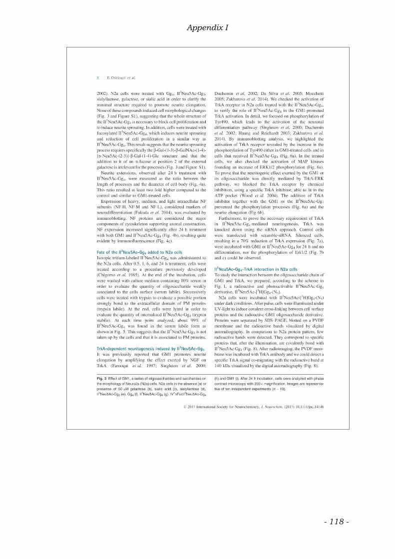
Appendix I
- 118 -

Appendix I
- 119 -

Appendix I
- 120 -

Appendix I
- 121 -
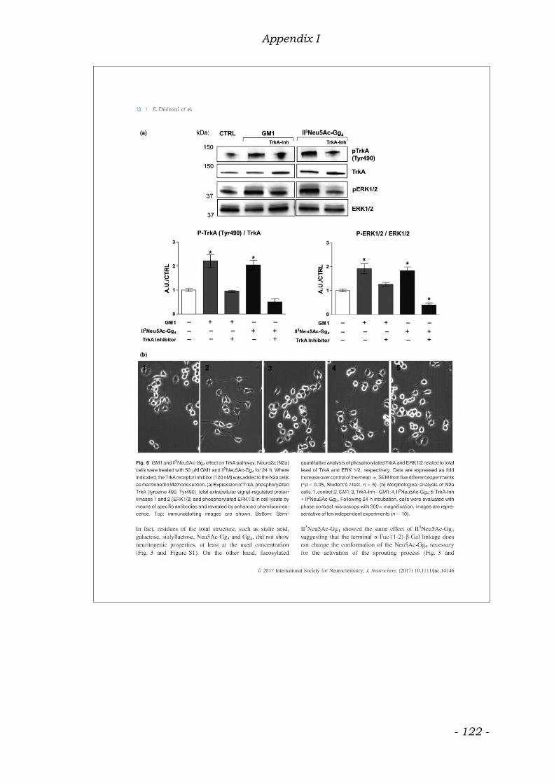
Appendix I
- 122 -

Appendix I
- 123 -

Appendix I
- 124 -

Appendix I
- 125 -

Appendix I
- 126 -
Appendix I
- 127 -