GlycoForum/GlycoScience/Science of Hyaluronan · 13/1/2010 · now focusing on the role of...
11
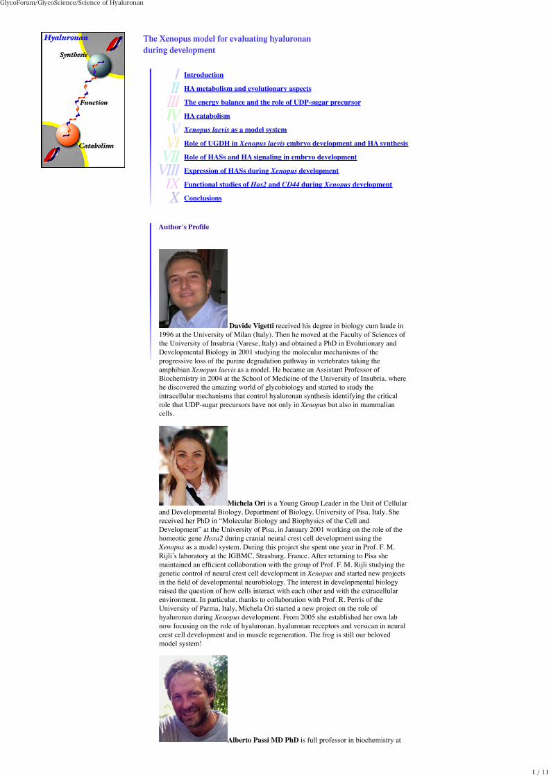
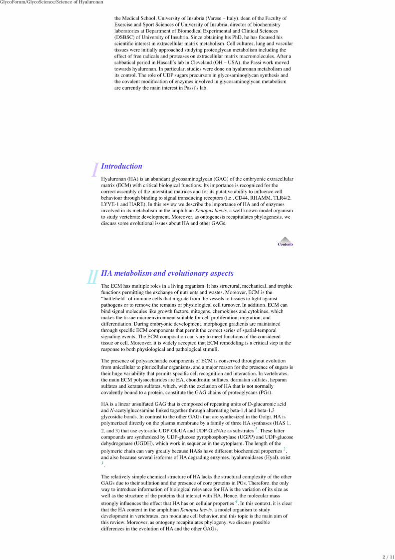
Introduction HA metabolism and evolutionary aspects The energy balance and the role of UDP-sugar precursor HA catabolism Xenopus laevis as a model system Role of UGDH in Xenopus laevis embryo development and HA synthesis Role of HASs and HA signaling in embryo development Expression of HASs during Xenopus development Functional studies of Has2 and CD44 during Xenopus development Conclusions Davide Vigetti received his degree in biology cum laude in 1996 at the University of Milan (Italy). Then he moved at the Faculty of Sciences of the University of Insubria (Varese, Italy) and obtained a PhD in Evolutionary and Developmental Biology in 2001 studying the molecular mechanisms of the progressive loss of the purine degradation pathway in vertebrates taking the amphibian Xenopus laevis as a model. He became an Assistant Professor of Biochemistry in 2004 at the School of Medicine of the University of Insubria, where he discovered the amazing world of glycobiology and started to study the intracellular mechanisms that control hyaluronan synthesis identifying the critical role that UDP-sugar precursors have not only in Xenopus but also in mammalian cells. Michela Ori is a Young Group Leader in the Unit of Cellular and Developmental Biology, Department of Biology, University of Pisa, Italy. She received her PhD in “Molecular Biology and Biophysics of the Cell and Development” at the University of Pisa, in January 2001 working on the role of the homeotic gene Hoxa2 during cranial neural crest cell development using the Xenopus as a model system. During this project she spent one year in Prof. F. M. Rijli’s laboratory at the IGBMC, Strasburg, France. After returning to Pisa she maintained an efficient collaboration with the group of Prof. F. M. Rijli studying the genetic control of neural crest cell development in Xenopus and started new projects in the field of developmental neurobiology. The interest in developmental biology raised the question of how cells interact with each other and with the extracellular environment. In particular, thanks to collaboration with Prof. R. Perris of the University of Parma, Italy, Michela Ori started a new project on the role of hyaluronan during Xenopus development. From 2005 she established her own lab now focusing on the role of hyaluronan, hyaluronan receptors and versican in neural crest cell development and in muscle regeneration. The frog is still our beloved model system! Alberto Passi MD PhD is full professor in biochemistry at GlycoForum/GlycoScience/Science of Hyaluronan 1 / 11
Transcript of GlycoForum/GlycoScience/Science of Hyaluronan · 13/1/2010 · now focusing on the role of...
Introduction
HA metabolism and evolutionary aspects
The energy balance and the role of UDP-sugar precursor
HA catabolism
Xenopus laevis as a model system
Role of UGDH in Xenopus laevis embryo development and HA synthesis
Role of HASs and HA signaling in embryo development
Expression of HASs during Xenopus development
Functional studies of Has2 and CD44 during Xenopus development
Conclusions
Davide Vigetti received his degree in biology cum laude in1996 at the University of Milan (Italy). Then he moved at the Faculty of Sciences ofthe University of Insubria (Varese, Italy) and obtained a PhD in Evolutionary andDevelopmental Biology in 2001 studying the molecular mechanisms of theprogressive loss of the purine degradation pathway in vertebrates taking theamphibian Xenopus laevis as a model. He became an Assistant Professor ofBiochemistry in 2004 at the School of Medicine of the University of Insubria, wherehe discovered the amazing world of glycobiology and started to study theintracellular mechanisms that control hyaluronan synthesis identifying the criticalrole that UDP-sugar precursors have not only in Xenopus but also in mammaliancells.
Michela Ori is a Young Group Leader in the Unit of Cellularand Developmental Biology, Department of Biology, University of Pisa, Italy. Shereceived her PhD in “Molecular Biology and Biophysics of the Cell andDevelopment” at the University of Pisa, in January 2001 working on the role of thehomeotic gene Hoxa2 during cranial neural crest cell development using theXenopus as a model system. During this project she spent one year in Prof. F. M.Rijli’s laboratory at the IGBMC, Strasburg, France. After returning to Pisa shemaintained an efficient collaboration with the group of Prof. F. M. Rijli studying thegenetic control of neural crest cell development in Xenopus and started new projectsin the field of developmental neurobiology. The interest in developmental biologyraised the question of how cells interact with each other and with the extracellularenvironment. In particular, thanks to collaboration with Prof. R. Perris of theUniversity of Parma, Italy, Michela Ori started a new project on the role ofhyaluronan during Xenopus development. From 2005 she established her own labnow focusing on the role of hyaluronan, hyaluronan receptors and versican in neuralcrest cell development and in muscle regeneration. The frog is still our belovedmodel system!
Alberto Passi MD PhD is full professor in biochemistry at
GlycoForum/GlycoScience/Science of Hyaluronan
1 / 11
the Medical School, University of Insubria (Varese – Italy), dean of the Faculty ofExercise and Sport Sciences of University of Insubria, director of biochemistrylaboratories at Department of Biomedical Experimental and Clinical Sciences(DSBSC) of University of Insubria. Since obtaining his PhD, he has focused hisscientific interest in extracellular matrix metabolism. Cell cultures, lung and vasculartissues were initially approached studying proteoglycan metabolism including theeffect of free radicals and proteases on extracellular matrix macromolecules. After asabbatical period in Hascall’s lab in Cleveland (OH – USA), the Passi work movedtowards hyaluronan. In particular, studies were done on hyaluronan metabolism andits control. The role of UDP sugars precursors in glycosaminoglycan synthesis andthe covalent modification of enzymes involved in glycosaminoglycan metabolismare currently the main interest in Passi’s lab.
IntroductionHyaluronan (HA) is an abundant glycosaminoglycan (GAG) of the embryonic extracellularmatrix (ECM) with critical biological functions. Its importance is recognized for thecorrect assembly of the interstitial matrices and for its putative ability to influence cellbehaviour through binding to signal transducing receptors (i.e., CD44, RHAMM, TLR4/2,LYVE-1 and HARE). In this review we describe the importance of HA and of enzymesinvolved in its metabolism in the amphibian Xenopus laevis, a well known model organismto study vertebrate development. Moreover, as ontogenesis recapitulates phylogenesis, wediscuss some evolutional issues about HA and other GAGs.
HA metabolism and evolutionary aspectsThe ECM has multiple roles in a living organism. It has structural, mechanical, and trophicfunctions permitting the exchange of nutrients and wastes. Moreover, ECM is the“battlefield” of immune cells that migrate from the vessels to tissues to fight againstpathogens or to remove the remains of physiological cell turnover. In addition, ECM canbind signal molecules like growth factors, mitogens, chemokines and cytokines, whichmakes the tissue microenvironment suitable for cell proliferation, migration, anddifferentiation. During embryonic development, morphogen gradients are maintainedthrough specific ECM components that permit the correct series of spatial-temporalsignaling events. The ECM composition can vary to meet functions of the consideredtissue or cell. Moreover, it is widely accepted that ECM remodeling is a critical step in theresponse to both physiological and pathological stimuli.
The presence of polysaccharide components of ECM is conserved throughout evolutionfrom unicellular to pluricellular organisms, and a major reason for the presence of sugars istheir huge variability that permits specific cell recognition and interaction. In vertebrates,the main ECM polysaccharides are HA, chondroitin sulfates, dermatan sulfates, heparansulfates and keratan sulfates, which, with the exclusion of HA that is not normallycovalently bound to a protein, constitute the GAG chains of proteoglycans (PGs).
HA is a linear unsulfated GAG that is composed of repeating units of D-glucuronic acidand N-acetylglucosamine linked together through alternating beta-1,4 and beta-1,3glycosidic bonds. In contrast to the other GAGs that are synthesized in the Golgi, HA ispolymerized directly on the plasma membrane by a family of three HA synthases (HAS 1,2, and 3) that use cytosolic UDP-GlcUA and UDP-GlcNAc as substrates 1. These lattercompounds are synthesized by UDP-glucose pyrophosphorylase (UGPP) and UDP-glucosedehydrogenase (UGDH), which work in sequence in the cytoplasm. The length of thepolymeric chain can vary greatly because HASs have different biochemical properties 2,and also because several isoforms of HA degrading enzymes, hyaluronidases (Hyal), exist3.
The relatively simple chemical structure of HA lacks the structural complexity of the otherGAGs due to their sulfation and the presence of core proteins in PGs. Therefore, the onlyway to introduce information of biological relevance for HA is the variation of its size aswell as the structure of the proteins that interact with HA. Hence, the molecular massstrongly influences the effect that HA has on cellular properties 4. In this context, it is clearthat the HA content in the amphibian Xenopus laevis, a model organism to studydevelopment in vertebrates, can modulate cell behavior, and this topic is the main aim ofthis review. Moreover, as ontogeny recapitulates phylogeny, we discuss possibledifferences in the evolution of HA and the other GAGs.
GlycoForum/GlycoScience/Science of Hyaluronan
2 / 11
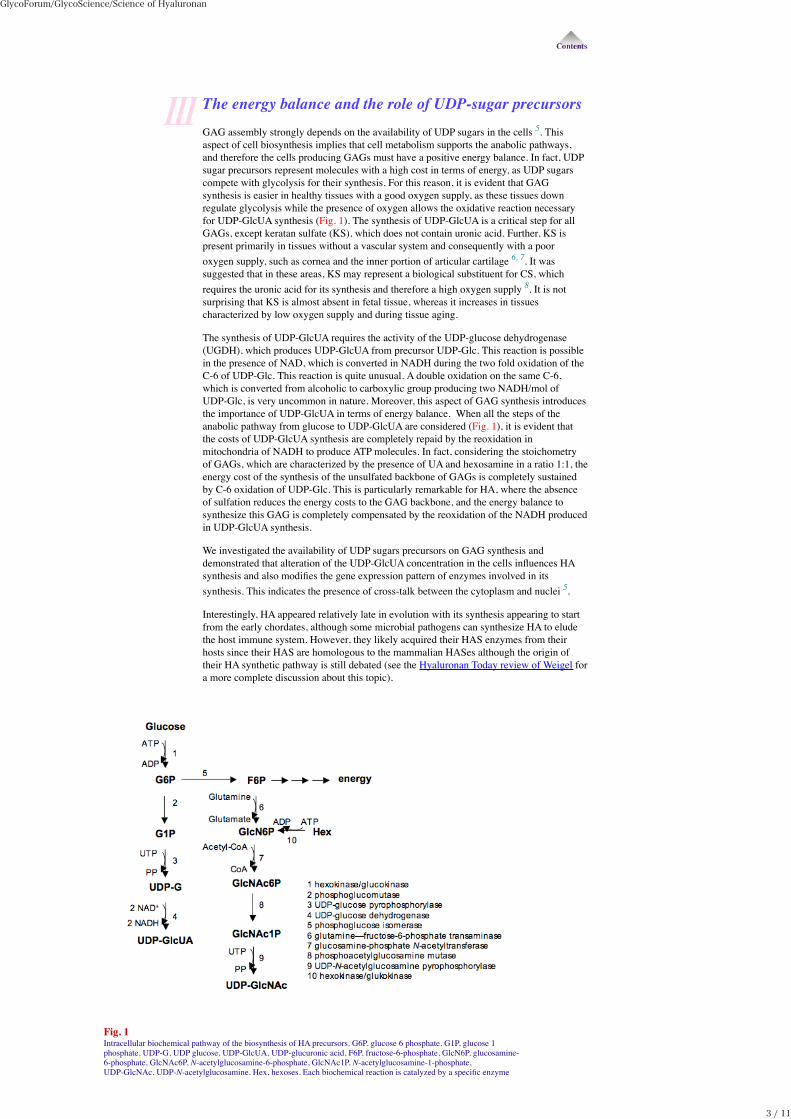
The energy balance and the role of UDP-sugar precursorsGAG assembly strongly depends on the availability of UDP sugars in the cells 5. Thisaspect of cell biosynthesis implies that cell metabolism supports the anabolic pathways,and therefore the cells producing GAGs must have a positive energy balance. In fact, UDPsugar precursors represent molecules with a high cost in terms of energy, as UDP sugarscompete with glycolysis for their synthesis. For this reason, it is evident that GAGsynthesis is easier in healthy tissues with a good oxygen supply, as these tissues downregulate glycolysis while the presence of oxygen allows the oxidative reaction necessaryfor UDP-GlcUA synthesis (Fig. 1). The synthesis of UDP-GlcUA is a critical step for allGAGs, except keratan sulfate (KS), which does not contain uronic acid. Further, KS ispresent primarily in tissues without a vascular system and consequently with a pooroxygen supply, such as cornea and the inner portion of articular cartilage 6, 7. It wassuggested that in these areas, KS may represent a biological substituent for CS, whichrequires the uronic acid for its synthesis and therefore a high oxygen supply 8. It is notsurprising that KS is almost absent in fetal tissue, whereas it increases in tissuescharacterized by low oxygen supply and during tissue aging.
The synthesis of UDP-GlcUA requires the activity of the UDP-glucose dehydrogenase(UGDH), which produces UDP-GlcUA from precursor UDP-Glc. This reaction is possiblein the presence of NAD, which is converted in NADH during the two fold oxidation of theC-6 of UDP-Glc. This reaction is quite unusual. A double oxidation on the same C-6,which is converted from alcoholic to carboxylic group producing two NADH/mol ofUDP-Glc, is very uncommon in nature. Moreover, this aspect of GAG synthesis introducesthe importance of UDP-GlcUA in terms of energy balance. When all the steps of theanabolic pathway from glucose to UDP-GlcUA are considered (Fig. 1), it is evident thatthe costs of UDP-GlcUA synthesis are completely repaid by the reoxidation inmitochondria of NADH to produce ATP molecules. In fact, considering the stoichometryof GAGs, which are characterized by the presence of UA and hexosamine in a ratio 1:1, theenergy cost of the synthesis of the unsulfated backbone of GAGs is completely sustainedby C-6 oxidation of UDP-Glc. This is particularly remarkable for HA, where the absenceof sulfation reduces the energy costs to the GAG backbone, and the energy balance tosynthesize this GAG is completely compensated by the reoxidation of the NADH producedin UDP-GlcUA synthesis.
We investigated the availability of UDP sugars precursors on GAG synthesis anddemonstrated that alteration of the UDP-GlcUA concentration in the cells influences HAsynthesis and also modifies the gene expression pattern of enzymes involved in itssynthesis. This indicates the presence of cross-talk between the cytoplasm and nuclei 5.
Interestingly, HA appeared relatively late in evolution with its synthesis appearing to startfrom the early chordates, although some microbial pathogens can synthesize HA to eludethe host immune system. However, they likely acquired their HAS enzymes from theirhosts since their HAS are homologous to the mammalian HASes although the origin oftheir HA synthetic pathway is still debated (see the Hyaluronan Today review of Weigel fora more complete discussion about this topic).
Fig. 1Intracellular biochemical pathway of the biosynthesis of HA precursors. G6P, glucose 6 phosphate, G1P, glucose 1phosphate, UDP-G, UDP glucose, UDP-GlcUA, UDP-glucuronic acid, F6P, fructose-6-phosphate, GlcN6P, glucosamine-6-phosphate, GlcNAc6P, N-acetylglucosamine-6-phosphate, GlcNAc1P, N-acetylglucosamine-1-phosphate,UDP-GlcNAc, UDP-N-acetylglucosamine. Hex, hexoses. Each biochemical reaction is catalyzed by a specific enzyme
GlycoForum/GlycoScience/Science of Hyaluronan
3 / 11
reported as numbered.
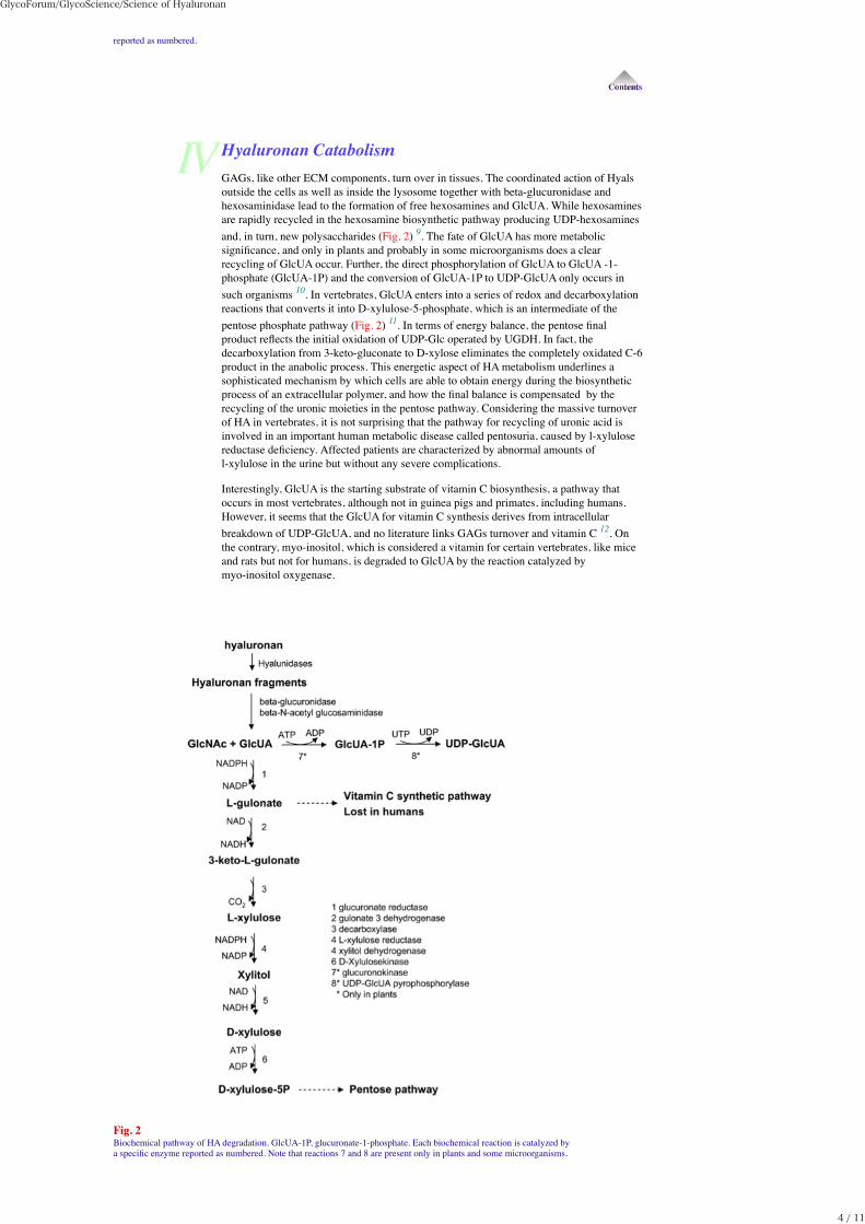
Hyaluronan CatabolismGAGs, like other ECM components, turn over in tissues. The coordinated action of Hyalsoutside the cells as well as inside the lysosome together with beta-glucuronidase andhexosaminidase lead to the formation of free hexosamines and GlcUA. While hexosaminesare rapidly recycled in the hexosamine biosynthetic pathway producing UDP-hexosaminesand, in turn, new polysaccharides (Fig. 2) 9. The fate of GlcUA has more metabolicsignificance, and only in plants and probably in some microorganisms does a clearrecycling of GlcUA occur. Further, the direct phosphorylation of GlcUA to GlcUA -1-phosphate (GlcUA-1P) and the conversion of GlcUA-1P to UDP-GlcUA only occurs insuch organisms 10. In vertebrates, GlcUA enters into a series of redox and decarboxylationreactions that converts it into D-xylulose-5-phosphate, which is an intermediate of thepentose phosphate pathway (Fig. 2) 11. In terms of energy balance, the pentose finalproduct reflects the initial oxidation of UDP-Glc operated by UGDH. In fact, thedecarboxylation from 3-keto-gluconate to D-xylose eliminates the completely oxidated C-6product in the anabolic process. This energetic aspect of HA metabolism underlines asophisticated mechanism by which cells are able to obtain energy during the biosyntheticprocess of an extracellular polymer, and how the final balance is compensated by therecycling of the uronic moieties in the pentose pathway. Considering the massive turnoverof HA in vertebrates, it is not surprising that the pathway for recycling of uronic acid isinvolved in an important human metabolic disease called pentosuria, caused by l-xylulosereductase deficiency. Affected patients are characterized by abnormal amounts ofl-xylulose in the urine but without any severe complications.
Interestingly, GlcUA is the starting substrate of vitamin C biosynthesis, a pathway thatoccurs in most vertebrates, although not in guinea pigs and primates, including humans.However, it seems that the GlcUA for vitamin C synthesis derives from intracellularbreakdown of UDP-GlcUA, and no literature links GAGs turnover and vitamin C 12. Onthe contrary, myo-inositol, which is considered a vitamin for certain vertebrates, like miceand rats but not for humans, is degraded to GlcUA by the reaction catalyzed bymyo-inositol oxygenase.
Fig. 2Biochemical pathway of HA degradation. GlcUA-1P, glucuronate-1-phosphate. Each biochemical reaction is catalyzed bya specific enzyme reported as numbered. Note that reactions 7 and 8 are present only in plants and some microorganisms.
GlycoForum/GlycoScience/Science of Hyaluronan
4 / 11
Xenopus leavis as model systemWhile researchers might argue the advantages of their particular model system, it isimportant to realize that complementary experimental approaches in different systems havegreatly increased our progress in understanding the overall processes occurring duringembryogenesis.
For many decades Amphibia were a favorite model system of embryologists due to thelarge size of the eggs, the large number of embryos, which develop outside of the mother,and the fact that the embryos exhibit a remarkable ability to heal after microsurgery. Onedisadvantage of traditional amphibian species is that they are seasonal breeders. Thismeant that investigators could not do their experiments throughout the year. Xenopuslaevis, the South African clawed frog, is a notable exception. Its ability to spawn wheninjected with gonadotropic hormone led to its common usage for human pregnancy tests inthe 1950s where an injection of pregnancy urine, which contains chorionic gonadotropin,would induce spawning. This led investigators to consider its use in experimentalembryology.
Xenopus embryos develop outside of the mother, which makes themextremely well suitedfor manipulations of gene activity via microinjection. Typically microinjection studies areused to examine the function of a gene of interest by overexpression of the wild type, or amutant sequence of the gene, or by loss of function approaches. Furthermore, theextremely reproducible cleavage patterns of the frog embryo and the availability ofdetailed fate mapping studies facilitate targeting of injected material to restricted lineagesof the developing embryo.
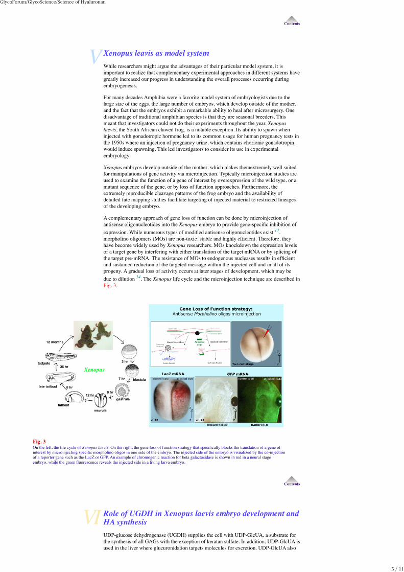
A complementary approach of gene loss of function can be done by microinjection ofantisense oligonucleotides into the Xenopus embryo to provide gene-specific inhibition ofexpression. While numerous types of modified antisense oligonucleotides exist 13,morpholino oligomers (MOs) are non-toxic, stable and highly efficient. Therefore, theyhave become widely used by Xenopus researchers. MOs knockdown the expression levelsof a target gene by interfering with either translation of the target mRNA or by splicing ofthe target pre-mRNA. The resistance of MOs to endogenous nucleases results in efficientand sustained reduction of the targeted message within the injected cell and in all of itsprogeny. A gradual loss of activity occurs at later stages of development, which may bedue to dilution 14. The Xenopus life cycle and the microinjection technique are described inFig. 3.
Fig. 3On the left, the life cycle of Xenopus laevis. On the right, the gene loss of function strategy that specifically blocks the translation of a gene ofinterest by microinjecting specific morpholino oligos in one side of the embryo. The injected side of the embryo is visualized by the co-injectionof a reporter gene such as the LacZ or GFP. An example of chromogenic reaction for beta galactosidase is shown in red in a neural stageembryo, while the green fluorescence reveals the injected side in a living larva embryo.
Role of UGDH in Xenopus laevis embryo development andHA synthesisUDP-glucose dehydrogenase (UGDH) supplies the cell with UDP-GlcUA, a substrate forthe synthesis of all GAGs with the exception of keratan sulfate. In addition, UDP-GlcUA isused in the liver where glucuronidation targets molecules for excretion. UDP-GlcUA also
GlycoForum/GlycoScience/Science of Hyaluronan
5 / 11
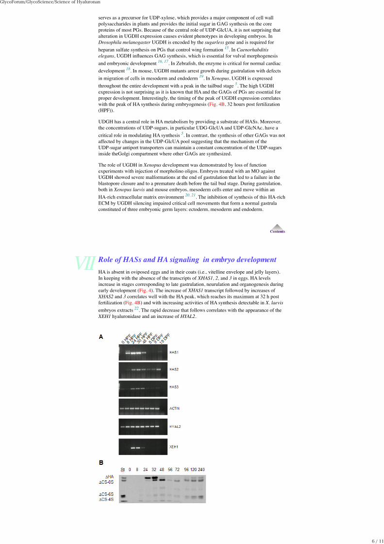
serves as a precursor for UDP-xylose, which provides a major component of cell wallpolysaccharides in plants and provides the initial sugar in GAG synthesis on the coreproteins of most PGs. Because of the central role of UDP-GlcUA, it is not surprising thatalteration in UGDH expression causes evident phenotypes in developing embryos. InDrosophila melanogaster UGDH is encoded by the sugarless gene and is required forheparan sulfate synthesis on PGs that control wing formation 15. In Caenorhabditiselegans, UGDH influences GAG synthesis, which is essential for vulval morphogenesisand embryonic development 16, 17. In Zebrafish, the enzyme is critical for normal cardiacdevelopment 18. In mouse, UGDH mutants arrest growth during gastrulation with defectsin migration of cells in mesoderm and endoderm 19. In Xenopus, UGDH is expressedthroughout the entire development with a peak in the tailbud stage 5. The high UGDHexpression is not surprising as it is known that HA and the GAGs of PGs are essential forproper development. Interestingly, the timing of the peak of UGDH expression correlateswith the peak of HA synthesis during embryogenesis (Fig. 4B, 32 hours post fertilization(HPF)).
UDGH has a central role in HA metabolism by providing a substrate of HASs. Moreover,the concentrations of UDP-sugars, in particular UDG-GlcUA and UDP-GlcNAc, have acritical role in modulating HA synthesis 5. In contrast, the synthesis of other GAGs was notaffected by changes in the UDP-GlcUA pool suggesting that the mechanism of theUDP-sugar antiport transporters can maintain a constant concentration of the UDP-sugarsinside theGolgi compartment where other GAGs are synthesized.
The role of UGDH in Xenopus development was demonstrated by loss of functionexperiments with injection of morpholino oligos. Embryos treated with an MO againstUGDH showed severe malformations at the end of gastrulation that led to a failure in theblastopore closure and to a premature death before the tail bud stage. During gastrulation,both in Xenopus laevis and mouse embryos, mesoderm cells enter and move within anHA-rich extracellular matrix environment 20, 21. The inhibition of synthesis of this HA-richECM by UGDH silencing impaired critical cell movements that form a normal gastrulaconstituted of three embryonic germ layers: ectoderm, mesoderm and endoderm.
Role of HASs and HA signaling in embryo developmentHA is absent in oviposed eggs and in their coats (i.e., vitelline envelope and jelly layers).In keeping with the absence of the transcripts of XHAS1, 2, and 3 in eggs. HA levelsincrease in stages corresponding to late gastrulation, neurulation and organogenesis duringearly development (Fig. 4). The increase of XHAS1 transcript followed by increases ofXHAS2 and 3 correlates well with the HA peak, which reaches its maximum at 32 h postfertilization (Fig. 4B) and with increasing activities of HA synthesis detectable in X. laevisembryos extracts 22. The rapid decrease that follows correlates with the appearance of theXEH1 hyaluronidase and an increase of HYAL2.
GlycoForum/GlycoScience/Science of Hyaluronan
6 / 11
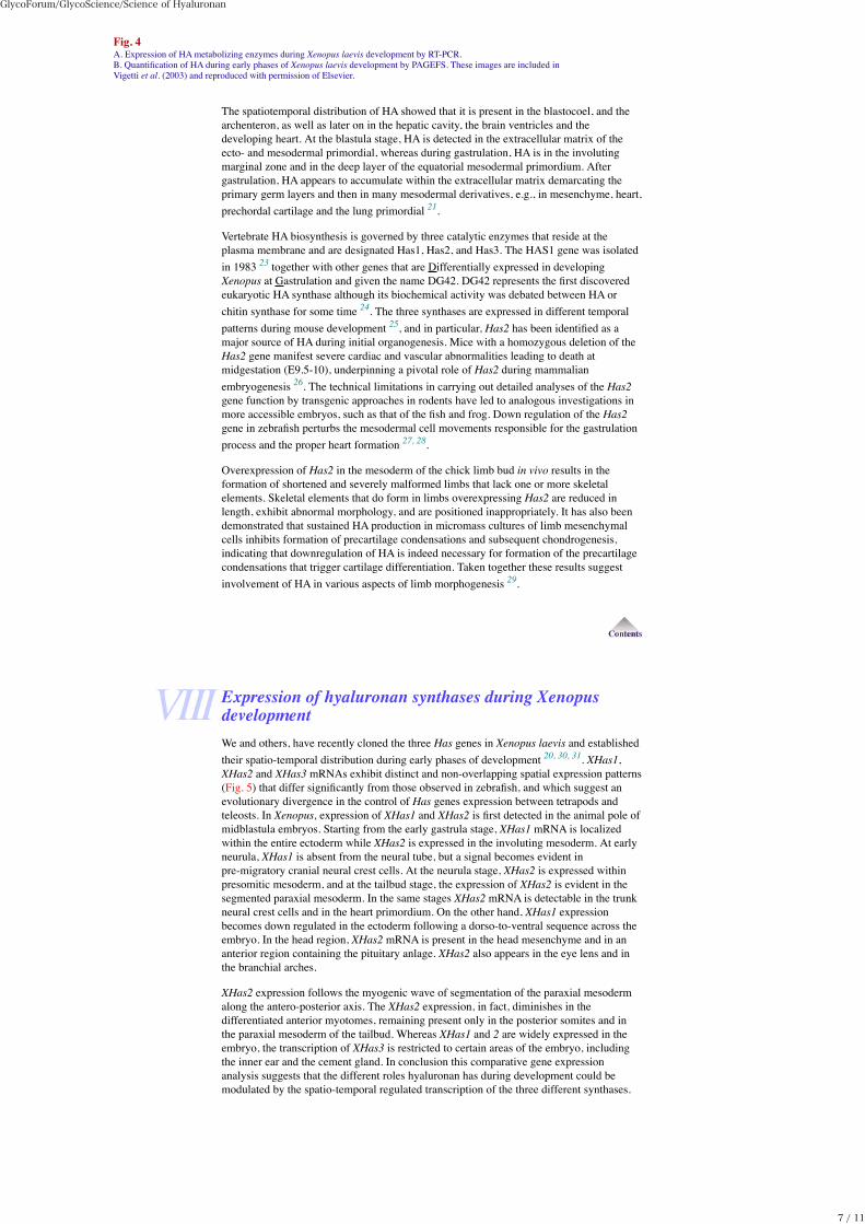
Fig. 4A. Expression of HA metabolizing enzymes during Xenopus laevis development by RT-PCR.B. Quantification of HA during early phases of Xenopus laevis development by PAGEFS. These images are included inVigetti et al. (2003) and reproduced with permission of Elsevier.
The spatiotemporal distribution of HA showed that it is present in the blastocoel, and thearchenteron, as well as later on in the hepatic cavity, the brain ventricles and thedeveloping heart. At the blastula stage, HA is detected in the extracellular matrix of theecto- and mesodermal primordial, whereas during gastrulation, HA is in the involutingmarginal zone and in the deep layer of the equatorial mesodermal primordium. Aftergastrulation, HA appears to accumulate within the extracellular matrix demarcating theprimary germ layers and then in many mesodermal derivatives, e.g., in mesenchyme, heart,prechordal cartilage and the lung primordial 21.
Vertebrate HA biosynthesis is governed by three catalytic enzymes that reside at theplasma membrane and are designated Has1, Has2, and Has3. The HAS1 gene was isolatedin 1983 23 together with other genes that are Differentially expressed in developingXenopus at Gastrulation and given the name DG42. DG42 represents the first discovered eukaryotic HA synthase although its biochemical activity was debated between HA orchitin synthase for some time 24. The three synthases are expressed in different temporalpatterns during mouse development 25, and in particular, Has2 has been identified as amajor source of HA during initial organogenesis. Mice with a homozygous deletion of theHas2 gene manifest severe cardiac and vascular abnormalities leading to death atmidgestation (E9.5-10), underpinning a pivotal role of Has2 during mammalianembryogenesis 26. The technical limitations in carrying out detailed analyses of the Has2gene function by transgenic approaches in rodents have led to analogous investigations inmore accessible embryos, such as that of the fish and frog. Down regulation of the Has2gene in zebrafish perturbs the mesodermal cell movements responsible for the gastrulationprocess and the proper heart formation 27, 28.
Overexpression of Has2 in the mesoderm of the chick limb bud in vivo results in theformation of shortened and severely malformed limbs that lack one or more skeletalelements. Skeletal elements that do form in limbs overexpressing Has2 are reduced inlength, exhibit abnormal morphology, and are positioned inappropriately. It has also beendemonstrated that sustained HA production in micromass cultures of limb mesenchymalcells inhibits formation of precartilage condensations and subsequent chondrogenesis,indicating that downregulation of HA is indeed necessary for formation of the precartilagecondensations that trigger cartilage differentiation. Taken together these results suggestinvolvement of HA in various aspects of limb morphogenesis 29.
Expression of hyaluronan synthases during XenopusdevelopmentWe and others, have recently cloned the three Has genes in Xenopus laevis and establishedtheir spatio-temporal distribution during early phases of development 20, 30, 31. XHas1,XHas2 and XHas3 mRNAs exhibit distinct and non-overlapping spatial expression patterns(Fig. 5) that differ significantly from those observed in zebrafish, and which suggest anevolutionary divergence in the control of Has genes expression between tetrapods andteleosts. In Xenopus, expression of XHas1 and XHas2 is first detected in the animal pole ofmidblastula embryos. Starting from the early gastrula stage, XHas1 mRNA is localizedwithin the entire ectoderm while XHas2 is expressed in the involuting mesoderm. At earlyneurula, XHas1 is absent from the neural tube, but a signal becomes evident inpre-migratory cranial neural crest cells. At the neurula stage, XHas2 is expressed withinpresomitic mesoderm, and at the tailbud stage, the expression of XHas2 is evident in thesegmented paraxial mesoderm. In the same stages XHas2 mRNA is detectable in the trunkneural crest cells and in the heart primordium. On the other hand, XHas1 expressionbecomes down regulated in the ectoderm following a dorso-to-ventral sequence across theembryo. In the head region, XHas2 mRNA is present in the head mesenchyme and in ananterior region containing the pituitary anlage. XHas2 also appears in the eye lens and inthe branchial arches.
XHas2 expression follows the myogenic wave of segmentation of the paraxial mesodermalong the antero-posterior axis. The XHas2 expression, in fact, diminishes in thedifferentiated anterior myotomes, remaining present only in the posterior somites and inthe paraxial mesoderm of the tailbud. Whereas XHas1 and 2 are widely expressed in theembryo, the transcription of XHas3 is restricted to certain areas of the embryo, includingthe inner ear and the cement gland. In conclusion this comparative gene expressionanalysis suggests that the different roles hyaluronan has during development could bemodulated by the spatio-temporal regulated transcription of the three different synthases.
GlycoForum/GlycoScience/Science of Hyaluronan
7 / 11
Fig. 5Schematic representation for the gene expression patterns of the three HA synthases during Xenopus embryodevelopment.
Functional studies of Has2 and CD44 during XenopusdevelopmentThe early lethality of Has2 knock out mouse embryos leaves the precise role of HA duringembryogenesis partially unresolved. To approach this problem we pursued a loss offunction study that allows us to delineate the XHas2 role in early developmental processes32. Tailbud Xenopus embryos that received a unilateral injection of XHas2 morpholino atthe two-cell stage failed to correctly organize their flank musculature. The embryos hadsmaller somites, and the segmental organization of the myotomes appeared disrupted.These observations raised the possibility that XHas2 loss of function could affect thestructural integrity of the inter- and intra-somitic extracellular matrix, which in turn couldinterfere with the establishment of correct cell-cell contacts between the formingmyoblasts.
As XHas2 is expressed in presomitic cells, we investigated whether the loss of Has2activity actually interferes with the early phases of the myogenic differentiation program.At the neurula stage, we observed a significant increase in the number of apoptotic cells inthe presomitic mesoderm while myoblast proliferation and the expression of the precociousmuscle markers appeared unaffected. These data strongly suggested that XHas2 activity isnot essential for the early myogenic commitment or myoblast proliferation, but could berequired for the survival and differentiation of the early muscle precursors. Indeed, the roleof XHas2 in preventing these cells from undergoing apoptosis seems to be restricted to aspecific developmental period, which coincides with the early steps of myogenicdifferentiation in the presomitic mesoderm. Proper assembly of the mesodermal ECM inthe presence of HA may be important for the maintenance of community effects, for properdiffusion/gradient formation of morphogens, or for undisturbed action of other instructivesignals involved in the differentiation and survival of developing myoblasts.
The effect of HA on survival and differentiation of presomitic mesodermal cells could beexerted through a structural supportive effect of the ECM. A more intriguing possibility isthat HA produced by myoblast cells could activate intracellular signals via binding tospecific receptors such as CD44 or RHAMM. To further investigate this aspect, weperformed functional knock down of XCD44. In Xenopus this receptor is expressed in thedeveloping musculature, but its deletion did not produce significant alteration in theformation of trunk muscle. Conversely, both XHas2 and XCD44 are involved in hypaxialmyoblast migration, which in Xenopus is required for the proper formation of the ventralbody wall musculature. The expression of XHas2 and XCD44 in this precursor cellpopulation suggests that the impaired migration may, at least in part, be determined by thefailure of hypaxial cells to contribute with their proper HA during the migratory processand that XCD44 function may be required for cell locomotion. It has been shown inzebrafish that Has2 is required to activate Rac1 signalling cascades that govern dorsal cellmigration during gastrulation and adaxial cell migration in the forming somites, allowingfor the hypothesis of an HA instructive role in inducing cells to migrate 27. We furthersuggest that, at least for hypaxial muscle cells, this HA role may be mediated by XCD44activation 32. Another interesting phenotype, observed in XHas2-deprived embryos, is analtered trunk NCC migration, corroborating a proposed pivotal role of HA during NCCdevelopment 33. It is noteworthy that Xenopus migrating trunk NCCs do not expressXCD44, and that depletion of the receptor in somitic cells does not influence the NCCmovements. Although the presence of HA in the extracellular environment seems to becrucial for allowing proper NCC migration, it is not clear if the impaired movement oftrunk NCCs observed in XHas2-depleted embryos was due to a failure of these cells tosynthesize and secrete HA, or to a structural deficit caused by its loss from the ECM of theNCC migratory pathways. An alternative possibility could be that the altered NCCmigration could result from a morphogenetic loss of the structural integrity of the somites
GlycoForum/GlycoScience/Science of Hyaluronan
8 / 11
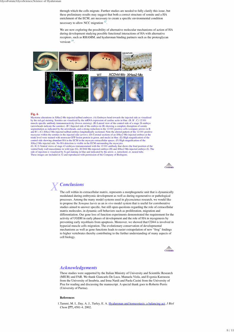
through which the cells migrate. Further studies are needed to fully clarify this issue, butthese preliminary results may suggest that both a correct structure of somite and a HAenrichment of the ECM, are necessary to create a specific environmental conditionnecessary to allow NCC migration 32.
We are now exploring the possibility of alternative molecular mechanisms of action of HAduring development studying possible functional interactions of HA with alternativereceptors, such as RHAMM, and hyaluronan binding partners such as the proteoglycanversican 34.
Fig. 6Myotome alterations in XHas2-Mo injected tailbud embryos. (A) Embryos bend towards the injected side as visualizedby the red-gal staining. Somites are visualized by the mRNA expression of cardiac actin in blue. (B, B’, C) 12/101muscle specific antibody immunoreactivity (brown staining). (B) Lateral view of the control side of a stage 26 embryo(arrowheads indicate the somites). (B’) Injected side of the embryo in (B) showing a complete disruption of somitesegmentation as indicated by the arrowheads, and a strong reduction in the 12/101 positive cells (compare arrows in Band B’). (C) XHas2-Mo injected tailbud embryo longitudinally sectioned. Note the altered pattern of the 12/101 positivemyocytes within the somites in the injected side (arrow). (D) Coronal sections of an XHas2-Mo injected embryo at thetrunk level were stained with neurocan-GFP fusion protein in green, and nuclei in blue. (E) High magnification of thecontrol side showing abundant HA in the ECM in the myocyte extracellular spaces. (F) High magnification of theXHas2-Mo injected side. No HA detection is visible in the ECM surrounding the myocytes.(G, H, I) Ventral views of stage 43 embryos immunostained with the 12/101 antibody that shows the final position of theventral body wall musculature in wild type (G), XCD44-Mo injected embryo (H) and XHas2-Mo injected embryo (I). Theside of injection is visualized by X-gal staining in blue and indicated by the arrow. n, notochord, nt, neural tube.These images are included in 32 and reproduced with permission of the Company of Biologists.
ConclusionsThe cell within its extracellular matrix, represents a morphogenetic unit that is dynamicallymodulated during embryonic development as well as during regenerative or pathologicalprocesses. Among the many model systems used in glycoscience research, we would liketo propose the Xenopus laevis as an in vivo model system that is useful for corroborativestudies aimed to answer specific, but still open questions regarding the role of extracellularmatrix molecules, in dynamic cell behaviors such as proliferation, migration anddifferentiation. Our gene loss of function experiments demonstrated the requirement for theactivity of UGDH in early phases of development and the role of HA in myogenesis bypreventing early myoblasts from apoptosis. Moreover, we showed that CD44 is involved inhypaxial muscle cells migration. The evolutionary conservation of developmentalmechanisms as well as gene functions leads to easier extrapolation of new “frog” findingsto higher vertebrates thereby contributing to the further understanding of many aspects ofcell biology.
AcknowledgementsThese studies were supported by the Italian Ministry of University and Scientific Research(MIUR) and FAR. We thank Giancarlo De Luca, Manuela Viola, and Evgenia Karousoufrom the University of Insubria, and Irma Nardi and Paola Casini from the University ofPisa for reading and discussing the manuscript. A special thank goes to Roberto Perris(University of Parma).
References
1.Tammi, M. I., Day, A. J., Turley, E. A. Hyaluronan and homeostasis: a balancing act. J BiolChem 277, 4581-4, 2002.
GlycoForum/GlycoScience/Science of Hyaluronan
9 / 11
2.Itano, N., Sawai, T., Yoshida, M., Lenas, P., Yamada, Y., Imagawa, M., Shinomura, T.,Hamaguchi, M., Yoshida, Y., Ohnuki, Y., Miyauchi, S., Spicer, A. P., McDonald, J. A.,Kimata, K. Three isoforms of mammalian hyaluronan synthases have distinct enzymaticproperties. J Biol Chem, 274, 25085-92,1999.
3.Stern, R., Jedrzejas, M. J. Hyaluronidases: their genomics, structures, and mechanisms ofaction. Chem Rev 106, 818-39, 2006.
4.Asari, A. Novel Functions of Hyaluronan Oligosaccharides. Science of Hyaluronan Today,2005.
5.Vigetti, D., Ori, M., Viola, M., Genasetti, A., Karousou, E., Rizzi, M., Pallotti, F., Nardi, I.,Hascall, V. C., De Luca, G., Passi, A. Molecular cloning and characterization ofUDP-glucose dehydrogenase from the amphibian Xenopus laevis and its involvement inhyaluronan synthesis. J Biol Chem 281, 8254-63, 2006.
6.Balduini, C., De Luca, G., Passi, A., Rindi, S., Salvini, R., Scott, J. E. Effect of oxygentension and lactate concentration on keratan sulphate and chondroitin sulphate biosynthesisin bovine cornea. Biochim Biophys Acta 1115, 187-91, 1992.
7.De Luca, G., Speziale, P., Balduini, C., Castellani, A. A. Biosynthesis ofglycosaminoglycans: uridine diphosphate glucose 4'-epimerase from cornea andepiphysial-plate cartilage. Connect Tissue Res 3, 39-47, 1975.
8.Scott, J. E., Haigh, M. Keratan sulphate and the ultrastructure of cornea and cartilage: a'stand-in' for chondroitin sulphate in conditions of oxygen lack? J Anat 158, 95-108, 1988.
9.MacNicholl, A. D., Wusteman, F. S., Winterburn, P. J., Powell, G. M., Curtis, C. G.Degradation of [3H]chondroitin 4-sulphate and re-utilization of the [3H]hexosaminecomponent by the isolated perfused rat liver. Biochem J 186, 279-86, 1980.
10.Feingold, D. S., Avigad, G. Sugar nucleotide transformations in plants. In TheBiochemistry of Plants: A Comprehensive Treatise, Vol. 3, Stumpf, P. K., Conn, E. E., Eds.Academic Press New York, USA, pp 101–170, 1980.
11.Hiatt, H. H. Pentosuria. In The Metabolic and Molecular Bases of Inherited DiseaseScriver, C. R., Beaudet, A. L., Sly, W. S., Valle, D., Eds. McGraw-Hill, New York , USA,Vol. Vol 1, pp 1589 – 1599, 2001.
12.Linster, C. L., Van Schaftingen, E. Vitamin C. Biosynthesis, recycling and degradation inmammals. Febs J 274, 1-22, 2007.
13.Dagle, J. M., Weeks, D. L. Oligonucleotide-based strategies to reduce gene expression.Differentiation 69, 75-82, 2001.
14.Heasman, J. Morpholino oligos: making sense of antisense? Dev Biol 243, 209-14, 2002.15.Hacker, U., Lin, X., Perrimon, N. The Drosophila sugarless gene modulates Wingless
signaling and encodes an enzyme involved in polysaccharide biosynthesis. Development,124, 3565-73, 1997.
16.Hwang, H. Y., Olson, S. K., Esko, J. D., Horvitz, H. R. Caenorhabditis elegans earlyembryogenesis and vulval morphogenesis require chondroitin biosynthesis. Nature 423,439-43, 2003.
17.Hwang, H. Y., Olson, S. K., Brown, J. R., Esko, J. D., Horvitz, H. R. The Caenorhabditiselegans genes sqv-2 and sqv-6, which are required for vulval morphogenesis, encodeglycosaminoglycan galactosyltransferase II and xylosyltransferase. J Biol Chem, 27811735-8, 2003.
18.Walsh, E. C., Stainier, D. Y. UDP-glucose dehydrogenase required for cardiac valveformation in zebrafish. Science 293, 1670-3, 2001.
19.Garcia-Garcia, M. J., Anderson, K. V. Essential role of glycosaminoglycans in Fgfsignaling during mouse gastrulation. Cell 114, 727-37, 2003.
20.Koprunner, M., Mullegger, J., Lepperdinger, G. Synthesis of hyaluronan of distinctlydifferent chain length is regulated by differential expression of Xhas1 and 2 during earlydevelopment of Xenopus laevis. Mech Dev 90, 275-8, 2000.
21.Mullegger, J., Lepperdinger, G. Hyaluronan is an abundant constituent of the extracellularmatrix of Xenopus embryos. Mol Reprod Dev 61, 312-6, 2002.
22.Meyer, M. F., Kreil, G. Cells expressing the DG42 gene from early Xenopus embryossynthesize hyaluronan. Proc Natl Acad Sci U S A 93, 4543-7, 1996.
23.Sargent, T. D., Dawid, I. B. Differential gene expression in the gastrula of Xenopus laevis.Science 222, 135-9, 1983.
24.Varki, A. Does DG42 synthesize hyaluronan or chitin?: A controversy aboutoligosaccharides in vertebrate development. Proc Natl Acad Sci U S A 93, 4523-5, 1996.
25.Tien, J. Y., Spicer, A. P. Three vertebrate hyaluronan synthases are expressed during mousedevelopment in distinct spatial and temporal patterns. Dev Dyn 233, 130-41, 2005.
26.Camenisch, T. D., Spicer, A. P., Brehm-Gibson, T., Biesterfeldt, J., Augustine, M. L.,Calabro, A., Jr., Kubalak, S., Klewer, S. E., McDonald, J. A. Disruption of hyaluronansynthase-2 abrogates normal cardiac morphogenesis and hyaluronan-mediatedtransformation of epithelium to mesenchyme. J Clin Invest 106, 349-60, 2000.
27.Bakkers, J., Kramer, C., Pothof, J., Quaedvlieg, N. E., Spaink, H. P., Hammerschmidt, M.Has2 is required upstream of Rac1 to govern dorsal migration of lateral cells duringzebrafish gastrulation. Development 131, 525-37, 2004.
28.Smith, K. A., Chocron, S., von der Hardt, S., de Pater, E., Soufan, A., Bussmann, J.,Schulte-Merker, S., Hammerschmidt, M., Bakkers, J. Rotation and asymmetricdevelopment of the zebrafish heart requires directed migration of cardiac progenitor cells.Dev Cell 14, 287-97, 2008.
29.Li, Y., Toole, B. P., Dealy, C. N., Kosher, R. A. Hyaluronan in limb morphogenesis. DevBiol 305, 411-20, 2007.
30.Nardini, M., Ori, M., Vigetti, D., Gornati, R., Nardi, I., Perris, R. Regulated geneexpression of hyaluronan synthases during Xenopus laevis development. Gene ExprPatterns 4, 303-8, 2004.
31.Vigetti, D., Viola, M., Gornati, R., Ori, M., Nardi, I., Passi, A., De Luca, G. Bernardini, G.,Molecular cloning, genomic organization and developmental expression of the Xenopuslaevis hyaluronan synthase 3. Matrix Biol 22, 511-7, 2003.
32.Ori, M., Nardini, M., Casini, P., Perris, R., Nardi, I. XHas2 activity is required duringsomitogenesis and precursor cell migration in Xenopus development. Development 133,631-40, 2006.
GlycoForum/GlycoScience/Science of Hyaluronan
10 / 11

33.Perris, R. The extracellular matrix in neural crest-cell migration. Trends Neurosci 20,23-31, 1997.
34.Casini, P., Ori, M., Avenoso, A., D'Ascola, A., Traina, P., Mattina, W., Perris, R., Campo,G. M. Calatroni, A., Nardi, I., Campo, S. Identification and gene expression of versicanduring early development of Xenopus. Int J Dev Biol. 52, 993-8, 2008.
January 13, 2010/ Copyright (c) Glycoforum. All Rights Reserved
GlycoForum/GlycoScience/Science of Hyaluronan
11 / 11