Glutamate Receptor-Like Channel3.3 Is Involved in ... · Glutamate Receptor-Like Channel3.3 Is...
13
Glutamate Receptor-Like Channel3.3 Is Involved in Mediating Glutathione-Triggered Cytosolic Calcium Transients, Transcriptional Changes, and Innate Immunity Responses in Arabidopsis 1[W][OA] Feng Li, Jing Wang, Chunli Ma, Yongxiu Zhao, Yingchun Wang, Agula Hasi, and Zhi Qi* College of Life Sciences, Inner Mongolia University, Hohhot 010021, China ORCID ID: 0000-0002-0224-5701 (Z.Q.). The tripeptide reduced glutathione (GSH; g-glutamate [Glu]-cysteine [Cys]-glycine) is a major endogenous antioxidant in both animal and plant cells. It also functions as a neurotransmitter mediating communication among neurons in the central nervous system of animals through modulating specific ionotropic Glu receptors (GLRs) in the membrane. Little is known about such signaling roles in plant cells. Here, we report that transient rises in cytosolic calcium triggered by exogenous GSH in Arabidopsis (Arabidopsis thaliana) leaves were sensitive to GLR antagonists and abolished in loss-of-function atglr3.3 mutants. Like the GSH biosynthesis-defective mutant PHYTOALEXIN DEFICIENT2, atglr3.3 showed enhanced susceptibility to the bacterial pathogen Pseudomonas syringae pv tomato DC3000. Pathogen-induced defense marker gene expression was also decreased in atglr3.3 mutants. Twenty-seven percent of genes that were rapidly responsive to GSH treatment of seedlings were defense genes, most of which were dependent on functional AtGLR3.3, while GSH suppressed pathogen propagation through the AtGLR3.3-dependent pathway. Eight previously identified putative AtGLR3.3 ligands, GSH, oxidized glutathione, alanine, asparagine, Cys, Glu, glycine, and serine, all elicited the AtGLR3.3-dependent cytosolic calcium transients, but only GSH and Cys induced the defense response, with the Glu- induced AtGLR3.3-dependent transcription response being much less apparent than that triggered by GSH. Together, these observations suggest that AtGLR3.3 is required for several signaling effects mediated by extracellular GSH, even though these effects may not be causally related. Reduced glutathione (GSH; g-Glu-Cys-Gly) is the most abundant short-chain peptide in cells and is enzymati- cally synthesized from Glu, Cys, and Gly. It is present in both intra- and extracellular compartments, although at least 90% is found inside the cell. Depending on the cell type, glutathione levels have been estimated to range from 1 to 10 mM, 90% of which is in reduced form, thus representing the major pool of endogenous nonprotein thiols within organisms (Noctor et al., 2011). Its Cys sulfur group can perform reversible redox reactions to form disulfides, notably with another glutathione Cys residue to produce oxidized glutathione (GSSG), which is continuously recycled at high rates to GSH by glutathi- one reductase existing in the cytosol and other cellular organelles. Therefore, glutathione serves a central role in free radical scavenging, maintaining the thiol status of proteins, and functions as a general antioxidant protect- ing cell membranes against oxidative stress and DNA against radiation (for review, see Franco and Cidlowski, 2009; Schmidt and Dringen, 2012). In plants, GSH typically accumulates in cells to milli- molar concentration. Intracellular GSH is essential for development and reactive oxygen species scavenging as well as oxidative signaling during stress responses (Foyer and Noctor, 2011; Noctor et al., 2012). Arabidopsis ( Arab- idopsis thaliana) knockout mutants completely deficit in GSH biosynthesis show either embryo- or seedling-lethal phenotypes (Cairns et al., 2006; Pasternak et al., 2008), demonstrating its indispensable role in plant develop- ment. Other Arabidopsis mutants that have various de- grees of reduction in endogenous GSH content show altered expression of stress-related genes (Ball et al., 2004; Mhamdi et al., 2010; Han et al., 2013a, 2013b), enhancing sensitivity to excessive zinc (Shanmugam et al., 2012) and cadmium (Jozefczak et al., 2012) as well as pathogens (Parisy et al., 2007; Dubreuil-Maurizi and Poinssot, 2012). In animal cells, GSH not only functions as an essential antioxidant as in plant cells, but GSH in the extracellular space of neurons in the central nervous system can act like a neurotransmitter or neuromodulator through specific interactions with Glu receptors (Levy et al., 1991; Juurlink, 1999; Dringen, 2000; Oja et al., 2000; Shaw et al., 2001; Aoyama et al., 2008). Glu receptors are either ionotropic (iGLRs) or metabotropic. iGLRs form ionic 1 This work was supported by the National Natural Science Foun- dation of China (grant no. 31171364), the Program for New Century Excellent Talents in University from the Ministry of Education, the Major Basic Science Research Open Program from the Inner Mongolia Science and Technology Department, and a Startup Grant from Inner Mongolia University (to Z.Q.). * Corresponding author; e-mail [email protected]. The author responsible for distribution of materials integral to the findings presented in this article in accordance with the policy de- scribed in the Instructions for Authors (www.plantphysiol.org) is: Zhi Qi ([email protected]). [W] The online version of this article contains Web-only data. [OA] Open Access articles can be viewed online without a subscrip- tion. www.plantphysiol.org/cgi/doi/10.1104/pp.113.217208 Plant Physiology Ò , July 2013, Vol. 162, pp. 1497–1509, www.plantphysiol.org Ó 2013 American Society of Plant Biologists. All Rights Reserved. 1497 www.plantphysiol.org on February 1, 2019 - Published by Downloaded from Copyright © 2013 American Society of Plant Biologists. All rights reserved.
Transcript of Glutamate Receptor-Like Channel3.3 Is Involved in ... · Glutamate Receptor-Like Channel3.3 Is...

Glutamate Receptor-Like Channel3.3 Is Involved inMediating Glutathione-Triggered Cytosolic CalciumTransients, Transcriptional Changes, and InnateImmunity Responses in Arabidopsis1[W][OA]
Feng Li, Jing Wang, Chunli Ma, Yongxiu Zhao, Yingchun Wang, Agula Hasi, and Zhi Qi*
College of Life Sciences, Inner Mongolia University, Hohhot 010021, China
ORCID ID: 0000-0002-0224-5701 (Z.Q.).
The tripeptide reduced glutathione (GSH; g-glutamate [Glu]-cysteine [Cys]-glycine) is a major endogenous antioxidant in bothanimal and plant cells. It also functions as a neurotransmitter mediating communication among neurons in the central nervoussystem of animals through modulating specific ionotropic Glu receptors (GLRs) in the membrane. Little is known about suchsignaling roles in plant cells. Here, we report that transient rises in cytosolic calcium triggered by exogenous GSH in Arabidopsis(Arabidopsis thaliana) leaves were sensitive to GLR antagonists and abolished in loss-of-function atglr3.3 mutants. Like the GSHbiosynthesis-defective mutant PHYTOALEXIN DEFICIENT2, atglr3.3 showed enhanced susceptibility to the bacterial pathogenPseudomonas syringae pv tomato DC3000. Pathogen-induced defense marker gene expression was also decreased in atglr3.3 mutants.Twenty-seven percent of genes that were rapidly responsive to GSH treatment of seedlings were defense genes, most of which weredependent on functional AtGLR3.3, while GSH suppressed pathogen propagation through the AtGLR3.3-dependent pathway. Eightpreviously identified putative AtGLR3.3 ligands, GSH, oxidized glutathione, alanine, asparagine, Cys, Glu, glycine, and serine, allelicited the AtGLR3.3-dependent cytosolic calcium transients, but only GSH and Cys induced the defense response, with the Glu-induced AtGLR3.3-dependent transcription response being much less apparent than that triggered by GSH. Together, theseobservations suggest that AtGLR3.3 is required for several signaling effects mediated by extracellular GSH, even thoughthese effects may not be causally related.
Reduced glutathione (GSH; g-Glu-Cys-Gly) is the mostabundant short-chain peptide in cells and is enzymati-cally synthesized from Glu, Cys, and Gly. It is present inboth intra- and extracellular compartments, although atleast 90% is found inside the cell. Depending on the celltype, glutathione levels have been estimated to rangefrom 1 to 10 mM, 90% of which is in reduced form, thusrepresenting the major pool of endogenous nonproteinthiols within organisms (Noctor et al., 2011). Its Cyssulfur group can perform reversible redox reactions toform disulfides, notably with another glutathione Cysresidue to produce oxidized glutathione (GSSG), which iscontinuously recycled at high rates to GSH by glutathi-one reductase existing in the cytosol and other cellular
organelles. Therefore, glutathione serves a central role infree radical scavenging, maintaining the thiol status ofproteins, and functions as a general antioxidant protect-ing cell membranes against oxidative stress and DNAagainst radiation (for review, see Franco and Cidlowski,2009; Schmidt and Dringen, 2012).
In plants, GSH typically accumulates in cells to milli-molar concentration. Intracellular GSH is essential fordevelopment and reactive oxygen species scavenging aswell as oxidative signaling during stress responses (Foyerand Noctor, 2011; Noctor et al., 2012). Arabidopsis (Arab-idopsis thaliana) knockout mutants completely deficit inGSH biosynthesis show either embryo- or seedling-lethalphenotypes (Cairns et al., 2006; Pasternak et al., 2008),demonstrating its indispensable role in plant develop-ment. Other Arabidopsis mutants that have various de-grees of reduction in endogenous GSH content showaltered expression of stress-related genes (Ball et al., 2004;Mhamdi et al., 2010; Han et al., 2013a, 2013b), enhancingsensitivity to excessive zinc (Shanmugam et al., 2012) andcadmium (Jozefczak et al., 2012) as well as pathogens(Parisy et al., 2007; Dubreuil-Maurizi and Poinssot, 2012).
In animal cells, GSH not only functions as an essentialantioxidant as in plant cells, but GSH in the extracellularspace of neurons in the central nervous system canact like a neurotransmitter or neuromodulator throughspecific interactions with Glu receptors (Levy et al., 1991;Juurlink, 1999; Dringen, 2000; Oja et al., 2000; Shawet al., 2001; Aoyama et al., 2008). Glu receptors are eitherionotropic (iGLRs) or metabotropic. iGLRs form ionic
1 This work was supported by the National Natural Science Foun-dation of China (grant no. 31171364), the Program for New CenturyExcellent Talents in University from the Ministry of Education, theMajor Basic Science Research Open Program from the Inner MongoliaScience and Technology Department, and a Startup Grant from InnerMongolia University (to Z.Q.).
* Corresponding author; e-mail [email protected] author responsible for distribution of materials integral to the
findings presented in this article in accordance with the policy de-scribed in the Instructions for Authors (www.plantphysiol.org) is:Zhi Qi ([email protected]).
[W] The online version of this article contains Web-only data.[OA] Open Access articles can be viewed online without a subscrip-
tion.www.plantphysiol.org/cgi/doi/10.1104/pp.113.217208
Plant Physiology�, July 2013, Vol. 162, pp. 1497–1509, www.plantphysiol.org � 2013 American Society of Plant Biologists. All Rights Reserved. 1497 www.plantphysiol.orgon February 1, 2019 - Published by Downloaded from
Copyright © 2013 American Society of Plant Biologists. All rights reserved.

cation channels permeable to Ca2+, Na+, and K+ and arenamed after their agonists N-methyl-D-Asp (NMDA),2-amino-3-hydroxy-5-methyl-4-isoxazolepropionate, andkainite (Flores-Soto et al., 2012). Metabotropic Glu re-ceptors are coupled to G proteins and functionallylinked either to the formation of inositol phosphates anddiacylglycerol or to the metabolism of cyclic nucleotides(Loane et al., 2012). The molecular structure of GSHtends to make it neuroactive, because all of its threeindividual amino acid residues can either function asneurotransmitters or interfere with glutamatergic neuro-transmission, with Glu and Gly being excitatory and in-hibitory neurotransmitters, respectively (Hnasko andEdwards, 2012). Furthermore, Cys is neurotoxic at highconcentrations (Slivka and Cohen, 1993). GSH canstimulate NMDA receptor-dependent membrane depo-larization and Ca2+ entry into rat neurons (Leslie et al.,1992; Janáky et al., 1993; Ogita et al., 1995; Varga et al.,1997), cultured hippocampal neurons (Chin et al., 2006),and pig cerebral cortical synaptic membranes (Janákyet al., 2000).
To the best of our knowledge, the first report of a po-tentially similar signaling role of extracellular GSH inplants to that found in the animal central nervous systemwas a study with tobacco (Nicotiana tabacum) expressingthe Ca2+ reporter luminescence protein aequorin in thecytosol (Gomez et al., 2004). The authors reported thatexogenous application of 5 mM GSH induces cytosolicCa2+ ([Ca2+]cyt) transient elevation in the leaf (Gomezet al., 2004). In Arabidopsis roots, our previous study alsodemonstrated that 1 mM GSH can induce strong [Ca2+]cytelevation (Qi et al., 2006). Furthermore, GSH can induceAtGLR3.3-dependent membrane depolarization (Qi et al.,2006). AtGLR3.3 is one of 20 Glu receptor genes in Arab-idopsis (Lacombe et al., 2001). The predicted proteinstructure of AtGLRs includes conserved transmembraneand extracellular ligand binding domains of iGLRs andG protein-coupled receptors (GPCRs; Chiu et al., 1999;Turano et al., 2001). Different plant GLR members aresuggested to play roles in various physiological pro-cesses, includingmineral nutrient homeostasis (Kim et al.,2001; Dubos et al., 2003; Kang et al., 2006; Aouini et al.,2012), carbon/nitrogen balance (Kang and Turano, 2003),root development (Li et al., 2006), stomata movement(Cho et al., 2009), abscisic acid sensing (Kang et al., 2004),gravitropism sensing (Miller et al., 2010), and pollen tubeelongation (Michard et al., 2011). Recently, pharmaco-logical approaches produced evidence that AtGLRs couldbe involved in innate immunity response in Arabidopsisseedlings (Kwaaitaal et al., 2011, 2012). In light of theestablished roles of GSH in plant defense responses(Ferrari et al., 2003; Parisy et al., 2007; Schlaeppi et al.,2008) and the requirement of AtGLR3.3 for GSH-inducedsignaling events in the roots (Qi et al., 2006), we hy-pothesized that GSH could mediate innate immunityresponses in the leaf through AtGLR3.3-dependentpathways. This study reports evidence that stronglysupports this notion and gives rise to several intriguingnew questions about this novel signaling cascade in thedefense response.
RESULTS
Exogenous GSH Induces AtGLR3.3-DependentHeterogeneous [Ca2+]cyt Rise
To investigate the signaling role of extracellular GSHin the leaf, we monitored how exogenous application ofGSH ([GSH]ext) affects [Ca2+]cyt in detached Arabidopsisleaves, using cytosolic expression of the Ca2+ reporterprotein aequorin. As observed previously (Qi et al., 2006),the background [Ca2+]cyt concentration was around 0.1mM (Fig. 1). Immediately after the delivery of a 10 mM
GSH solution into the detached leaf, a rise in [Ca2+]cytwas observed (Fig. 1). Increasing the [GSH]ext to 100 and1,000 mM further promoted the response, which showeda strong concentration-dependent pattern (Fig. 1).
In our previous study with Arabidopsis roots, wedemonstrated that 1 mM [GSH]ext induced membranedepolarization and that this effect largely depends onAtGLR3.3 (Qi et al., 2006), one of 20 Glu receptor-likegenes in Arabidopsis. We hypothesized that the ob-served [GSH]ext-induced [Ca2+]cyt rise in this study wasalso mediated by AtGLR3.3. As an initial test of thishypothesis, we pretreated the leaf with the iGLRsantagonists (2R)-amino-5-phosphonopentanoate (AP5)and 6,7-dinitroquinoxaline-2,3-dione. Both of them,
Figure 1. [GSH]ext-induced concentration-dependent [Ca2+]cyt rise inthe leaf. A, Representative recording curve of 10, 100, and 1,000 mM
GSH-induced [Ca2+]cyt transient rise in a detached leaf. B, Averagedpeak values with SE of the responses (n = 5), which are significantlydifferent to each other at P , 0.01. The dash line indicates the back-ground [Ca2+]cyt.
1498 Plant Physiol. Vol. 162, 2013
Li et al.
www.plantphysiol.orgon February 1, 2019 - Published by Downloaded from Copyright © 2013 American Society of Plant Biologists. All rights reserved.

especially AP5, significantly suppressed the [GSH]ext-induced responses (Fig. 2). We next introduced aequo-rin gene into the atglr3.3-1 and atglr3.3-2 knockoutmutant backgrounds through cross pollination andmonitored its response to [GSH]ext. The GSH-induced[Ca2+]cyt rise observed in the wild type was completelyabsent in the mutants (Fig. 3).We next used pharmacological approaches to investi-
gate which Ca2+ mobility pathways contribute to the[GSH]ext-induced and AtGLR3.3-dependent [Ca2+]cyt rise.Removal of extracellular Ca2+ and including 1 mM Ca2+
chelator EGTA in the recording buffer significantly, butnot completely, suppressed 100 mM GSH-induced [Ca2+]cytrise (Fig. 2). This indicates that extracellular Ca2+ influxthrough certain plasma membrane Ca2+-permeable chan-nels partially, but not fully, contributes to the observedresponse. cAMP-gated calcium channels have been iden-tified in the plasma membrane of Arabidopsis leafprotoplasts (Ali et al., 2007). Alloxan can interrupt thecAMP-dependent Ca2+ influx pathway by inhibitingadenylate cyclase activity and so blocking cAMP produc-tion. Pretreatment for 1 h with 1 mM alloxan significantlyinhibited the rise in [Ca2+]cyt (Fig. 2). Our previous studyimplicated Cyclic Nucleotide Gated Channel2 (CNGC2)in mediating the [Ca2+]cyt rise associated with innate im-munity (Ma et al., 2009). However, in this study, wefound a normal response of the cngc2 mutant expressingaequorin to the [GSH]ext (data not shown).
Chelating extracellular Ca2+ did not abolish the GSH-induced [Ca2+]cyt response (Fig. 2), implicating Ca2+ re-lease from certain internal Ca2+ storage pools in theresponse. 1,4,5-trisphosphate (IP3)-mediated Ca2+ releasefrom internal Ca2+ pools has been demonstrated in Arab-idopsis guard cells (Tang et al., 2007). Neomycin can blockthe IP3-mediated Ca2+ mobility pathway by inhibitingthe activity of phospholipase C, which catalyzes pro-duction of IP3 from phosphatidylinositol 4,5 bisphos-phate. One-hour 1 mM neomycin pretreatment alsosignificantly suppressed the [GSH]ext-induced Ca2+ re-sponse (Fig. 2). Taken together, these data demon-strated that the [GSH]ext-induced Ca2+ response fullydepended on AtGLR3.3 but was caused by increasedCa2+ mobility through various pathways.
AtGLR3.3 Mediates Part of the Early TranscriptionalResponse of the Leaf to [GSH]ext
Ligand-induced [Ca2+]cyt rise is often linked to down-stream transcriptional responses (Dodd et al., 2010). Tofocus on the early transcriptional responses to [GSH]extand to establish the importance of AtGLR3.3 for the GSH-mediated signaling cascades, we treated the wild-typeand atglr3.3-2 seedlings for 1 h with either the control(CK) recording buffer or the buffer containing 100 mM
GSH. Two independent biological replicates were con-ducted for whole genome transcriptional profiling anal-ysis with Agilent Arabidopsis microarrays. Under the CKcondition, there were 35 genes showing at least 3-folddifferential expression between the mutant and the wildtype (Supplemental Table S1). As expected,AtGLR3.3wasthe lowest expressed transcript in the atglr3.3 mutantcompared with the wild type, with 47- and 50-foldchanges in the two biological replicates, respectively(Supplemental Table S1).
GSH treatment of the wild type altered expression of97 genes, with at least 3-fold transcript abundancechange (Fig. 4A; Supplemental Tables S2 and S3). By
Figure 2. Pharmacological study of the [GSH]ext-induced [Ca2+]cyttransient response in the leaf. A, Representative recording curve of 100mM [GSH]ext-induced [Ca2+]cyt rise in the CK and pretreated with 1 mM
various Ca2+ mobility pathway blockers as indicated. B, Averaged peakvalues of these responses with SE (n = 4 for EGTA and n = 5 for the rest).The asterisk stands for the SD to the CK at P , 0.01. The averagebackground [Ca2+]cyt was around 0.08 mM.
Figure 3. [GSH]ext-induced [Ca2+]cyt response was absent in theatglr3.3mutant. Averaged recording curves with SE of 100 mM [GSH]ext-induced [Ca2+]cyt rise in the leaf cells of the ecotype Columbia (Col-0) andatglr3.3-1 and atglr3.3-2 mutants expressing aequorin (n = 10 for each).
Plant Physiol. Vol. 162, 2013 1499
Glutathione and Glutamate Receptor in Innate Immunity
www.plantphysiol.orgon February 1, 2019 - Published by Downloaded from Copyright © 2013 American Society of Plant Biologists. All rights reserved.

comparing the genes modulated by GSH in the atglr3.3mutant, we found that 70 (Supplemental Table S2) ofthe 97 genes showed no significant change in the mutant.The remaining 27 genes showed the same expressiontrends in response to GSH in both the wild type and themutant (Fig. 4A; Supplemental Tables S3 and S4). Thus,this expression profiling analysis indicated that 72% (70/97) of the genes rapidly modulated by GSH in the wildtype were dependent on the AtGLR3.3 pathway and28% (27/97) independent of it.
To validate the transcriptional profiling data set, inde-pendent biological samples were produced, and eightgenes selected from the list of genes regulated by GSHin the wild type were chosen for quantitative reversetranscript (qRT)-PCR analysis (Supplemental Tables S2and S3). All eight genes showed significantly increasedexpression in response to GSH, an effect that wassignificantly attenuated in the atglr3.3-2 mutant back-ground (Supplemental Fig. S1). The trends of geneexpression in Supplemental Figure S1 were consistentwith those revealed by the microarray (SupplementalTable S2). These observations provide further evi-dence that early transcriptional responses induced byGSH are at least partially acting through AtGLR3.3.
GSH-Regulated AtGLR3.3-Dependent Defenseand Signaling-Related Genes
To explore which cellular pathways were regulated byGSH in an AtGLR3.3-dependent manner, we performedcategorization of these genes by Gene Ontology anno-tation analysis combined with manual checking ofavailable literature associated with each of the 70 genesin Supplemental Table S2 through searching The Arabi-dopsis Information Resource (http://www.arabidopsis.org). This analysis showed that 27%, 20%, 20%, 20%,and 16% of these genes were functionally categorizedas Defense, Signaling, Unknown, Transcription Factor,and Others, respectively (Fig. 4B; Supplemental TableS2). There were eight genes that were common to the
categories Defense and Transcription Factor. All of thegenes in the Defense category have been demonstratedusing corresponding mutants to play roles during de-fense responses or found to be induced by variouspathogens at transcriptional level (Table I; SupplementalTable S2). These genes notably include the essential sal-icylic acid (SA) biosynthesis gene ISOCHORISMATESYNTHASE1 (ICS1; Wildermuth et al., 2001) and itskey regulator SYSTEMIC ACQUIRED RESISTANCEDEFICIENT1 (SARD1; Zhang et al., 2010) as well as thetranscription factors CALMODULIN BINDING PROTEIN60-LIKEG (CBP60G) (Wang et al., 2009), WRKY33, andWRKY46 (Birkenbihl et al., 2012; Hu et al., 2012; Moreauet a., 2012), all of which are essential genes for medi-ating defense response of Arabidopsis.
Genes in the Signaling category include a kinase(At1g67000), receptor-like proteins RLP7 and RLP20, cal-cium homeostasis-related proteins such as Glu receptorsAtGLR1.2, AtGLR2.8, and AtGLR2.9, and CALMODULINLIKE37. These proteins could be involved in relaying theGSH-induced AtGLR3.3-dependent rise in [Ca2+]cyt tran-sient to downstream signaling cascades (Fig. 3). In addi-tion, this category includes At3g28580, an AAA-ATPase,which was up-regulated 6-fold by GSH in an AtGLR3.3-dependent manner (Supplemental Table S2). This com-ponent has recently been reported to play a central rolein mediating plant responses to reactive oxygen speciesin conditions of environmental stress (Simková et al.,2012). Taken together, this analysis demonstrated that theAtGLR3.3-dependent early transcriptional responses ofArabidopsis to [GSH]ext primarily involved cellular de-fense and signaling, functional categories that account for47% of the modulated genes (Supplemental Table S2).
AtGLR3.3 Is Required for GSH-Mediated Innate ImmunityResponse in Arabidopsis Leaf
Arabidopsis mutants defective in GSH biosynthesisshow altered defense responses to various pathogens
Figure 4. Overview of the GSH- and Glu-mediated AtGLR3.3-dependent gene expression. A, Number of overlapping genesmodulated by GSH and Glu in the ecotype Columbia (Col-0) and atglr3.3 mutant. B, Functional category of the genes mod-ulated by GSH with AtGLR3.3 dependence. C, Functional category of the genes modulated by Glu with AtGLR3.3 dependence.The numbers in and the percentage symbol outside the pie chart indicate the number and percentage of the genes in thecategory, respectively. U, Unknown; TF, Transcription Factor; S, Signaling; O, Others; D, Defense.
1500 Plant Physiol. Vol. 162, 2013
Li et al.
www.plantphysiol.orgon February 1, 2019 - Published by Downloaded from Copyright © 2013 American Society of Plant Biologists. All rights reserved.

(Glazebrook and Ausubel, 1994; Roetschi et al., 2001;van Wees et al., 2003; Bohman et al., 2004; Parisy et al.,2007; Dubreuil-Maurizi and Poinssot, 2012). Innateimmunity responses involve complicated signalingnetworks. We hypothesized that the atglr3.3 mutantsmay have overlapping defense phenotypes with Arab-idopsis mutants, such PHYTOALEXIN DEFICIENT2-1(pad2-1), defective in GSH biosynthesis. To test itspathogen phenotype, we inoculated mature leaves ofthe wild type and two allelic knockout mutants (atglr3.3-1and atglr3.3-2) with the hemibiotrophic model bacterialpathogen Pseudomonas syringae pv tomato DC3000 (PstDC3000). Pathogen growth was monitored at 0, 1, 2, and3 d post inoculation (DPI). Figure 5 showed that the twoatglr3.3mutants had significantly higher susceptibility tothe pathogen infection than the wild type, indicatingthat AtGLR3.3 has a role in innate immunity of Arabi-dopsis. To validate our experimental system, we reex-amined the pathogen phenotype of pad2-1 along withthe wild type. Supplemental Figure S3 showed that thepathogen population per unit in the mutant at 2 DPI wassignificantly higher than that of the wild type, which wasconsistent with the previous report (Glazebrook andAusubel, 1994).To investigate the role of AtGLR3.3 in the response,
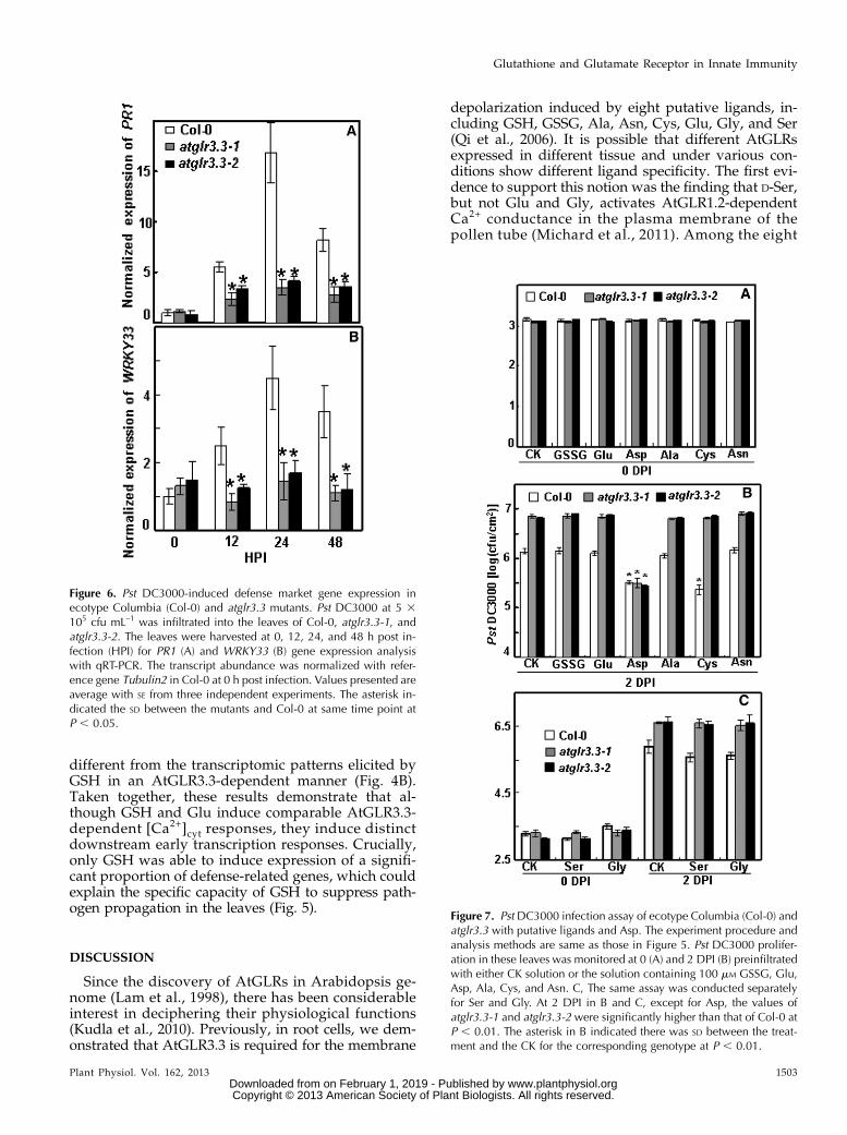
we monitored expression of two defense marker genes,PATHOGENESIS-RELATEDGENE1 (PR1) andWRKY33, inresponse to pathogen infection in both the wild typeand the mutants at 0, 12, 24, and 48 h post inoculation.The pathogen-induced expression of the defense markergenes was significantly attenuated in both allelic atglr3.3loss-of-function mutants (Fig. 6). To investigate the
physiological significance of the GSH-mediated AtGLR3.3-dependent defense gene expression (Fig. 4; Table I), wenext examined the effect of [GSH]ext on pathogen propa-gation inside the leaves of the wild type and the mutants.One day prior to the pathogen infiltration, the leaves wereinfiltrated with the CK buffer or the CK buffer con-taining100 mM GSH. On the day after, all leaves wereinfiltrated with the pathogen suspension at 5 3 105
colony-forming units (cfu) mL–1. The GSH pretreatmentsignificantly suppressed pathogen propagation in thewild-type leaf. However, suppression of propagationby [GSH]ext was not evident in the atglr3.3mutants (Fig.5). Thus, [GSH]ext promotes innate immunity responsesin the leaf through AtGLR3.3-dependent pathways.
Ligand Specificity of AtGLR3.3 in the InnateImmune Response
Our previous study with Arabidopsis root and hy-pocotyl cells suggests that there are up to eight puta-tive ligands of AtGLR3.3: GSH, GSSG, Ala, Asn, Cys,Gly, Glu, and Ser (Qi et al., 2006; Stephens et al., 2008).Among these, Glu plays a dominant role (Stephenset al., 2008). Given the results shown in Figure 5, wehypothesized that if any of the eight putative ligandsare true biological ligands of AtGLR3.3 in vivo, theyshould be able to evoke AtGLR3.3-dependent innateimmunity response in the leaf by exogenous applica-tion, in a similar fashion to GSH (Figs. 3–5). To test thishypothesis, we infiltrated the leaf of the wild type andtwo alleles of mutants with CK or CK solution con-taining 100 mM GSSG, Ala, Asn, Cys, Gly, and Glu
Table I. Defense genes induced by GSH in ecotype Columbia but not glr3.3-2
Gene ID FC01a FC02a Average Annotation Reference
Having confirmative role in defenseAT5G13320 17.9 6.1 12 GRETCHEN HAGEN 3.12; SA biosynthesis Okrent et al., 2009AT2G46400 15.7 4.9 10.3 WRKY46 transcription factor Hu et al., 2012AT1G33960 12.5 3 7.75 AVIRULENCE-INDUCED GENE1 Reuber and Ausubel, 1996AT1G74710 5.8 4.8 5.3 Isochorismate Synthase1; SA biosynthesis Wildermuth et al., 2001AT1G72520 3.3 6.6 4.95 Lipoxygenase4; jasmonic acid biosynthesis Acosta and Farmer, 2010AT2G35980 5.1 3.9 4.5 Yellow-leaf-specific gene9 Po-Wen et al., 2013AT5G26920 4.4 3.4 3.9 CBP60G Wang et al., 2009AT1G73805 3.6 3.5 3.55 SARD1; SA biosynthesis Zhang et al., 2010AT2G38470 3.4 3.4 3.4 WRKY33 transcription factor Birkenbihl et al., 2012
Having putative role in defenseAT5G36970 14.8 23.1 18.95 HAIRPIN-INDUCED1 LIKE25; induced by pathogens Varet et al., 2002AT5G47850 7 13.2 10.1 CRINKLY44 related4; protein kinase; response to insect Little et al., 2007AT1G21240 7.5 3.3 5.4 Wall associated kinase3; induced by SA He et al., 1999AT2G18060 3.5 6.3 4.9 NAC (NAM, ATAF1/2 and CUC2) domain containing protein37;
induced by chitinLibault et al., 2007
AT3G50930 3.7 5.7 4.7 ATPases associated with diverse cellular activities type; inducedby bacterial pathogen
Navarro et al., 2004
AT3G23230 3 6.3 4.65 Ethylene response factor98; induced by pathogen and jasmonic acid McGrath et al., 2005AT2G43000 3.7 5.1 4.4 NAC domain containing protein42; camalexin biosynthesis Saga et al., 2012AT1G14540 4 3.8 3.9 Peroxidase4; induced by oxidative stress and fungus infection Rasul et al., 2012AT1G18570 3.2 4.1 3.65 MYB51 transcription factor; induced by pathogen Maekawa et al., 2012AT4G33050 4 3.1 3.55 Embryo sac development arrest39; response to chitin and virus Libault et al., 2007
aFold changes of two replicates.
Plant Physiol. Vol. 162, 2013 1501
Glutathione and Glutamate Receptor in Innate Immunity
www.plantphysiol.orgon February 1, 2019 - Published by Downloaded from Copyright © 2013 American Society of Plant Biologists. All rights reserved.

1 d before the pathogen inoculation. We also includedAsp, a nonexciting amino acid, as a control. At 2 DPI,consistent with the previous results (Fig. 5), the atglr3.3mutants showed enhanced bacterial growth (Fig. 7).Among the compounds tested, only Cys and Asp wereable to act similarly to GSH to significantly suppresspathogen propagation in the wild-type leaf (Fig. 7).The suppressing effects of Cys, but not Asp, were an-nulled in the atglr3.3 loss-of-function mutants (Fig. 7).Thus, of the two amino acids, only the suppressiveeffect of Cys acts through an AtGLR3.3-dependentpathway, similar to GSH.
The Eight Putative Ligands, but Not Asp, Inducean AtGLR3.3-Dependent [Ca2+]cyt Rise
It was unexpected that the eight ligands and Aspwould have distinct effects on the plant’s innate im-munity responses (Figs. 5 and 7). We hypothesizedthat the difference could be related to (1) varyingability to initiate the transient rise in [Ca2+]cyt and (2)different dependence on AtGLR3.3 in the leaves. Wetherefore examined effects of seven ligands and Aspon the [Ca2+]cyt in the leaf cells of the wild type andatglr3.3 mutants. All the tested ligands, except Aspbut including Cys, evoked significant [Ca2+]cyt re-sponses (Fig. 8). These were of varying amplitudes,but all were dependent on AtGLR3.3 (Fig. 8). Thisresult demonstrated that innate immunity responsestriggered by the ligands GSH and Cys are not sim-ply correlated with their ability to induce [Ca2+]cyttransients.
The Glu-Mediated Early Transcriptional ResponseIs Distinct to That Induced by GSH
Glu is the principle exciting amino acid that acti-vates GLRs in the central nervous system (Flores-Sotoet al., 2012). It also evokes strong [Ca2+]cyt responses inthe leaf (Fig. 8A), which are at least comparable with, ifnot stronger than, those induced by GSH (Figs. 2 and3). However, Glu and GSH had essentially distincteffects on the plant defense responses. Most notably,Glu had no suppressive effect on the pathogen prop-agation (Figs. 5 and 7).
To further explore the influence of the two com-pounds, we compared Glu-elicited transcriptomic re-sponses with those induced by GSH (Fig. 4B; Table I;Supplemental Table S2). In total, treatment of wild-typeleaves with 100 mM Glu modulated expression of 70genes, 69 of which showed no significant changes inresponse to Glu in the atglr3.3mutant and can thereforebe categorized as Glu-modulated AtGLR3.3-dependentgenes (Fig. 4A; Supplemental Tables S5, S6, and S7).Among the 69 genes, only six genes were common tothe GSH-modulated AtGLR3.3-dependent group (Fig.4A; Supplemental Tables S2 and S5). Functional catego-rization of the 69 Glu-modulated AtGLR3.3-dependentgenes showed that 37%, 16%, 23%, 3%, and 6% of thesegenes were classified as Unknown, Others, Signaling,Transcription Factor, and Defense, respectively (Fig. 4C).The Defense section only included four genes: PR1,FLAVIN-DEPENDENT MONOOXYGENASE1 (Mishinaand Zeier, 2006), CELL WALL-ASSOCIATED KINASE1,and CELL WALL-ASSOCIATED KINASE3 (Brutus et al.,2010; De Lorenzo et al., 2011). Hence, it was strikingly
Figure 5. Role of AtGLR3.3 in the innate immunity and GSH-triggered defense response in the leaf. A, Leaves of ecotypeColumbia (Col-0) and atglr3.3-1 and atglr3.3-2 mutants were infiltrated with either CK solution or the solution containing100 mM GSH 1 d before the inoculation of Pst DC3000 at 5 3 105 cfu mL–1. The pathogen proliferation was monitored at 0, 1,and 2 DPI. Values presented are average with SE from at least four leaves from different plants. At 1, 2, and 3 DPI, there is SD
between the Col-0 and two mutants at P, 0.01. Two asterisks indicated significance at P, 0.01. Five independent experimentswere conducted and showed comparable results. B, Leaf symptoms of the Col-0, atglr3.3-1, and atglr3.3-2 plants preinfiltratedwith CK solution or the solution containing 100 mM GSH 3 DPI with Pst DC3000. C, Same leaves in B were kept 1 d longer, e.g.4 DPI, in a sealed agar plate.
1502 Plant Physiol. Vol. 162, 2013
Li et al.
www.plantphysiol.orgon February 1, 2019 - Published by Downloaded from Copyright © 2013 American Society of Plant Biologists. All rights reserved.
different from the transcriptomic patterns elicited byGSH in an AtGLR3.3-dependent manner (Fig. 4B).Taken together, these results demonstrate that al-though GSH and Glu induce comparable AtGLR3.3-dependent [Ca2+]cyt responses, they induce distinctdownstream early transcription responses. Crucially,only GSH was able to induce expression of a signifi-cant proportion of defense-related genes, which couldexplain the specific capacity of GSH to suppress path-ogen propagation in the leaves (Fig. 5).
DISCUSSION
Since the discovery of AtGLRs in Arabidopsis ge-nome (Lam et al., 1998), there has been considerableinterest in deciphering their physiological functions(Kudla et al., 2010). Previously, in root cells, we dem-onstrated that AtGLR3.3 is required for the membrane
depolarization induced by eight putative ligands, in-cluding GSH, GSSG, Ala, Asn, Cys, Glu, Gly, and Ser(Qi et al., 2006). It is possible that different AtGLRsexpressed in different tissue and under various con-ditions show different ligand specificity. The first evi-dence to support this notion was the finding that D-Ser,but not Glu and Gly, activates AtGLR1.2-dependentCa2+ conductance in the plasma membrane of thepollen tube (Michard et al., 2011). Among the eight
Figure 6. Pst DC3000-induced defense market gene expression inecotype Columbia (Col-0) and atglr3.3 mutants. Pst DC3000 at 5 3105 cfu mL–1 was infiltrated into the leaves of Col-0, atglr3.3-1, andatglr3.3-2. The leaves were harvested at 0, 12, 24, and 48 h post in-fection (HPI) for PR1 (A) and WRKY33 (B) gene expression analysiswith qRT-PCR. The transcript abundance was normalized with refer-ence gene Tubulin2 in Col-0 at 0 h post infection. Values presented areaverage with SE from three independent experiments. The asterisk in-dicated the SD between the mutants and Col-0 at same time point atP , 0.05.
Figure 7. Pst DC3000 infection assay of ecotype Columbia (Col-0) andatglr3.3 with putative ligands and Asp. The experiment procedure andanalysis methods are same as those in Figure 5. Pst DC3000 prolifer-ation in these leaves was monitored at 0 (A) and 2 DPI (B) preinfiltratedwith either CK solution or the solution containing 100 mM GSSG, Glu,Asp, Ala, Cys, and Asn. C, The same assay was conducted separatelyfor Ser and Gly. At 2 DPI in B and C, except for Asp, the values ofatglr3.3-1 and atglr3.3-2 were significantly higher than that of Col-0 atP , 0.01. The asterisk in B indicated there was SD between the treat-ment and the CK for the corresponding genotype at P , 0.01.
Plant Physiol. Vol. 162, 2013 1503
Glutathione and Glutamate Receptor in Innate Immunity
www.plantphysiol.orgon February 1, 2019 - Published by Downloaded from Copyright © 2013 American Society of Plant Biologists. All rights reserved.

putative ligands of AtGLR3.3, GSH has establishedphysiological function as an essential antioxidant andsignaling molecule involved in plant defense responses(Noctor et al., 2012). It led us to form hypotheses thatAtGLR3.3 may play a role in innate immunity and inmediating GSH-induced defense responses. Resultspresented in this study support this hypothesis andopen up several intriguing avenues of research forfurther exploration.
The GSH-Induced AtGLR3.3-Dependent [Ca2+]cytTransient Involves Several Calcium Stores
It was previously observed that exogenous appli-cation of GSH at millimolar concentrations induced a[Ca2+]cyt rise in tobacco leaf and Arabidopsis root cells(Gomez et al., 2004; Qi et al., 2006). However, theidentity of the proteins involved in this response wasnot known. In this study, we first demonstrated that aslittle as 10 mM GSH could evoke significant elevation ofthe [Ca2+]cyt (Fig. 1), suggesting that the cell has sensitivemechanisms to detect extracellular GSH at physiologicalconcentrations. Furthermore, using a pharmacologi-cal approach and a transfer DNA (T-DNA) insertionknockout mutant expressing the Ca2+ reporter pro-tein aequorin, we demonstrated that the response fullydepended on AtGLR3.3 in Arabidopsis leaf cells (Fig. 3).The result is consistent with the previous observationthat AtGLR3.3 is required for GSH-induced membranedepolarization in the roots (Qi et al., 2006). In animalcells, iGLRs form cation channels permeable to Ca2+, K+,
and Na+ (Flores-Soto et al., 2012). Recently, AtGLR3.4,the closest homolog of AtGLR3.3, has been demonstratedto form an amino acid-gated Ca2+ channel when heter-ologously expressed in the plasma membrane of humanembryonic kidney cells (Vincill et al., 2012).
It is possible that AtGLR3.3 forms similar Ca2+ channelsto AtGLR3.4 in the plasma membrane of the leaf cells andis gated by extracellular GSH to conduct Ca2+ influx intothe cytosol. However, chelating extracellular Ca2+ didn’tcompletely abolish the GSH-mediated [Ca2+]cyt response(Fig. 2). This implies that mobilization of internal Ca2+
pools also contributes to the response. AtGLRs are pre-dicted to contain conserved domains of both iGLRs andGPCRs (Turano et al., 2001). It is possible that theAtGLR3.3, similar to GPCRs, is coupled to certain internalCa2+ mobility-signaling pathways, such as IP3-mediatedCa2+ release from internal Ca2+ pools (Loane et al.,2012).Support for this notion comes from the observation thatpharmacologically interfering with IP3 production signif-icantly suppressed the GSH-mediated response (Fig. 2). Insummary, the observed [Ca2+]cyt elevation could beexplained by (1) direct influx of extracellular Ca2+ throughthe AtGLR3.3 channel into the cytosol and (2) indirectactivation of internal Ca2+-releasing pathways throughunknown intermediate signal transducers.
Role of AtGLR3.3 in Innate Immunity and GSH-InducedDefense Responses
Two alleles of atglr3.3 loss-of-function mutants showedconsistently enhanced susceptibility to PstDC3000 (Fig. 5)and decreased expression of defense-relatedmarker genesin response to the pathogen (Fig. 6). This implicatesAtGLR3.3 as a novel genetic component of the innateimmunity system in the leaf. Theoretically, any of thepreviously identified eight ligands could be biologicallysignificant ligands of the receptor. By testing the effects ofthe exogenous application of the ligands individually onpathogen propagation, we found that both GSH and Cyswere able to significantly suppress pathogen growth in anAtGLR3.3-dependent manner (Fig. 7). The common fea-ture that GSH and Cys share, and which is absent fromthe other potential ligands, is a thiol group. It is possiblethat it is these groups that interact with AtGLR3.3, whichin turn evokes specific downstream signaling cascadesthat ultimately lead to the defense responses. It is welldocumented that cytosolic GSH can move into theextracellular space during cell death in animal cells(Hammond et al., 2004; Schmidt and Dringen, 2012). Inplants, pathogen infection increases GSH production(Vanacker et al., 2000; Parisy et al., 2007). Possibly, cyto-solic GSH can be exported to the extracellular spacethrough oligopeptide transporters (Koh et al., 2002) dur-ing the interaction between the plant cells and pathogens.
GSH serves as reservoir for Cys, and the two flankingamino acids Glu and Gly protect the sulfhydryl groupof Cys against oxidation (Meyer, 2008; Hell and Wirtz,2011). It is known that externally supplied Cys, afterbeing transported into the cytosol, is transiently formed
Figure 8. Effects of the seven putative ligands of AtGLR3.3 and Asp onthe [Ca2+]cyt in the leaf cells of ecotype Columbia (Col-0) and atglr3.3expressing aequorin. Averaged recording curves with SE of 100 mM Glu(A) and Asp (B) induced [Ca2+]cyt rise in the leaf cells of Col-0 andatglr3.3-1 and atglr3.3-2 mutants expressing aequorin (n = 8 for Gluand n = 5 for Asp). C, the average peak values with SE of the response tothe indicated ligands (n = 8 for GSSG and Glu, n = 5 for Asp, Cys, Ala,Asn, and Ser, and n = 7 for Gly). The asterisk indicated SD at P , 0.01between the mutants and Col-0 for the same treatment. The dash lineindicated the background [Ca2+]cyt.
1504 Plant Physiol. Vol. 162, 2013
Li et al.
www.plantphysiol.orgon February 1, 2019 - Published by Downloaded from Copyright © 2013 American Society of Plant Biologists. All rights reserved.

into GSH (Hell andWirtz, 2011). Therefore, it is possiblethat in addition to its direct binding to the AtGLR3.3,Cys can reinforce the GSH production and indirectlyinduce the defense response. Conversion of Cys to GSHmay have contributed to the induced resistance tobacteria (preinfiltration 1 d prior to the pathogen inoc-ulation), but the much shorter incubations used for theluminescence studies (minutes) mean that effects of Cyson [Ca2+]cyt are likely to be direct. It is a very intriguingquestion how perception of extracellular GSH and Cysby the membrane-anchored AtGLR3.3 relays into down-stream signaling cascade.The suppressing effect of Asp on the pathogen pro-
pagation through the AtGLR3.3-independent pathwaywas unexpected (Fig. 7). Asp is closely linked to severalmetabolites that play essential roles in primary metabolicpathways such as the tricarboxylic acid cycle that con-tributes to cellular energy metabolism (Kirma et al., 2012).It was possible that excess Asp could interrupt the energyproduction process and in turn decrease the energy in theplant cells available for the pathogen consumption. It isalso possible that excess Asp drives production of certaindefense compounds to either directly limit the pathogenpropagation or trigger the plant cells’ innate immunityresponses. However, Glu, another amino acid that isclosely linked to the tricarboxylic acid cycle, did notsuppress pathogen propagation.Our study gives rise to questions about the physiolog-
ical ligands of AtGLR3.3. In animal cells, [GSH]ext can in-duce release of other compounds, for example dopaminefrom mouse striatal slices (Janáky et al., 2007) and taurinefrom developingmouse hippocampus (Janáky et al., 2008).We cannot exclude the possibility that [GSH]ext inducesrelease of other compounds from the plant cells to activatethe AtGLR3.3-dependent innate immunity response.
GSH-Mediated AtGLR3.3-Dependent EarlyTranscription Response
Several studies report that [GSH]ext induces stress-associated gene expression, and partial and whole genomedata have been presented for GSH- and glutathione re-ductase-deficient mutants (Ball et al., 2004; Mhamdi et al.,2010; Han et al., 2013b). In this study, we analyzed theearly transcription responses of the plant cells to [GSH]extwith the aim of studying gene regulation effects of GSHacting as a signalingmolecule rather than in its well-knownrole as an antioxidant. Because we only had two biologicalreplicates, we set very strict standards as detailed in“Materials and Methods” to process the raw data. Its va-lidity was supported by two findings: first, the AtGLR3.3transcript was the most down-regulated in the atglr3.3mutant compared with that in the wild type (SupplementalTable S1); second, real-time qRT-PCR with independent bi-ological samples were largely consistent with themicroarraydata set (Supplemental Fig. S1). The GSH treatment mod-ulated expression of 97 genes in the wild type, 70 of whichwere not significantly changed in atglr.3.3, accounting for72% of the total (Fig. 4A; Supplemental Table S2). It clearly
indicated that AtGLR3.3 plays an essential role inthe early transcriptional responses of the leaf toGSH. The genetic mechanism of the GSH-mediatedand AtGLR3.3-independent gene expression is unknown.In animal cells, in addition to the GLRs, GSH has beenshown to have profound regulative effects on theryanodine receptor (Zable et al., 1997), death receptor (Moritoet al., 2003), and GPCRs (Wang et al., 2006). It is possiblethat certain unknown receptors in Arabidopsis mediatethe AtGLR3.3-independent GSH signaling cascade.
For the GSH-modulated AtGLR3.3-dependent genes,we did standard Gene Ontology annotation analysiscombined with manual literature checking. It revealedthat 27% of the genes have either confirmative roles indefense or are induced by biotic stresses (Table I;Supplemental Table S2), which accounted for the largestsubgroup in the early transcription response. It makesstrong sense in context of the defense function of GSHobserved in this study (Fig. 5). Particularly interesting isthe finding that GSH induced expression of ICS1 andSARD1 in an AtGLR3.3-dependent manner, two essen-tial genes for SA biosynthesis (Wildermuth et al., 2001;Zhang et al., 2010). A regulatory role for SA in GSHproduction in plants has been described (Srivastava andDwivedi, 1998; Yoshida et al., 2009). Our finding thatGSH can strongly induce ICS1 expression (Table I) im-plies that there is a positive AtGLR3.3-dependent feed-back mechanism for GSH to enhance SA biosynthesisunder stress conditions. Hydrogen peroxide inducesproduction of SA in tobacco (Leon et al., 1995), while theGSH-deficient mutant pad2-1 lost pathogen Phytophthorabrassicae-triggered expression of ICS1 (Dubreuil-Mauriziand Poinssot, 2012). Recently, it has been reported thathydrogen peroxide-triggered accumulation of SA in acatalase-deficient mutant can be largely blocked byblocking glutathione synthesis, and that is associatedwith the failure to up-regulate ICS1 (Han et al., 2013a).It will be very interesting to investigate the role ofAtGLR3.3 in the interaction between SA and GSHhomeostasis.
Correlation of the [Ca2+]cyt Transient Rise with theDefense Response
A large number of environmental and internal signalsare able to evoke [Ca2+]cyt transients in plant cells(Kudla et al., 2010). In most of these cases, two funda-mental questions remain: (1) the molecular componentunderlying the transient [Ca2+]cyt rise and (2) how theCa2+ responses are linked to the downstream physio-logical function mediated by the signaling. In this study,we provided genetic evidence that the transient [Ca2+]cytrise induced by the eight putative ligands were fullydependent on AtGLR3.3 (Figs. 3 and 8). However, thelink between the GSH-induced transient [Ca2+]cyt riseand the defense functions of GSH still remain elusive.
It is possible that the two effects of GSH are notrelated. Evidence to support this argument is thatGlu-induced AtGLR3.3-dependent [Ca2+]cyt transient
Plant Physiol. Vol. 162, 2013 1505
Glutathione and Glutamate Receptor in Innate Immunity
www.plantphysiol.orgon February 1, 2019 - Published by Downloaded from Copyright © 2013 American Society of Plant Biologists. All rights reserved.

rise was stronger than that of GSH (Fig. 8), even thoughunlike GSH, Glu did not induce significant amount ofdefense gene expression and defense responses (Figs.4C and 7). Key points are the still unknown structure ofthe Glu receptor complex and the mechanism throughwhich it specifically recognizes different ligands. In ani-mal cells, functioning iGLRs are composed of differentsubunits encoded by individual iGLR genes and acti-vated by different ligands. A typical case in this contextis the NMDA iGLR, which is composed of four subunits,two from NMDA Receptor A (NRA), NRB, NRC, andNRD and two NR1s. NRA to NRD is activated by Gluand NR1 by Gly (Flores-Soto et al., 2012).
It is possible that the AtGLRs are also formed bydifferent subunits that recognize different ligands. GSHand Cys could activate a unique subunit through theirspecific thiol group and induce additional signalingcascades that occur alongside, rather than downstream,of the transient [Ca2+]cyt rise. Our previous study with thecross-desensitization technique indirectly reveals that theAtGLRs are composed by AtGLR3.3 and other subunitsspecifically activated by different putative ligands (Stephenset al., 2008).
The situation could be even more complex. It has beenproposed for some time that transient [Ca2+]cyt rises in-duced by different signals could be different in strength,frequency, and spatial distribution (Kudla et al., 2010). Arecent study provides evidence in support of this andshows that transient [Ca2+]cyt rises with different strengthand spatial distribution are linked to different transcrip-tion responses (Wheeler et al., 2012). Such concepts couldexplain why GSH and Glu induce comparable changesin [Ca2+]cyt but distinct gene expression responses. Tocompletely elucidate this mechanism, studies will requiremore precise measurement and subcellular imaging ofdynamic changes in [Ca2+]cyt. Whether the two GSH-mediated AtGLR3.3-dependent effects are related or not,our data suggest that the GSH-related functions ofAtGLR3.3 are involved in signaling. The atglr3.3 mutantshowed wild-type growth on cadmium (SupplementalFig. S2), a condition in which GSH plays an importantbut nonsignaling role as a precursor molecule of phyto-chelatins that sequester cadmium (Cobbett et al., 1998;Jozefczak et al., 2012).
CONCLUSION
Glu receptors play vital roles in mediating informationexchange among neurons in the central nervous system.Their complex functions are determined by their versatilestructures, subunit composition, various ligand types, andbinging sites as well as binding affinity in different tissuesand under different physiological conditions (Flores-Sotoet al., 2012; Popescu, 2012). In this study, we providedevidence to support a novel role of AtGLR3.3 in the innateimmunity and GSH-mediated defense response in Arab-idopsis leaf. Our findings promote understanding of theAtGLR gene family and the related signaling roles of ex-tracellular GSH and raise intriguing questions for furtherstudy.
MATERIALS AND METHODS
Arabidopsis AtGLR3.3 Mutants Expressing Aequorin
The Arabidopsis (Arabidopsis thaliana) atglr3.3-1 and atglr3.3-2 (At1g42540)homozygous knockout mutants used in this study were described previously(Qi et al., 2006) and were SALK_040458 and SALK_066009, respectively (Alonsoet al., 2003), obtained from The Arabidopsis Biological Resource Center. To intro-duce the aequorin gene into the mutant background, mutants were crossed with theaequorin transgenic Arabidopsis line (Knight et al., 1991). In the F2 segregationpopulation, homozygous T-DNA insertions were detected with gene-specificprimers (SALK_040458, 59-TTGTTTGTTGCAGCATTGAG-39, 59-GCCAATCTT-GAGTTCTTTCCC-39 and SALK_066009, 59-AAGCACCAGACATCTTACGC-39,59-TCAGATTGGTCCAATTTTAGC-39) and the T-DNA left border-specific primerLBb1.3 (59-ATTTTGCCGATTTCGGAAC-39). The presence of active aequorin wasvalidated by measuring luminescence intensity in the leaf, as described below.
[Ca2+]cyt Measurement in Arabidopsis LeafExpressing Aequorin
To measure dynamic changes in [Ca2+]cyt in the leaf, Arabidopsis seeds weresurface sterilized with 100% ethanol (v/v) for 15 min and dried on sterilized filterpaper in a fume hood. Sterilized seeds were sown on 0.7% (w/v) agar mediumcontaining one-half-strength Murashige and Skoog basic salts with 1% (w/v) Sucand 5 mM MES, pH 5.7. After 3-d treatment at 4°C, the plates were placed in agrowth chamber at 22°C to 24°C , 12-h light/12-h dark, and a light intensity of 100to 120 mmol E m–2 s–1. One leaf per 12-d-old seedlings was cut and placed in a1.5-mL centrifuge tube with 100 mL control buffer (CK buffer) composed of 1 mM
KCl, 1 mM CaCl2, and 5 mM MES, pH 5.7. We observed that leaves from precutseedlings had a very weak [Ca2+]cyt response. To ensure consistent results, therefore,care was taken to sample only one leaf per seedling. Under dim light, coelenterazinehcp (Promega) was added into the buffer to a final concentration of 1 mM. After12- to 14-h incubation in the dark, the tube with the leaf was put into a single tubeluminometer with a detection limit of 10–21 mol luciferase (Turner Biosystem, 20/20)in a roomwith dim light. The tubewas left to stand for 3 to 5min to allow the leaf torecover from potential disturbances of [Ca2+]cyt triggered by the handling. For thetreatment, 100 mL control buffer with 2 times the final concentration of GSH wasquickly delivered into the tube by positioning the pipette tip on the internal walls toenable the treatment solution to diffuse into the solution with the leaf. This proce-dure avoided directly dropping the treatment solution into the solution with the leafand so aimed to minimize disturbance of the leaf [Ca2+]cyt background.
Luminescence intensity was measured at 1-Hz frequency. At the end of eachrecording, 500 mL ice-cold 1 M CaCl2 in 20% ethanol (v/v) was delivered intothe tube to saturate the Ca2+ and aequorin protein-dependent luminescence.The absolute [Ca2+]cyt concentration was calculated based on the method describedpreviously (Rentel and Knight, 2004). To explore the contribution of external Ca2+
to the observed [Ca2+]cyt rise, we incubated the leaf in Ca2+-free CK buffer with1 mM EGTA, a Ca2+ chelator. The GSH treatment was also performed in the samebuffer. To address effects of AP5, alloxan, neomycin, and 6,7-dinitroquinoxaline-2,3-dione on the [Ca2+]cyt responses, these chemicals were added into the recordingbuffer at the indicated concentration 1 h before the GSH treatments. All chemicalswere purchased from Sigma.
Arabidopsis Seedling Treatment and RNA Extractionfor Complementary DNA Microarray
Arabidopsis seedlings were cultured on 0.7% (w/v) agar medium con-taining one-half-strength Murashige and Skoog basic salt with 1% Suc (w/v)and 5 mM MES, pH 5.7. Ten-day-old seedlings were equilibrated in the CK bufferfor 24 h. Then, these seedlings were either treated with the CK buffer or the CKbuffer containing100 mM GSH and 100 mM Glu for 1 h, respectively. Total RNAwasextracted from each sample using EasyPure Plant RNA kit (Transgene). FurtherRNA purification, probe preparation, hybridization, andmicroarray scanning weredone at the National GeneChip Engineering Center (http://www.ebioserve.com).Labeled samples were hybridized with one Agilent Arabidopsis 4344k array byfollowing the manufacturer’s instructions. Four biologically independent sets ofArabidopsis seedlings were prepared, two for the microarray experiment and twofor real-time qRT-PCR analysis.
The raw data were normalized by quantile algorithm using Gene SpringSoftware 11.0. The following criteria were used in the National GeneChipEngineering Center for defining significantly differentially expressed genes: (1)genes that are statistically significant at the level of P , 0.05 after false
1506 Plant Physiol. Vol. 162, 2013
Li et al.
www.plantphysiol.orgon February 1, 2019 - Published by Downloaded from Copyright © 2013 American Society of Plant Biologists. All rights reserved.

discovery rate correction and labeled “Present” call by the GeneChip Opera-tion System output and (2) genes showing a change in the expression levelbetween treatment and control at least 3-fold in the two replicates. The GeneOntology annotation analysis was conducted using the online program athttp://www.Arabidopsis.org/tools/bulk/go/index.jsp. The raw data of themicroarray experiment was deposited at http://www.ebi.ac.uk/miamex-press/ with access number E–MEXP–3885.
qRT-PCR Analysis
Tissue preparation and RNA extraction were as for the microarray analyses.Total RNA was treated with RNAse-free DNAse to remove all DNA contami-nation (Takara). One microgram of RNA samples were used for complementaryDNA synthesis with oligo(dT) primer and PrimeScript II reverse transcriptase(Takara) following a standardprotocol. DNAcontaminationof the complementaryDNAwas further tested by PCRwith a pair of primers spanning an intron. Beforethe qRT-PCR analysis, all primers were tested for their specificity and primerdimer formation by conducting semiquantitative reverse transcript PCR. qRT-PCR was conducted on a Biorad Chromo 4 real-time PCR system using SYBRGreen Premix Ex Taq II kit (Takara). The Arabidopsis Tubulin2 gene (At5g62690)was used as a constitutive reference to normalize the gene expression. Relativeexpression to the wild type under CK condition was presented in Figure 6 andSupplemental Figure S1. Primer sequences for the PCR are listed in SupplementalTable S8.
Bacterial Growth Assay
To prepare Arabidopsis plants for the assay, we grew two wild-type andtwomutant plants in the samepot tominimize side effects of growth environmentdifference on the pathogen propagation results. The pots were maintained in agrowth chamber with 50 to 70% humidity at 22°C to 24°C, 12-h light/12-h dark,and a light intensity of 100 to 120 mmol E m–2 s–1. Four-week-old leaves wereused in this study. For each plant, we chose two leaves for syringe-based infil-tration. One day before the pathogen inoculation, we infiltrated the leaf witheither the CK or CK containing100 mM GSH, GSSG, Ala, Asn, Cys, Gly, Glu, Ser,and Asp.
The bacterial strain used in this study was Pst DC3000. The –80°C stock ofthe pathogen was first streaked onto a low-salt Luria-Bertani plate (tryptone10 g L–1, yeast [Saccharomyces cerevisiae] extract 5 g L–1, and NaCl 5 g L–1) with50 mg mL–1 rifampin. Plates with bacteria were cultured at 28°C for 38 to 42 h,then subcultured in liquid Luria-Bertani solution growing up to an opticaldensity at 600 nm of 0.8 to 1.0. After collection by centrifuging, the pathogenwas resuspended in sterile water to an optical density at 600 nm of 0.001,corresponding to 5 3 105 cfu mL–1 bacteria, and infiltrated into the leaf with aneedless 1-mL syringe. At 0, 1, 2, and 3 DPI, the inoculated leaf was taken andbriefly rinsed in 75% ethanol (v/v) and sterile water. A leaf disc with 0.5-cmdiameter was sampled from each of four to eight different leaves from sepa-rate plants for each treatments of each genotype and ground in sterile water. Astandard colony-forming unit dilution assay was conducted (Katagiri et al.,2002). Five independent biological replicates were performed.
Supplemental Data
The following materials are available in the online version of this article.
Supplemental Figure S1. qRT-PCR analysis of GSH-induced AtGLR3.3-dependent gene expression.
Supplemental Figure S2. Root elongation responses of ecotype Columbiaand atglr3.3 to Cd2+.
Supplemental Figure S3. Pst DC3000 infection assay of Arabidopsis pad2-1 mutants.
Supplemental Table S1. Differential expressed gene in the atglr3.3-2 mu-tant compared with that in ecotype Columbia.
Supplemental Table S2. Genes modulated by GSH in ecotype Columbiabut not atglr3.3-2 mutant.
Supplemental Table S3. Genes modulated by GSH in ecotype Columbiaand atglr3.3-2 mutant.
Supplemental Table S4. Genes modulated by GSH in atglr3.3-2 but notecotype Columbia.
Supplemental Table S5. Genes modulated by Glu in ecotype Columbiabut not atglr3.3-2 mutant.
Supplemental Table S6. Genes modulated by Glu in ecotype Columbiaand atglr3.3-2 mutant.
Supplemental Table S7. Genes modulated by Glu in atglr3.3-2 but notecotype Columbia.
Supplemental Table S8. Primers used for the quantitative PCR.
ACKNOWLEDGMENTS
We thank Shengyang He (Michigan State University) for the strain of bacterialpathogen PstDC3000, Dr. Marc R. Knight (Durham University) for the Arabidopsistransgenic seeds expressing the aequorin gene, Felix Mauch (University of Fribourg)for the pad2-1mutant seeds, Turner BiosystemUSA for donation of the luminometerinstrument, and Dr. Huifeng Zhu (Capital Normal University-Beijing) for initialtechnical assistance on the bacterial growth assay.
Received March 1, 2013; accepted May 6, 2013; published May 8, 2013.
LITERATURE CITED
Acosta IF, Farmer EE (2010) Jasmonates. The Arabidopsis Book 8: e0129,doi/10.1199/tab.0129
Ali R, Ma W, Lemtiri-Chlieh F, Tsaltas D, Leng Q, von Bodman S,Berkowitz GA (2007) Death don’t have no mercy and neither doescalcium: Arabidopsis CYCLIC NUCLEOTIDE GATED CHANNEL2 andinnate immunity. Plant Cell 19: 1081–1095
Alonso JM, Stepanova AN, Leisse TJ, Kim CJ, Chen H, Shinn P, StevensonDK, Zimmerman J, Barajas P, Cheuk R, et al (2003) Genome-wide inser-tional mutagenesis of Arabidopsis thaliana. Science 301: 653–657
Aouini A, Hernould M, Ariizumi T, Matsukura C, Ezura H, Asamizu E(2012) Overexpression of the tomato glutamate receptor-like genes SlGLR1.1and SlGLR3.5 hinders Ca2+ utilization and promotes hypersensitivity to Na+
and K+ stresses. Plant Biotechnol 29: 229–235Aoyama K, Watabe M, Nakaki T (2008) Regulation of neuronal glutathione
synthesis. J Pharmacol Sci 108: 227–238Ball L, Accotto GP, Bechtold U, Creissen G, Funck D, Jimenez A, Kular B,
Leyland N, Mejia-Carranza J, Reynolds H, et al (2004) Evidence for adirect link between glutathione biosynthesis and stress defense geneexpression in Arabidopsis. Plant Cell 16: 2448–2462
Birkenbihl RP, Diezel C, Somssich IE (2012) Arabidopsis WRKY33 is akey transcriptional regulator of hormonal and metabolic responses to-ward Botrytis cinerea infection. Plant Physiol 159: 266–285
Bohman S, Staal J, Thomma BP, Wang M, Dixelius C (2004) Character-isation of an Arabidopsis-Leptosphaeria maculans pathosystem: resistancepartially requires camalexin biosynthesis and is independent of salicylicacid, ethylene and jasmonic acid signalling. Plant J 37: 9–20
Brutus A, Sicilia F, Macone A, Cervone F, De Lorenzo G (2010) A domainswap approach reveals a role of the plant wall-associated kinase 1 (WAK1) asa receptor of oligogalacturonides. Proc Natl Acad Sci USA 107: 9452–9457
Cairns NG, Pasternak M, Wachter A, Cobbett CS, Meyer AJ (2006) Mat-uration of Arabidopsis seeds is dependent on glutathione biosynthesiswithin the embryo. Plant Physiol 141: 446–455
Chin TY, Chueh SH, Tao PL (2006) S-Nitrosoglutathione and glutathioneact as NMDA receptor agonists in cultured hippocampal neurons. ActaPharmacol Sin 27: 853–860
Chiu J, DeSalle R, Lam HM, Meisel L, Coruzzi G (1999) Molecular evo-lution of glutamate receptors: a primitive signaling mechanism thatexisted before plants and animals diverged. Mol Biol Evol 16: 826–838
Cho D, Kim SA, Murata Y, Lee S, Jae SK, Nam HG, Kwak JM (2009) De-regulated expression of the plant glutamate receptor homolog AtGLR3.1impairs long-term Ca2+-programmed stomatal closure. Plant J 58: 437–449
Cobbett CS, May MJ, Howden R, Rolls B (1998) The glutathione-deficient,cadmium-sensitive mutant, cad2-1, of Arabidopsis thaliana is deficient ing-glutamylcysteine synthetase. Plant J 16: 73–78
De Lorenzo G, Brutus A, Savatin DV, Sicilia F, Cervone F (2011) Engi-neering plant resistance by constructing chimeric receptors that recog-nize damage-associated molecular patterns (DAMPs). FEBS Lett 585:1521–1528
Plant Physiol. Vol. 162, 2013 1507
Glutathione and Glutamate Receptor in Innate Immunity
www.plantphysiol.orgon February 1, 2019 - Published by Downloaded from Copyright © 2013 American Society of Plant Biologists. All rights reserved.

Dodd AN, Kudla J, Sanders D, Merchant S, Briggs W, Ort D (2010) Thelanguage of calcium signaling. Annu Rev Plant Biol 61: 593–620
Dringen R (2000) Metabolism and functions of glutathione in brain. ProgNeurobiol 62: 649–671
Dubos C, Huggins D, Grant GH, Knight MR, Campbell MM (2003) A role forglycine in the gating of plant NMDA-like receptors. Plant J 35: 800–810
Dubreuil-Maurizi C, Poinssot B (2012) Role of glutathione in plant sig-naling under biotic stress. Plant Signal Behav 7: 210–212
Ferrari S, Plotnikova JM, De Lorenzo G, Ausubel FM (2003) Arabidopsis localresistance to Botrytis cinerea involves salicylic acid and camalexin and requiresEDS4 and PAD2, but not SID2, EDS5 or PAD4. Plant J 35: 193–205
Flores-Soto ME, Chaparro-Huerta V, Escoto-Delgadillo M, Vazquez-Valls E, González-Castañeda RE, Beas-Zarate C (2012) Structure andfunction of NMDA-type glutamate receptor subunits. Neurologia 27:301–310
Foyer CH, Noctor G (2011) Ascorbate and glutathione: the heart of theredox hub. Plant Physiol 155: 2–18
Franco R, Cidlowski JA (2009) Apoptosis and glutathione: beyond an an-tioxidant. Cell Death Differ 16: 1303–1314
Glazebrook J, Ausubel FM (1994) Isolation of phytoalexin-deficient mu-tants of Arabidopsis thaliana and characterization of their interactionswith bacterial pathogens. Proc Natl Acad Sci USA 91: 8955–8959
Gomez LD, Noctor G, Knight MR, Foyer CH (2004) Regulation of calciumsignalling and gene expression by glutathione. J Exp Bot 55: 1851–1859
Hammond CL, Madejczyk MS, Ballatori N (2004) Activation of plasmamembrane reduced glutathione transport in death receptor apoptosis ofHepG2 cells. Toxicol Appl Pharmacol 195: 12–22
Han Y, Chaouch S, Mhamdi A, Queval G, Zechmann B, Noctor G (2013a)Functional analysis of Arabidopsis mutants points to novel roles forglutathione in coupling H2O2 to activation of salicylic acid accumulationand signaling. Antioxid Redox Signal 18: 2106–2121
Han Y, Mhamdi A, Chaouch S, Noctor G (2013b) Regulation of basal andoxidative stress-triggered jasmonic acid-related gene expression byglutathione. Plant Cell Environ 36: 1135–1146
He ZH, Cheeseman I, He D, Kohorn BD (1999) A cluster of five cell wall-associated receptor kinase genes, Wak1-5, are expressed in specific or-gans of Arabidopsis. Plant Mol Biol 39: 1189–1196
Hell R, Wirtz M (2011) Molecular biology, biochemistry and cellularphysiology of cysteine metabolism. The Arabidopsis Book 9: e0154, doi/10.1199/tab.0154
Hnasko TS, Edwards RH (2012) Neurotransmitter corelease: mechanismand physiological role. Annu Rev Physiol 74: 225–243
Hu Y, Dong Q, Yu D (2012) Arabidopsis WRKY46 coordinates withWRKY70 and WRKY53 in basal resistance against pathogen Pseudomo-nas syringae. Plant Sci 185-186: 288–297
Janáky R, Dohovics R, Saransaari P, Oja SS (2007) Modulation of [3H]dopamine release by glutathione in mouse striatal slices. NeurochemRes 32: 1357–1364
Janáky R, Shaw CA, Oja SS, Saransaari P (2008) Taurine release in de-veloping mouse hippocampus is modulated by glutathione and gluta-thione derivatives. Amino Acids 34: 75–80
Janáky R, Shaw CA, Varga V, Hermann A, Dohovics R, Saransaari P, OjaSS (2000) Specific glutathione binding sites in pig cerebral corticalsynaptic membranes. Neuroscience 95: 617–624
Janáky R, Varga V, Saransaari P, Oja SS (1993) Glutathione modulates theN-methyl-D-aspartate receptor-activated calcium influx into cultured ratcerebellar granule cells. Neurosci Lett 156: 153–157
Jozefczak M, Remans T, Vangronsveld J, Cuypers A (2012) Glutathione isa key player in metal-induced oxidative stress defenses. Int J Mol Sci 13:3145–3175
Juurlink BH (1999) Management of oxidative stress in the CNS: the manyroles of glutathione. Neurotox Res 1: 119–140
Kang J, Mehta S, Turano FJ (2004) The putative glutamate receptor 1.1(AtGLR1.1) in Arabidopsis thaliana regulates abscisic acid biosynthesisand signaling to control development and water loss. Plant Cell Physiol45: 1380–1389
Kang J, Turano FJ (2003) The putative glutamate receptor 1.1 (AtGLR1.1)functions as a regulator of carbon and nitrogen metabolism in Arabi-dopsis thaliana. Proc Natl Acad Sci USA 100: 6872–6877
Kang S, Kim HB, Lee H, Choi JY, Heu S, Oh CJ, Kwon SI, An CS (2006)Overexpression in Arabidopsis of a plasma membrane-targeting gluta-mate receptor from small radish increases glutamate-mediated Ca2+ in-flux and delays fungal infection. Mol Cells 21: 418–427
Katagiri F, Thilmony R, He SY (2002) The Arabidopsis thaliana-Pseudomonas syringae interaction. The Arabidopsis Book 1: 0039, doi/10.1199/tab.0039
Kim SA, Kwak JM, Jae SK, Wang MH, Nam HG (2001) Overexpression ofthe AtGluR2 gene encoding an Arabidopsis homolog of mammalianglutamate receptors impairs calcium utilization and sensitivity to ionicstress in transgenic plants. Plant Cell Physiol 42: 74–84
Kirma M, Araújo WL, Fernie AR, Galili G (2012) The multifaceted role ofaspartate-family amino acids in plant metabolism. J Exp Bot 63: 4995–5001
Knight MR, Campbell AK, Smith SM, Trewavas AJ (1991) Transgenicplant aequorin reports the effects of touch and cold-shock and elicitorson cytoplasmic calcium. Nature 352: 524–526
Koh S, Wiles AM, Sharp JS, Naider FR, Becker JM, Stacey G (2002) An oligo-peptide transporter gene family in Arabidopsis. Plant Physiol 128: 21–29
Kudla J, Batistic O, Hashimoto K (2010) Calcium signals: the lead currencyof plant information processing. Plant Cell 22: 541–563
Kwaaitaal M, Huisman R, Maintz J, Reinstädler A, Panstruga R (2011)Ionotropic glutamate receptor (iGluR)-like channels mediate MAMP-induced calcium influx in Arabidopsis thaliana. Biochem J 440: 355–365
Kwaaitaal M, Maintz J, Cavdar M, Panstruga R (2012) On the ligandbinding profile and desensitization of plant ionotropic glutamate re-ceptor (iGluR)-like channels functioning in MAMP-triggered Ca2+ influx.Plant Signal Behav 7: 1373–1377
Lacombe B, Becker D, Hedrich R, DeSalle R, Hollmann M, Kwak JM,Schroeder JI, Le Novère N, Nam HG, Spalding EP, et al (2001) Theidentity of plant glutamate receptors. Science 292: 1486–1487
Lam HM, Chiu J, Hsieh MH, Meisel L, Oliveira IC, Shin M, Coruzzi G(1998) Glutamate-receptor genes in plants. Nature 396: 125–126
Leon J, Lawton MA, Raskin I (1995) Hydrogen peroxide stimulates sali-cylic acid biosynthesis in tobacco. Plant Physiol 108: 1673–1678
Leslie SW, Brown LM, Trent RD, Lee YH, Morris JL, Jones TW, RandallPK, Lau SS, Monks TJ (1992) Stimulation of N-methyl-D-aspartatereceptor-mediated calcium entry into dissociated neurons by reducedand oxidized glutathione. Mol Pharmacol 41: 308–314
Levy DI, Sucher NJ, Lipton SA (1991) Glutathione prevents N-methyl-D-aspartate receptor-mediated neurotoxicity. Neuroreport 2: 345–347
Li J, Zhu S, Song X, Shen Y, Chen H, Yu J, Yi K, Liu Y, Karplus VJ, Wu P,et al (2006) A rice glutamate receptor-like gene is critical for the divisionand survival of individual cells in the root apical meristem. Plant Cell 18:340–349
Libault M, Wan J, Czechowski T, Udvardi M, Stacey G (2007) Identifi-cation of 118 Arabidopsis transcription factor and 30 ubiquitin-ligasegenes responding to chitin, a plant-defense elicitor. Mol Plant MicrobeInteract 20: 900–911
Little D, Gouhier-Darimont C, Bruessow F, Reymond P (2007) Oviposi-tion by pierid butterflies triggers defense responses in Arabidopsis.Plant Physiol 143: 784–800
Loane DJ, Stoica BA, Faden AI (2012) Metabotropic glutamate receptor-mediated signaling in neuroglia. Wiley Interdiscip Rev Membr TranspSignal 1: 136–150
Ma W, Qi Z, Smigel A, Walker RK, Verma R, Berkowitz GA (2009) Ca2+,cAMP, and transduction of non-self perception during plant immuneresponses. Proc Natl Acad Sci USA 106: 20995–21000
Maekawa S, Sato T, Asada Y, Yasuda S, Yoshida M, Chiba Y, Yamaguchi J(2012) The Arabidopsis ubiquitin ligases ATL31 and ATL6 control thedefense response as well as the carbon/nitrogen response. Plant MolBiol 79: 217–227
McGrath KC, Dombrecht B, Manners JM, Schenk PM, Edgar CI, MacleanDJ, Scheible WR, Udvardi MK, Kazan K (2005) Repressor- andactivator-type ethylene response factors functioning in jasmonate sig-naling and disease resistance identified via a genome-wide screen ofArabidopsis transcription factor gene expression. Plant Physiol 139:949–959
Meyer AJ (2008) The integration of glutathione homeostasis and redoxsignaling. J Plant Physiol 165: 1390–1403
Mhamdi A, Hager J, Chaouch S, Queval G, Han Y, Taconnat L,Saindrenan P, Gouia H, Issakidis-Bourguet E, Renou JP, et al (2010)Arabidopsis GLUTATHIONE REDUCTASE1 plays a crucial role in leafresponses to intracellular hydrogen peroxide and in ensuring appro-priate gene expression through both salicylic acid and jasmonic acidsignaling pathways. Plant Physiol 153: 1144–1160
Michard E, Lima PT, Borges F, Silva AC, Portes MT, Carvalho JE,Gilliham M, Liu LH, Obermeyer G, Feijó JA (2011) Glutamate
1508 Plant Physiol. Vol. 162, 2013
Li et al.
www.plantphysiol.orgon February 1, 2019 - Published by Downloaded from Copyright © 2013 American Society of Plant Biologists. All rights reserved.

receptor-like genes form Ca2+ channels in pollen tubes and are regulatedby pistil D-serine. Science 332: 434–437
Miller ND, Durham Brooks TL, Assadi AH, Spalding EP (2010) Detectionof a gravitropism phenotype in glutamate receptor-like 3.3 mutants ofArabidopsis thaliana using machine vision and computation. Genetics186: 585–593
Mishina TE, Zeier J (2006) The Arabidopsis flavin-dependent mono-oxygenase FMO1 is an essential component of biologically induced systemicacquired resistance. Plant Physiol 141: 1666–1675
Moreau M, Degrave A, Vedel R, Bitton F, Patrit O, Renou JP, Barny MA,Fagard M (2012) EDS1 contributes to nonhost resistance of Arabidopsisthaliana against Erwinia amylovora. Mol Plant Microbe Interact 25: 421–430
Morito N, Yoh K, Itoh K, Hirayama A, Koyama A, Yamamoto M,Takahashi S (2003) Nrf2 regulates the sensitivity of death receptor sig-nals by affecting intracellular glutathione levels. Oncogene 22: 9275–9281
Navarro L, Zipfel C, Rowland O, Keller I, Robatzek S, Boller T, Jones JD(2004) The transcriptional innate immune response to flg22. Interplayand overlap with Avr gene-dependent defense responses and bacterialpathogenesis. Plant Physiol 135: 1113–1128
Noctor G, Mhamdi A, Chaouch S, Han Y, Neukermans J, Marquez-GarciaB, Queval G, Foyer CH (2012) Glutathione in plants: an integratedoverview. Plant Cell Environ 35: 454–484
Noctor G, Queval G, Mhamdi A, Chaouch S, Foyer CH (2011) Glutathione.The Arabidopsis Book 9: e0142, doi/10.1199/tab.0142
Ogita K, Enomoto R, Nakahara F, Ishitsubo N, Yoneda Y (1995) A possiblerole of glutathione as an endogenous agonist at the N-methyl-D-aspartaterecognition domain in rat brain. J Neurochem 64: 1088–1096
Oja SS, Janáky R, Varga V, Saransaari P (2000) Modulation of glutamatereceptor functions by glutathione. Neurochem Int 37: 299–306
Okrent RA, Brooks MD, Wildermuth MC (2009) Arabidopsis GH3.12(PBS3) conjugates amino acids to 4-substituted benzoates and is in-hibited by salicylate. J Biol Chem 284: 9742–9754
Parisy V, Poinssot B, Owsianowski L, Buchala A, Glazebrook J, Mauch F(2007) Identification of PAD2 as a g-glutamylcysteine synthetase high-lights the importance of glutathione in disease resistance of Arabidopsis.Plant J 49: 159–172
Pasternak M, Lim B, Wirtz M, Hell R, Cobbett CS, Meyer AJ (2008) Re-stricting glutathione biosynthesis to the cytosol is sufficient for normalplant development. Plant J 53: 999–1012
Popescu GK (2012) Modes of glutamate receptor gating. J Physiol 590:73–91
Po-Wen C, Singh P, Zimmerli L (2013) Priming of the Arabidopsis pattern-triggered immunity response upon infection by necrotrophic Pecto-bacterium carotovorum bacteria. Mol Plant Pathol 14: 58–70
Qi Z, Stephens NR, Spalding EP (2006) Calcium entry mediated byGLR3.3, an Arabidopsis glutamate receptor with a broad agonist profile.Plant Physiol 142: 963–971
Rasul S, Dubreuil-Maurizi C, Lamotte O, Koen E, Poinssot B, Alcaraz G,Wendehenne D, Jeandroz S (2012) Nitric oxide production mediatesoligogalacturonide-triggered immunity and resistance to Botrytis cinereain Arabidopsis thaliana. Plant Cell Environ 35: 1483–1499
Rentel MC, Knight MR (2004) Oxidative stress-induced calcium signalingin Arabidopsis. Plant Physiol 135: 1471–1479
Reuber TL, Ausubel FM (1996) Isolation of Arabidopsis genes that differ-entiate between resistance responses mediated by the RPS2 and RPM1disease resistance genes. Plant Cell 8: 241–249
Roetschi A, Si-Ammour A, Belbahri L, Mauch F, Mauch-Mani B (2001)Characterization of an Arabidopsis-Phytophthora pathosystem: resistancerequires a functional PAD2 gene and is independent of salicylic acid,ethylene and jasmonic acid signalling. Plant J 28: 293–305
Saga H, Ogawa T, Kai K, Suzuki H, Ogata Y, Sakurai N, Shibata D, OhtaD (2012) Identification and characterization of ANAC042, a transcriptionfactor family gene involved in the regulation of camalexin biosynthesisin Arabidopsis. Mol Plant Microbe Interact 25: 684–696
Schlaeppi K, Bodenhausen N, Buchala A, Mauch F, Reymond P (2008)The glutathione-deficient mutant pad2-1 accumulates lower amounts ofglucosinolates and is more susceptible to the insect herbivore Spodopteralittoralis. Plant J 55: 774–786
Schmidt MM, Dringen R (2012) GSH synthesis and metabolism. InGruetter R, Choi I, eds, Advance in Neurobiology, Vol 4. Springer, NewYork, pp 1029–1050.
Shanmugam V, Tsednee M, Yeh KC (2012) ZINC TOLERANCE INDUCEDBY IRON 1 reveals the importance of glutathione in the cross-homeostasisbetween zinc and iron in Arabidopsis thaliana. Plant J 69: 1006–1017
Shaw C, Janaky R, Ogita K (2001) Glutathione-mediated signal transduc-tion in mammalian CNS. J Neurochem 78: 120
Simková K, Moreau F, Pawlak P, Vriet C, Baruah A, Alexandre C, HennigL, Apel K, Laloi C (2012) Integration of stress-related and reactiveoxygen species-mediated signals by Topoisomerase VI in Arabidopsisthaliana. Proc Natl Acad Sci USA 109: 16360–16365
Slivka A, Cohen G (1993) Brain ischemia markedly elevates levels of theneurotoxic amino acid, cysteine. Brain Res 608: 33–37
Srivastava MK, Dwivedi UN (1998) Salicylic acid modulates glutathionemetabolism in pea seedlings. J Plant Physiol 153: 409–414
Stephens NR, Qi Z, Spalding EP (2008) Glutamate receptor subtypesevidenced by differences in desensitization and dependence on theGLR3.3 and GLR3.4 genes. Plant Physiol 146: 529–538
Tang RH, Han S, Zheng H, Cook CW, Choi CS, Woerner TE, Jackson RB,Pei ZM (2007) Coupling diurnal cytosolic Ca2+ oscillations to the CAS-IP3 pathway in Arabidopsis. Science 315: 1423–1426
Turano FJ, Panta GR, Allard MW, van Berkum P (2001) The putativeglutamate receptors from plants are related to two superfamilies ofanimal neurotransmitter receptors via distinct evolutionary mecha-nisms. Mol Biol Evol 18: 1417–1420
van Wees SC, Chang HS, Zhu T, Glazebrook J (2003) Characterization ofthe early response of Arabidopsis to Alternaria brassicicola infection us-ing expression profiling. Plant Physiol 132: 606–617
Vanacker H, Carver TL, Foyer CH (2000) Early H2O2 accumulation inmesophyll cells leads to induction of glutathione during the hyper-sensitive response in the barley-powdery mildew interaction. PlantPhysiol 123: 1289–1300
Varet A, Parker J, Tornero P, Nass N, Nürnberger T, Dangl JL, Scheel D,Lee J (2002) NHL25 and NHL3, two NDR1/HIN1-1ike genes in Arabidopsisthaliana with potential role(s) in plant defense. Mol Plant Microbe In-teract 15: 608–616
Varga V, Jenei Z, Janáky R, Saransaari P, Oja SS (1997) Glutathione is anendogenous ligand of rat brain N-methyl-D-aspartate (NMDA) and2-amino-3-hydroxy-5-methyl-4-isoxazolepropionate (AMPA) receptors.Neurochem Res 22: 1165–1171
Vincill ED, Bieck AM, Spalding EP (2012) Ca2+ conduction by an aminoacid-gated ion channel related to glutamate receptors. Plant Physiol 159:40–46
Wang L, Tsuda K, Sato M, Cohen JD, Katagiri F, Glazebrook J (2009)Arabidopsis CaM binding protein CBP60g contributes to MAMP-inducedSA accumulation and is involved in disease resistance against Pseudo-monas syringae. PLoS Pathog 5: e1000301
Wang M, Yao Y, Kuang D, Hampson DR (2006) Activation of family CG-protein-coupled receptors by the tripeptide glutathione. J Biol Chem281: 8864–8870
Wheeler DG, Groth RD, Ma H, Barrett CF, Owen SF, Safa P, Tsien RW(2012) CaV1 and CaV2 channels engage distinct modes of Ca2+ signalingto control CREB-dependent gene expression. Cell 149: 1112–1124
Wildermuth MC, Dewdney J, Wu G, Ausubel FM (2001) Isochorismatesynthase is required to synthesize salicylic acid for plant defence. Nature414: 562–565
Yoshida S, Tamaoki M, Ioki M, Ogawa D, Sato Y, Aono M, Kubo A, SajiS, Saji H, Satoh S, et al (2009) Ethylene and salicylic acid control glu-tathione biosynthesis in ozone-exposed Arabidopsis thaliana. PhysiolPlant 136: 284–298
Zable AC, Favero TG, Abramson JJ (1997) Glutathione modulates ryanodinereceptor from skeletal muscle sarcoplasmic reticulum. Evidence for redoxregulation of the Ca2+ release mechanism. J Biol Chem 272: 7069–7077
Zhang Y, Xu S, Ding P, Wang D, Cheng YT, He J, Gao M, Xu F, Li Y, ZhuZ, et al (2010) Control of salicylic acid synthesis and systemic acquiredresistance by two members of a plant-specific family of transcriptionfactors. Proc Natl Acad Sci USA 107: 18220–18225
Plant Physiol. Vol. 162, 2013 1509
Glutathione and Glutamate Receptor in Innate Immunity
www.plantphysiol.orgon February 1, 2019 - Published by Downloaded from Copyright © 2013 American Society of Plant Biologists. All rights reserved.


![Glutamate carboxypeptidase II gene knockout attenuates ... · metabotropic glutamate receptor (mGluR3) [- 7–9]. Acti vating mGluR3 by NAAG reduces the synaptic glutamate ... (Leica](https://static.fdocuments.net/doc/165x107/5c4d740293f3c34aee567cc7/glutamate-carboxypeptidase-ii-gene-knockout-attenuates-metabotropic-glutamate.jpg)