Glucosamine for Osteoarthritis
13
Review Article Glucosamine for Osteoarthritis: Biological Effects, Clinical Efficacy, and Safety on Glucose Metabolism Juan Salazar, 1 Luis Bello, 1 Mervin Chávez, 1 Roberto Añez, 1 Joselyn Rojas, 1,2 and Valmore Bermúdez 1 1 Endocrine and Metabolic Diseases Research Center, Faculty of Medicine, University of Zulia, Maracaibo 4004, Venezuela 2 Institute of Clinical Immunology, University of Los Andes, M´ erida 5101, Venezuela Correspondence should be addressed to Valmore Berm´ udez; [email protected] Received 29 September 2013; Accepted 20 December 2013; Published 11 February 2014 Academic Editor: Jiri Vencovsky Copyright © 2014 Juan Salazar et al. is is an open access article distributed under the Creative Commons Attribution License, which permits unrestricted use, distribution, and reproduction in any medium, provided the original work is properly cited. Osteoarthritis is a chronic degenerative disorder that currently represents one of the main causes of disability within the elderly population and an important presenting complaint overall. e pathophysiologic basis of osteoarthritis entails a complex group of interactions among biochemical and mechanical factors that have been better characterized in light of a recent spike in research on the subject. is has led to an ongoing search for ideal therapeutic management schemes for these patients, where glucosamine is one of the most frequently used alternatives worldwide due to their chondroprotective properties and their long-term effects. Its use in the treatment of osteoarthritis is well established; yet despite being considered effective by many research groups, controversy surrounds their true effectiveness. is situation stems from several methodological aspects which hinder appropriate data analysis and comparison in this context, particularly regarding objectives and target variables. Similar difficulties surround the assessment of the potential ability of glucosamine formulations to alter glucose metabolism. Nevertheless, evidence supporting diabetogenesis by glucosamine remains scarce in humans, and to date, this association should be considered only a theoretical possibility. 1. Introduction Osteoarticular disease currently represents one of the most common presenting complaints in clinical practice, gener- ating severe impacts in the quality of life of patients and representing a heavy economic burden for public health systems [1]. Within this group, osteoarthritis (OA) is the most prevalent articular disorder, with a prevalence of up to 80% in subjects over 65 years of age [2]. Nevertheless, variability in diagnostic criteria, variations of methodology in epidemiologic studies, and a relative scarcity of research in the subject have obscured the true scope of this issue [3]. Venezuela does not escape this scenario. As described in epidemiologic reports by the National Center of Rheumatic Disease during the 1995–2010 period, OA is the third osteoarticular disorder most frequently diagnosed at first consultation, representing 16,22% ( = 13, 983)[4]. Still, said report does not specify whether these figures include diagnoses made in primary attention consultations, which should ideally detect most of these patients. OA, also called osteoarthrosis, is a chronic arthropathy characterized by the degeneration and loss of articular car- tilage, resulting in disruption of its mechanical properties and those of subchondral bone as well as modifications in the surrounding soſt tissue. Although this process can develop in all osteoarticular structures, the knee remains the most accessibly assessed and the one with the most specific evaluation criteria [3, 5]. Nonmodifiable risk factors associated with OA include advanced age, female gender, and ethnicity; while articular overload, intense physical activity, and obesity are modifiable risk factors. Altogether, these components must all be included in the integral management of this kind of patients [5]. Currently recognized pathophysiologic mechanisms involve not only articular lesion and degeneration but also a coexisting chronic inflammatory process which favors Hindawi Publishing Corporation Arthritis Volume 2014, Article ID 432463, 13 pages http://dx.doi.org/10.1155/2014/432463
Transcript of Glucosamine for Osteoarthritis

Review ArticleGlucosamine for Osteoarthritis: Biological Effects, ClinicalEfficacy, and Safety on Glucose Metabolism
Juan Salazar,1 Luis Bello,1 Mervin Chávez,1 Roberto Añez,1
Joselyn Rojas,1,2 and Valmore Bermúdez1
1 Endocrine and Metabolic Diseases Research Center, Faculty of Medicine, University of Zulia,Maracaibo 4004, Venezuela
2 Institute of Clinical Immunology, University of Los Andes, Merida 5101, Venezuela
Correspondence should be addressed to Valmore Bermudez; [email protected]
Received 29 September 2013; Accepted 20 December 2013; Published 11 February 2014
Academic Editor: Jiri Vencovsky
Copyright © 2014 Juan Salazar et al. This is an open access article distributed under the Creative Commons Attribution License,which permits unrestricted use, distribution, and reproduction in any medium, provided the original work is properly cited.
Osteoarthritis is a chronic degenerative disorder that currently represents one of the main causes of disability within the elderlypopulation and an important presenting complaint overall. The pathophysiologic basis of osteoarthritis entails a complex group ofinteractions among biochemical and mechanical factors that have been better characterized in light of a recent spike in researchon the subject. This has led to an ongoing search for ideal therapeutic management schemes for these patients, where glucosamineis one of the most frequently used alternatives worldwide due to their chondroprotective properties and their long-term effects. Itsuse in the treatment of osteoarthritis is well established; yet despite being considered effective bymany research groups, controversysurrounds their true effectiveness.This situation stems from several methodological aspects which hinder appropriate data analysisand comparison in this context, particularly regarding objectives and target variables. Similar difficulties surround the assessmentof the potential ability of glucosamine formulations to alter glucose metabolism. Nevertheless, evidence supporting diabetogenesisby glucosamine remains scarce in humans, and to date, this association should be considered only a theoretical possibility.
1. Introduction
Osteoarticular disease currently represents one of the mostcommon presenting complaints in clinical practice, gener-ating severe impacts in the quality of life of patients andrepresenting a heavy economic burden for public healthsystems [1]. Within this group, osteoarthritis (OA) is themost prevalent articular disorder, with a prevalence of upto 80% in subjects over 65 years of age [2]. Nevertheless,variability in diagnostic criteria, variations of methodologyin epidemiologic studies, and a relative scarcity of research inthe subject have obscured the true scope of this issue [3].
Venezuela does not escape this scenario. As described inepidemiologic reports by the National Center of RheumaticDisease during the 1995–2010 period, OA is the thirdosteoarticular disorder most frequently diagnosed at firstconsultation, representing 16,22% (𝑛 = 13, 983) [4]. Still,said report does not specify whether these figures include
diagnoses made in primary attention consultations, whichshould ideally detect most of these patients.
OA, also called osteoarthrosis, is a chronic arthropathycharacterized by the degeneration and loss of articular car-tilage, resulting in disruption of its mechanical propertiesand those of subchondral bone as well as modificationsin the surrounding soft tissue. Although this process candevelop in all osteoarticular structures, the knee remainsthe most accessibly assessed and the one with the mostspecific evaluation criteria [3, 5]. Nonmodifiable risk factorsassociated with OA include advanced age, female gender, andethnicity; while articular overload, intense physical activity,and obesity are modifiable risk factors. Altogether, thesecomponents must all be included in the integral managementof this kind of patients [5].
Currently recognized pathophysiologic mechanismsinvolve not only articular lesion and degeneration but alsoa coexisting chronic inflammatory process which favors
Hindawi Publishing CorporationArthritisVolume 2014, Article ID 432463, 13 pageshttp://dx.doi.org/10.1155/2014/432463

2 Arthritis
the progressive loss of hyaline cartilage through numerousmolecular mediators [6]. Furthermore, chondral structuresmay not be the sole target of this deterioration, since allcomponents of the articular surface seem to be affected [7]. Inrecent times, this paradigm shift has led to an ongoing reviewof therapeutic management schemes for these patients,wherein glucosamine supplements remain cornerstone pre-scriptions in clinical practice, both by primary attention ofphysicians and specialists [8]. Nevertheless, their biochemicalfeatures and implications render it necessary to delve furtherinto their repercussions over carbohydrate metabolism,considering the array of endocrine-metabolic adverse effectsthey have been linked to.
2. Pathophysiology of Osteoarthritis
Despite the great prevalence and impact of OA in the adultpopulation, its specific etiology remains unknown; andmuchlike most chronic diseases, a constellation of risk factorshave been proposed to interact amongst each other in thiscase, both biochemically andmechanically, ultimately leadingto the onset and progression of this disease [9]. Articularcartilage, a fundamental component of the osteoarticular sys-tem, is the main degradation target, yet other structures arealso affected including subchondral, capsular, synovial, andperiarticular soft tissue [10, 11]. Still, the principal disruptionoccurs within the chondrocyte, with an unbalance betweenthe synthesis and degradation of extracellularmatrix, becauseof an excessive local release of proteolytic enzymes, and aprogressive deceleration of cartilage reparation [12, 13].
In addition, a vast catalogue of bioactive molecules issynthesized at the chondral level, including proinflammatorycytokines IL-1, IL-8, IL-17, IL-18, and TNF-𝛼, as well as freeradicals (nitric oxide), growth factors (TGF-𝛽), and lipidicmediators (Prostaglandin E
2, Leukotriene B
4) [14]. This
inflammatory component develops primarily at the synovialmembrane, coexisting with other degenerative mechanisms,and has led research efforts to contemplate therapeutic inter-ventions directed to the stimulation of cartilage synthesis,modulation of inflammation, and regulation of chondrocytemetabolism [6, 15].
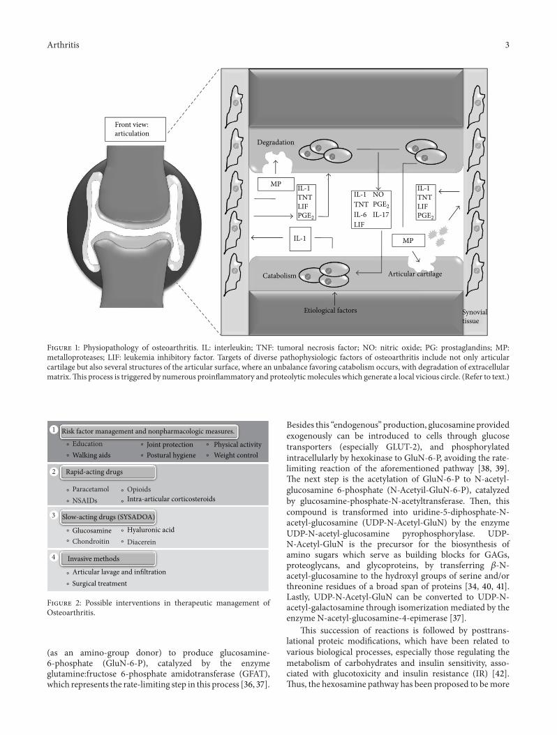
Notably, Aspden et al. [16] have suggested consideringOA as a systemic disease, where the main disruption wouldinvolve lipid metabolism and stromal cell differentiation,a concept stemmed from the common embryologic originshared by all structures constituting the articular cavity.Nonetheless, current views remain focused on the localpathology, where novel pathophysiologic pathways and fac-tors are constantly discovered, generating potential therapeu-tic targets [17]; Figure 1 depicts the main pathophysiologicroutes of OA.
3. Therapeutic Management of Osteoarthritis
Along with pharmacologic agents, nonpharmacologic mea-sures remain a cornerstone of OA treatment, fundamentally,the management of all risk factors involved and possiblecomorbidities such as obesity, diabetes, and menopause [18].
Therefore, patient education, physical activity, physiotherapy,articular protection, postural hygiene, and weight control areessential injury- and pain-limiting tools, which are generallyincluded in all clinical guidelines for the integralmanagementof OA patients, albeit receiving varying degrees of recom-mendation throughout different regions worldwide [5, 18–21].
On the other hand, the main objective of drug use inOA is symptomatic management, reducing both pain andunderlying inflammation [22]. Various management guide-lines have categorized these drugs as Symptom ModifyingOsteoarthritis Drugs (SMOADS) [23, 24] which are dividedinto 2 subgroups:
(i) rapid-acting drugs including analgesics, nonsteroidalanti-inflammatory drugs (NSAIDs), and intraarticu-lar glucocorticoids and opioids;
(ii) slow-acting drugs or SYSADOA (Symptomatic SlowActing Drugs for Osteoarthritis).
Regarding the first subgroup, paracetamol is consideredthe initial drug for the management of knee OA [25, 26],with NSAIDs being broadly recommended if no satisfactoryresults are observed after first-line management, althoughtheir adverse effect profiles should be considered prior toprescription [27, 28]. Lastly, intra-articular opioids and glu-cocorticoids are only implemented in very specific situationswhere initial treatment has been inefficient [19]. In general,utilization of drugs in this subgroup depends on their safetyprofile, patient consent, cost-effectiveness, and other factorsrelevant to the specific clinical evolution of patients [5, 23].
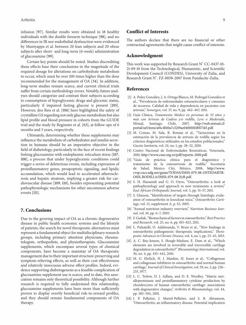
Findings reported in the 90s decade about articularcartilage, its metabolic activity, and regenerative capacity [29,30] have led to the proposal of chondromodulating and/orchondroprotective substances, which constitute the groupof slow-acting drugs or SYSADOA, including cartilaginousmatrix precursors (glucosamine, chondroitin, and hyaluronicacid) and cytokine modulators (diacerein and metallopro-tease inhibitors) [24]. These drugs, particularly glucosamine(GluN), have raised controversy regarding their utilization,due to inconsistencies in findings on their effectiveness in thetreatment of these patients [19, 31, 32]. Figure 2 summarizesthe therapeutic management of OA patients.
4. What Is Glucosamine? Molecular Aspects
GluN (2-amino-2-deoxy-D-glucose) is an aminomono-saccharide derived principally from chitin, a compoundfound in the exoskeleton of certain marine invertebrates[33]. GluN is an essential noncellular component ofconnective tissue, cartilage, ligaments, and other structures[24, 34] (Figure 3). The main compounds including GluNare glucosamine hydrochloride, glucosamine sulfate, andN-acetylglucosamine [34]. The latter can be organicallysynthesized through the hexosamine pathway, an alternativemetabolic route to glycolysis, which is esteemed to consumeup to 5% of glucose in adipocyte cultures [35].
Thismetabolic pathway is essential for the biosynthesis ofamino sugars, utilizing fructose-6-phosphate and glutamine
Arthritis 3
Degradation
Catabolism Articular cartilage
Front view:articulation
MP
MP
Etiological factors
IL-1
IL-1
TNT IL-1TNTLIF
LIFPGE2
IL-1TNTLIFPGE2
PGE2
IL-6
NO
IL-17
Synovial tissue
Figure 1: Physiopathology of osteoarthritis. IL: interleukin; TNF: tumoral necrosis factor; NO: nitric oxide; PG: prostaglandins; MP:metalloproteases; LIF: leukemia inhibitory factor. Targets of diverse pathophysiologic factors of osteoarthritis include not only articularcartilage but also several structures of the articular surface, where an unbalance favoring catabolism occurs, with degradation of extracellularmatrix.This process is triggered by numerous proinflammatory and proteolyticmolecules which generate a local vicious circle. (Refer to text.)
Risk factor management and nonpharmacologic measures.
Rapid-acting drugs
Slow-acting drugs (SYSADOA)
Invasive methods
Glucosamine
Articular lavage and infiltrationSurgical treatment
Hyaluronic acid
Intra-articular corticosteroids
Education
ParacetamolNSAIDs
Chondroitin
Opioids
Diacerein
1
2
3
4
Walking aidsJoint protectionPostural hygiene
Physical activityWeight control
∘
∘
∘
∘
∘
∘
∘
∘
∘
∘
∘
∘
∘
∘
∘
∘
Figure 2: Possible interventions in therapeutic management ofOsteoarthritis.
(as an amino-group donor) to produce glucosamine-6-phosphate (GluN-6-P), catalyzed by the enzymeglutamine:fructose 6-phosphate amidotransferase (GFAT),which represents the rate-limiting step in this process [36, 37].
Besides this “endogenous” production, glucosamine providedexogenously can be introduced to cells through glucosetransporters (especially GLUT-2), and phosphorylatedintracellularly by hexokinase to GluN-6-P, avoiding the rate-limiting reaction of the aforementioned pathway [38, 39].The next step is the acetylation of GluN-6-P to N-acetyl-glucosamine 6-phosphate (N-Acetyil-GluN-6-P), catalyzedby glucosamine-phosphate-N-acetyltransferase. Then, thiscompound is transformed into uridine-5-diphosphate-N-acetyl-glucosamine (UDP-N-Acetyl-GluN) by the enzymeUDP-N-acetyl-glucosamine pyrophosphorylase. UDP-N-Acetyl-GluN is the precursor for the biosynthesis ofamino sugars which serve as building blocks for GAGs,proteoglycans, and glycoproteins, by transferring 𝛽-N-acetyl-glucosamine to the hydroxyl groups of serine and/orthreonine residues of a broad span of proteins [34, 40, 41].Lastly, UDP-N-Acetyl-GluN can be converted to UDP-N-acetyl-galactosamine through isomerization mediated by theenzyme N-acetyl-glucosamine-4-epimerase [37].
This succession of reactions is followed by posttrans-lational proteic modifications, which have been related tovarious biological processes, especially those regulating themetabolism of carbohydrates and insulin sensitivity, asso-ciated with glucotoxicity and insulin resistance (IR) [42].Thus, the hexosamine pathway has been proposed to bemore

4 Arthritis
H
H
H
H
H
CCC
C
C
OH
OH
NH2
CH2OHC C
H
H
H
HC
C
OHOH
NH2
CH2OH
CHOH
O
O
HO
Proteoglycan
N-glucosamine
Figure 3: Chemical structure of glucosamine.
than a simple glucosensor, as it may be a potential mediatorimplicated in the pathogenesis of Type 2 Diabetes Mellitus(T2DM) [35, 43]. The main reactions in the hexosaminepathway are shown in Figure 4.
The rate-limiting step in the regulation of this routeinvolves GFAT, which is the only ammonium-independentenzyme of the amidotransferase subfamily [44]. It is alsostrongly inhibited by the final product of this metabolicpathway (UDP-N-Acetyl-GluN) through an allosteric mech-anism [45]. Therefore, its activity is influenced by UDP-N-Acetyl-GluN intracellular concentrations and intensified byProtein Kinase A (PKA)-dependent phosphorylation [46,47]. Moreover, its affinity for fructose-6-phosphate is low, sothe concentration of this substrate plays an important role inthe start-up of this reaction [40].
Ultimately, the plasmatic concentration of glucosamine inhealthy subjects is approximately 0.04mmol/L, rising up to0.06mmol/L in those taking common doses of glucosaminesupplements [34, 48]. It should be noted that the oralroute offers only 20% the plasmatic concentrations whichwould be obtained through intravenous administration[34, 49, 50]. It has been suggested that the pharmacokineticsand pharmacodynamics of glucosamine in humans closelyresemble those of experimental rat models [51].
5. Glucosamine: Effective forOsteoarticular Disease?
When evaluating the effectiveness of a drug or therapeutictool, it is important to consider the target variables susceptibleto modification or “end points,” which in the case of clinicalassays on patients with knee OA are represented mainly bypain andmeasurement of articular space [52]. Based on theseand other manifestations, several indices or score systemshave been created to allow researchers to assess the severityand evolution of the disease when under a given therapy[53, 54],TheWesternOntario andMcMaster Universities OA(WOMAC) index and the Lequesne functional severity indexare some of the most frequently used across various clinicalassays [52].
Another aspect worth considering when assessing effec-tiveness is the type of supplement prescribed; currently,glucosamine hydrochloride and sulfate are the most com-mercialized in our country and worldwide [55]. However,several studies have reported that when comparing bothformulations, glucosamine sulfate exhibits more favorableresults, especially in its crystalline form [24, 56, 57].These dif-fering formulations, as well as differences in pharmaceuticalmanufacturing, are responsible for distinct pharmacokinetic

Arthritis 5
P
P
P
P
P
P
P
HK
UDPGalNAc UDP-GlucNAc
GlucN-6-
GlucNAc-1-
GlucNAc-6-UDPPi
Glu
Glucose
ADP ATP
Glucose
Glycolysis
Proteoglycans
Glycolipids
Glycoproteins
GFAT
Gluc-6-
Fruc-6-
Fruc-6-Glutamine
Glucosamine
Glucosamine
Glycogensynthesis
GlucN-6-
Figure 4: Glucosamine biosynthesis. HK: hexokinase; Gluc-6-P: glucose-6-phosphate; Fruc-6-P: fructose-6-phosphate; GFAT: glucosaminefructose-6-phosphate amindotransferase; GlucN-6-P: Glucosamine-6-phosphate; GlucNAc-6-P: N-acetyl-glucosamine-6-phosphate;GlucNAc-1-P: N-acetyl-glucosamine-1-phosphate; UDPGalNAc: uridine diphosphate (UDP)-N-acetyl-galactosamine; UDP-GlucNAc:UDP-N-acetyl-glucosamine. Glucosamine may be obtained from exogenous supplements, or it may be endogenously synthesized fromglucose through the hexosamine pathway, an alternate pathway to glycolysis. Its product is uridine 5-diphosphate-N-acetyl-glucosamine(UDP-N-Acetyl-GluN), which is a precursor for glycosaminoglycans, proteoglycans, and glycoproteins. (Refer to text.)
features which must be taken into consideration, as theycould influence comparisons between reports [58].
Regarding dosages, although each presentation showsspecific characteristics, therapeutic effects are obtained withdoses ranging between 1,250–1,500mg daily [59, 60]. Astheir name implies, SYSADOA offer a slow onset of relief—approximately 2 weeks—and their effects may remain activefor as long as 2 months after their omission [61]. Notably,the European Medicines Agency has suggested that at least6 months of treatment are required for the evaluation ofSYSADOA effectiveness for articular pain and 2 years arenecessary to assess modifications of articular structures [58].
All elements considered a true overarching feature ofresearch in the evaluation of these supplements as theiroverwhelming heterogeneity with respect to objectives, for-mulations, combinations with other compounds, and time ofuse, amongmany other variables of utmost importance whencomparing studies. Indeed, the heterogeneity in outcomemeasures is particularly noteworthy and unjustified, con-sidering most rheumatologic diseases have been discussedin OMERACT conferences, whose purpose is unificationof evaluation criteria for clinical assays in this field [62].RegardingOA, sinceOMERACT3 in 1996, the 3main aspectsto be evaluated in all Phase III studies are pain, physicalfunction, and global patient assessment, as per the simplifiedOARSI criteria in each of its scenarios and thus allowingfor result unification and facilitating comparisons betweenstudies. Only after considering these fundamental aspects canthe novel variables in OA progression be considered [52].
This line of research ranges from clinical assays tometa-analyses, encompassing hundreds of patients (Table 1).Parallel studies by Reginster et al. and Pavelka et al. [63, 64]
demonstrated the disease-modifying ability of glucosaminesulfate supplements, by ascertaining improvement of symp-tomatology and prevention of articular space loss in kneeOA patients at a 3-year follow-up. Furthermore, results froma subsequent follow-up on these patients at an average of5 years suggested that treatment with glucosamine sulfatefor at least 12 months may prevent the need for kneearthroplasty, revealing the profound extent of the disease-modifying power of this compound [65].
The effects in the short-medium termhave been evaluatedby studies such as the GUIDE Trial [66], which confirmsprevious reports regarding the significant improvement glu-cosamine sulfate yields on symptoms of kneeOA, in the rangeor even superior to what exerted by a first line NSAID oracetaminophen.
Nevertheless, it must be noted that in other reports,benefits do not seem to be present in all analyzed subjects, butonly in specific subgroups with distinctive clinical features.This has been exemplified by Clegg et al. [67], who after uti-lizing glucosamine hydrochloride in their study—a valuablemethodological aspect for the comparison of results [58]—could not prove this version of the supplement to reducepain after 24 weeks in knee OA patients with mild articularpain. These variations in the utilized supplements are indeedvery influential. Great-scale research has shown that theuse of different commercial brands could factor into results,as suggested by Towheed et al. [31], in their meta-analysisof over 20 randomized controlled trials; only formulationsof glucosamine sulfate manufactured by Rotta Laboratoriesdisplayed effectiveness in the symptomatic management ofpatients with OA of the knee, while with other presentations,no statistically significant results were obtained.

6 Arthritis
Table1:Stud
ieso
nthee
ffectivenesso
fGlucosaminefor
Oste
oarthritis.
Author
[reference]
Samplea
nd/ora
mou
ntof
trials
Supp
lementtype,do
ses
Con
clusio
ns
Reginstere
tal.[63]
Patie
ntstreated
with
GS:106,
placebo:106
GS(1500m
gOD)
Inthisrand
omized,dou
ble-blindplacebo-controlledtrial,tre
atmentw
ithGSpreventedlossof
artic
ular
spacea
ndim
proved
symptom
sasa
ssessedby
WOMAC
scoringata3
-yearfollow-up.
Pavelkae
tal.[64]
Patie
ntstreated
with
GS:101,
placebo:101
GS(1500m
gOD)
Thisrand
omized,dou
ble-blindplacebo-controlledtrialalso
foun
dtre
atmentw
ithGSto
preventlosso
farticular
spacea
ndam
elioratep
ainas
assessed
byWOMAC
scoringandthe
Lequ
esne
Indexata3
-yearfollow-up.
Bruyeree
tal.[65]
Patie
ntstreated
with
GS:144,
placebo:131
GS(1500m
gOD)
Thisplacebo-controlledprospectives
tudy
suggeststhattre
atmentw
ithGSfora
tleast12
mon
thsa
ndup
to3yearsm
aypreventthe
need
toperfo
rmkn
eearthroplastyin
anaverage
follo
w-upof
5yearsa
fterd
rugdiscon
tinuatio
n.
Herrero-Beaum
ont
etal.[66]
Patie
ntstreated
with
GS:106,
acetam
inop
hen:
108,placebo:104
GS(1500m
gOD),
acetam
inop
hen
(3gOD)
Inthisrand
omized,dou
ble-blind,placebo-controlledstu
dy,dailyconsum
ptionof
1500
mgof
GSproved
moree
ffectivethanplaceboin
thes
ymptom
aticmanagem
ento
fkneeO
A.
Non
etheless,the
effectsof
acetam
inop
henweres
imilar.
Clegg
etal.[67]
Patie
ntstreated
with
GH:317,
CS:318,
GH+CS
:317,
celecoxib:318,
Placebo:313
GH(1500m
gOD)
Inthisrand
omized,dou
bled
ummystu
dy,neither
treatmentw
ithglucosam
inea
lone
nor
combinedwith
CSredu
cedpain
inthea
verage
OApatie
ntgrou
pdu
ring24
weeks.H
owever,
combinedtherapymay
beeffectiv
eintheg
roup
ofpatie
ntsw
ithmod
eratetosevere
knee
pain.
McA
lindo
netal.[68]
15rand
omized,dou
ble-blind,
placebo-controlledtrialswith≥4
weeks
oftre
atment
GS,GH,orS
Cversus
Placebo
Inthismeta-analysis,
trialsindicatethesec
ompo
unds
have
amod
eratetostr
ongeffecto
verO
Asymptom
s,bu
tmetho
dology
issuesm
ayexaggeratethisbeneficialeffect.N
otwith
stand
ingthis,
they
appear
tobe
safeandhave
apositive
impactover
symptom
atolog
y.
Towheed
etal.[31]
25rand
omized,con
trolledtrials
(4963patie
nts)
GS(1500m
gOD)
Thismeta-analysissuggeststhatGSpreparations
byRo
ttaLabo
ratorie
smay
bemoree
ffective
than
placeboin
them
anagem
ento
fpainandartic
ular
functio
nalityas
assessed
bythe
Lequ
esne
Indexin
subjectswith
symptom
aticOANevertheless,otherformulations
didno
tshow
anyeffectiv
eness(statisticalsig
nificance)atm
anagingpain
norfun
ctionalityor
rigidity
asassessed
bytheW
OMAC
scale.
Wandeletal.[69]
10larges
calerand
omized
placebo-controlledtrials
(3803patie
nts)
GS,GH,orS
Cversus
placebo
Thismeta-analysissuggestsneith
erglucosam
inen
orchon
droitin
alon
e,no
rtheircombinatio
n,isableto
redu
ceartic
ular
pain,n
orcanthey
mod
ifyartic
ular
spaceincomparis
onto
placebo.
OD:onced
aily;G
S:glucosam
ines
ulfate;C
S:chon
droitin
sulfate;O
A:oste
oarthritis;GH:glucosamineh
ydrochlorid
e.

Arthritis 7
In addition to these findings, one of the most con-troversial reports surrounding the effectiveness of thesesupplements was issued by Wandel et al. [69], who intheir meta-analysis of 10 large-scale randomized controlledtrials (3,803 patients) concluded that neither glucosaminenor chondroitin sulfate, neither alone nor combined, couldsignificantly improve pain nor reduction of articular spacewhen compared to placebo, consequently arguing againstprescribing these agents in patients withOA.This paper arosenumerous criticisms from several specialists and experts inthe matter [70, 71], who fundamentally refuted the method-ology used in their recollection and analysis of data, as well astheir results, sustaining such claims by emphasizing the greatvariability and heterogeneity of the studies analyzed [58].Notably, in the report of a post-publication meeting, the BMJeditor withdrew support from the inappropriate conclusionsof this meta-nalysis, which were not adequately supportedby their data. This illustrates the high degree of controversyattributed to the utilization of these compounds in patientswith OA [60].
Although most studies tend to favor the effectiveness ofthese compounds in subjects with OA at least through min-imal or indirect evidence, especially as disease-progressionmodulators [72], no evidence exists of chondroprotectiveeffects of glucosamine in a preventive context [73]. This fitswith the main findings of in vitro studies, which suggesta predominantly anticatabolic effect in cell cultures [74].Several molecular mechanisms are implied, including theinhibition of catabolic enzymes, such as metalloproteases,phospholipase A
2, and aggrecanase-2 as well as the rkversal
of the effects of IL-1𝛽 and cyclooxigenase-2, and inhibi-tion of NF-𝜅B signaling [75–77]. This impact in energeticmetabolism and oxidative stress appears to be triggerednot only with the consumption of glucosamine alone, butalso when accompanied with chondroitin sulfate [78]. Theseeffects have been observed to be more consistent withglucosamine sulfate rather than hydrochloride [79].
Nevertheless, a great proportion of these experimentalreports employ glucosamine concentrations much higherthan those obtained through the oral ingestion of supple-ments, hindering the extrapolation of these findings to in vivostudies [80]. Regarding studies in animalmodels, findings aresimilar to in vitro results, with modifications predominantlyin synovial inflammation, cartilage degradation, and boneresorption, primarily through the repression of proinflam-matory cytokine genes [81]. Ultimately, the heterogeneityin experimental reports resembles its clinical counterpart,with important differences in the types of supplements used,as well as doses and other characteristics which should beunified in future studies. Harmonizing these criteria is apriority in order to accurately and successfully extrapolatemolecular mechanisms to human subjects in the clinicalscenario.
6. Glucosamine: Safety on Glucose Metabolism
Amidst the few adverse effects reported regarding glu-cosamine supplements [20], the most common are gas-trointestinal complaints, including pain, diarrhea, nausea,
and pyrosis [82]. On the other hand, although no fulminantevents have been described, several cases of allergic reac-tions have been documented, including angioedema [83],asthmatic crises [84], and photosensitivity [85]. Lastly, muchlike the controversy surrounding their potential efficacy astherapeutic agents, the consequences of these supplements oncarbohydrate metabolism and insulin levels have become oneof the most debated topics in rheumatology in recent years[86].
The key point in this matter resides in several findingswhich associate the hexosamine pathway with the develop-ment of IR, with reports as early as the year 1991, whenMarshall et al. [35] outlined such a hypothesis after exploringthe role of glutamine in their experimental models for IR. Asdescribed previously, the final product of thismetabolic path-way isUDP-N-Acetyl-GluN (Figure 4), which is precursor forGAG, proteoglycans and glycoproteins. It must be noted thatthesemacromolecules are synthesized in specific cytoplasmicorganelles (endoplasmic reticulumandGolgi apparatus) [87],while in the nucleus and cytosol, UDP-N-Acetyl-GluN alsoserves as a substrate for the enzymatic action of O-N-Acetyl-GluN transferase (OGT), which is able to transfer N-Acetyl-GluN to the serine/threonine residues of various proteinsin a process known as reversible posttranslational proteicmodification [88]. The target proteins of this process includethe insulin receptor substrate (IRS) types 1 and 2, as well asGLUT-4 [89, 90]. Numerous research groups suggest, albeitnot in a definitive way, that thesemodificationsmay representthe molecular basis for IR associated with the hexosaminepathway [87], since it may antagonize the phosphorylationcascade of insulin signaling [42, 90].
Besides these cytosolic mechanisms, O-GluNacylationmay also target many transcription factors, therefore regulat-ing the expression of proteins in the nucleus [91, 92]. Severaltranscription factors have been shown to be involved throughexperimentalmodels aswell as genes such as those of glucose-6-phosphatase and phosphoenolpyruvate carboxykinase, keyenzymes from the gluconeogenesis pathway [93]. Thus, agreat proportion of current research is focused on the roleplayed by OGT in post-translational modifications, as itseffects do not seem to be limited to insulin signaling—acting as “metabosensor” mechanism—and it may be partof a wide array of alarm responses or stress reactions in thecardiovascular system [39, 94–96].
Despite these findings in animal models, reports inhumans stand divided and although some research suggestsmetabolic effects for these supplements [50, 104], mostclinical trials and meta-analyses suggest this link is not asmuch clear in humans (Table 2). Moreover, a great part ofthese studies—including clinical assays [100, 101] and meta-analyses [102, 103]—were carried out on subjects with alreadyimpaired glucose metabolism, obscuring the interpretationof their results. Nevertheless, several studies have failed tofind associations between GluN administration and insulinresistance as assessed by its gold standard test, the euglycemicinsulin clamp (EIC). Such is the case of Monauni et al.,with their study on 10 healthy subjects who were assessedthrough determination of glycemia, application of glucosetolerance test, and the EIC while undergoing glucosamine

8 Arthritis
Table2:Stud
iesrelatingGlucosamineu
sewith
theo
nsetof
Insulin
Resis
tance.
Author
[reference]
Samplea
nd/ora
mou
ntof
trials
Supp
lementtype,
doses.
Con
clusio
ns
Mon
auni
etal.[97]
10healthysubjects
Glucosamine
(infusio
n:1.6
micromol/m
in−1 /k
g−1 5
micromol/m
in−1 /k
g−1 )
Type
notspe
cifie
d
IVGTT
andEICwerep
erform
eddu
ringeither
asalineinfusionor
alow
(1.6micromol)o
rhigh
(5micromol)G
luNinfusio
n.GluNdidno
tchangeg
lucose
utilizatio
nor
intracellular
metabolism
,nor
diditaffectreadilyreleasableinsulin
levels,
GSIS,or
thetim
econ
stant
ofsecretion,bu
titincreased
both
theg
lucose
thresholdof
GSISandplasmafastin
gglucose.Th
ese
effectswerep
resent
athigh
GluNdo
ses.
Pouw
elsetal.[98]
18healthysubjects
GS
(infusio
n:4m
icromol/dL)
EICwas
perfo
rmed
throug
hout
atleast300
minutes
durin
ginfusio
nof
GluN(4
micromol/dL);
90–240
min,0–300
min,ord
uringsalin
einfusion.
GluNhadno
effecto
ninsulin
-indu
ced
glucoseu
ptake.
Mun
iyappa
etal.[99]
40lean
subjectsand40
obese
subjects
GH(500
mgTID)
versus
Placebo
Thisstu
dyfoun
dno
differences
inEICbetweenpatie
ntsreceiving
GHandplaceboaft
er6
weeks
oftherapy,with
inbo
ththeleanandob
eseg
roup
s.
Biggee
etal.[50]
16patie
ntsw
ithexclu
sive
diagno
seso
fOA,treated
with
GS
GS(1500m
gOD)
Inthisstu
dy,carrie
dou
tinsubjectswith
outany
metabolicdisorders(such
asTD
M2andIFG),
3ou
tof16individu
alsd
isplayeddisrup
tionof
oralglucosetolerance
after
treatmentw
ithGS.
Thissuggeststhen
ecessityfora
perio
dof
timefor
thissupp
lementsto
exertm
etabolic
mod
ificatio
nsin
thisgrou
pof
patie
ntsa
swellastria
lsin
poorlycontrolledsubjects.
Scroggieetal.[100]
Patie
ntstreated
with
GC:
22,
placebo:12
GH(1500m
gOD)+
CS(1200m
gOD)
versus
Placebo
Thisrand
omized,dou
ble-blind,placebo-controlledclinicaltria
lcarrie
dou
tinpatie
ntsw
ithcontrolledTD
M2determ
ined
thattheo
raladm
instratio
nof
GHatrecommendeddo
sesd
idno
talterg
lycemiccontrolinthisgrou
pof
patie
nts.
Albertetal.[101]
12patie
ntsinar
ando
mized,
doub
le-blin
d,placebo-controlled,
cross-over
trial
Glucosamine
(1500m
gOD)
Type
notspe
cifie
d
Thisstu
dyinferred
glucosam
inea
tcom
mon
lyused
doses,may
notsignificatively
affect
glycem
iccontrol,lip
idprofi
le,or
apoA
Ilevels
indiabeticpatie
ntsa
fter2
weeks
oftre
atment.
Simon
etal.[102]
23trialswith
different
metho
dologies:
glucosam
ineIV
(infused):2,
oralglucosam
ine:21
GSor
GHversus
placebo
Thismeta-analysisinclu
dedtrialswith
both
IVandoralform
ulations,and
even
long
-term
repo
rts,concluding
thatglucosam
inec
onsumptionathabitualdo
sesm
ayno
taffectthe
metabolism
ofno
rmoglycemic,“prediabetic,”or
diabeticsubjectsandthatcurrently
nodefin
itive
motives
arev
alid
fortheirrestric
tionin
theseg
roup
sofind
ividuals.
Dostro
vsky
etal.[103]
11trialswith
different
metho
dologies:
RCT:
6,prospectives
tudies:5
GS,GH,orS
Cversus
placebo
Thismeta-analysishigh
lighted
3trialswhere
OAglucosam
inew
asused,followed
bymod
ificatio
nsin
insulin
sensitivityandbasalglycemia.A
ddition
ally,
studies
thatinclu
ded
subjectswith
IFGor
IRshow
edgreaterimpactover
carboh
ydratemetabolism
.Thus,this
popu
latio
nshou
ldbe
atargeto
ffurther
research.
IVGTT
:intraveno
usglucosetolerancetest;
EIC:
euglycem
icinsulin
clamp;
GluN:glucosamine;.
GSIS:glucose-stimulated
insulin
secretion;
TID:three
times
aday;OD:oncedaily
;GS:glucosam
inesulfate;C
S:chon
droitin
sulfate;O
A:oste
oarthrosis;
GH:glucosaminehydrochloride;TD
M2:type
2diabestesm
ellitus;apo
AI:apolipop
rotein
AI;IFG:impaire
dfasting
glucose;IV:intraveno
us;R
CT:rando
mized
controlled
trials;
AO:adm
inistered
orally;IR:
insulin
resis
tance.
Arthritis 9
infusion [97]. Similar results were obtained in 18 healthyindividuals with the double forearm technique [98], and nodifferences in IR nor endothelial dysfunction were evidencedby Muniyappa et al. between 20 lean subjects and 20 obesesubjects after short- and long-term (6-week) administrationof glucosamine [99].
Certain key points should be noted. Studies discreditingthese effects base their conclusions in the magnitude of therequired dosage for alterations on carbohydrate metabolismto occur, which must be over 100 times higher than the doserecommended for the management of OA [34]. In addition,long-term studies remain scarce, and current clinical trialssuffer from certain methodology errors. Notably, future anal-yses should categorize and contrast their subjects accordingto consumption of hypoglycemic drugs and glycemic status,particularly if impaired fasting glucose is present [105].However, dos Reis et al. [106] have highlighted the safety ofcrystallineGS regarding not only glucosemetabolismbut alsolipid profile and blood pressure in cohorts from the GUIDEtrial and the study by Reginster et al. [63], at follow-ups of 6months and 3 years, respectively.
Ultimately, determining whether these supplements mayinfluence the metabolism of carbohydrates and insulin secre-tion in humans should be an imperative objective in thefield of diabetology, particularly in the face of recent findingslinking glucosamine with endoplasmic reticulum stress [107,108], a process that under hyperglycemic conditions couldtrigger a series of deleterious events, including expression ofproinflammatory genes, proapoptotic signaling, and lipidicaccumulation, which would lead to accelerated atheroscle-rosis and hepatic steatosis, implying a greater risk for car-diovascular disease [109, 110], besides representing potentialpathophysiologic mechanisms for other uncommon adverseevents [111].
7. Conclusions
Due to the growing impact of OA as a chronic degenerativedisease in public health economic systems and the lifestyleof patients, the search for novel therapeutic alternatives mustrepresent a fundamental object formultidisciplinary researchgroups, including primary attention physicians, rheuma-tologists, orthopedists, and physiotherapists. Glucosaminesupplements, which encompass several types of chemicalcomponents, have become a mainstay of OA therapeuticmanagement due to their important structure-preserving andsymptom-relieving effects, as well as their cost effectivenessand relatively innocuous adverse effect profiles. Indeed, evi-dence supporting diabetogenesis as a feasible complication ofglucosamine supplement use is scarce, and to date, this asso-ciation remains only theoretical possibility. Although furtherresearch is required to fully understand this relationship,glucosamine supplements have been more than sufficientlyproven to display overtly beneficial risk-to-reward profiles,and they should remain fundamental components of OAtherapy.
Conflict of Interests
The authors declare that there are no financial or othercontractual agreements that might cause conflict of interests.
Acknowledgment
This work was supported by Research Grant N∘ CC-0437-10-21-09-10 from the Technological, Humanistic, and ScientificDevelopment Council (CONDES), University of Zulia, andResearch Grant N∘. FZ-0058-2007 from Fundacite-Zulia.
References
[1] A. Poley Gonzalez, J. A. Ortega Blanco,M. Pedregal Gonzalez etal., “Prevalencia de enfermedades osteoarticulares y consumode recursos. Calidad de vida y dependencia en pacientes conartrosis,” Semergen, vol. 37, no. 9, pp. 462–467, 2011.
[2] Guıa Clınica, Tratamiento Medico en personas de 55 anos ymas con Artrosis de Cadera y/o rodilla, Leve o Moderada,Minsal, Santiago, Chile, 2007, http://web.minsal.cl/portal/url/item/a01c4b10a7c5219ae04001011f017145.pdf.
[3] M. Comas, M. Sala, R. Roman et al., “Variaciones en laestimacion de la prevalencia de artrosis de rodilla segun loscriterios diagnosticos utilizados en los estudios poblacionales,”Gaceta Sanitaria, vol. 24, no. 1, pp. 28–32, 2010.
[4] Centro Nacional de Enfermedades Reumaticas, Venezuela,2010, http://www.cner.org.ve/pdf/reporte 2010.pdf.
[5] “Guıa de practica clınica para el diagnostico ytratamiento de la osteoartrosis de rodilla,” Secretariade Salud, Mexico City, Mexico, 2008, http://www.cvsp.cucs.udg.mx/guias/TODAS/IMSS 079 08 OSTEOARTR-OSIS RODILLA/IMSS 079 08 EyR.pdf.
[6] S. H. Hassanali and G. O. Oyoo, “Osteoarthritis: a look atpathophysiology and approach to new treatments: a review,”East African Orthopaedic Journal, vol. 5, pp. 51–57, 2011.
[7] S. Glasson, “Identification of targets through histologic evalu-ation of osteoarthritis in knockout mice,” Osteoarthritis Carti-lage, vol. 13, supplement A, p. S3, 2005.
[8] “Annual nutrition industry overview,” Nutrition Business Jour-nal, vol. 10, pp. 6–7, 2005.
[9] F.Guilak, “Biomechanical factors in osteoarthritis,”Best Practiceand Research, vol. 25, no. 6, pp. 815–823, 2011.
[10] L. Pulsatelli, O. Addimanda, V. Brusi et al., “New findings inosteoarthritis pathogenesis: therapeutic implications,” Thera-peutic Advances in Chronic Disease, vol. 4, no. 1, pp. 23–43, 2013.
[11] A. C. Bay-Jensen, S. Hoegh-Madsen, E. Dam et al., “Whichelements are involved in reversible and irreversible cartilagedegradation in osteoarthritis?”Rheumatology International, vol.30, no. 4, pp. 435–442, 2010.
[12] M. G. Ehrlich, H. J. Mankin, H. Jones et al., “Collagenaseand collagenase inhibitors in osteoarthritic and normal humancartilage,” Journal of Clinical Investigation, vol. 59, no. 2, pp. 226–233, 1977.
[13] L. C. Tetlow, D. J. Adlam, and D. E. Woolley, “Matrix met-alloproteinase and proinflammatory cytokine production bychondrocytes of human osteoarthritic cartilage: associationswith degenerative changes,” Arthritis & Rheumatology, vol. 44,pp. 585–594, 2001.
[14] J. P. Pelletier, J. Martel-Pelletier, and S. B. Abramson,“Osteoarthritis, an inflammatory disease. Potential implication

10 Arthritis
for the selection of new therapeutic targets,” Arthritis &Rheumatology, vol. 44, pp. 1237–1247, 2001.
[15] J. P. Schroeppel, J. D. Crist, H. C. Anderson, and J. Wang,“Molecular regulation of articular chondrocyte function and itssignificance in osteoarthritis,”Histology andHistopathology, vol.26, no. 3, pp. 377–394, 2011.
[16] R. M. Aspden, B. A. A. Scheven, and J. D. Hutchison,“Osteoarthritis as a systemic disorder including stromal celldifferentiation and lipid metabolism,” The Lancet, vol. 357, no.9262, pp. 1118–1120, 2001.
[17] W. B. van den Berg, “Osteoarthritis year 2010 in review:pathomechanisms,” Osteoarthritis and Cartilage, vol. 19, no. 4,pp. 338–341, 2011.
[18] M. V. Goycochea, V. M. Lopez, M. Colin-Marin et al., “Guıaclınica en osteoartrosis de rodilla y cadera,” Revista Medica delInstitutoMexicano del Seguro Social, vol. 41, suplement, pp. S99–S107, 2003.
[19] The Royal Australian College of General Practitioners,Guideline for the Non-Surgical Management of Hipand Knee Osteoarthritis, 2009, http://www.nhmrc.gov.au/files nhmrc/publications/attachments/cp117-hip-knee-osteo-arthritis.pdf.
[20] W. Zhang, R. W. Moskowitz, G. Nuki et al., “OARSI recom-mendations for the management of hip and knee osteoarthritis,part II: OARSI evidence-based, expert consensus guidelines,”Osteoarthritis and Cartilage, vol. 16, no. 2, pp. 137–162, 2008.
[21] R. Christensen, E. M. Bartels, A. Astrup, and H. Bliddal, “Effectof weight reduction in obese patients diagnosed with kneeosteoarthritis: a systematic review and meta-analysis,” Annalsof the Rheumatic Diseases, vol. 66, no. 4, pp. 433–439, 2007.
[22] T. P. Stitik, E. Altschuler, and P. M. Foye, “Pharmacotherapyof osteoarthritis,” American Journal of Physical Medicine andRehabilitation, vol. 85, supplement, no. 11, pp. S15–S28, 2006.
[23] K. M. Jordan, N. K. Arden, M. Doherty et al., “EULARrecommendations 2003: an evidence based approach to themanagement of knee osteoarthritis: report of a Task Force of theStanding Committee for International Clinical Studies Includ-ing Therapeutic Trials (ESCISIT),” Annals of the RheumaticDiseases, vol. 62, no. 12, pp. 1145–1155, 2003.
[24] E. Mongil, I. Sanchez, F. Torre et al., “Farmacos de accion lenta(Sysadoa) en el tratamiento de la osteoartrosis,” Revista de laSociedad Espanola del Dolor, vol. 7, pp. 485–496, 2006.
[25] B. L. Kidd, R. M. Langford, T. Wodehouse et al., “Currentapproaches in the treatment of arthritic pain,”Arthritis ResearchandTherapy, vol. 9, no. 3, article 214, 2007.
[26] Ministry of Health Singapore (MOH), “Clinical prac-tice guidelines Osteoarthritis of the knee,” 2007,http://www.moh.gov.sg/cpg.
[27] National Prescribing Service (NPS), “Analgesic choicesin persistent pain. Prescribing Practice Review,” 2006,http://www.nps.org.au/publications/health-professional/prescribing-practice-review/2006/analgesic-choices-in-persistent-pain.
[28] E. M. Antman, J. S. Bennett, A. Daugherty, C. Furberg, H.Roberts, and K. A. Taubert, “Use of nonsteroidal antiinflamma-tory drugs: an update for clinicians: a scientific statement fromthe American Heart Association,” Circulation, vol. 115, no. 12,pp. 1634–1642, 2007.
[29] S. Tanaka, C. Hamanishi, H. Kikuchi, and K. Fukuda, “Factorsrelated to degradation of articular cartilage in osteoarthritis: areview,” Seminars in Arthritis and Rheumatism, vol. 27, no. 6, pp.392–399, 1998.
[30] F. J. Blanco, R. Guitian, E. Vazquez-Martul, F. J. de Toro, and F.Galdo, “Osteoarthritis chondrocytes die by apoptosis,” Arthritis& Rheumatology, vol. 41, pp. 284–289, 1998.
[31] T. E. Towheed, L. Maxwell, T. P. Anastassiades et al.,“Glucosamine therapy for treating osteoarthritis,” CochraneDatabase of Systematic Reviews, no. 2, Article ID CD002946,2005.
[32] S. C. Vlad, M. P. LaValley, T. E. McAlindon, and D. T. Felson,“Glucosamine for pain in osteoarthritis: why do trial resultsdiffer?” Arthritis and Rheumatism, vol. 56, no. 7, pp. 2267–2277,2007.
[33] E. A. Heath-Heckman and M. J. McFall-Ngai, “The occurrenceof chitin in the hemocytes of invertebrates,”Zoology, vol. 114, no.4, pp. 191–198, 2011.
[34] J.W.Anderson, R. J. Nicolosi, and J. F. Borzelleca, “Glucosamineeffects in humans: a review of effects on glucose metabolism,side effects, safety considerations and efficacy,” Food and Chem-ical Toxicology, vol. 43, no. 2, pp. 187–201, 2005.
[35] S. Marshall, V. Bacote, and R. R. Traxinger, “Discovery of ametabolic pathway mediating glucose-induced desensitizationof the glucose transport system: role of hexosamine in theinduction of insulin resistance,”The Journal of Biological Chem-istry, vol. 266, no. 8, pp. 4706–4712, 1991.
[36] R. Kornfeld, “Studies on L-glutamine D-fructose 6-phosphate amidotransferase. I. Feedback inhibition byuridine diphosphate-N-acetylglucosamine,” The Journal ofBiological Chemistry, vol. 242, no. 13, pp. 3135–3141, 1967.
[37] G. Wu, T. E. Haynes, W. Yan, and C. J. Meininger, “Pres-ence of glutamine:fructose-6-phosphate amidotransferase forglucosamine-6-phosphate synthesis in endothelial cells: effectsof hyperglycaemia and glutamine,” Diabetologia, vol. 44, no. 2,pp. 196–202, 2001.
[38] M. Uldry, M. Ibberson, M. Hosokawa, and B.Thorens, “GLUT2is a high affinity glucosamine transporter,” FEBS Letters, vol.524, no. 1–3, pp. 199–203, 2002.
[39] N. Fulop, R. B. Marchase, and J. C. Chatham, “Role of proteinO-linked N-acetyl-glucosamine in mediating cell function andsurvival in the cardiovascular system,”Cardiovascular Research,vol. 73, no. 2, pp. 288–297, 2007.
[40] E. D. Schleicher and C. Weigert, “Role of the hexosaminebiosynthetic pathway in diabetic nephropathy,” Kidney Interna-tional, Supplement, vol. 58, supplement 77, pp. S13–S18, 2000.
[41] G. D. Holt and G. W. Hart, “The subcellular distribution ofterminal N-acetylglucosamine moieties. Localization of a novelprotein-saccharidie linkage, O-linked GlcNAc,” The Journal ofBiological Chemistry, vol. 261, no. 17, pp. 8049–8057, 1986.
[42] R. J. Copeland, J.W. Bullen, andG.W.Hart, “Cross-talk betweenGlcNAcylation and phosphorylation: roles in insulin resis-tance and glucose toxicity,” American Journal of Physiology—Endocrinology and Metabolism, vol. 295, no. 1, pp. E17–E28,2008.
[43] W. B. Dias and G.W. Hart, “O-GlcNAcmodification in diabetesandAlzheimer’s disease,”Molecular BioSystems, vol. 3, no. 11, pp.766–772, 2007.
[44] S. Milewski, “Glucosamine-6-phosphate synthase: the multi-facets enzyme,” Biochimica et Biophysica Acta, vol. 1597, no. 2,pp. 173–192, 2002.
[45] G. L. McKnight, S. L. Mudri, S. L. Mathewes et al., “Molecularcloning, cDNA sequence, and bacterial expression of humanglutamine: fructose-6-phosphate amidotransferase,” The Jour-nal of Biological Chemistry, vol. 267, no. 35, pp. 25208–25212,1992.

Arthritis 11
[46] P. P. Sayeski and J. E. Kudlow, “Glucose metabolism to glu-cosamine is necessary for glucose stimulation of transforminggrowth factor-𝛼 gene transcription,” The Journal of BiologicalChemistry, vol. 271, no. 25, pp. 15237–15243, 1996.
[47] J. Zhou, Q. K. Huynh, R. T. Hoffman et al., “Regulation ofglutamine: fructose-6-phosphate amidotransferase by cAMP-dependent protein kinase,” Diabetes, vol. 47, no. 12, pp. 1836–1840, 1998.
[48] M. J. Pouwels, J. R. Jacobs, P. N. Span, J. A. Lutterman, P. Smits,andC. J. Tack, “Short-term glucosamine infusion does not affectinsulin sensitivity in humans,” Journal of Clinical Endocrinologyand Metabolism, vol. 86, no. 5, pp. 2099–2103, 2001.
[49] I. Setnikar and L. C. Rovati, “Absorption, distribution,metabolism and excretion of glucosamine sulfate: a review,”Arzneimittel-Forschung/Drug Research, vol. 51, no. 9, pp.699–725, 2001.
[50] B. A. Biggee, C. M. Blinn, M. Nuite, J. E. Silbert, and T. E.McAlindon, “Effects of oral glucosamine sulphate on serumglucose and insulin during an oral glucose tolerance test ofsubjects with osteoarthritis,” Annals of the Rheumatic Diseases,vol. 66, no. 2, pp. 260–262, 2007.
[51] A. Aghazadeh-Habashi, S. Sattari, F. Pasutto, and F. Jamali, “Sin-gle dose pharmacokinetics and bioavailability of glucosamine inthe rat,” Journal of Pharmacy and Pharmaceutical Sciences, vol.5, no. 2, pp. 181–184, 2002.
[52] T. Pham, D. V. der Heijde, M. Lassere et al., “Outcome variablesfor osteoarthritis clinical trials: the OMERACT-OARSI set ofresponder criteria,” Journal of Rheumatology, vol. 30, no. 7, pp.1648–1654, 2003.
[53] N. Bellamy, W. W. Buchanan, C. H. Goldsmith, J. Campbell,and L. W. Stitt, “Validation study of WOMAC: a health statusinstrument for measuring clinically important patient relevantoutcomes to antirheumatic drug therapy in patients withosteoarthritis of the hip or knee,” Journal of Rheumatology, vol.15, no. 12, pp. 1833–1840, 1988.
[54] M. Dougados, “Monitoring osteoarthritis progression and ther-apy,” Osteoarthritis and Cartilage, vol. 12, supplement A, pp.S55–S60, 2004.
[55] M. J. Divins, “Mercado de antiartrosicos,” Farmacia Profesional,vol. 24, no. 1, pp. 40–44, 2010.
[56] E. J. Uitterlinden, H. Jahr, J. L. M. Koevoet et al., “Glucosaminedecreases expression of anabolic and catabolic genes in humanosteoarthritic cartilage explants,” Osteoarthritis and Cartilage,vol. 14, no. 3, pp. 250–257, 2006.
[57] L. C. Rovati, F. Girolami, and S. Persiani, “Crystalline glu-cosamine sulfate in the management of knee osteoarthritis:efficacy, safety, and pharmacokinetic properties,” TherapeuticAdvances in Musculoskeletal Disease, vol. 4, no. 3, pp. 167–180,2012.
[58] R.Calvo, “Sulfato de glucosamina y condroitın sulfato, farmacospara el tratamiento de la artrosis, acusados de no presentareficacia clınica. ¿Culpables?” Gaceta Medica de Bilbao, vol. 109,no. 4, pp. 158–164, 2012.
[59] C. Black, C. Clar, R. Henderson et al., “The clinical effectivenessof glucosamine and chondroitin supplements in slowing orarresting progression of osteoarthritis of the knee: a systematicreview and economic evaluation,” Health Technology Assess-ment, vol. 13, no. 52, pp. 1–123, 2009.
[60] Sociedad Uruguaya de Reumatologıa (SUR), “Utilidad delos antiartrosicos de accion lenta en el tratamiento de laartrosis,” Carta Reumatologica, vol. 3, no. 1, pp. 1–40, 2012,
http://www.reumauruguay.org/web/images/carta reumato-logica 2012.pdf.
[61] F. Abad Santos, D. Ochoa, and A. Garcıa Garcıa, “Actualizacionde la eficacia de condroitın sulfato y sulfato de glucosaminaen el tratamiento de la artrosis,” Actualidad en farmacologıa yterapeutica, vol. 9, pp. 97–108, 2011.
[62] P. Tugwell, M. Boers, P. Brooks, L. Simon, V. Strand, and L.Idzerda, “OMERACT: an international initiative to improveoutcome measurement in rheumatology,” Trials, vol. 8, article38, 2007.
[63] J. Y. Reginster, R. Deroisy, L. C. Rovati et al., “Long-termeffects of glucosamine sulphate on osteoarthritis progression: arandomised, placebo-controlled clinical trial,” The Lancet, vol.357, no. 9252, pp. 251–256, 2001.
[64] K. Pavelka, J. Gatterova, M. Olejarova, S. Machacek, G. Gia-covelli, and L. C. Rovati, “Glucosamine sulfate use and delayof progression of knee osteoarthritis: a 3-year, randomized,placebo-controlled, double-blind study,” Archives of InternalMedicine, vol. 162, no. 18, pp. 2113–2123, 2002.
[65] O. Bruyere, K. Pavelka, L. C. Rovati et al., “Total joint replace-ment after glucosamine sulphate treatment in knee osteoarthri-tis: results of a mean 8-year observation of patients fromtwo previous 3-year, randomised, placebo-controlled trials,”Osteoarthritis and Cartilage, vol. 16, no. 2, pp. 254–260, 2008.
[66] G. Herrero-Beaumont, J. A. Roman Ivorra, M. D. C. Trabado etal., “Glucosamine sulfate in the treatment of knee osteoarthritissymptoms: a randomized, double-blind, placebo-controlledstudy using acetaminophen as a side comparator,” Arthritis andRheumatism, vol. 56, no. 2, pp. 555–567, 2007.
[67] D. O. Clegg, D. J. Reda, C. L. Harris et al., “Glucosamine,chondroitin sulfate, and the two in combination for painfulknee osteoarthritis,”The New England Journal of Medicine, vol.354, no. 8, pp. 795–808, 2006.
[68] T. E. McAlindon, M. P. La Valley, J. P. Gulin, and D. T. Felson,“Glucosamine and chondroitin for treatment of osteoarthritis: asystematic quality assessment and meta-analysis,” Journal of theAmerican Medical Association, vol. 283, no. 11, pp. 1469–1475,2000.
[69] S. Wandel, P. Juni, B. Tendal et al., “Effects of glucosamine,chondroitin, or placebo in patients with osteoarthritis of hip orknee: network meta-analysis,” British Medical Journal, vol. 341,Article ID c4675, 2010.
[70] G. Giacovelli and L. C. Rovati, “Glucosamine and osteoarthritis.Conclusions not supported by methods and results,” BritishMedical Journal, vol. 341, Article ID c6338, 2010.
[71] J. Reginster, R. D. Altman, and M. C. Hochberg, “Glucosamineand osteoarthritis. Prescribed regimen is effective,” BritishMedical Journal, vol. 341, Article ID c6335, 2010.
[72] J. Reginster, A. Neuprez, M. Lecart, N. Sarlet, and O. Bruyere,“Role of glucosamine in the treatment for osteoarthritis,”Rheumatology International, vol. 32, no. 10, pp. 2959–2967, 2012.
[73] Y. Herontin, X. Chevalier, G. Herrero-Beaumont et al., “Physio-logical effects of oral glucosamine on joint health: current statusand consensus on future research priorities,” BMC ResearchNotes, vol. 6, article 115, 2013.
[74] Y.Henrotin, A.Mobasheri, andM.Marty, “Is there any scientificevidence for the use of glucosamine in the management ofhuman osteoarthritis?” Arthritis Research and Therapy, vol. 14,no. 1, article 201, 2012.
[75] K. Imagawa, M. C. de Andres, K. Hashimoto et al., “The epige-netic effect of glucosamine and a nuclear factor-kappa B (NF-kB) inhibitor on primary human chondrocytes—implications

12 Arthritis
for osteoarthritis,” Biochemical and Biophysical Research Com-munications, vol. 405, no. 3, pp. 362–367, 2011.
[76] A. R. Shikhman, K. Kuhn, N. Alaaeddine, and M. Lotz,“N-acetylglucosamine prevents IL-1𝛽-mediated activation ofhuman chondrocytes,” Journal of Immunology, vol. 166, no. 8,pp. 5155–5160, 2001.
[77] M. B. Goldring and M. Otero, “Inflammation in osteoarthritis,”Current Opinion in Rheumatology, vol. 23, no. 5, pp. 471–478,2011.
[78] P. Chan, J. P. Caron, and M. W. Orth, “Short-term geneexpression changes in cartilage explants stimulated with inter-leukin 1𝛽 plus glucosamine and chondroitin sulfate,” Journal ofRheumatology, vol. 33, no. 7, pp. 1329–1340, 2006.
[79] V. Calamia, C. Ruiz-Romero, B. Rocha et al., “Pharmacopro-teomic study of the effects of chondroitin and glucosaminesulfate on human articular chondrocytes,” Arthritis ResearchandTherapy, vol. 12, no. 4, article R138, 2010.
[80] B. A. Biggee, C. M. Blinn, T. E. McAlindon, M. Nuite, andJ. E. Silbert, “Low levels of human serum glucosamine afteringestion of glucosamine sulphate relative to capability forperipheral effectiveness,” Annals of the Rheumatic Diseases, vol.65, no. 2, pp. 222–226, 2006.
[81] N. Ivanovska and P. Dimitrova, “Bone resorption and remodel-ing inmurine collagenase-induced osteoarthritis after adminis-tration of glucosamine,” Arthritis Research andTherapy, vol. 13,no. 2, article R44, 2011.
[82] E. C.Huskisson, “Glucosamine and chondroitin for osteoarthri-tis,” Journal of International Medical Research, vol. 36, no. 6, pp.1161–1179, 2008.
[83] V. Matheu, M. T. Gracia Bara, R. Pelta, E. Vivas, and M. Rubio,“Immediate-hypersensitivity reaction to glucosamine sulfate,”Allergy, vol. 54, no. 6, p. 643, 1999.
[84] A. F. Tallia and D. A. Cardone, “Asthma exacerbation associ-ated with glucosamine-chondroitin supplement,” Journal of theAmerican Board of Family Practice, vol. 15, no. 6, pp. 481–484,2002.
[85] T. Danao-Camara, “Potential side effects of treatment withglucosamine and chondroitin,” Arthritis and Rheumatism, vol.43, no. 12, p. 2853, 2000.
[86] J. J. Manson and A. Rahman, “This house believes that weshould advise our patients with osteoarthritis of the knee to takeglucosamine,” Rheumatology, vol. 43, no. 1, pp. 100–101, 2004.
[87] M. G. Buse, “Hexosamines, insulin resistance, and the com-plications of diabetes: current status,” American Journal ofPhysiology—Endocrinology and Metabolism, vol. 290, no. 1, pp.E1–E8, 2006.
[88] W. A. Lubas, D. W. Frank, M. Krause, and J. A. Hanover, “O-linked GlcNAc transferase is a conserved nucleocytoplasmicprotein containing tetratricopeptide repeats,” The Journal ofBiological Chemistry, vol. 272, no. 14, pp. 9316–9324, 1997.
[89] F. Andreozzi, C. D’Alessandris, M. Federici et al., “Activa-tion of the hexosamine pathway leads to phosphorylation ofinsulin receptor substrate-1 on Ser307 and Ser612 and impairsthe phosphatidylinositol 3-kinase/akt/mammalian target ofrapamycin insulin biosynthetic pathway in RIN pancreatic 𝛽-cells,” Endocrinology, vol. 145, no. 6, pp. 2845–2857, 2004.
[90] M. G. Buse, K. A. Robinson, B. A. Marshall, R. C. Hresko, andM. M. Mueckler, “Enhanced O-GlcNAc protein modificationis associated with insulin resistance in GLUT1-overexpressingmuscles,” American Journal of Physiology—Endocrinology andMetabolism, vol. 283, no. 2, pp. E241–E250, 2002.
[91] X. Yang, K. Su, M. D. Roos, Q. Chang, A. J. Paterson, and J. E.Kudlow, “O-linkage of N-acetylglucosamine to Sp1 activationdomain inhibits its transcriptional capability,” Proceedings of theNational Academy of Sciences of theUnited States of America, vol.98, no. 12, pp. 6611–6616, 2001.
[92] N. Lamarre-Vincent and L. C. Hsieh-Wilson, “Dynamic glyco-sylation of the transcription factor CREB: a potential role ingene regulation,” Journal of the American Chemical Society, vol.125, no. 22, pp. 6612–6613, 2003.
[93] M. P. Housley, N. D. Udeshi, J. T. Rodgers et al., “A PGC-1𝛼-O-GlcNAc transferase complex regulates FoxO transcriptionfactor activity in response to glucose,” The Journal of BiologicalChemistry, vol. 284, no. 8, pp. 5148–5157, 2009.
[94] B. Laczy, B. G. Hill, K.Wang et al., “Protein O-GlcNAcylation: anew signaling paradigm for the cardiovascular system,” Amer-ican Journal of Physiology—Heart and Circulatory Physiology,vol. 296, no. 1, pp. H13–H28, 2009.
[95] G. A. Ngoh, H. T. Facundo, A. Zafir, and S. P. Jones, “O-GlcNAcsignaling in the cardiovascular system,” Circulation Research,vol. 107, no. 2, pp. 171–185, 2010.
[96] V. V. Lima, K. Spitler, H. Choi, R. C. Webb, and R. C. Tostes,“O-GlcNAcylation and oxidation of proteins: is signalling in thecardiovascular system becoming sweeter?” Clinical Science, vol.123, no. 8, pp. 473–486, 2012.
[97] T.Monauni,M. G. Zenti, A. Cretti et al., “Effects of glucosamineinfusion on insulin secretion and insulin action in humans,”Diabetes, vol. 49, no. 6, pp. 926–935, 2000.
[98] J. L. Stumpf and S. W. Lin, “Effect of glucosamine on glucosecontrol,”Annals of Pharmacotherapy, vol. 40, no. 4, pp. 694–698,2006.
[99] R. Muniyappa, R. J. Karne, G. Hall et al., “Oral glucosaminefor 6 weeks at standard doses does not cause or worsen insulinresistance or endothelial dysfunction in lean or obese subjects,”Diabetes, vol. 55, no. 11, pp. 3142–3150, 2006.
[100] D. A. Scroggie, A. Albright, and M. D. Harris, “The effectof glucosamine-chondroitin supplementation on glycosylatedhemoglobin levels in patients with type 2 diabetes mellitus: aplacebo-controlled, double-blinded, randomized clinical trial,”Archives of Internal Medicine, vol. 163, no. 13, pp. 1587–1590,2003.
[101] S. G. Albert, R. F. Oiknine, S. Parseghian, A. D. Mooradian, M.J. Haas, and T.McPherson, “The effect of glucosamine on serumHDL cholesterol and apolipoprotein AI levels in people withdiabetes,” Diabetes Care, vol. 30, no. 11, pp. 2800–2803, 2007.
[102] R. R. Simon, V. Marks, A. R. Leeds, and J. W. Anderson,“A comprehensive review of oral glucosamine use and effectson glucose metabolism in normal and diabetic individuals,”Diabetes/Metabolism Research and Reviews, vol. 27, no. 1, pp. 14–27, 2011.
[103] N. R. Dostrovsky, T. E. Towheed, R. W. Hudson, and T. P. Anas-tassiades, “The effect of glucosamine on glucose metabolism inhumans: a systematic review of the literature,”Osteoarthritis andCartilage, vol. 19, no. 4, pp. 375–380, 2011.
[104] T. Pham, A. Cornea, K. E. Blick, A. Jenkins, and R. H. Scofield,“Oral glucosamine in doses used to treat osteoarthritis worsensinsulin resistance,”American Journal of theMedical Sciences, vol.333, no. 6, pp. 333–339, 2007.
[105] P. D. Marshall, S. Poddar, and E. M. Tweed, “Clinical inquiries:do glucosamine and chondroitin worsen blood sugar control indiabetes?” Journal of Family Practice, vol. 55, no. 12, pp. 1091–1093, 2006.

Arthritis 13
[106] R. P. dos Reis, G. Giacovelli, F. Girolami, R. Andre, A. Bonazzi,and L. C. Rovati, “Crystalline glucosamine sulfate in the treat-ment of osteoarthritis: evidence of long-term cardiovascularsafety from clinical trials,” Open Rheumatology Journal, vol. 5,no. 1, pp. 69–77, 2011.
[107] G. A. Raciti, C. Iadicicco, L. Ulianich et al., “Glucosamine-induced endoplasmic reticulum stress affects GLUT4 expres-sion via activating transcription factor 6 in rat and humanskeletal muscle cells,” Diabetologia, vol. 53, no. 5, pp. 955–965,2010.
[108] A. T. Sage, L. A. Walter, Y. Shi et al., “Hexosamine biosyn-thesis pathway flux promotes endoplasmic reticulum stress,lipid accumulation, and inflammatory gene expression in hep-atic cells,” American Journal of Physiology—Endocrinology andMetabolism, vol. 298, no. 3, pp. E499–E511, 2010.
[109] D. R. Beriault, S. Sharma, Y. Shi, M. I. Khan, and G. H. Wer-stuck, “Glucosamine-supplementation promotes endoplasmicreticulum stress, hepatic steatosis and accelerated atherogenesisin apoE-/-mice,”Atherosclerosis, vol. 219, no. 1, pp. 134–140, 2011.
[110] D. R. Beriault and G. H. Werstuck, “The role of glucosamine-induced ER stress in diabetic atherogenesis,” ExperimentalDiabetes Research, vol. 2012, Article ID 187018, 11 pages, 2012.
[111] V. Ebrahim, M. Albeldawi, and D. J. Chiang, “Acute liver injuryassociated with glucosamine dietary supplement,” BMJ CaseReports, 2012.