![Nasal Septal Schwannoma – A Rare ause of Unilateral Nasal ... · Schwannomas of the nasal septum is excep-tionally rare[11,12]. A case of Schwannoma of nasal septum was first described](https://static.fdocuments.net/doc/165x107/5e82705b149bda43a714c9c2/nasal-septal-schwannoma-a-a-rare-ause-of-unilateral-nasal-schwannomas-of-the.jpg)
Glomus Tumor of the Nasal Septum - clinicsinsurgery.com · Title: Glomus Tumor of the Nasal Septum...
2
Remedy Publications LLC., | http://clinicsinsurgery.com/ Clinics in Surgery 2018 | Volume 3 | Article 2079 1 Glomus Tumor of the Nasal Septum OPEN ACCESS *Correspondence: Hsin-Chien Chen, Department of Otolaryngology-Head and Neck Surgery, Tri-Service General Hospital, National Defense Medical Center, 325, Section 2, Cheng-Kung Road, Taipei, 114, Taiwan, Tel: 886-2-87927192; Fax: 886-2-87927193; E-mail: [email protected] Received Date: 06 Aug 2018 Accepted Date: 14 Aug 2018 Published Date: 16 Aug 2018 Citation: Chen P-J, Chen H-C. Glomus Tumor of the Nasal Septum. Clin Surg. 2018; 3: 2079. Copyright © 2018 Hsin-Chien Chen. This is an open access article distributed under the Creative Commons Attribution License, which permits unrestricted use, distribution, and reproduction in any medium, provided the original work is properly cited. Clinical Image Published: 16 Aug, 2018 Po-Jun Chen and Hsin-Chien Chen* Department of Otolaryngology-Head and Neck Surgery, Tri-Service General Hospital, Taiwan Keywords Glomus tumor; Nasal septum; SMA Pathologic Clinic A 52-year-old man visited our outpatient clinic with localized pain when he dug leſt nostril. On physical examination, there is a submucosal nodule about 5 mm X 5 mm in size over the caudal end of nasal septum. e patient received laser-assisted excision of the lesion under local anesthesia. e histologic findings showed a well-circumscribed nodule localized in subepithelial stromal connective tissue (Figure 1A) and the tumor cells were uniformly round and small cells associated with conspicuous vasculature (Figure 1B). Immunohistochemical staining with Smooth Muscle Alfa-Actin (SMA) was positive for tumor cells (Figure 1C) but CD34 was negative for tumor cells (Figure 1D). e histopathologic diagnosis was compatible with glomus tumor. ere were no signs of recurrence aſter post-operative follow-up (Figure 1E). Glomus tumors are rare soſt-tissue neoplasm’s that mostly present in adults [1]. It typically occur in soſt tissue of the distal extremities, particularly the subungual sites [1,2]. ese tumors are typically painful and oſten causing paroxysmal pain while temperature and pressure changes [2]. ey rarely occur in the head and neck region and usually present as a small and painful nodule in superficial soſt tissue [3]. In the literature review, 32 cases of intranasal glomus tumor had been reported [3]. e typical histological presentation of glomus tumor is a perivascular and homogenous round cells around with blood vessels [1,2]. ey are composed with glomus cell, smooth cells, and vasculature. In the common form of glomus tumor, little vasculature and scant smooth muscle were found. e glomangiomas have more vascular component, and glomangiomyomas are made up of remarkable vascular and smooth muscle constituent [1,2]. Glomus tumors are characteristically and diffusely immunoreactive for smooth muscle α-actin, muscle specific actin and h-caldesmon [1,2]. Besides, vimentin and collagen type IV are expressed and variable expression of CD34 and desmin has also been reported [1,2]. ese staining can be performed to exclude differential diagnosis such as vascular leiomyoma or glomangiopericytoma [1]. About 1% of glomus tumors are reported to be malignant [1]. e treatment of choice for symptomatic glomus tumors is total surgical excision. Laser excision and sclerotherapy have also been reported [4]. e goal of surgical excision is the completely removal of the tumor capsule to relief the painful sensation and to minimize the recurrent rate. Figure 1: (A) The pathology showed a well-defined tumor located at the submucosa and muscle layers (H & E, original magnification X 40). (B) Histopathology shows vascular channels separated by stroma contending glomus cells with small, regular, round and indistinct nucleoli (H & E, original magnification X 400). (C) Immunohistochemical analysis with SMA was positive for the tumor cells (original magnification X 400). (D) CD34 showed positive staining for blood vessels, but negative for tumor cells. (Original magnification X 400). (E) There was no evidence of recurrence following surgery on nasal septum (arrow).
Transcript of Glomus Tumor of the Nasal Septum - clinicsinsurgery.com · Title: Glomus Tumor of the Nasal Septum...
Remedy Publications LLC., | http://clinicsinsurgery.com/
Clinics in Surgery
2018 | Volume 3 | Article 20791
Glomus Tumor of the Nasal Septum
OPEN ACCESS
*Correspondence:Hsin-Chien Chen, Department of Otolaryngology-Head and Neck
Surgery, Tri-Service General Hospital, National Defense Medical Center, 325,
Section 2, Cheng-Kung Road, Taipei, 114, Taiwan, Tel: 886-2-87927192; Fax:
886-2-87927193;E-mail: [email protected]
Received Date: 06 Aug 2018Accepted Date: 14 Aug 2018Published Date: 16 Aug 2018
Citation: Chen P-J, Chen H-C. Glomus Tumor
of the Nasal Septum. Clin Surg. 2018; 3: 2079.
Copyright © 2018 Hsin-Chien Chen. This is an open access article
distributed under the Creative Commons Attribution License, which permits unrestricted use, distribution,
and reproduction in any medium, provided the original work is properly
cited.
Clinical ImagePublished: 16 Aug, 2018
Po-Jun Chen and Hsin-Chien Chen*
Department of Otolaryngology-Head and Neck Surgery, Tri-Service General Hospital, Taiwan
KeywordsGlomus tumor; Nasal septum; SMA
Pathologic ClinicA 52-year-old man visited our outpatient clinic with localized pain when he dug left nostril. On
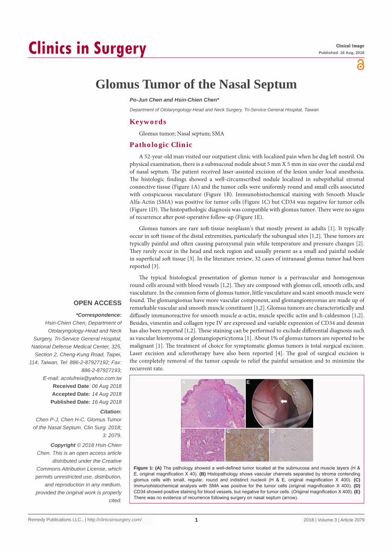
physical examination, there is a submucosal nodule about 5 mm X 5 mm in size over the caudal end of nasal septum. The patient received laser-assisted excision of the lesion under local anesthesia. The histologic findings showed a well-circumscribed nodule localized in subepithelial stromal connective tissue (Figure 1A) and the tumor cells were uniformly round and small cells associated with conspicuous vasculature (Figure 1B). Immunohistochemical staining with Smooth Muscle Alfa-Actin (SMA) was positive for tumor cells (Figure 1C) but CD34 was negative for tumor cells (Figure 1D). The histopathologic diagnosis was compatible with glomus tumor. There were no signs of recurrence after post-operative follow-up (Figure 1E).
Glomus tumors are rare soft-tissue neoplasm’s that mostly present in adults [1]. It typically occur in soft tissue of the distal extremities, particularly the subungual sites [1,2]. These tumors are typically painful and often causing paroxysmal pain while temperature and pressure changes [2]. They rarely occur in the head and neck region and usually present as a small and painful nodule in superficial soft tissue [3]. In the literature review, 32 cases of intranasal glomus tumor had been reported [3].
The typical histological presentation of glomus tumor is a perivascular and homogenous round cells around with blood vessels [1,2]. They are composed with glomus cell, smooth cells, and vasculature. In the common form of glomus tumor, little vasculature and scant smooth muscle were found. The glomangiomas have more vascular component, and glomangiomyomas are made up of remarkable vascular and smooth muscle constituent [1,2]. Glomus tumors are characteristically and diffusely immunoreactive for smooth muscle α-actin, muscle specific actin and h-caldesmon [1,2]. Besides, vimentin and collagen type IV are expressed and variable expression of CD34 and desmin has also been reported [1,2]. These staining can be performed to exclude differential diagnosis such as vascular leiomyoma or glomangiopericytoma [1]. About 1% of glomus tumors are reported to be malignant [1]. The treatment of choice for symptomatic glomus tumors is total surgical excision. Laser excision and sclerotherapy have also been reported [4]. The goal of surgical excision is the completely removal of the tumor capsule to relief the painful sensation and to minimize the recurrent rate.
Figure 1: (A) The pathology showed a well-defined tumor located at the submucosa and muscle layers (H & E, original magnification X 40). (B) Histopathology shows vascular channels separated by stroma contending glomus cells with small, regular, round and indistinct nucleoli (H & E, original magnification X 400). (C) Immunohistochemical analysis with SMA was positive for the tumor cells (original magnification X 400). (D) CD34 showed positive staining for blood vessels, but negative for tumor cells. (Original magnification X 400). (E) There was no evidence of recurrence following surgery on nasal septum (arrow).
Hsin-Chien Chen, et al., Clinics in Surgery - Otolaryngology
Remedy Publications LLC., | http://clinicsinsurgery.com/ 2018 | Volume 3 | Article 20792
References1. Gombos Z, Zhang PJ. Glomus tumor. Arch Pathol Lab Med.
2008;132(9):1448-52.
2. Mravic M, LaChaud G, Nguyen A, Scott MA, Dry SM, James AW. Clinical and histopathological diagnosis of glomus tumor: an institutional experience of 138 cases. Int J Surg Pathol. 2015;23(3):181-8.
3. Chirila M, Rogojan L. Glomangioma of the nasal septum: A case report and review. Ear Nose Throat J. 2013;92(4-5):E7-9.
4. Lee SH, Roh MR, Chung KY. Subungual glomus tumors: Surgical approach and outcome based on tumor location. Dermatol Surg. 2013;39(7):1017-22.