Global and regional parameters of myocardial viability by speckle tracking echocardiography
62
New Non-invasive Methods for Quantification of Regional and Global Myocardial Function in the Normal and Ischemic Left Ventricle Thomas M. Helle-Valle, MD Institute for Surgical Research and Department of Cardiology Oslo University Hospital, Rikshospitalet and Faculty of Medicine, University of Oslo Oslo, Norway 2013
Transcript of Global and regional parameters of myocardial viability by speckle tracking echocardiography
New Non-invasive Methods for Quantification of
Regional and Global Myocardial Function in the
Normal and Ischemic Left Ventricle
Thomas M. Helle-Valle, MD
Institute for Surgical Research and Department of Cardiology
Oslo University Hospital, Rikshospitalet and
Faculty of Medicine, University of Oslo
Oslo, Norway
2013

© Thomas M. Helle-Valle, 2013 Series of dissertations submitted to the Faculty of Medicine, University of Oslo No. 1559 ISBN 978-82-8264-502-7 All rights reserved. No part of this publication may be reproduced or transmitted, in any form or by any means, without permission. Cover: Inger Sandved Anfinsen. Printed in Norway: AIT Oslo AS. Produced in co-operation with Akademika publishing. The thesis is produced by Akademika publishing merely in connection with the thesis defence. Kindly direct all inquiries regarding the thesis to the copyright holder or the unit which grants the doctorate.
3
Contents
Contents ...................................................................................................................................... 3
Acknowledgements ..................................................................................................................... 4
List of papers............................................................................................................................... 7
Selected Abbreviations ............................................................................................................... 8
Introduction ................................................................................................................................. 9
Aims of the Thesis .................................................................................................................... 17
General aims ......................................................................................................................... 17
Specific aims ......................................................................................................................... 17
Material ..................................................................................................................................... 18
Experimental Study ............................................................................................................... 18
Paper I ............................................................................................................................... 18
Clinical Studies ..................................................................................................................... 18
Paper I ............................................................................................................................... 18
Paper II .............................................................................................................................. 18
Paper III ............................................................................................................................ 19
Methods..................................................................................................................................... 20
Experimental Study ............................................................................................................... 20
Clinical Studies ..................................................................................................................... 24
Statistics ................................................................................................................................ 28
Summary of Results .................................................................................................................. 30
Paper I ................................................................................................................................... 30
Paper II .................................................................................................................................. 31
Paper III ................................................................................................................................ 32
Discussion ................................................................................................................................. 33
Methodological considerations ............................................................................................. 35
LV Rotation, Torsion and Strain by STE.............................................................................. 37
Comparison with previous studies ........................................................................................ 39
Radial strain by MDCT ......................................................................................................... 41
Limitations ............................................................................................................................ 42
Conclusions ............................................................................................................................... 45
Reference List ........................................................................................................................... 46

4
Acknowledgements
The present thesis is based on studies carried out at the Department of Cardiology and
at the Institute for Surgical Research, Oslo University Hospital, Rikshospitalet, during
the years 2004 – 2007, and at Johns Hopkins University Hospital, Baltimore, USA,
during the years 2007-2008.
The work was supported by the Norwegian Council of Cardiovascular Diseases
from 2003 - 2006 and by the Department of Cardiology, Rikshospitalet, from 2006 -
2007. In addition, without the financial support from Medinnova AS, Unger-Vetlesen
Foundation, Gidske og Peter Jacob Sørensens Foundation, Alf og Aagot Helgesens
Foundation, Dr Alexander Malthes Foundation, Caroline Musæus Aarsvolds
Foundation and from the Norwegian Society of Cardiology, an immensely
informative and inspiring year as a research fellow at Johns Hopkins University
Hospital would not have been possible.
The success of scientific research depends on the teamwork of individuals with
complementary skills. The present thesis is the result of such teamwork. First of all, I
have been extremely privileged to have had Professor Otto A. Smiseth as my formal
supervisor since 2006. Otto is one of the world’s foremost authorities on
cardiovascular mechanics and his academic achievements are recognized worldwide.
To work with Otto has been immensely inspiring and without his vast knowledge,
bright ideas, enthusiasm, support and patience, my scientific achievements would
simply not have been possible. Despite Otto’s busy schedule, he has always been
available for encouraging discussions, even if that sometimes meant short meetings
during coffee breaks at conferences, in hotel lobbies, at airports, or while walking
with him to his next meeting. Nevertheless, because of Otto’s ability to focus and to
extract the scientific essence of our data, even short meetings were highly productive.
I also wish to thank Otto for giving me the opportunity to spend an unforgettable and
highly informative year as a research fellow at Johns Hopkins University Hospital.
Professor Thor Edvardsen became my co-supervisor in 2006. His knowledge
within the field of cardiac imaging, his experience in experimental and clinical
research and his ability to overcome the practical difficulties frequently met while
carrying out various research projects, were absolutely essential to my research.
Despite his busy schedule, his open door policy frequently initiated spontaneous and

5
highly fruitful discussions – sometimes even on non-scientific topics. I also wish to
thank Thor for his generosity and friendship.
I was fortunate to have Professor Halfdan Ihlen as my formally supervisor until
his retirement in 2006. Halfdan has a unique position in Norwegian cardiology and
have played a central role in the development of various echocardiographic
techniques. His experience with both clinical work and research, his personal
qualities, his enthusiasm for our scientific work, his encouragements during
adversities and his focus on the balance between independent and cooperative work,
made him to an outstanding mentor.
The experimental research performed at Institute for Surgical Research was
complex and time consuming and required 100% teamwork. Without the genuine
knowledge, support and advices of my colleagues and friends Anders Opdahl, Espen
Remme, Trond Vartdal, Erik Lyseggen, Eirik Pettersen and Helge Skulstad, the
experimental part of my research would not have been possible. The development and
use of complex mathematical models and algorithms were a central part of my
research. Thanks to engineers Espen Remme, Jonas Crosby and Stein-Inge Rabben
these issues were elegantly solved. I also wish to thank Drs Ketil Lunde, Ola Gjesdal,
Einar Hopp and Hans-Jørgen Smith for important and generous contributions. I am
also indebted to Dr Brage H Amundsen and Professor Hans Torp from Department of
Circulation and Medical Imaging, NTNU, for their important contributions to Paper 1
in the present thesis. Thanks to the statistician Are Hugo Pripp for helping me with
the linear mixed-model statistical method in Paper 2.
I feel much honored to have had the opportunity to be a part of Professor João
A. C. Lima’s research team at Johns Hopkins University Hospital in Baltimore. João
is heading one of the world’s leading centers when it comes to cardiac imaging and
the use of MRI and CT. His personal qualities are unique and the invaluable
experiences I gained as a research fellow are too many to be listed. Due to João’s
support, enthusiasm, humor and friendship, I am looking forward to continuing our
ongoing scientific collaboration. I would also like to express my deepest gratitude to
my great colleagues and friends at Johns Hopkins University Hospital, Drs Veronica
Fernandes, Boas D. Rosen, Wen-Chung Yu, Ilan Gottlieb, Andrea Vavere and
engineer Elzbieta Chamera. Their enthusiasm, support and cooperation were essential
for the development of the new multimodality tissue tracking method and the

6
subsequent publication (Paper 3 in my Thesis). A special thanks to Kathleen Lensch
for solving any problem at any time.
Thanks to Professor Ansgard Aasen for office facilities and for providing a
stimulating environment at the Institute for Surgical Research; to Vivi Bull Stubberud
for keeping the operation room in order; to Roger Ødegård and Eldrid Winther-
Larssen for technical assistance; and to Dr Svend Aakhus, Pia Bryde, Johanna
Andreassen and Richard Massey at the Echo-lab for, among other tasks, helping me
improving my echocardiographic imaging skills.
Finally, I wish to thank my loving and inspiring parents, who passed away too
early, but who still live inside of me; my family and friends for vitalizing my life; and
my parents-in-law for their generosity and support whenever needed. Last, but not
least, my deepest gratitude goes to my wife Anita and to our daughters Sara and Maja
for reminding me every day that there is more to the heart than strain, torsion and
twist - I love you!

7
List of papers
I. Helle-Valle T, Crosby J, Edvardsen T, Lyseggen E, Amundsen BH, Smith HJ, Rosen
BD, Lima JA, Torp H, Ihlen H, Smiseth OA. New Noninvasive Method for Assessment
of Left Ventricular Rotation: Speckle Tracking Echocardiography. Circulation. 2005;
112; 3149-3156.
II. Helle-Valle T, Remme EW, Lyseggen E, Pettersen E, Vartdal T, Opdahl A, Smith HJ,
Osman N, Ihlen H, Edvardsen T, Smiseth OA. Clinical Assessment of Left Ventricular
Rotation and Strain – A Novel Approach for Quantification of Function in Infarcted
Myocardium and its Border Zones. Am J Physiol Heart Circ Physiol. 2009
Jul;297(1):H257-67
III. Helle-Valle T; Yu WC; Fernandes VRF; Rosen BD; Lima JAC. Usefulness of Radial
Strain Mapping by Multidetector Computer Tomography (MDCT) to Quantify Regional
Myocardial Function in Patients with Healed Myocardial Infarction. Am J Cardiol. 2010
Aug 15;106(4):483-91.

8
Selected Abbreviations
2-D = two-dimensional
CVD = cardiovascular disease
ECG = electrocardiogram
ED = end-diastole
ES = end-systole
ICD = implantable cardioverter-defibrillator
dP/dt = time derivative of left ventricular pressure
LAD = left anterior descending coronary artery
LE MRI = late enhancement magnetic resonance imaging
LV = left ventricle/left ventricular
LVP = left ventricular pressure
MDCT = multidetector computer tomography
MRI = magnetic resonance imaging
MTT = multimodality tissue tracking
RCA = right coronary artery
ROI = region of interest
STE = speckle tracking echocardiography
9
Introduction
Left ventricular (LV) dysfunction is a progressive and malignant condition and among the
leading causes of morbidity and mortality worldwide. Optimal handling strategies for patients
with suspected or established LV dysfunction depend on the ability to determine its mechanism
and extent and non-invasive methods for assessment of LV function are therefore pivotal.
However, normal LV function is a highly complex sequence of multiple interrelated events
involving factors such as active and passive myocardial tissue properties, coordinated electro-
mechanical coupling, loading conditions, geometry and LV fiber orientation. Because
cardiovascular diseases, such as coronary artery disease, arrhythmias, hypertension, diabetes,
valvular disease, myocarditis, congenital heart disease and cardiomyopathies may affect LV
function through different mechanisms, an unambiguous definition of LV dysfunction is
difficult. Consequently, non-invasive modalities that allow accurate and reproducible
quantification of markers reflecting different aspects of LV mechanics and function are needed.1-
4 The aim of the present Thesis is to introduce new non-invasive quantitative methods for
assessment of regional and global LV function and to use these methods to study LV mechanics
in the setting of myocardial ischemia, the most common cause of LV dysfunction.
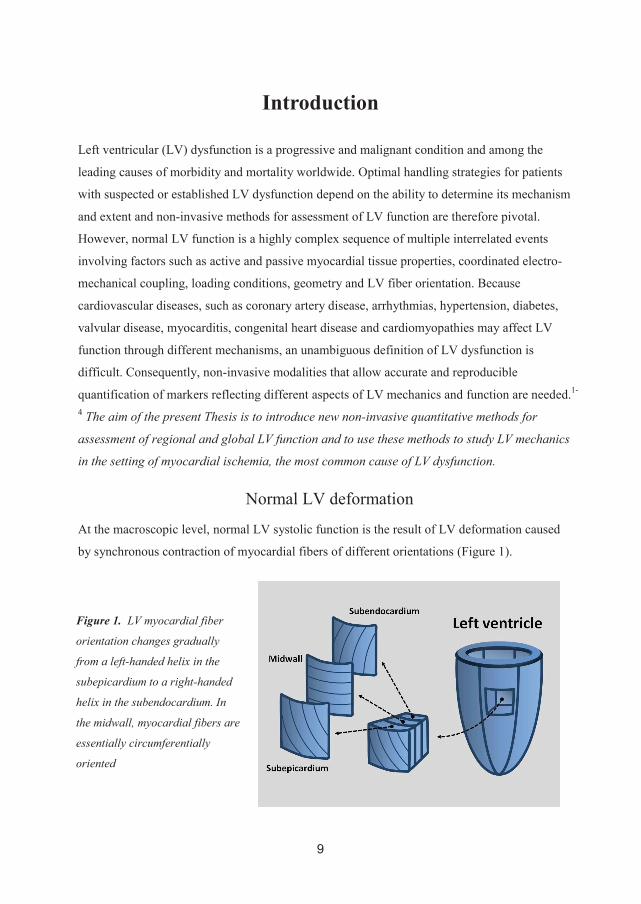
Normal LV deformation
At the macroscopic level, normal LV systolic function is the result of LV deformation caused
by synchronous contraction of myocardial fibers of different orientations (Figure 1).
Figure 1. LV myocardial fiber
orientation changes gradually
from a left-handed helix in the
subepicardium to a right-handed
helix in the subendocardium. In
the midwall, myocardial fibers are
essentially circumferentially
oriented
10
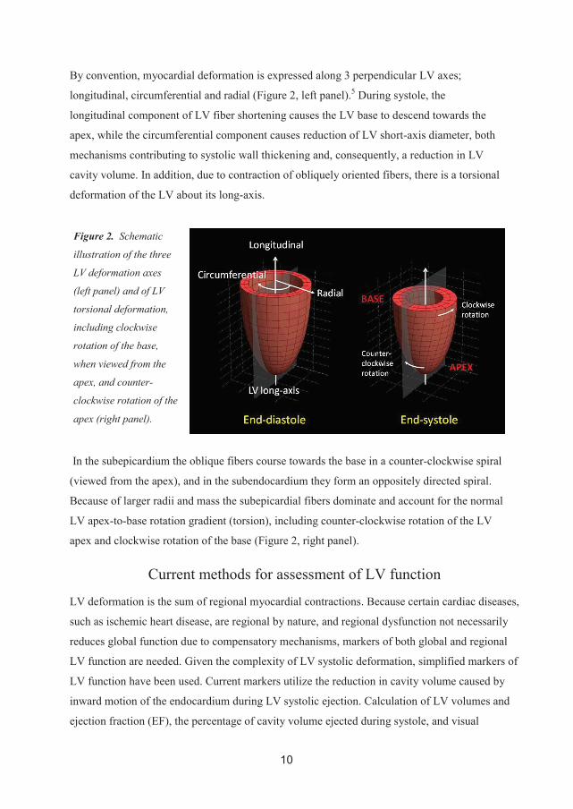
By convention, myocardial deformation is expressed along 3 perpendicular LV axes;
longitudinal, circumferential and radial (Figure 2, left panel).5 During systole, the
longitudinal component of LV fiber shortening causes the LV base to descend towards the
apex, while the circumferential component causes reduction of LV short-axis diameter, both
mechanisms contributing to systolic wall thickening and, consequently, a reduction in LV
cavity volume. In addition, due to contraction of obliquely oriented fibers, there is a torsional
deformation of the LV about its long-axis.
In the subepicardium the oblique fibers course towards the base in a counter-clockwise spiral
(viewed from the apex), and in the subendocardium they form an oppositely directed spiral.
Because of larger radii and mass the subepicardial fibers dominate and account for the normal
LV apex-to-base rotation gradient (torsion), including counter-clockwise rotation of the LV
apex and clockwise rotation of the base (Figure 2, right panel).
Current methods for assessment of LV function
LV deformation is the sum of regional myocardial contractions. Because certain cardiac diseases,
such as ischemic heart disease, are regional by nature, and regional dysfunction not necessarily
reduces global function due to compensatory mechanisms, markers of both global and regional
LV function are needed. Given the complexity of LV systolic deformation, simplified markers of
LV function have been used. Current markers utilize the reduction in cavity volume caused by
inward motion of the endocardium during LV systolic ejection. Calculation of LV volumes and
ejection fraction (EF), the percentage of cavity volume ejected during systole, and visual
Figure 2. Schematic
illustration of the three
LV deformation axes
(left panel) and of LV
torsional deformation,
including clockwise
rotation of the base,
when viewed from the
apex, and counter-
clockwise rotation of the
apex (right panel).

11
evaluation of LV regional wall motion (wall motion score index) and thickening are therefore the
most widely used markers of global and regional LV function. These markers can be measured
non-invasively from cine loops obtained by transthoracic echocardiography, magnetic resonance
imaging (MRI) or multidetector computed tomography (MDCT). The most widely used modality
is echocardiography, which is a bedside technique that is highly available, portable, safe and
inexpensive and allows for real-time visualization of cardiac morphology, hemodynamics and
motion.
In patients with poor acoustic window, MRI and MDCT are alternatives to
echocardiography for assessment of LV volumes and wall motion. Cardiac MRI has been
considered the clinical “gold standard” but is a complex, expensive and time consuming
modality of limited availability that cannot be performed in patients with implanted pacemakers
or defibrillators. MDCT has recently emerged as a promising noninvasive clinical tool for
assessment of cardiac morphology, coronary anatomy and myocardial perfusion, viability and
scarring.6, 7 Retrospective ECG-gated MDCT allows for image reconstruction in any phase of the
cardiac cycle and, thus, for construction of cine loops. Recent studies have demonstrated that LV
volumes, wall motion and wall thickening can be assessed accurately when compared to
measures by MRI, echocardiography and SPECT.8, 9 MDCT has the advantage of being fast,
relatively inexpensive and providing images of high spatial resolution. Although radiation is an
important limitation, the introduction of new generations multidetector and dual source MDCT
scanners have dramatically reduced this problem.10
Echocardiographic assessment of LV volumes, EF, wall motion and thickening has proven
to be clinically useful in most cardiac diseases. The method, however, has important limitations
by being subjective, highly observer and image quality dependent, insensitive to subtle changes
in myocardial function and predominantly reflecting the radial component of myocardial
deformation.11-14 Due to the progressive nature of LV dysfunction and the potential benefit of
early detection, quantitative markers of subtle, subclinical myocardial dysfunction are needed.
Studies have shown that changes in the normal temporal pattern of the LV deformation sequence,
particularly in the setting of myocardial ischemia or dyssynchrony, precedes changes in the
amplitude of wall motion and wall thickening.15-17 Consequently, there is a need for additional
methods for non-invasive quantification of the magnitude and timing of regional and global
myocardial deformation.
12
Measures of LV deformation
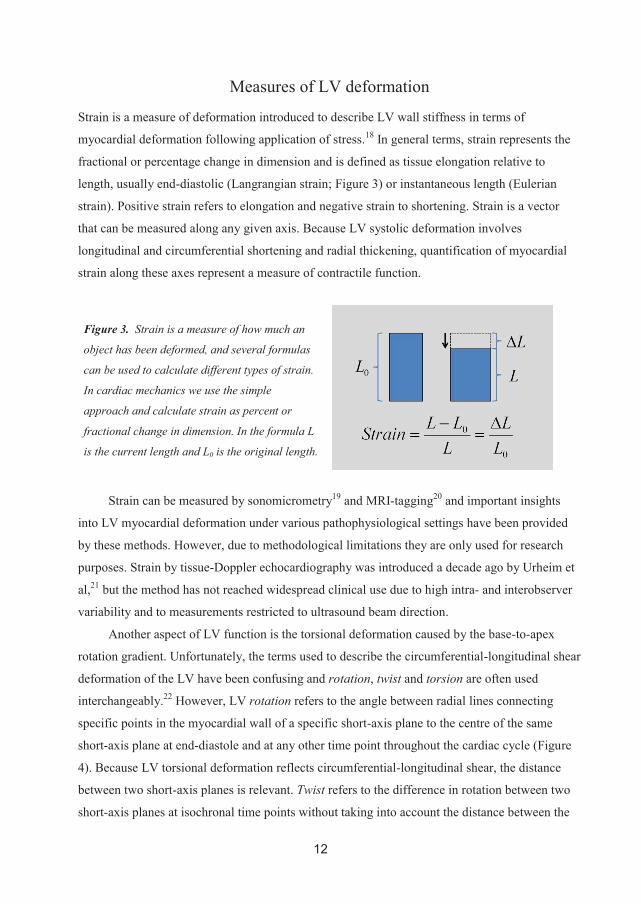
Strain is a measure of deformation introduced to describe LV wall stiffness in terms of
myocardial deformation following application of stress.18 In general terms, strain represents the
fractional or percentage change in dimension and is defined as tissue elongation relative to
length, usually end-diastolic (Langrangian strain; Figure 3) or instantaneous length (Eulerian
strain). Positive strain refers to elongation and negative strain to shortening. Strain is a vector
that can be measured along any given axis. Because LV systolic deformation involves
longitudinal and circumferential shortening and radial thickening, quantification of myocardial
strain along these axes represent a measure of contractile function.
Strain can be measured by sonomicrometry19 and MRI-tagging20 and important insights
into LV myocardial deformation under various pathophysiological settings have been provided
by these methods. However, due to methodological limitations they are only used for research
purposes. Strain by tissue-Doppler echocardiography was introduced a decade ago by Urheim et
al,21 but the method has not reached widespread clinical use due to high intra- and interobserver
variability and to measurements restricted to ultrasound beam direction.
Another aspect of LV function is the torsional deformation caused by the base-to-apex
rotation gradient. Unfortunately, the terms used to describe the circumferential-longitudinal shear
deformation of the LV have been confusing and rotation, twist and torsion are often used
interchangeably.22 However, LV rotation refers to the angle between radial lines connecting
specific points in the myocardial wall of a specific short-axis plane to the centre of the same
short-axis plane at end-diastole and at any other time point throughout the cardiac cycle (Figure
4). Because LV torsional deformation reflects circumferential-longitudinal shear, the distance
between two short-axis planes is relevant. Twist refers to the difference in rotation between two
short-axis planes at isochronal time points without taking into account the distance between the
Figure 3. Strain is a measure of how much an
object has been deformed, and several formulas
can be used to calculate different types of strain.
In cardiac mechanics we use the simple
approach and calculate strain as percent or
fractional change in dimension. In the formula L
is the current length and L0 is the original length.

13
two planes, while torsion refers to the rotational difference between two short-axis planes (twist)
when normalized to the instantaneous distance between the two planes. Because the dynamic
distance between apical and basal short-axis planes is difficult to assess by 2-dimensional
echocardiography, the terms torsion and twist are frequently used synonymously. For simplicity,
in the present Thesis, LV rotation is used as a common term for LV rotation, twist and torsion
unless stated otherwise.
The magnitude and characteristics of LV rotation have been described in different clinical
and experimental studies using MRI- tagging and other complex methodologies.23-29 It is well
established that LV rotation is sensitive to changes in both regional and global LV function,25, 30-
41 and an important marker with respect to LV ejection and filling.30, 42-45 Although LV rotation
reflects a fundamental property of normal LV function and has been proposed as a useful clinical
marker of impaired LV function,39, 46-51 LV rotation has not been incorporated into the clinical
assessment of LV function due to methodological limitations.
Figure 4. Cylindrical LV model illustrating direction and magnitude of basal (dark blue arrow)
and apical (light blue arrow) rotation. Because the dynamic distance between apical and basal
short-axis planes is difficult to measure by 2-dimensional echocardiography, in Paper 1, LV torsion
was calculated simply as the rotational difference between apical and basal rotation (i.e. twist).

14
Assessment of LV deformation
At the initiation of the present studies, speckle tracking echocardiography (STE) had recently
been introduced as a promising angle-independent method for automated tracking of natural
acoustic markers (speckles) visualized by conventional gray-scale imaging.52, 53 Because
speckles represent myocardial “finger prints” and can be tracked in any direction throughout the
cardiac cycle (Figure 5), myocardial motion and deformation can be measured by STE in the
circumferential and radial direction from LV short axis recordings, and in the longitudinal and
transversal direction from LV long axis recordings. Consequently, the introduction of STE as a
non-invasive method to quantify LV motion and deformation represented an important
breakthrough which allowed for LV strain and rotation to be brought into the clinical arena.
Recently, Ogawa et al54 introduced another echocardiographic speckle tracking algorithms
based on automated tracking of “on-screen” pixel patterns. Because the algorithm uses cine
loops stored in the AVI format, it does not discriminate between different imaging modalities
and, thus, represented a promising multimodality technique for automated tracking of myocardial
motion and deformation.
Figure 5. End-diastolic and end-systolic apical short-axis echocardiographic images from an
animal experiment. Stable speckle patterns or “finger prints” suitable for automated tracking are
indicated (white squares) at end-diastole (left panel) and end-systole (right panel).
15
Assessment of LV function during Myocardial Infarction
Myocardial ischemia caused by coronary artery stenosis or occlusion is the most common cause
of LV dysfunction and involves a wide spectrum of pathophysiological conditions, ranging from
acute ischemia to chronic infarction. Detection and quantification of myocardial ischemia
provides important diagnostic, therapeutic and prognostic information in both the acute and
chronic phases, even when the initial event was not recognized.55 Detection of reversibly injured
myocardium in the setting of acute (stunning) or chronic (hibernation) ischemia and assessment
of total infarct size and transmurality are important in order to determine potential benefit from
revascularization,56-58 and for prediction of long-term improvement in contractile function.59, 60
Recent studies have also emphasized the prognostic importance of quantifying the infarct border
zone (peri-infarct zone) in addition to infarct core, as the former is a substrate for malignant
ventricular arrhythmias and its extent an independent marker of sudden death.61
Late-enhanced contrast MRI is the reference method for accurate visualization and
quantification of myocardial scarring,62 but clinical use is limited by restricted availability. An
indirect way of measuring infarct extent is to study its effects on myocardial deformation.
Changes in myocardial contraction patterns caused by ischemia were first elegantly demonstrated
by Tennant and Wiggers in 1935.63 Impaired systolic contraction patterns are observed within
seconds of acute ischemia and characteristic changes in the magnitude and timing of myocardial
deformation occur as myocardial infarction evolves as a wavefront of necrosis extending from
the subendocardium to the subepicardium within hours.64, 65 In patients with suspected or
established ischemic heart disease, non-invasive assessment of LV regional and global
deformation has proven to be relevant with respect to diagnosis, treatment selection and
prognosis, as mortality increases exponentially with decreasing LVEF and linearly with
increasing wall motion abnormality.66 However, the sensitivity and specificity of determining
ischemia visually by conventional echocardiography are moderate and show substantial
variability in evaluating the extent and severity of regional dysfunction.67, 68 Because of loading
conditions and remodeling, low LVEF does not necessarily reflect the extent of reduced
contractile function due to compensatory regional hyperkinesis or mitral regurgitation.14
Similarly, regional wall motion might be misleading owing to viable but dysfunctional
myocardium.
Furthermore, because myocardial infarcts demonstrate a highly irregular morphology and
abrupt changes in infarct transmurality are commonly present,69, 70 methods that allow accurate
mapping of total infarct size and distribution from function analysis are needed.

16
Summary
Optimal handling strategies for patients with suspected or established cardiac disease require
non-invasive methods for assessment of the extent and mechanism of LV dysfunction. Although
current volumetric and wall motion indices by echocardiography, cardiac MRI or MDCT are
useful, additional quantitative and sensitive methods for assessment of myocardial mechanics are
needed. Functional analysis is of particular importance in the setting of myocardial ischemia, the
most common cause of LV dysfunction. In the present Thesis we introduce and validate LV
rotation and strain by STE, and radial strain mapping by multimodality tissue tracking MDCT, as
new non-invasive methods for quantitative assessment of the magnitude and timing of regional
and global LV deformation. By detailed analysis of regional strain and rotation in the infarcted
LV, we demonstrate how ischemic and non-ischemic myocardium affects regional and global
deformation indices and how these novel methods can be applied in a clinical setting.
17
Aims of the Thesis
General aims
To introduce new non-invasive methods for quantitative assessment of regional and global LV
function, and to study myocardial function in the infarcted LV
Specific aims
To introduce and validate speckle tracking echocardiography (STE) as a noninvasive method
for assessment of LV rotation and torsion (Paper I)
To determine how LV rotation and circumferential strain by STE can be used in combination
to assess myocardial function in the infarcted LV (Paper II)
To introduce and validate radial strain mapping by multimodality tissue tracking
multidetector computer tomography (MDCT) as a new noninvasive method for quantification
of myocardial function (Paper III)
To investigate whether radial strain mapping by MDCT can be used to assess myocardial
infarct distribution in patients with healed myocardial infarction (Paper III)

18
Material
Experimental Study
Paper I
Thirteen mongrel dogs of either sex were studied. Baseline data were obtained from all
animals, whereas dobutamine intervention was not performed in the first 4 experiments.
In 5 dogs, recordings during ischemia could not be obtained due to sustained ventricular
fibrillation shortly after LAD occlusion. To explore the importance of an intact
pericardium for LV rotation, short-axis recordings by STE were performed immediately
before and after pericardiotomy in 6 dogs. The study was approved by the National
Animal Experimentation Board and the animals were supplied by the Center for
Comparative Medicine, Oslo University Hospital, Rikshospitalet.
Clinical Studies
Paper I
The study population consisted of 29 healthy volunteers (14 women; 33±6 years). The
study protocol was approved by the National Committee for Medical Research Ethics of
Norway and all participants gave written, informed consent.
Paper II
The study population consisted of 15 healthy volunteers (7 women; 34±7 years) and 23
patients selected from a database at OUS, Rikshospitalet, consisting of patients
reexamined ~6 months after percutaneous coronary intervention (PCI) due to first time
acute ST-elevation myocardial infarction. All patients with single coronary artery disease
and echocardiographic short-axis recordings of the LV apex obtained from the apical
window were included. According to angiographic findings, 18 patients had left
descending coronary artery occlusion, 3 had circumflex coronary artery occlusion, and 2
had right coronary artery occlusion. All study subjects had given written, informed
consent, and the protocol was approved by the Regional Ethics Committee.

19
Paper III
Available multidetector computer tomography (MDCT) data from patients with ischemic
heart disease (n=20; 7 women; 56±11 years) who participated in a previously performed
study at Johns Hopkins University Hospital, Baltimore, involving subjects with ischemic
or idiopathic LV dysfunction were selected. These patients were referred for implantable
cardioverter-defibrillator (ICD) placement and scheduled to undergo magnetic resonance
(MR) imaging and MDCT imaging as part of a prospective study relating image patterns
to ICD firings. All subjects had given written, informed consent and the study protocol
was approved by the Johns Hopkins institutional review committee.
20
Methods
Experimental Study
Animal Model
The animal model used in Paper I is a well established model for integrated studies of
cardiac mechanics and hemodynamics.71-73 The reasons for using dogs, is their tolerance
to extensive instrumentation, the appropriate size of the heart and the anatomical and
functional resemblance to the human heart. The dogs were anesthetized, artificially
ventilated and cannulated. After a median sternotomy, the pericardium was split and the
edges of the pericardial incision were loosely resutured after instrumentation. Inflatable
vascular occluders were placed around the proximal third of the LAD. Invasive pressure
data were digitized at 200Hz. Recordings were obtained with the dogs in a supine
position and with the ventilator turned off. At the end of the experiment, the animal was
euthanized with an intracardiac injection of pentobarbital.
Sonomicrometry
Sonomicrometry is considered as the gold standard for assessment of myocardial
deformation (function) in experimental studies.74 The method uses small (2 mm in
diameter) piezoelectric transducers (“crystals”) that can both transmit and receive
ultrasound signals (≥1 MHz). To perform a single distance measurement, one crystal will
transmit a burst of ultrasound, and a second crystal will receive this ultrasound signal.
The crystals are connected with a wire to a digital sonomicrometer and the elapsed time
from transmission to reception is a direct and linear representation of the separation of the
crystals. The resulting transit time is converted to a distance if the speed of sound in the
material being measured is known (~1540 m/s in cardiac tissue). By repeating the
measurement of transit-time hundreds of times per second, the instantaneous distance
between crystals implanted in the myocardium can be assessed with high temporal
resolution. Spatial resolution is determined by the distance between crystals. Because
sonomicrometry involves implantation of foreign objects into the myocardium, it has
been speculated that the method leads to injury and alters myocardial function. However,
21
studies using tissue-Doppler imaging have indicated that carefully implanted crystals
have minimal effect on systolic or diastolic regional function.75
LV Rotation and Torsion by Sonomicrometry
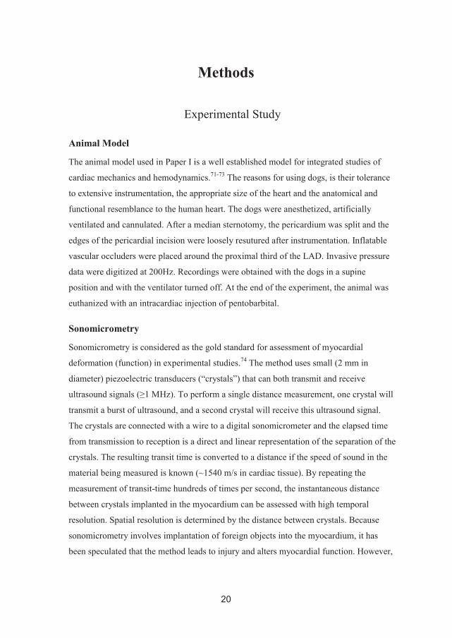
For the estimation of LV rotation and torsion by sonomicrometry, 12 crystals were
implanted in the LV in a grid-like manner to create 3 parallel short-axis planes (Figure 6).
One crystal was implanted at the tip of the apex, and 11 crystals in the LV circumference
at basal (n=3), equatorial (n=4) and apical (n=4) short-axis levels (Figure 6). Using the
signals obtained from the 3-dimensional grid of crystals, the coordinates of each crystal
were automatically determined by triangulation as a function of time. Parallel apical,
equatorial and basal LV planes were constructed by interpolation of the corresponding
crystal coordinates, and the in-plane positions were approximated by interpolation. The
center of rotation for each LV plane was determined as the center of a best-fitted-circle
through the interpolated coordinates. For each plane, the angular movements of the
interpolated coordinates were averaged. Apical and basal rotation was calculated by
measuring the difference in angular movement between the equatorial level (minimal
rotation) and the apical and basal levels. LV torsion was estimated as the difference in
angular movement between apical and basal planes at isochronal points (Figure 7).
Figure 6. A schematic
representation of the LV
with implanted sonomicro-
metric crystals (filled
circles) at the basal,
equatorial and apical
22
Echocardiographic Recordings
LV short-axis recordings were obtained using a Vivid 7 scanner (GE Vingmed).
Transducer frequencies (1.7-2.0 MHz), sampling rates (60-110 frames per second), focus
(mid-ventricular), sector depth (minimal) and sector width (narrow) were adjusted to
optimize image quality. Short-axis echocardiograms were recorded in the same planes as
used for sonomicrometry (basal, equatorial and apical), using the position of the crystals
as reference. Echocardiographic recordings were done immediately before the
sonomicrometry recordings.
Speckle Tracking Echocardiography
Speckle formation is a natural phenomenon which occurs when light or sound is scattered
back from microrough surfaces. In echocardiography, a speckle pattern is the result of
constructive and destructive interference of ultrasound backscattered from tissue
structures smaller than a wavelength and the pattern is seen as characteristic intensity
variations in an image. Although a speckle pattern is not a direct representation of the
underlying tissue, its feature remains stable as long as the distances between scatters are
unchanged. In echocardiography imaging, the distances between cardiac scatterers
change constantly due to tissue deformation. However, given the high temporal resolution
of modern scanners, the extent of myocardial deformation from one frame to the next is
limited, resulting in moderate inter-frame degrading of a speckle pattern. Based on the
assumption that speckle patterns are sufficiently preserved between frames and therefore
represent myocardial “finger prints”, block-matching algorithms were developed for
tracking local tissue motion from the displacement of speckle patterns visible in
echocardiographic cine recordings. At the initiation of the experimental study (Paper I),
no commercial STE software was available.
LV Rotation and Torsion by speckle tracking echocardiography
The speckle tracking algorithm used in the experimental study was originally developed
by DrTech Hans Torp at NTNU for LV long-axis tracking, but in collaboration with
DrIng Jonas Crosby, the algorithm was further modified for short-axis analyses. The
algorithm, using minimum SAD (sum of absolute differences) of the B-mode pixel data,76
was used to track the position of a kernel region (a selected region of interest (ROI) with
a unique speckle pattern) frame by frame throughout the cardiac cycle. To avoid drifting,
the tracking algorithm was applied both forward and backward, and the results were
23
averaged. Nine ROIs were automatically superimposed on the echo image at end-diastole
and positioned to fit the circular shaped LV. In our experimental study this superimposed
circle was aligned with the subepicardial LV circumference at the approximate same
transmural level as the implanted crystals. Rotation was estimated as the average angular
displacement of all ROIs relative to the center of a best fitted circle through the same
ROIs, frame by frame. Torsion was estimated as the difference between apical and basal
rotation at isochronal points (Figure 7).
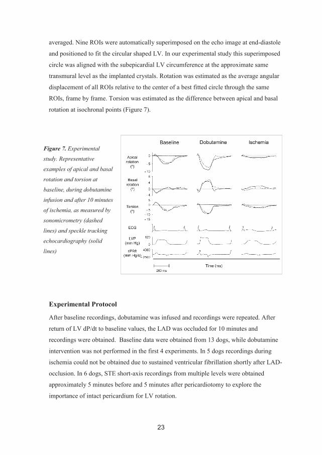
Experimental Protocol
After baseline recordings, dobutamine was infused and recordings were repeated. After
return of LV dP/dt to baseline values, the LAD was occluded for 10 minutes and
recordings were obtained. Baseline data were obtained from 13 dogs, while dobutamine
intervention was not performed in the first 4 experiments. In 5 dogs recordings during
ischemia could not be obtained due to sustained ventricular fibrillation shortly after LAD-
occlusion. In 6 dogs, STE short-axis recordings from multiple levels were obtained
approximately 5 minutes before and 5 minutes after pericardiotomy to explore the
importance of intact pericardium for LV rotation.
Figure 7. Experimental
study. Representative
examples of apical and basal
rotation and torsion at
baseline, during dobutamine
infusion and after 10 minutes
of ischemia, as measured by
sonomicrometry (dashed
lines) and speckle tracking
echocardiography (solid
lines)
24
Clinical Studies
Magnetic Resonance Imaging (MRI)
MRI is considered as the reference method for assessment of cardiac anatomy, chamber
volumes and function. The method is based on transmitting and receiving radio frequency
signals from atomic nuclei spinning in a strong magnetic field. By adding weaker
magnetic fields, MRI can generate cross-sectional 2-dimensional images in any plane.
Due to its high spatial resolution, high signal-to-noise ratio and acceptable temporal
resolution, MRI provides high-quality images of the LV. Myocardial tagging is a MRI
technique that is considered as the reference method for quantification of regional LV
function. By modulating the magnetization gradient, the signal from the myocardium can
be nulled in a grid pattern prior to the onset of image acquisition. During imaging, the
dark appearing grids move with the tissue and, thus, represent tissue markers. Areas of
myocardium that are not contracting appropriately will demonstrate decreased grid
deformation during the cardiac cycle. Intravenous contrast agents can be used to enhance
the signal. Gadolinium chelates causes a shortening of T1 relaxation time, making
gadolinium appear bright on T1-weighted images. In myocardial regions of infarction or
fibrosis, gadolinium is retained for a longer period due to tissue damage and infarcted
myocardium will therefore appear bright in images obtained 10-20 minutes after contrast
injection (late enhancement).
MRI recordings
MRI scans were performed using 1.5 T scanners (Magnetom Vision Plus, Siemens, and
Sigma CV/I, GE Medical Systems). Three long-axis and 8-10 short-axis cine loops
covering the entire LV were obtained during breath hold (12-20 seconds) with a slice
thickness of 7-10 mm and time resolution 35-40 ms. Multiple tagged short-axis
recordings (8-10 slices) were obtained by prescribing parallel striped tags in 2 orthogonal
orientations with a slice thickness of 7-10mm and a temporal resolution of 35-40 ms.
Multiple late enhancement (LE) short-axis images (8-10 slices) with a thickness of ~10
mm were obtained 10-20 minutes after gadolinium injection.
LV rotation, torsion, strain, infarction and volume by MRI
LV rotation (Papers I-II), circumferential strain (Paper II) and radial strain (Paper III) by
MRI tagging were analyzed by Harmonic Phase Imaging (HARP, version 1.0, Diagnosoft
25
Inc. Palo Alto, CA).77 Myocardial infarct size by LE MRI was analyzed by PACS, Sectra,
Sweden (Paper II) and by HARP (Paper III). LV volumes and EF (Paper III) were
calculated by the true Simpson’s method using QMass (Version 4.2, Medis, NL). LV
torsion was calculated as the difference between apical and basal rotation at isochronal
points. To ensure anatomical correspondence between short-axis recordings obtained by
MRI and STE we used LV intracavitary diameter, wall thickness and anatomic
landmarks. In Paper I, LV apical and basal rotation by MRI-tagging was calculated as the
average of measurements obtained in the mid- and subendocardial layer to be consistent
with measurements by STE. LV torsion was estimated as the difference between apical
and basal rotation at isochronal points. In Paper II, LV apical rotation and strain by MRI
and STE were compared according to a 4-segment model (in 3 patients, tagged MRI
images could not be analyzed due to poor image quality). For comparison of LV apical
infarct distribution by LE MRI and rotation and strain by STE, a detailed 36-segment LV
model was used (12 segments for each of 3 short-axes). In Paper III, radial strain by MRI
tagging was compared to measures by MDCT using an 18-segment LV model.
Echocardiographic recordings
In Papers I-II, using a Vivid 7 scanner (GE Vingmed Ultrasound, Horten, Norway), LV
long- and short-axis recordings were obtained during breath holds with the individual in a
supine left lateral position, within 5-15 minutes of the MRI examination. In order to
obtain optimal speckle quality at reproducible and representative short-axis levels,
projection of the LV base was obtained from a standard parasternal probe position, while
projections of the LV apex, just proximal to the level with luminal closure at end-systole,
was recorded from a anterior or anterolateral position, distal to the conventional
parasternal apical window. An effort was made to make the LV cross-sections as circular
as possible. Transducer frequencies (1.7-2.0 MHz), sampling rates (60-110 frames per
second), focus (mid-ventricular), sector depth (minimal) and sector width (narrow) were
adjusted to optimize image quality.
LV rotation and strain by STE
In Paper I we used the same Matlab-based software as in the experimental study. The
analyses were performed in the same manner and the same criteria for adjustments or
rejections of ROIs were used. However, in the clinical study the quality of the speckles
improved progressively from the epi- to the endocardium and assessment of rotation was

26
therefore limited to the mid- and subendocardial layers. In Paper II LV apical rotation and
circumferential strain were assessed using commercially available speckle tracking
software (EchoPAC, GE Vingmed Ultrasound).52 For each apical short-axis recording the
endocardial border was manually traced in the frame where its complete contour was
identified the best, and the automatically applied epicardial border was adjusted to cover
the myocardium without including the pericardium. After successful tracking, rotation
and strain were automatically calculated for numerous locations evenly distributed along
the circumference and end-systolic rotation and strain were extracted from 24 evenly
distributed segments that corresponded to the MRI segments.
Rotation and Strain in a Mathematical Simulation Model of the Ischemic LV
In Paper II we used the CMISS finite element analysis program (Bioengineering institute,
University of Auckland, Auckland, New Zealand) to construct a finite element model to
study mechanisms of changes in strain and rotation in the ischemic LV. The model
included LV geometry, fiber-orientation, passive elastic myocardial properties, active
systolic tension and stretch ratio along the fiber direction and time varying intracellular
Ca2+ concentration.78-80 According to a 12-segment apical short-axis model, an antero-
septal infarct was included by abolishing active fiber force and increasing passive
stiffness. Both transmural and non-transmural infarcts were simulated. The 3-dimensional
deformation of the LV was simulated using physiological pressure-volume boundary
conditions.
Multidetector Computer Tomography (MDCT)
MDCT has emerged as a noninvasive method for assessment of coronary anatomy and
calcium burden, chamber volumes, wall motion, and myocardial scarring and perfusion.
In MDCT, a two-dimensional array of detector elements replaces the linear array of
detector elements used in typical conventional and helical CT scanners. The 2-
dimensional detector array allows CT scanners to acquire multiple slices or sections
simultaneously, which greatly increase the speed of CT image acquisition. A complete
examination of the entire heart can be performed within seconds with excellent spatial
resolution and signal-to-noise ratio and with acceptable temporal resolution and radiation
dosage (4-6 mSv).81
27
MDCT recordings
In Paper III, retrospective ECG-gated MDCT images were obtained by a 64-slice scanner
(Aquillion, Toshiba, Toshiba Medical Systems). Gantry rotation time was 400 ms to 500
ms depending on the heart rate and no beta-blocking agents were used. A bolus non-
ionic contrast was administered and a navigation tool was used to obtain the highest
possible temporal resolution. Two to 5 consecutive heart beats were used to reconstruct
the R-R interval into ~20 phases and dedicated software (Vital Images, Minnetonka,
Minnesota) was used to reformat the images into multiple long- and short-axis cine
recordings. Image level-window was adjusted to optimize the contrast between tissue and
blood and the recordings were stored in the AVI format for further analysis.
Multimodality tissue tracking (MTT)
The MTT algorithm is an automated pixel pattern-matching technique originally
developed by Toshiba Medical Systems for STE.54 However, in contrast to the speckle
tracking algorithms used in Papers I-II, the MTT algorithm utilizes “on-screen” pixel
information provided by cine loops stored in the DICOM or AVI format. By a template
matching technique the motion of stable pixel patterns can be tracked frame by frame.
Common determinants of accurate tracking include spatial resolution and signal-to-noise
ratio. Because MDCT technology provides images of superb spatial resolution and high
signal-to-noise ratio, the endo- and epicardial borders are well defined and their excursion
in the radial direction suitable for automated tracking. By including the entire myocardial
wall in the strain calculation, problems related to the transmural strain gradient are
minimized.
Radial strain by MDCT
In LV basal, mid and apical short-axis MDCT cines, the endocardial and epicardial
borders were drawn in an early-systolic frame, and the endocardial and epicardial
displacement tracked throughout the cardiac cycle. After successful tracking, the
magnitude and timing of peak systolic radial strain for each of 12 equiangular segments
were extracted and used for further analyses. The RV inferior septal insertion point was
used as reference for reproducible segmentation. Global end-systole was determined as
the time of aortic valve closure. Strain values were not extracted from regions of
inadequate tracking. We used a conventional 18-segment LV model (6 segments per
short-axis) to compare radial strain by STE and MRI tagging, and a 36-segment LV

28
model (12 segments per short-axis) to compare radial strain mapping by MDCT and
infarct extent by LE MRI (Figure 8).
Statistics
Values are expressed as mean±SD, unless otherwise stated. Statistical differences were
considered significant at P<0.05. Variables from independent measurements were
compared by a least square linear regression and by the Bland-Altman method (Papers I-
III).82 We used the unpaired t-test (Paper I) or one-way repeated-measures ANOVA
followed by Bonferroni correction for predefined relevant comparisons (Papers I-II). In
Paper III the linear mixed-model statistical method83 with random intercept was used to
assess the significance of infarct extent in explaining radial strain variations (SPSS 13
and 16, SPSS Inc., Chicago). Different relevant covariance structures were tested and the
Figure 8. Representative example of 12-segment radial strain analysis of the LV mid-
ventricular short-axis by MTT MDCT in a patient with myocardial infarction of the
anterior, lateral and posterior wall, as demonstrated by late enhancement MRI. While
remote segments (J-L) demonstrate substantial wall thickening, infarct center segments
(C-G) show severely impaired thickening. Note that ischemic border zone segments (B, H)
show less thickening than non-ischemic border zone segments (A, I).

29
analysis with lowest Akaike’s Information Score selected. To determine inter- (Papers I-
III) and intraobserver (Paper III) variability, recordings were randomly selected and
independently analyzed. The reproducibility was expressed by the intraclass correlation
coefficient and by the Bland-Altman method.
30
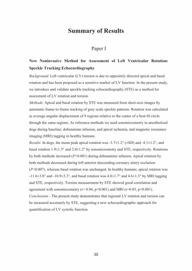
Summary of Results
Paper I
New Noninvasive Method for Assessment of Left Ventricular Rotation:
Speckle Tracking Echocardiography
Background: Left ventricular (LV) torsion is due to oppositely directed apical and basal
rotation and has been proposed as a sensitive marker of LV function. In the present study,
we introduce and validate speckle tracking echocardiography (STE) as a method for
assessment of LV rotation and torsion.
Methods: Apical and basal rotation by STE was measured from short-axis images by
automatic frame-to-frame tracking of gray scale speckle patterns. Rotation was calculated
as average angular displacement of 9 regions relative to the center of a best-fit circle
through the same regions. As reference methods we used sonomicrometry in anesthetized
dogs during baseline, dobutamine infusion, and apical ischemia, and magnetic resonance
imaging (MRI) tagging in healthy humans.
Results: In dogs, the mean peak apical rotation was -3.7±1.2° (±SD) and -4.1±1.2°, and
basal rotation 1.9±1.5° and 2.0±1.2° by sonomicrometry and STE, respectively. Rotations
by both methods increased (P<0.001) during dobutamine infusion. Apical rotation by
both methods decreased during left anterior descending coronary artery occlusion
(P<0.007), whereas basal rotation was unchanged. In healthy humans, apical rotation was
-11.6±3.8° and -10.9±3.3°, and basal rotation was 4.8±1.7° and 4.6±1.3° by MRI tagging
and STE, respectively. Torsion measurement by STE showed good correlation and
agreement with sonomicrometry (r= 0.94, p<0.001) and MRI (r=0.85, p<0.001).
Conclusions - The present study demonstrates that regional LV rotation and torsion can
be measured accurately by STE, suggesting a new echocardiographic approach for
quantification of LV systolic function.
31
Paper II
Clinical Assessment of Left Ventricular Rotation and Strain – A Novel
Approach for Quantification of Function in Infarcted Myocardium and its
Border Zones
Background: Left ventricular (LV) circumferential strain and rotation have been
introduced as clinical markers of myocardial function. This study investigates how
regional LV apical rotation and strain can be used in combination to assess function in the
infarcted ventricle.
Methods: In healthy subjects (n = 15) and patients with myocardial infarction (n = 23),
LV apical segmental rotation and strain were measured from apical short-axis recordings
by speckle tracking echocardiography (STE) and MRI tagging. Infarct extent was
determined by late gadolinium enhancement MRI. To investigate mechanisms of changes
in strain and rotation, we used a mathematical finite element simulation model of the LV.
Results: Mean apical rotation and strain by STE were lower in patients than in healthy
subjects (9.0 ± 4.9 vs. 12.9 ± 3.5° and -13.9 ± 10.7 vs. -23.8 ± 2.3%, respectively, P <
0.05). In patients, regional strain was reduced in proportion to segmental infarct extent (r
= 0.80, P < 0.0001). Regional rotation, however, was similar in the center of the infarct
and in remote viable myocardium. Minimum and maximum rotations were found at the
infarct borders: minimum rotation at the border zone opposite to the direction of apical
rotation, and maximum rotation at the border zone in the direction of rotation. The
simulation model reproduced the clinical findings and indicated that the dissociation
between rotation and strain was caused by mechanical interactions between infarcted and
viable myocardium.
Conclusion: Systolic strain reflects regional myocardial function and infarct extent,
whereas systolic rotation defines infarct borders in the LV apical region. Regional
rotation, however, has limited ability to quantify regional myocardial dysfunction.

32
Paper III
Usefulness of Radial Strain Mapping by Multidetector Computer
Tomography (MDCT) to Quantify Regional Myocardial Function in Patients
with Healed Myocardial Infarction
Background: We introduce and evaluate strain mapping by multidetector computer
tomography (MDCT) as a new non-invasive method for assessment of myocardial
function. Methods: In patients (n=16) with healed myocardial infarction, peak systolic
radial strain was measured by automated pixel pattern matching analysis of multiple left
ventricular (LV) 64-slice MDCT short-axis recordings. For comparison, radial strain and
myocardial infarct extent were measured by tagged magnetic resonance imaging (MRI)
and late enhancement (LE) MRI, respectively.
Results: In a linear mixed model analysis, myocardial infarct extent was a strong
predictor of segmental strain by MDCT (β=-0.44, P<0.0001). Strain was significantly
different between non-infarcted (0%), non-transmurally (0-50%) and transmurally
(>50%) infarcted segments (P<0.001), and between infarcted and non-infarcted border
zone segments (P<0.001). There was a close relationship between strain by MDCT and
by tagged MRI (r=0.68, P<0.0001 and mean difference -7.4±11.7%). Mean time-to-peak
systolic strain was 324±42 by MDCT and 335±56 ms by tagged MRI (mean difference
11±40 ms).
Conclusion: To our knowledge this is the first study to demonstrate that regional
myocardial function can be quantified by MDCT imaging, indicating that assessment of
radial strain by MDCT might be a useful tool in the evaluation of patients with
cardiovascular diseases.

33
Discussion
Accurate and reproducible assessment of LV function is pivotal for the majority of
patients with cardiac diseases and is of particular importance in the setting of myocardial
ischemia, the most common cause of LV dysfunction.84 In the present thesis we have
introduced and validated LV rotation and circumferential strain by STE and radial strain
by multimodality tissue-tracking MDCT as new non-invasive methods for quantification
of regional and global LV function. In Paper I we demonstrated that STE allows for
quantitative assessment of LV rotation and torsion (Figure 9), indices that reflect
fundamental properties of LV function. In an animal model, that allowed global and
regional LV contractility to be altered, we showed that while LV torsion reflects global
LV function, LV rotation is a more regional marker of LV function.
However, when analyzing the apical in-plane distribution of regional rotation and strain
in a combined clinical and simulation study of the infarcted LV (Paper II), we observed
that while a close association existed between regional circumferential strain and infarct
transmurality, regional rotation did not provide a site-specific measure of regional
Figure 9. Experimental data –
rotation: A, correlation
between rotation measured by
sonomicrometry and STE. B,
Agreement between rotation
measured by sonomicrometry
and STE.

34
function, but defined infarct borders (Figure 10). The simulation model reproduced the
clinical findings and supported our hypothesis that the dissociation between rotation and
strain was caused by mechanical interactions between infarcted and viable myocardium.
Although echocardiography is the cornerstone for diagnosing and monitoring
cardiac diseases, STE analysis is challenging in a significant number of patients due to
insufficient image quality.85 MDCT has rapidly emerged as a comprehensive, accurate
and fast modality for assessment of cardiac and coronary anatomy, myocardial perfusion
and scarring as well as volumes and wall motion. In Paper III we introduced and
validated radial strain mapping by multimodality tissue tracking MDCT as a new method
for quantification of global and regional LV function. In patients with healed myocardial
infarction, the magnitude and timing of myocardial thickening was quantified in a highly
detailed fashion by a novel automated pixel pattern matching technique from LV short-
axis recordings by MDCT. The validity of our approach was confirmed by the use of
Figure 10. Corresponding apical short-axis images by MRI tagging, late enhancement
MRI and STE in a patient with LAD related myocardial infarction, demonstrating that
while circumferential strain (bottom) reflects infarct transmurality, segments of maximum
and minimum rotation reflect the two infarct borders (segments 2 and 12)

35
tagged MRI as a reference method. When the relationship between myocardial infarct
extent and radial strain by MDCT was studied, significant differences in radial strain
were observed between non-infarcted, non-transmurally infarcted and transmurally
infarcted segments, and between infarcted and non-infarcted border zone segments
(Figure 11). Furthermore, a significant correlation was observed between global LV
radial strain and LV infarct size.
Methodological considerations
Validation of LV Rotation, Torsion and Strain by STE
In Paper I, we demonstrated that LV basal and apical rotation and LV torsion could be
measured non-invasively by automated tracking of speckles from echocardiographic LV
short-axis recordings. The validity of our approach was tested using sonomicrometry as
reference method in an animal model and MRI-tagging in humans. With sonomicrometry,
the implanted myocardial crystals served as anatomic landmarks which ensured that the
LV cross-sectional planes studied by the two methods were the same, and measurements
could be done only a few seconds apart. Furthermore, in the animal model comparison
between the methods could be done under a wide range of experimental settings known
to alter LV rotation. The STE method showed dynamics, magnitudes and timing of peak
basal and apical rotation and torsion that were closely related to measurements by
sonomicrometry. The same relationship was found when STE was compared to MRI in
healthy volunteers.
Figure 11. A. Correlation between segmental infarct extent and radial strain by MDCT.
B. Radial strain for non-infarcted, non-transmurally infarcted and transmurally infarcted
segments. C. Radial strain for remote, border zone and infarct centre segments.
36
In Paper II, global and regional LV apical rotation and strain by STE was measured
in healthy subjects and in patients with myocardial infarction using MRI-tagging as a
reference method. Regional LV apical rotation and strain by STE demonstrated a close
relationship to measures by MRI according to an apical 4-segment model. Because of
limited spatial resolution of MRI tagging and impaired tag quality due to myocardial
scarring, the 24-segment model used by STE could not be validated.
Two different STE algorithms were used in Papers I-II. At the time when the
studies included in Paper I were initiated, no commercially speckle tracking software was
available. We therefore used a Matlab-based algorithm developed by engineers at NTNU,
Trondheim, Norway. The same software was used in the first validation study of strain by
STE, published shortly after Paper I.86 Just before onset of the study included in Paper II,
commercially available software was introduced (EchoPAC, GE Vingmed) and given the
obvious advantages of commonly available software, this was used in Paper II. To our
knowledge, the two software have not been compared with respect to quantification of
strain and rotation. However, because both algorithms use the block-matching approach,
showed close relationship with their reference methods and demonstrated similar
magnitude of global LV apical rotation in healthy subjects, there are good reasons to
believe that the two algorithms are comparable.
Feasibility and reproducibility of STE analysis of mean LV basal and apical
rotation (Paper I), and local LV apical rotation and strain (Paper II) were excellent
(>90%). The typical time spent to complete a short-axis analysis, including adjustments
and re-tracking if needed, was typically less than 60 seconds.
Validation of LV Strain by MDCT
To our knowledge, Paper III is the first study to demonstrate that regional myocardial
function can be quantified in an automated and detailed fashion by MDCT imaging.
Because MDCT technology provides images of high spatial resolution and high signal-to-
noise ratio, the endo- and epicardial borders are well defined and their excursions suitable
for automated feature tracking. In patients with healed myocardial infarction, the
magnitude and timing of radial strain was quantified by a novel automated pixel pattern
matching technique from MDCT LV short-axis recordings. The validity of our approach
was tested by the use of tagged MRI as a reference method, and the magnitude and timing
of radial strain by MDCT showed good correlation and agreement with measures by the
37
reference method. Radial strain by MDCT was measured with excellent intra- and inter-
observer reproducibility.
Due to noise caused by ICD leads and motion artifacts, MDCT recordings could not
be analyzed in 4 patients. Of the remaining studies, 97% of LV segments could be
analyzed. The typical time spent to quantify regional strain from a LV short-axis MDCT
cine by MTT was less than 60 seconds.
LV Rotation, Torsion and Strain by STE
LV Rotation and Torsion
In Paper I, LV rotation at the base was predominantly clockwise in both the experimental
and clinical study, consistent with previous findings.26, 33, 35, 36, 87-91 Dobutamine infusion
caused a significant increase in clockwise rotation with no change in time to peak
rotation. Apical ischemia caused no significant change in basal rotation reflecting that
there was no impairment of LV function between equator and base. Some studies have
indicated that basal LV rotation is minimal.24, 25 However, in patients with aortic stenosis
reduced magnitude of basal rotation compared to normals has been observed.
Furthermore, in patients with chronic heart failure, 6 months of treatment was associated
with an increase in basal rotation, while apical rotation was unchanged, indicating that
measurement of basal rotation may be of clinical relevance. However, when compared to
the reference methods, assessment of rotation by STE was less accurate for the basal level
than for the apical level. Therefore, deviation in basal rotation may be difficult to assess
by STE, and larger clinical studies have to be done in order to answer this question.
Counterclockwise rotation during LV ejection was demonstrated at the apical level
in healthy individuals (Papers I-II) and was also found in the dog model. During
dobutamine infusion there was an increase in apical rotation, and a tendency towards a
decrease in time to peak rotation, whereas during LAD occlusion apical rotation was
reduced. The same finding was observed in Paper II, where apical rotation was
significantly lower in patients compared to healthy subjects. Changes in apical rotation by
dobutamine and apical ischemia are in keeping with previous studies, using other
methodologies.30-32, 42, 45
Although systolic apical rotation was predominantly counterclockwise, there was a
small, clockwise rotation during isovolumic contraction. The oppositely directed rotation
was demonstrated by both sonomicrometry and STE (Papers I-II) but not by MRI. This

38
phenomenon has been described previously and might be attributed to earlier activation
of subendocardial fibers (right-handed helix) than subepicardial fibers.37, 43, 92, 93 The
reason why this motion was not recognized by MRI-tagging is probably the relatively low
temporal resolution of the method (35 ms).
As demonstrated in Paper I, the LV apex and base showed, in principle, inversed
rotational courses throughout the cardiac cycle. Because LV torsion is calculated as the
rotational difference between apex and base at isochronal time points, and the magnitude
of apical rotation far exceeds that of basal rotation, LV torsion is predominantly
determined by apical rotation. This was demonstrated in the experimental study, where
apical ischemia caused no change in basal rotation while both apical rotation and LV
torsion were reduced. Furthermore, in a recent study by our group, using both
experimental and clinical data, we showed that apical rotation represents the dominant
contribution to LV torsion over a wide range of hemodynamic conditions, suggesting that
apical rotation by STE may serve as a simple and feasible clinical index of LV torsion.94
Dissociation Between Regional Rotation and Strain
In Paper II, we demonstrated that regional LV apical rotation and strain reflect entirely
different features of infarcted myocardium. Reductions in end-systolic strain in post-
infarct patients corresponded to transmural extent of infarcted myocardium, as evidenced
by a strong correlation between systolic strain and percent infarction within segments.
These observations were in keeping with previous studies and confirmed that systolic
strain reflects regional contractile function.60, 95-98 This was in contrast to regional rotation
which did not reflect regional contractility. Maximum and minimum values of regional
rotation, however, were markers of the anatomical locations of the infarct borders. The
simulation study reproduced the clinical findings and provided insights into the
mechanism of the apparent inconsistency between regional apical rotation and strain:
While oblique LV fibers running from the base to the apex account for the overall
counter-clockwise rotation of the apex, the circumferential fibers affect the in-plane
distribution of regional rotation, which in the normal LV is homogeneous. Due to the
imbalance of circumferentially oriented contractile forces in the setting of regional
ischemia, the ischemic myocardium will be pulled from both sides by non-infarcted
myocardium. Consequently, the counter-clockwise infarct border is pulled in the counter-
clockwise direction (same direction as overall apical rotation) while the clockwise infarct
border is pulled in the opposite, clockwise direction. In total, these mechanisms cause

39
hyper-rotation of the counter-clockwise border zone, and hypo-rotation of the clockwise
border zone (Figure 12).
Comparison with previous studies
Rotation and Torsion in the Normal LV
The magnitudes of LV apical and basal rotation and torsion, as reported in previous
experimental and clinical studies, differ substantially, and a number of factors may
explain this variance.23-27, 32, 33, 35-37, 40, 42, 44-47, 47, 87, 88, 91, 99-102 As demonstrated by Henson
et al.103 apparent differences in torsion between mice and humans are due to different
sizes of their ventricles. When systolic torsion angle was normalized for LV length,
torsion was essentially similar in the two species. Furthermore, since torsion angle is a
non-linear function of ventricular length, its magnitude depends critically on
measurement level relative to the LV base or other reference points. The relatively high
values of apical rotation measured by Gibbons Kroeker et al.24 is most likely explained by
their measurement technique, which recorded rotation at the distal part of the LV apex.
Another factor that may explain some of the variance in torsion magnitude in different
Figure 12. Schematic representations of
the LV viewed from the apex towards the
base. Ischemic myocardium (gray area),
direction of rotation (open arrows),
circumferential forces acting on the
ischemic region (solid arrows) and
ischemic margins at onset of systole
(dotted lines) are indicated. Upper panels
illustrate rotational deformation of an
apical short-axis slice in an LV
consisting of circumferential fibers only.
Lower panels illustrate rotational
deformation of an apical short-axis slice
in an LV consisting of circumferential
and oblique myocardial fibers. Open
arrow length indicates mangitude of
rotation.

40
studies is the existence of a marked transmural gradient with subendocardial values
almost twice the subepicardial.23 In the dog model we measured subepicardial rotation,
and during baseline conditions torsion was approximately 6º. These values are in the
same range as was reported by Buchalter et al,32 who measured both subepicardial and
subendocardial torsion in anesthetized dogs by MRI tagging. Furthermore, the
hemodynamic and contractile status of the heart in different animal models may vary and
give rise to differences in torsion. In our study, a significant reduction in rotation at the
apical level and a trend towards a reduction at the base was observed during the 2- to 3-
hour period before the baseline recordings. This was associated with reductions in LVP
and LV dP/dtmax, indicating that there was some reduction in LV systolic function. Most
likely the decrease in rotation reflects impairment of LV function due to the extensive
surgical instrumentation.
LV Rotation and Strain in the Ischemic LV
Several studies have confirmed that ischemia reduces LV rotation, but no studies have
investigated the interaction between regional shortening and rotation in the ischemic
ventricle. In an experimental MRI tagging study by Buchalter et al the relationship
between distribution of LV torsion and ischemia was examined.32 Local torsion was
measured as the rotational difference between corresponding apical and basal locations
before and during LAD-occlusion. Local torsion was further grouped into 2 LV sections,
an anterior and a posterior, only the former showing reduced torsion during ischemia.
Because LAD-occlusion primarily affects the anterior LV wall, the observations made by
Buchalter et al. of reduced torsion of the anterior section during ischemia, can be
interpreted as inconsistent with our results. However, according to the depiction of the
distribution of apical ischemia in each animal in their study, the anterior and posterior
sections did not represent strictly ischemic and non-ischemic myocardium, respectively:
In all animals the lateral non-ischemic border-zone, which in our study represents
myocardium of hypo-rotation, was located in the anterior (“ischemic”) section, while the
septal ischemic border-zone, which in our study represents myocardium of hyper-rotation
was located in the posterior (“non-ischemic”) section. Consequently, the apparent
discrepancies between our observations and the findings by Buchalter et al, could be
explained by their inclusion of non-ischemic myocardium of hypo-rotation in the anterior
LV section, and ischemic myocardium of hyper-rotation in the posterior LV section.

41
Radial strain by MDCT
The clinical use of MDCT for assessment of cardiac anatomy, coronary angiography and
myocardial perfusion is rapidly increasing. By acquiring retrospective ECG-gated data,
cine loops can also be constructed from the same dataset. Conventional non-invasive LV
strain analysis uses 3 short-axis slices to construct a 16-segment model.104 Given the
heterogeneous distribution of myocardial infarct transmurality64, 69, 70 and the high quality
of MDCT images, in Paper III we used a 36-segment LV model to obtain a more detailed
mapping of the relationship between myocardial deformation and infarct morphology.
The construction of narrower segments ensured that each category contained
morphologically more homogeneous segment and reduced the likelihood of including
“contaminated” segments, particularly in the non-infarcted and infarcted border zone
groups. In our study, the close inverse relationship observed between radial strain and
infarct transmurality allowed for discrimination between segments of various infarct
transmurality and between infarcted and non-infarcted border zone segments. These
observations were in keeping with the STE data presented in Paper II regarding
circumferential strain mapping.
Although sonomicrometry studies have demonstrated that the three strain
components respond equally to acute ischemia, controversies exist regarding the
sensitivity of the various strain components in the setting of chronic infarction. A strong
inverse relationship between infarct transmurality and strain has consistently been
demonstrated in the circumferential or longitudinal directions, while conflicting reports
exist regarding the sensitivity of radial strain.67, 96, 105-111 A possible explanation for this
inconsistency relates to methodological issues, as radial strain has been the most
challenging strain component to measure non-invasively, showing greater variability and
less reproducibility when compared to assessment of circumferential or longitudinal
strain.67, 96, 110, 112 Current strain imaging methods utilize intramyocardial markers for
tissue tracking. Because radial strain increases by almost 100% when moving from the
subepicardium towards the subendocardium, the magnitude of radial strain measurements
strongly depends on the transmural location of the myocardial markers used for
tracking.111, 113-116 Consequently, given the limited number of MR tags across the LV wall
and the reduction in speckle quality in echocardiographic images when moving towards
the subepicardium, radial strain imaging by these techniques is challenging.87 In this
perspective, the introduction of radial strain by MDCT is an important supplement to

42
strain analyses by other modalities. Furthermore, recent patient studies of cardiac
resynchronization therapy have indicated that radial strain may characterize LV
mechanical dyssynchrony in a more sensitive manner than longitudinal strain, further
supporting the relevance of radial strain measurements by MDCT.117, 118
Limitations
Material
The number of study subjects included in Papers I-III was relatively small. The healthy
subjects in Papers I-II were relatively young and patients included in Paper II had mostly
LAD-related infarctions. In Paper III, no subjects with preserved LV function were
included. Consequently, to determine the clinical applicability of our findings, larger
studies are need that should include older subjects and patients with a wider range of
myocardial dysfunction and infarct localizations.
Sonomicrometry
A limitation with the sonomicrometry model was the lack of extra-cardiac reference for
the crystal positions, which represented a fundamental difference between STE and
sonomicrometry. While STE provided a measure relative to a fixed reference point
outside the heart (the echocardiography transducer), in our model sonomicrometry had no
such external reference. Therefore, sonomicrometry provided no direct measure of
rotation. To compensate for this limitation we used the LV equatorial plane, which has
essentially zero rotation, as reference plane. This assumption is justified by a number of
previous studies in different species, including dogs, that show only minimal rotation at
the LV equator,26, 35, 36, 87, 90, 91, 103 and was supported by the observation in the present
study that rotation at the equatorial level by STE was minimal. For assessment of torsion,
however, the difference between apical and basal rotation, no assumptions about the
equatorial plane was needed.
Speckle tracking echocardiography
Several important factors may influence the accuracy of speckle tracking. The quality of
the recordings must be high in order to achieve correct tracking, and requires proper
adjustment of frame rate, probe frequency and focus. In Paper I, tracking quality was
evaluated visually and individual ROIs of poor tracking were manually moved to areas of
43
better speckle quality, or deleted. Since rotational displacement is relatively
homogenously distributed around the LV circumference in healthy myocardium, deletion
of one or maximum two ROIs will have minimal effect on the average rotation.23, 32, 46 In
Paper II, using commercially available software, inadequate tracking was also visually
evaluated and the endocardial delineation manually adjusted in regions of poor tracking
or drawn in another frame until better tracking was achieved. In this study, only apical
short-axes were analyzed and no regions had to be excluded.
A fundamental problem with speckle tracking in LV short-axis images is that
longitudinal motion of the LV causes myocardium to move in and out of the image plane.
As a consequence, speckles generated from the ultrasound beam (2-3mm of thickness)
will represent myocardium from different cross-sectional levels during the cardiac cycle.
The experimental study (Paper I) demonstrated that this problem was most pronounced at
the LV base where the longitudinal displacement was approximately 4 mm from ED to
ES, while LV apex was essentially stationary. The fact that the relationship between STE
and the reference methods was best at the apical level might well be attributed to this
phenomenon, further supported by the successful tracking of all apical segments in Paper
II.
A limitation in the clinical studies was that speckle quality in some cases was
suboptimal in the subepicardial layer of the LV. Therefore, for the validation of LV
rotation against MRI in Paper I, we compared measurements from the mid and inner wall
layers. In Paper II, a different tracking algorithm was used, and the automatically applied
epicardial delineation was adjusted to cover the myocardium without including the
epicardium. Hopefully technical developments in the ultrasound technology will resolve
the problem with suboptimal subepicardial speckle quality. In the dog study we had direct
access to the heart via a sternotomy and by using ultrasound gel as standoff we obtained
satisfactory tracking quality in all layers of the LV wall.
One limitation for the clinical routine use of STE is the selection of reproducible
anatomical landmarks for measuring apical rotation. One approach is to move the cross-
sectional image plane as far distally as possible. Another approach could be to measure at
the most distal level that does not have luminal closure during systole. At the basal level,
reproducible image planes were easier to obtain, using the fibrous mitral ring for
orientation. Selection of imaging plane is a challenge and clinical testing of STE in
patients are needed to determine if reproducible measures can be obtained from ventricles
44
that may change in size and geometry over time. Also, given the recent advances in 3-
dimensional echocardiography imaging, it is likely that this problem will be solved.
Given the mechanical complexity of 3-dimensional myocardial deformation and
interaction, in Paper II a more complete approach would have been to include measures
of regional shear strain and torsion in our analysis. In principle such measures can be
estimated by combining analyses from multiple 2-dimensional cross-sectional recordings.
However, by current echocardiographic techniques this method is challenging,
particularly with respect to determining the distance between slices and to our approach
of detailed strain- and rotation mapping. Hopefully, 3-dimensional echocardiography
imaging will allow simultaneous calculation of regional rotation, strain, shear strain and
torsion in the near future.
MRI tagging
Although strain by tagged MRI is considered as the reference method for non-invasive
quantification of myocardial function, strain analysis in the infarcted LV may be
challenging due to suboptimal tag lines within fibrotic tissue. Assessment of strain in the
radial direction is further challenged by signal-to-noise limitations in most 1.5 T MRI
systems, which only allow one or two tag planes to be inserted between the endo and
epicardium,87 and by post-infarct wall thinning in transmurally infarcted segments.
MDCT
Due to the radiation involved in MDCT imaging, the inclusion of patients must be
carefully weighed and subjects with preserved LV function are therefore currently not
likely to undergo protocols needed for cine construction. In our study, mean radiation
dose was 10.8±2.4 mSv, which is similar to the effective radiation doses in abdominal CT
(10 mSv), but more than in a diagnostic catheterization (7 mSv).81 Although the
feasibility of MTT analysis of subjects with preserved LV function has not been proven, a
substantial number of remote segments demonstrated radial strain values similar to those
previously reported in healthy subjects, indicates that adequate tracking is feasible also in
the healthy LV. Finally, the use of a 64-slice MDCT scanner in our study in part limited
the temporal and spatial resolution of our cine recordings.
45
Conclusions
In the present thesis we have demonstrated that
the magnitude and timing of apical and basal LV rotation and torsion can be
measured accurately by STE;
a close association exists between regional circumferential strain and infarct
transmurality and between locations of maximum and minimum rotation and infarct
borders;
the magnitude and timing of LV radial strain can be measured accurately by MDCT;
when analyzed in a detailed fashion, a close association exists between the
distribution of radial strain and infarct transmurality.
These findings indicate that assessment of rotation and circumferential strain by STE and
radial strain by MDCT may have potentials to become clinical noninvasive tools for
detection and quantification of LV ischemia.

46
Reference List
1. Cohn JN, Duprez DA. Time to foster a rational approach to preventing cardiovascular morbid events. J Am Coll Cardiol. 2008;52:327-329.
2. Fraser AG, Buser PT, Bax JJ, Dassen WR, Nihoyannopoulos P, Schwitter J, Knuuti JM, Hoher M, Bengel F, Szatmari A. The future of cardiovascular imaging and non-invasive diagnosis: a joint statement from the European Association of Echocardiography, the Working Groups on Cardiovascular Magnetic Resonance, Computers in Cardiology, and Nuclear Cardiology, of the European Society of Cardiology, the European Association of Nuclear Medicine, and the Association for European Paediatric Cardiology. Eur Heart J. 2006;27:1750-1753.
3. Howard BV, Rodriguez BL, Bennett PH, Harris MI, Hamman R, Kuller LH, Pearson TA, Wylie-Rosett J. Prevention Conference VI: Diabetes and Cardiovascular disease: Writing Group I: epidemiology. Circulation. 2002;105:e132-e137.
4. Voelkel NF, Quaife RA, Leinwand LA, Barst RJ, McGoon MD, Meldrum DR, Dupuis J, Long CS, Rubin LJ, Smart FW, Suzuki YJ, Gladwin M, Denholm EM, Gail DB. Right ventricular function and failure: report of a National Heart, Lung, and Blood Institute working group on cellular and molecular mechanisms of right heart failure. Circulation. 2006;114:1883-1891.
5. D'hooge J, Heimdal A, Jamal F, Kukulski T, Bijnens B, Rademakers F, Hatle L, Suetens P, Sutherland GR. Regional strain and strain rate measurements by cardiac ultrasound: principles, implementation and limitations. Eur J Echocardiogr. 2000;1:154-170.
6. Mahnken AH, Koos R, Katoh M, Spuentrup E, Busch P, Wildberger JE, Kuhl HP, Gunther RW. Sixteen-slice spiral CT versus MR imaging for the assessment of left ventricular function in acute myocardial infarction. Eur Radiol. 2005;15:714-720.
7. Sibley CT, Lima JA. Assessment of ventricular structure and function with multidetector CT and MRI. Curr Cardiol Rep. 2008;10:67-71.
8. Butler J, Shapiro MD, Jassal DS, Neilan TG, Nichols J, Ferencik M, Brady TJ, Hoffmann U, Cury RC. Comparison of multidetector computed tomography and two-dimensional transthoracic echocardiography for left ventricular assessment in patients with heart failure. Am J Cardiol. 2007;99:247-249.
9. Cury RC, Nieman K, Shapiro MD, Butler J, Nomura CH, Ferencik M, Hoffmann U, Abbara S, Jassal DS, Yasuda T, Gold HK, Jang IK, Brady TJ. Comprehensive assessment of myocardial perfusion defects, regional wall motion, and left ventricular function by using 64-section multidetector CT. Radiology. 2008;248:466-475.

47
10. Sabarudin A, Sun Z, Ng KH. A systematic review of radiation dose associated with different generations of multidetector CT coronary angiography. J Med Imaging Radiat Oncol. 2012;56:5-17.
11. Carabello BA, Nolan SP, McGuire LB. Assessment of preoperative left ventricular function in patients with mitral regurgitation: value of the end-systolic wall stress-end-systolic volume ratio. Circulation. 1981;64:1212-1217.
12. Gunther S, Grossman W. Determinants of ventricular function in pressure-overload hypertrophy in man. Circulation. 1979;59:679-688.
13. Malm S, Frigstad S, Sagberg E, Larsson H, Skjaerpe T. Accurate and reproducible measurement of left ventricular volume and ejection fraction by contrast echocardiography: a comparison with magnetic resonance imaging. J Am Coll Cardiol. 2004;44:1030-1035.
14. Marciniak A, Claus P, Sutherland GR, Marciniak M, Karu T, Baltabaeva A, Merli E, Bijnens B, Jahangiri M. Changes in systolic left ventricular function in isolated mitral regurgitation. A strain rate imaging study. Eur Heart J. 2007;28:2627-2636.
15. Cucchini F, Baldi G, Barilli AL, Di Donato M, Visioli O. Tardokinesis in coronary artery disease: evidence with instantaneous analysis of left ventricular ejection. Eur J Cardiol. 1981;12:153-166.
16. Ehring T, Heusch G. Left ventricular asynchrony: an indicator of regional myocardial dysfunction. Am Heart J. 1990;120:1047-1057.
17. Guth BD, Schulz R, Heusch G. Time course and mechanisms of contractile dysfunction during acute myocardial ischemia. Circulation. 1993;87:IV35-IV42.
18. Mirsky I, Parmley WW. Assessment of passive elastic stiffness for isolated heart muscle and the intact heart. Circ Res. 1973;33:233-243.
19. Bugge-Asperheim B, Lekven J, Kiil F. Effect of saline infusion on stroke volume and end-systolic volume at various levels of adrenergic activity in dogs. Scand J Clin Lab Invest. 1972;29:15-24.
20. Zerhouni EA, Parish DM, Rogers WJ, Yang A, Shapiro EP. Human heart: tagging with MR imaging--a method for noninvasive assessment of myocardial motion. Radiology. 1988;169:59-63.
21. Urheim S, Edvardsen T, Torp H, Angelsen B, Smiseth OA. Myocardial strain by Doppler echocardiography. Validation of a new method to quantify regional myocardial function. Circulation. 2000;102:1158-1164.
22. Sengupta PP, Tajik AJ, Chandrasekaran K, Khandheria BK. Twist mechanics of the left ventricle: principles and application. JACC Cardiovasc Imaging. 2008;1:366-376.

48
23. Buchalter MB, Weiss JL, Rogers WJ, Zerhouni EA, Weisfeldt ML, Beyar R, Shapiro EP. Noninvasive quantification of left ventricular rotational deformation in normal humans using magnetic resonance imaging myocardial tagging. Circulation. 1990;81:1236-1244.
24. Gibbons Kroeker CA, Ter Keurs HE, Knudtson ML, Tyberg JV, Beyar R. An optical device to measure the dynamics of apex rotation of the left ventricle. Am J Physiol Heart Circ Physiol. 1993;265:H1444-H1449.
25. Knudtson ML, Galbraith PD, Hildebrand KL, Tyberg JV, Beyar R. Dynamics of left ventricular apex rotation during angioplasty: a sensitive index of ischemic dysfunction. Circulation. 1997;96:801-808.
26. Nichols K, Kamran M, Cooke CD, Faber TL, Garcia EV, Bergmann SR, Depuey EG. Feasibility of detecting cardiac torsion in myocardial perfusion gated SPECT data. J Nucl Cardiol. 2002;9:500-507.
27. Mirro MJ, Rogers EW, Weyman AE, Feigenbaum H. Angular displacement of the papillary muscles during the cardiac cycle. Circulation. 1979;60:327-333.
28. Beyar R, Yin FC, Hausknecht M, Weisfeldt ML, Kass DA. Dependence of left ventricular twist-radial shortening relations on cardiac cycle phase. Am J Physiol. 1989;257:H1119-H1126.
29. Ingels NB, Jr., Daughters GT, Stinson EB, Alderman EL. Measurement of midwall myocardial dynamics in intact man by radiography of surgically implanted markers. Circulation. 1975;52:859-867.
30. Gibbons Kroeker CA, Tyberg JV, Beyar R. Effects of load manipulations, heart rate, and contractility on left ventricular apical rotation. An experimental study in anesthetized dogs. Circulation. 1995;92:130-141.
31. Kroeker CA, Tyberg JV, Beyar R. Effects of ischemia on left ventricular apex rotation. An experimental study in anesthetized dogs. Circulation. 1995;92:3539-3548.
32. Buchalter MB, Rademakers FE, Weiss JL, Rogers WJ, Weisfeldt ML, Shapiro EP. Rotational deformation of the canine left ventricle measured by magnetic resonance tagging: effects of catecholamines, ischaemia, and pacing. Cardiovasc Res. 1994;28:629-635.
33. Fuchs E, Muller MF, Oswald H, Thony H, Mohacsi P, Hess OM. Cardiac rotation and relaxation in patients with chronic heart failure. Eur J Heart Fail. 2004;6:715-722.
34. Maier SE, Fischer SE, McKinnon GC, Hess OM, Krayenbuehl HP, Boesiger P. Evaluation of left ventricular segmental wall motion in hypertrophic cardiomyopathy with myocardial tagging. Circulation. 1992;86:1919-1928.
35. Nagel E, Stuber M, Lakatos M, Scheidegger MB, Boesiger P, Hess OM. Cardiac rotation and relaxation after anterolateral myocardial infarction. Coron Artery Dis. 2000;11:261-267.
49
36. Sandstede JJ, Johnson T, Harre K, Beer M, Hofmann S, Pabst T, Kenn W, Voelker W, Neubauer S, Hahn D. Cardiac systolic rotation and contraction before and after valve replacement for aortic stenosis: a myocardial tagging study using MR imaging. AJR Am J Roentgenol. 2002;178:953-958.
37. Yun KL, Niczyporuk MA, Daughters GT, Ingels NB, Jr., Stinson EB, Alderman EL, Hansen DE, Miller DC. Alterations in left ventricular diastolic twist mechanics during acute human cardiac allograft rejection. Circulation. 1991;83:962-973.
38. DeAnda A, Jr., Komeda M, Nikolic SD, Daughters GT, Ingels NB, Miller DC. Left ventricular function, twist, and recoil after mitral valve replacement. Circulation. 1995;92:II458-II466.
39. Hansen DE, Daughters GT, Alderman EL, Ingels NB, Stinson EB, Miller DC. Effect of volume loading, pressure loading, and inotropic stimulation on left ventricular torsion in humans. Circulation. 1991;83:1315-1326.
40. Tibayan FA, Rodriguez F, Langer F, Zasio MK, Bailey L, Liang D, Daughters GT, Ingels NB, Jr., Miller DC. Alterations in left ventricular torsion and diastolic recoil after myocardial infarction with and without chronic ischemic mitral regurgitation. Circulation. 2004;110:II109-II114.
41. Sorger JM, Wyman BT, Faris OP, Hunter WC, McVeigh ER. Torsion of the left ventricle during pacing with MRI tagging. J Cardiovasc Magn Reson. 2003;5:521-530.
42. Rademakers FE, Buchalter MB, Rogers WJ, Zerhouni EA, Weisfeldt ML, Weiss JL, Shapiro EP. Dissociation between left ventricular untwisting and filling. Accentuation by catecholamines. Circulation. 1992;85:1572-1581.
43. McDonald IG. The shape and movements of the human left ventricle during systole. A study by cineangiography and by cineradiography of epicardial markers. Am J Cardiol. 1970;26:221-230.
44. Moon MR, Ingels NB, Jr., Daughters GT, Stinson EB, Hansen DE, Miller DC. Alterations in left ventricular twist mechanics with inotropic stimulation and volume loading in human subjects. Circulation. 1994;89:142-150.
45. Dong SJ, Hees PS, Huang WM, Buffer SA, Jr., Weiss JL, Shapiro EP. Independent effects of preload, afterload, and contractility on left ventricular torsion. Am J Physiol. 1999;277:H1053-H1060.
46. Hansen DE, Daughters GT, Alderman EL, Ingels NB, Jr., Miller DC. Torsional deformation of the left ventricular midwall in human hearts with intramyocardial markers: regional heterogeneity and sensitivity to the inotropic effects of abrupt rate changes. Circ Res. 1988;62:941-952.
47. DeAnda A, Jr., Moon MR, Yun KL, Daughters GT, Ingels NB, Jr., Miller DC. Left ventricular torsional dynamics immediately after mitral valve replacement. Circulation. 1994;90:II339-II346.

50
48. Delhaas T, Kotte J, van der Toorn A, Snoep G, Prinzen FW, Arts T. Increase in left ventricular torsion-to-shortening ratio in children with valvular aortic stenosis. Magn Reson Med. 2004;51:135-139.
49. MacGowan GA, Burkhoff D, Rogers WJ, Salvador D, Azhari H, Hees PS, Zweier JL, Halperin HR, Siu CO, Lima JA, Weiss JL, Shapiro EP. Effects of afterload on regional left ventricular torsion. Cardiovasc Res. 1996;31:917-925.
50. Moon MR, DeAnda A, Jr., Daughters GT, Ingels NB, Miller DC. Effects of chordal disruption on regional left ventricular torsional deformation. Circulation. 1996;94:II143-II151.
51. Tischler M, Niggel J. Left ventricular systolic torsion and exercise in normal hearts. J Am Soc Echocardiogr. 2003;16:670-674.
52. Leitman M, Lysyansky P, Sidenko S, Shir V, Peleg E, Binenbaum M, Kaluski E, Krakover R, Vered Z. Two-dimensional strain-a novel software for real-time quantitative echocardiographic assessment of myocardial function. J Am Soc Echocardiogr. 2004;17:1021-1029.
53. Reisner SA, Lysyansky P, Agmon Y, Mutlak D, Lessick J, Friedman Z. Global longitudinal strain: a novel index of left ventricular systolic function. J Am Soc Echocardiogr. 2004;17:630-633.
54. Ogawa K, Hozumi T, Sugioka K, Matsumura Y, Nishiura M, Kanda R, Abe Y, Takemoto Y, Yoshiyama M, Yoshikawa J. Usefulness of automated quantitation of regional left ventricular wall motion by a novel method of two-dimensional echocardiographic tracking. Am J Cardiol. 2006;98:1531-1537.
55. Kwong RY, Chan AK, Brown KA, Chan CW, Reynolds HG, Tsang S, Davis RB. Impact of unrecognized myocardial scar detected by cardiac magnetic resonance imaging on event-free survival in patients presenting with signs or symptoms of coronary artery disease. Circulation. 2006;113:2733-2743.
56. Kim RJ, Wu E, Rafael A, Chen EL, Parker MA, Simonetti O, Klocke FJ, Bonow RO, Judd RM. The use of contrast-enhanced magnetic resonance imaging to identify reversible myocardial dysfunction. N Engl J Med. 2000;343:1445-1453.
57. Orn S, Manhenke C, Anand IS, Squire I, Nagel E, Edvardsen T, Dickstein K. Effect of left ventricular scar size, location, and transmurality on left ventricular remodeling with healed myocardial infarction. Am J Cardiol. 2007;99:1109-1114.
58. Tarantini G, Razzolini R, Cacciavillani L, Bilato C, Sarais C, Corbetti F, Marra MP, Napodano M, Ramondo A, Iliceto S. Influence of transmurality, infarct size, and severe microvascular obstruction on left ventricular remodeling and function after primary coronary angioplasty. Am J Cardiol. 2006;98:1033-1040.

51
59. Choi KM, Kim RJ, Gubernikoff G, Vargas JD, Parker M, Judd RM. Transmural extent of acute myocardial infarction predicts long-term improvement in contractile function. Circulation. 2001;104:1101-1107.
60. Gerber BL, Garot J, Bluemke DA, Wu KC, Lima JA. Accuracy of contrast-enhanced magnetic resonance imaging in predicting improvement of regional myocardial function in patients after acute myocardial infarction. Circulation. 2002;106:1083-1089.
61. Yan AT, Shayne AJ, Brown KA, Gupta SN, Chan CW, Luu TM, Di Carli MF, Reynolds HG, Stevenson WG, Kwong RY. Characterization of the peri-infarct zone by contrast-enhanced cardiac magnetic resonance imaging is a powerful predictor of post-myocardial infarction mortality. Circulation. 2006;114:32-39.
62. Gibbons RJ, Valeti US, Araoz PA, Jaffe AS. The quantification of infarct size. J Am Coll Cardiol. 2004;44:1533-1542.
63. Tennant R, Wiggers CJ. The effect of coronary occlusion on myocardial contraction. Am J Physiol. 1935;112:351-361.
64. Reimer KA, Lowe JE, Rasmussen MM, Jennings RB. The wavefront phenomenon of ischemic cell death. 1. Myocardial infarct size vs duration of coronary occlusion in dogs. Circulation. 1977;56:786-794.
65. Reimer KA, Jennings RB. The "wavefront phenomenon" of myocardial ischemic cell death. II. Transmural progression of necrosis within the framework of ischemic bed size (myocardium at risk) and collateral flow. Lab Invest. 1979;40:633-644.
66. Pierard LA, Albert A, Chapelle JP, Carlier J, Kulbertus HE. Relative prognostic value of clinical, biochemical, echocardiographic and haemodynamic variables in predicting in-hospital and one-year cardiac mortality after acute myocardial infarction. Eur Heart J. 1989;10:24-31.
67. Gjesdal O, Helle-Valle T, Hopp E, Lunde K, Vartdal T, Aakhus S, Smith HJ, Ihlen H, Edvardsen T. Noninvasive separation of large, medium, and small myocardial infarcts in survivors of reperfused ST-elevation myocardial infarction: a comprehensive tissue Doppler and speckle-tracking echocardiography study. Circ Cardiovasc Imaging. 2008;1:189-96, 2.
68. Kvitting JP, Wigstrom L, Strotmann JM, Sutherland GR. How accurate is visual assessment of synchronicity in myocardial motion? An In vitro study with computer-simulated regional delay in myocardial motion: clinical implications for rest and stress echocardiography studies. J Am Soc Echocardiogr. 1999;12:698-705.
69. Eaton LW, Bulkley BH. Expansion of acute myocardial infarction: its relationship to infarct morphology in a canine model. Circ Res. 1981;49:80-88.

52
70. Page DL, Caulfield JB, Kastor JA, DeSanctis RW, Sanders CA. Myocardial changes associated with cardiogenic shock. N Engl J Med. 1971;285:133-137.
71. Edvardsen T, Urheim S, Skulstad H, Steine K, Ihlen H, Smiseth OA. Quantification of left ventricular systolic function by tissue Doppler echocardiography: added value of measuring pre- and postejection velocities in ischemic myocardium. Circulation. 2002;105:2071-2077.
72. Skulstad H, Edvardsen T, Urheim S, Rabben SI, Stugaard M, Lyseggen E, Ihlen H, Smiseth OA. Postsystolic shortening in ischemic myocardium: active contraction or passive recoil? Circulation. 2002;106:718-724.
73. Urheim S, Edvardsen T, Steine K, Skulstad H, Lyseggen E, Rodevand O, Smiseth OA. Postsystolic shortening of ischemic myocardium: a mechanism of abnormal intraventricular filling. Am J Physiol Heart Circ Physiol. 2003;284:H2343-H2350.
74. Bugge-Asperheim B, Leraand S, Kiil F. Local dimensional changes of the myocardium measured by ultrasonic technique. Scand J Clin Lab Invest. 1969;24:361-371.
75. Korinek J, Vitek J, Sengupta PP, Romero-Corral A, Krishnamoorthy VK, McMahon EM, Khandheria BK, Belohlavek M. Does implantation of sonomicrometry crystals alter regional cardiac muscle function? J Am Soc Echocardiogr. 2007;20:1407-1412.
76. Bohs LN, Trahey GE. A novel method for angle independent ultrasonic imaging of blood flow and tissue motion. IEEE Trans Biomed Eng. 1991;38:280-286.
77. Garot J, Bluemke DA, Osman NF, Rochitte CE, McVeigh ER, Zerhouni EA, Prince JL, Lima JA. Fast determination of regional myocardial strain fields from tagged cardiac images using harmonic phase MRI. Circulation. 2000;101:981-988.
78. Hunter PJ, McCulloch AD, Ter Keurs HE. Modelling the mechanical properties of cardiac muscle. Prog Biophys Mol Biol. 1998;69:289-331.
79. Nash MP, Hunter PJ. Computational mechanics of the heart: from tissue structure to ventricular function. J Elasticity. 2000;61:113-141.
80. Streeter DD, Jr., Spotnitz HM, Patel DP, Ross J, Jr., Sonnenblick EH. Fiber orientation in the canine left ventricle during diastole and systole. Circ Res. 1969;24:339-347.
81. Einstein AJ, Knuuti J. Cardiac imaging: does radiation matter? Eur Heart J. 2012;33:573-578.
82. Bland JM, Altman DG. Statistical methods for assessing agreement between two methods of clinical measurement. Lancet. 1986;1:307-310.

53
83. Fitzmaurice GM, Ravichandran C. A primer in longitudinal data analysis. Circulation. 2008;118:2005-2010.
84. Curtis JP, Sokol SI, Wang Y, Rathore SS, Ko DT, Jadbabaie F, Portnay EL, Marshalko SJ, Radford MJ, Krumholz HM. The association of left ventricular ejection fraction, mortality, and cause of death in stable outpatients with heart failure. J Am Coll Cardiol. 2003;42:736-742.
85. Marwick TH, Leano RL, Brown J, Sun JP, Hoffmann R, Lysyansky P, Becker M, Thomas JD. Myocardial strain measurement with 2-dimensional speckle-tracking echocardiography: definition of normal range. JACC Cardiovasc Imaging. 2009;2:80-84.
86. Amundsen BH, Helle-Valle T, Edvardsen T, Torp H, Crosby J, Lyseggen E, Stoylen A, Ihlen H, Lima JA, Smiseth OA, Slordahl SA. Noninvasive myocardial strain measurement by speckle tracking echocardiography: validation against sonomicrometry and tagged magnetic resonance imaging. J Am Coll Cardiol. 2006;47:789-793.
87. Moore CC, Lugo-Olivieri CH, McVeigh ER, Zerhouni EA. Three-dimensional systolic strain patterns in the normal human left ventricle: characterization with tagged MR imaging. Radiology. 2000;214:453-466.
88. Nagel E, Stuber M, Burkhard B, Fischer SE, Scheidegger MB, Boesiger P, Hess OM. Cardiac rotation and relaxation in patients with aortic valve stenosis. Eur Heart J. 2000;21:582-589.
89. Setser RM, Kasper JM, Lieber ML, Starling RC, McCarthy PM, White RD. Persistent abnormal left ventricular systolic torsion in dilated cardiomyopathy after partial left ventriculectomy. J Thorac Cardiovasc Surg. 2003;126:48-55.
90. Fogel MA, Weinberg PM, Hubbard A, Haselgrove J. Diastolic biomechanics in normal infants utilizing MRI tissue tagging. Circulation. 2000;102:218-224.
91. Lorenz CH, Pastorek JS, Bundy JM. Delineation of normal human left ventricular twist throughout systole by tagged cine magnetic resonance imaging. J Cardiovasc Magn Reson. 2000;2:97-108.
92. Azhari H, Buchalter M, Sideman S, Shapiro E, Beyar R. A conical model to describe the nonuniformity of the left ventricular twisting motion. Ann Biomed Eng. 1992;20:149-165.
93. Durrer D, van Dam RT, Freud GE, Janse MJ, Meijler FL, Arzbaecher RC. Total excitation of the isolated human heart. Circulation. 1970;41:899-912.
94. Opdahl A, Helle-Valle T, Remme EW, Vartdal T, Pettersen E, Lunde K, Edvardsen T, Smiseth OA. Apical Rotation by Speckle Tracking Echocardiography: A Simplified Bedside Index of Left Ventricular Twist. J Am Soc Echocardiogr. 2008.
95. Becker M, Hoffmann R, Kuhl HP, Grawe H, Katoh M, Kramann R, Bucker A, Hanrath P, Heussen N. Analysis of myocardial deformation based on
54
ultrasonic pixel tracking to determine transmurality in chronic myocardial infarction. Eur Heart J. 2006;27:2560-2566.
96. Chan J, Hanekom L, Wong C, Leano R, Cho GY, Marwick TH. Differentiation of subendocardial and transmural infarction using two-dimensional strain rate imaging to assess short-axis and long-axis myocardial function. J Am Coll Cardiol. 2006;48:2026-2033.
97. Croisille P, Moore CC, Judd RM, Lima JA, Arai M, McVeigh ER, Becker LC, Zerhouni EA. Differentiation of viable and nonviable myocardium by the use of three-dimensional tagged MRI in 2-day-old reperfused canine infarcts. Circulation. 1999;99:284-291.
98. Edvardsen T, Gerber BL, Garot J, Bluemke DA, Lima JA, Smiseth OA. Quantitative assessment of intrinsic regional myocardial deformation by Doppler strain rate echocardiography in humans: validation against three-dimensional tagged magnetic resonance imaging. Circulation. 2002;106:50-56.
99. Hansen DE, Daughters GT, Alderman EL, Stinson EB, Baldwin JC, Miller DC. Effect of acute human cardiac allograft rejection on left ventricular systolic torsion and diastolic recoil measured by intramyocardial markers. Circulation. 1987;76:998-1008.
100. Stuber M, Scheidegger MB, Fischer SE, Nagel E, Steinemann F, Hess OM, Boesiger P. Alterations in the local myocardial motion pattern in patients suffering from pressure overload due to aortic stenosis. Circulation. 1999;100:361-368.
101. Rothfeld JM, LeWinter MM, Tischler MD. Left ventricular systolic torsion and early diastolic filling by echocardiography in normal humans. Am J Cardiol. 1998;81:1465-1469.
102. Stuber M, Nagel E, Fischer SE, Spiegel MA, Scheidegger MB, Boesiger P. Quantification of the local heartwall motion by magnetic resonance myocardial tagging. Comput Med Imaging Graph. 1998;22:217-228.
103. Henson RE, Song SK, Pastorek JS, Ackerman JJ, Lorenz CH. Left ventricular torsion is equal in mice and humans. Am J Physiol Heart Circ Physiol. 2000;278:H1117-H1123.
104. Cerqueira MD, Weissman NJ, Dilsizian V, Jacobs AK, Kaul S, Laskey WK, Pennell DJ, Rumberger JA, Ryan T, Verani MS. Standardized myocardial segmentation and nomenclature for tomographic imaging of the heart: a statement for healthcare professionals from the Cardiac Imaging Committee of the Council on Clinical Cardiology of the American Heart Association. Circulation. 2002;105:539-542.
105. Becker M, Lenzen A, Ocklenburg C, Stempel K, Kuhl H, Neizel M, Katoh M, Kramann R, Wildberger J, Kelm M, Hoffmann R. Myocardial deformation
55
imaging based on ultrasonic pixel tracking to identify reversible myocardial dysfunction. J Am Coll Cardiol. 2008;51:1473-1481.
106. Derumeaux G, Loufoua J, Pontier G, Cribier A, Ovize M. Tissue Doppler imaging differentiates transmural from nontransmural acute myocardial infarction after reperfusion therapy. Circulation. 2001;103:589-596.
107. Gotte MJ, van Rossum AC, Twisk JWR, Kuijer JPA, Marcus JT, Visser CA. Quantification of regional contractile function after infarction: strain analysis superior to wall thickening analysis in discriminating infarct from remote myocardium. J Am Coll Cardiol. 2001;37:808-817.
108. Jones CJ, Raposo L, Gibson DG. Functional importance of the long axis dynamics of the human left ventricle. Br Heart J. 1990;63:215-220.
109. Langeland S, D'hooge J, Wouters PF, Leather HA, Claus P, Bijnens B, Sutherland GR. Experimental validation of a new ultrasound method for the simultaneous assessment of radial and longitudinal myocardial deformation independent of insonation angle. Circulation. 2005;112:2157-2162.
110. Reant P, Labrousse L, Lafitte S, Bordachar P, Pillois X, Tariosse L, Bonoron-Adele S, Padois P, Deville C, Roudaut R, Dos SP. Experimental validation of circumferential, longitudinal, and radial 2-dimensional strain during dobutamine stress echocardiography in ischemic conditions. J Am Coll Cardiol. 2008;51:149-157.
111. Villarreal FJ, Lew WY, Waldman LK, Covell JW. Transmural myocardial deformation in the ischemic canine left ventricle. Circ Res. 1991;68:368-381.
112. Abraham TP, Pinheiro AC. Speckle-derived strain a better tool for quantification of stress echocardiography? J Am Coll Cardiol. 2008;51:158-160.
113. Bogaert J, Rademakers FE. Regional nonuniformity of normal adult human left ventricle. Am J Physiol Heart Circ Physiol. 2001;280:H610-H620.
114. Delfino JG, Fornwalt BK, Eisner RL, Leon AR, Oshinski JN. Determination of transmural, endocardial, and epicardial radial strain and strain rate from phase contrast MR velocity data. J Magn Reson Imaging. 2008;27:522-528.
115. Gallagher KP, Osakada G, Matsuzaki M, Miller M, Kemper WS, Ross J, Jr. Nonuniformity of inner and outer systolic wall thickening in conscious dogs. Am J Physiol. 1985;249:H241-H248.
116. Rademakers FE, Rogers WJ, Guier WH, Hutchins GM, Siu CO, Weisfeldt ML, Weiss JL, Shapiro EP. Relation of regional cross-fiber shortening to wall thickening in the intact heart. Three-dimensional strain analysis by NMR tagging. Circulation. 1994;89:1174-1182.
117. Helm RH, Leclercq C, Faris OP, Ozturk C, McVeigh E, Lardo AC, Kass DA. Cardiac dyssynchrony analysis using circumferential versus longitudinal

56
strain: implications for assessing cardiac resynchronization. Circulation. 2005;111:2760-2767.
118. Suffoletto MS, Dohi K, Cannesson M, Saba S, Gorcsan J, III. Novel speckle-tracking radial strain from routine black-and-white echocardiographic images to quantify dyssynchrony and predict response to cardiac resynchronization therapy. Circulation. 2006;113:960-968.

I


II


III