Ulcerative colitis management (PDF) | Ulcerative colitis ...
Dr.Mohamed Alshekhani
2015
1

2

3

4

Introduction:• MC :a common cause of chronic nonbloody diarrhea, esp in elderly.
• 10- 20% with chronic diarrhea are diagnosed with MC.
• A normal or nearly normal endoscopic picture is typically seen & only histology can confirm the diagnosis,differentiating between its major subtypes: collagenous colitis (CC) or lymphocytic colitis (LC).
• Affected individuals present with frequent loose or watery stools, leading to urgency, fecal incontinence, severely affecting QOL.
• Abdominal pain & weight loss are common.
• Patients with active MC have a The only drug tested in multiple (RCT) is budesonide, being highly effective & achieves clinical remission In 80%.
• Symptom relapse occurs in 60-80% of patients after withdrawal necessitating a discussion regarding maintenance therapy.
• The cause is unknown, ? a luminal agent triggers an immunologic response in the mucosa of genetically predisposed individuals. 5
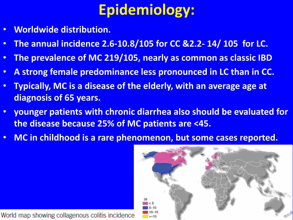
Epidemiology:• Worldwide distribution.
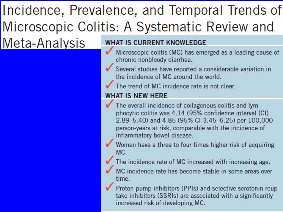
• The annual incidence 2.6-10.8/105 for CC &2.2- 14/ 105 for LC.
• The prevalence of MC 219/105, nearly as common as classic IBD
• A strong female predominance less pronounced in LC than in CC.
• Typically, MC is a disease of the elderly, with an average age at diagnosis of 65 years.
• younger patients with chronic diarrhea also should be evaluated for the disease because 25% of MC patients are <45.
• MC in childhood is a rare phenomenon, but some cases reported.
6

7

Clinical presentations:• LC &CC are not distinguishable from each other.
• The key clinical feature is chronic non-bloody diarrhea, typically watery, leading to urgency (70%) &ultimately, fecal incontinence (40%).
• In severe cases, bowel movements can be > 15/ day &nocturnal diarrhea is common (50%).
• Despite considerable fluid loss, serious dehydration, electrolyte changes, or other complications are rare.
• The natural is not well investigated but the risk of relapse is high (60– 80%) after cessation of budesonide treatment, indicating that the course is chronic relapsing.
• Active disease leads to impaired health-related QoL (mainly stool consistency) & often results in severe social handicap.
8

Clinical presentations:• Disease activity defined as 3 or more stools/day or 1 or more
watery stools/day.
• Abdominal pain is a common symptom in MC.
• Abdominal discomfort or cramps may occur in up to 50%, like IBD.
• 43% of MC patients fulfilled the ROME II criteria.
• Intermediate or severe pain is observed in periods with active disease &ameliorated with budesonide.
• Weight loss can occur in active disease in nearly half of patients.
• Significant weight loss. concomitant celiac should be ruled out.
• Associated with other AI diseases; most commonly celiac disease (12%), AI thyroid disease (10.3%), Sjögren (3.4%), DM (1.7%), skin/joint diseases (6.0%),in most cases, preceded MC with earlier onset of colitis and more severe GI symptoms.
• The overall malignancy & mortality in CC is not different with the general pop with increased risk of LC in women,?smokiing
9

Histopathology:• MC is a histologic diagnosis, but only in patients with chronic
diarrhea.
• This clinical information always should be provided to the pathologist.
• The history of diarrhea per se does not identify patients at higher risk of abnormal histology, but those > 60 had a markedly increased likelihood of a specific histologic abnormality&MC was the most common diagnosis.
• Histology is necessary to make the diagnosis, differentiating between the 2 major subtypes (ie, LC/CC)&to rule out other causes of chronic diarrhea.
• On endoscopy,mucosa of the colon is almost always normal, but occasionally may show subtle changes, such as edema, erythema, altered vascular pattern, or even mucosal defects.
• In MC, the morphologic findings may be patchy¬ continuous.
10

Histopathology:• In CC a collagenous band > 10 m in thickness was more common in
the right colon ( highest in the cecum & ascending colon), less frequent in the sigmoid & rectum, whereas the mononuclear inflammation in the lamina propria was found to be distributed evenly among the different segments.
• It is recommended to take multiple biopsy specimens throughout the whole colon because biopsy specimens obtained only from the rectum or from the rectum & sigmoid colon may miss 41-21% of cases, respectively.
• The biopsy specimens should be submitted, preferably, in separate contaIiners.
11

Histopathology: LC• The predominant histologic feature of LC is intraepithelial
lymphocytosis (ie, an increased number of surface intraepithelial lymphocytes [IELs] with little or no crypt architectural distortion) ; 20 or more IELs/ 100 surface epithelial cells (normal, < 5)
• The terminal ileum may be affected in LC (&CC) with a mean villous IEL >5.25
• The surface epithelium may show degenerative/or regenerative changes, such as vacuolization, flattening&loss of mucin.
• Compared with healthy individuals, the cellularity in the lamina propria is increased diffusely.
• The inflammatory infiltrate consists mainly of lymphocytes and plasma cells,but eosinophils /neutrophils also may be present, sometimes within the epithelium.
• In general,H&E-stained slides are sufficient to make the diagnosis,& immunohistochemistry to identify intraepithelial T cells by their positivity for CD3 is not routinely needed. 12
Histopathology: CC• The predominant feature is a thickened collagen band, exceeding
10 mm(N< 3 m m) under the surface epithelium, most evident between the crypts.
• It has an irregular jagged appearance at the deeper border&maycontain entrapped capillaries, red blood cells&inflammatory cells.
• Damage to the surface epithelium usually is pronounced &more common than in LC, with sloughing of surface epithelial cells from subepithelial collagen being a characteristic finding.
• An increased number of IELs is seen, but not to the same amount as in LC&cellularity in the lamina propria is increased by a predominantly mononuclear inflammatory infiltrate& IBD-like features,such as active crypt inflammation with occasional crypt abscess formation, may occur.
• In most cases, H&E is enough without problems.
• In borderline cases,other stains, or IHC with Abs against tenascin, not present in normal adult mucosa may be helpful 13

Histopathology: Variants & DD• Not fullfilling the criteria:
• Borderline LC, MC incomplete, MC not otherwise specified, or paucicellular LC.
• it may be difficult to separate these milder forms from IBS.
14

Pathophysiology
• The etiology & pathophysiology is not well understood but is likely to be multifactorial, involving mucosal immune responses to luminal factors in a genetically predisposed individual.
15

Genetics• Familial occurrence reported, but the exact role of genetic factors
remains to be defined.
• HLA studies: association of MC with HLA-DQ2 or DQ1/3 and a higher frequency of HLADR3DQ2 haplotype & TNF2 allele carriage
• In contrast to Crohn’ s disease, functional polymorphism in the nucleotide-binding oligomerization domain-containing protein 2 & caspase recruitment domain-containing protein 15 (NOD2/ CARD15 ) gene has not been detected.
16
epithelial barrier dysfunction• Leading to increased transmucosal permeability of antigens&
bacteria, promoting intestinal inflammation.
• Mucosal barrier dysfunction persisted despite short-term, clinically effective treatment with budesonide.
17

Diarrhea mechanism• It is likely a combination of osmotic &secretory components with
normal anal function & rectal compliance.
18
Risk factors: Drugs• Considering a drug to cause MC should include clinical
improvement, &disappearance of histologic findings after withdrawal &recurrence after rechallenge.
• Drug with a high likelihood to cause MC were acarbose, aspirin, cyclo3 fort, lansoprazole, NSAIDs, ranitidine, sertraline, ticlopidine,
• PPI&NSAIDs identified as the 2 drugs with the highest likelihood to cause MC.
19

Risk factors: Smoking• It is an equal risk factor for MC for men& women .
• Smokers develop the disease earlier than nonsmokers.
20
Treatment:• The primary aim of therapy is to:
• Achieve clinical remission
• Improve the patient’ s QoL.
• The possibility of drug-induced MC always should be taken into account&the patient’ s medication should be reviewed carefully for drugs with high likelihood to cause MC.
• Discontinuation of such drugs, if possible, may lead to resolution of symptoms.
• Smoking cessation can be considered.
• It is advisable that patients register their bowel movements in a diary before initiation and during follow-up treatment.
21
Treatment:• The strongest treatment evidence is currently available for
budesonide, a locally active corticosteroid with extensive first-pass metabolism in the liver & low systemic exposure.
22
23

24
25
26

Summary:
• A chronic IBD characterized by chronic nonbloody diarrhea &specific histopathology features.
• Active disease, defined as 3 or more stools or 1 or more watery stools per day, significantly reduces quality of life.
• The incidence &prevalence of microscopic colitis to be comparable with those of Crohn’s disease &ulcerative colitis.
• Microscopic colitis is still under-recognized in clinical practice:
• Most know little about its etiology &pathophysiology.
• There are many challenges to the diagnosis& treatment.
27

BO4:
• 1. Which one of the following statements is correct regarding microscopic colitis (MC)?
• A. Th e male to female incidence rate ratio is > 3.0.
• B. Th e median age at diagnosis is > 70 years.
• C. MC is associated with the use of proton pump inhibitors (PPIs).
• D. Use of selective serotonin reuptake inhibitors (SSRIs) appears to be protective against MC.
28

BO4:
• 2. Which one of the following statements is correct regarding incidence rates in microscopic colitis (MC)?
• A. Th e incidence rate of collagenous colitis (CC) is < 5 per 100,000 person-years.
• B. Th e incidence rate of CC is signifi cantly higher than the incidence rate of lymphocytic colitis (LC).
• C. Th e overall incidence rate of MC has been steadily increasing in the United States since the year 2000.
• D. The incidence rate of MC decreases with increased age.
29

BO4:
• 3. An 82-year-old male individual taking naproxen and citalopram daily presents for chronic diarrhea. He has no family history of inflammatory bowel disease. Physical exam is unremarkable, and laboratory tests and fecal fat testing are normal. Colonoscopy revealsmoderate left - sided diverticulitis, and biopsies from the left and right colon reveal LC. Which one of the following statements is correct regarding this patient?
• A. His condition will predictably and significantly improve if he stops taking naproxen.
• B. SSRI use is a more likely contributor to his condition than NSAID use.
• C. His 85-year-old brother has a higher risk of developing LC, based on his age.
• D. He has developed MC as a result of his diverticulitis.30

Thanks for attention
31