
GIT 4th 2015 CRC.
61
Colorectal neoplasia: Dr.Mohamed Alshekhani Professor in Medicine MBChB-CABM-FRCP-EBGH 2015 1
-
Upload
shaikhani -
Category
Health & Medicine
-
view
145 -
download
6
Transcript of GIT 4th 2015 CRC.

Colorectal neoplasia:
Dr.Mohamed AlshekhaniProfessor in Medicine
MBChB-CABM-FRCP-EBGH2015
1

CRC: Epidemiology
• Incidence higher for men (F/M 1.4:1), with considerable geographic variation; the majority (60%) from developed regions.
• Blacks have higher incidence & mortality.
2

3

4

5

6

Predisposing factors: IBD
• Increased risk depending on:
• Duration of IBD; progressively increases after 10 years (2%), 20 years (8%)& 30 years (18%).
• The extent & location: Pancolitis& distal colitis confer high& intermediate colorectal cancer risks, respectively,proctitis has minimal (if any) increased risk.
• A concomitant diagnosis of primary sclerosing cholangitis& family history of CRC (especially <50 years).
• Age of onset of disease: not important.
7

8

Predisposing factors: T2DM
• 30% higher risk due to higher concentrations of circulating insulin and/or insulin-like growth factors related to obesity.
9

Predisposing factors: Ureterosigmoidostomy
• Associated with a markedly elevated risk (100-500 times higher than general population), with a prolonged latency period that can exceed 50 years.
10

Predisposing factors:Cholecystectomy
• Associated with modesty increased risk, BZ of altered fecal bile acid composition.
11

Predisposing factors : Non-syndromic Family History
• Influenced by:
• The degree of relationship
• Number of relatives affected
• Age at diagnosis of family member.
• First-degree relative or two second-degree relatives at any age: lifetime risk is elevated *2-3.
• When 2 first-degree relatives are affected, increased by *3-4.
• Diagnosis of CRC in a first-degree relative at age 50 years or younger increase risk by *3-4.
• A family history of CR adenoma appears to confer similarly increased CRC risks.
12

Predisposing factors : Hereditary Colorectal Cancer Syndromes
• 1. Hereditary Nonpolyposis Colorectal Cancer:
• The most common type of hereditary CRC (2-3% of all cases).
• Autosomal dominant.
• Colorectal adenomas develop at a relatively young age ( 20- 30 years) &progress to CRC more quickly than sporadic adenomas. Lifetime risk is 80%, with a mean age at diagnosis of 44 ys.
• Clinicopathologic features include proximal colon location (up to 2/3), poorly differentiated histology& MSI high phenotype.
• Endometrial AC is the 2ND most common ca in HNPCC,30-60%.
• Additional HNPCC-related malignancies are gastric, biliary tract, urinary tract, ovarian &small-bowel cancer.
13

14

15

Predisposing factors : 2.Familial Adenomatous Polyposis
• Incidence is 1/6000-13,000 live birdis, <1% of CRC.
• Germline mutations in APC gene,Autosomal dominant.
• Characterized by hundreds-thousands of adenomatous polyps distributed throughout the colorectum.
• Mean age at polyp diagnosis of 16 years.
• Without intervention, nearly all patients will develop colorectal cancer, with a mean age at cancer diagnosis of 39 years.
16

Predisposing factors : 2.Familial Adenomatous Polyposis
• Duodenal adenomas, particularly in the periampullary region, found in 50-90% & a lifetime risk for duodenal cancer of 4- 12%.
• Other malignancies: papillary thyroid cancer, childhood hepatoblastoma, gastric adenocarcinoma.
• Benign: gastric fundic gland polyps, small-bowel adenomas, osteomas, desmoid tumors, supernumerary teeth, congenital hypertrophy of the retinal pigment epithelium.
• APC mutational analysis can be used to confirm the diagnosis.
17

Predisposing factors : 3.Attenuated FAP
• AFAP is a subset of FAP.
• Affected individuals develop fewer (<100), often more proximally distributed, colorectal adenomas.
• Clinical presentation may be delayed until 4th or 5th decade of life.
• The cumulative CRC risk appears to be slightly lower for AFAP (70%) than for classic FAP (100%).
• Extracolonic features include UGIT polyps but less prominent than in classic FAP.
• Germline mutations are typically found in the extreme distal or proximal ends of the APC gene.
18

Predisposing factors : 4.MTH-Associated Polyposis
• Characterized by heterozygous & homozygous mutations in MTH gene (involved in DNA base-excision repair).
• These mutations may be found in 1% (heterozygous) & 0.5- 1% (homozygous) of general population.
• It is autosomal recessive, with biallelic MTH mutations predisposing to somatic alterations in APC and/or other key growth-regulating genes.
• The clinical spectrum of MAP often resembles AFAP.
• Genetic testing for MTH mutations is not yet routinely performed.
19
Predisposing factors : 5.Peutz-Jeghcrs Syndrome
• The estimated incidence is 1/8300- 280,000 live birdis.
• Mutations in STK11/LKBI gene inherited with AD pattern.
• Clinical manifestations consist of mucocutaneous melanocytic macules (particularly in perioral region& over buccal mucosa) & GIT hamartomatous polyps, which may present during adolescence or early adulthood with symptoms of abdominal pain, bleeding,obstruction, or intussusception.
20
Predisposing factors : 5.Peutz-Jeghcrs Syndrome
• PJS-associated hamartomatous polyps are most commonly located in SI (60- 90%) but in the colorectum, biliary tract, resp tract, or GIT.
• Colorectal adenomas can also be observed.
• Cancer risk markedly increased, with 50-90% of affected individuals developing at least one malignancy over life time: colorectal cancer, 40%; gastric, 30- 60%; pancreas, 35%; small intestine, 15- 30%; esophagus, 0.5-33%; breast, 54%; ovary, 21%;lung, 15%; cervix, 10%; uterus, 9%; testes, 9%.
21

Predisposing factors : 6.Peutz-Jeghcrs Syndrome
• Proposed diagnostic criteria require a combination of:
• Histologically confirmed hamartomatous polyp(s) with distinctive PJS features (arborizing smooth muscle fibers)
• Mucocutaneous melanocytic macules
• &/or a Known family history of PJS.
• STK11/LKB1 mutational analysis can also be performed to confirm a suspected PJS diagnosis.
22

Predisposing factors : 7.Juvenile Polyposis Syndrome
• The incidence is 1/16,000 -100,000 live births.
• Mutations in BMPRIA, SMAD4,PTEN genes.
• Benign juvenile hamartomatous polyps are relatively common, occurring as solitary, sporadic lesions in up to 2% of children <10 ys.
• In contrast, patients with JPS have multiple polyps that are distributed throughout the GIT.
• Abdominal pain, diarrhea, bleeding, obstruction, or intussusception may occur during childhood or adolescence, with an average age at JPS diagnosis of 18.5 years.
• 2/3 of untreated patients with JPS develop CRC at 60 years.
• Other Cans: gastric, duodenal,pancreatic cancer.
• Some may also have hereditary hemorrhagic telangiectasia, which can present with digital clubbing.
23
Predisposing factors : Juvenile Polyposis Syndrome
• Clinical diagnosis of JPS can be made based on any of:
• (1) Three or more juvenile polyps in the colorectum
• (2) Multiple juvenile polyps throughout the GIT, or 3 or any number of juvenile polyps coupled with a family history of JPS.
• Genetic testing is not yet routinely performed in clinical practice.
24

Predisposing factors : 8.Hyperplastic Polyposis
• A familial predisposition to multiple and/or large hyperplastic polyps in the colorectum.
• CRC risk is increased to an unknown degree.
• Diagnosis made if any of the following criteria are met:
• (1) 5 or more histologically confirmed hyperplastic polyps located proximal to the sigmoid colon, of which two or more are larger than 1 cm in diameter.
• (2) Any number of hyperplastic polyps located proximal to the sigmoid colon in a person who has one or more first-degree relatives with documented HPP.
• (3) >30 hyperplastic polyps distributed throughout the colorectum.
25

26

CRC Screening: Average Risk
• ¾ of all CRCs are diagnosed in average-risk persons (persons without previously identified CRC risk factors).
• 90% occur during the sixth decade of life or later.
• Clinicians should perform an individualized assessment of risk for CRC in all adults.
• Those with average risk should begin screening at age 50 years (age 40 years for black or DCs)& continue at intervals based on the method of screening chosen &the results of screening until age 75 years or until there is a life expectancy of <10 years.
• Multiple test options are used.
27

28

29

30

CRC Screening: Average Risk
• The tests include:
• (1) tests that detect premalignant adenomas & CRC
• (flexible sigmoidoscopy,colonoscopy, double-contrast barium enema, CT colonography)
• (2) tests that primarily detect colorectal cancer (stool assays).
• Existing data are insufficient to support CT colonography or stool DNA testing for average-risk CRC screening.
• The ACP guidance statement recommends using a stool-based test, flexible sigmoidoscopy,or optical colonoscopy for screening in patients at average risk.
31

CRC Screening: Average Risk
• 1.Guaiac Fecal Occult Blood Test:
• Uses a reagent that changes color in presence of peroxidase, which is found in human blood, animal blood& other dietary sources.
• Requires collection of two samples from three consecutive stools (six samples total).
• Samples can be obtained at home & mailed in to a clinical laboratory for processing & analysis.
• Results in statistically significant reductions in CRC incidence 17-20% & mortality 15-33% with regular screening.
32

CRC Screening: Average Risk
• Limitations of gFOBT screening are:
• Low sensitivity for advanced adenomas (11- 41%)
• Diet & medication interactions that may produce false-positive or false-negative results.
• The need for annual testing if negative
• The need for appropriate diagnostic follow-up (ideally by colonoscopy) if testing is positive to achieve maximum benefit.
33

CRC Screening: Average Risk
• 2.Fecal Immunochemical Test:
• Incorporate antibodies directed against the human globin protein, improving its specificity over gFOBT.
• FIT sensitivity 60- 85% for CRC&25-50% for advanced adenomas.
• Sample collection can be completed at home& pretest dietary restrictions arc not necessary.
• As with gFOBT screening, FIT screening requires annual testing for negative results & appropriate diagnostic follow-up for a positive result.
34

CRC Screening: Average Risk
• 3.Stool DNA Test:
• Stool DNA (sDNA) assays tar get molecular alterations that originate directly from lesion of interest.
• Currendy, an entire stool specimen (30 g minimum) must be submitted for evaluation.
• It demonstrated moderate sensitivity for CRC (25-58%) & lower sensitivity for adenomas 1 cm or larger (8-45%).
• At present, sDNA testing is not routinely available for colorectal cancer screening.
35

CRC Screening: Average Risk
• 4.Flexible Sigmoidoscopy
• Endoscopic evaluation of the lower colorectum (up to 60 cm).
• Performed after limited bowel preparation (two enemas) without conscious sedation.
• Adenomas of any size or polyps larger than 5 mm should undergo full colonoscopy.
• Reduces mortality by 60%.
• Proximal neoplasia is more common in blacks & older women, proceeding directly to colonoscopy for screening may be more prudent in these patients.
• A potential complication is bowel perforation, which occurs in<1/ 20,000.
36

CRC Screening: Average Risk
• 5.CT Colonography
• Evaluates entire colorectum,&limited extracolonic structures in the abdomen &pelvis.
• Dietary restrictions, full cathartic preparation, oral contrast ingestion, air insufflation are required.
• It has estimated sensitivities of > 90% for CRC & 85-93% for polyps 1 cm or larger.
• Diagnostic and/or therapeutic colonoscopy is recommended for one or more polyps 1 cm or larger, or three or more polyps 6 mm or larger.
• Bowel perforation can occur from CT colonography, but extremely low (<1/10,000).
37

CRC Screening: Average Risk
• 6.Colonoscopy:
• Evaluates entire colorectum.
• Dietary restrictions , full cathartic preparation are required prior to the procedure&conscious sedation is needed.
• Therapeutic polypectomy can be completed during the baseline procedure.
• Results in substantial reductions in CRC incidence (53- 72%) & mortality (31%).
38

CRC Screening: Average Risk
• If no polyps are detected during a high-quality baseline colonoscopy (cecal intubation accomplished, mucosa well visualized with minimal fecal debris,withdrawal time >6 minutes), the next colonoscopy can be performed in 10 years.
• Miss rates:5% for CRC, 2-12% for polyps 1 cm or larger, >20% for polyps 6 mm or larger.
• Risks: significant bleeding (2-6/1000) bowel perforation (1/1000).
39

Surveillance: Postpolypectomy
• Patients with one or > adenomas detected during CRC screening are at increased risk for developing subsequent advanced CR neoplasia.
• The strongest predictors of future risk depends on:
• Multiplicity
• Size
• Morphology
• histology.
40

Surveillance: Postpolypectomy
• Low-risk patients (1-2 adenomas,<1 cm, tubular morphology & low-grade dysplasia), surveillance colonoscopy repeated in 5-10 ys.
• High-risk patients (>3 adenomas, >1 cm, villous morphology, or high-grade dysplasia), surveillance colonoscopy repeated in 3 years, assuming complete removal of the index lesion(s).
• If >10 adenomas are detected during a single evaluation, the next colonoscopy should be repeated in < 3 years &possibility of an underlying hereditary cancer syndrome should be considered.
41

Surveillance: Postpolypectomy
• Patients with large, sessile adenomas removed piecemeal should undergo repeat colonoscopy at a relatively short interval (2-6 mondis) to ensure complete resection.
• Patients with small, hyperplastic,rectal polyps only (not meeting criteria for hyperplastic polyposis syndrome) should undergo surveillance according to average-risk screening guidelines.
42

Surveillance: Post CRC treatment
• Patients with CRC who receive potentially curative treatment (such as surgery, chemotherapy, radiation therapy, or a combination of these) should be monitored aggressively to detect early recurrence & perform surveillance for metachronous CR neoplasia( in the remaining colorectal segment).
• Preoperative clearing colonoscopy (or CT colonography for obstructing tumors) is recommended to identify synchronous malignancies, which occur in 2-7% of all patients with CRC.
43

Surveillance: Post CRC treatment
• Following treatment,surveillance colonoscopy should be repeated 1 year after the clearing examination.
• If normal findings are observed, subsequent surveillance colonoscopies can be performed at 3 years& 5 years after the clearing examination, respectively. If adenomas are detected, surveillance intervals can be tailored according to postpolypectomyguidelines.
44

45

CRC: Clinical presentations
• Symptoms are often related to anatomic location& tumor size.
• For tumors originating in the Proximal colon (cecum to splenic flexure): common symptoms include nonspecific abdominal pain, unintentional weight loss& occult GIB.
• Tumors in distal colon (descending & sigmoid) or rectum: can present with increased stool frequency (small caliber), incomplete evacuation, constipation, or hematochezia.
46

CRC: Clinical presentations
• Clinical signs that may be related to CRC are anemia, fistula, mass lesion, or unexplained abscess.
• At presentation, 40% have localized disease, 36% have regional metastases, 19% have distant metastases& 5% are unstaged.
• Relatively frequent sites for distant metastases are the liver, peritoneal cavity, lungs; adrenal glands, ovaries, and skeletal system are less commonly involved.
• Metastases to the brain or central nervous system are rare.
47

CRC: Diagnosis & Staging
• Colonoscopy is the initial test of choice for evaluating symptoms suggestive of CRC because biopsies can be obtained.
• Staging is determined by depth of tumor invasion, extent of nodal involvement,&presence or absence of distant metastases.
• For CRC, the clinical (or preoperative) stage is generally based on physical exam, abdomino-pelvic CT & chest radiography findings.
• The rectal cancer staging usually includes EUS or pelvic MRI in addition to the tests for colon cancers.
• The final pathologic stage is determined following surgery.
• The 5-year relative survival rate is 65%, with estimates of 90%, 70%, & 12% for localized, regional& distant stages of disease, respectively.
48

49

50

BO5 1:
• Most of the CRCs occur in:
• Average risk persons.
• Those with HNPCC.
• FAB.
• AFAP.
• PJS.
51
BO5 2:
• The highest risk for developing CRC is among:
• Post cholecystectomy patients.
• Post ureterosigmoidostmy patients.
• FAB.
• AFAP.
• PJS.
52

BO5 3:
• All are reasons not to do cholecystectomy in asymptomatic gall stones, except:
• Associated morbidity.
• Associated mortality.
• Risk of CRC.
• Weight gain.
• Missing other diagnoses.
53

BO5 4:
• The 2nd most common cancer in FAB after CRC is:
• Fundal gastric caner.
• Periampullary cancer.
• Ovarian cancer.
• Breast cancer.
• Uninary bladder cancer.
54

BO5 5 :
• The ideal method for CRC screening is:
• Sigmoidoscopy.
• Fecal occult blood test.
• CT colonogarphy.
• Colonoscopy.
• Barium enema.
55
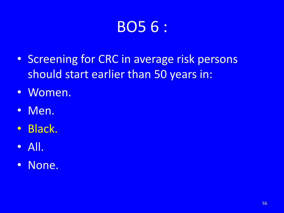
BO5 6 :
• Screening for CRC in average risk persons should start earlier than 50 years in:
• Women.
• Men.
• Black.
• All.
• None.
56

BO5 7 :
• All are risk for CRC except:
• FAP.
• AFAP.
• PJS.
• Family history of colorectal adneoma.
• Family history of CRC.
57

BO5 8 :
• The following are advised to prevent CRC in average risk persons:
• Increase fruit intake.
• Increase vegetables.
• Avoid smoking.
• NSAIDs use.
• Physical activity.
58
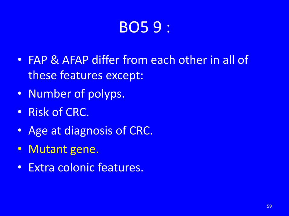
BO5 9 :
• FAP & AFAP differ from each other in all of these features except:
• Number of polyps.
• Risk of CRC.
• Age at diagnosis of CRC.
• Mutant gene.
• Extra colonic features.
59

BO5 10 :
• The strongest predictors of future CRC risk depends on all the following except:
• Multiplicity.
• Size.
• Morphology.
• Histology.
• None of the above.
60

Thanks for attention
61