
Girls homozygous for an IL-2–inducible T cell kinase ... · In addition to SAP and XIAP, the SLAM...
9
Research article 1350 The Journal of Clinical Investigation http://www.jci.org Volume 119 Number 5 May 2009 Girls homozygous for an IL-2–inducible T cell kinase mutation that leads to protein deficiency develop fatal EBV-associated lymphoproliferation Kirsten Huck, 1 Oliver Feyen, 1 Tim Niehues, 2 Franz Rüschendorf, 3 Norbert Hübner, 3 Hans-Jürgen Laws, 1 Tanja Telieps, 1 Stefan Knapp, 4 Hans-Heinrich Wacker, 5 Alfons Meindl, 6 Hassan Jumaa, 7 and Arndt Borkhardt 1 1 Department of Pediatric Oncology, Hematology and Clinical Immunology, Centre for Child and Adolescent Health, Heinrich Heine University, Düsseldorf, Germany. 2 Centre for Child and Adolescent Health, HELIOS Klinikum Krefeld, Krefeld, Germany. 3 Max Delbrück Center for Molecular Medicine (MDC), Berlin, Germany. 4 Structural Genomics Consortium, Nuffield Department of Medicine, and Department of Clinical Pharmacology, University of Oxford, Headington, Oxford, United Kingdom. 5 Joint Practice for Hematopathology, Kiel, Germany. 6 Klinikum rechts der Isar, Technische Universität, Munich, Germany. 7 Department of Molecular Immunology, Max Planck Institute of Immunobiology, Freiburg, Germany. The fatal immune dysregulation that sometimes follows EBV infection in boys has been linked to mutations in two X chromosome–encoded genes, SLAM-associated protein (SAP) and X-linked inhibitor of apoptosis (XIAP). In this study we describe 2 girls from a consanguineous Turkish family who died after developing severe immune dysregulation and therapy-resistant EBV-positive B cell proliferation following EBV infection. SNP array–based genome-wide linkage analysis revealed IL-2–inducible T cell kinase (ITK) as a candidate gene for this immunodeficiency syndrome. Both girls harbored a homozygous missense mutation that led to sub- stitution of a highly conserved residue (R335W) in the SH2 domain of ITK. Characteristics of ITK deficiency in mouse models, such as absence of NKT cells and high levels of eomesodermin in CD8 + cells, were seen in either one or both of the girls. Two lines of evidence suggested that R335W caused instability of the ITK pro- tein. First, in silico modeling of the mutant protein predicted destabilization of the SH2 domain. Additionally, Western blot analysis revealed that, unlike wild-type ITK, the R335W mutant was nearly undetectable when expressed in 293 T cells. Our results suggest that ITK deficiency causes what we believe to be a novel immu- nodeficiency syndrome that leads to a fatal inadequate immune response to EBV. Because ITK deficiency resembles EBV-associated lymphoproliferative disorders in boys, we suggest that this molecular cause should be considered during diagnosis and treatment. Introduction Severe immune dysregulation in boys after primary infection with EBV, presenting as fatal mononucleosis, hemophagocyto- sis, hypogammaglobulinemia, and lymphoproliferation, is the hallmark of X-linked lymphoproliferative disease (XLP), a well- known inherited immunodeficiency disorder (1). Mutations in the SH2 domain protein 1A (SH2D1A), also referred to as sig- naling lymphocytic activation molecule family member 1–associ- ated (SLAM-associated) protein (SAP), account for approximately 60% of XLP cases (2–4). In addition, mutations in the X-linked inhibitor of apoptosis (XIAP) protein have been shown to be caus- ative in 3 XLP families (5). Recently, several studies have demon- strated an almost complete absence of NKT cells in lymph nodes, liver, spleen, and thymus of SAP-deficient mice as well as in the blood of patients with XLP carrying mutations in SAP and XIAP (5–8). As the lack of NKT cells is a shared feature of SAP and XIAP deficiency, a critical role has been postulated for NKT cells in the response to EBV infection. In addition to SAP and XIAP, the SLAM receptor family mem- ber 1, the tyrosine kinase Fyn, and 2 members of the Tec family kinases, namely the resting lymphocyte kinase (RLK) and the IL-2– inducible T cell kinase (ITK), have been shown to be required for the development of NKT cells in mice (9–13). We examined a consanguineous Turkish family in which 2 girls died after showing a fatal EBV-associated lymphoprolifera- tive disorder clinically resembling XLP. In both girls we found homozygous mutations in the SH2 domain of ITK. The identified mutation R335W results in protein destabilization, lack of ITK in lymph node biopsies, and absence of NKT cells. Thus, we believe ITK deficiency to be a novel primary immunodeficiency character- ized by fatal immune dysregulation following EBV infection. Results Patients Patient 1. The female index patient was the first child of healthy consanguineous Turkish parents. At the age of 5 years 2 months, immunoglobulin levels, measured during an episode of aph- thous stomatitis and subsequent candida stomatitis, were nor- mal. From the age of 6 years, she successively developed recurrent Conflict of interest: The authors have declared that no conflict of interest exists. Nonstandard abbreviations used: BTK, Bruton’s tyrosine kinase; CRP, C-reactive protein; EBNA, Epstein-Barr nuclear antigen; Eomes, eomesodermin; FACS, fluores- cence-activated cell sorting; ITK, IL-2–inducible T cell kinase; LMP1, latent membrane protein 1; SAP, SLAM-associated protein; SLAM, signaling lymphocytic activation molecule family member 1; VCA, viral capsid antigen; XIAP, X-linked inhibitor of apoptosis; XLP, X-linked lymphoproliferative disease. Citation for this article: J. Clin. Invest. 119:1350–1358 (2009). doi:10.1172/JCI37901.
Transcript of Girls homozygous for an IL-2–inducible T cell kinase ... · In addition to SAP and XIAP, the SLAM...

Research article
1350 TheJournalofClinicalInvestigation http://www.jci.org Volume 119 Number 5 May 2009
Girls homozygous for an IL-2–inducible T cell kinase mutation that leads to protein deficiency develop fatal EBV-associated
lymphoproliferationKirsten Huck,1 Oliver Feyen,1 Tim Niehues,2 Franz Rüschendorf,3 Norbert Hübner,3
Hans-Jürgen Laws,1 Tanja Telieps,1 Stefan Knapp,4 Hans-Heinrich Wacker,5 Alfons Meindl,6 Hassan Jumaa,7 and Arndt Borkhardt1
1Department of Pediatric Oncology, Hematology and Clinical Immunology, Centre for Child and Adolescent Health, Heinrich Heine University, Düsseldorf, Germany. 2Centre for Child and Adolescent Health, HELIOS Klinikum Krefeld, Krefeld, Germany. 3Max Delbrück Center for Molecular Medicine (MDC), Berlin, Germany. 4Structural Genomics Consortium, Nuffield Department of Medicine, and Department of Clinical Pharmacology, University of Oxford, Headington,
Oxford, United Kingdom. 5Joint Practice for Hematopathology, Kiel, Germany. 6Klinikum rechts der Isar, Technische Universität, Munich, Germany. 7Department of Molecular Immunology, Max Planck Institute of Immunobiology, Freiburg, Germany.
ThefatalimmunedysregulationthatsometimesfollowsEBVinfectioninboyshasbeenlinkedtomutationsintwoXchromosome–encodedgenes,SLAM-associatedprotein(SAP)andX-linkedinhibitorofapoptosis(XIAP).Inthisstudywedescribe2girlsfromaconsanguineousTurkishfamilywhodiedafterdevelopingsevereimmunedysregulationandtherapy-resistantEBV-positiveBcellproliferationfollowingEBVinfection.SNParray–basedgenome-widelinkageanalysisrevealedIL-2–inducibleTcellkinase(ITK)asacandidategeneforthisimmunodeficiencysyndrome.Bothgirlsharboredahomozygousmissensemutationthatledtosub-stitutionofahighlyconservedresidue(R335W)intheSH2domainofITK.CharacteristicsofITKdeficiencyinmousemodels,suchasabsenceofNKTcellsandhighlevelsofeomesodermininCD8+cells,wereseenineitheroneorbothofthegirls.TwolinesofevidencesuggestedthatR335WcausedinstabilityoftheITKpro-tein.First,insilicomodelingofthemutantproteinpredicteddestabilizationoftheSH2domain.Additionally,Westernblotanalysisrevealedthat,unlikewild-typeITK,theR335Wmutantwasnearlyundetectablewhenexpressedin293Tcells.OurresultssuggestthatITKdeficiencycauseswhatwebelievetobeanovelimmu-nodeficiencysyndromethatleadstoafatalinadequateimmuneresponsetoEBV.BecauseITKdeficiencyresemblesEBV-associatedlymphoproliferativedisordersinboys,wesuggestthatthismolecularcauseshouldbeconsideredduringdiagnosisandtreatment.
IntroductionSevere immune dysregulation in boys after primary infection with EBV, presenting as fatal mononucleosis, hemophagocyto-sis, hypogammaglobulinemia, and lymphoproliferation, is the hallmark of X-linked lymphoproliferative disease (XLP), a well-known inherited immunodeficiency disorder (1). Mutations in the SH2 domain protein 1A (SH2D1A), also referred to as sig-naling lymphocytic activation molecule family member 1–associ-ated (SLAM-associated) protein (SAP), account for approximately 60% of XLP cases (2–4). In addition, mutations in the X-linked inhibitor of apoptosis (XIAP) protein have been shown to be caus-ative in 3 XLP families (5). Recently, several studies have demon-strated an almost complete absence of NKT cells in lymph nodes, liver, spleen, and thymus of SAP-deficient mice as well as in the blood of patients with XLP carrying mutations in SAP and XIAP (5–8). As the lack of NKT cells is a shared feature of SAP and XIAP
deficiency, a critical role has been postulated for NKT cells in the response to EBV infection.
In addition to SAP and XIAP, the SLAM receptor family mem-ber 1, the tyrosine kinase Fyn, and 2 members of the Tec family kinases, namely the resting lymphocyte kinase (RLK) and the IL-2– inducible T cell kinase (ITK), have been shown to be required for the development of NKT cells in mice (9–13).
We examined a consanguineous Turkish family in which 2 girls died after showing a fatal EBV-associated lymphoprolifera-tive disorder clinically resembling XLP. In both girls we found homozygous mutations in the SH2 domain of ITK. The identified mutation R335W results in protein destabilization, lack of ITK in lymph node biopsies, and absence of NKT cells. Thus, we believe ITK deficiency to be a novel primary immunodeficiency character-ized by fatal immune dysregulation following EBV infection.
Results
PatientsPatient 1. The female index patient was the first child of healthy consanguineous Turkish parents. At the age of 5 years 2 months, immunoglobulin levels, measured during an episode of aph-thous stomatitis and subsequent candida stomatitis, were nor-mal. From the age of 6 years, she successively developed recurrent
Conflictofinterest: The authors have declared that no conflict of interest exists.
Nonstandardabbreviationsused: BTK, Bruton’s tyrosine kinase; CRP, C-reactive protein; EBNA, Epstein-Barr nuclear antigen; Eomes, eomesodermin; FACS, fluores-cence-activated cell sorting; ITK, IL-2–inducible T cell kinase; LMP1, latent membrane protein 1; SAP, SLAM-associated protein; SLAM, signaling lymphocytic activation molecule family member 1; VCA, viral capsid antigen; XIAP, X-linked inhibitor of apoptosis; XLP, X-linked lymphoproliferative disease.
Citationforthisarticle: J. Clin. Invest. 119:1350–1358 (2009). doi:10.1172/JCI37901.
research article
TheJournalofClinicalInvestigation http://www.jci.org Volume 119 Number 5 May 2009 1351
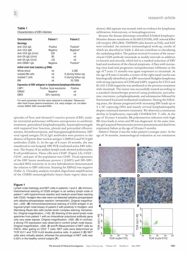
episodes of fever and elevated C-reactive protein (CRP), nodu-lar interstitial pulmonary infiltrates unresponsive to antibiotic treatment, generalized lymphadenopathy, hepatosplenomegaly with impaired liver function, pleural and pericardial effusion, anemia, thrombocytopenia, and hypogammaglobulinemia. EBV viral capsid antigen (VCA) IgG antibodies were positive in the absence of Epstein-Barr nuclear antigen 1 (EBNA1) and VCA IgM antibodies (Table 1). At the age of 6 years 10 months, she was transferred to our hospital. EBV PCR confirmed active EBV infec-tion. The biopsy of an axillary lymph node showed polymorphic B cell proliferation. These cells were CD20+ (Figure 1A), CD22+, CD10–, and part of the population was CD30+. Focal expression of the EBV latent membrane protein 1 (LMP1) and 30% EBV-encoded RNA reactivity in in situ hybridization demonstrated the relation to EBV infection. Staining for EBNA2 was negative (Table 1). Clonality analysis revealed oligoclonal amplification of the CDRIII immunoglobulin heavy-chain region (data not
shown). BM aspirate was normal, with no evidence for lymphoma infiltration, histiocytosis, or hemophagocytosis.
Because the disease phenotype resembled X-linked lymphopro-liferative disease mutations in SLAM (CD150), SAP, natural killer cell receptor 2B4 (2B4), TNFRSF6 (also known as FAS), and EAT were excluded. An extensive immunological work-up, results of which are described in Table 2, did not contribute to elucidating the underlying defect. The patient received 4 courses of the mono-clonal CD20 antibody rituximab at weekly intervals in addition to foscavir and steroids, which led to a marked reduction of EBV load and resolution of the clinical symptoms. A flare with increas-ing virus load and progressive intrapulmonary infiltrates at the age of 7 years 11 months was again responsive to rituximab. At the age of 8 years 2 months, a tumor of the right nasal concha was histologically identified as an EBV-associated Hodgkin lymphoma with strong expression of CD30 and LMP1, negative for CD15 and Ki-A10. CD20 negativity was attributed to the previous treatment with rituximab. The tumor was successfully treated according to a standard chemotherapy protocol using prednisone, procarba-zine, vincristine, cyclophosphamide, and adriamycine followed by fractionated local and mediastinal irradiation. During the follow-ing years, the disease progressed with increasing EBV loads up to 3 × 10⁷ copies/μg DNA and mainly cervical lymphadenopathy despite continued intensive treatment. We observed a continuous decline in lymphocytes, especially CD45RACD4+ T cells. At the age of 10 years 4 months, BK polyomavirus infection with high BK virus loads in urine and PB was diagnosed. At the same time, the girl acquired Pneumocystis jirovecii pneumonia and died from respiratory failure at the age of 10 years 5 months.
Patient 2. Patient 2 was the index patient’s younger sister. At the age of 16 months, immunological evaluation at our institution
Table 1Characteristics of EBV infection
Characteristic Patient1 Patient2Serology Anti–VCA IgG Positive PositiveA
Anti–VCA IgM Negative NegativeA
Anti–VCA IgA (U/ml) 320 ndAnti–EA IgG (U/ml) 80 ndAnti–EA IgA (U/ml) <20 ndAnti–EBNA1 IgG Negative PositiveA
Initialviralload(copies/μgDNA)Whole blood nd 5,280Isolated NK cells nd 9 (during follow-up)Isolated T cells nd 0 (during follow-up)BM nd 33,7000
ExpresionofEBVantigensinlymphoma/lymphoproliferationLMP1 Positive, focal expression PositiveEBNA2 Negative ndEBER 30% reactivity nd
For each parameter the first value obtained is indicated. AMeasured after fresh frozen plasma transfusion. EA, early antigen; nd, not deter-mined; EBER, EBV-encoded RNA.
Figure 1Lymph node histology and NKT cells in patients 1 and 2. (A) Immuno-histochemical staining of CD20 antigen in an axillary lymph node of patient 1 with hyperimmune reaction and medium-sized blast reacting with CD20. Hodgkin-like cells were not detected (alkaline phosphatase anti–alkaline-phosphatase reaction; hematoxilin). Original magnifica-tion, ×200. (B) Immunohistochemical staining of CD20 antigen in an inguinal lymph node biopsy of patient 2 with positivity in Hodgkin- and Sternberg-Reed–like cells (avidin-biotin complex staining; hematoxi-lin). Original magnification, ×100. (C) Staining of the same lymph node specimen from patient 1 with an intracellular polyclonal antibody gave only very weak signals. Original magnification, ×500. (D) In contrast, a strong ITK expression was observed in control lymph node tissue. Original magnification, ×500. (E and F) Measurement of NKT cells by FACS. After gating on CD3+ T cells, NKT cells were determined as TCR Vβ11 and TCR Vα24 double-positive cells. In patient 2 (E) NKT cells were virtually absent, whereas the percentage of NKT cells was 0.35% in the healthy control subject (F).
research article
1352 TheJournalofClinicalInvestigation http://www.jci.org Volume 119 Number 5 May 2009
Table 2Clinical and immunological characteristics
Category Parameter Patient1 Patient2 ReferencevaluesA
Lymphocyte subsetsB,C Leukocytes, no./μl 8,700 3,100 5,200–11,000 Lymphocytes, no./μl 3,480 732 2,300–5,400 CD3+, % (no./μl) 32 (1,114) 74 (541) 56–75 (1,400–3,700) CD3+CD4+, % (no./μl) 14 (487) 58 (424) 28-47 (700–2,200) CD3+CD8+, % (no./μl) 14 (487) 12 (88) 16–30 (490–1,300) CD20+, % (no./μl) 60 (2,088) 0 2–76 (390–1,400) CD56+CD3–, % (no./μl) 8 (278) 5 (37) 4–17 (130–720) CD3+HLA-DR+, % 13 6 Not established TCRαβ+CD4–/CD8–, % nd 0.8 Not established TCRγδ+CD3+, % nd 4.6 Not established CD4+CD45RA+, % (no./μl) 33 (161) 5 (21) 53–86 (430–1,500) CD4+CD45RO+, % (no./μl) 77 (375) 94 (403) 9–26 (220–660) CD8+CD45RA+, % (no./μl) nd 54 (48) 69–97 (380–1,100) CD8+CD45RO+, % (no./μl) nd 46 (40) Not established
T cell function (cpm)D Medium 3,400 nd 600 Mitogen stimulation PHA 67,600 nd 53,800 PWM 21,200 nd 21,900 OKT3 24,200 nd 66,200 SAC 100 nd 10,000 Antigen stimulation Medium 8,100 nd 800 Tetanus toxoid 6,900 nd 1,100 PPD 13,100 nd 4,100 Candida 12,400 nd 1,300
TCR repertoireE Diversified nd
NK cell function Intracellular perforin and granzyme Normal Normal B expressionB
NK cell lysisF Initially reduced, nd normal during follow up
Serum immunoglobulins IgG (mg/dl) 355 816G 600–1,300 IgA (mg/dl) 37 102G 60–220 IgM (mg/dl) 28 90G 40–160 IgE (IU/ml) nd <5
Specific antibodies Tetanus toxoid, diphtheria toxoid, Present PresentG
pneumococci, Hib Isohemagglutinins Present PresentG
Cytokines sCD25 (U/ml) nd 5,500 IL-1β (pg/ml) nd <5
Exclusion of known CD40 and CD40L expressionB Normal Normal immunodeficiencies SLAM (CD150), SAP, TNFRSF6, 2B4 (FasR) No mutation nd
Miscellaneous Fibrinogen (mg/dl) 432 281 180–350 Ferritin (mg/dl) 186 7310 9–59 Triglycerides (mg/dl) 432 370 11–127 LDH (U/l) 682 748 <308 Coombs test Negative Negative Negative
For each parameter, the first value obtained is indicated; unless otherwise denoted, this was before initiation of IVIG or immunomodulatory therapy. AUnless otherwise indicated, reference values are age-related laboratory normal values. For lymphocyte subsets, reference values are taken from Shearer et al. (38). Reference values for B cells refer to CD3–CD19+ cells. Reference values for T cell function refer to values measured from a control subject on the same day as patient 1. BObtained by FACS analysis. CIn patient 2, immunophenotyping of lymphocyte subsets was done after the start of therapy with dexamethasone and rituximab. D[3H]-thymidine incorporation was measured and expressed as the mean of triple measurements minus the medium values (values from cells in culture medium that were not stimulated with antigen or mitogen). EObtained by FACS analysis with monoclonal Vβ antibodies in patient 1. FNK cell–mediated lysis of [51Cr]-labeled K562 target cells. GObtained after fresh frozen plasma transfusion. PHA, phytohemagglutinin; PWM, pokeweed mitogen; OKT3, anti-CD3; SAC, Staphylococcus aureus Cowan I; PPD, purified protein derivative; Hib, Haemophilus influenzae type b; sCD25, soluble CD25; 2B4, natural killer cell receptor 2B4; LDH, lactate dehydrogenase.
research article
TheJournalofClinicalInvestigation http://www.jci.org Volume 119 Number 5 May 2009 1353
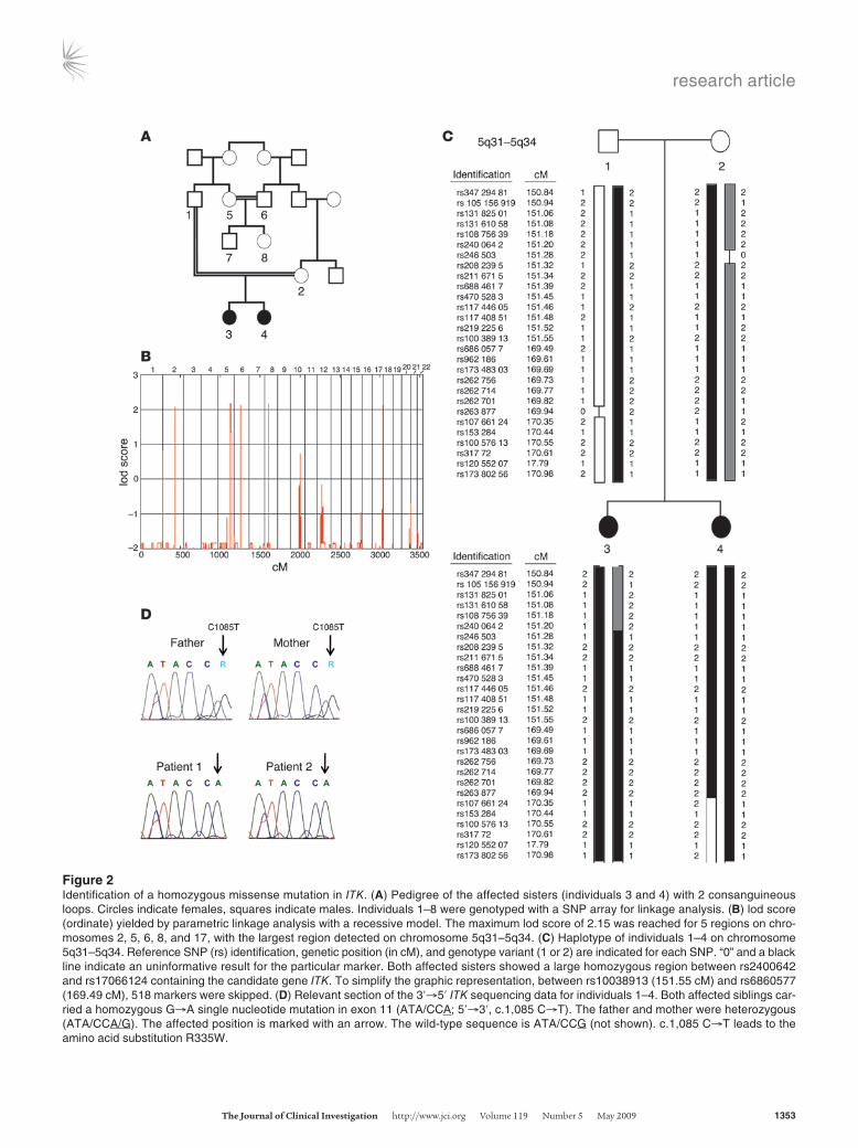
Figure 2Identification of a homozygous missense mutation in ITK. (A) Pedigree of the affected sisters (individuals 3 and 4) with 2 consanguineous loops. Circles indicate females, squares indicate males. Individuals 1–8 were genotyped with a SNP array for linkage analysis. (B) lod score (ordinate) yielded by parametric linkage analysis with a recessive model. The maximum lod score of 2.15 was reached for 5 regions on chro-mosomes 2, 5, 6, 8, and 17, with the largest region detected on chromosome 5q31–5q34. (C) Haplotype of individuals 1–4 on chromosome 5q31–5q34. Reference SNP (rs) identification, genetic position (in cM), and genotype variant (1 or 2) are indicated for each SNP. “0” and a black line indicate an uninformative result for the particular marker. Both affected sisters showed a large homozygous region between rs2400642 and rs17066124 containing the candidate gene ITK. To simplify the graphic representation, between rs10038913 (151.55 cM) and rs6860577 (169.49 cM), 518 markers were skipped. (D) Relevant section of the 3′→5′ ITK sequencing data for individuals 1–4. Both affected siblings car-ried a homozygous G→A single nucleotide mutation in exon 11 (ATA/CCA; 5′→3′, c.1,085 C→T). The father and mother were heterozygous (ATA/CCA/G). The affected position is marked with an arrow. The wild-type sequence is ATA/CCG (not shown). c.1,085 C→T leads to the amino acid substitution R335W.

research article
1354 TheJournalofClinicalInvestigation http://www.jci.org Volume 119 Number 5 May 2009
showed normal blood count, CRP, IgG and IgG subclasses, specific vaccination antibodies, hemagglutinins, and immunophenotyp-ing of lymphocyte subsets. She was EBV VCA IgG positive and VCA IgM negative at that time. However, at the age of 16 months, the presence of maternal VCA IgG antibodies could not be excluded. At the age of 5 years, she developed recurrent episodes of fever and elevated CRP and erythrocyte sedimentation rate unresponsive to antibiotic treatment. Within 4 months the symptoms progressed to a life-threatening condition with profound pancytopenia, retro-peritoneal and abdominal lymphadenopathy, hepatosplenomegaly, severely impaired liver function, ascites, and pleural effusion. At that time, aged 5 years 4 months, she was transferred to our hospi-tal. PCR showed an active EBV infection with a virus load of 3,920 copies/μg DNA in PB and 33,700 copies/μg in BM. EBV VCA IgG and anti-EBNA1 antibodies were positive, whereas VCA IgM anti-bodies were absent. In isolated T and NK cells, no relevant EBV load could be detected (Table 1). The biopsy specimen of an ingui-nal lymph node showed a proliferation of Hodgkin- and Sternberg-Reed–like giant cells positive for CD20 (Figure 1B) and LMP1 and negative for CD30 and CD15. BM aspirate was normal. Further laboratory results are summarized in Table 2. Within the first day after admission to our hospital, the girl developed multiple organ failure requiring mechanical ventilation and hemodialysis for sev-eral days. Treatment with ganciclovir, steroids, and rituximab at weekly intervals led to an improvement of symptoms and a stable virus load not exceeding 5,000 copies/μg DNA but did not induce a complete remission as initially seen in the sister. At the age of 5 years 9 months, she received haploidentical PB stem cell trans-plantation. During aplasia severe nasopharyngeal hemorrhage led to an acute airway obstruction with respiratory insufficiency and cardiovascular arrest. Unfortunately, she died 7 days after resusci-tation from the consequences of ischemic brain injury.
Molecular findingsConsanguinity of the parents and the similar clinical course in both daughters were suggestive for a genetic basis of the disease. We therefore initiated a genome-wide linkage analysis with 8 fam-ily members including patient 1 (individual 3), patient 2 (indi-vidual 4), father (individual 1), mother (individual 2), an aunt (individual 5), an uncle (individual 6), a male cousin (individual 7), and a female cousin (individual 8) (Figure 2A). Parametric linkage analysis with a recessive model revealed genome-wide 5 regions on chromosomes 2, 5, 6, 8, and 17 where the maximal possible lod score of 2.15 was reached (Figure 2B). The largest segment was on chromosome 5q31–5q34, where the homozygous region between recombinant markers rs2400642 and rs17066124 spanned 17.2 Mb (Figure 2C). Within this region we identified 78 positional candidate genes including ITK, which appeared the most likely candidate for the development of the disease. Sequencing of ITK revealed the homozygous missense mutation C1085T in both affected individuals (Figure 2D). The parents, aunt, and male cous-in (individuals 1, 2, 5, and 7) were identified as heterozygous car-riers, whereas individuals 6 and 8 showed the wild-type sequence. To our knowledge, C1085T has not been published as a SNP, and the C1085T mutation was not present in 100 screened children of Turkish descent and 100 children of German descent (data not shown). Comparison of ITK sequences between different species revealed complete conservation of this residue from human to frog Xenopus (data not shown). The discovered mutation leads to the substitution of an arginine by tryptophan residue at position 335
(R335W) in ITK. R335 is located in the BG loop of the ITK SH2 domain (Figure 3C). Due to the distant location of the mutated residue to the phosphotyrosine binding pocket, it is not likely that this mutation impairs recruitment of the SH2 domain substrate. Attempts to functionally express the R335W mutant SH2 domain in bacteria failed due to aggregation of the recombinant protein testing 8 different expression constructs, whereas wild-type pro-tein was stable and expressed functionally at high levels (data not shown). In silico prediction of protein stability revealed a severe destabilization of the SH2 domain by the mutation. We therefore expressed the R335W mutant and wild-type ITK along with differ-ent controls in 293 T cells. By Western blot analysis, the R335W mutant protein was nearly undetectable (Figure 3A). To control that similar amounts of ITK mRNA were expressed in R335W and wild-type transfected cells, we performed quantitative RT-PCR after DNase digestion of the transfected plasmid. We found equal mRNA expression levels for R335W mutated and wild-type ITK (data not shown), which underlines the protein destabilizing effect of the R335W mutant. As a result of this protein instability, using a polyclonal ITK antibody, we found extremely low staining levels in the lymph node biopsy from patient 1 as compared with normal lymph node tissue (Figure 1, C and D).
Studies in Itk–/– mice suggest that ITK is required for repress-ing the T box transcription factor eomesodermin (Eomes) in CD8+ single-positive T cells (24). In agreement with the low ITK levels detected in the biopsy, quantitative RT-PCR revealed a 1,000- to 6,000-fold overexpression of Eomes in both affected children as compared with their heterozygous parents and 2 healthy male controls (Figure 3B). Finally, functional ITK has been shown to be essential for the development of NKT cells. After gating on CD3+ T cells, NKT cells were determined as TCR Vβ11 and TCR Vα24 double-positive cells. In patient 2, NKT cells were virtually absent (Figure 1E), whereas the percentage of NKT cells in the healthy control subject shown in Figure 1F was 0.35%. Unfortunately we were not able to determine in retrospect the level of NKT cells in patient 1 due to the lack of appropriate material. However, the heterozygous parents showed a low percentage of NKT cells, which was 0.15% in the mother and 0.08% in the father, but both were healthy throughout their lives.
DiscussionWe report what we believe is a novel primary immunodeficiency, which leads to an XLP-like phenotype in girls, caused by a homo-zygous mutation in ITK on chromosome 5q31–5q32. This is the first molecular cause of an autosomal recessively inherited lym-phoproliferative disease.
Both ITK-deficient individuals suffered from uncontrolled EBV infection meeting several criteria for secondary hemophagocytic lymphohistiocytosis, but without evidence of hemophagocytosis in BM and lymph nodes. In contrast to SAP deficiency, in which the development of preferably Burkitt lymphoma is not restricted to EBV-positive individuals, and XIAP deficiency, in which no case of lymphoma occurred so far, the leading symptom was an atypical, generalized EBV-positive B cell proliferation with progression to Hodgkin lymphoma in patient 1 (5, 14). It is not clear whether the progressive decline of lymphocytes in patient 1, which led to Pneu-mocystis jirovecii and BK virus infection, was due to ITK deficiency itself, chronic EBV infection, or the long-lasting virostatic therapy.
The non-receptor tyrosine kinase ITK is a member of the Tec kinase family, which also includes Bruton’s tyrosine kinase (BTK),

research article
TheJournalofClinicalInvestigation http://www.jci.org Volume 119 Number 5 May 2009 1355
Tec protein tyrosine kinase (TEC), RLK, and BM kinase X-linked (BMX) (15). Tec kinases contain a conserved SH3, SH2 and catalytic (kinase) domain. The SH2 domain of non-receptor tyrosine kinases is a non-catalytic protein interaction module, which by engaging with a phosphotyrosine-containing signaling partner regulates
activity of the catalytic domain (16). Our results suggest that R335W mutant ITK causes a profound instability of the ITK protein rather than an impairment of ITK activation. Interestingly, mutations at exactly analogous positions within the SH2 domain have been described in BTK (Y361C) (17) and SAP (Q99P) (18), both leading
Figure 3Evidence of severe destabilization of the mutant ITK protein. (A) Western blot with transiently transfected 293 T cells using a monoclonal ITK antibody. Cyclophilin A was used as negative control (lane 1). No expression was detected for the R335W mutant (lane 5) and an ITK variant with complete deletion of the SH2 domain (lane 3), whereas wild-type ITK (lane 4) and the kinase-inactivating mutant (F511Y) (lane 2) showed strong ITK expression. (B) Results of quantitative real-time PCR for EOMES mRNA in CD8+ single-positive T cells on a logarithmic scale. We found 1,000- to 6,000-fold higher mRNA levels of EOMES in both affected children compared with their heterozygous parents or healthy controls. Error bars denote the standard deviation. P1, patient 1; P2, patient 2; F, father; M, mother; C1, control 1; C2, control 2. (C) Domain architecture of ITK and the location of the mutated residue (R335) (highlighted by a red label) in the SH2 domain. Shown is the NMR minimized average struc-ture (protein data bank code 2etz). Protein instability probably results from a compromised salt bridge network (dotted lines) as well as surface exposure of the introduced tryptophan side chain. A phosphotyrosine substrate peptide and the corresponding binding pocket are located on the opposite side of the molecule. The main structural elements are labeled. (D) Site of the BG loop mutations in ITK (R335W, red), BTK (K374N, blue), and SAP (Q99P, yellow).

research article
1356 TheJournalofClinicalInvestigation http://www.jci.org Volume 119 Number 5 May 2009
to protein instability and the clinical phenotype of atypical X-linked agammaglobulinemia (XLA) and XLP, respectively (Figure 3D).
Given the pivotal role of ITK in a number of T cell signaling pathways, absence of Itk has been studied in great detail in mice, but no human disease with ITK deficiency has been described yet. The previously published experimental data in Itk–/– mouse cells greatly facilitated the choice of ITK among our candidate gene list of 78 genes after linkage analysis. Loss of Itk in mice leads to major immunological abnormalities, e.g., decreased responses to TCR stimulation, T cell proliferation, production of IL-2 and other effector cytokines, as well as altered thymic selection of CD4+ and CD8+ cells by modulation of TCR signal strength (19–23). In a set of elegant experiments, different research groups demonstrated that Tec kinase–deficient mice develop a large population of abnormal CD8+ T cells exhibiting innate immune properties but with high expression of the memory markers CD44 and CD122 (24–26).
We also found a clearly increased memory phenotype in our patients pointing to a profoundly altered differentiation of the conventional T cell lineage (Table 2). Perhaps the most impor-
tant paralleling feature between Itk-deficient mice and humans is the absence or reduced numbers of NKT cells. Itk–/– mice show a block of NKT cell maturation and reduced peripheral survival of NKT cells (8, 10, 11).
In mice, NKT cells have been shown to provide a protective innate-type immune response to several microbes (27) includ-ing viruses, e.g., HSV1 and HSV2 (28, 29) and hepatitis B virus (30). Strong evidence for a paramount biological relevance of NKT cells in humans comes from the obser-vation that boys with XLP due to SAP or XIAP deficiency show a severely reduced number, or even a complete absence, of NKT cells (5, 6). Thus, a critical role for NKT cells in the immune response to EBV infection in humans has been postulated (5, 6). The absence of NKT cells in patient 2 and the concomitant inability to clear EBV infection in the 2 ITK-deficient sisters strengthens the hypothesis of a role for NKT cells in the con-trol of EBV infection. The fact that both heterozygous parents have low but still detectable numbers of NKT cells also suggests that ITK is required for the selection and/or survival of NKT cells not only in mice, but also in humans. Rigaud et al. found normal numbers of NKT cells in 8 patients with acute infectious mononucleosis, show-ing that the absence of NKT cells is not an immediate consequence
of acute EBV infection (5), whereas to our knowledge there are no data on NKT cell numbers in patients with chronic active EBV infection. EBV affects more than 90% of the world’s population, but why the adequate immunological control of this successful member of the herpes family seems to be critically dependent on this numerically rather tiny cell population remains obscure.
However, as there are autoimmune conditions in which patients with a severely reduced number of NKT cells are not especially sus-ceptible to EBV infection (31), the lack of NKT cells is likely to con-tribute to the impaired immune response to EBV but may not be the single underlying factor. Other evidence for a protective role of invariant NKT cells in human infection is rather limited. In 2003 Levy et al. reported a girl with severe varicella infection after vari-cella vaccination, in whom an absolute deficiency of invariant NKT cells was the only detectable immunologic abnormality (32).
The immunophenotypical as well as functional analyses in our cases were performed following several months of active EBV infection. Thus, it is not possible to clearly distinguish between the impact of EBV infection and ITK deficiency itself. In the future,
Table 35′→3′ primer sequences for nested amplification of the coding regions of ITK
Exon Primer Upstream Downstream1 Ex GAAAGGATGTGGTTTCGGCCTTTG TTTCCTGTGCTCTATTTTATGCTATG In TCAGAGGAGAAGCTCAGCTATGTTGGC TTAGCCCAGTCCAAACATAAAATGC2 Ex GCCAATGGATCTTATCTAGCAGTAG CTGCACCCGGCTGTGACTGAAG In ATATTGTCTTCTTTCTTACATGAATGG TTTTAACCACAGTCCAGGGAATGG3 Ex CACTCTCGGCTCAGTAGGTCTG TCTCACACCACACTCTATTCTATTG In GTCAACTATCTCCATGCACGCTG AACCCTACTATAGGATCAAGAGTG4 Ex GGCTCACTCAGCCAGGTCTAATC TGCATTCTGTTCCTGACACCCTC In GAGGTGTTATAAAATTGGTAGCCCT AATGATTAAAGGCCAAAAGCTTTTACT5 Ex CAAGTCAGGTTTCACTGTGTCTTAT GACATGCAAATAGGAACATGCCAAT In CCCCTTTCTTCTGTTGTTTTCCTG GGTAGCCTGTGGGCCCACCTG6 Ex CCAGAGGAAGGCAGACTGTCTC CTGTGGGAGATAGGACAAATCATC In CAGACACCCCGATGAAAGGAGG TCGCACCACACCTCAAACACGG7 Ex ATATTCCCCAATCTTTAAATGACTTTTA AGTCAACCAATAATTTATTCCTAACTTA In TGGATTAAACCTATAAAATGATATAAAAC AGGATTCCTTAGGACTGTATTTTCC8 Ex ATGACCATATGATTTTCTAGCATTGTC GCCTGCCAAATCGCTGGGATTC In TATAAGTAAATAATGAAACTTTAAAATATGT CAGTGCACTTAATTCATTTTATTTTATGT9 Ex CCTACAGTATTTCCTCCTTCTGTG CTACCTCTTGCACTGTCTAACTTTG In GTGGAGCTGGAGGCATAAGCCT GCTCACACACTTCAGAAGTGTTTCT10 Ex GTTATAAGACAAAGATAATAAGAACTTAA AGAAGGCAGAGCTCAGGCAGTAA In GTGTCTCAACATTGCTTCTTAATAATC TTGCTCACCTGCTGCTCACCTC11 Ex TAGTGATTTAAGTTAGATGGTTGCTAG ATTTGTAGTCTGAGGAACAGGTAG In AGCAAAGCCCTAACCACTGCTTC ATTTGCCCCAGACCTTTAGGGAC12 Ex GGCTAAAATTCTAGTTAGGGCTTTAT GCATTTCAAGAACACTGAACCGAT In TGGTATCCCAATACCTTATATCTACT CTGCATATCCTTGTTCTGAGCACT13 Ex AGACAAAATGACCATTGGCTATTTTG AGATTATCTGTAATGTATTATTTATTAATG In TTGGGAGACTGAGTTTAGGCCATC CCAGTCATTTCAGTGAGAACTGTC14/15A Ex TGCAGTAAAGCAAAGGACTGTGATT CAGGGTTCAGTGTGGGTAGGGTT In GAGACTCCTTAACTACTGATGACTC CAGCTGGCCTGAAGCCCAGATC16 Ex AATCCTAATGCAAGGAGTCTGTAATT TGCCTCATCCTTCTGAGAGGGTT In TTGCTGTCTGTGGGCTTTGTCATT GGAAGGAGGGAGGTCCTCAAATT17 Ex AATCCACAGGGGATGCTGCTATTA GCACCACATGTGACAAGAGGCTA In TGGATTTACCTATGACTCATAAGTAC AGCTCCCAGCTTGGTGGCTGAC
AThe short exons 14 and 15 were amplified together, including intron 14/15. Ex, external primers; In, inter-nal primers.

research article
TheJournalofClinicalInvestigation http://www.jci.org Volume 119 Number 5 May 2009 1357
the identification and examination of further patients with ITK deficiency will certainly contribute to elucidating the functional implications of ITK deficiency in different lymphocyte lineages on host defense. We recommend that in all children with EBV-associ-ated lymphoproliferative disorders, ITK deficiency should be con-sidered because it may show striking clinical and immunological similarities to SAP or XIAP deficiency in boys.
MethodsThe studies were reviewed and approved by the institutional review board (ethics committee) of Heinrich Heine University. The parents gave informed consent to carry out the investigations described below.
Linkage analysis. Samples were genotyped with the Affymetrix Human Mapping 250K Sty I array according to the manufacturer’s guidelines (Affymetrix). Genotype calling was performed with the Dynamic Model algorithm implemented in the Affymetrix software GTYPE version 4.1. The Call rate was above 94% for all samples; the average call rate was 97.15%.
Quality control and data conversion were managed by ALOHOMORA (http://gmc.mdc-berlin.de/alohomora/) (33). The correct relationship of individuals within the families was verified with the Graphical Representa-tion of Relationships software (http://www.sph.umich.edu/csg/abecasis/GRR/index.html) using 14,339 autosomal markers (34). Mendelian errors were detected by PedCheck (http://watson.hgen.pitt.edu/register/soft_doc.html) and genotypes deleted in all individuals, while unlikely genotypes (double recombinants), identified with Merlin (http://www.sph.umich.edu/csg/abecasis/Merlin/), were deleted in the individuals in which they appeared (35, 36). Parametric multipoint lod score analysis and haplotyp-ing were performed using Merlin. We used a recessive model with com-plete penetrance and a trait locus allele frequency of 0.0001. Marker allele frequencies for a European population as well as genetic positions were extracted from the Affymetrix SNP annotation file (version na23) (http://www.affymetrix.com/support/technical/byproduct.affx?product=500k).
Of the 238,304 SNPs on the array, 238,230 had a physical and genetic position. To reduce the impact of linkage disequilibrium between closely linked markers on the lod score analysis, the pedigree was recalculated with smaller sets of markers (86,162 and 67,334 SNPs) with a minimal distance of 10,000 bp and minimal minor allele frequencies of 0.05 and 0.1. Graphi-cal plots of the Merlin haplotypes were created using HaploPainter (http://haplopainter.sourceforge.net) (37).
ITK staining in lymph node tissue. The polyclonal ITK antibody ab32113 (Abcam) was used for staining of ITK in lymph node tissue.
ITK sequencing and expression of ITK constructs in 293T cells. Genomic DNA was extracted from PB leukocytes. Mutation analysis was performed by direct sequencing of PCR fragments obtained after nested amplification of the exonic and flanking intron region coding sequences of ITK with 17 exons. Primers to amplify the genomic DNA samples were designed accord-ing to GenBank sequences (Table 3). Direct cycle sequencing of all PCR fragments was performed with BigDye Terminator v3.1 cycle sequencing kit (Applied Biosystems) and analyzed by capillary electrophoresis on an ABI Prism 3130 Genetic Analyzer (Applied Biosystems). Analyzed sequences were compared with the cDNA and genomic DNA sequences in GenBank accession numbers NM_005546 (human ITK mRNA). The mutation nomenclature used follows the recommendation of the Human Genome Variation Society (http://www.hgvs.org/mutnomen).
Confluent 293T cells were transfected with either full-length mutated ITK or wild-type ITK plasmids, which were purchased from GenScript. Transient transfections were performed by polyethylenimine transfection using stan-dard parameters, and Western blotting was performed 24 hours thereafter.
ITK Western blot. Postnuclear supernatant equivalents of 1 × 10⁶ (or 0.5 × 10⁶ stimulated) cells were separated by 12% SDS-PAGE and blotted onto PVDF
membranes (1 h, 100 V, Hybond-P; GE Healthcare) and blocked with 5% nonfat dry milk in PBS-Tween (0.05% Tween-20 in PBS) for 1 hour. After washing with PBS-Tween, the blots were incubated overnight with the pri-mary antibody ITK 2F12 (catalog 2380; Cell Signaling Technology) at 4°C. Blots were washed again with PBS-Tween, incubated with the HRP-cou-pled secondary antibodies (1:20,000) goat anti-rabbit (catalog RPN4301) and sheep anti-mouse IgG (catalog RPN4201V) horseradish peroxide (GE Healthcare) for 1 hour at room temperature, washed again, and then developed with a chemiluminescence reagent (Super Signal West Pico Che-miluminescent Substrate; Thermo Scientific). Western blots were stripped with Re-Blot Plus (Millipore) and reprobed with ITK (Y402; catalog 1595-1; Epitomics) (for protocol, see above, beginning with blocking) to ensure the specificity of the antibody used first.
Quantitative real-time PCR analysis of Eomes expression. CD8+ T cells were isolated from PB using Miltenyi Biotec beads and columns. Total RNA was prepared from sorted cells using RNase Mini kit (Qiagen) and reverse transcribed into cDNA using Quantitect Reverse Transcription Kit (Qia-gen) according to the manufacturer’s instructions. Quantitative PCR was performed using the primer and probe set for Eomes (Applied Biosystems), with S18 as a housekeeping gene. Data were analyzed using the ΔΔCT method and normalized to S18. The relative gene expression levels were then determined by comparing them with the expression found in the CD8+ population of the father, which were set as 1.
Immunophenotyping by flow cytometry. Whole blood was anticoagulated with EDTA and processed within 24 hours. A differential white blood cell count was done automatically. Four-color fluorescence-activated cell sort-ing (FACS) analysis was performed with a FACS Calibur flow cytometer with CellQuest version 3.2 software (BD). Lymphocyte subpopulations were analyzed by FACS staining using FITC-, PE-, peridinin-chlorophyll protein– (PerCP-), and allophycocyanin-labeled monoclonal antibodies (BD or Immunotech) following the manufacturer’s instructions. Antibod-ies (clone) against the following epitopes were used: CD3 (SK7; BD), CD4 (SK3; BD), CD8 (SK1; BD), TCRab (WT31; BD), TCRgd (11F2; BD), TCR Vα24 (C15; Immunotech), TCR Vβ11 (C21; Immunotech), CD45RA (L48; BD), CD45RO (UCHL-1; BD), CD56 (MY31; BD), HLA-DR (L243; BD), and CD20 (L27; BD). Conserved thawed cells of both patients and healthy controls were CD3 positively selected using whole-blood CD3 MicroBeads (Miltenyi Biotec) following the manufacturer’s instructions. After gating on 1 × 10⁵ CD3+ T cell lymphocytes, the percentage of NKT cells (CD3+ TCR Vα24+ TCR Vβ11+) was determined by FACS.
Prediction of protein stability. For in silico prediction of protein stability, the Eris program was used (http://troll.med.unc.edu/eris).
AcknowledgmentsSupported by grants from the European Community (LSHB-CT-2004-005276, to A. Borkhardt) and from the German Ministry for Science and Education (NGFN2, to N. Hübner). The Structural Genomics Consortium is a registered charity that receives funds from the Canadian Institutes for Health Research, Canadian Foundation for Innovation, Genome Canada through the Ontario Genomics Institute, GlaxoSmithKline, Karolinska Institutet, Knut and Alice Wallenberg Foundation, Ontario Innovation Trust, Ontario Ministry for Research and Innovation, Merck, Novartis Research Foundation, Swedish Agency for Innovation Systems, Swedish Foundation for Strategic Research, and Wellcome Trust. We thank S. Bellert in Düsseldorf for excellent technical assistance, M. Gombert in Düsseldorf for assistance with real-time PCR, H. Hanenberg in Düsseldorf for providing stored patient material, H.K. Müller-Hermelink in Würzburg for evaluation of histological samples, M. Schneider in Ulm and S. Ehl in Freiburg for NK cell

research article
1358 TheJournalofClinicalInvestigation http://www.jci.org Volume 119 Number 5 May 2009
cytotoxicity assays, and D. Dilloo and all other members of the BM transplantation team in Düsseldorf involved in patient care.
Received for publication October 28, 2008, and accepted in revised form February 11, 2009.
Address correspondence to: Arndt Borkhardt, Department of Pedi-atric Oncology, Hematology and Clinical Immunology, Centre for Child and Adolescent Health, Heinrich Heine University, Mooren-str. 5, 40225 Düsseldorf, Germany. Phone: 49-211-811-7680; Fax: 49-211-811-7607; E-mail: [email protected].
1. Purtilo, D.T., Grierson, H.L., Davis, J.R., and Okano, M. 1991. The X-linked lymphoproliferative disease: from autopsy toward cloning the gene 1975-1990. Pediatr. Pathol. 11:685–710.
2. Coffey, A.J., et al. 1998. Host response to EBV infec-tion in X-linked lymphoproliferative disease results from mutations in an SH2-domain encoding gene. Nat. Genet. 20:129–135.
3. Nichols, K.E., et al. 1998. Inactivating mutations in an SH2 domain-encoding gene in X-linked lympho-proliferative syndrome. Proc. Natl. Acad. Sci. U. S. A. 95:13765–13770.
4. Sayos, J., et al. 1998. The X-linked lymphoprolif-erative-disease gene product SAP regulates signals induced through the co-receptor SLAM. Nature. 395:462–469.
5. Rigaud, S., et al. 2006. XIAP deficiency in humans causes an X-linked lymphoproliferative syndrome. Nature. 444:110–114.
6. Pasquier, B., et al. 2005. Defective NKT cell devel-opment in mice and humans lacking the adapter SAP, the X-linked lymphoproliferative syndrome gene product. J. Exp. Med. 201:695–701.
7. Chung, B., Aoukaty, A., Dutz, J., Terhorst, C., and Tan, R. 2005. Signaling lymphocytic activation molecule-associated protein controls NKT cell functions. J. Immunol. 174:3153–3157.
8. Nichols, K.E., et al. 2005. Regulation of NKT cell development by SAP, the protein defective in XLP. Nat. Med. 11:340–345.
9. Borowski, C., and Bendelac, A. 2005. Signaling for NKT cell development: the SAP-FynT connection. J. Exp. Med. 201:833–836.
10. Gadue, P., Morton, N., and Stein, P.L. 1999. The Src family tyrosine kinase Fyn regulates natural killer T cell development. J. Exp. Med. 190:1189–1196.
11. Felices, M., and Berg, L.J. 2008. The Tec kinases Itk and Rlk regulate NKT cell maturation, cytokine pro-duction, and survival. J. Immunol. 180:3007–3018.
12. Au-Yeung, B.B., and Fowell, D.J. 2007. A key role for Itk in both IFN gamma and IL-4 production by NKT cells. J. Immunol. 179:111–119.
13. Gadue, P., and Stein, P.L. 2002. NK T cell precur-sors exhibit differential cytokine regulation and require Itk for efficient maturation. J. Immunol. 169:2397–2406.
14. Sumegi, J., et al. 2000. Correlation of mutations of the SH2D1A gene and epstein-barr virus infection with clinical phenotype and outcome in X-linked lymphoproliferative disease. Blood. 96:3118–3125.
15. Gibson, S., et al. 1993. Identification, cloning, and characterization of a novel human T-cell-specific tyrosine kinase located at the hematopoietin com-plex on chromosome 5q. Blood. 82:1561–1572.
16. Pletneva, E.V., Sundd, M., Fulton, D.B., and Andreot-ti, A.H. 2006. Molecular details of Itk activation by prolyl isomerization and phospholigand binding: the NMR structure of the Itk SH2 domain bound to a phosphopeptide. J. Mol. Biol. 357:550–561.
17. Saffran, D.C., et al. 1994. Brief report: a point mutation in the SH2 domain of Bruton’s tyrosine kinase in atypical X-linked agammaglobulinemia. N. Engl. J. Med. 330:1488–1491.
18. Morra, M., et al. 2001. Characterization of SH2D1A missense mutations identified in X-linked lym-phoproliferative disease patients. J. Biol. Chem. 276:36809–36816.
19. Schwartzberg, P.L., Finkelstein, L.D., and Readinger, J.A. 2005. TEC-family kinases: regulators of T-help-er-cell differentiation. Nat. Rev. Immunol. 5:284–295.
20. Liu, K.Q., Bunnell, S.C., Gurniak, C.B., and Berg, L.J. 1998. T cell receptor-initiated calcium release is uncoupled from capacitative calcium entry in Itk-deficient T cells. J. Exp. Med. 187:1721–1727.
21. Berg, L.J., Finkelstein, L.D., Lucas, J.A., and Schwartzberg, P.L. 2005. Tec family kinases in T lymphocyte development and function. Annu. Rev. Immunol. 23:549–600.
22. Liao, X.C., and Littman, D.R. 1995. Altered T cell receptor signaling and disrupted T cell develop-ment in mice lacking Itk. Immunity. 3:757–769.
23. Lucas, J.A., Miller, A.T., Atherly, L.O., and Berg, L.J. 2003. The role of Tec family kinases in T cell devel-opment and function. Immunol. Rev. 191:119–138.
24. Atherly, L.O., et al. 2006. The Tec family tyrosine kinases Itk and Rlk regulate the development of conventional CD8+ T cells. Immunity. 25:79–91.
25. Broussard, C., et al. 2006. Altered development of CD8+ T cell lineages in mice deficient for the Tec kinases Itk and Rlk. Immunity. 25:93–104.
26. Dubois, S., Waldmann, T.A., and Muller, J.R. 2006. ITK and IL-15 support two distinct subsets of CD8+
T cells. Proc. Natl. Acad. Sci. U. S. A. 103:12075–12080. 27. Kinjo, Y., and Kronenberg, M. 2005. Valpha14i
NKT cells are innate lymphocytes that participate in the immune response to diverse microbes. J. Clin. Immunol. 25:522–533.
28. Ashkar, A.A., and Rosenthal, K.L. 2003. Interleukin-15 and natural killer and NKT cells play a critical role in innate protection against genital herpes sim-plex virus type 2 infection. J. Virol. 77:10168–10171.
29. Grubor-Bauk, B., Simmons, A., Mayrhofer, G., and Speck, P.G. 2003. Impaired clearance of herpes sim-plex virus type 1 from mice lacking CD1d or NKT cells expressing the semivariant V alpha 14-J alpha 281 TCR. J. Immunol. 170:1430–1434.
30. Kakimi, K., Guidotti, L.G., Koezuka, Y., and Chisa-ri, F.V. 2000. Natural killer T cell activation inhib-its hepatitis B virus replication in vivo. J. Exp. Med. 192:921–930.
31. Miyake, S., and Yamamura, T. 2007. NKT cells and autoimmune diseases: unraveling the complexity. Curr. Top. Microbiol. Immunol. 314:251–267.
32. Levy, O., et al. 2003. Disseminated varicella infec-tion due to the vaccine strain of varicella-zoster virus, in a patient with a novel deficiency in natural killer T cells. J. Infect. Dis. 188:948–953.
33. Ruschendorf, F., and Nurnberg, P. 2005. ALO-HOMORA: a tool for linkage analysis using 10K SNP array data. Bioinformatics. 21:2123–2125.
34. Abecasis, G.R., Cherny, S.S., Cookson, W.O., and Cardon, L.R. 2001. GRR: graphical representation of relationship errors. Bioinformatics. 17:742–743.
35. O’Connell, J.R., and Weeks, D.E. 1998. PedCheck: a program for identification of genotype incom-patibilities in linkage analysis. Am. J. Hum. Genet. 63:259–266.
36. Abecasis, G.R., Cherny, S.S., Cookson, W.O., and Cardon, L.R. 2002. Merlin--rapid analysis of dense genetic maps using sparse gene flow trees. Nat. Genet. 30:97–101.
37. Thiele, H., and Nurnberg, P. 2005. HaploPainter: a tool for drawing pedigrees with complex haplo-types. Bioinformatics. 21:1730–1732.
38. Shearer, W.T., et al. 2003. Lymphocyte subsets in healthy children from birth through 18 years of age: the Pediatric AIDS Clinical Trials Group P1009 study. J. Allergy Clin. Immunol. 112:973–980.


![Expansion of the Receptor-Like Kinase/Pelle Gene Family · Expansion of the Receptor-Like Kinase/Pelle Gene Family and Receptor-Like Proteins in Arabidopsis1[w] Shin-Han Shiu and](https://static.fdocuments.net/doc/165x107/6062fe504860f365ba0e2c31/expansion-of-the-receptor-like-kinasepelle-gene-expansion-of-the-receptor-like.jpg)

