Giant submandibular sialolipoma masquerading as huge ...
5
CASE REPORT Open Access Giant submandibular sialolipoma masquerading as huge goitre: a case report Sentilnathan Subramaniam 1 , Syamim Johan 1 , Firdaus Hayati 2* , Chiak Yot Ng 3 , Nornazirah Azizan 4 , Jitt Aun Chuah 1 and Irfan Mohamad 5 Abstract Background: Sialolipoma is a rare tumour which may arise from both major and minor salivary glands and has recently been described as a variant of salivary gland lipomatous lesions. Case presentation: We report a 54-year-old male who presented with a 7-year history of large right anterior neck swelling. He was clinically euthyroid and had no compressive or infiltrative symptoms. He sought medical attention due to the discomfort exerted by the weight of the mass and was keen for excision. The swelling appeared like a goitre but physical examination proved otherwise. Imaging was suggestive of a benign tumour arising from the right parapharyngeal fossa. The mass was surgically excised and was noted to be adherent to part of the submandibular gland. Histopathological examination revealed a new variant of benign adipocytic tumour of salivary gland or sialolipoma arising from the submandibular gland. Besides being the largest sialolipoma to be reported, there are also no reports of giant submandibular sialolipomas masquerading as a huge goitre in appearance. Conclusion: Submandibular sialolipomas can present in really large sizes and appear as a giant goitre. It is important to differentiate between benign lipomas from liposarcomas and tailor the management accordingly. Surgical enucleation is the preferred choice of treatment for these benign tumours with low recurrence rates. Keywords: Case report, Lipoma, Minor salivary gland, Parotid gland, Sialolipoma Background A huge anterior neck swelling is almost always associ- ated with goitre by a commoner especially from the en- demic region. However, that is not always the case. History, clinical examination and imaging allows us to ascertain the origin of the mass. Large anterior neck swellings theoretically can arise from the skin, subcuta- neous tissue, muscle and various other structures in the region. Common causes that we usually encounter are thyroglossal cyst, lymphangioma, branchial cyst, cystic hygroma, lymphadenopathy and goitres. Lipomatous le- sions of the head and neck region are generally uncom- mon and may arise from any structure that contains fat tissue. Sialolipoma is a rare variant of lipomatous tumour occurring within the salivary gland with less than 100 cases reported globally. Histologically, it is composed of predominantly mature adipocytes inter- mingled with various benign salivary gland parenchyma [1]. However, the histogenesis of sialolipoma remains unknown and is postulated to be of a hamartomatous origin [2]. We report a 54-year old gentleman who pre- sented with a long history of right anterior neck swelling which clinically appears like a giant goitre and discuss our management strategies. Case presentation A 54-year-old gentleman presented with a 7-year history of right neck swelling which had progressively increased in size without compressive or infiltrative symptoms. He complained of a pulling discomfort exerted by the © The Author(s). 2020 Open Access This article is licensed under a Creative Commons Attribution 4.0 International License, which permits use, sharing, adaptation, distribution and reproduction in any medium or format, as long as you give appropriate credit to the original author(s) and the source, provide a link to the Creative Commons licence, and indicate if changes were made. The images or other third party material in this article are included in the article's Creative Commons licence, unless indicated otherwise in a credit line to the material. If material is not included in the article's Creative Commons licence and your intended use is not permitted by statutory regulation or exceeds the permitted use, you will need to obtain permission directly from the copyright holder. To view a copy of this licence, visit http://creativecommons.org/licenses/by/4.0/. The Creative Commons Public Domain Dedication waiver (http://creativecommons.org/publicdomain/zero/1.0/) applies to the data made available in this article, unless otherwise stated in a credit line to the data. * Correspondence: [email protected] 2 Department of Surgery, Faculty of Medicine and Health Sciences, Universiti Malaysia Sabah, Kota Kinabalu, Sabah, Malaysia Full list of author information is available at the end of the article Subramaniam et al. BMC Surgery (2020) 20:130 https://doi.org/10.1186/s12893-020-00787-8
Transcript of Giant submandibular sialolipoma masquerading as huge ...

CASE REPORT Open Access
Giant submandibular sialolipomamasquerading as huge goitre: a case reportSentilnathan Subramaniam1, Syamim Johan1, Firdaus Hayati2* , Chiak Yot Ng3, Nornazirah Azizan4,Jitt Aun Chuah1 and Irfan Mohamad5
Abstract
Background: Sialolipoma is a rare tumour which may arise from both major and minor salivary glands and hasrecently been described as a variant of salivary gland lipomatous lesions.
Case presentation: We report a 54-year-old male who presented with a 7-year history of large right anterior neckswelling. He was clinically euthyroid and had no compressive or infiltrative symptoms. He sought medical attentiondue to the discomfort exerted by the weight of the mass and was keen for excision. The swelling appeared like agoitre but physical examination proved otherwise. Imaging was suggestive of a benign tumour arising from theright parapharyngeal fossa. The mass was surgically excised and was noted to be adherent to part of thesubmandibular gland. Histopathological examination revealed a new variant of benign adipocytic tumour of salivarygland or sialolipoma arising from the submandibular gland. Besides being the largest sialolipoma to be reported,there are also no reports of giant submandibular sialolipomas masquerading as a huge goitre in appearance.
Conclusion: Submandibular sialolipomas can present in really large sizes and appear as a giant goitre. It isimportant to differentiate between benign lipomas from liposarcomas and tailor the management accordingly.Surgical enucleation is the preferred choice of treatment for these benign tumours with low recurrence rates.
Keywords: Case report, Lipoma, Minor salivary gland, Parotid gland, Sialolipoma
BackgroundA huge anterior neck swelling is almost always associ-ated with goitre by a commoner especially from the en-demic region. However, that is not always the case.History, clinical examination and imaging allows us toascertain the origin of the mass. Large anterior neckswellings theoretically can arise from the skin, subcuta-neous tissue, muscle and various other structures in theregion. Common causes that we usually encounter arethyroglossal cyst, lymphangioma, branchial cyst, cystichygroma, lymphadenopathy and goitres. Lipomatous le-sions of the head and neck region are generally uncom-mon and may arise from any structure that contains fat
tissue. Sialolipoma is a rare variant of lipomatoustumour occurring within the salivary gland with lessthan 100 cases reported globally. Histologically, it iscomposed of predominantly mature adipocytes inter-mingled with various benign salivary gland parenchyma[1]. However, the histogenesis of sialolipoma remainsunknown and is postulated to be of a hamartomatousorigin [2]. We report a 54-year old gentleman who pre-sented with a long history of right anterior neck swellingwhich clinically appears like a giant goitre and discussour management strategies.
Case presentationA 54-year-old gentleman presented with a 7-year historyof right neck swelling which had progressively increasedin size without compressive or infiltrative symptoms. Hecomplained of a pulling discomfort exerted by the
© The Author(s). 2020 Open Access This article is licensed under a Creative Commons Attribution 4.0 International License,which permits use, sharing, adaptation, distribution and reproduction in any medium or format, as long as you giveappropriate credit to the original author(s) and the source, provide a link to the Creative Commons licence, and indicate ifchanges were made. The images or other third party material in this article are included in the article's Creative Commonslicence, unless indicated otherwise in a credit line to the material. If material is not included in the article's Creative Commonslicence and your intended use is not permitted by statutory regulation or exceeds the permitted use, you will need to obtainpermission directly from the copyright holder. To view a copy of this licence, visit http://creativecommons.org/licenses/by/4.0/.The Creative Commons Public Domain Dedication waiver (http://creativecommons.org/publicdomain/zero/1.0/) applies to thedata made available in this article, unless otherwise stated in a credit line to the data.
* Correspondence: [email protected] of Surgery, Faculty of Medicine and Health Sciences, UniversitiMalaysia Sabah, Kota Kinabalu, Sabah, MalaysiaFull list of author information is available at the end of the article
Subramaniam et al. BMC Surgery (2020) 20:130 https://doi.org/10.1186/s12893-020-00787-8
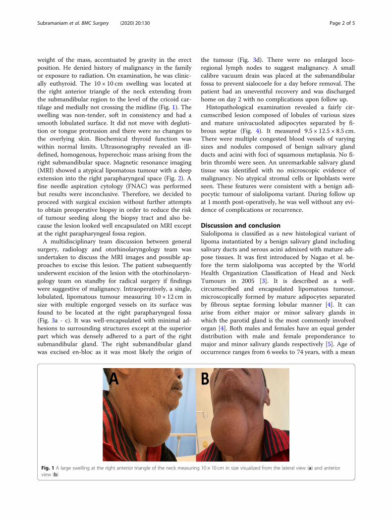
weight of the mass, accentuated by gravity in the erectposition. He denied history of malignancy in the familyor exposure to radiation. On examination, he was clinic-ally euthyroid. The 10 × 10 cm swelling was located atthe right anterior triangle of the neck extending fromthe submandibular region to the level of the cricoid car-tilage and medially not crossing the midline (Fig. 1). Theswelling was non-tender, soft in consistency and had asmooth lobulated surface. It did not move with degluti-tion or tongue protrusion and there were no changes tothe overlying skin. Biochemical thyroid function waswithin normal limits. Ultrasonography revealed an ill-defined, homogenous, hyperechoic mass arising from theright submandibular space. Magnetic resonance imaging(MRI) showed a atypical lipomatous tumour with a deepextension into the right parapharyngeal space (Fig. 2). Afine needle aspiration cytology (FNAC) was performedbut results were inconclusive. Therefore, we decided toproceed with surgical excision without further attemptsto obtain preoperative biopsy in order to reduce the riskof tumour seeding along the biopsy tract and also be-cause the lesion looked well encapsulated on MRI exceptat the right parapharyngeal fossa region.A multidisciplinary team discussion between general
surgery, radiology and otorhinolaryngology team wasundertaken to discuss the MRI images and possible ap-proaches to excise this lesion. The patient subsequentlyunderwent excision of the lesion with the otorhinolaryn-gology team on standby for radical surgery if findingswere suggestive of malignancy. Intraoperatively, a single,lobulated, lipomatous tumour measuring 10 × 12 cm insize with multiple engorged vessels on its surface wasfound to be located at the right parapharyngeal fossa(Fig. 3a - c). It was well-encapsulated with minimal ad-hesions to surrounding structures except at the superiorpart which was densely adhered to a part of the rightsubmandibular gland. The right submandibular glandwas excised en-bloc as it was most likely the origin of
the tumour (Fig. 3d). There were no enlarged loco-regional lymph nodes to suggest malignancy. A smallcalibre vacuum drain was placed at the submandibularfossa to prevent sialocoele for a day before removal. Thepatient had an uneventful recovery and was dischargedhome on day 2 with no complications upon follow up.Histopathological examination revealed a fairly cir-
cumscribed lesion composed of lobules of various sizesand mature univacuolated adipocytes separated by fi-brous septae (Fig. 4). It measured 9.5 × 12.5 × 8.5 cm.There were multiple congested blood vessels of varyingsizes and nodules composed of benign salivary glandducts and acini with foci of squamous metaplasia. No fi-brin thrombi were seen. An unremarkable salivary glandtissue was identified with no microscopic evidence ofmalignancy. No atypical stromal cells or lipoblasts wereseen. These features were consistent with a benign adi-pocytic tumour of sialolipoma variant. During follow upat 1 month post-operatively, he was well without any evi-dence of complications or recurrence.
Discussion and conclusionSialolipoma is classified as a new histological variant oflipoma instantiated by a benign salivary gland includingsalivary ducts and serous acini admixed with mature adi-pose tissues. It was first introduced by Nagao et al. be-fore the term sialolipoma was accepted by the WorldHealth Organization Classification of Head and NeckTumours in 2005 [3]. It is described as a well-circumscribed and encapsulated lipomatous tumour,microscopically formed by mature adipocytes separatedby fibrous septae forming lobular manner [4]. It canarise from either major or minor salivary glands inwhich the parotid gland is the most commonly involvedorgan [4]. Both males and females have an equal genderdistribution with male and female preponderance tomajor and minor salivary glands respectively [5]. Age ofoccurrence ranges from 6 weeks to 74 years, with a mean
Fig. 1 A large swelling at the right anterior triangle of the neck measuring 10 × 10 cm in size visualized from the lateral view (a) and anteriorview (b)
Subramaniam et al. BMC Surgery (2020) 20:130 Page 2 of 5

of 39.4 years [4]. Tumour size ranged between 10 to 90mm (mean: 46.2 mm) [4]. In our case, the sialolipomaarises from the submandibular gland and it is the largestof its kind to be reported till date.The aetiology of anterior neck swellings can be divided
by onset or duration, namely, congenital, acute, subacuteand chronic. In this case, a 7-year history of a progres-sively growing neck swelling is suggestive of its chron-icity. The initial primary diagnosis for these swellingscan be of a thyroid pathology, laryngocele, thyroglossalduct cyst, lipoma, liposarcoma or even parathyroid car-cinoma [6]. However, given such diagnoses, it is veryrare for a submandibular sialolipoma to appear like alarge goitre as depicted in our case. Undoubtedly, historytaking and physical examination are of utmost import-ance in order to clinch the diagnosis.Imaging modalities in lipomatous tumours provide a
preoperative diagnosis and allows the surgical team toplan the operation besides looking for features to suggestmalignancy. Simple ultrasonography can be useful butits findings vary depending on the expertise of the oper-ator. In good hands, it can predict the tumour origin as
in our case. Computed tomography (CT) and MRI aremore accurate in depicting the tumour texture, locationand compressive or infiltrative features compared toultrasonography. Additionally, MRI allows us to differen-tiate between lipoma and liposarcoma by looking at theintensity of the fat signal and is superior to the conven-tional CT in this aspect. MRI findings suggestive of lipo-sarcoma are namely thickened or nodular septa(typically thicker than 2mm), its association with non-adipose masses and prominent foci of high T2 signaland prominent areas of enhancement [7]. Fine needle as-piration cytology is useful as the initial first-line proced-ure to diagnose salivary gland lesions however itsaccuracy is less than 50% in lipomatous tumours [8].This reduced accuracy is due to various other lesions ofthe salivary gland which may contain a significantamount of adipose tissue such as lipomatous pleo-morphic adenoma, lipomatosis and lipoadenoma [8].Presence of a fibrous capsule in the histology of sialoli-poma helps in distinguishing it from the others.The treatment of choice in managing sialolipomas is
surgical excision of the involved salivary gland. Guided
Fig. 2 a Axial MRI on T1-weighted fat suppression sequence showing hyperintense soft tissue component in the anterior portion of the masswith nodular irregular septation within the mass. b Post contrast axial MRI on T1-weighted fat suppression sequence showing avidly enhancingsoft tissue component in the anterior portion of the mass with enhancing nodular irregular septation within the mass. c Axial MRI on T1-weighted fat suppression sequence showing hyperintense soft tissue component in the anterior portion of the mass with nodular irregularseptation within the mass. The right submandibular gland (blue star) is being displaced anteriorly by the mass. d Coronal MRI on T1-weighted fatsuppression sequence showing the mass insinuating into the right parapharyngeal space from the right margin of the right anterior neck,elevating the right pterygoid muscles (blue star)
Subramaniam et al. BMC Surgery (2020) 20:130 Page 3 of 5

Fig. 3 a Elliptical incision made and a portion of skin containing the previous FNAC tract was removed en-bloc with the tumour. This portion ofskin is used to manipulate the tumor without handling the tumor itself besides providing better cosmesis by reducing excess skin. b Enucleationof the lesion with its capsule by creating a lower flap. An ultrasonic dissection device was used to seal and divide the peritumoral vessels andachieve haemostasis. c Creation of an upper flap and dissection of the engorged peritumoral vessels is demonstrated here. d Macroscopicappearance of the lesion showing a lipomatous tumour with intact capsule measuring 10 × 12 cm in size. The right submandibular gland wasexcised en bloc (black arrow)
Fig. 4 Microscopic features of sialolipoma showing a presence of lobules of mature adipose tissue separated by fibrous septae (a to d) with apresence of salivary glands acini (a & b) and higher magnification showing mature univacuolated adipocytes with no lipoblast (d)
Subramaniam et al. BMC Surgery (2020) 20:130 Page 4 of 5

by MRI, access to the tumour can be decided and this isa very crucial step in planning the surgery. In certaincases, the lipoma may be intramuscular hence posing agreater challenge to achieve complete tumour excision.Bleeding from the muscle is a common occurrence in-traoperatively and meticulous haemostasis is importantin such cases. Other possible risks and complicationspertaining to this surgery include injury to the surround-ing vessels and nerves, namely the facial artery, marginalmandibular nerve (branch of the facial nerve), lingualnerve and hypoglossal nerve. Post-operative salivary fis-tula and sialocoele are complications that need to beavoided. Nevertheless, proper planning and completesurgical excision provides good surgical outcomes as therisk of recurrence is zero [9].In conclusion, submandibular sialolipomas can present
in really large sizes and appear as a giant goitre. The at-tending surgeon should be able to differentiate betweenbenign lipomatous tumours and liposarcomas throughhistory, physical examination and imaging. Completesurgical enucleation with the involved salivary gland isthe mainstay of treatment with low recurrence rates.
AbbreviationsMRI: Magnetic resonance imaging; FNAC: Fine needle aspiration cytology;CT: Computed tomography
AcknowledgementsWe would like to thank the Director General of Health Malaysia for hispermission to publish this article as a case report.
Authors’ contributionsSS and FH conducted the literature search and drafted the manuscript. SS,SJ, and FH contributed to the conception and design of the work. SS, SJ, andJAC were involved in the management of the patient. CYN provided the MRIfigures and descriptions. NA provided the histology figures and descriptions.IM provided the literature review and justification on the role ofotorhinolaryngology team in managing this case. All authors reviewed themanuscript and gave approval for publication of the final version.
FundingThe study did not receive any funding.
Availability of data and materialsNot applicable.
Ethics approval and consent to participateNo ethical clearance required as it only involves a case report.
Consent for publicationWritten and signed informed consent for publication of this case wasobtained from the patient, including radiological and intraoperative images.A copy of the consent document can be provided upon request.
Competing interestsThe authors have no conflicts of interest to declare.
Author details1Department of Surgery, Queen Elizabeth Hospital, Ministry of HealthMalaysia, Kota Kinabalu, Sabah, Malaysia. 2Department of Surgery, Faculty ofMedicine and Health Sciences, Universiti Malaysia Sabah, Kota Kinabalu,Sabah, Malaysia. 3Department of Medicine, Faculty of Medicine and HealthSciences, Universiti Malaysia Sabah, Kota Kinabalu, Sabah, Malaysia.4Department of Pathobiology and Medical Diagnostic, Faculty of Medicine
and Health Sciences, Universiti Malaysia Sabah, Kota Kinabalu, Sabah,Malaysia. 5Department of Otorhinolaryngology-Head and Neck Surgery,School of Medical Sciences, Universiti Sains Malaysia, Kota Bharu, Kelantan,Malaysia.
Received: 13 April 2020 Accepted: 31 May 2020
References1. Qayyum S, Meacham R, Sebelik M, Zafar N. Sialolipoma of the parotid gland:
case report with literature review comparing major and minor salivarygland sialolipomas. J Oral Maxillofac Pathol. 2013;17(1):95–7.
2. Parente P, Longobardi G, Bigotti G. Hamartomatous sialolipoma of thesubmandibular gland: case report. Br J Oral Maxillofac Surg. 2008;46(7):599–600.
3. Nagao T, Sugano I, Ishida Y, et al. Sialolipoma: a report of seven cases of anew variant of salivary gland lipoma. Histo-pathology. 2001;38:30–6.
4. Ruangritchankul K, Connor S, Oakley R. Oncocytic sialolipoma of parotid gland:case report and literature review. Head Neck Pathol. 2019;13(4):548–53.
5. Ghafar MA, Tuang G, Mohammad NY, Nadarajan C, Abdullah B. Sialolipomaof the parotid gland: an uncommon lipoma variant of salivary gland. ActaMedica Bulgarica. 2018;45(1):39–41.
6. Haynes J, Arnold KR, Aguirre-Oskins C, Chandra S. Evaluation of neck massesin adults. Am Fam Physician. 2015;91(10):698–706.
7. Gaskin CM, Helms CA. Lipomas, lipoma variants, and well-differentiatedliposarcomas (atypical lipomas): results of MRI evaluations of 126consecutive fatty masses. AJR Am J Roentgenol. 2004;182:733–9.
8. Nonaka CF, Pereira KM, de Andrade Santos PP, de Almeida FR, da CostaMiguel MC. Sialolipoma of minor salivary glands. Ann Diagn Pathol. 2011;15(1):6–11.
9. Sato K, Gotoh C, Uchida H, Kawashima H, Yoshida M, Kitano Y, Kishimoto H.Sialolipoma of the submandibular gland in a child. J Pediatr Surg. 2011;46(2):408–10.
Publisher’s NoteSpringer Nature remains neutral with regard to jurisdictional claims inpublished maps and institutional affiliations.
Subramaniam et al. BMC Surgery (2020) 20:130 Page 5 of 5