
Giant-cell tumor of the tendon sheath: when must we ... · Giant cell tumor of the tendon sheath is...
15
Giant-cell tumor of the tendon sheath: when must we suspect it? C. Santos Montón, J. M. Alonso Sánchez, D. C. Cuellar, P. A. Chaparro García, R. Corrales, J. D. Albillo Labarra; Salamanca/ES Learning objectives The purpose of this exhibit is: 1. To review the pathophysiology of the giant-cell tumor of the tendon sheath (GCTTS). 2. To explain the imaging features of the GCTTS in the different radiologic techniques. 3. To establish a differential diagnosis between GCTTS and other soft- tissue masses. Background The giant cell tumor of the tendon sheath is a benign lesion that arises from the tendon sheath. It is in doubt whether the mass is a true neoplasm or a reactive mass to trauma. GCTTS most commonly occurs in the flexor tendon sheath of the hand and wrist, in the volar aspect of the first three fingers, followed by the ankle and foot region. Findings and procedure details - Pathophysiology of the giant-cell tumor of the tendon sheath. Giant cell tumor of the tendon sheath (GCTTS) is an uncommon and usually benign peritendinous fibrous mass. It is a hypervascular lesion arising from the synovium of the tendon sheath. The lesion is typically small and it appears well-encapsulated and lobulated. Microscopically
Transcript of Giant-cell tumor of the tendon sheath: when must we ... · Giant cell tumor of the tendon sheath is...

Giant-cell tumor of the tendon sheath: when must we
suspect it?
C. Santos Montón, J. M. Alonso Sánchez, D. C. Cuellar, P. A.
Chaparro García, R. Corrales, J. D. Albillo Labarra; Salamanca/ES
Learning objectives
The purpose of this exhibit is:
1. To review the pathophysiology of the giant-cell tumor of the tendon sheath (GCTTS).
2. To explain the imaging features of the GCTTS in the different radiologic techniques.
3. To establish a differential diagnosis between GCTTS and other soft-tissue masses.
Background
The giant cell tumor of the tendon sheath is a benign lesion that arises from the tendon sheath. It is in doubt whether the mass is a true neoplasm or a reactive mass to trauma. GCTTS most commonly occurs in the flexor tendon sheath of the hand and wrist, in the volar aspect of the first three fingers, followed by the ankle and foot region.
Findings and procedure details
- Pathophysiology of the giant-cell tumor of the tendon sheath.
Giant cell tumor of the tendon sheath (GCTTS) is an uncommon and usually benign peritendinous fibrous mass. It is a hypervascular lesion arising from the synovium of the tendon sheath. The lesion is typically small and it appears well-encapsulated and lobulated. Microscopically

it is composed of synovial cells, histiocytes, multinucleated giant cells, inflammatory cells, macrophages, xanthoma cells, and collagen.
GCTTS is also known as the localized form of the pigmented villonodular synovitis. Both entities have the same histologic features and they are classified as “fibrohistiocytic tumors” in the World Health Organization classification system for bone and soft tissue tumors of 2013. The localized form can arise in or extrinsic to a joint, and the diffuse form predominantly originates outside the joint. The giant cell tumor of the tendon sheath appears as a rubbery, multinodular, well-encapsulated, brown, orange, or yellow mass; the color depends on the proportion of foam cells and degree of hemosiderin deposition. A collagenous capsule surrounds the lesion.
- Demographics, clinical presentation, prognosis and treatment.
Giant cell tumor of the tendon sheath is approximately 1.6% of all soft-tissue tumors. It may occur at any age, with a peak incidence between the third and fifth decades and a slight female predominance. Patients may report a history of trauma to the affected area, for which there is a debate whether the tumor is a true neoplasm or a pseudoneoplastic inflammatory response to soft tissue trauma. The tumor usually surrounds a nearby tendon or more rarely the neurovascular bundles.
They are most common in the hand, particularly in the volar aspect of the first three fingers (80%). The second most common site is the ankle-foot complex. In children, however, GCTTS may have an equal predilection for the upper and lower extremities.
GCTTS presents as a slowly growing mass of soft tissue and can cause varying degrees of pain or be asymptomatic. Infrequent multifocal lesions have been described.
Malignancy is unusual in giant cell tumors of the tendon sheath but it has been described.
The treatment is a local excision with clear margins to minimize the risk of recurrence. Surgery is curative, although recurrence after resection is not uncommon and may be seen in approximately 7-27% of patients. Radiation theraphy after the resection may be useful in rare aggressive lesions.

- X-ray findings, sonographic appearance and MRI findings.
Radiographs may show normal findings or a soft-tissue mass in patients with a GCTTS. Pressure erosions of underlying bone are present in about 20% of patients. Periosteal reaction, osteopenia, calcification, degenerative or cystic changes, and intraosseous invasion may be seen.
On ultrasonography GCTTS usually appears as a solid, homogeneous, hypoechoic mass. It is very helpful to evaluate the relationship with the adjacent tendon. Internal vascularity can be demonstrated by Doppler examination in most cases. US can provide exact information about the tumor and its relationship with the surrounding tissue, which indicates that it can be used as the first method to diagnose a giant cell tumor of the tendon sheath.
MRI typically shows a well-defined mass adjacent to or enveloping a tendon. Characteristically, the mass is hypointense on T1-weighted images, approximately equal to skeletal muscle. On T2 weighted images there is usually low signal due to chronic hemorrhage with hemosiderin deposition. The most specific feature of GCTTS is the blooming artefact on gradient echo sequences; GCTTS typically contains hemosiderin which causes a blooming artifact on gradient-echo images. There may be areas of low signal and high signal on T2 weighted images due to the presence of hemosiderin and fluid respectively. Uniform enhancement can be seen post intravenous gadolinium.
- Pitfalls and mimics.
The differential diagnosis includes any soft-tissue mass with low signal intensity on T1-weighted and T2-weighted images, such as a desmoid tumor (particularly if deep) or pigmented villonodular synovitis, which more commonly involves the larger joints. Aside from possessing similar signal characteristics, giant cell tumor of the tendon sheath also resembles histologically pigmented villonodular synovitis.
In the differential diagnoses we must include desmoid tumor, fibroma, cavernous hemangioma, ganglion cyst, granuloma, fibrosarcoma, malignant fibrous histiocytoma, synovial cell sarcoma, chondroma,

granuloma, and tophaceous gout. But none of these entities usually contain hemosiderin.
The fibroma of the tendon sheath could have a similar appearance on MRI to the GCTTS. Fibroma of the tendon sheath and GCTTS can be difficult to differentiate on clinical examination and imaging. In some cases, demographic and location information can help with the differential diagnosis. Both lesions occur most commonly in the upper extremities. Fibromas can present as soft-tissue masses that are similar to giant cell tumors within or adjacent to tendon sheaths. They usually appear isointense to skeletal muscle on T1-weighted sequences and isointense to hypointense on T2-weighted sequences, but they may have areas of high T2 signal, depending on their mixture of fibrous and myxoid composition.
Conclusion
GCTTS are solid masses that have close contact with the tendon sheath. Ultrasound examination can provide exact information about this tumor and its relationship with the surrounding structures, so it should be the first technique to start the study of the mass.
Radiographs may show normal findings, a soft tissue mass or less
commonly bone erosions. On MRI they are typically hypointense on T1
and T2-weighted images with a homogeneous enhancement after
contrast administration.

References
1. Jelinek JS, Kransdorf MJ, Shmookler BM, Aboulafia AA, Malawer MM. Giant cell tumor of the tendon sheath: MR findings in nine cases. AJR Am J Roentgenol. 1994;162(4):919-22.
2. Middleton WD, patel V, Teefey SA, Boyer MI. Giant cell tumors of the tendon sheath: analysis of sonographic findings. AJR Am J Roentgenol. 2004;183(2):337-9.
3. Teh J, Whiteley G. MRI of soft tissue masses of the hand and wrist. Br J Radiol. 2007;80(949):47-63.
4. Ly JQ, Carlosn CL, LaGatta LM, Beall DP. Giant cell tumor of the peroneus tendon sheath. AJR Am J Roentgenol. 2003;180(5):1442.
5. Wang Y, Tang J, Luo Y. The value of sonography in diagnosing giant cell tumors of the tendón sheath. J Ultrasound Med.2007;26(10):1333-40.
6. Llauger J, Palmer J, Rosón N, Cremades R, Bagué S. Pigmented villonodular synovitis and giant cell tumors of the tendon sheath: radiologic and pathologic features. AJR Am J Roentgenol. 1999;172(4):1087-91.

Fig. 1: 50-year-old woman with a soft tissue mass in the second finger
of the left hand. Radiography shows a mass in the distal
interphalangeal junction without bone erosions. Histologic examination
reveals a giant cell tumor of the tendon sheath.
Fig. 2: Lumbosacral radiography shows an expansive lytic lesion in the
left transverse apophysis of the 3rd lumbar vertebra. The histologic
examination reveals a giant cell tumor of the tendon sheath.

Fig. 3: Axial CT scan, bone window, shows an expansive lytic lesion
which affects the left transverse apophysis, pedicle and superior
zygapophyseal joint of the 3rd lumbar vertebra. The histologic
examination reveals a giant cell tumor of the tendon sheath
Fig. 4: Axial T1-weighted image shows an isointense mass located in
the left transverse apophysis, pedicle and superior zygapophyseal joint
of the 3rd lumbar vertebra. The histologic examination reveals a giant
cell tumor of the tendon sheath.

Fig. 5: Axial contrast-enhanced T1-weighted image shows a
enhancement of the mass located in the left transverse apophysis,
pedicle and superior zygapophyseal joint of the 3rd lumbar vertebra.
The histologic examination reveals a giant cell tumor of the tendon
sheath.
Fig. 6: Axial T2-weighted image shows a hyperintense mass located in
the left transverse apophysis, pedicle and superior zygapophyseal joint
of the 3rd lumbar vertebra. The histologic examination reveals a giant
cell tumor of the tendon sheath.

Fig. 7: Ultrasonographic image of the first finger of a 69-year-old
woman: Hypoechoic vascularized soft tissue mass around the flexor
tendon that proved to be a giant cell tumor.
Fig. 8: Giant cell tumor of the tendon sheath in the first finger of a 69-
year-old woman. Axial T1-weighted MR image shows a hypointense
multilobulated mass which surrounds flexor digitorum tendon.

Fig. 9: Giant cell tumor of the tendon sheath in the first finger of a 69-
year-old woman. Axial T2-weighted MR image shows a hypointense
multilobulated mass which surrounds flexor digitorum tendon.
Fig. 10: Giant cell tumor of the tendon sheath in the first finger of a 69-
year-old woman. Axial contrast-enhanced T1-weighted MR image
shows a multilobulated mass with intense contrast enhancement which
surrounds flexor digitorum tendon.

Fig. 11: Giant cell tumor of the tendon sheath in the first finger of a 69-
year-old woman. Sagittal T2-weighted MR image shows a hypointense
multilobulated mass which surrounds flexor digitorum tendon.
Fig. 12: 38-year-old woman with a soft tissue mass in the great toe of
the right foot. Radiography shows a mass in the interphalangeal
junction without bone erosions. Histologic examination reveals a giant
cell tumor of the tendon sheath
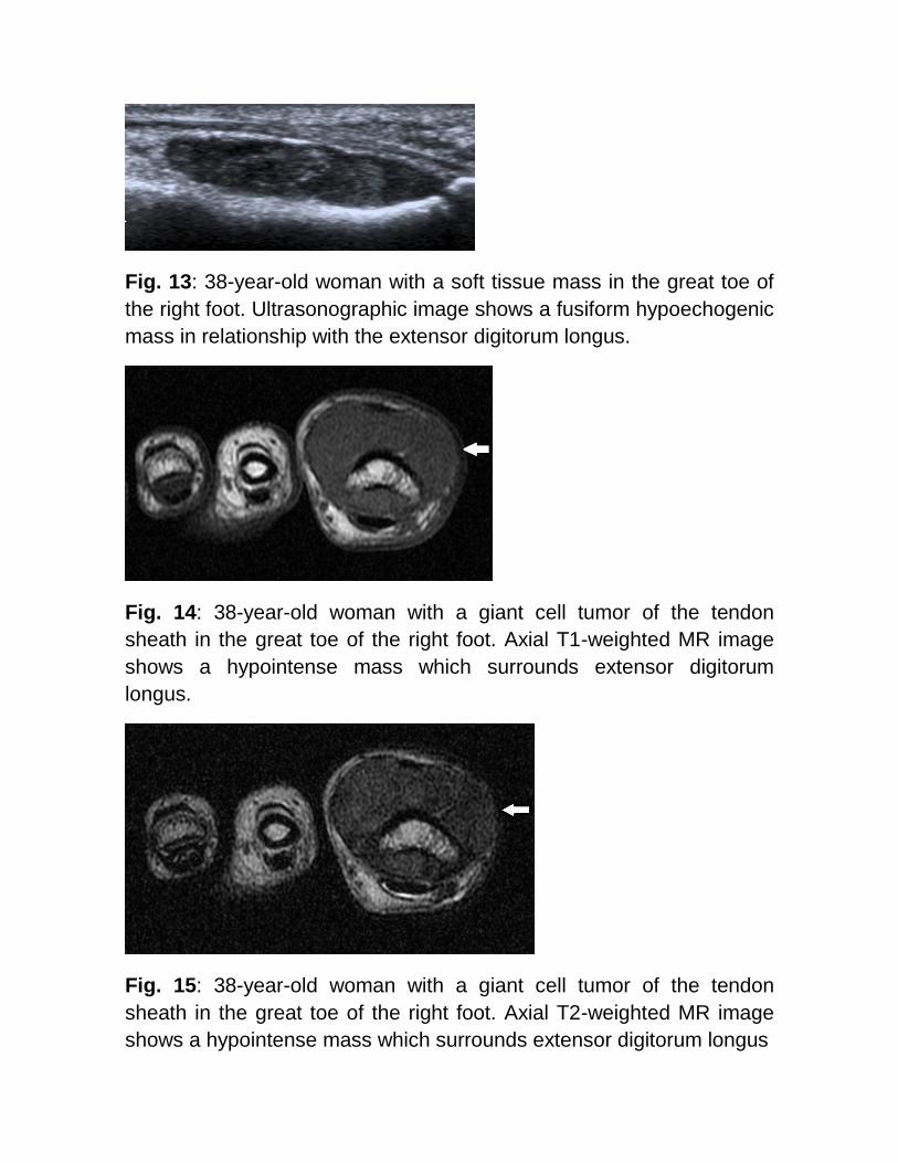
Fig. 13: 38-year-old woman with a soft tissue mass in the great toe of
the right foot. Ultrasonographic image shows a fusiform hypoechogenic
mass in relationship with the extensor digitorum longus.
Fig. 14: 38-year-old woman with a giant cell tumor of the tendon
sheath in the great toe of the right foot. Axial T1-weighted MR image
shows a hypointense mass which surrounds extensor digitorum
longus.
Fig. 15: 38-year-old woman with a giant cell tumor of the tendon
sheath in the great toe of the right foot. Axial T2-weighted MR image
shows a hypointense mass which surrounds extensor digitorum longus

Fig. 16: 38-year-old woman with a giant cell tumor of the tendon
sheath in the great toe of the right foot. Axial contrast-enhanced T1-
weighted MR image shows a mass with intense contrast enhancement
which surrounds extensor digitorum longus.
Fig. 17: 20-year-old woman who presents a soft tissue mass in the
right knee. Sagittal gradient echo T2-weighted MR image shows a
hyperintense mass (arrow) in Hoffa´s fat pad with an area of low signal
intensity that reveals hemosiderin deposition. The histologic diagnosis
after resection confirmed the diagnosis of giant cell tumor of the tendon
sheath.

Fig. 18: 20-year-old woman who presents a soft tissue mass in the
right knee. Axial T1-weighted MR image shows a hypointense mass
(arrow) in Hoffa´s fat pad. The histologic diagnosis after resection
confirmed the diagnosis of giant cell tumor of the tendon sheath.
Fig. 19: 20-year-old woman who presents a soft tissue mass in the
right knee. Axial contrast-enhanced T1-weighted MR image shows a
mass (arrow) in Hoffa´s fat pad with slight contrast enhancement. The
histologic diagnosis after resection confirmed the diagnosis of giant cell
tumor of the tendon sheath.

Fig. 20: 20-year-old woman who presents a soft tissue mass in the
right knee. Axial T2-weighted MR image shows a hypointense mass
(arrow) in Hoffa´s fat pad. The histologic diagnosis after resection
confirmed the diagnosis of giant cell tumor of the tendon sheath.


![Giant cell tumor of tendon sheath in the hand: analysis of risk ......and being giant cell tumor of the tendon sheath (GCTT S) the most common form [1–5]. The pathogenesis of GCTTS](https://static.fdocuments.net/doc/165x107/60935d76623e6068eb220bb6/giant-cell-tumor-of-tendon-sheath-in-the-hand-analysis-of-risk-and-being.jpg)


