Giant Axonal Neuropathy A Disorder of Intermediate Filament Organization By: Emily Gilles Alberts,...
20
Giant Axonal Giant Axonal Neuropathy Neuropathy A Disorder of Intermediate Filament A Disorder of Intermediate Filament Organization Organization By: Emily Gilles Alberts, et al. Molecular Biology of the Cell, 4 th Edition
-
Upload
vanessa-newton -
Category
Documents
-
view
219 -
download
2
Transcript of Giant Axonal Neuropathy A Disorder of Intermediate Filament Organization By: Emily Gilles Alberts,...

Giant Axonal NeuropathyGiant Axonal NeuropathyA Disorder of Intermediate Filament OrganizationA Disorder of Intermediate Filament Organization
By: Emily Gilles
Alberts, et al. Molecular Biology of the Cell, 4th Edition
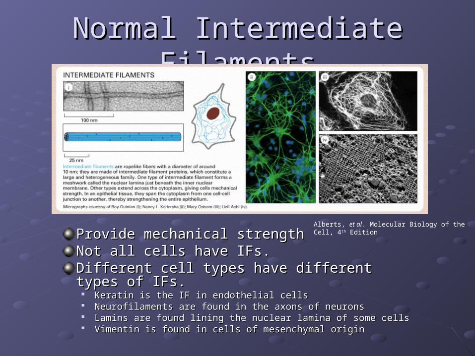
Normal Intermediate FilamentsNormal Intermediate Filaments
Provide mechanical strengthProvide mechanical strengthNot all cells have IFs. Not all cells have IFs. Different cell types have different types of IFs.Different cell types have different types of IFs.
Keratin is the IF in endothelial cellsKeratin is the IF in endothelial cells Neurofilaments are found in the axons of neuronsNeurofilaments are found in the axons of neurons Lamins are found lining the nuclear lamina of some cellsLamins are found lining the nuclear lamina of some cells Vimentin is found in cells of mesenchymal originVimentin is found in cells of mesenchymal origin
Alberts, et al. Molecular Biology of the Cell, 4th Edition
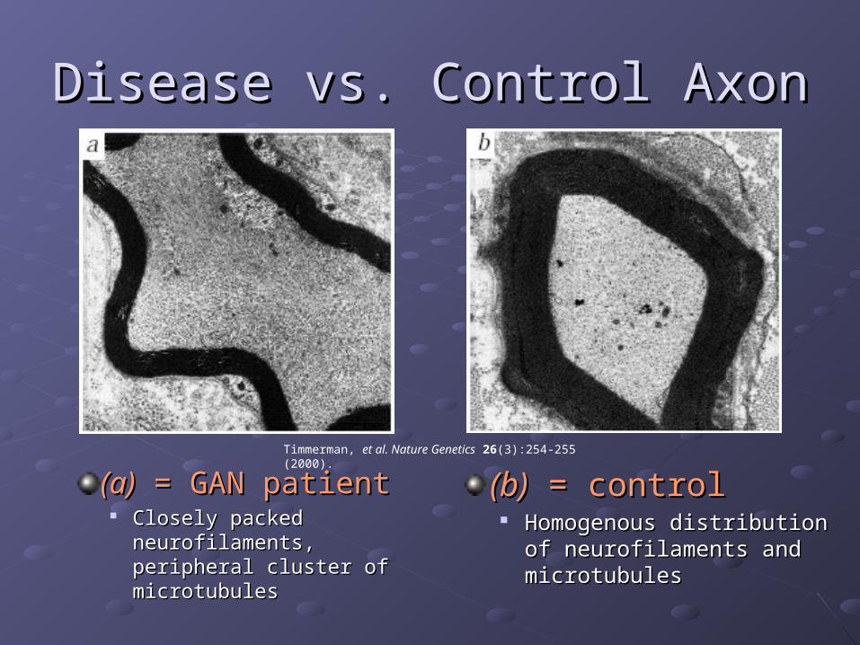
Disease vs. Control AxonDisease vs. Control Axon
(a)(a) = GAN patient = GAN patient Closely packed Closely packed
neurofilaments, neurofilaments, peripheral cluster of peripheral cluster of microtubulesmicrotubules
(b)(b) = control = control Homogenous distribution of Homogenous distribution of
neurofilaments and neurofilaments and microtubulesmicrotubules
Timmerman, et al. Nature Genetics 26(3):254-255 (2000).
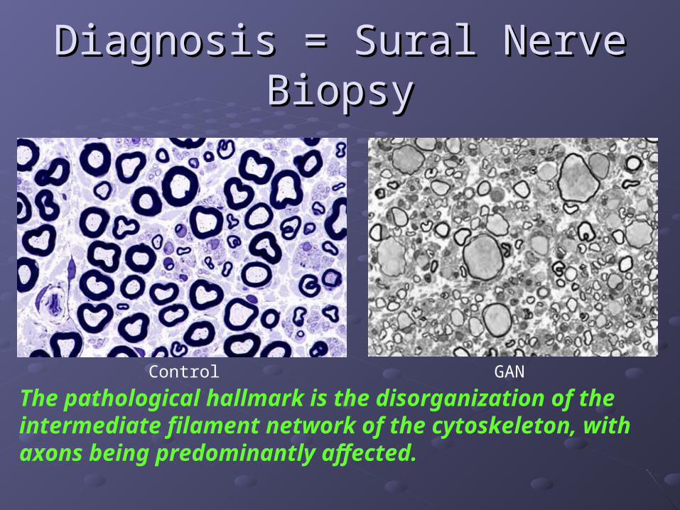
Diagnosis = Sural Nerve BiopsyDiagnosis = Sural Nerve Biopsy
The pathological hallmark is the disorganization of the intermediate filament network of the cytoskeleton, with axons being predominantly affected.
Control GAN

The typical clinical case:The typical clinical case:
Disease onset typically occurs Disease onset typically occurs between ages 4-7.between ages 4-7.Death occurs before the age of 30.Death occurs before the age of 30.Symptoms begin as clumsiness of Symptoms begin as clumsiness of gait and progressive weakness gait and progressive weakness starting at the lower limbs.starting at the lower limbs.Dysarthria, nystagmas, facial Dysarthria, nystagmas, facial weakness, and mental retardation weakness, and mental retardation soon become apparent.soon become apparent.Kinky hair may or may not be Kinky hair may or may not be present.present.Rare, autosomal recessiveRare, autosomal recessive
Treiber-Held, et al. Neuropediatrics 25(2):89-93 (1994).
Maia, et al. Neuropediatrics 19(1):10-15 (1988).

Homozygosity MappingHomozygosity Mapping
Ben Hamida, et al. Neurogenetics 1(2):129-133 (1997).
Flanigan, et al. Annals of Neurology 43(1):143-148 (1998).
What is homozygosity mapping?
• Exactly like positional cloning
• Fewer affected individuals can be used
• Offspring must be from consanguineous parents
In addition to being homozygous at the disease allele, there is a greatly increased likelihood of being homozygous by descent in adjacent regions of the genome.
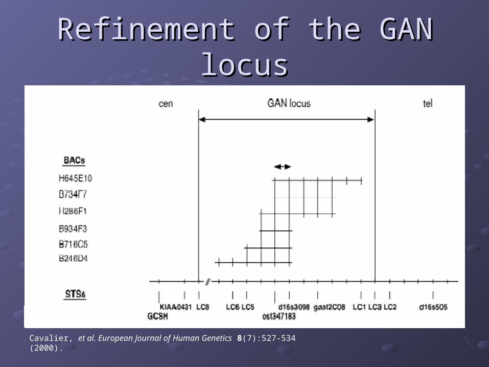
Refinement of the GAN locusRefinement of the GAN locus
Cavalier, et al. European Journal of Human Genetics 8(7):527-534 (2000).

The Gene: GigaxoninThe Gene: Gigaxonin
Bomont, et al. Nature Genetics 26(3):370-374 (2000).
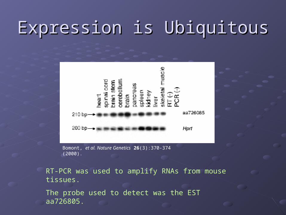
Expression is UbiquitousExpression is Ubiquitous
Bomont, et al. Nature Genetics 26(3):370-374 (2000).
RT-PCR was used to amplify RNAs from mouse tissues.
The probe used to detect was the EST aa726805.
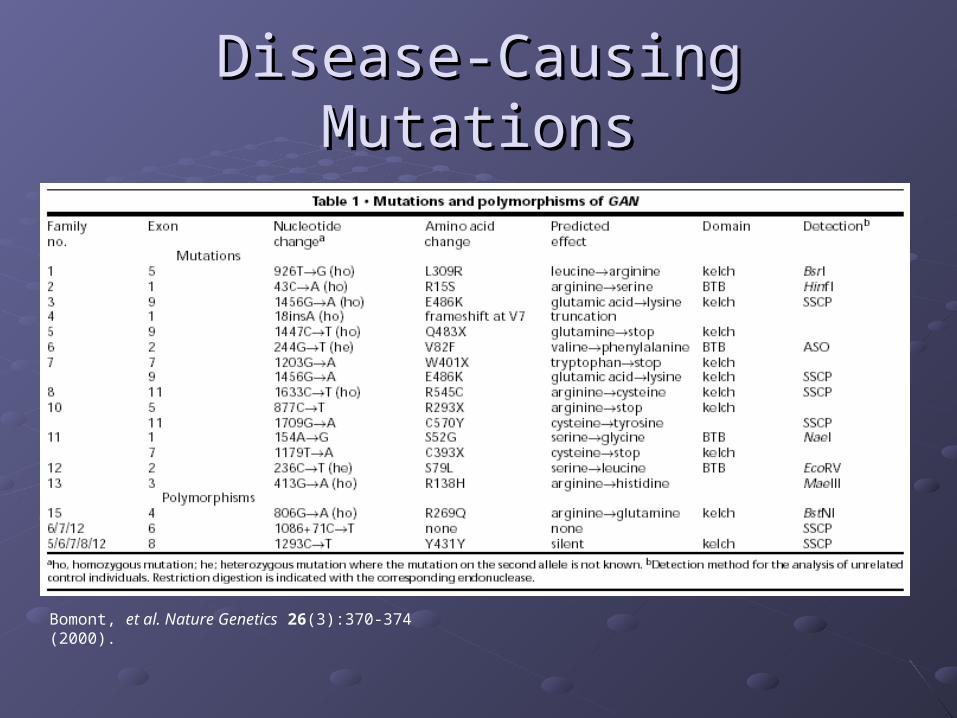
Disease-Causing MutationsDisease-Causing Mutations
Bomont, et al. Nature Genetics 26(3):370-374 (2000).

Additional Mutations DiscoveredAdditional Mutations Discovered
Bomont, et al. Human Mutations 21(4):446-451 (2003).
Kuhlenbaumer, et al Neurology 58(8):1273-1276 (2002).

Cytoskeletal Elements are Cross-LinkedCytoskeletal Elements are Cross-Linked
Actin filaments, microtubules, and intermediate Actin filaments, microtubules, and intermediate filaments interact via cross-linking proteinsfilaments interact via cross-linking proteinsPicture below shows intermediate filaments Picture below shows intermediate filaments (blue) linked to microtubules (red) via plectin (blue) linked to microtubules (red) via plectin (green)(green)
Alberts, et al. Molecular Biology of the Cell, 4th Edition

Yeast Two-Hybrid SetupYeast Two-Hybrid Setup
Blue circle = DNA Blue circle = DNA binding domainbinding domain
Yellow square = Yellow square = gigaxoningigaxonin
Red square = protein Red square = protein from human brain from human brain cDNAcDNA
Blue semi-circle = Blue semi-circle = activator domainactivator domain
Criekinge WV and Beyaert R. Biological Proceedures Online 2:1-38 (1999)

Gigaxonin colocalizes with MAP1B-LCGigaxonin colocalizes with MAP1B-LC
HA-Gig and flag-MAP1B-HA-Gig and flag-MAP1B-LC were cotransfected LC were cotransfected into cos7 cellsinto cos7 cells
Gigaxonin and MAP1B-Gigaxonin and MAP1B-LC colocalize togetherLC colocalize together
D shows diffuse D shows diffuse accumulation of accumulation of gigaxonin in cytoplasm gigaxonin in cytoplasm when transfected alonewhen transfected alone
Ding, et al. Journal of Cell Biology 158(3):427-433 (2002).

In vivoIn vivo Results Similar to Results Similar to in vitroin vitro
Gigaxonin colocalizes with MAP1B-LC Gigaxonin colocalizes with MAP1B-LC in vivoin vivo..
Ding, et al. Journal of Cell Biology 158(3):427-433 (2002).
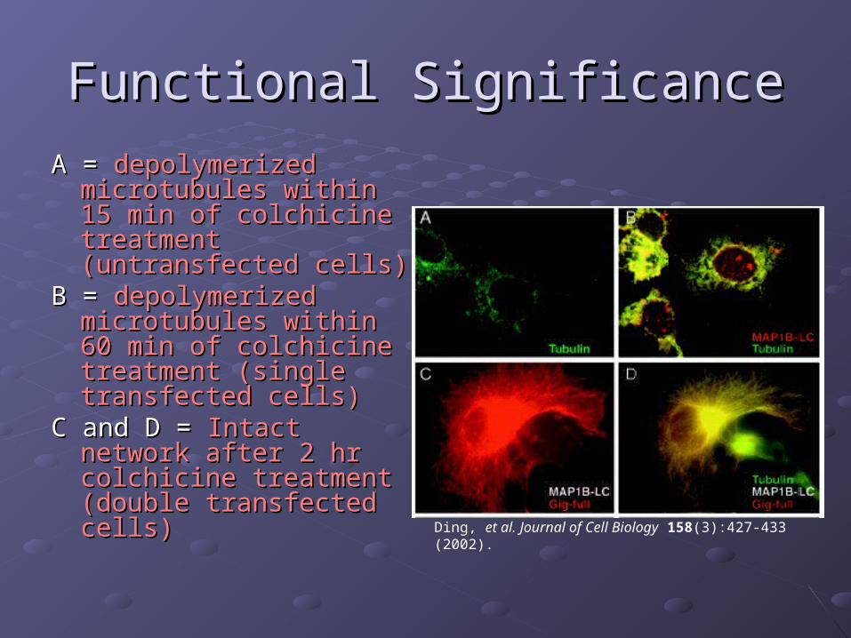
Functional SignificanceFunctional Significance
A = A = depolymerized depolymerized microtubules within 15 microtubules within 15 min of colchicine min of colchicine treatment (untransfected treatment (untransfected cells)cells)
B = B = depolymerized depolymerized microtubules within 60 microtubules within 60 min of colchicine min of colchicine treatment (single treatment (single transfected cells) transfected cells)
C and D = C and D = Intact network Intact network after 2 hr colchicine after 2 hr colchicine treatment (double treatment (double transfected cells)transfected cells) Ding, et al. Journal of Cell Biology 158(3):427-433 (2002).

Molecular Diagnosis/TreatmentMolecular Diagnosis/Treatment
Molecular Diagnosis is only available on a Molecular Diagnosis is only available on a research basis.research basis.
There is no cure and treatment is based There is no cure and treatment is based on alleviating treatable symptoms.on alleviating treatable symptoms.
Current diagnosis based on:• nerve biopsy showing thinly myelnated, enlarged axons
• nerve conduction studies showing reduced nerve conduction velocity (NCV), severely reduced compound motor action potentials (CMAP) and absent sensory nerve action potentials (SNAP)
• abnormal visual evoked responses
• EEG showing increased slow wave activity
• MRI showing cerebellar and white matter abnormalities

SummarySummaryGiant Axonal Neuropathy is a rare, autosomal recessive Giant Axonal Neuropathy is a rare, autosomal recessive disease. Diagnosis is mainly based on the appearance of disease. Diagnosis is mainly based on the appearance of giant axons containing aggregated neurofilaments in a sural giant axons containing aggregated neurofilaments in a sural nerve biopsy.nerve biopsy.The disease locus was mapped to chromosome 16q21 by The disease locus was mapped to chromosome 16q21 by homozygosity mapping.homozygosity mapping.Using additional markers the disease locus was refined to a Using additional markers the disease locus was refined to a smaller, 590kb region.smaller, 590kb region.Gigaxonin was identified by searching an EST database. Gigaxonin was identified by searching an EST database. cDNA of the gene was cloned from a cDNA human brain cDNA of the gene was cloned from a cDNA human brain library.library.Several different point mutations throughout the gene can Several different point mutations throughout the gene can result in the disease phenotype.result in the disease phenotype.Gigaxonin binds to MAP1B-LC, as determined fluorescently Gigaxonin binds to MAP1B-LC, as determined fluorescently tagged antibodies and immunoprecipitations. This interaction tagged antibodies and immunoprecipitations. This interaction improves the stability of the microtubule network.improves the stability of the microtubule network.Molecular treatment cannot be addressed until more is known Molecular treatment cannot be addressed until more is known about the gigaxonin gene product.about the gigaxonin gene product.

ReferencesReferencesAlberts, et al. Molecular Biology of the Cell, 4th Edition
Ding, et al. Journal of Cell Biology 158(3):427-433 (2002).
Criekinge WV and Beyaert R. Biological Procedures Online 2:1-38 (1999)
Kuhlenbaumer, et al Neurology 58(8):1273-1276 (2002).
Bomont, et al. Human Mutations 21(4):446-451 (2003).
Bomont, et al. Nature Genetics 26(3):370-374 (2000).
Timmerman, et al. Nature Genetics 26(3):254-255 (2000).
Ben Hamida, et al. Neurogenetics 1(2):129-133 (1997).
Flanigan, et al. Annals of Neurology 43(1):143-148 (1998).
Cavalier, et al. European Journal of Human Genetics 8(7):527-534 (2000).
Treiber-Held, et al. Neuropediatrics 25(2):89-93 (1994).
Maia, et al. Neuropediatrics 19(1):10-15 1988.

Identifying Gigaxonin Binding PartnersIdentifying Gigaxonin Binding Partners
Ubiquitous expression Ubiquitous expression was confirmed with was confirmed with immunoblotimmunoblot
Transfected cos7 Transfected cos7 cells expressed cells expressed gigaxoningigaxonin
HA epitope tag at C- HA epitope tag at C- and N- terminal was and N- terminal was recognized by recognized by αα-HA -HA antibodyantibody
Ding, et al. Journal of Cell Biology 158(3):427-433 (2002).