GI Emergency Interventions: Upper & Lower … Emergency Interventions: Upper & Lower Endoscopy....
36
St. James Healthcare Surgical Services - Endoscopy Butte, Montana GI Emergency Interventions: Upper & Lower Endoscopy
Transcript of GI Emergency Interventions: Upper & Lower … Emergency Interventions: Upper & Lower Endoscopy....
St. James Healthcare
Surgical Services -
Endoscopy
Butte, Montana
GI Emergency Interventions: Upper & Lower Endoscopy
OBJECTIVES:
Identify patients at risk for GI Bleeding complications.
Demonstrate competencies related to hemostasis during a GI Emergency.
Give examples of collaborative action with health team members during a GI emergency.
Identify areas of practice improvement following completion of this learning module.

TEST YOUR KNOWLEDGE: True/False
Rapid clinical decisions and appropriate interventions can make the difference between resolving a GI bleed and prolonged hospitalization.
Effective hemostasis
involves ONE intervention.
Hematochezia
is gastric (vomit) or duodenal frank red or old “coffee”
color.
Being prepared for a GI Bleed intervention involves having your “tool box”
ready for immediate use.
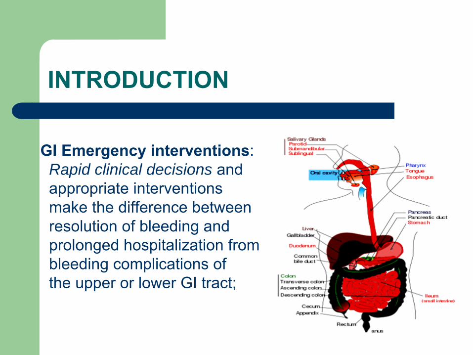
INTRODUCTION
GI Emergency interventions:Rapid clinical decisions and appropriate interventions make the difference between resolution of bleeding andprolonged hospitalization from bleeding complications of the upper or lower GI tract;
GI EMERGENCY: TRIAGE
Triage for acute or induced GI bleeding:Identifying the level of risk involves -
•
Collaborative Team Work –
GI ENDO Team and designated referral center all working quickly together.
•
Often involves a decision to proceed with an elective outpatient intervention versus a later emergency endoscopy and possible hospitalization.

Identifying the High Risk Factors?
Screening guidelines would include the following risk factors:
Co-morbid conditions (poor health status)
Age (elderly)
Recent bleeding events
Severity of endoscopic findings, such as a large bleeding visible vessel (vs. a clean small tear)

ACUTE GI BLEED: Signs & Symptoms
Signs:•
Hematemesis – gastric
(vomit) or duodenal frank red/“coffee”
color
•
Melena –
gastric (vomit) or bowel (stool) stained black “tarry”
by blood
pigment•
Hematochezia –
red blood, i.e. lower bowel or hemorrhoids
Symptoms:•Skin = pallor, diaphoresis
•Neuro = lightheaded or syncope
•Pulmonary = dyspnea(exertional)
•Cardiac = tachycardia and hypotension

ACUTE GI BLEED
Nursing Assessment:•
Mental Status•
Vital Signs: HR, BP, RR•
Pallor –
skin, eyelids, nails•
Lab (CBC, Chemistry, Coagulopathy)Interventions and Preparation:•
IV Fluids/Blood Transfusion•
Oxygen Supplement•
Warm Blankets •
Music Therapy ♫ ♫ ♫•
Urgent Endoscopsy
Create a Calm Environment

UPPER GI BLEEDING
CAUSES OF UPPER BLEEDING
•
Peptic Ulcer Disease (PUD)•
Esophageal or Gastric Varices•
ArterioVenous
Malformation (AVM)•
Mallory-Weiss Tear•
Tumors •
Dieulafoy
Lesion•
Other Causes, i.e. esophagitis
or mucosal abnormality, bleeding complications during upper endoscopy

HAVE YOUR TOOLS READY!
•
Lavage
Tube or Irrigation Set-Up•
Sclerotherapy
Needle + Meds, i.e. Epinephrine 1:10,000 [10 ml]
•
HemoClips•
Band Ligator, Endo-Loop (ligation device)•
Heater Probe (7 Fr/10 Fr)/BICAP (7 Fr/10 Fr)•
ERBE –
APC, Valley Lab•
Other -
Tatto, Snare Box, Biopsy (lesion, inflammation), Roth Net Retrieval Device
•
Examples preferred in your tool box?
So, Let’s Have An Intervention!

HEMOSTASIS TECHNIQUES
Anticipate dual therapy during an active, uncontrolled, bleed to achieve effective hemostasis, and to reduce re-
bleeding and the need for surgery -i.e. Sclerotherapy
+ Hemoclips
+ BICAP/Heater Probe

INJECTION SCLEROTHERAPY: GI ENDO PROTOCOL
Purpose: Tissue injection to control bleeding Epinephrine 1:10,000 [10 ml]: Epinephrine 1:1,000 [1 ml] + NaCl
0.9 % [9 ml]
= Epinephrine 1:10,000 [10 ml]
Needle exits from protective sheath
Syringe leur-locksto injection port
SclerotherapyInjection is madeto the bleed site during endoscopy
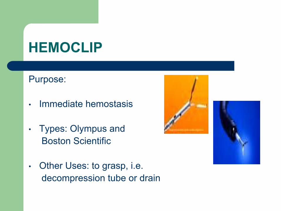
HEMOCLIP
Purpose:
•
Immediate hemostasis
•
Types: Olympus and Boston Scientific
•
Other Uses: to grasp, i.e. decompression tube or drain
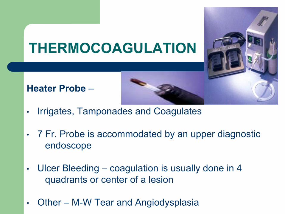
THERMOCOAGULATION
Heater Probe
–
•
Irrigates, Tamponades
and Coagulates
•
7 Fr. Probe is accommodated by an upper diagnosticendoscope
•
Ulcer Bleeding –
coagulation is usually done in 4 quadrants or center of a lesion
•
Other –
M-W Tear and Angiodysplasia

THERMOCOAGULATION
Argon Plasma Coagulation (APC):•
Colon Prep required
•
Coagulation with continuous movement (“painting”)
•
Not the first line of therapy in active bleeding (due to dissipation)

THERMOCOAGULATION: APC Settings –
standard settings
SITE • Esophagus• Stomach
FLOW • 1.4 LPM
COAG • A-60 Watt• Blue Pedal
Note: recommended settings may
differ in hospital settings; follow
department policy or
standard work
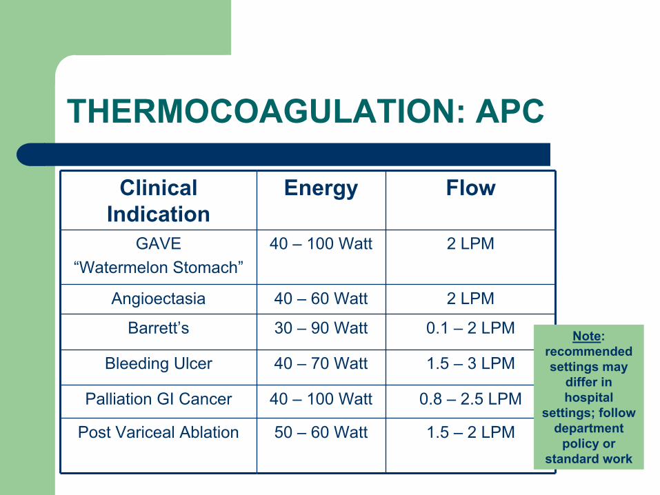
THERMOCOAGULATION: APC
Clinical Indication
Energy Flow
GAVE “Watermelon Stomach”
40 –
100 Watt 2 LPM
Angioectasia 40 –
60 Watt 2 LPM
Barrett’s 30 –
90 Watt 0.1 –
2 LPM
Bleeding Ulcer 40 –
70 Watt 1.5 –
3 LPM
Palliation GI Cancer 40 –
100 Watt 0.8 –
2.5 LPM
Post Variceal
Ablation 50 –
60 Watt 1.5 –
2 LPM
Note: recommended settings may
differ in hospital
settings; follow department
policy or standard work
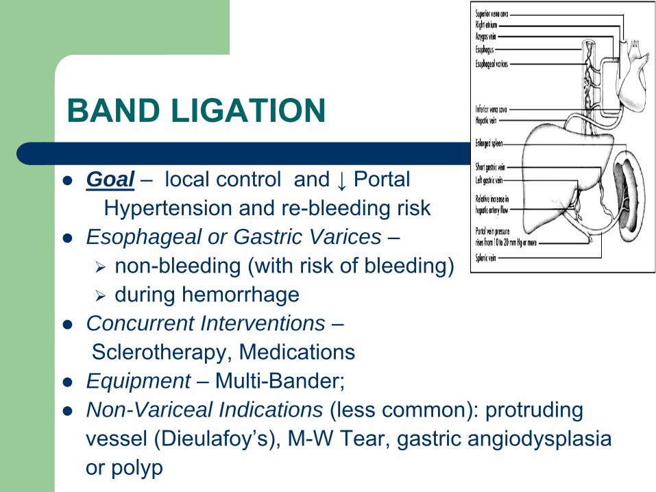
BAND LIGATION
Goal –
local control and ↓
Portal Hypertension and re-bleeding risk
Esophageal or Gastric Varices –
non-bleeding (with risk of bleeding)
during hemorrhage
Concurrent Interventions –Sclerotherapy, Medications
Equipment –
Multi-Bander;
Non-Variceal Indications (less common): protruding vessel (Dieulafoy’s), M-W Tear, gastric angiodysplasiaor polyp
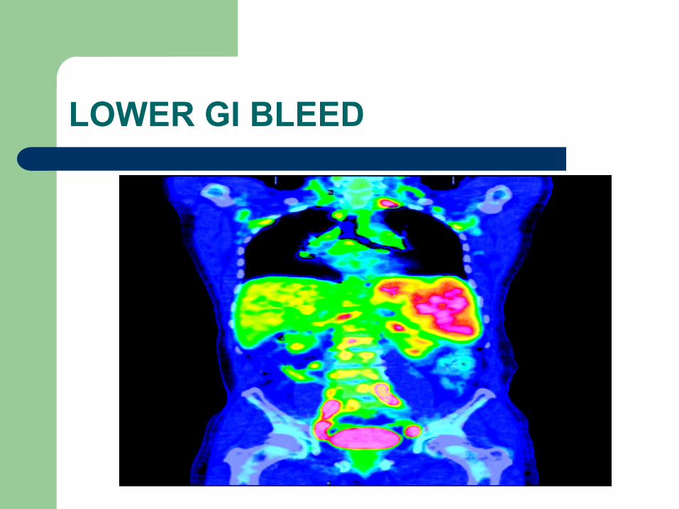
LOWER GI BLEED
CAUSES OF LOWER GI BLEEDING:
Causes:•
Diverticulosis•
Colon Cancer or Polyps•
Colitis (i.e. IBD, Infectious, Ischemic, Radiation)•
Angiodysplasia•
Post-Polypectomy, Anorectal
Fistula,•
Anorectal, i.e. hemorrhoids•
Obscure (unknown origin)

HAVE YOUR TOOLS READY!
•
Sclerotherapy
Needle + Meds, i.e. NaCl
0.9 % + Epinephrine 1:10,000 [10 ml]
•
HemoClips•
Endo-Loop, Snare•
ERBE Unit, Valley Lab•
Heater Probe (7 Fr/10 Fr)/Bicap
(7 Fr/10 Fr)•
Concurrent Intervention –
Tatto, Biopsy (lesion, inflammation), Roth Net Retrieval Device
•
Examples preferred in your tool box?
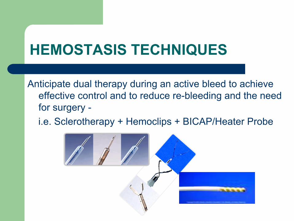
HEMOSTASIS TECHNIQUES
Anticipate dual therapy during an active bleed to achieve effective control and to reduce re-bleeding and the need for surgery -i.e. Sclerotherapy
+ Hemoclips
+ BICAP/Heater Probe
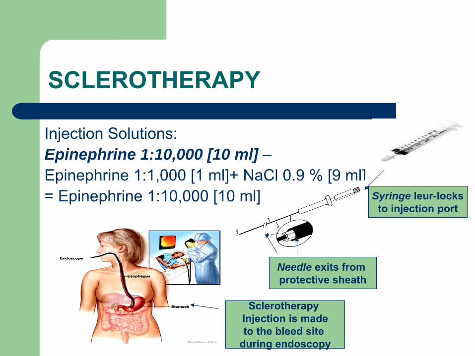
SCLEROTHERAPY
Injection Solutions:Epinephrine 1:10,000 [10 ml] –Epinephrine 1:1,000 [1 ml]+ NaCl
0.9 % [9 ml]
= Epinephrine 1:10,000 [10 ml]
Needle exits from protective sheath
Syringe leur-locksto injection port
SclerotherapyInjection is madeto the bleed site
during endoscopy
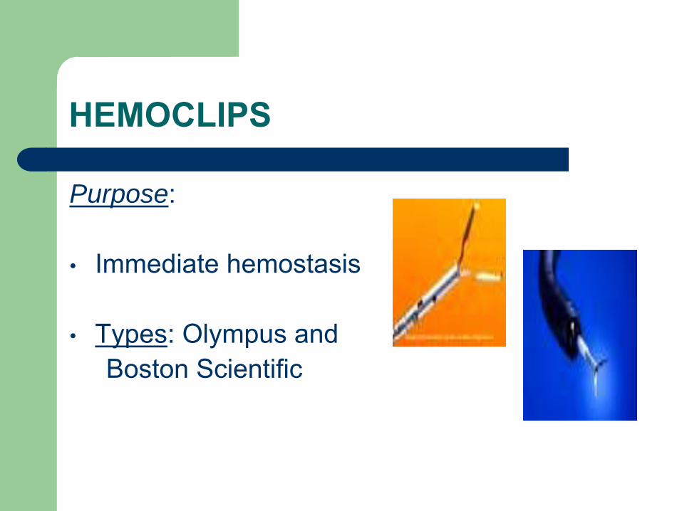
HEMOCLIPS
Purpose:
•
Immediate hemostasis
•
Types: Olympus and Boston Scientific
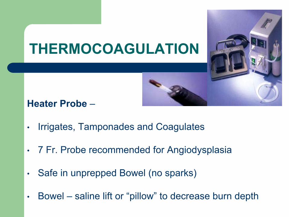
THERMOCOAGULATION
Heater Probe
–
•
Irrigates, Tamponades
and Coagulates
•
7 Fr. Probe recommended for Angiodysplasia
•
Safe in unprepped
Bowel (no sparks)
•
Bowel –
saline lift or “pillow”
to decrease
burn depth

THERMOCOAGULATION
Argon Plasma Coagulation (APC) –•
Colon Prep required
•
Coagulation with continuous movement (“painting”)
•
Saline lift or “pillow”
to reduce burn depth or injury (witha sclero
needle)
•
Not the first line of therapy in active bleeding (due to dissipation)
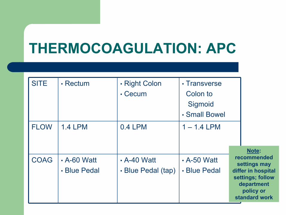
THERMOCOAGULATION: APC
SITE • Rectum • Right Colon• Cecum
• Transverse Colon to Sigmoid
• Small Bowel
FLOW 1.4 LPM 0.4 LPM 1 –
1.4 LPM
COAG • A-60 Watt• Blue Pedal
• A-40 Watt• Blue Pedal (tap)
• A-50 Watt• Blue Pedal
Note: recommended settings may
differ in hospital settings; follow
department policy or
standard work
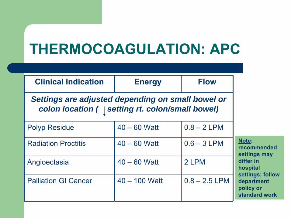
THERMOCOAGULATION: APC
Clinical Indication Energy Flow
Settings are adjusted depending on small bowel or colon location ( setting rt. colon/small bowel)
Polyp Residue 40 –
60 Watt 0.8 –
2 LPM
Radiation Proctitis 40 –
60 Watt 0.6 –
3 LPM
Angioectasia 40 –
60 Watt 2 LPM
Palliation GI Cancer 40 –
100 Watt 0.8 –
2.5 LPM
Note: recommended settings may differ in hospital settings; follow department policy or standard work
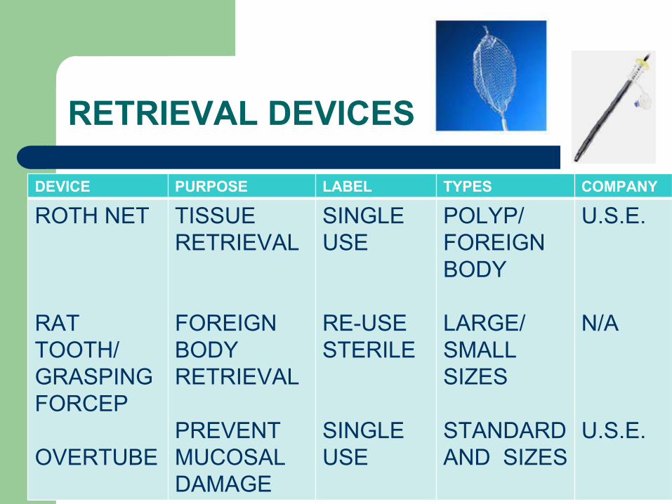
RETRIEVAL DEVICES
DEVICE PURPOSE LABEL TYPES COMPANY
ROTH NET
RAT TOOTH/GRASPINGFORCEP
OVERTUBE
TISSUE RETRIEVAL
FOREIGN BODY RETRIEVAL
PREVENT MUCOSAL DAMAGE
SINGLE USE
RE-USESTERILE
SINGLE USE
POLYP/FOREIGN BODY
LARGE/SMALLSIZES
STANDARD AND SIZES
U.S.E.
N/A
U.S.E.
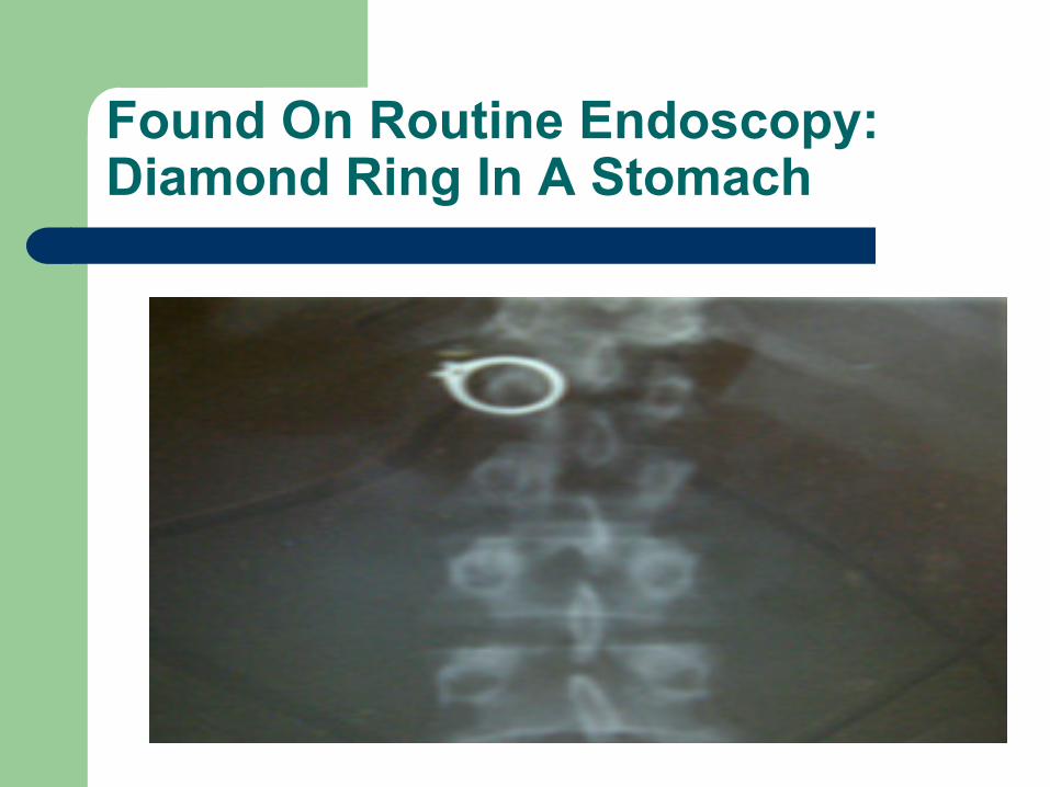
Found On Routine Endoscopy: Diamond Ring In A Stomach

References:
Ananya
Das, MD, Richard C.K
Wong. Prediction of outcome of acute GI hemorrhage: a review of risk scores and predictive models. Volume 60, Issue 1, Pages 85-93 (July)
General Practice Notebook (2009). Retrieved online at www.gpnotebook.co.uk
ASGE (2007 –
2009) Online Guidelines and Positions Statements; retrieved online at www.asge.org
SGNA (2007 –
2009) Online Guidelines and Position Statements; retrieved online at www.sgna.org
OLYMPUS (2009). Retrieved online at www.olympus.com.
U.S. Endoscopy (2009). Retrieved online at www.olympus.com.
ERBE U.S.A. (2009). Retrieved online at www.olympus.com.
Valley Lab (2009). Retrieved online at www.valleylab.com.
VMMC GI Webpage (2009). Retrieved online at www.vmmc.org.
TEST YOUR KNOWLEDGE: True/False
Rapid clinical decisions and appropriate interventions can make the difference between resolving a GI bleed and prolonged hospitalization.
Effective hemostasis
involves ONE intervention.
Hematochezia
is gastric (vomit) or duodenal frank red or old “coffee”
color.
Being prepared for a GI Bleed intervention involves having your “tool box”
ready for immediate use.
POST QUESTIONS:
After completing this online learning module:
Describe three things that will improve or change your everyday practice.
List the learning content you would like covered in future online modules relative to GI Bleeding Emergency Interventions.
Skills Lab session of ½
-
1 hour was attended by me on GI Emergency Interventions:
YES □
NO □

The End
Susan DePasquale, CGRN, MSN (2011)