Genomics and Proteomics - University of Torontosites.utoronto.ca/acdclab/pubs/PM/22908226.pdf ·...
15
Diamandis Morrissey, Keith A. Jarvi and Eleftherios P. Trudel, Theodorus van der Kwast, Colm Smith, Andrei P. Drabovich, Dominique George S. Karagiannis, Ihor Batruch, Chris Punit Saraon, Natasha Musrap, Daniela Cretu, Castration-resistant Prostate Cancer and during the Development of High Grade Cell Lines Reveals a Role for Protein S Androgen-independent Prostate Cancer Proteomic Profiling of Genomics and Proteomics: doi: 10.1074/jbc.M112.384438 originally published online August 20, 2012 2012, 287:34019-34031. J. Biol. Chem. 10.1074/jbc.M112.384438 Access the most updated version of this article at doi: . JBC Affinity Sites Find articles, minireviews, Reflections and Classics on similar topics on the Alerts: When a correction for this article is posted • When this article is cited • to choose from all of JBC's e-mail alerts Click here Supplemental material: http://www.jbc.org/content/suppl/2012/08/20/M112.384438.DC1.html http://www.jbc.org/content/287/41/34019.full.html#ref-list-1 This article cites 48 references, 14 of which can be accessed free at at Univ of Toronto - OCUL on June 26, 2013 http://www.jbc.org/ Downloaded from
Transcript of Genomics and Proteomics - University of Torontosites.utoronto.ca/acdclab/pubs/PM/22908226.pdf ·...

DiamandisMorrissey, Keith A. Jarvi and Eleftherios P.Trudel, Theodorus van der Kwast, Colm Smith, Andrei P. Drabovich, DominiqueGeorge S. Karagiannis, Ihor Batruch, Chris Punit Saraon, Natasha Musrap, Daniela Cretu, Castration-resistant Prostate Cancer
andduring the Development of High Grade Cell Lines Reveals a Role for Protein SAndrogen-independent Prostate Cancer Proteomic Profiling ofGenomics and Proteomics:
doi: 10.1074/jbc.M112.384438 originally published online August 20, 20122012, 287:34019-34031.J. Biol. Chem.
10.1074/jbc.M112.384438Access the most updated version of this article at doi:
.JBC Affinity SitesFind articles, minireviews, Reflections and Classics on similar topics on the
Alerts:
When a correction for this article is posted•
When this article is cited•
to choose from all of JBC's e-mail alertsClick here
Supplemental material:
http://www.jbc.org/content/suppl/2012/08/20/M112.384438.DC1.html
http://www.jbc.org/content/287/41/34019.full.html#ref-list-1
This article cites 48 references, 14 of which can be accessed free at
at Univ of Toronto - OCUL on June 26, 2013http://www.jbc.org/Downloaded from

Proteomic Profiling of Androgen-independent ProstateCancer Cell Lines Reveals a Role for Protein S during theDevelopment of High Grade and Castration-resistantProstate Cancer□S
Received for publication, May 22, 2012, and in revised form, August 17, 2012 Published, JBC Papers in Press, August 20, 2012, DOI 10.1074/jbc.M112.384438
Punit Saraon‡§1, Natasha Musrap‡§, Daniela Cretu‡§, George S. Karagiannis‡§, Ihor Batruch‡, Chris Smith‡,Andrei P. Drabovich‡, Dominique Trudel¶, Theodorus van der Kwast§¶, Colm Morrissey�, Keith A. Jarvi‡**,and Eleftherios P. Diamandis‡§‡‡2
From the ‡Samuel Lunenfeld Research Institute, Department of Pathology and Laboratory Medicine, Mount Sinai Hospital,Toronto, Ontario M5T 3L9, Canada, the §Department of Laboratory Medicine and Pathobiology, University of Toronto, Toronto,M5S 1A8 Ontario, Canada, the ¶Department of Pathology, University Health Network, University of Toronto, Toronto, M5G 2C4Ontario, Canada, the �Department of Urology, University of Washington, Seattle, Washington 98467, the **Department of Surgery(Division of Urology), Mount Sinai Hospital, Toronto M5T 3L9, Ontario, Canada, and the ‡‡Department of Clinical Biochemistry,University Health Network, Toronto, M5G 2C4 Ontario, Canada
Background: The mechanisms that cause castration-resistant prostate cancer remain unknown.Results:Using high throughput proteomics and subsequent clinical validation, we identified Protein S as being elevated in highgrade/advanced prostate cancer.Conclusion: Protein S is elevated in aggressive prostate cancer.Significance: Protein S expression could serve as a biomarker of aggressive prostate cancer.
Androgen deprivation constitutes the principal therapy foradvanced and metastatic prostate cancers. However, this thera-peutic intervention usually results in the transition to a moreaggressive androgen-independent prostate cancer. The elucida-tion of molecular alterations during the progression to andro-gen independence is an integral step toward discovering moreeffective targeted therapies. With respect to identifying crucialmediators of this transition, we compared the proteomes ofandrogen-independent (PC3, DU145, PPC1, LNCaP-SF, and22Rv1) and androgen-dependent (LNCaP and VCaP) and/ornormal prostate epithelial (RWPE) cell lines using mass spec-trometry. We identified more than 100 proteins that were dif-ferentially secreted in the androgen-independent cell lines. Ofthese, Protein S (PROS1) was elevated in the secretomes of all ofthe androgen-independent prostate cancer cell lines, with nodetectable secretion in normal and androgen-dependent celllines. Using quantitative PCR, we observed significantlyhigher (p < 0.05) tissue expression levels of PROS1 in pros-tate cancer samples, further indicating its importance inprostate cancer progression. Similarly, immunohistochemis-try analysis revealed elevation of PROS1 in high grade pros-tate cancer (Gleason grade >8), and further elevation in cas-tration-resistant metastatic prostate cancer lesions. We alsoobserved its significant (p < 0.05) elevation in high gradeprostate cancer seminal plasma samples. Taken together,
these results show that PROS1 is elevated in high grade andcastration-resistant prostate cancer and could serve as apotential biomarker of aggressive disease.
Prostate cancer is themost commonly diagnosed solid organtumor and the second leading cause of death due to canceramongmen inNorthAmerica (1). It is observed as either a slowgrowing indolent tumor or as amore advanced aggressive form;however, the current screening methods are not able to differ-entiate between these two forms. Prostate-specific antigen isthe most widely used biomarker for prostate cancer. However,due to a documented lack of specificity, it may also be elevatedin other non-cancerous pathologies, including benign prostatehyperplasia and prostatitis (2, 3). This inevitably leads to over-diagnosis and, ultimately, to overtreatment.Current treatments depend on whether the tumor is local-
ized or has metastasized. Localized prostate cancers are usuallytreated with either targeted radiation or radical prostatectomy.For more advanced and metastatic cancers, the mainstay treat-ment is androgen deprivation therapy, which is initially veryeffective at reducing tumor volume and growth (4, 5). However,persisting androgen deprivation often results in the selectionand accumulation of cancerous cells that have acquired resis-tance, leading to the development of androgen-independent orcastration-resistant prostate cancer (4, 5). This genotypic andphenotypic alteration is accompanied by increased mortalityrates because there are no alternative targeted therapies. Thus,the understanding of molecular alterations during the progres-sion to androgen independence becomes of utmost importancefor catalyzing the development of targeted and efficient thera-peutic options.
□S This article contains supplemental Tables S1–S10 and Fig. 1.1 Supported by the Ontario Graduate Scholarship and the Scace Graduate
Prostate Cancer Fellowship.2 To whom correspondence should be addressed: Mount Sinai Hospital,
Joseph & Wolf Lebovic Ctr., 60 Murray St. (Box 32), Flr. 6, Rm. L6-201,Toronto, Ontario M5T 3L9, Canada. Tel.: 416-586-8443; Fax: 416-619-5521;E-mail: [email protected].
THE JOURNAL OF BIOLOGICAL CHEMISTRY VOL. 287, NO. 41, pp. 34019 –34031, October 5, 2012© 2012 by The American Society for Biochemistry and Molecular Biology, Inc. Published in the U.S.A.
OCTOBER 5, 2012 • VOLUME 287 • NUMBER 41 JOURNAL OF BIOLOGICAL CHEMISTRY 34019 at Univ of Toronto - OCUL on June 26, 2013http://www.jbc.org/Downloaded from
The androgen receptor (AR)3 signaling cascade has beenextensively studied with respect to the progression of andro-gen-independent prostate cancer. Overall, AR signaling resis-tance has been attributed to AR gene amplifications, AR genemutations, changes in co-regulators or steroidogenic enzymes,or alternative proteins via outlaw pathways (4–10). However,recent interest has shifted toward the identification of novel“bypass” pathways, the so-called AR-independent pathways,which can play a role in this process as well (11, 12).High throughput proteomics is a growing field that can vir-
tually be used to study the proteomic signature of any biologicalmaterial (13). Numerous studies have been conducted usingproteomics to identify biomarkers for prostate cancer using celllines, tissues, and biological fluids, including expressed pros-tatic secretions (14–21).Previously, we performed proteomic analysis of the condi-
tioned media of three prostate cancer cell lines (LNCaP, PC3,and 22Rv1) and identified over 1000 proteins that could serve aspotential biomarkers for prostate cancer diagnosis (16). In thecurrent study, we performed proteomic analysis of the condi-tioned media (secretome analysis) of five androgen indepen-dent cell lines (PC3, DU145, PPC1, LNCaP-SF, and 22Rv1), twoandrogen-dependent cell lines (LNCaP and VCaP), and onenormal prostate epithelial cell line (RWPE). In total, we identi-fied over 3000 proteins, with over 100 proteins being differen-tially secreted between the AIPC and non-AIPC cell lines. Ofthese, Protein S (PROS1) was elevated in all five AIPC cell lines,with no observed secretion in the normal and androgen-depen-dent prostate cancer cell lines. Subsequently, we observedPROS1 overexpression in high grade prostate cancer tissue andseminal plasma. In addition, PROS1 expression was highly ele-vated in castration-resistant metastatic prostate cancer speci-mens. These results demonstrate that PROS1 could serve as apotential biomarker for high grade prostate cancers as well asproviding new areas for the therapeutic intervention for thetreatment of AIPC patients.
MATERIALS AND METHODS
Cells and Reagents—The human prostate cancer cell linesPC3, DU145, LNCaP, 22Rv1, and VCaP and the normal pros-tate epithelial cell line RWPE-1 were purchased from theAmerican Type Culture Collection (ATCC, Manassas, VA).The LNCaP-SF cells were kindly provided by Dr. Atsushi Miz-okami, and the PPC-1 cell line was provided by Dr. AaronSchimmer. Cell culture media specified by the ATCC for eachof the cell lines were used as follows: Dulbecco’s modifiedEagle’s medium (DMEM) (ATCC) with 10% fetal bovine serum(Thermo Scientific) was used for PC3, DU145, and VCaP;Roswell ParkMemorial Institute (RPMI) (ATCC)with 10%FBSwas used for 22RV1, PPC-1, and LNCaP cells. The normalRWPE cell line was grown in keratinocyte serum-free media(ATCC) supplemented with bovine pituitary extract andrecombinant epidermal growth factor. The LNCaP-SF cellswere grown in DMEM supplemented with 10% charcoal-stripped fetal bovine serum (Invitrogen). All cells were main-
tained at 37 °C with 5% CO2 in a humidified incubator. Allexperiments were performed within the first five passages fromthe initiation of all cultures.Cell Culture—Cells were cultured in six T-175 flasks and
allowed to grow in 30 ml of their respective media until theyreached 70% confluence. Generally, cells were allowed to growfor 48 h for them to achieve this confluence. The medium wasthen removed, and cells were washed three times with 20 ml ofPBS (Invitrogen). Following the washes, 30 ml of Chinese ham-ster ovary serum-free medium (Invitrogen) were added to eachof the flasks, which were then cultured for 2 days. After furthergrowth, the conditionedmediumwas collected and centrifugedat 2000 � g for 5 min to remove cellular debris. The resultingsupernatant was transferred to a fresh tube, and total proteinwas measured using a Coomassie Blue (Bradford) total proteinassay. Each flask contained roughly 500 �g of total protein, ofwhich two were combined to generate a total of 1 mg. Becausewe initially grew sixT-175 flasks, wewere able to combine themin three replicates containing a total protein level of roughly 1mg. The triplicates were then further processed following thesample preparation protocol below.Sample Preparation—Each sample containing 1 mg of total
protein was initially dialyzed using a 3.5 kDa molecular masscut-off membrane (Spectrum Laboratories, Inc., Compton,CA) in 5 liters of 1 mM ammonium bicarbonate buffer solutionat 4 °C overnight. The buffer solution was changed twice, andthe sample was then frozen at �80 °C and lyophilized to dry-ness using a Modulyo freeze dryer (Thermo Electron Corp.).The resulting dry protein product was denatured in 8 M ureaand reduced with 200 mM dithiothreitol at 50 °C for 30 min.Samples were then alkylated in 500 mM iodoacetamide whileshaking in the dark for 1 h. Each replicate was then furtherdesalted using a NAP5 column (GE Healthcare), frozen, andlyophilized again to complete dryness. Finally, the sampleswerethen digested with trypsin (Promega; sequencing-grade modi-fied porcine trypsin) at 37 °C overnight using a 1:50 trypsin/protein concentration ratio. The samples, now containing tryp-tic peptides, were acidified with formic acid before strongcation exchange (SCX).SCX on a High Pressure Liquid Chromatography (HPLC)
System—To reduce sample complexity, the samples were sub-jected to SCX using an Agilent 1100 HPLC system. The HPLCsystem was connected to a PolySULPHOETHYL ATM columnwith a 200-Å pore size and a diameter of 5�m(TheNest GroupInc., Southborough, MA). A total of 12 fractions per replicatecontaining the greatest number of peptides based on peakintensity were eluted and collected to perform mass spectro-metric analysis. The HPLC fractions were C18-extracted usingZipTipC18 micropipette tips (Millipore, Billerica, MA) andeluted in 5 �l of Buffer B (90% acetonitrile, 0.1% formic acid,10% water, and 0.02% trifluoroacetic acid). An additional 80 �lof Buffer A (95% water, 0.1% formic acid, 5% acetonitrile, and0.02% trifluoroacetic acid) were added to each zipped fraction.Mass Spectrometry (LC-MS/MS)—40 �l of each fraction
were injected into an autosampler on the EASY-nLC system(Proxeon Biosystems, Odense, Denmark). Peptides were firstcollected onto a 3-cm C18 column (inner diameter of 200 �m)and were then eluted onto a resolving 5-cm analytic C18 col-
3 The abbreviations used are: AR, androgen receptor; AIPC, androgen-inde-pendent prostate cancer; SCX, strong cation exchange chromatography.
PROS1 Is Elevated in Aggressive Prostate Cancer
34020 JOURNAL OF BIOLOGICAL CHEMISTRY VOLUME 287 • NUMBER 41 • OCTOBER 5, 2012 at Univ of Toronto - OCUL on June 26, 2013http://www.jbc.org/Downloaded from

umn (inner diameter of 75 �m) with an 8-�m tip (New Objec-tive). This HPLC system was coupled online to an LTQ-Or-bitrap XL (Thermo Fisher Scientific) mass spectrometer, usinga nano-electrospray ionization source (Proxeon Biosystems) indata-dependentmode. The fractions were run on a 55-min gra-dient, and eluted peptides were subjected to one full MS1 scan(450–1450m/z) in the Orbitrap at 60,000 resolution, followedby six data-dependent MS2 scans in the linear ion trap. Thefollowing parameters were enabled: monoisotopic precursorselection, charge state screening, and dynamic exclusion. Inaddition, charge states of �1 and �4 and unassigned chargestates were not subjected to MS2 fragmentation.Protein Identification and Data Analysis—RAW files were
uploaded onto Mascot Daemon (version 2.2), and extract_msnwas used to generate DAT and Mascot generic files. Mascotgeneric files were further searched on GPM (Global ProteomeMachine Manager, version 2006.06.01) against the concate-nated nonredundant IPI.Human version 3.62 database withparent and fragment tolerances of 7 ppm to generateX!Tandemfiles. The DAT and X!Tandem files were then merged andsearched with Scaffold (Proteome Software Inc., Portland, OR;version 2.0) to generate a file containing protein lists. The finalScaffold files contained normalized spectral counts for each ofthe cell lines, which were subsequently used as a semiquantita-tivemeasure to compare protein secretion among each cell line.ProteinCenter (Thermo Fisher Scientific) was used to
retrieve Gene Ontology annotations and pathway analysesfrom the Kyoto Encyclopedia of Genes and Genomes. To visu-alize and assess networks of overexpressed and underexpressedcandidates, Ingenuity Pathway Analysis (Ingenuity Systems,Redwood City, CA) software was used. Using this software,pathway analysis was performed, obtaining information oncanonical pathways and molecular networks that have beenaltered, which were determined by Fisher’s exact test.RNA Extraction and Quantitative PCR—Total RNAwas iso-
lated from cells using an RNeasy kit (Qiagen). cDNA was gen-erated from 1 �g of total RNA using the Superscript II cDNAsynthesis kit (Invitrogen). LuCaP 96 and LuCaP 96AI xenografttissues were frozen in liquid nitrogen and then ground into afine powder using amortar and pestle. RNAwas extracted fromthe resulting powdered tissue using anRNeasy kit (Qiagen), andthe final RNA product was used for further cDNA synthesis.Quantitative PCR was conducted using 1� SYBR reagent
(Applied Biosystems), and transcript levels of PROS1 TWSG1,LTBP1, GBA, PAM, and GAS6 were measured on a 7500 ABIsystem. For clinical validation, the TissueScan Prostate CancercDNAArray II was used (Origene). Quantitative PCR was con-ducted on these samples using the same SYBRGreen reagent asmentioned above. The following primer sequences were used:PROS1, GGCTCCTACTATCCTGGTTCTG (forward) andCAAGGCAAGCATAACACCAGTGC (reverse); GAS6, CCT-TCCATGAGAAGGACCTCGT (forward) and GAAGCACT-GCATCCTCGTGTTC (reverse); TBP, TGTATCCACAGTG-AATCTTGGTTG (forward) and GGTTCGTGGCTCTCTT-ATCCTC (reverse); TWSG1, CTTTGGGACGAGTGCTGT-GACT (forward) and GAGAAGGGATCGGTTCATGCAG(reverse); LTBP1, TGAATGCCAGCACCGTCATCTC (for-ward) and CTGGCAAACACTCTTGTCCTCC (reverse);
GBA, TGCTGCTCTCAACATCCTTGCC (forward) and TAG-GTGCGGATGGAGAAGTCAC (reverse); PAM, TGAAGGCA-CCTGGGAACCAGAA (forward) and CTCTGTGGAAAATC-ACCAGGTTAT (reverse).Western Blotting—Protein expression of PROS1, TWSG1,
LTBP1, GBA, and PAM was assessed using Western blot anal-ysis. Roughly, 30 �g of total protein from LuCaP 96 and LuCaP96AI were loaded onto an SDS-polyacrylamide gel (4–15%;Bio-Rad) and transferred onto PVDF membranes (Bio-Rad).Membraneswere then incubatedwith 5%blocking solution (2.5g of skim milk powder in Tris buffer solution containing 0.1%Tween (TBST)) overnight at 4 °C. Membranes were incubatedwith rabbit polyclonal antibody against PROS1 (1:1000),TWSG1 (1:500), LTBP1 (1:100), GBA (1:1000; Sigma), or PAM(1:1000; Protein Tech) for 1 h at room temperature. The mem-branes were then washed six times (three 15-min washes fol-lowed by three 5-min washes) with TBST. Membranes werethen incubated with goat anti-rabbit secondary antibody con-jugated to alkaline phosphatase (1:3000; Jackson Laboratories)or goat anti-mouse secondary antibody conjugated to alkalinephosphatase (1:3000; Jackson Laboratories) for 1 h at roomtemperature. After washing with TBST, proteins were detectedusing the ECL detection reagent (Siemens). The expression ofGAPDH (Cell Signaling Technology) was used as an internalstandard.Immunohistochemistry—Prostate cancer tissue microarrays
consisting of eight normal and 40 cancer cores were purchasedfromUS BioMax (Rockville,MD). Themetastatic prostate can-cer tissue microarray was developed and provided by the GUCancer Research laboratories at the University of Washington(Seattle, WA). Human tissue microarrays of fixed paraffin-em-bedded metastatic tissues from 23 rapid autopsy patients whodied of prostate cancer (consisting of three tissue microarrayblocks with two replicate cores per metastatic site) were usedfor immunohistochemical analyses. All patients had castration-resistant prostate cancer at the time of autopsy, defined by thepresence of a rising serum prostate-specific antigen followingmedical or surgical castration.Tissue microarrays were deparaffinized in xylene and rehy-
drated using ethanol. Endogenous peroxidase was inactivatedusing hydrogen peroxide for 10 min and washed with PBS.Antigen retrieval was then performed using citrate buffer in amicrowave for 10 min. Slides were then blocked for 5 min incasein and incubated overnight with the following antibodies:PROS1 (1:500; Epitomics), TWSG1 (1:50; Abgent), LTBP1(1:200; Abgent). Rabbit IgG was used on a duplicate slide toserve as a negative control. Following 10 min of PBS washing,slides were placed in secondary antibody for 30 min using theBGX kit (Biogenex, Fremont, CA). After a 10-min wash in PBS,slides were developed with the addition of 3,3�-diaminobenzi-dine for 5 min. Slides were then counterstained with hematox-ylin, dehydrated, and coverslipped.Seminal Plasma and ELISA—Semen was collected from sus-
pected prostate cancer patients prior to biopsy, allowed toliquefy at room temperature for 1 h, and centrifuged at 13,000rpm for 15 min to separate seminal plasma from cells and cel-lular debris. The seminal plasma was aliquoted into 1.5-mlEppendorf tubes and frozen at�80 °Cuntil further use. Clinical
PROS1 Is Elevated in Aggressive Prostate Cancer
OCTOBER 5, 2012 • VOLUME 287 • NUMBER 41 JOURNAL OF BIOLOGICAL CHEMISTRY 34021 at Univ of Toronto - OCUL on June 26, 2013http://www.jbc.org/Downloaded from
information for the patients can be found in supplementalTable S1.PROS1 protein levels in seminal plasma were assessed using
an enzyme-linked immunosorbent assay (American Diagnos-tica). Briefly, seminal plasma samples were diluted 10 times,and the ELISA was performed according to the manufacturer’sinstructions.Wound Repair/Scratch Assay—To assess wound repair and
cell migration, LNCaP cells were grown to full confluence in6-well plates in RPMIwith 10% FBS. Upon reaching full conflu-ence, the cells were incubated with 10 �g/ml mitomycin-C for2 h to inhibit cell proliferation, and a standardized scratch wasmade down the middle of each well using a 200-�l pipette. Themedium was removed, and cells were washed three times withPBS to remove any debris caused by the initial scratch. Scratch-induced cells were then either treated with 2 �g/ml humanpurified PROS1 (Enzyme Research Laboratories) or leftuntreated. Wound closure was measured by determining thedistance of cells at the ends of the wound 24 and 48 h post-scratching. After 48 h, cells were fixed and stained with CrystalViolet dye, and the number of migrating cells was measured bycounting the number of cells found within the middle of thewound.Statistical Analysis—All gene expression studies on cell lines
and xenografts consisting of normalized expressionswere com-pared using a two-tailed t test (GraphPad Prism Software).Gene expression studies on human prostate cancer and normaltissue were compared using a non-parametric Mann-WhitneyTest (GraphPad Prism Software). Finally, �2 tests were used tocompare different groups from the immunohistochemistrydata. Differences were considered significant if p values wereless than 0.05. All data are expressed as means � S.E.
RESULTS
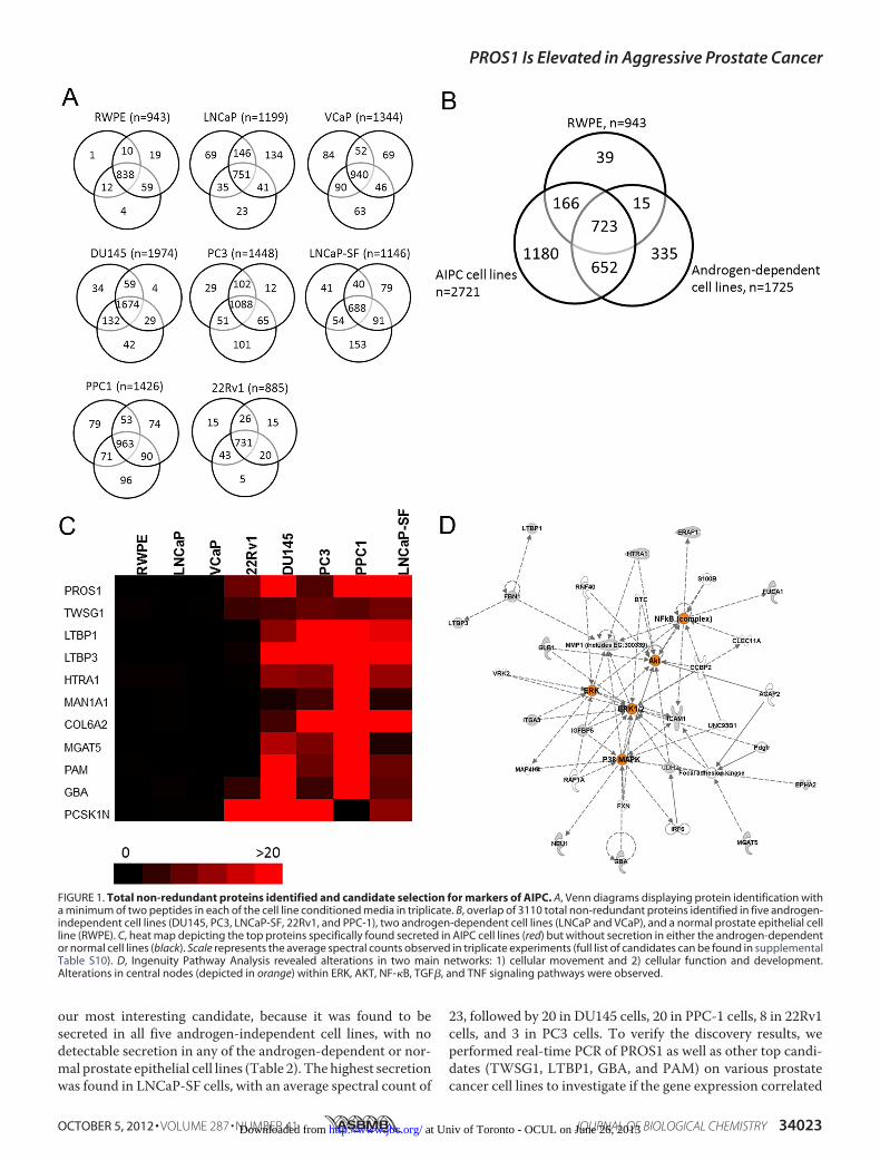
Proteomic Profiling of Prostate Cancer Cell Line ConditionedMedia—To identify modulators of androgen-independentprostate cancer that could serve as potential biomarkers ofaggressive disease, we performed an in depth proteomic analy-sis of the conditioned media of five androgen-independentprostate cancer cell lines (DU145, PC3, LNCaP-SF, PPC-1, and22Rv1), two androgen-dependent cell lines (LNCaP andVCaP),and one “near normal” prostate epithelial cell line (RWPE).Briefly, cells were grown in serum-free media; the conditionedmedia were collected, reduced, alkylated, and trypsin-digested;and peptides were subjected to two-dimensional liquid chro-matography (LC), which consisted of SCX chromatography onan HPLC system, followed by reverse-phase LC, prior to tan-demmass spectrometry (MS/MS). After performing the analy-sis, we identified, with a minimum of two peptides, 1974 pro-teins in DU145 cells, 1448 proteins in PC3 cells, 1146 proteinsin LNCaP-SF cells, 1426 proteins in PPC-1 cells, 885 proteins in22Rv1 cells, 1199 proteins in LNCaP cells, 1344 proteins inVCaP cells, and 943 proteins in RWPE cells, with adequatereproducibility among the triplicates (Fig. 1A). In total, weidentified 3110 non-redundant proteins with at least two pep-tides in the conditioned media of the cell lines combined. 723proteins (of 3110; 23.2%) were common to all cell lines. Amongthe androgen-independent cell lines, 1180 proteins were found
to be unique, whereas 335 and 39 proteins were unique to theandrogen-dependent cell lines and normal RWPE cell line,respectively (Fig. 1B). These data are summarized in Table 1,and complete lists of proteins identifiedwithin each cell line arepresented in supplemental Tables S2–S9.To identify gene ontology classifications, which include
molecular function, biological process, and cellular compo-nents, we utilized the Protein Center database. The top cellularlocalization annotations of the proteins identified within eachof the cell lines were cytoplasmic, membrane-bound, ornuclear. Over 67% of proteins were annotated as being eithercell surface, extracellular, ormembrane-bound.Themajority ofproteins were functionally annotated as either being proteinbinding or as having catalytic activity. Finally, the top biologicalprocesses of the proteins weremetabolic processes, followed byregulation of biological processes and response to stimuli (sup-plemental Fig. 1).Prioritization of Candidate Markers of Androgen Indepen-
dence—In order to identify candidates of androgen-indepen-dent and aggressive prostate cancers, we compared the differ-ential protein expression based on normalized spectral countsbetween our androgen-independent, androgen-dependent,and/or normal RWPE cell lines. Because we were interested infinding proteins that are elevated during AIPC, we decided toset as cut-offs proteins that were found with a minimum of twopeptides within our androgen-independent cell line proteomesand with less than two peptides in either the androgen-depen-dent or normal prostate cell line proteomes. This filter resultedin the selection of 1180 proteins that were unique to at least oneAIPC cell line. The top candidates included proteins that wereexpressed in multiple AIPC cell lines, with minimal or no spec-tral counts in the androgen-dependent or normal cells (Fig. 1C).To further prioritize the candidate list of proteins, a final dataset of 57 proteins was derived, consisting of proteins that weresecreted in at least three AIPC cell lines with aminimumof twospectral counts, and with less than one spectral count in allandrogen-dependent cell lines and the normal cell line. Table 2summarizes these candidates with respect to the observedspectral counts in each of the cell lines as well as the numberof AIPC cell lines that displayed positive results. The listcontains proteins that were previously studied in the contextof prostate cancer progression, including MGAT5, PAM,GBA, ROBO1, CD59, MMP1, IGFBP4, CDH2, TGFB2,ICAM1, EPHA2, and IGFBP5 (22–33), thus providing fur-ther confirmation for the robustness of our quantificationmethod.We next subjected our candidate list to preclustering path-
way analysis using Ingenuity Pathway Analysis. This analysisrevealed twomajor networks that the candidates were enrichedfor: 1) cellular movement and 2) cellular function and develop-ment (Fig. 1D). The candidates had central nodes in theNF-�B,AKT, ERK, p38 MAPK, TGF�, and TNF signaling cascades, allof which have been previously documented to be associatedwith prostate cancer progression (34–39).Protein S Is Elevated in Androgen-independent Prostate Can-
cer Cell Lines and Is Activated in the LuCaP 96AI Androgen-independent Prostate Cancer Xenograft Model—Based on ourinitial discovery results, the vitaminK-dependent Protein Swas
PROS1 Is Elevated in Aggressive Prostate Cancer
34022 JOURNAL OF BIOLOGICAL CHEMISTRY VOLUME 287 • NUMBER 41 • OCTOBER 5, 2012 at Univ of Toronto - OCUL on June 26, 2013http://www.jbc.org/Downloaded from
our most interesting candidate, because it was found to besecreted in all five androgen-independent cell lines, with nodetectable secretion in any of the androgen-dependent or nor-mal prostate epithelial cell lines (Table 2). The highest secretionwas found in LNCaP-SF cells, with an average spectral count of
23, followed by 20 in DU145 cells, 20 in PPC-1 cells, 8 in 22Rv1cells, and 3 in PC3 cells. To verify the discovery results, weperformed real-time PCR of PROS1 as well as other top candi-dates (TWSG1, LTBP1, GBA, and PAM) on various prostatecancer cell lines to investigate if the gene expression correlated
FIGURE 1. Total non-redundant proteins identified and candidate selection for markers of AIPC. A, Venn diagrams displaying protein identification witha minimum of two peptides in each of the cell line conditioned media in triplicate. B, overlap of 3110 total non-redundant proteins identified in five androgen-independent cell lines (DU145, PC3, LNCaP-SF, 22Rv1, and PPC-1), two androgen-dependent cell lines (LNCaP and VCaP), and a normal prostate epithelial cellline (RWPE). C, heat map depicting the top proteins specifically found secreted in AIPC cell lines (red) but without secretion in either the androgen-dependentor normal cell lines (black). Scale represents the average spectral counts observed in triplicate experiments (full list of candidates can be found in supplementalTable S10). D, Ingenuity Pathway Analysis revealed alterations in two main networks: 1) cellular movement and 2) cellular function and development.Alterations in central nodes (depicted in orange) within ERK, AKT, NF-�B, TGF�, and TNF signaling pathways were observed.
PROS1 Is Elevated in Aggressive Prostate Cancer
OCTOBER 5, 2012 • VOLUME 287 • NUMBER 41 JOURNAL OF BIOLOGICAL CHEMISTRY 34023 at Univ of Toronto - OCUL on June 26, 2013http://www.jbc.org/Downloaded from

with the protein expression data. As expected, we observed sig-nificantly elevated transcript levels of each of the candidates inandrogen-independent cell lines (PC3 and DU145), comparedwith the normal (RWPE) and androgen-dependent cells(LNCaP and VCaP) (Fig. 2A).To determine whether PROS1 and other top candidates may
play a role during the progression of androgen-independentprostate cancer, LuCaP96 and its androgen-independent coun-terpart, LuCaP 96AI, xenografts were utilized. The transcriptand protein expression of PROS1, TWSG1, LTBP1, PAM, andGBA were assessed using quantitative PCR and Western blot,respectively. It was shown that all of the candidates displayedsignificant increases in their gene expression in the LuCaP 96AIxenograft-derived cells (p � 0.05) (Fig. 2B). Protein validationof these candidates usingWestern blotting supported our data,demonstrating an up-regulation of our identified enzymes inLuCaP 96AI cells (Fig. 2C). Taken together, our cell line andxenograft data clearly demonstrate that PROS1 and other topcandidates are elevated at both the protein and transcript levelin androgen-independent prostate cancer cells and couldpotentially serve as biomarkers and/or therapeutic targets foraggressive prostate cancer.Protein S Expression Is Elevated in High Grade Prostate
Cancer—We then sought to investigate whether PROS1 wasalso overexpressed in prostate cancer patients. To examine this,we measured its transcript levels in normal and human tumortissue samples. Using real-time PCR, we found that PROS1transcript levels were significantly elevated (p � 0.017) by over2-fold in prostate cancer compared with normal tissue (Fig.4A).
In addition, using immunohistochemistry, we assessed theprotein expression level of PROS1 in normal (n � 8) and clini-cally localized prostate cancers of varying grade (n � 40). Wedevised a scoring system to assess protein expression, wherebyeach core was scored with a 0, 1, 2, or 3, which corresponds tono staining and low, moderate, or high staining, respectively.After analysis, we observed a stair-wise expression pattern ofPROS1 staining, with minimal staining in normal cores, mod-erate staining in low grade prostate cancer (Gleason grade �7),and increased staining in high grade prostate cancer (Gleason�8) (Fig. 3A). Normal cores depicted very little positive stain-ing, with only 13% of cores displaying a score of 2 or greater,whereas low grade prostate cancer cores had intermediateexpression, staining positively in 25% of cases. Finally, highgrade prostate cancer cores displayed the greatest expressionlevels, with 43% of cores staining positively (Fig. 3B). Similarly,we also assessed the expression of TWSG1 and LTBP1 on thesesamples. Both TWSG1 and LTBP1 displayed elevated expres-sion in cases of high grade prostate cancer; however, the expres-sion pattern was not as prominent at PROS1 (Fig. 3). Theseresults indicate that PROS1 is overexpressed in high gradeprostate cancers and may therefore play a role in the progres-sion of prostate cancer to an advanced stage disease.Protein S Is Elevated in the Seminal Plasma of Intermediate
and High Grade Prostate Cancer Patients—After observingincreased gene and protein expression of PROS1 in high gradeprostate cancer tissue specimens, we wanted to determine ifelevated PROS1 expression could also be reflected with ele-vated seminal plasma secretion. We chose to analyze seminalplasma because it represents the proximal fluid in which pros-
TABLE 1Total number of proteins identified in triplicate analysis of cell line conditioned medium
RWPE LNCaP VCaP 22Rv1 PPC1 DU145 PC3 LNCaP-SF
Total non-redundant proteins (with �2 peptides) 943 1199 1344 885 1426 1974 1448 1146No. of peptides identified with . . . aOnly 2 peptides 243 429 477 434 548 689 598 425Only 3 peptides 110 88 125 108 136 198 150 107Only 4 peptides 77 79 99 86 127 123 112 79�5 peptides 513 603 643 227 615 964 588 535
a Based on the average triplicate spectral count value.
TABLE 2Top candidate proteins elevated in androgen-independent cell lines based on average normalized spectral count valuesFifty-seven proteins were foundwith aminimumof two peptides in at least three AIPC cell lines andwith less than one peptide in androgen-dependent and normal prostateepithelial cell lines. For a complete list of the 52 proteins, see supplemental Table S10.
RWPE(normal)
LNCaP(AD)a
VCaP(AD)
22Rv1(AI)b
DU145(AI)
PC3(AI)
PPC1(AI)
LNCaP-SF(AI)
Number of positiveAIPC cell lines
Previously studied duringprostate cancer progression?
PROS1 0 0 0 8 20 3 20 23 5TWSG1 1 0 0 5 6 8 7 9 5GBA 0 1 0 4 25 5 37 7 5 Ref. 28PGRMC1 0 1 0 3 3 2 5 4 5LTBP1 0 0 0 0 11 58 161 18 4LTBP3 0 0 0 1 27 28 48 22 4HTRA1 1 1 0 0 11 10 72 12 4MAN1A1 0 0 0 0 2 5 93 3 4COL6A2 0 0 0 0 5 38 23 30 4MGAT5 0 0 0 0 14 10 61 3 4 Ref. 26PAM 0 0 0 0 32 8 34 8 4 Ref. 27PCSK1N 0 0 0 30 20 24 0 11 4CHID1 0 0 0 0 2 7 50 11 4ROBO1 0 1 0 2 0 9 25 10 4 Ref. 29CD59 1 1 0 0 2 9 16 6 4 Ref. 30B4GALT4 0 0 0 0 6 6 14 5 4B3GAT3 0 0 0 3 7 1 10 3 4
a AD, androgen-dependent cell lines.b AI, androgen-independent cell lines.
PROS1 Is Elevated in Aggressive Prostate Cancer
34024 JOURNAL OF BIOLOGICAL CHEMISTRY VOLUME 287 • NUMBER 41 • OCTOBER 5, 2012 at Univ of Toronto - OCUL on June 26, 2013http://www.jbc.org/Downloaded from

tatic secretions should be enriched for. We assessed PROS1levels using ELISA in a variety of seminal plasmas taken fromcontrol (n � 8), prostatitis (n � 8), low grade (Gleason �6, n �8), and intermediate/high grade (Gleason �7, n � 13) prostatecancer patients. Clinical information of the seminal plasmasamples can be found in supplemental Table S1. Based on ouranalysis, we observed a statistically significant (p � 0.05) eleva-tion of PROS1 in seminal plasma from intermediate and highgrade prostate cancer patients (Fig. 4B). The area under thecurve of PROS1 being able to distinguish benign (negativebiopsy and prostatitis) and low grade prostate cancers fromintermediate/high grade prostate cancer patients in seminalplasma was 0.875 (confidence interval � 0.744–1.0, p � 0.001)(Fig. 4C). These results suggest a potential role of PROS1 as a
biomarker, to assist in the differential diagnosis of high gradefrom low grade prostate cancer and/or benign conditions.PROS1-stimulated Prostate Cancer Cells Have Increased
Migratory Potential—To explore whether PROS1 has a role inprostate cancer growth or migration, we performed in vitroscratch assays, in which LNCaP cells were grown to full conflu-ence, treatedwithmitomycin-C for 2 h, and scratched to inducewounding. Cells were then either treated with 1–2 �g/ml ofhuman purified PROS1 or left untreated to serve as controls,and the amount of wound closure as well as the number ofmigrating cells was assessed. We observed that 24 and 48 hpostscratch, there was a statistically significant (p � 0.05)increase in wound closure in PROS1-treated LNCaP cells (Fig.5A). Specifically, over 40% of the original wound was healed in
FIGURE 2. Analysis of gene and protein expression levels of PROS1, TWSG1, LTBP1, GBA, and PAM on prostate cancer cell lines and LuCaP96/96AIxenografts. A, gene expression profiles of top candidates on normal (RWPE), androgen-dependent (LNCaP and VCaP), and androgen-independent (DU145and PC3) cell lines. B and C, gene and protein expression levels of top candidates are increased in the LuCaP 96AI androgen-independent xenograft model(*, p � 0.05, Mann-Whitney test). Error bars, S.E.
PROS1 Is Elevated in Aggressive Prostate Cancer
OCTOBER 5, 2012 • VOLUME 287 • NUMBER 41 JOURNAL OF BIOLOGICAL CHEMISTRY 34025 at Univ of Toronto - OCUL on June 26, 2013http://www.jbc.org/Downloaded from

PROS1-treated cells compared with less than 20% wound clo-sure in non-treated control cells (Fig. 5B). In addition, to assesscell migration during PROS1 stimulation, we fixed and stainedcells after 24 and 48 h of inducing the wound and counted cellsthat were found within default squares within the originalwound gap. After analysis, we observed a significant increase(p� 0.05) in the number ofmigrating cells during PROS1 stim-ulation (Fig. 5B). Taken together, these data are indicative thatPROS1 has a direct role on prostate cancer cellular processes,including growth and migration.PROS1 Transcript Levels Increase after Continuous Growth
in Androgen-deprived Conditions—During androgen depriva-tion, prostate cancer cells undergo apoptosis due to the absenceof key growth stimuli, in particular androgens. To assess theeffect of androgen deprivation on PROS1 expression, we grewLNCaP cells in androgen-deprived conditions for varyingtimes, including 1, 2, and 5 days, and extracted total RNA toexamine the expression levels of PROS1. Interestingly, PROS1gene expression levels were increased in a time-dependent
manner (Fig. 5C).We also observed an increase in PROS1 tran-script level in PC3 androgen-deprived cells (data not shown).Previously, we assessed PROS1 expression in the LuCaP
96/LuCaP 96AI xenograftmodel and observed elevated expres-sion in the androgen-independent xenograft. We measuredtranscript levels of GAS6, a known homologue of PROS1,which has previously been linked to prostate cancer progres-sion (45, 46), on these xenografts and found its elevation inLuCaP 96AI cells (p � 0.05). Taken together, these results fur-ther support the involvement of PROS1 andGAS6 in the devel-opment of AIPC.PROS1 Is Highly Expressed in Cases of Castration-resistant
Metastatic Prostate Cancers—Thus far, we have demonstratedthat PROS1 is elevated 1) in vitro, in androgen-independent celllines; 2) in localized high grade disease, both at the tissue andseminal plasma level; and 3) in an androgen-independent xeno-graft model. Based on these findings, we aimed to examinePROS1 expression in various castration-resistant metastaticprostate cancer human samples to identify whether it is dys-
FIGURE 3. Protein expression of PROS1, TWSG1, and LTBP1 in human prostate cancer tissues. A, representative immunohistochemistry images of casesof normal, low grade (Gleason �7), and high grade (Gleason �8) prostate cancer specimens, under light microscopy (�20). B, immunohistochemical stainingwas quantified using a scoring scale of 0, 1, 2, and 3 corresponding to no staining and low, moderate, and high staining, respectively, as blindly determined bya pathologist. Positive cores were determined to be ones that stained with an intensity of 2 or greater.
PROS1 Is Elevated in Aggressive Prostate Cancer
34026 JOURNAL OF BIOLOGICAL CHEMISTRY VOLUME 287 • NUMBER 41 • OCTOBER 5, 2012 at Univ of Toronto - OCUL on June 26, 2013http://www.jbc.org/Downloaded from

regulatedduringprostate cancermetastasis aswell.Usingourpre-vious scoring system in a tissuemicroarray, containing castration-resistantmetastatic lesions to thebone (n�72), lymphnodes (n�28), and lung and liver (n � 19), we demonstrated substantiallyincreased PROS1 staining in all metastatic sites (Fig. 6). Specifi-cally,weobserved15.8%of lungand livermetastasescontainingnostainingor low staining and84.2%of coreshaving intense staining.With respect to the lymph node metastasis cores, we found 25%with lowstainingand75%with intense staining.Withinbonemet-astatic lesions, 29% of cores displayed low staining, whereas 71%had intense staining. In contrast, in normal prostate cores, PROS1staining was very low because themajority of the samples (87.5%)had little or no staining, and a small proportion (12.5%) hadmod-
erate PROS1 expression. Following statistical analysis, PROS1expression was found significantly up-regulated in lung and liver(p�0.001), lymphnode (p�0.002), andbone (p�0.002)prostatecancermetastatic lesions comparedwith normal prostate samples(Table 3). In conclusion, PROS1 expression is up-regulated in cas-tration-resistant prostate cancermetastases and, thus, could serveas a potential biomarker and therapeutic target for aggressivedisease.
DISCUSSION
In the present study, we aimed to delineate the proteomes ofseveral prostate cancer and a near normal prostate cell lineconditioned media to identify proteins that are elevated during
FIGURE 4. Expression of PROS1 in seminal plasma specimens of varying Gleason grade. A, using the TissueScan Prostate Cancer cDNA Array II consisting of eightnormal and 36 prostate cancer specimens, the gene expression profiles of PROS1 showed significantly elevated mRNA expression in cancer compared with normalspecimens (*, p �0.05, Mann-Whitney test). B, ELISA analysis of PROS1 protein levels in seminal plasma from negative biopsy (n �8) (positive prostate-specific antigentest), prostatitis (n � 8), low grade prostate cancer (Gleason �6) (n � 8), and intermediate and high grade prostate cancer (Gleason �7) (n � 13) patients (*, p � 0.05,Mann-Whitney test). C, the area under the curve (AUC) of PROS1 being able to distinguish benign (negative biopsy and prostatitis) and low grade prostate cancers fromintermediate/high grade prostate cancer patients in seminal plasma was 0.875 (confidence interval (CI) � 0.744–1.0, p � 0.001).
PROS1 Is Elevated in Aggressive Prostate Cancer
OCTOBER 5, 2012 • VOLUME 287 • NUMBER 41 JOURNAL OF BIOLOGICAL CHEMISTRY 34027 at Univ of Toronto - OCUL on June 26, 2013http://www.jbc.org/Downloaded from

androgen-independent prostate cancer. Specifically, by usingLC-MS/MS, we performed proteomic analysis on five andro-gen-independent (DU145, PC3, 22Rv1, PPC1, and LNCaP-SF),two androgen-dependent (LNCaP and VCaP), and one nearnormal (RWPE) prostate epithelial cell line. After performingexperiments in triplicates and using various protein identifica-tion search engines (X!Tandem,Mascot, and Scaffold), wewereable to identify between 885 and 1974 proteins with at least twopeptides in each of the cell line conditioned media. In total, weidentified a 3110-non-redundant protein data set, which, to ourknowledge, is the most comprehensive one to date. To inter-nally validate our approach, we observed among our top candi-dates, 12 proteins (MGAT5, PAM, GBA, ROBO1, CD59,MMP1, IGFBP4, CDH2, TGFB2, ICAM1, EPHA2, andIGFBP5) that have previously been studied or implicated inprostate cancer progression (22–33). For example,MGAT5hasbeen shown to mediate enhanced invasion and metastaticpotential for prostate cancer cells through many in vitro inva-sion assays and xenograft animal models.After analyzing our candidates, we decided to further inves-
tigate the anticoagulation factor PROS1, because it was foundsecreted in only the androgen-independent cell lines, with no
detectable secretions in androgen-dependent or normal pros-tate epithelial cell lines. After performing clinical validation ona variety of tissue and seminal plasma samples, we observedelevation of PROS1 specifically in high grade cases, in additionto its elevation in castration-resistant metastatic prostate can-cer cases, which supported our initial goal of identifying mark-ers of aggressive prostate cancer.To our knowledge, this is the first study documenting a role
for PROS1with respect to prostate cancer pathogenesis. In fact,after performingmultiple searches, the only other study thatwecould find assessing PROS1 in any form of cancer was a studyevaluating various coagulation factor expressions during colo-rectal cancer development. In the aforementioned studies,PROS1 staining in colorectal cancer cells was performed; how-ever, the investigators did not go into further detail aside fromtheir immunohistochemistry analysis (40). Although PROS1has been highly studiedwith respect to the coagulation cascade,recent studies have shown that PROS1 could act as a ligand fora family of receptor tyrosine kinases, consisting of Tyro3, Axl,andMer (TAM receptors) (41–44). Interestingly, GAS6, whichshares 40% amino acid identity with PROS1, is a known ligandfor these TAM receptors (41). We also observed GAS6 secre-
FIGURE 5. LNCaP cells were treated with human purified PROS1, and a wound repair scratch assay was performed. PROS1-treated cells (1 or 2 �g/ml) hadsignificantly increased wound repair (A) and an increase in migrating cells (B) compared with untreated cells 24 and 48 h postscratch (*, p � 0.05, Mann-Whitneytest). C, LNCaP cells were grown in androgen-depleted conditions for a 5-day period. PROS1 gene expression levels increased the longer the cells were grownin androgen-depleted conditions (*, p � 0.05, Mann-Whitney test). A similar expression profile was observed with PC3 cells (data not shown). D, PROS1 (Fig. 2B)and its homologue GAS6 transcript levels were increased in the LuCaP 96AI androgen-independent xenograft model (*, p � 0.05, Mann-Whitney test). Errorbars, S.E.
PROS1 Is Elevated in Aggressive Prostate Cancer
34028 JOURNAL OF BIOLOGICAL CHEMISTRY VOLUME 287 • NUMBER 41 • OCTOBER 5, 2012 at Univ of Toronto - OCUL on June 26, 2013http://www.jbc.org/Downloaded from

tion in two AIPC cell lines (DU145 and LNCaP-SF), with nodetectable secretions in either the androgen-dependent or nor-mal prostate cell lines. Previously, GAS6 has been shown tohave increased affinity for the Axl protein (41). In regard toprostate cancer pathology, Sainaghi et al. (45) demonstratedthat Axl could be activated by GAS6 in DU145 and PC3 cells,resulting in the phosphorylation of theMEK protein, leading toincreased cell survival and proliferation. Shiozawa et al. (46)went a step further and observed that upon GAS6/Axl stimula-tion, prostate cancer cells had increasedmetastatic and invasiveproperties, particularly to bone. Both of these studies demon-strate that TAM receptors, specifically Axl, and their corre-sponding ligands (GAS6) are able to promote prostate cancer
tumorogenesis. The question of whether PROS1 is able to act ina similar way has yet to be elucidated. However, from our anal-ysis, we also observed increased cell migration upon stimulationwith PROS1 as well as its elevated expression in high grade andmetastatic disease, further providing evidence that it may be pro-viding a survival advantage for prostate cancer cells. However, themechanistic role of PROS1and its potential downstreamsignalingcascade still requires further exploration.PROS1 shares 40% amino acid identity with GAS6, and both
proteins have identical structural domains, including a �-glu-tamic acid domain, which is integral for vitaminK binding, fourepidermal growth factor (EGF)-like modules, and two tandemlaminin G domains that are structurally related to those of thesex hormone binding globulin (41). PROS1, but notGAS6, con-tains a unique thrombin cleavage domain, which is importantfor its functions within the coagulation cascade (41). Interest-ingly, two recent studies conducted on mouse neuronal cellsshowed that upon chemically induced cell injury, PROS1 wasable to activate AKT and induce an anti-apoptotic cascade,resulting in reduced cell death (47, 48). The response was spe-cific to PROS1 binding to the TYRO3 receptor and not Axl orMer. These results marked a novel role for PROS1 outside ofthe coagulation cascade as a signaling molecule. Also, it wasobserved that the lamininGdomains in particular were integralfor the binding of PROS1 and subsequent activation of itsdownstream signaling pathway (47, 48).The novel role of PROS1 as a signaling molecule in the neu-
ronal mouse model provides an interesting explanation as to
FIGURE 6. Expression of PROS1 in castration-resistant metastatic prostate cancer to the bone, lymph node, liver, and lungs. Representative immuno-histochemistry images of PROS1 staining in normal prostate (A), liver metastasis (B), bone metastasis (C), lymph node metastasis (D), and lung metastasis (E) ofthe prostate are shown. Images were taken under light microscopy (�20). F, immunohistochemical staining was quantified using a scoring scale of 0, 1, 2, and3, corresponding to no staining and low, moderate, and high staining, respectively, as blindly determined by a pathologist. Positive cores were determined tobe ones that stained with an intensity of 2 or greater.
TABLE 3PROS1 expression in castration-resistant metastatic prostate cancers
Tissue typeNumber of
positive coresaStaining
percentagep value comparedwith normalb
%Normalprostate
1/8 12.5 NAc
Lung andlivermetastasis
16/19 84.2 0.0009
Lymphnodemetastasis
21/28 75.0 0.0026
Bonemetastasis
51/72 70.8 0.0022
a Positive staining means a score of 2 or higher, based on the pathologist’s score(see Fig. 3 legend).
b p value was calculated using a �2 test.c Not applicable.
PROS1 Is Elevated in Aggressive Prostate Cancer
OCTOBER 5, 2012 • VOLUME 287 • NUMBER 41 JOURNAL OF BIOLOGICAL CHEMISTRY 34029 at Univ of Toronto - OCUL on June 26, 2013http://www.jbc.org/Downloaded from
why it may possibly become activated in high grade and aggres-sive prostate cancer. During aggressive prostate cancer, andro-gen deprivation is usually the gold standard therapy.Many cellsundergo apoptosis during this treatment; however, some areable to evade the therapy and continue growing in the absence ofandrogens. A possible hypothesis for our observed increase inPROS1expression in aggressiveprostate cancer couldbedue to itspotential involvement in activating downstream anti-apoptoticpathways, which in turn provide a survival advantage for cancercells and promote their progression to AIPC. Further experimen-tation needs to be conducted to determine more precisely thefunctional roleofPROS1and itspotentialdownstreamsignaling inaggressive disease. In addition, PROS1 as a therapeutic targetbecomes of interest because the development of potential mole-cules that can possibly inhibit PROS1 signaling function withoutaltering its coagulation properties presents an interesting avenueof therapeutic intervention.Overall, in this study, we demonstrate that PROS1 is a novel
marker of high grade and castration-resistant metastatic pros-tate cancer, which warrants further functional validationthrough the use of relevant in vitro and in vivo models. Ourpresent findings provide sufficient evidence that PROS1 playsan important role in prostate cancer pathogenesis and suggestan interesting area for therapeutic intervention for a diseasethat lacks targeted treatments.
Acknowledgments—We acknowledge the support of the UniversityHealth Network Pathology Research Program for performing theimmunohistochemistry experiments. We thank Apostolos Dimitro-manolakis for assistance in analyzing the proteomics data and withstatistical analysis. We also thank the patients and their families whowere willing to participate in the Prostate Cancer Donor Program, forwithout them research of this nature would not be possible. We alsoacknowledge Lawrence True, Eva Corey, Celestia Higano, and RobertVessella and the rapid autopsy teams in the Urology Department atthe University of Washington. The donated material is the result ofwork supported by resources from the PacificNorthwest Prostate Can-cer SPORE (P50CA97186), PO1 NIH, National Institutes of Health,Grant PO1CA085859, and the Richard M. Lucas Foundation.
REFERENCES1. Gronberg, H. (2003) Prostate cancer epidemiology. Lancet 361, 859–8642. Diamandis, E. P. (1998) Prostate-specific antigen. Its usefulness in clinical
medicine. Trends Endocrinol. Metab. 9, 310–3163. Sardana, G., Dowell, B., and Diamandis, E. P. (2008) Emerging biomarkers
for the diagnosis and prognosis of prostate cancer. Clin. Chem. 54,1951–1960
4. Denmeade, S. R., and Isaacs, J. T. (2002) A history of prostate cancertreatment. Nat. Rev. Cancer 2, 389–396
5. Saraon, P., Jarvi, K., and Diamandis, E. P. (2011) Molecular alterationsduring progression of prostate cancer to androgen independence. Clin.Chem. 57, 1366–1375
6. Chodak, G. W., Kranc, D. M., Puy, L. A., Takeda, H., Johnson, K., andChang, C. (1992) Nuclear localization of androgen receptor in heteroge-neous samples of normal, hyperplastic and neoplastic human prostate.J. Urol. 147, 798–803
7. Crawford, E. D., and Petrylak, D. (2010) Castration-resistant prostate can-cer. Descriptive yet pejorative? J. Clin. Oncol. 28, e408
8. Debes, J. D., and Tindall, D. J. (2004) Mechanisms of androgen-refractoryprostate cancer. N. Engl. J. Med. 351, 1488–1490
9. Ruizeveld de Winter, J. A., Trapman, J., Vermey, M., Mulder, E., Zegers,
N. D., and van der Kwast, T. H. (1991) Androgen receptor expression inhuman tissues. An immunohistochemical study. J. Histochem. Cytochem.39, 927–936
10. Sadi, M. V., Walsh, P. C., and Barrack, E. R. (1991) Immunohistochemicalstudy of androgen receptors inmetastatic prostate cancer. Comparison ofreceptor content and response to hormonal therapy. Cancer 67,3057–3064
11. Edwards, J., and Bartlett, J. M. (2005) The androgen receptor and signaltransduction pathways in hormone-refractory prostate cancer. Part 1.Modifications to the androgen receptor. BJU Int. 95, 1320–1326
12. Edwards, J., and Bartlett, J. M. (2005) The androgen receptor and signaltransduction pathways in hormone-refractory prostate cancer. Part 2. An-drogen receptor cofactors and bypass pathways. BJU Int. 95, 1327–1335
13. Karagiannis, G. S., Pavlou, M. P., and Diamandis, E. P. (2010) Cancersecretomics reveal pathophysiological pathways in cancer molecular on-cology.Mol Oncol 4, 496–510
14. Alaiya, A. A., Al-Mohanna, M., Aslam, M., Shinwari, Z., Al-Mansouri, L.,Al-Rodayan, M., Al-Eid, M., Ahmad, I., Hanash, K., Tulbah, A., Bin Mah-fooz, A., and Adra, C. (2011) Proteomics-based signature for human be-nign prostate hyperplasia and prostate adenocarcinoma. Int. J. Oncol. 38,1047–1057
15. Khan, A. P., Poisson, L. M., Bhat, V. B., Fermin, D., Zhao, R., Kalyana-Sundaram, S., Michailidis, G., Nesvizhskii, A. I., Omenn, G. S., Chinnai-yan, A. M., and Sreekumar, A. (2010) Quantitative proteomic profiling ofprostate cancer reveals a role for miR-128 in prostate cancer. Mol CellProteomics 9, 298–312
16. Sardana, G., Jung, K., Stephan, C., and Diamandis, E. P. (2008) Proteomicanalysis of conditioned media from the PC3, LNCaP, and 22Rv1 prostatecancer cell lines. Discovery and validation of candidate prostate cancerbiomarkers. J. Proteome Res. 7, 3329–3338
17. Skvortsov, S., Schafer, G., Stasyk, T., Fuchsberger, C., Bonn, G. K., Bartsch,G., Klocker, H., and Huber, L. A. (2011) Proteomics profiling of microdis-sected low andhigh grade prostate tumors identifies LaminA as a discrim-inatory biomarker. J. Proteome Res. 10, 259–268
18. Tyson,D. R., andOrnstein, D. K. (2008) Proteomics of cancer of hormone-dependent tissues. Adv. Exp. Med. Biol. 630, 133–147
19. Vellaichamy, A., Dezso, Z., JeBailey, L., Chinnaiyan, A. M., Sreekumar, A.,Nesvizhskii, A. I., Omenn, G. S., and Bugrim, A. (2010) “Topological sig-nificance” analysis of gene expression and proteomic profiles from pros-tate cancer cells reveals key mechanisms of androgen response. PLoS One5, e10936
20. Zheng, Y., Xu, Y., Ye, B., Lei, J., Weinstein, M. H., O’Leary, M. P., Richie,J. P.,Mok, S. C., and Liu, B. C. (2003) Prostate carcinoma tissue proteomicsfor biomarker discovery. Cancer 98, 2576–2582
21. Drake, R. R., Elschenbroich, S., Lopez-Perez, O., Kim, Y., Ignatchenko, V.,Ignatchenko, A., Nyalwidhe, J. O., Basu, G., Wilkins, C. E., Gjurich, B.,Lance, R. S., Semmes, O. J., Medin, J. A., and Kislinger, T. (2010) In depthproteomic analyses of direct expressed prostatic secretions. J. ProteomeRes. 9, 2109–2116
22. Tsui, K. H., Chang, P. L., Feng, T. H., Chung, L. C., Sung, H. C., and Juang,H. H. (2008) Evaluating the function of matriptase and N-acetylgluco-saminyltransferase V in prostate cancer metastasis. Anticancer Res. 28,1993–1999
23. Trendel, J. A., Ellis, N., Sarver, J. G., Klis, W. A., Dhananjeyan, M.,Bykowski, C. A., Reese, M. D., and Erhardt, P. W. (2008) Catalyticallyactive peptidylglycine �-amidating monooxygenase in the media of an-drogen-independent prostate cancer cell lines. J. Biomol. Screen. 13,804–809
24. Zhang, Y., Kim, K. H., Zhang, W., Guo, Y., Kim, S. H., and Lu, J. (2012)Galbanic acid decreases androgen receptor abundance and signaling andinduces G1 arrest in prostate cancer cells. Int. J. Cancer 130, 200–212
25. Latil, A., Chene, L., Cochant-Priollet, B., Mangin, P., Fournier, G., Ber-thon, P., and Cussenot, O. (2003) Quantification of expression of netrins,slits and their receptors in human prostate tumors. Int J Cancer 103,306–315
26. Jarvis, G. A., Li, J., Hakulinen, J., Brady, K. A., Nordling, S., Dahiya, R., andMeri, S. (1997) Expression and function of the complement membraneattack complex inhibitor protectin (CD59) in human prostate cancer. Int.
PROS1 Is Elevated in Aggressive Prostate Cancer
34030 JOURNAL OF BIOLOGICAL CHEMISTRY VOLUME 287 • NUMBER 41 • OCTOBER 5, 2012 at Univ of Toronto - OCUL on June 26, 2013http://www.jbc.org/Downloaded from

J. Cancer 71, 1049–105527. Cao, J., Chiarelli, C., Richman,O., Zarrabi, K., Kozarekar, P., andZucker, S.
(2008) Membrane type 1 matrix metalloproteinase induces epithelial-to-mesenchymal transition in prostate cancer. J. Biol. Chem. 283, 6232–6240
28. Damon, S. E., Maddison, L., Ware, J. L., and Plymate, S. R. (1998) Overex-pression of an inhibitory insulin-like growth factor-binding protein(IGFBP), IGFBP-4, delays onset of prostate tumor formation. Endocrinol-ogy 139, 3456–3464
29. Jennbacken, K., Tesan, T., Wang, W., Gustavsson, H., Damber, J. E., andWelen, K. (2010) N-cadherin increases after androgen deprivation and isassociated with metastasis in prostate cancer. Endocr. Relat. Cancer 17,469–479
30. Blanchere,M.,Mestayer, C., Saunier, E., Broshuis,M., andMowszowicz, I.(2001) Transforming growth factor � in the human prostate. Its role instromal-epithelial interactions in non-cancerous cell culture. Prostate 46,311–318
31. Wolff, J. M., Stephenson, R. N., Chisholm, G. D., and Habib, F. K. (1995)Levels of circulating intercellular adhesion molecule-1 in patients withmetastatic cancer of the prostate and benign prostatic hyperplasia. Eur. J.Cancer 31A, 339–341
32. Taddei, M. L., Parri, M., Angelucci, A., Onnis, B., Bianchini, F., Giannoni,E., Raugei, G., Calorini, L., Rucci, N., Teti, A., Bologna,M., andChiarugi, P.(2009) Kinase-dependent and -independent roles of EphA2 in the regula-tion of prostate cancer invasion and metastasis. Am. J. Pathol. 174,1492–1503
33. Miyake, H., Pollak, M., and Gleave, M. E. (2000) Castration-induced up-regulation of insulin-like growth factor-binding protein-5 potentiates in-sulin-like growth factor-I activity and accelerates progression to androgenindependence in prostate cancer models. Cancer Res. 60, 3058–3064
34. Suh, J., and Rabson, A. B. (2004) NF-�B activation in human prostatecancer. Important mediator or epiphenomenon? J. Cell. Biochem. 91,100–117
35. Blando, J. M., Carbajal, S., Abel, E., Beltran, L., Conti, C., Fischer, S., andDiGiovanni, J. (2011) Cooperation between Stat3 and Akt signaling leadsto prostate tumor development in transgenic mice. Neoplasia 13,254–265
36. Kinkade, C.W., Castillo-Martin,M., Puzio-Kuter, A., Yan, J., Foster, T. H.,Gao, H., Sun, Y., Ouyang, X., Gerald,W. L., Cordon-Cardo, C., andAbate-Shen, C. (2008) Targeting AKT/mTOR and ERKMAPK signaling inhibitshormone-refractory prostate cancer in a preclinical mouse model. J. Clin.Invest. 118, 3051–3064
37. Recchia, A. G., Musti, A. M., Lanzino, M., Panno, M. L., Turano, E., Zum-pano, R., Belfiore, A., Ando, S., and Maggiolini, M. (2009) A cross-talk
between the androgen receptor and the epidermal growth factor receptorleads to p38MAPK-dependent activation of mTOR and cyclin D1 expres-sion in prostate and lung cancer cells. Int. J. Biochem. Cell Biol. 41,603–614
38. Zhang, F., Lee, J., Lu, S., Pettaway, C. A., and Dong, Z. (2005) Blockade oftransforming growth factor-� signaling suppresses progression of andro-gen-independent human prostate cancer in nude mice. Clin. Cancer Res.11, 4512–4520
39. Davis, J. S., Nastiuk, K. L., and Krolewski, J. J. (2011) TNF is necessary forcastration-induced prostate regression, whereas TRAIL and FasL are dis-pensable.Mol. Endocrinol. 25, 611–620
40. Sierko, E., Wojtukiewicz, M. Z., Zawadzki, R., Zimnoch, L., and Kisiel, W.(2010) Expression of protein C (PC), protein S (PS) and thrombomodulin(TM) in human colorectal cancer. Thromb. Res. 125, e71–e5
41. Hafizi, S., and Dahlback, B. (2006) Gas6 and protein S. Vitamin K-depen-dent ligands for the Axl receptor tyrosine kinase subfamily. FEBS J. 273,5231–5244
42. Hafizi, S., and Dahlback, B. (2006) Signaling and functional diversitywithin the Axl subfamily of receptor tyrosine kinases. Cytokine GrowthFactor Rev. 17, 295–304
43. Hall, M. O., Obin, M. S., Heeb, M. J., Burgess, B. L., and Abrams, T. A.(2005) Both protein S and Gas6 stimulate outer segment phagocytosis bycultured rat retinal pigment epithelial cells. Exp. Eye Res. 81, 581–591
44. Lemke, G., and Rothlin, C. V. (2008) Immunobiology of the TAM recep-tors. Nat. Rev. Immunol. 8, 327–336
45. Sainaghi, P. P., Castello, L., Bergamasco, L., Galletti, M., Bellosta, P., andAvanzi, G. C. (2005) Gas6 induces proliferation in prostate carcinoma celllines expressing the Axl receptor. J. Cell. Physiol. 204, 36–44
46. Shiozawa, Y., Pedersen, E. A., Patel, L. R., Ziegler, A. M., Havens, A. M.,Jung, Y., Wang, J., Zalucha, S., Loberg, R. D., Pienta, K. J., and Taichman,R. S. (2010) GAS6/AXL axis regulates prostate cancer invasion, prolifera-tion, and survival in the bone marrow niche. Neoplasia 12, 116–127
47. Guo, H., Barrett, T.M., Zhong, Z., Fernandez, J. A., Griffin, J. H., Freeman,R. S., and Zlokovic, B. V. (2011) Protein S blocks the extrinsic apoptoticcascade in tissue plasminogen activator/N-methyl D-aspartate-treatedneurons via Tyro3-Akt-FKHRL1 signaling pathway. Mol. Neurodegener.6, 13
48. Zhong, Z., Wang, Y., Guo, H., Sagare, A., Fernandez, J. A., Bell, R. D.,Barrett, T. M., Griffin, J. H., Freeman, R. S., and Zlokovic, B. V. (2010)Protein S protects neurons from excitotoxic injury by activating the TAMreceptor Tyro3-phosphatidylinositol 3-kinase-Akt pathway through itssex hormone-binding globulin-like region. J. Neurosci. 30, 15521–15534
PROS1 Is Elevated in Aggressive Prostate Cancer
OCTOBER 5, 2012 • VOLUME 287 • NUMBER 41 JOURNAL OF BIOLOGICAL CHEMISTRY 34031 at Univ of Toronto - OCUL on June 26, 2013http://www.jbc.org/Downloaded from

Citations http://www.jbc.org/content/287/41/34019#otherarticles
This article has been cited by 1 HighWire-hosted articles:
at Univ of Toronto - OCUL on June 26, 2013http://www.jbc.org/Downloaded from