2007 Genomic RNA sequence of feline coronavirus strain FCoV C1Je

pathogens
Article
Genomic Recombination Leading to DecreasedVirulence of Group B Streptococcus in a Mouse Modelof Adult Invasive DiseaseSarah Teatero 1,†, Paul Lemire 2,†,‡, Ken Dewar 3, Jessica Wasserscheid 3, Cynthia Calzas 2,Gustavo V. Mallo 1,4, Aimin Li 1, Taryn B.T. Athey 1, Mariela Segura 2 and Nahuel Fittipaldi 1,4,*
1 Public Health Ontario Laboratory, 661 University Avenue, Suite 17-100, Toronto, ON M5G 1M1, Canada;[email protected] (S.T.); [email protected] (G.V.M.); [email protected] (A.L.);[email protected] (T.B.T.A.); [email protected] (N.F.)
2 Laboratory of Immunology, Faculty of Veterinary Medicine, University of Montreal, 3200 Sicotte Street,Saint-Hyacinthe, QC J2S 2M2, Canada; [email protected] (P.L.); [email protected] (C.C.);[email protected] (M.S.)
3 McGill University and Genome Quebec Innovation Centre, 740 Dr. Penfield Avenue Rm 7104, Montreal,QC H3A 0G1, Canada; [email protected] (K.D.); [email protected] (J.W.)
4 Department of Laboratory Medicine and Pathobiology, Faculty of Medicine, University of Toronto,27 King’s College Circle, Toronto, ON M5S 1A1, Canada
* Correspondence: [email protected]; Tel.: +1-647-792-3429† These authors contributed equally to this work.‡ Present address: Department of Laboratory Medicine and Pathobiology, Faculty of Medicine,
University of Toronto, 27 King’s College Circle, Toronto, ON M5S 1A1, Canada.
Academic Editor: Michael OttoReceived: 5 July 2016; Accepted: 1 August 2016; Published: 5 August 2016
Abstract: Adult invasive disease caused by Group B Streptococcus (GBS) is increasing worldwide.Whole-genome sequencing (WGS) now permits rapid identification of recombination events,a phenomenon that occurs frequently in GBS. Using WGS, we described that strain NGBS375,a capsular serotype V GBS isolate of sequence type (ST)297, has an ST1 genomic background but hasacquired approximately 300 kbp of genetic material likely from an ST17 strain. Here, we examined thevirulence of this strain in an in vivo model of GBS adult invasive infection. The mosaic ST297 strainshowed intermediate virulence, causing significantly less systemic infection and reduced mortalitythan a more virulent, serotype V ST1 isolate. Bacteremia induced by the ST297 strain was similar tothat induced by a serotype III ST17 strain, which was the least virulent under the conditions tested.Yet, under normalized bacteremia levels, the in vivo intrinsic capacity to induce the productionof pro-inflammatory cytokines was similar between the ST297 strain and the virulent ST1 strain.Thus, the diminished virulence of the mosaic strain may be due to reduced capacity to disseminateor multiply in blood during a systemic infection which could be mediated by regulatory factorscontained in the recombined region.
Keywords: group B Streptococcus; Streptococcus agalactiae; recombination; invasive bacterial infection;adult infection; cytokines
1. Introduction
Group B Streptococcus (GBS, also known as Streptococcus agalactiae) is a major agent of neonatalinfections and also causes invasive disease in adults [1]. GBS strains are classified into 10 serotypes(Ia, Ib, and II-IX), based on an antigenic reaction directed against the capsular polysaccharide [2,3].Historically, an overabundance of serotype III strains has been observed among neonatal infections [4,5].On the other hand, the increase in adult GBS infections observed in the last decades has been driven
Pathogens 2016, 5, 54; doi:10.3390/pathogens5030054 www.mdpi.com/journal/pathogens

Pathogens 2016, 5, 54 2 of 11
mainly by serotype V GBS strains [4–6]. A multi-locus sequence typing (MLST) scheme is widely usedto classify GBS strains [7]. More than 700 sequence types (STs) have been described, grouped into arelatively small number of major genetic lineages or clonal complexes (CCs) [8]. However, only a fewsuccessful GBS clonal complexes are associated with human infections world-wide [9].
Recombination involving vast areas of the GBS genome occurs frequently [10–12]. For example,strains of ST17 (the founder ST of CC17) associated with neonatal invasive disease were, until recently,exclusively typed as serotype III [13]. However, we and others have described exchange of DNAmaterial including genes involved in capsule biosynthesis in strains of serotype III CC17, givingrise to serotype IV CC17 strains [14,15]. The surface adhesin HvgA, associated with GBS neonatalmeningitis [16], was until very recently considered unique to CC17 strains. However, we recentlydiscovered that a non-CC17 strain (NGBS375) has acquired a genomic region of approximately 300 Kbpcontaining gene hvgA and other genes encoding key GBS virulence factors [15]. Strain NGBS375 is aserotype V isolate belonging to ST297 (a member of CC1). It has been shown that the vast majority ofinvasive serotype V GBS strains causing bacteremia in non-pregnant adults belong to ST1, the founderST of CC1 [17].
Here, we tested the hypothesis that recombination confers this ST297 strain with a uniquevirulence potential which impacts disease pathogenesis. We report that genomic recombinationresults in impaired ability of the ST297 isolate to induce systemic disease in a mouse model of adultinvasive disease.
2. Results
2.1. Genome Comparisons Show that Strain NGBS375 Has an ST1 Genome Backbone but Has Acquired anArea of 308,916 bp by Homologous Recombination from an ST17 Donor
Our previous data suggested that the genome of strain NGBS375 (ST297) may have acquired alarge genomic region from a putative CC17 donor [15]. To study the issue in more detail, we sequencedto closure the genome of strain NGBS375, as well as the genome of one arbitrarily chosen strain ofST1 (NGBS357). We next compared the genome of NGBS375 (ST297) to that of NGBS357 (ST1) and tothe genomes of several other GBS strains representing seven other STs. For most STs, we identified>10,000 single-nucleotide polymorphisms (SNPs) relative to the genome of strain NGBS375 (ST297)(Table 1). However, we noticed that for strain NGBS128 (ST17), these polymorphisms were unevenlydistributed, with a low density of SNPs (fewer than 100 unique polymorphisms) mapping betweenpositions 1,760,000 and 2,060,000 bp of the NGBS375 (ST297) genome (Figure 1A). On the other hand, instrain NGBS357 (ST1), this area had a high concentration of SNPs, but few SNPs mapped to other regionsof the NGBS375 (ST297) genome (Figure 1A and Table 1). These data strongly suggested that NGBS375possesses an ST1 genomic background but has acquired a large block of ST17 gene content. UsingBayesian analysis of recombination [18], we precisely defined a 308,916 bp region having undergonerecombination in the genome of the mosaic ST297 strain (nucleotides 1,750,311–2,059,227) (Figure 1B).
Table 1. Number of single-nucleotide polymorphisms (SNPs) identified relative to ST297 strain NGBS375.
Strain ST a No. of SNPs b
NGBS572 452 15,244NEM316 23 13,951
A909 7 13,800NGBS061 459 11,1422603V/R 110 10,987NGBS128 17 21,022NGBS357 1 2645
a ST: Sequence type; b SNP: Single-nucleotide polymorphism.

Pathogens 2016, 5, 54 3 of 11
Pathogens 2016, 5, 54 3 of 11
2.2. Genetic Content in the Recombined Region of ST297Strain NGBS375
The area of recombination in ST297 strain NGBS375 contains 301 genes, including several encoding known and putative virulence factors, membrane-associated proteins and transcriptional regulators (Supplementary Table S2). In addition to hvgA, the gene encoding a adhesin implicated in invasion of epithelial barriers [16], this region contains a gene encoding a C3 degrading proteinase, the gene encoding the immunomodulatory protein GAPDH [19,20], and gene cspA, encoding a serine protease that inactivates CXC chemokines [21]. Of particular interest, this region contains 19 genes predicted to encode transcriptional regulators, including rgfCA, a two-component system that mediates GBS binding to extracellular matrix components such as fibrinogen [22].
2.3. Ortholog Analysis of GBS Strains
Although NGBS375 is a mosaic of ST1 and ST17 strains, it is highly unlikely that it is derived directly from the ST1 and ST17 strains used here for genome comparison. That is, other differences in gene content may exist between the three strains. We assessed this hypothesis by performing ortholog gene cluster analysis. Genome annotation predicted 2090 protein-coding sequences (CDSs), 80 tRNA genes, and 21 rRNA genes in strain NGBS375 (ST297), while 2155 CDSs, 80 tRNA genes, and 21 rRNA genes were predicted for strain NGBS357 (ST1). ST17 strain NGBS128 had 21 rRNA and 80 tRNA-encoding regions, and 1981 CDSs [23]. PGAP analysis revealed 1803 orthologous gene clusters common to all three strains (Figure 2). There were 99 gene clusters unique to NGBS375 (ST297), 107 gene clusters unique to NGBS128 (ST17) and 128 gene clusters unique to strain NGBS357 (ST1) (Supplementary Tables S3–S5). The majority of unique gene clusters in each strain were associated with mobile genetic elements (MGE) (see Supplementary Tables S3–S5). Interestingly, the ST1 strain also possessed the erythromycin resistance determinant ermTR, and the MGE-associated virulence factor esxA [24].
Figure 1. The genome of strain NGBS375 is a mosaic of ST1 and ST17 genomic content. (A) Genome atlas of strain NGBS375 (ST297). Depicted data from innermost to outermost circles represent genome size in Mbp (circle 1), percent G+C content (circle 2), GC skew, or (G-C)/(G+C), averaged over a moving window of 10,000 bp, with excess G and excess C shown in green and purple, respectively (circle 3). Circle 4 shows annotated coding sequences (CDSs) on the forward/positive-strand (dark blue), while circle 5 shows reverse/negative-strand encoded CDSs (light blue). Distribution of SNPs identified in strains NGBS572, NEM316, A909, NGBS061, and 2603VR, relative to the genome of strain NGBS375 (ST297) are shown in grey in circles 6, 7, 8, 9, and 10, respectively. Circle 11 shows SNPs identified in ST1 strain NGBS357 (red) relative to the ST297 strain. Circle 12 shows SNPs identified in ST17 strain NGBS128 (blue) relative to the ST297 strain. Reference landmarks are shown in circle 13: Mobile genetic elements are depicted in black; genes used in the GBS MLST scheme are shown in light blue; hvgA gene and other genes of interest, in orange; (B) Areas of recombination based on the genomes of ten ST1 GBS strains, ten ST17 GBS strains, and mosaic NGBS375 (ST297). Each horizontal
ST17
ST297
ST1
sdhA adhP
tkt
glcKatr
pheS
glnA
0.5 Mb 1 Mb 1.5 Mb 2 Mb
csrRS
hvgA
cpsAneuA
ST297NGBS375
2,172,875 bp
0.2 Mbp
0.4 Mbp
0.6 Mbp
0.8 Mbp
1 Mbp1.2 Mbp
1.4 Mbp
1.6 Mbp
1.8 Mbp
2 Mbp
tetM
srtAciaR�sA
rgfCAcspA
yesNM
A B
Figure 1. The genome of strain NGBS375 is a mosaic of ST1 and ST17 genomic content. (A) Genomeatlas of strain NGBS375 (ST297). Depicted data from innermost to outermost circles represent genomesize in Mbp (circle 1), percent G+C content (circle 2), GC skew, or (G-C)/(G+C), averaged over a movingwindow of 10,000 bp, with excess G and excess C shown in green and purple, respectively (circle 3).Circle 4 shows annotated coding sequences (CDSs) on the forward/positive-strand (dark blue), whilecircle 5 shows reverse/negative-strand encoded CDSs (light blue). Distribution of SNPs identified instrains NGBS572, NEM316, A909, NGBS061, and 2603VR, relative to the genome of strain NGBS375(ST297) are shown in grey in circles 6, 7, 8, 9, and 10, respectively. Circle 11 shows SNPs identified inST1 strain NGBS357 (red) relative to the ST297 strain. Circle 12 shows SNPs identified in ST17 strainNGBS128 (blue) relative to the ST297 strain. Reference landmarks are shown in circle 13: Mobile geneticelements are depicted in black; genes used in the GBS MLST scheme are shown in light blue; hvgA geneand other genes of interest, in orange; (B) Areas of recombination based on the genomes of ten ST1GBS strains, ten ST17 GBS strains, and mosaic NGBS375 (ST297). Each horizontal band represents abacterial strain. The panel shows a horizontal representation of the recombinant segments that werepredicted for each strain. The horizontal scale represents the length of the NGBS375 genome. Colorsare arbitrarily assigned; fragments of the same color and in the same column are from the same originacross different strains. The area of recombination in NGBS375 (shown in red) is from genome position1,750,311 to 2,059,227 bp. ST17 strains used: NGBS317, NGBS398, NGBS169, NGBS470, NGBS299,NGBS500, NGBS534, NGBS291, NGBS238, and NGBS636. ST1 strains used: NGBS180, NGBS246,NGBS444, NGBS267, NGBS283, NGBS303, NGBS348, NGBS380, NGBS425, and NGBS558.
2.2. Genetic Content in the Recombined Region of ST297Strain NGBS375
The area of recombination in ST297 strain NGBS375 contains 301 genes, including several encodingknown and putative virulence factors, membrane-associated proteins and transcriptional regulators(Supplementary Table S2). In addition to hvgA, the gene encoding a adhesin implicated in invasionof epithelial barriers [16], this region contains a gene encoding a C3 degrading proteinase, the geneencoding the immunomodulatory protein GAPDH [19,20], and gene cspA, encoding a serine proteasethat inactivates CXC chemokines [21]. Of particular interest, this region contains 19 genes predictedto encode transcriptional regulators, including rgfCA, a two-component system that mediates GBSbinding to extracellular matrix components such as fibrinogen [22].
2.3. Ortholog Analysis of GBS Strains
Although NGBS375 is a mosaic of ST1 and ST17 strains, it is highly unlikely that it is deriveddirectly from the ST1 and ST17 strains used here for genome comparison. That is, other differencesin gene content may exist between the three strains. We assessed this hypothesis by performingortholog gene cluster analysis. Genome annotation predicted 2090 protein-coding sequences (CDSs),

Pathogens 2016, 5, 54 4 of 11
80 tRNA genes, and 21 rRNA genes in strain NGBS375 (ST297), while 2155 CDSs, 80 tRNA genes,and 21 rRNA genes were predicted for strain NGBS357 (ST1). ST17 strain NGBS128 had 21 rRNAand 80 tRNA-encoding regions, and 1981 CDSs [23]. PGAP analysis revealed 1803 orthologous geneclusters common to all three strains (Figure 2). There were 99 gene clusters unique to NGBS375 (ST297),107 gene clusters unique to NGBS128 (ST17) and 128 gene clusters unique to strain NGBS357 (ST1)(Supplementary Tables S3–S5). The majority of unique gene clusters in each strain were associatedwith mobile genetic elements (MGE) (see Supplementary Tables S3–S5). Interestingly, the ST1 strainalso possessed the erythromycin resistance determinant ermTR, and the MGE-associated virulencefactor esxA [24].
Pathogens 2016, 5, 54 4 of 11
band represents a bacterial strain. The panel shows a horizontal representation of the recombinant segments that were predicted for each strain. The horizontal scale represents the length of the NGBS375 genome. Colors are arbitrarily assigned; fragments of the same color and in the same column are from the same origin across different strains. The area of recombination in NGBS375 (shown in red) is from genome position 1,750,311 to 2,059,227 bp. ST17 strains used: NGBS317, NGBS398, NGBS169, NGBS470, NGBS299, NGBS500, NGBS534, NGBS291, NGBS238, and NGBS636. ST1 strains used: NGBS180, NGBS246, NGBS444, NGBS267, NGBS283, NGBS303, NGBS348, NGBS380, NGBS425, and NGBS558.
Figure 2. Orthologous gene clusters identified in the genome of strains NGBS357 (ST1), NGBS128 (ST17), and NGBS375 (ST297). PGAP was used to identify orthologous gene clusters between strains. Numbers of shared gene clusters are shown in overlapping areas.
2.4. Altered Virulence of the Mosaic GBS Strain in An Adult Mouse Model of Systemic Infection
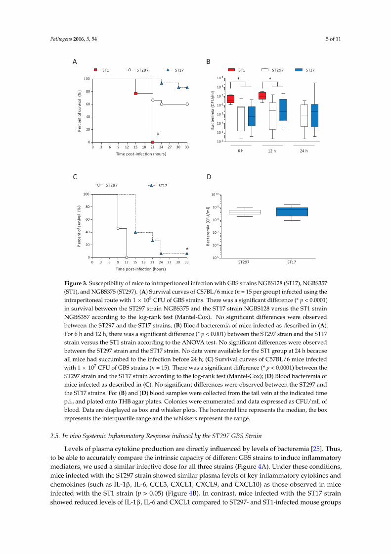
Acquisition or exchange of large areas of genome content can greatly impact several traits of bacterial pathogenic organisms, including virulence. To begin to investigate how genetic exchange affects the virulence of the ST297 isolate, we performed experimental infections of adult mice. As comparators, we used one ST1 strain (NGBS357) and one ST17 strain (NGBS128). All mice infected with the ST1 strain died by 21 h post-infection (p.i.) (Figure 3A). On the other hand, 60% of mice survived the infection in the ST297 group (p < 0.0001 vs. ST1-infected mice). At a dose of 105 CFU, the survival curve of ST297-infected mice was similar to the one observed in mice infected with the ST17 strain (p > 0.05). In agreement with these results, bacteremia levels at 6 and 12 h p.i. were similar in these two groups, but significantly lower than those observed in animals infected with the highly virulent ST1 strain (Figure 3B). To better dissect possible differences between the ST297 and the ST17 strains, mouse mortality was monitored after infection with a higher bacterial dose (107 CFU). Under these conditions, mice infected with the ST297 strain showed significantly greater mortality levels than animals infected with the ST17 strain (p < 0.0001; Figure 3C), with 100% mortality at 12 h p.i. Yet, at 6 h p.i., bacteremia levels were similar between the two groups (Figure 3D). Taken together, these data indicate that the mosaic ST297 strain has an intermediate virulence between ST1 and ST17 strains, but induces bacteremia levels comparable to the ST17 strain.
107
99
12820
29 153
1803NGBS128 NGBS357
NGBS375
Figure 2. Orthologous gene clusters identified in the genome of strains NGBS357 (ST1), NGBS128(ST17), and NGBS375 (ST297). PGAP was used to identify orthologous gene clusters between strains.Numbers of shared gene clusters are shown in overlapping areas.
2.4. Altered Virulence of the Mosaic GBS Strain in An Adult Mouse Model of Systemic Infection
Acquisition or exchange of large areas of genome content can greatly impact several traits ofbacterial pathogenic organisms, including virulence. To begin to investigate how genetic exchangeaffects the virulence of the ST297 isolate, we performed experimental infections of adult mice.As comparators, we used one ST1 strain (NGBS357) and one ST17 strain (NGBS128). All mice infectedwith the ST1 strain died by 21 h post-infection (p.i.) (Figure 3A). On the other hand, 60% of micesurvived the infection in the ST297 group (p < 0.0001 vs. ST1-infected mice). At a dose of 105 CFU,the survival curve of ST297-infected mice was similar to the one observed in mice infected with theST17 strain (p > 0.05). In agreement with these results, bacteremia levels at 6 and 12 h p.i. were similarin these two groups, but significantly lower than those observed in animals infected with the highlyvirulent ST1 strain (Figure 3B). To better dissect possible differences between the ST297 and the ST17strains, mouse mortality was monitored after infection with a higher bacterial dose (107 CFU). Underthese conditions, mice infected with the ST297 strain showed significantly greater mortality levelsthan animals infected with the ST17 strain (p < 0.0001; Figure 3C), with 100% mortality at 12 h p.i. Yet,at 6 h p.i., bacteremia levels were similar between the two groups (Figure 3D). Taken together, thesedata indicate that the mosaic ST297 strain has an intermediate virulence between ST1 and ST17 strains,but induces bacteremia levels comparable to the ST17 strain.
Pathogens 2016, 5, 54 5 of 11Pathogens 2016, 5, 54 5 of 11
Figure 3. Susceptibility of mice to intraperitoneal infection with GBS strains NGBS128 (ST17), NGBS357 (ST1), and NGBS375 (ST297). (A) Survival curves of C57BL/6 mice (n = 15 per group) infected using the intraperitoneal route with 1 × 10⁵ CFU of GBS strains. There was a significant difference (* p < 0.0001) in survival between the ST297 strain NGBS375 and the ST17 strain NGBS128 versus the ST1 strain NGBS357 according to the log-rank test (Mantel-Cox). No significant differences were observed between the ST297 and the ST17 strains; (B) Blood bacteremia of mice infected as described in (A). For 6 h and 12 h, there was a significant difference (* p < 0.001) between the ST297 strain and the ST17 strain versus the ST1 strain according to the ANOVA test. No significant differences were observed between the ST297 strain and the ST17 strain. No data were available for the ST1 group at 24 h because all mice had succumbed to the infection before 24 h; (C) Survival curves of C57BL/6 mice infected with 1 × 10⁷ CFU of GBS strains (n = 15). There was a significant difference (* p < 0.0001) between the ST297 strain and the ST17 strain according to the log-rank test (Mantel-Cox); (D) Blood bacteremia of mice infected as described in (C). No significant differences were observed between the ST297 and the ST17 strains. For (B) and (D) blood samples were collected from the tail vein at the indicated time p.i., and plated onto THB agar plates. Colonies were enumerated and data expressed as CFU/mL of blood. Data are displayed as box and whisker plots. The horizontal line represents the median, the box represents the interquartile range and the whiskers represent the range.
2.5. In vivo Systemic Inflammatory Response induced by the ST297 GBS Strain
Levels of plasma cytokine production are directly influenced by levels of bacteremia [25]. Thus, to be able to accurately compare the intrinsic capacity of different GBS strains to induce inflammatory mediators, we used a similar infective dose for all three strains (Figure 4A). Under these conditions, mice infected with the ST297 strain showed similar plasma levels of key inflammatory cytokines and chemokines (such as IL-1β, IL-6, CCL3, CXCL1, CXCL9, and CXCL10) as those observed in mice
A
0 3 6 9 12 15 18 21 24 27 30 330
20
40
60
80
100
ST1 ST297 ST17
Time post-infec�on (hours)
)%( lavivrus fo tnecre
P
6 h 12 h 24 h
B
0 3 6 9 12 15 18 21 24 27 30 330
20
40
60
80
100
ST297 ST17
Time post-infec�on (hours)
)%( lavivrus fo tnecre
P
C D
10 2
10 3
10 4
10 5
10 6
10 7
10 8
10 9
ST1 ST297 ST17
)lm/
UFC( ai
m er etcaB
ST297 ST1710 5
10 6
10 7
10 8
10 9
10 10
Bact
erem
ia (C
FU/m
l)
* *
*
*
Figure 3. Susceptibility of mice to intraperitoneal infection with GBS strains NGBS128 (ST17), NGBS357(ST1), and NGBS375 (ST297). (A) Survival curves of C57BL/6 mice (n = 15 per group) infected using theintraperitoneal route with 1 ˆ 105 CFU of GBS strains. There was a significant difference (* p < 0.0001)in survival between the ST297 strain NGBS375 and the ST17 strain NGBS128 versus the ST1 strainNGBS357 according to the log-rank test (Mantel-Cox). No significant differences were observedbetween the ST297 and the ST17 strains; (B) Blood bacteremia of mice infected as described in (A).For 6 h and 12 h, there was a significant difference (* p < 0.001) between the ST297 strain and the ST17strain versus the ST1 strain according to the ANOVA test. No significant differences were observedbetween the ST297 strain and the ST17 strain. No data were available for the ST1 group at 24 h becauseall mice had succumbed to the infection before 24 h; (C) Survival curves of C57BL/6 mice infectedwith 1 ˆ 107 CFU of GBS strains (n = 15). There was a significant difference (* p < 0.0001) between theST297 strain and the ST17 strain according to the log-rank test (Mantel-Cox); (D) Blood bacteremia ofmice infected as described in (C). No significant differences were observed between the ST297 andthe ST17 strains. For (B) and (D) blood samples were collected from the tail vein at the indicated timep.i., and plated onto THB agar plates. Colonies were enumerated and data expressed as CFU/mL ofblood. Data are displayed as box and whisker plots. The horizontal line represents the median, the boxrepresents the interquartile range and the whiskers represent the range.
2.5. In vivo Systemic Inflammatory Response induced by the ST297 GBS Strain
Levels of plasma cytokine production are directly influenced by levels of bacteremia [25]. Thus,to be able to accurately compare the intrinsic capacity of different GBS strains to induce inflammatorymediators, we used a similar infective dose for all three strains (Figure 4A). Under these conditions,mice infected with the ST297 strain showed similar plasma levels of key inflammatory cytokines andchemokines (such as IL-1β, IL-6, CCL3, CXCL1, CXCL9, and CXCL10) as those observed in miceinfected with the ST1 strain (p > 0.05) (Figure 4B). In contrast, mice infected with the ST17 strainshowed reduced levels of IL-1β, IL-6 and CXCL1 compared to ST297- and ST1-infected mouse groups

Pathogens 2016, 5, 54 6 of 11
(p < 0.05). Interestingly, infection with the ST17 strain resulted in higher plasma levels of the chemokineCCL3 than those observed in the other groups.
Pathogens 2016, 5, 54 6 of 11
(p<0.05). Interestingly, infection with the ST17 strain resulted in higher plasma levels of the chemokine CCL3 than those observed in the other groups.
Figure 4. Plasma levels of pro-inflammatory cytokines in mice infected with GBS strains NGBS128 (ST17), NGBS357 (ST1), and NGBS375 (ST297). C57BL/6 mice were infected using the intraperitoneal route with 1 × 10⁷ CFU of GBS strains, and euthanized at 6 h p.i.. Mock-infected mice (vehicle solution only) were used as non-infected controls. (A) Bacteremia in mice infected as previously described. Blood samples were collected by cardiac puncture, and plated onto THB agar plates. Colonies were enumerated and data expressed as CFU/mL of blood. Data are displayed as box and whisker plots. The horizontal line represents the median, the box represents the interquartile range and the whiskers represent the range. No significant differences were observed between the strains; (B) Plasma was
.
A
IL -1β
Prod
uc�o
n (ng
/ml)
Prod
uc�o
n (ng
/ml)
IL -6
CCL3 CXCL1
CXCL10CXCL9
B
Prod
uc�o
n (ng
/ml)
Prod
uc�o
n (ng
/ml)
Prod
uc�o
n ( ng
/ml)
Prod
uc�o
n (ng
/ml)
ST1 ST297 ST1710 5
10 6
10 7
10 8
10 9
10 10
)lm/
UFC( ai
me re tcaB
Mock ST1 ST297 ST170
1
2
3
4
Mock ST1 ST297 ST170
200
400
600
800
1000
1200
Mock ST1 ST297 ST170
5
10
15
20
25
Mock ST1 ST297 ST170
200
400
600
800
Mock ST1 ST297 ST170
2
4
6
Mock ST1 ST297 ST170
2
4
6
8
10
* *
**
Figure 4. Plasma levels of pro-inflammatory cytokines in mice infected with GBS strains NGBS128(ST17), NGBS357 (ST1), and NGBS375 (ST297). C57BL/6 mice were infected using the intraperitonealroute with 1 ˆ 107 CFU of GBS strains, and euthanized at 6 h p.i.. Mock-infected mice (vehiclesolution only) were used as non-infected controls. (A) Bacteremia in mice infected as previouslydescribed. Blood samples were collected by cardiac puncture, and plated onto THB agar plates.Colonies were enumerated and data expressed as CFU/mL of blood. Data are displayed as box andwhisker plots. The horizontal line represents the median, the box represents the interquartile rangeand the whiskers represent the range. No significant differences were observed between the strains;(B) Plasma was collected and production of IL-1β, IL-6, CCL3, CXCL1, CXCL9 and CXCL10 wasmeasured by ELISA. Data are displayed as box and whisker plots from two independent experiments(total n = 20; 10 mice per group, per experiment). The horizontal line represents the median, the boxrepresents the interquartile range and the whiskers represent the range. * p < 0.05 indicates statisticallysignificant differences between the ST1 strain, and the ST297 strain, versus the ST17 strain according toANOVA test. No significant differences were observed between the ST1 strain and the ST297 strain.

Pathogens 2016, 5, 54 7 of 11
3. Discussion
Here, we confirm that strain NGBS375, a previously described serotype V ST297 strain isolatedfrom a case of adult invasive GBS infection, has a bona fide CC1 genome background, but has acquireda large genetic region from a presumed CC17 donor by means of recombination. The genomesequence of this strain is now available (NCBI accession number CP102503). Among other importantexchanged virulence factors, the mosaic strain acquired the gene encoding the virulence factor HvgA.This surface-exposed protein mediates bacterial adhesion, and has been associated with bacterialdissemination across intestinal and blood-brain barriers, leading to meningitis in neonates [16]. Whileour data does not permit to predict how the virulence of the ST297 strain would compare to that ofST1 or ST17 strains in a neonatal model of infection, interestingly, acquisition hvgA did not increasethe virulence of the ST297 mosaic strain in a model of adult GBS disease. Indeed, this strain showedhigher virulence in intraperitoneal-infection model than an hvgA-positive ST17 strain, but it was lessvirulent than an ST1 hvgA-negative strain. The mosaic strain also showed lower bacteremia than theST1 strain at 6 h and 12 h p.i., indicating that hvgA does not contribute to bacterial disseminationwhen accompanying an ST1 genomic background and/or in the context of the adult mouse modelused. The latter hypothesis seems more likely, as an hvgA-positive ST17 strain (NGBS128) had thelowest virulence profile. This suggests that HvgA is not essential for GBS virulence in adults. Otherfactors present in the recombined area (such as cspA, cfb, rgfCA, dltABCD) might be more relevant forvirulence in an adult model of invasive GBS disease and/or during the septic shock phase of GBSdisease. One limitation of our study is that we used only one serotype V ST297 strain. Assessmentof the virulence of more recombinant isolates when they become available would be important toconfirm this speculation. In addition, because strains tested here are non-isogenic, ascertaining thecontribution to virulence of each of the ST17-derived factors is challenging, due to the significantbackground effects of the large area of recombination.
It is well recognized that the exacerbated inflammatory response induced early during thesystemic phase of GBS infection is mostly responsible for the clinical outcome, including suddendeath [26,27]. Yet, under normalized bacteremia levels, the ST297 and the highly virulent ST1 strainshad similar intrinsic capacity to induce an inflammatory cascade of cytokines and chemokines. Thus,the diminished pathogenic capacity of the ST297 strain may be related to a reduced bacteremia whencompared to ST1-infected mice. In contrast, the lower pathogenicity of the ST17 strain appears to be,at least in part, related to a reduced capacity to induce expression of key mediators of inflammationand septic shock, namely IL-1β, IL-6 and CXCL1. In a GBS-infection model, IL-1β deficiency has beenassociated with selective impairment in the production of the neutrophil chemokine CXCL1 [28]. Thisis an agreement with our data showing decreased IL-1β production concomitant to reduced CXCL1release during infection with the ST17 strain. Increased systemic and local levels of IL-1 β and IL-6,together with tumor necrosis factor alpha, are known to correlate with a more severe development ofsepsis in mice [29]. Furthermore, very high cytokine concentrations, such as IL-1β and IL-8, are foundin septic newborn infants [26]. Thus, reduced levels of these inflammatory mediators might improveor delay the clinical outcome of ST17-infected animals compared to ST1- or ST297-infected ones.Interestingly, and in contrast to above-mentioned pro-inflammatory mediators, the CCL3 chemokinewas highly upregulated in ST17-infected mice. In a number of model systems, CCL3 has been shownto play an important role in the recruitment of mononuclear cells [30,31]. In a Klebsiella pneumoniaeinfection model, CCL3 was shown to promote bacterial phagocytosis and killing, thus contributing topathogen clearance [32]. The clinical course of disease, number of circulating bacteria, and systemicand local inflammation are all interconnected, and under complex regulation mechanisms that seem todiffer between the different GBS STs [26,33].
Recombination in GBS has long been known to play important roles in the emergence of virulentstrains causing disease in humans. Here, we show that the genome of strain NGBS375 (ST297) is amosaic resulting from a recombination event involving acquisition of approximately 300 Kbp of ST17genetic material in an ST1 genomic background, leading to reduced virulence in a murine model of

Pathogens 2016, 5, 54 8 of 11
adult disease. A better understanding of the dynamic expression of virulence traits in the differentGBS genomic backgrounds, and of the cellular immunology in the context of GBS adult infection,is required to elucidate adult GBS disease pathogenesis.
4. Materials and Methods
4.1. Bacterial Strains and Growth Conditions
GBS strains NGBS375 (serotype V, ST297), NGBS357 (serotype V, ST1), and NGBS128 (serotype III,ST17) were used in this study. The ST297 and ST1 strains were isolated from the blood of unrelatedadult patients in Toronto, Canada, in 2011 [15]. Strain NGBS128 (ST17) was isolated in 2010 in Toronto,Canada, from a case of late onset disease [2,15]. Strains were grown in Todd Hewitt broth (THB) oragar (THA) (Becton Dickinson, Mississauga, ON, Canada) or on sheep blood agar plates at 37 ˝C for18 h. Inocula for in vivo infections were prepared as described elsewhere [34]. Briefly, GBS colonieswere inoculated in THB and incubated at 37 ˝C with shaking for 8 h, then 10 µL of a 1/1000 dilutionof the 8 h-cultures were inoculated into 30 mL of THB followed by incubation with shaking for 12 hat 37 ˝C. Bacteria were then washed and resuspended in 20 mL of THB to obtain an OD600 nm of 0.5,which corresponds to approximately 2 ˆ 108 CFU/mL. Final suspensions were enumerated by platingappropriate dilutions onto THA.
4.2. Genome Sequencing and Analysis
The genomes of strains NGBS375 (ST297) and NGBS357 (ST1) were sequenced using PacBiosequencing (Pacific Biosciences, Menlo Park, CA, USA) as previously described [35]. Two cells ofsequence were generated for each isolate. To assess base-calling accuracy in the PacBio assembly,Illumina short-reads for the two strains were generated. Genomic libraries were prepared usingNextera XT kits (Illumina, San Diego, CA, USA) and sequenced as paired-end reads in an IlluminaHiSeq 2500 or MiSeq instrument (Illumina). Both genome assemblies were completely concordant withfull length perfectly aligning Illumina short-reads. The genome of NGBS375 (ST297) was circularized.Circularization of NGBS357 could not be achieved due to the presence of a repetitive region of >80 kb inits genome. However, a single contig was obtained (Supplementary Figure S1). Genome assemblies areavailable in GenBank under Accession numbers CP012504 (NGBS357, ST1), and CP102503 (NGBS375,ST297). Supplementary Table S1 presents genome closure statistics. The genome sequences of strainsNGBS128 (ST17), NGBS572 (ST452), NEM316 (ST23), A909 (ST7), NGBS061 (ST459), 2603VR (ST110)were retrieved from GenBank (Accession numbers CP012480, CP007632, AL732656, NC_007432,CP007631, and NC_004116, respectively). Short-read Illumina data for strain NGBS128 were retrievedfrom NCBI sequence-read archive (SRA, accession number SAMN04007140). Identification oftRNA-encoding regions and annotation were performed with Prokka [36]. Ortholog analysis wasperformed using PGAP [37]. Polymorphisms relative to strain NGBS375 were identified usingVAAL [38] for strains for which Illumina data was available, otherwise with Mauve [39]. Recombinationanalysis was performed using BRATNextGen [18] run with 20 iterations and 100 replicates, with ap-value of 0.05 as the significance cut-off. Genome visualizations were created using BRIG [40]. Shortreads have been made available at SRA under Study Accession PRJNA295774 and PRJNA274384 forthe ST17 and ST1 strains, respectively.
4.3. Mouse Experimental Infections
All animal work was approved by the Animal Welfare Committee of the University of Montreal.Female, 5-6-week-old, C57BL/6 mice (Charles River Laboratories, Wilmington, MA, USA) wereacclimatized to a 12-h-light/12-h-dark cycle with free access to food and water. On the day of theexperiment, animals were divided into 4 groups. Group 1 (n = 15 mice) received 0.5 mL of an NGBS375(ST297) suspension (105 CFU/mL) by intraperitoneal injection. Groups 2 and 3 (n = 15 each) receivedthe same dose of strains NGBS357 (ST1) and NGBS128 (ST17), respectively. Group 4 (n = 7) received

Pathogens 2016, 5, 54 9 of 11
0.5 mL of sterile THB. Three independent preliminary trials were performed to establish the optimalbacterial doses and time points (data not shown). Mice were monitored daily to record clinical signssuch as depression, rough appearance of hair coat, and swollen eyes, and/or mortality. Numbers ofviable bacteria in blood were quantified at 6 h, 12 h and 24 h p.i. Briefly, blood samples (8 µL) werecollected from the tail vein, serially diluted in PBS and plated onto THA plates as described above,and colonies enumerated. A second experiment was carried out to contrast the virulence of strainsNGBS128 (ST1, n = 15 mice) and NGBS375 (ST297, n = 15 mice) at a higher infectious dose (107 CFU).Experiments were carried out as described above. To measure plasma cytokine levels, mice wereinfected (intraperitoneal route) with 107 CFU of the different GBS strains (n = 10 for each strain in twoindependent experiments). Mock-infected mice were used as negative controls. Mice were euthanizedat 6 h p.i. and blood collected by cardiac puncture. Bacteremia levels were quantified as describedabove and plasma conserved at ´80 ˝C for cytokine analyses.
4.4. Cytokine Quantification by ELISA
Levels of IL-1β, IL-6, CCL3 (MIP-1α), CXCL1 (KC), CXCL9 (MIG) and CXCL10 (IP-10) in theplasma of infected mice were measured by sandwich ELISA using pair-matched antibodies fromR&D Systems (Minneapolis, MN, USA), according to the manufacturer’s recommendations. Twofolddilutions of recombinant mouse cytokines were used to generate standard curves. Sample dilutionsgiving OD readings in the linear portion of the appropriate standard curve were used to quantify thelevels of each cytokine.
4.5. Statistical Analyses
Data from experimental infections are presented as Kaplan-Meier survival curves, mean ˘ SEMor geometric mean with 95% confidence interval where appropriate. Prism v.5 (Graphpad, San Diego,CA, USA) was used for data analysis. Log-rank (Mantel-Cox) tests were used to compare the survivalcurves. For bacteremia and plasma cytokine levels, an ANOVA test was performed. p < 0.05 wasconsidered as the cut-off for statistical significance.
Supplementary Materials: The following files are available online at www.mdpi.com/2076-0817/5/3/54/s1,Figure S1: Genome atlas of the complete genome of NGBS357 (ST1), Table S1: Whole-genome sequencing statisticsfor genome closure, Table S2: Gene content in recombined region of NGBS375 (ST297), Table S3: Unique geneclusters found in NGBS375 (ST297), Table S4: Unique genes in NGBS128 (ST17), Table S5: Unique genes inNGBS357 (ST1).
Acknowledgments: We thank the staff at Public Health Ontario Genome Core facility for Illumina sequencing ofour samples. This work was funded by Public Health Ontario.
Author Contributions: S.T., P.L., M.S. and N.F. designed research. S.T., P.L., K.D., J.W., C.C. and A.L. performedresearch. S.T., G.M., T.B.T.A. and N.F. analyzed data. S.T., P.L. and N.F. wrote the manuscript. All authors reviewedand approved the manuscript.
Conflicts of Interest: The authors declare no conflict of interest.
References
1. Farley, M.M. Group b streptococcal disease in nonpregnant adults. Clin. Infect. Dis. 2001, 33, 556–561.[CrossRef] [PubMed]
2. Berti, F.; Campisi, E.; Toniolo, C.; Morelli, L.; Crotti, S.; Rosini, R.; Romano, M.R.; Pinto, V.; Brogioni, B.;Torricelli, G.; et al. Structure of the type IX group B Streptococcus capsular polysaccharide and its evolutionaryrelationship with types V and VII. J. Biol. Chem. 2014, 289, 23437–23448. [CrossRef] [PubMed]
3. Cieslewicz, M.J.; Chaffin, D.; Glusman, G.; Kasper, D.; Madan, A.; Rodrigues, S.; Fahey, J.; Wessels, M.R.;Rubens, C.E. Structural and genetic diversity of group B Streptococcus capsular polysaccharides. Infect. Immun.2005, 73, 3096–3103. [CrossRef] [PubMed]
4. Lamagni, T.L.; Keshishian, C.; Efstratiou, A.; Guy, R.; Henderson, K.L.; Broughton, K.; Sheridan, E. Emergingtrends in the epidemiology of invasive group B streptococcal disease in England and Wales, 1991–2010.Clin. Infect. Dis. 2013, 57, 682–688. [CrossRef] [PubMed]

Pathogens 2016, 5, 54 10 of 11
5. Joubrel, C.; Tazi, A.; Six, A.; Dmytruk, N.; Touak, G.; Bidet, P.; Raymond, J.; Trieu Cuot, P.; Fouet, A.;Kerneis, S.; et al. Group B Streptococcus neonatal invasive infections, France 2007–2012. Clin. Microbiol. Infect.2015, 21, 910–916. [CrossRef] [PubMed]
6. Phares, C.R.; Lynfield, R.; Farley, M.M.; Mohle-Boetani, J.; Harrison, L.H.; Petit, S.; Craig, A.S.; Schaffner, W.;Zansky, S.M.; Gershman, K.; et al. Epidemiology of invasive group b streptococcal disease in the UnitedStates, 1999–2005. JAMA J. Am. Med. Assoc. 2008, 299, 2056–2065. [CrossRef] [PubMed]
7. Jones, N.; Bohnsack, J.F.; Takahashi, S.; Oliver, K.A.; Chan, M.S.; Kunst, F.; Glaser, P.; Rusniok, C.; Crook, D.W.;Harding, R.M.; et al. Multilocus sequence typing system for group B Streptococcus. J. Clin. Microbiol. 2003, 41,2530–2536. [CrossRef] [PubMed]
8. Honsa, E.; Fricke, T.; Stephens, A.J.; Ko, D.; Kong, F.; Gilbert, G.L.; Huygens, F.; Giffard, P.M. Assignment ofStreptococcus agalactiae isolates to clonal complexes using a small set of single nucleotide polymorphisms.BMC Microbiol. 2008, 8, 140. [CrossRef] [PubMed]
9. Da Cunha, V.; Davies, M.R.; Douarre, P.E.; Rosinski-Chupin, I.; Margarit, I.; Spinali, S.; Perkins, T.; Lechat, P.;Dmytruk, N.; Sauvage, E.; et al. Streptococcus agalactiae clones infecting humans were selected and fixedthrough the extensive use of tetracycline. Nat. Commun. 2014, 5, 4544. [CrossRef] [PubMed]
10. Luan, S.L.; Granlund, M.; Sellin, M.; Lagergard, T.; Spratt, B.G.; Norgren, M. Multilocus sequence typing ofSwedish invasive group B Streptococcus isolates indicates a neonatally associated genetic lineage and capsuleswitching. J. Clin. Microbiol. 2005, 43, 3727–3733. [CrossRef] [PubMed]
11. Lefebure, T.; Stanhope, M.J. Evolution of the core and pan-genome of Streptococcus: Positive selection,recombination, and genome composition. Genome Biol. 2007, 8, R71. [CrossRef] [PubMed]
12. Brochet, M.; Rusniok, C.; Couve, E.; Dramsi, S.; Poyart, C.; Trieu-Cuot, P.; Kunst, F.; Glaser, P. Shaping abacterial genome by large chromosomal replacements, the evolutionary history of Streptococcus agalactiae.Proc. Natl. Acad. Sci. USA 2008, 105, 15961–15966. [CrossRef] [PubMed]
13. Sorensen, U.B.; Poulsen, K.; Ghezzo, C.; Margarit, I.; Kilian, M. Emergence and global dissemination ofhost-specific Streptococcus agalactiae clones. mBio 2010, 1, e00178-10. [CrossRef] [PubMed]
14. Bellais, S.; Six, A.; Fouet, A.; Longo, M.; Dmytruk, N.; Glaser, P.; Trieu-Cuot, P.; Poyart, C. Capsularswitching in group B Streptococcus CC17 hypervirulent clone: A future challenge for polysaccharide vaccinedevelopment. J. Infect. Dis. 2012, 206, 1745–1752. [CrossRef] [PubMed]
15. Teatero, S.; McGeer, A.; Low, D.E.; Li, A.; Demczuk, W.; Martin, I.; Fittipaldi, N. Characterization of invasivegroup B Streptococcus strains from the greater Toronto area, Canada. J. Clin. Microbiol. 2014, 52, 1441–1447.[CrossRef] [PubMed]
16. Tazi, A.; Disson, O.; Bellais, S.; Bouaboud, A.; Dmytruk, N.; Dramsi, S.; Mistou, M.Y.; Khun, H.; Mechler, C.;Tardieux, I.; et al. The surface protein HvgA mediates group B Streptococcus hypervirulence and meningealtropism in neonates. J. Exp. Med. 2010, 207, 2313–2322. [CrossRef] [PubMed]
17. Flores, A.R.; Galloway-Pena, J.; Sahasrabhojane, P.; Saldana, M.; Yao, H.; Su, X.; Ajami, N.J.; Holder, M.E.;Petrosino, J.F.; Thompson, E.; et al. Sequence type 1 group B Streptococcus, an emerging cause of invasivedisease in adults, evolves by small genetic changes. Proc. Natl. Acad. Sci. USA 2015, 112, 6431–6436.[CrossRef] [PubMed]
18. Marttinen, P.; Hanage, W.P.; Croucher, N.J.; Connor, T.R.; Harris, S.R.; Bentley, S.D.; Corander, J. Detection ofrecombination events in bacterial genomes from large population samples. Nucleic Acids Res. 2012, 40, e6.[CrossRef] [PubMed]
19. Seifert, K.N.; McArthur, W.P.; Bleiweis, A.S.; Brady, L.J. Characterization of group B streptococcalglyceraldehyde-3-phosphate dehydrogenase: Surface localization, enzymatic activity, and protein-proteininteractions. Can. J. Microbiol. 2003, 49, 350–356. [CrossRef] [PubMed]
20. Madureira, P.; Baptista, M.; Vieira, M.; Magalhaes, V.; Camelo, A.; Oliveira, L.; Ribeiro, A.;Tavares, D.; Trieu-Cuot, P.; Vilanova, M.; et al. Streptococcus agalactiae GAPDH is a virulence-associatedimmunomodulatory protein. J. Immunol. 2007, 178, 1379–1387. [CrossRef] [PubMed]
21. Bryan, J.D.; Shelver, D.W. Streptococcus agalactiae CspA is a serine protease that inactivates chemokines.J. Bacteriol. 2009, 191, 1847–1854. [CrossRef] [PubMed]
22. Al Safadi, R.; Mereghetti, L.; Salloum, M.; Lartigue, M.F.; Virlogeux-Payant, I.; Quentin, R.; Rosenau, A.Two-component system rgfA/C activates the fbsb gene encoding major fibrinogen-binding protein in highlyvirulent CC17 clone group B Streptococcus. PLoS ONE 2011, 6, e14658. [CrossRef] [PubMed]
Pathogens 2016, 5, 54 11 of 11
23. Teatero, S.; Ramoutar, E.; McGeer, A.; Li, A.; Melano, R.G.; Wasserscheid, J.; Dewar, K.; Fittipaldi, N. Clonalcomplex 17 group B Streptococcus strains causing invasive disease in neonates and adults originate from thesame genetic pool. Sci. Rep. 2016, 6, 20047. [CrossRef] [PubMed]
24. Puymege, A.; Bertin, S.; Guedon, G.; Payot, S. Analysis of Streptococcus agalactiae pan-genome for prevalence,diversity and functionality of integrative and conjugative or mobilizable elements integrated in thetrna(lys ctt) gene. Mol. Genet. Genom. MGG 2015, 290, 1727–1740. [CrossRef] [PubMed]
25. Puliti, M.; Uematsu, S.; Akira, S.; Bistoni, F.; Tissi, L. Toll-like receptor 2 deficiency is associated withenhanced severity of group B streptococcal disease. Infect. Immun. 2009, 77, 1524–1531. [CrossRef] [PubMed]
26. Wennekamp, J.; Henneke, P. Induction and termination of inflammatory signaling in group B streptococcalsepsis. Immunol. Rev. 2008, 225, 114–127. [CrossRef] [PubMed]
27. Landwehr-Kenzel, S.; Henneke, P. Interaction of Streptococcus agalactiae and cellular innate immunity incolonization and disease. Front. Immunol. 2014, 5, 519. [CrossRef] [PubMed]
28. Biondo, C.; Mancuso, G.; Midiri, A.; Signorino, G.; Domina, M.; Lanza Cariccio, V.; Venza, M.; Venza, I.;Teti, G.; Beninati, C. Essential role of interleukin-1 signaling in host defenses against group B Streptococcus.mBio 2014, 5, e01428-14. [CrossRef] [PubMed]
29. Puliti, M.; Bistoni, F.; Orefici, G.; Tissi, L. Exacerbation of group B streptococcal sepsis and arthritis in diabeticmice. Microbes Infect. 2006, 8, 2376–2383. [CrossRef] [PubMed]
30. Fahey, T.J., 3rd; Tracey, K.J.; Tekamp-Olson, P.; Cousens, L.S.; Jones, W.G.; Shires, G.T.; Cerami, A.; Sherry, B.Macrophage inflammatory protein 1 modulates macrophage function. J. Immunol. 1992, 148, 2764–2769.[PubMed]
31. Didier, P.J.; Paradis, T.J.; Gladue, R.P. The CC chemokine MIP-1alpha induces a selective monocyte infiltrationfollowing intradermal injection into nonhuman primates. Inflammation 1999, 23, 75–86. [CrossRef] [PubMed]
32. Lindell, D.M.; Standiford, T.J.; Mancuso, P.; Leshen, Z.J.; Huffnagle, G.B. Macrophage inflammatory protein1alpha/CCL3 is required for clearance of an acute Klebsiella pneumoniae pulmonary infection. Infect. Immun.2001, 69, 6364–6369. [CrossRef] [PubMed]
33. De Francesco, M.A.; Gargiulo, F.; Negrini, R.; Gelmi, M.; Manca, N. Different sequence strains ofStreptococcus agalactiae elicit various levels of cytokine production. Immunol. Investig. 2008, 37, 741–751.[CrossRef] [PubMed]
34. Lemire, P.; Calzas, C.; Segura, M. The NOD2 receptor does not play a major role in the pathogenesis of groupB Streptococcus in mice. Microb. Pathog. 2013, 65, 41–47. [CrossRef] [PubMed]
35. Teatero, S.; McGeer, A.; Li, A.; Gomes, J.; Seah, C.; Demczuk, W.; Martin, I.; Wasserscheid, J.; Dewar, K.;Melano, R.G.; et al. Population structure and antimicrobial resistance of invasive serotype IV group BStreptococcus, Toronto, Ontario, Canada. Emerg. Infect. Dis. 2015, 21, 585–591. [CrossRef] [PubMed]
36. Seemann, T. Prokka: Rapid prokaryotic genome annotation. Bioinformatics 2014, 30, 2068–2069. [CrossRef][PubMed]
37. Zhao, Y.; Wu, J.; Yang, J.; Sun, S.; Xiao, J.; Yu, J. Pgap: Pan-genomes analysis pipeline. Bioinformatics 2012, 28,416–418. [CrossRef] [PubMed]
38. Nusbaum, C.; Ohsumi, T.K.; Gomez, J.; Aquadro, J.; Victor, T.C.; Warren, R.M.; Hung, D.T.; Birren, B.W.;Lander, E.S.; Jaffe, D.B. Sensitive, specific polymorphism discovery in bacteria using massively parallelsequencing. Nat. Methods 2009, 6, 67–69. [CrossRef] [PubMed]
39. Darling, A.E.; Mau, B.; Perna, N.T. Progressivemauve: Multiple genome alignment with gene gain, loss andrearrangement. PLoS ONE 2010, 5, e11147. [CrossRef] [PubMed]
40. Alikhan, N.F.; Petty, N.K.; Ben Zakour, N.L.; Beatson, S.A. Blast ring image generator (BRIG): Simpleprokaryote genome comparisons. BMC Genom. 2011, 12, 402. [CrossRef] [PubMed]
© 2016 by the authors; licensee MDPI, Basel, Switzerland. This article is an open accessarticle distributed under the terms and conditions of the Creative Commons Attribution(CC-BY) license (http://creativecommons.org/licenses/by/4.0/).