Genetics of CHAPTER 1 Neonatal Jaundice · 1 Genetics of Neonatal Jaundice Jon F. Watchko and Zhili...
28
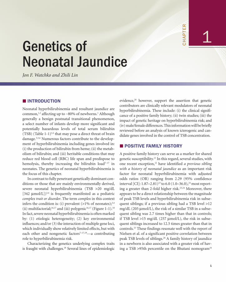
1 Genetics of Neonatal Jaundice Jon F. Watchko and Zhili Lin 1 CHAPTER INTRODUCTION Neonatal hyperbilirubinemia and resultant jaundice are common, 1,2 affecting up to ∼80% of newborns. 1 Although generally a benign postnatal transitional phenomenon, a select number of infants develop more significant and potentially hazardous levels of total serum bilirubin (TSB) (Table 1-1) 3,4 that may pose a direct threat of brain damage. 3,5,6 Numerous factors contribute to the develop- ment of hyperbilirubinemia including genes involved in: (i) the production of bilirubin from heme; (ii) the metab- olism of bilirubin; and (iii) heritable conditions that may reduce red blood cell (RBC) life span and predispose to hemolysis, thereby increasing the bilirubin load 7–17 in neonates. The genetics of neonatal hyperbilirubinemia is the focus of this chapter. In contrast to fully penetrant genetically dominant con- ditions or those that are mainly environmentally derived, severe neonatal hyperbilirubinemia (TSB >20 mg/dL [342 μmol/L]) 3,4 is frequently manifested as a pediatric complex trait or disorder. The term complex in this context infers the condition is: (i) prevalent (>1% of neonates); 3,4 (ii) multifactorial; 16,17 and (iii) polygenic 16,17 (Figure 1-1). 18 In fact, severe neonatal hyperbilirubinemia is often marked by: (1) etiologic heterogeneity; (2) key environmental influences; and/or (3) the interaction of multiple gene loci, which individually show relatively limited effects, but with each other and nongenetic factors 7–17,19 —a contributing role to hyperbilirubinemia risk. Characterizing the genetics underlying complex traits is fraught with challenges. 18 Several lines of epidemiologic evidence, 20 however, support the assertion that genetic contributors are clinically relevant modulators of neonatal hyperbilirubinemia. These include: (i) the clinical signifi- cance of a positive family history; (ii) twin studies; (iii) the impact of genetic heritage on hyperbilirubinemia risk; and (iv) male/female differences. This information will be briefly reviewed before an analysis of known icterogenic and can- didate genes involved in the control of TSB concentration. POSITIVE FAMILY HISTORY A positive family history can serve as a marker for shared genetic susceptibility. 21 In this regard, several studies, with one recent exception, 22 have identified a previous sibling with a history of neonatal jaundice as an important risk factor for neonatal hyperbilirubinemia with adjusted odds ratios (OR) ranging from 2.29 (95% confidence interval [CI]: 1.87–2.81) 23 to 6.0 (1.0–36.0), 24 most report- ing a greater than 2-fold higher risk. 23,24 Moreover, there appears to be a direct relationship between the magnitude of peak TSB levels and hyperbilirubinemia risk in subse- quent siblings; if a previous sibling had a TSB level >12 mg/dL (205 μmol/L), the risk of a similar TSB in a subse- quent sibling was 2.7 times higher than that in controls; if TSB level >15 mg/dL (257 μmol/L), the risk in subse- quent siblings increased to 12.5 times greater than that in controls. 25 These findings resonate well with the report of Nielsen et al. of a significant positive correlation between peak TSB levels of siblings. 26 A family history of jaundice in a newborn is also associated with a greater risk of hav- ing a TSB >95th percentile on the Bhutani nomogram 16
Transcript of Genetics of CHAPTER 1 Neonatal Jaundice · 1 Genetics of Neonatal Jaundice Jon F. Watchko and Zhili...
1
Genetics of Neonatal Jaundice Jon F. Watchko and Zhili Lin
1
CH
AP
TE
R
INTRODUCTION �
Neonatal hyperbilirubinemia and resultant jaundice are common, 1 , 2 affecting up to ∼80% of newborns. 1 Although generally a benign postnatal transitional phenomenon, a select number of infants develop more significant and potentially hazardous levels of total serum bilirubin (TSB) ( Table 1-1 ) 3 , 4 that may pose a direct threat of brain damage. 3 , 5 , 6 Numerous factors contribute to the develop-ment of hyperbilirubinemia including genes involved in: (i) the production of bilirubin from heme; (ii) the metab-olism of bilirubin; and (iii) heritable conditions that may reduce red blood cell (RBC) life span and predispose to hemolysis, thereby increasing the bilirubin load 7 – 17 in neonates. The genetics of neonatal hyperbilirubinemia is the focus of this chapter.
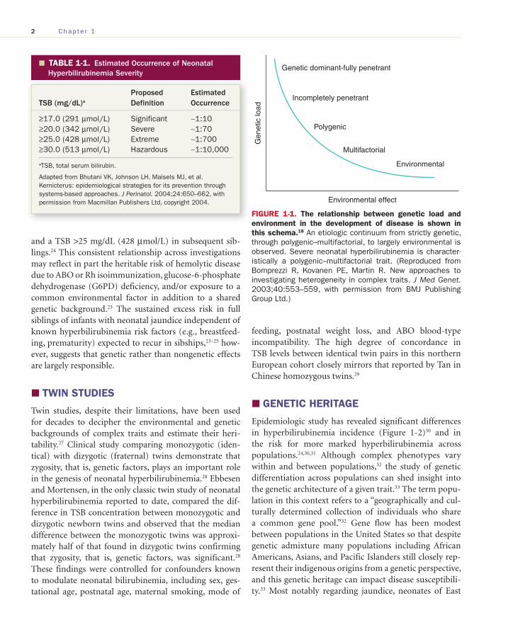
In contrast to fully penetrant genetically dominant con-ditions or those that are mainly environmentally derived, severe neonatal hyperbilirubinemia (TSB >20 mg/dL [342 μmol/L]) 3 , 4 is frequently manifested as a pediatric complex trait or disorder . The term complex in this context infers the condition is: (i) prevalent (>1% of neonates); 3 , 4 (ii) multifactorial; 16 , 17 and (iii) polygenic 16 , 17 ( Figure 1-1 ). 18 In fact, severe neonatal hyperbilirubinemia is often marked by: (1) etiologic heterogeneity; (2) key environmental influences; and/or (3) the interaction of multiple gene loci, which individually show relatively limited effects, but with each other and nongenetic factors 7 – 17 , 19 —a contributing role to hyperbilirubinemia risk.
Characterizing the genetics underlying complex traits is fraught with challenges. 18 Several lines of epidemiologic
evidence, 20 however, support the assertion that genetic contributors are clinically relevant modulators of neonatal hyperbilirubinemia. These include: (i) the clinical signifi-cance of a positive family history; (ii) twin studies; (iii) the impact of genetic heritage on hyperbilirubinemia risk; and (iv) male/female differences. This information will be briefly reviewed before an analysis of known icterogenic and can-didate genes involved in the control of TSB concentration.
POSITIVE FAMILY HISTORY �
A positive family history can serve as a marker for shared genetic susceptibility. 21 In this regard, several studies, with one recent exception, 22 have identified a previous sibling with a history of neonatal jaundice as an important risk factor for neonatal hyperbilirubinemia with adjusted odds ratios (OR) ranging from 2.29 (95% confidence interval [CI]: 1.87–2.81) 23 to 6.0 (1.0–36.0), 24 most report-ing a greater than 2-fold higher risk. 23 , 24 Moreover, there appears to be a direct relationship between the magnitude of peak TSB levels and hyperbilirubinemia risk in subse-quent siblings; if a previous sibling had a TSB level >12 mg/dL (205 μmol/L), the risk of a similar TSB in a subse-quent sibling was 2.7 times higher than that in controls; if TSB level >15 mg/dL (257 μmol/L), the risk in subse-quent siblings increased to 12.5 times greater than that in controls. 25 These findings resonate well with the report of Nielsen et al. of a significant positive correlation between peak TSB levels of siblings. 26 A family history of jaundice in a newborn is also associated with a greater risk of hav-ing a TSB >95th percentile on the Bhutani nomogram 16
Stev_Ch01_001-028.indd 1Stev_Ch01_001-028.indd 1 3/20/12 11:18:03 AM3/20/12 11:18:03 AM
2 Chap t e r 1
and a TSB >25 mg/dL (428 μmol/L) in subsequent sib-lings. 24 This consistent relationship across investigations may reflect in part the heritable risk of hemolytic disease due to ABO or Rh isoimmunization, glucose-6-phosphate dehydrogenase (G6PD) deficiency, and/or exposure to a common environmental factor in addition to a shared genetic background. 23 The sustained excess risk in full siblings of infants with neonatal jaundice independent of known hyperbilirubinemia risk factors (e.g., breastfeed-ing, prematurity) expected to recur in sibships, 23 – 25 how-ever, suggests that genetic rather than nongenetic effects are largely responsible.
TWIN STUDIES �
Twin studies, despite their limitations, have been used for decades to decipher the environmental and genetic backgrounds of complex traits and estimate their heri-tability. 27 Clinical study comparing monozygotic (iden-tical) with dizygotic (fraternal) twins demonstrate that zygosity, that is, genetic factors, plays an important role in the genesis of neonatal hyperbilirubinemia. 28 Ebbesen and Mortensen, in the only classic twin study of neonatal hyperbilirubinemia reported to date, compared the dif-ference in TSB concentration between monozygotic and dizygotic newborn twins and observed that the median difference between the monozygotic twins was approxi-mately half of that found in dizygotic twins confirming that zygosity, that is, genetic factors, was significant. 28
These findings were controlled for confounders known to modulate neonatal bilirubinemia, including sex, ges-tational age, postnatal age, maternal smoking, mode of
feeding, postnatal weight loss, and ABO blood-type incompatibility. The high degree of concordance in TSB levels between identical twin pairs in this northern European cohort closely mirrors that reported by Tan in Chinese homozygous twins. 29
GENETIC HERITAGE �
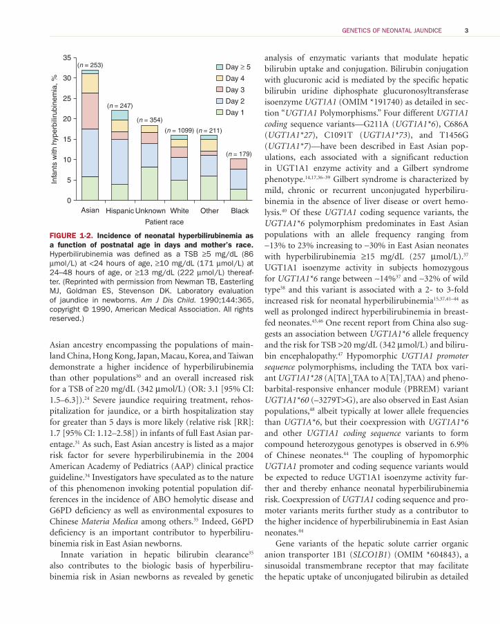
Epidemiologic study has revealed significant differences in hyperbilirubinemia incidence ( Figure 1-2 ) 30 and in the risk for more marked hyperbilirubinemia across populations. 24 , 30 , 31 Although complex phenotypes vary within and between populations, 32 the study of genetic differentiation across populations can shed insight into the genetic architecture of a given trait. 33 The term popu-lation in this context refers to a “geographically and cul-turally determined collection of individuals who share a common gene pool.” 32 Gene flow has been modest between populations in the United States so that despite genetic admixture many populations including African Americans, Asians, and Pacific Islanders still closely rep-resent their indigenous origins from a genetic perspective, and this genetic heritage can impact disease susceptibili-ty. 33 Most notably regarding jaundice, neonates of East
TABLE 1-1. � Estimated Occurrence of Neonatal Hyperbilirubinemia Severity
TSB (mg/dL) a Proposed Definition
Estimated Occurrence
≥17.0 (291 μmol/L) Significant ∼1:10≥20.0 (342 μmol/L) Severe ∼1:70≥25.0 (428 μmol/L) Extreme ∼1:700≥30.0 (513 μmol/L) Hazardous ∼1:10,000
a TSB, total serum bilirubin.
Adapted from Bhutani VK, Johnson LH, Maisels MJ, et al. Kernicterus: epidemiological strategies for its prevention through systems-based approaches. J Perinatol. 2004;24:650–662, with permission from Macmillan Publishers Ltd, copyright 2004.
Genetic dominant-fully penetrant
Incompletely penetrant
Polygenic
Multifactorial
Environmental
Gen
etic
load
Environmental effect
FIGURE 1-1. The relationship between genetic load and environment in the development of disease is shown in this schema. 18 An etiologic continuum from strictly genetic, through polygenic–multifactorial, to largely environmental is observed. Severe neonatal hyperbilirubinemia is character-istically a polygenic–multifactorial trait. (Reproduced from Bomprezzi R, Kovanen PE, Martin R. New approaches to investigating heterogeneity in complex traits. J Med Genet.2003;40:553–559, with permission from BMJ Publishing Group Ltd. )
Stev_Ch01_001-028.indd 2Stev_Ch01_001-028.indd 2 3/20/12 11:18:03 AM3/20/12 11:18:03 AM
GENETICS OF NEONATAL JAUNDICE 3
Asian ancestry encompassing the populations of main-land China, Hong Kong, Japan, Macau, Korea, and Taiwan demonstrate a higher incidence of hyperbilirubinemia than other populations 30 and an overall increased risk for a TSB of ≥20 mg/dL (342 μmol/L) (OR: 3.1 [95% CI: 1.5–6.3]). 24 Severe jaundice requiring treatment, rehos-pitalization for jaundice, or a birth hospitalization stay for greater than 5 days is more likely (relative risk [RR]: 1.7 [95% CI: 1.12–2.58]) in infants of full East Asian par-entage. 31 As such, East Asian ancestry is listed as a major risk factor for severe hyperbilirubinemia in the 2004 American Academy of Pediatrics (AAP) clinical practice guideline. 34 Investigators have speculated as to the nature of this phenomenon invoking potential population dif-ferences in the incidence of ABO hemolytic disease and G6PD deficiency as well as environmental exposures to Chinese Materia Medica among others. 35 Indeed, G6PD deficiency is an important contributor to hyperbiliru-binemia risk in East Asian newborns.
Innate variation in hepatic bilirubin clearance 35 also contributes to the biologic basis of hyperbiliru-binemia risk in Asian newborns as revealed by genetic
analysis of enzymatic variants that modulate hepatic bilirubin uptake and conjugation. Bilirubin conjugation with glucuronic acid is mediated by the specific hepatic bilirubin uridine diphosphate glucuronosyltransferase isoenzyme UGT1A1 (OMIM *191740) as detailed in sec-tion “ UGT1A1 Polymorphisms.” Four different UGT1A1 coding sequence variants—G211A ( UGT1A1*6 ), C686A ( UGT1A1*27 ), C1091T ( UGT1A1*73 ), and T1456G ( UGT1A1*7 )—have been described in East Asian pop-ulations, each associated with a significant reduction in UGT1A1 enzyme activity and a Gilbert syndrome phenotype. 14 , 17 , 36 – 39 Gilbert syndrome is characterized by mild, chronic or recurrent unconjugated hyperbiliru-binemia in the absence of liver disease or overt hemo-lysis. 40 Of these UGT1A1 coding sequence variants, the UGT1A1*6 polymorphism predominates in East Asian populations with an allele frequency ranging from ∼13% to 23% increasing to ∼30% in East Asian neonates with hyperbilirubinemia ≥15 mg/dL (257 μmol/L). 37 UGT1A1 isoenzyme activity in subjects homozygous for UGT1A1*6 range between ∼14% 37 and ∼32% of wild type 38 and this variant is associated with a 2- to 3-fold increased risk for neonatal hyperbilirubinemia 15 , 37 , 41 – 44 as well as prolonged indirect hyperbilirubinemia in breast-fed neonates. 45 , 46 One recent report from China also sug-gests an association between UGT1A1*6 allele frequency and the risk for TSB >20 mg/dL (342 μmol/L) and biliru-bin encephalopathy. 47 Hypomorphic UGT1A1 promoter sequence polymorphisms, including the TATA box vari-ant UGT1A1*28 (A[TA]
6 TAA to A[TA]
7 TAA) and pheno-
barbital-responsive enhancer module (PBREM) variant UGT1A1*60 (–3279T>G), are also observed in East Asian populations, 48 albeit typically at lower allele frequencies than UGT1A*6 , but their coexpression with UGT1A1*6 and other UGT1A1 coding sequence variants to form compound heterozygous genotypes is observed in 6.9% of Chinese neonates. 44 The coupling of hypomorphic UGT1A1 promoter and coding sequence variants would be expected to reduce UGT1A1 isoenzyme activity fur-ther and thereby enhance neonatal hyperbilirubinemia risk. Coexpression of UGT1A1 coding sequence and pro-moter variants merits further study as a contributor to the higher incidence of hyperbilirubinemia in East Asian neonates. 44
Gene variants of the hepatic solute carrier organic anion transporter 1B1 ( SLCO1B1 ) (OMIM *604843), a sinusoidal transmembrane receptor that may facilitate the hepatic uptake of unconjugated bilirubin as detailed
35
30
25
20
15
10
5
0
(n = 247)
(n = 253)
(n = 354)
(n = 1099) (n = 211)
(n = 179)
Asian Hispanic Unknown
Infa
nts
with
hyp
erbi
lirub
inem
ia, %
White Other Black
Day 1
Day 2
Day 3
Day 4
Day ≥ 5
Patient race
FIGURE 1-2. Incidence of neonatal hyperbilirubinemia as a function of postnatal age in days and mother’s race. Hyperbilirubinemia was defined as a TSB ≥5 mg/dL (86 μmol/L) at <24 hours of age, ≥10 mg/dL (171 μmol/L) at 24–48 hours of age, or ≥13 mg/dL (222 μmol/L) thereaf-ter. (Reprinted with permission from Newman TB, Easterling MJ, Goldman ES, Stevenson DK. Laboratory evaluation of jaundice in newborns. Am J Dis Child. 1990;144:365, copyright © 1990, American Medical Association. All rights reserved.)
Stev_Ch01_001-028.indd 3Stev_Ch01_001-028.indd 3 3/20/12 11:18:04 AM3/20/12 11:18:04 AM
4 Chap t e r 1
in section “ SLCO1B1 Polymorphisms,” are also prevalent in East Asian populations. 15 , 49 Nonsynonymous SLCO1B1gene variants that limit the efficacy of hepatic bilirubin uptake could ultimately impair hepatic bilirubin clear-ance and predispose to hyperbilirubinemia. One putative allele, SLCO1B1*1b (A388G), is reported to enhance neo-natal hyperbilirubinemia risk in Taiwanese neonates, 15 an effect not seen however in Thai 50 or Malaysian Chinese newborns. 51 Coupling of icterogenic UGT1A1 and SLCO1B1 variants is reported to enhance neonatal hyper-bilirubinemia risk in Taiwanese newborns, 15 one that is further increased when the infant is also exclusively breastfed. 15
In contrast to infants of East Asian heritage, African American neonates as a group show a lower overall inci-dence of clinically significant hyperbilirubinemia 6 , 22 , 30 , 52 – 57
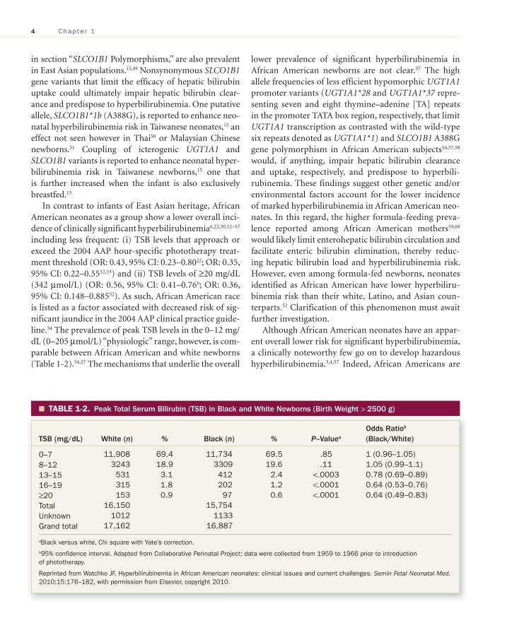
including less frequent: (i) TSB levels that approach or exceed the 2004 AAP hour-specific phototherapy treat-ment threshold (OR: 0.43, 95% CI: 0.23–0.80 22 ; OR: 0.35, 95% CI: 0.22–0.55 52 , 53 ) and (ii) TSB levels of ≥20 mg/dL (342 μmol/L) (OR: 0.56, 95% CI: 0.41–0.76 6 ; OR: 0.36, 95% CI: 0.148–0.885 52 ). As such, African American race is listed as a factor associated with decreased risk of sig-nificant jaundice in the 2004 AAP clinical practice guide-line. 34 The prevalence of peak TSB levels in the 0–12 mg/dL (0–205 μmol/L) “physiologic” range, however, is com-parable between African American and white newborns ( Table 1-2 ). 54 , 57 The mechanisms that underlie the overall
lower prevalence of significant hyperbilirubinemia in African American newborns are not clear. 57 The high allele frequencies of less efficient hypomorphic UGT1A1promoter variants ( UGT1A1*28 and UGT1A1*37 repre-senting seven and eight thymine–adenine [TA] repeats in the promoter TATA box region, respectively, that limit UGT1A1 transcription as contrasted with the wild-type six repeats denoted as UGT1A1*1 ) and SLCO1B1 A388G gene polymorphism in African American subjects 16 , 57 , 58
would, if anything, impair hepatic bilirubin clearance and uptake, respectively, and predispose to hyperbili-rubinemia. These findings suggest other genetic and/or environmental factors account for the lower incidence of marked hyperbilirubinemia in African American neo-nates. In this regard, the higher formula-feeding preva-lence reported among African American mothers 59 , 60
would likely limit enterohepatic bilirubin circulation and facilitate enteric bilirubin elimination, thereby reduc-ing hepatic bilirubin load and hyperbilirubinemia risk. However, even among formula-fed newborns, neonates identified as African American have lower hyperbiliru-binemia risk than their white, Latino, and Asian coun-terparts. 52 Clarification of this phenomenon must await further investigation.
Although African American neonates have an appar-ent overall lower risk for significant hyperbilirubinemia, a clinically noteworthy few go on to develop hazardous hyperbilirubinemia. 3 , 4 , 57 Indeed, African Americans are
TABLE 1-2. � Peak Total Serum Bilirubin (TSB) in Black and White Newborns (Birth Weight > 2500 g)
TSB (mg/dL) White ( n ) % Black ( n ) % P –Value a Odds Ratio b (Black/White)
0–7 11,908 69.4 11,734 69.5 .85 1 (0.96–1.05)8–12 3243 18.9 3309 19.6 .11 1.05 (0.99–1.1)13–15 531 3.1 412 2.4 <.0003 0.78 (0.69–0.89)16–19 315 1.8 202 1.2 <.0001 0.64 (0.53–0.76)≥20 153 0.9 97 0.6 <.0001 0.64 (0.49–0.83)Total 16,150 15,754Unknown 1012 1133Grand total 17,162 16,887
a Black versus white, Chi square with Yate’s correction.
b 95% confidence interval. Adapted from Collaborative Perinatal Project; data were collected from 1959 to 1966 prior to introduction of phototherapy.
Reprinted from Watchko JF. Hyperbilirubinemia in African American neonates: clinical issues and current challenges. Semin Fetal Neonatal Med. 2010;15:176–182, with permission from Elsevier, copyright 2010.
Stev_Ch01_001-028.indd 4Stev_Ch01_001-028.indd 4 3/20/12 11:18:04 AM3/20/12 11:18:04 AM
GENETICS OF NEONATAL JAUNDICE 5
overrepresented in the US Pilot Kernicterus Registry 3 , 4 , 55 accounting for more than 25% of US kernicterus cases, 3 , 4 , 57 and black race is an independent risk factor for bilirubin encephalopathy (OR: 19.0; 95% CI: 2.5–144.7) in the United Kingdom and Ireland as well. 61 G6PD deficiency accounts for ∼60% of African American newborns with kernicterus 3 , 4 , 57 with late-preterm gesta-tion and ABO hemolytic disease being other clinically important clinical contributors to severe hyperbiliru-binemia risk in African American neonates. 57 Clinical study designed to enhance the identification of African American newborns predisposed to develop hazardous hyperbilirubinemia is of particular merit including the potential utility of birth hospitalization point of care G6PD screening. 57 , 62
Regarding genetic heritage, it is important to recog-nize that ethnicity does not properly capture or charac-terize an individual’s genotype or even genetic variation among individuals; more accurate assessment will be obtained by genotyping specific disease- associated alleles.32,33 In the absence of being able to perform geno-typing studies routinely, however, population affiliation will continue to be of clinical value in broadly assessing risk. 17 , 34
MALE SEX �
Several reports demonstrate that male neonates have higher TSB levels than female neonates 2 , 23 , 63 , 64 and are overrepresented in: (i) infant cohorts readmitted to the hospital for management of neonatal jaundice 2 , 64 , 65 (OR: 2.89 [95% CI: 1.46–5.74]) 2 ; (ii) the US Pilot Kernicterus Registry, a database of voluntarily reported cases of ker-nicterus, where there is an ∼2-fold greater predominance of males ( n = 84) than females ( n = 38) 3 ; and (iii) autop-sied cases of kernicterus (male:female ratio 127:90). 66 Others have failed to demonstrate sex as a significant risk factor for hyperbilirubinemia (>95th percentile on Bhutani nomogram). 16 In general, however, the current literature suggests both an increased risk for marked hyperbilirubinemia and an increased susceptibility to bilirubin-induced injury in male neonates. Regarding the former, the prevalence of the Gilbert syndrome is reportedly more than 2-fold higher in males (12.4%) than in females (4.8%). 67 The UGT1A1 gene variants that underlie Gilbert syndrome detailed below would be expected to enhance the risk of neonatal hyperbili-rubinemia, particularly when coexpressed with other
icterogenic conditions, 12 , 15 , 16 including G6PD deficiency which given its X-linked nature is also more prevalent in males. In addition, several clinical studies suggest greater male susceptibility to bilirubin-induced central nervous system (CNS) injury, 68 – 71 a phenomenon also noted in the Gunn rat model of neonatal hyperbilirubinemia and kernicterus. 72 , 73 A potential role for sex hormones in this process remains unexplored but merits study as gonado-tropin surges during late embryonic and early postnatal life impact CNS development. 74 Innate gender-based neu-ronal differences independent of circulating sex steroids may also contribute to this sexually dimorphic vulner-ability to CNS injury. 75
SPECIFIC GENES AND THEIR �VARIANTS THAT MODULATE NEONATAL BILIRUBIN CONCENTRATION
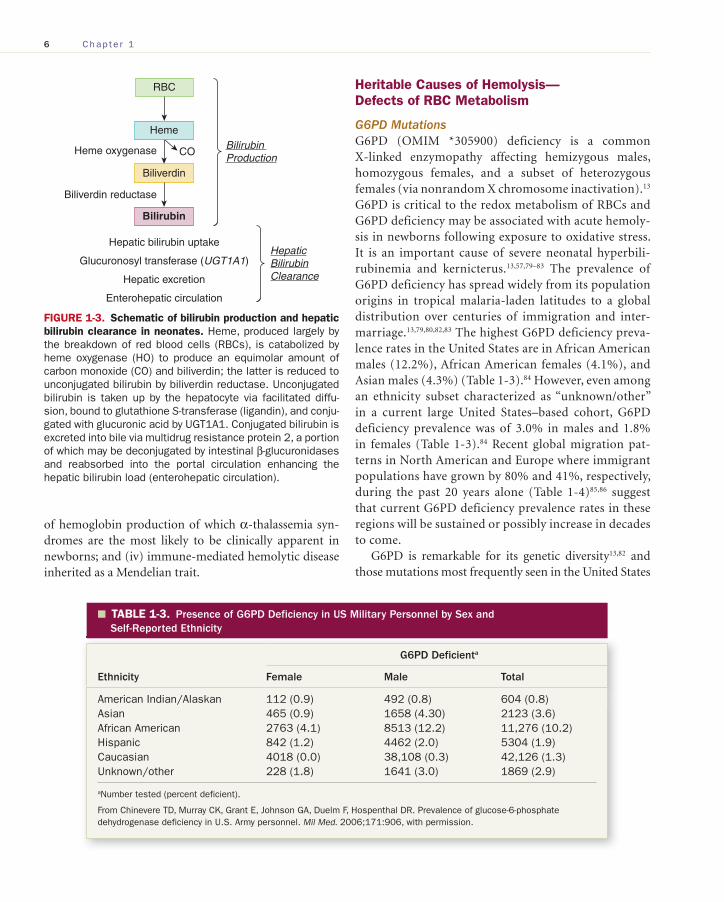
Numerous genes are involved in controlling neonatal bilirubin concentration and can be categorized as those that modulate: (i) heme production (namely, condi-tions that predispose to hemolysis and/or reduce RBC life span); (ii) the catabolism of heme to bilirubin (heme oxygenase [HO]; biliverdin reductase); (iii) hepatic bilirubin uptake (SLCO1B1); (iv) hepatocyte bilirubin binding (glutathione S -transferase [GST; ligandin]); and (v) hepatic bilirubin clearance (UGT1A1). Specific gene mutations and polymorphisms related to each cat-egory are reviewed below in sequence as schematized in Figure 1-3 . Regulatory genes, particularly those of the nuclear receptor superfamily that modulate the expres-sion of genes involved in bilirubin metabolism, will also be detailed. 76
HERITABLE CONDITIONS THAT MAY �CAUSE HEMOLYSIS IN NEONATES
The reduced life span of normal newborn RBCs (70–90 days as opposed to 120 days in the adult) 77 , 78 contributes to enhanced bilirubin production in neonates. Heritable hemolytic disorders accelerate RBC turnover and are major risk factors for severe hyperbilirubinemia. 3 The heritable causes of hemolysis in the newborns are many, but can be broadly grouped into: (i) defects of RBC metabolism, of which G6PD and pyruvate kinase (PK) deficiency are notable causes; (ii) defects of RBC mem-brane structure, of which congenital spherocytosis is an important and underrecognized contributor; (iii) defects
Stev_Ch01_001-028.indd 5Stev_Ch01_001-028.indd 5 3/20/12 11:18:04 AM3/20/12 11:18:04 AM
6 Chap t e r 1
of hemoglobin production of which α-thalassemia syn-dromes are the most likely to be clinically apparent in newborns; and (iv) immune-mediated hemolytic disease inherited as a Mendelian trait.
Heritable Causes of Hemolysis—Defects of RBC Metabolism
G6PD Mutations G6PD (OMIM *305900) deficiency is a common X-linked enzymopathy affecting hemizygous males, homozygous females, and a subset of heterozygous females (via nonrandom X chromosome inactivation). 13
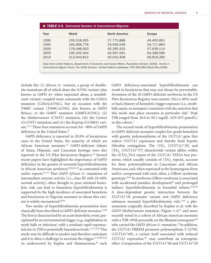
G6PD is critical to the redox metabolism of RBCs and G6PD deficiency may be associated with acute hemoly-sis in newborns following exposure to oxidative stress. It is an important cause of severe neonatal hyperbili-rubinemia and kernicterus. 13 , 57 , 79 – 83 The prevalence of G6PD deficiency has spread widely from its population origins in tropical malaria-laden latitudes to a global distribution over centuries of immigration and inter-marriage. 13 , 79 , 80 , 82 , 83 The highest G6PD deficiency preva-lence rates in the United States are in African American males (12.2%), African American females (4.1%), and Asian males (4.3%) ( Table 1-3 ). 84 However, even among an ethnicity subset characterized as “unknown/other” in a current large United States–based cohort, G6PD deficiency prevalence was of 3.0% in males and 1.8% in females ( Table 1-3 ). 84 Recent global migration pat-terns in North American and Europe where immigrant populations have grown by 80% and 41%, respectively, during the past 20 years alone ( Table 1-4 ) 85 , 86 suggest that current G6PD deficiency prevalence rates in these regions will be sustained or possibly increase in decades to come.
G6PD is remarkable for its genetic diversity 13 , 82 and those mutations most frequently seen in the United States
Heme
Heme oxygenase CO
Biliverdin
Biliverdin reductase
Bilirubin Production
RBC
Bilirubin
Hepatic bilirubin uptake
Glucuronosyl transferase (UGT1A1)
Hepatic excretion
Enterohepatic circulation
HepaticBilirubinClearance
FIGURE 1-3. Schematic of bilirubin production and hepatic bilirubin clearance in neonates. Heme, produced largely by the breakdown of red blood cells (RBCs), is catabolized by heme oxygenase (HO) to produce an equimolar amount of carbon monoxide (CO) and biliverdin; the latter is reduced to unconjugated bilirubin by biliverdin reductase. Unconjugated bilirubin is taken up by the hepatocyte via facilitated diffu-sion, bound to glutathione S -transferase (ligandin), and conju-gated with glucuronic acid by UGT1A1. Conjugated bilirubin is excreted into bile via multidrug resistance protein 2, a portion of which may be deconjugated by intestinal β-glucuronidases and reabsorbed into the portal circulation enhancing the hepatic bilirubin load (enterohepatic circulation).
TABLE 1-3. � Presence of G6PD Deficiency in US Military Personnel by Sex and Self-Reported Ethnicity
Ethnicity
G6PD Deficient a
Female Male Total
American Indian/Alaskan 112 (0.9) 492 (0.8) 604 (0.8)Asian 465 (0.9) 1658 (4.30) 2123 (3.6)African American 2763 (4.1) 8513 (12.2) 11,276 (10.2)Hispanic 842 (1.2) 4462 (2.0) 5304 (1.9)Caucasian 4018 (0.0) 38,108 (0.3) 42,126 (1.3)Unknown/other 228 (1.8) 1641 (3.0) 1869 (2.9)
a Number tested (percent deficient).
From Chinevere TD, Murray CK, Grant E, Johnson GA, Duelm F, Hospenthal DR. Prevalence of glucose-6-phosphate dehydrogenase deficiency in U.S. Army personnel. Mil Med. 2006;171:906, with permission.
Stev_Ch01_001-028.indd 6Stev_Ch01_001-028.indd 6 3/20/12 11:18:04 AM3/20/12 11:18:04 AM
GENETICS OF NEONATAL JAUNDICE 7
include the: (i) African A– variants, a group of double-site mutations all of which share the A376G variant (also known as G6PD A+ when expressed alone, a nondefi-cient variant) coupled most commonly with the G202A mutation (G202A;A376G), but on occasion with the T968C variant (T968C;A376G, also known as G6PD Betica), or the G680T mutation (G680T;A376G); (ii) the Mediterranean (C563T) mutation; (iii) the Canton(G1376T) mutation; and (iv) the Kaiping (G1388A) vari-ant. 13 , 53 These four mutations account for ∼90% of G6PD deficiency in the United States. 87
G6PD deficiency is reported in 20.8% of kernicterus cases in the United States, the majority of which are African American neonates. 3 , 5 G6PD-deficient infants of Asian, Hispanic, and Caucasian heritage were also reported in the US Pilot Kernicterus Registry. 3 , 5 Several recent papers have highlighted the importance of G6PD deficiency in the genesis of neonatal hyperbilirubinemia in African American newborns 57 , 80 , 88 – 90 as contrasted with earlier reports. 91 – 93 That G6PD African A– mutations of intermediate enzyme activity (i.e., class III with 10–60% normal activity), often thought to pose minimal hemo-lytic risk, can lead to hazardous hyperbilirubinemia is supported by the high incidence of associated hemolysis and kernicterus in Nigerian neonates in whom this vari-ant is widely encountered. 80 , 94
Two modes of hyperbilirubinemia presentation have classically been described in G6PD-deficient neonates. 79 , 80
The first is characterized by an acute hemolytic event, pre-cipitated by an environmental trigger (e.g., naphthalene in moth balls or infection) with a resultant rapid exponen-tial rise in TSB to potentially hazardous levels. 13 , 57 , 79 , 80 This mode may be difficult to predict and therefore anticipate and it is often a challenge to ascertain the trigger. 57 , 79 , 80 , 95 , 96
As underscored by Kaplan and Hammerman, 80 such
G6PD deficiency-associated hyperbilirubinemia can result in kernicterus that may not always be preventable. Seventeen of the 26 G6PD-deficient newborns in the US Pilot Kernicterus Registry were anemic (Hct ≤ 40%) and/or had a history of hemolytic trigger exposure (i.e., moth-ball, sepsis, or urosepsis) consistent with the assertion that this mode may place neonates at particular risk. 5 Peak TSB ranged from 28.0 to 50.1 mg/dL (479–857 μmol/L) in this cohort. 5
The second mode of hyperbilirubinemia presentation in G6PD-deficient neonates couples low-grade hemolysis with genetic polymorphisms of the UGT1A1 gene that reduce UGT1A1 expression and thereby limit hepatic bilirubin conjugation. The (TA)
7 [ UGT1A1*28 ] and
(TA) 8 [ UGT1A1*37 ] dinucleotide variant alleles within
the A(TA) n TAA repeat of the UGT1A1 TATAA box pro-
moter, which usually consists of (TA) 6 repeats, account
for these polymorphisms in Caucasians and African Americans, and, when expressed in the homozygous form and/or coexpressed with each other, a Gilbert syndrome genotype. 58 , 97 In newborns Gilbert syndrome is associated with accelerated jaundice development 98 and prolonged indirect hyperbilirubinemia in breastfed infants. 41 , 45 , 99
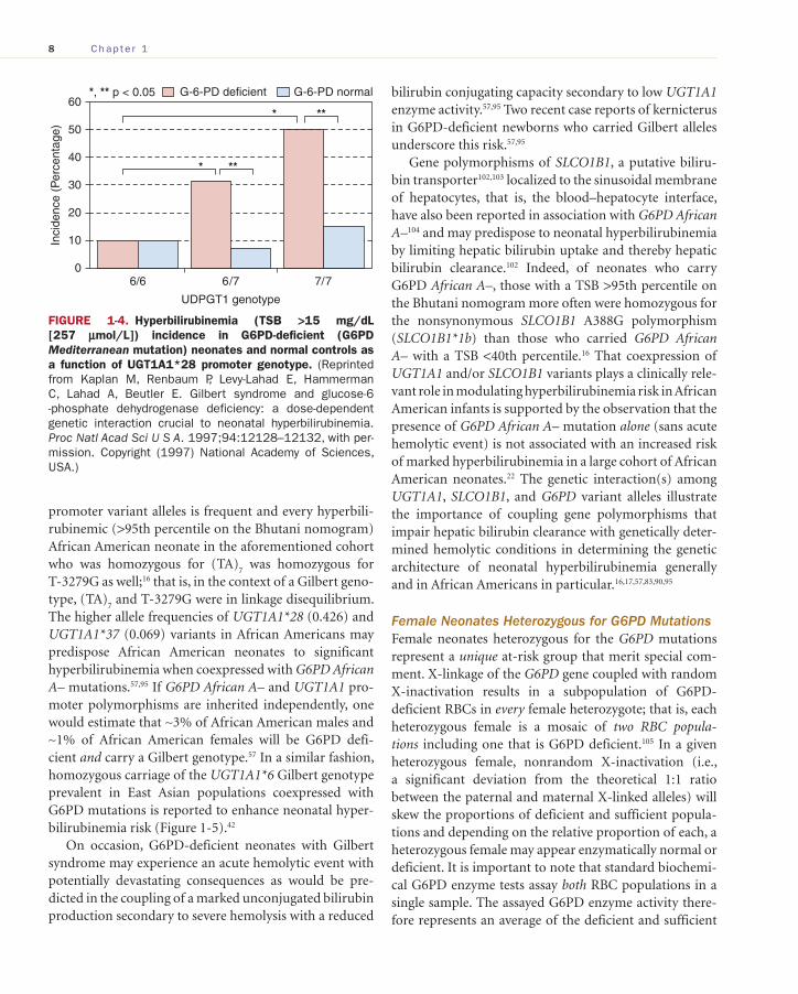
A dose-dependent genetic interaction between the UGT1A1*28 promoter variant and G6PD deficiency enhances neonatal hyperbilirubinemia risk, 12 , 90 a phe-nomenon originally described by Kaplan et al. with the G6PD Mediterranean mutation ( Figure 1-4 ) 12 and more recently noted in a cohort of African American neonates with a TSB >95th percentile on the Bhutani nomogram 100
who carried the G6PD African A– mutation. 16 In addition, the UGT1A1 PBREM promoter polymorphism T-3279G ( UGT1A1*60 ), a variant itself associated with reduced UGT1A1 expression, 101 may contribute an icterogenic effect. Coexpression of the UGT1A1*60 and UGT1A1*28
TABLE 1-4. � Estimated Number of International Migrants
Year World North America Europe
1990 155,518,065 27,773,888 49,400,6611995 165,968,778 33,595,046 54,717,8642000 178,498,563 40,395,432 57,639,1142005 195,245,404 45,597,061 64,398,5852010 213,943,812 50,042,408 69,819,282
Data from United Nations, Department of Economic and Social Affairs, Population Division (2009). Trends in International Migrant Stock: The 2008 Revision. (United Nations database, POP/DB/MIG/Stock/Rev.2008).
Stev_Ch01_001-028.indd 7Stev_Ch01_001-028.indd 7 3/20/12 11:18:05 AM3/20/12 11:18:05 AM
8 Chap t e r 1
promoter variant alleles is frequent and every hyperbili-rubinemic (>95th percentile on the Bhutani nomogram) African American neonate in the aforementioned cohort who was homozygous for (TA)
7 was homozygous for
T-3279G as well; 16 that is, in the context of a Gilbert geno-type, (TA)
7 and T-3279G were in linkage disequilibrium.
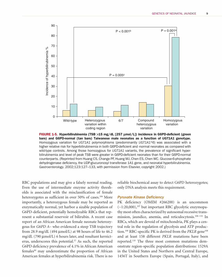
The higher allele frequencies of UGT1A1*28 (0.426) and UGT1A1*37 (0.069) variants in African Americans may predispose African American neonates to significant hyperbilirubinemia when coexpressed with G6PD African A– mutations. 57 , 95 If G6PD African A– and UGT1A1 pro-moter polymorphisms are inherited independently, one would estimate that ∼3% of African American males and ∼1% of African American females will be G6PD defi-cient and carry a Gilbert genotype. 57 In a similar fashion, homozygous carriage of the UGT1A1*6 Gilbert genotype prevalent in East Asian populations coexpressed with G6PD mutations is reported to enhance neonatal hyper-bilirubinemia risk ( Figure 1-5 ). 42
On occasion, G6PD-deficient neonates with Gilbert syndrome may experience an acute hemolytic event with potentially devastating consequences as would be pre-dicted in the coupling of a marked unconjugated bilirubin production secondary to severe hemolysis with a reduced
bilirubin conjugating capacity secondary to low UGT1A1 enzyme activity. 57 , 95 Two recent case reports of kernicterus in G6PD-deficient newborns who carried Gilbert alleles underscore this risk. 57 , 95
Gene polymorphisms of SLCO1B1 , a putative biliru-bin transporter 102 , 103 localized to the sinusoidal membrane of hepatocytes, that is, the blood–hepatocyte interface, have also been reported in association with G6PD African A– 104 and may predispose to neonatal hyperbilirubinemia by limiting hepatic bilirubin uptake and thereby hepatic bilirubin clearance. 102 Indeed, of neonates who carry G6PD African A– , those with a TSB >95th percentile on the Bhutani nomogram more often were homozygous for the nonsynonymous SLCO1B1 A388G polymorphism ( SLCO1B1*1b ) than those who carried G6PD African A– with a TSB <40th percentile. 16 That coexpression of UGT1A1 and/or SLCO1B1 variants plays a clinically rele-vant role in modulating hyperbilirubinemia risk in African American infants is supported by the observation that the presence of G6PD African A– mutation alone (sans acute hemolytic event) is not associated with an increased risk of marked hyperbilirubinemia in a large cohort of African American neonates. 22 The genetic interaction(s) among UGT1A1 , SLCO1B1 , and G6PD variant alleles illustrate the importance of coupling gene polymorphisms that impair hepatic bilirubin clearance with genetically deter-mined hemolytic conditions in determining the genetic architecture of neonatal hyperbilirubinemia generally and in African Americans in particular. 16 , 17 , 57 , 83 , 90 , 95
Female Neonates Heterozygous for G6PD Mutations Female neonates heterozygous for the G6PD mutations represent a unique at-risk group that merit special com-ment. X-linkage of the G6PD gene coupled with random X-inactivation results in a subpopulation of G6PD-deficient RBCs in every female heterozygote; that is, each heterozygous female is a mosaic of two RBC popula-tions including one that is G6PD deficient. 105 In a given heterozygous female, nonrandom X-inactivation (i.e., a significant deviation from the theoretical 1:1 ratio between the paternal and maternal X-linked alleles) will skew the proportions of deficient and sufficient popula-tions and depending on the relative proportion of each, a heterozygous female may appear enzymatically normal or deficient. It is important to note that standard biochemi-cal G6PD enzyme tests assay both RBC populations in a single sample. The assayed G6PD enzyme activity there-fore represents an average of the deficient and sufficient
60
* **
* **
Inci
denc
e (P
erce
ntag
e) 50
40
30
20
UDPGT1 genotype
6/6 6/7 7/7
10
0
*, ** p < 0.05 G-6-PD deficient G-6-PD normal
FIGURE 1-4. Hyperbilirubinemia (TSB >15 mg/dL [257 μmol/L]) incidence in G6PD-deficient (G6PD Mediterranean mutation) neonates and normal controls as a function of UGT1A1*28 promoter genotype. (Reprinted from Kaplan M, Renbaum P, Levy-Lahad E, Hammerman C, Lahad A, Beutler E. Gilbert syndrome and glucose-6-phosphate dehydrogenase deficiency: a dose-dependent genetic interaction crucial to neonatal hyperbilirubinemia. Proc Natl Acad Sci U S A. 1997;94:12128–12132, with per-mission. Copyright (1997) National Academy of Sciences, USA.)
Stev_Ch01_001-028.indd 8Stev_Ch01_001-028.indd 8 3/20/12 11:18:05 AM3/20/12 11:18:05 AM
GENETICS OF NEONATAL JAUNDICE 9
RBC populations and may give a falsely normal reading. Even the use of intermediate enzyme activity thresh-olds is associated with the misclassification of female heterozygotes as sufficient in over 50% of cases. 106 More importantly, a heterozygous female may be reported as enzymatically normal, yet harbor a sizable population of G6PD-deficient, potentially hemolyzable RBCs that rep-resent a substantial reservoir of bilirubin. A recent case report of an African American female neonate heterozy-gous for G6PD A– who evidenced a steep TSB trajectory from 28.9 mg/dL (494 μmol/L) at 98 hours of life to 46.2 mg/dL (790 μmol/L) 6 hours later, and resultant kernict-erus, underscores this potential. 57 As such, the reported G6PD deficiency prevalence of 4.1% in African American females 84 may underestimate the proportion of African American females at hyperbilirubinemia risk. There is no
reliable biochemical assay to detect G6PD heterozygotes; only DNA analysis meets this requirement.
Pyruvate Kinase Deficiency PK deficiency (OMIM #266200) is an uncommon (∼1:20,000), 107 but important RBC glycolytic enzymopa-thy most often characterized by autosomal recessive trans-mission, jaundice, anemia, and reticulocytosis. 108 – 110 In RBCs, which are devoid of mitochondria, PK plays a cen-tral role in the regulation of glycolysis and ATP produc-tion. 108 RBC-specific PK is derived from the PKLR gene 108 and at least 158 different PKLR mutations have been reported. 110 The three most common mutations dem-onstrate region-specific population distributions: 1529A in the United States and Northern and Central Europe, 1456T in Southern Europe (Spain, Portugal, Italy), and
90
80
70
60
50
40
30
20
10
0 Homozygous
variationHeterozygous variation withincoding region
Wild-type
Inci
denc
e of
hyp
erbi
lirub
inem
ia %
6/7 Compound heterozygous
variation
P = 0.005c
P < 0.001b P = 0.001a
FIGURE 1-5. Hyperbilirubinemia (TSB >15 mg/dL [257 μmol/L]) incidence in G6PD-deficient (green bars) and G6PD-normal (tan bars) Taiwanese male neonates as a function of UGT1A1 genotype. Homozygous variation for UGT1A1 polymorphisms (predominantly UGT1A1*6 ) was associated with a higher relative risk for hyperbilirubinemia in both G6PD-deficient and -normal neonates as compared with wild-type controls. Among those homozygous for UGT1A1 variants, the prevalence of significant hyper-bilirubinemia and level of peak TSB were greater in G6PD-deficient neonates than for their G6PD-normal counterparts. (Reprinted from Huang CS, Change PF, Huang MJ, Chen ES, Chen WC. Glucose-6-phosphate dehydrogenase deficiency, the UDP-glucuronosyl transferase 1A1 gene, and neonatal hyperbilirubinemia. Gastroenterology. 2002;123:127–133, with permission from Elsevier, copyright 2002.)
Stev_Ch01_001-028.indd 9Stev_Ch01_001-028.indd 9 3/20/12 11:18:05 AM3/20/12 11:18:05 AM
10 Chap t e r 1
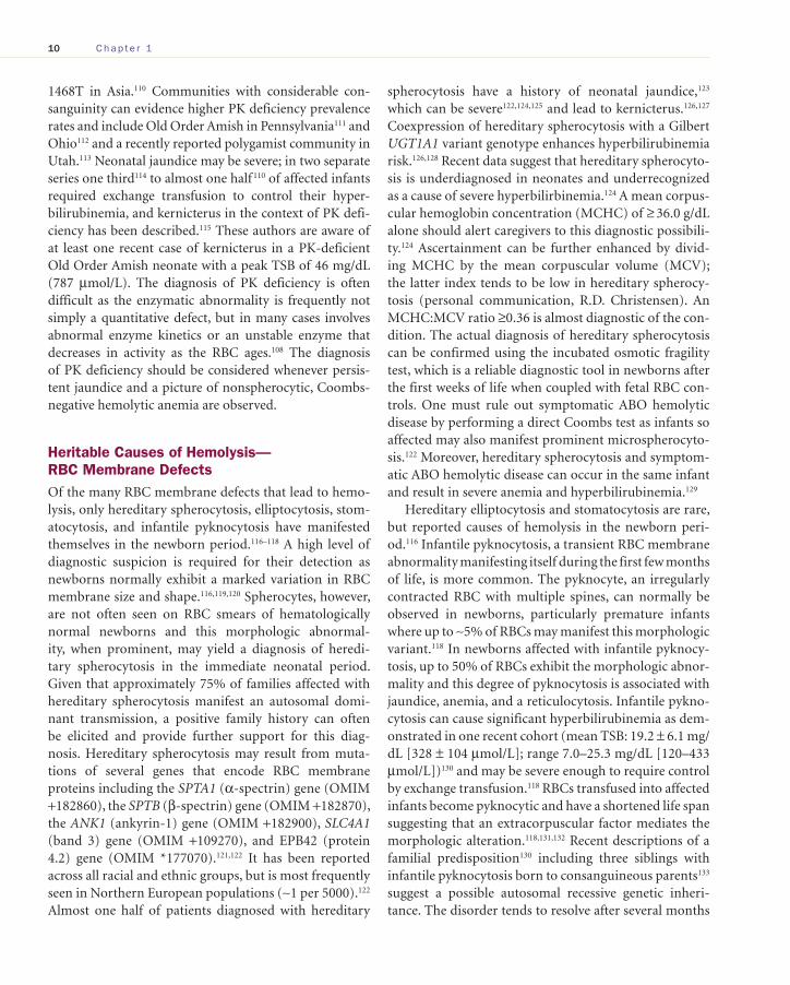
1468T in Asia. 110 Communities with considerable con-sanguinity can evidence higher PK deficiency prevalence rates and include Old Order Amish in Pennsylvania 111 and Ohio 112 and a recently reported polygamist community in Utah. 113 Neonatal jaundice may be severe; in two separate series one third 114 to almost one half 110 of affected infants required exchange transfusion to control their hyper-bilirubinemia, and kernicterus in the context of PK defi-ciency has been described. 115 These authors are aware of at least one recent case of kernicterus in a PK-deficient Old Order Amish neonate with a peak TSB of 46 mg/dL (787 μmol/L). The diagnosis of PK deficiency is often difficult as the enzymatic abnormality is frequently not simply a quantitative defect, but in many cases involves abnormal enzyme kinetics or an unstable enzyme that decreases in activity as the RBC ages. 108 The diagnosis of PK deficiency should be considered whenever persis-tent jaundice and a picture of nonspherocytic, Coombs-negative hemolytic anemia are observed.
Heritable Causes of Hemolysis—RBC Membrane Defects Of the many RBC membrane defects that lead to hemo-lysis, only hereditary spherocytosis, elliptocytosis, stom-atocytosis, and infantile pyknocytosis have manifested themselves in the newborn period. 116 – 118 A high level of diagnostic suspicion is required for their detection as newborns normally exhibit a marked variation in RBC membrane size and shape. 116 , 119 , 120 Spherocytes, however, are not often seen on RBC smears of hematologically normal newborns and this morphologic abnormal-ity, when prominent, may yield a diagnosis of heredi-tary spherocytosis in the immediate neonatal period. Given that approximately 75% of families affected with hereditary spherocytosis manifest an autosomal domi-nant transmission, a positive family history can often be elicited and provide further support for this diag-nosis. Hereditary spherocytosis may result from muta-tions of several genes that encode RBC membrane proteins including the SPTA1 (α-spectrin) gene (OMIM +182860), the SPTB (β-spectrin) gene (OMIM +182870), the ANK1 (ankyrin-1) gene (OMIM +182900), SLC4A1 (band 3) gene (OMIM +109270), and EPB42 (protein 4.2) gene (OMIM *177070). 121 , 122 It has been reported across all racial and ethnic groups, but is most frequently seen in Northern European populations (∼1 per 5000). 122 Almost one half of patients diagnosed with hereditary
spherocytosis have a history of neonatal jaundice, 123 which can be severe 122 , 124 , 125 and lead to kernicterus. 126 , 127 Coexpression of hereditary spherocytosis with a Gilbert UGT1A1 variant genotype enhances hyperbilirubinemia risk. 126 , 128 Recent data suggest that hereditary spherocyto-sis is underdiagnosed in neonates and underrecognized as a cause of severe hyperbilirbinemia. 124 A mean corpus-cular hemoglobin concentration (MCHC) of ≥ 36.0 g/dL alone should alert caregivers to this diagnostic possibili-ty. 124 Ascertainment can be further enhanced by divid-ing MCHC by the mean corpuscular volume (MCV); the latter index tends to be low in hereditary spherocy-tosis (personal communication, R.D. Christensen). An MCHC:MCV ratio ≥0.36 is almost diagnostic of the con-dition. The actual diagnosis of hereditary spherocytosis can be confirmed using the incubated osmotic fragility test, which is a reliable diagnostic tool in newborns after the first weeks of life when coupled with fetal RBC con-trols. One must rule out symptomatic ABO hemolytic disease by performing a direct Coombs test as infants so affected may also manifest prominent microspherocyto-sis. 122 Moreover, hereditary spherocytosis and symptom-atic ABO hemolytic disease can occur in the same infant and result in severe anemia and hyperbilirubinemia. 129
Hereditary elliptocytosis and stomatocytosis are rare, but reported causes of hemolysis in the newborn peri-od. 116 Infantile pyknocytosis, a transient RBC membrane abnormality manifesting itself during the first few months of life, is more common. The pyknocyte, an irregularly contracted RBC with multiple spines, can normally be observed in newborns, particularly premature infants where up to ∼5% of RBCs may manifest this morphologic variant. 118 In newborns affected with infantile pyknocy-tosis, up to 50% of RBCs exhibit the morphologic abnor-mality and this degree of pyknocytosis is associated with jaundice, anemia, and a reticulocytosis. Infantile pykno-cytosis can cause significant hyperbilirubinemia as dem-onstrated in one recent cohort (mean TSB: 19.2 ± 6.1 mg/dL [328 ± 104 μmol/L]; range 7.0–25.3 mg/dL [120–433 μmol/L]) 130 and may be severe enough to require control by exchange transfusion. 118 RBCs transfused into affected infants become pyknocytic and have a shortened life span suggesting that an extracorpuscular factor mediates the morphologic alteration. 118 , 131 , 132 Recent descriptions of a familial predisposition 130 including three siblings with infantile pyknocytosis born to consanguineous parents 133 suggest a possible autosomal recessive genetic inheri-tance. The disorder tends to resolve after several months
Stev_Ch01_001-028.indd 10Stev_Ch01_001-028.indd 10 3/20/12 11:18:05 AM3/20/12 11:18:05 AM
GENETICS OF NEONATAL JAUNDICE 11
of life. Pyknocytosis may also occur in other conditions including G6PD deficiency and hereditary elliptocytosis.
Heritable Causes of Hemolysis—Hemoglobinopathies Defects in hemoglobin structure or synthesis infrequently manifest themselves in the neonatal period. Of these, the α-thalassemia syndromes are the most likely to be clini-cally apparent in newborns. Thalassemias are inherited dis-orders of hemoglobin synthesis. Each human diploid cell contains four copies of the α-globin gene and, thus, four α-thalassemia syndromes have been described reflecting the presence of defects in one, two, three, or four α-globin genes. Silent carriers have one abnormal α-globin chain and are asymptomatic. α-Thalassemia trait is associated with two α-thalassemia mutations, can be detected by a low MCV of <95 μ 3 (normal infants 100–120 μ 3 ), 134 and in neonates is not associated with hemolysis. Hemoglobin H disease, prevalent in Asian and Mediterranean popu-lations, results from the presence of three α-thalassemia mutations and can cause hemolysis and anemia in neo-nates. 135 An increasing number of infants with Hemoglobin H disease have been reported in the United States since the early 1990s reflecting recent immigration patterns. 136 , 137 Homozygous α-thalassemia (total absence of α-chain synthesis) often results in profound hemolysis, anemia, hydrops fetalis, and almost always stillbirth or death in the immediate neonatal period, although survival throughout childhood has been reported. 136
The pure β-thalassemias do not manifest themselves in the newborn period and the γ-thalassemias are: (i) incompatible with life (homozygous form); (ii) associ-ated with transient mild to moderate neonatal anemia if one or two genes are involved that resolves when β-chain synthesis begins; or (iii) in combination with impaired β-chain synthesis, associated with severe hemolytic ane-mia and marked hyperbilirubinemia. 138
Immune-Mediated Hemolytic Disease of the Newborn Immune-mediated hemolytic disease can develop in the neonate of a heterospecific RBC antigen mother/infant pair when maternally derived antibody binds to the neonatal RBC antigen. The ABO (OMIM #110300) and RHD/CE (OMIM #111680 and 111700) blood group sys-tems are the most commonly encountered in this regard, albeit minor RBC groups can also be associated with
immune-mediated hemolytic disease of the newborn. ABO antigen status is under the control of at least three alleles on chromosome 9q34; A and B are codominant; O is recessive. The antibody type is also under genetic control. For all intents and purposes, symptomatic ABO hemolytic disease is limited to infants of blood group A or B born to mothers of blood group O, who show marked jaundice, a positive direct Coombs test, and often microspherocytosis on an RBC smear. 139 It is of interest that the frequency distribution of blood types A, B, and O differs across populations. Some previous studies suggest that ABO hemolytic disease is more frequent in African American newborns, 140 – 143 including evidence that a positive direct Coombs test is more common in African American heterospecific mother/infant pairs. 141
The RH antigen types are determined by three closely linked loci on chromosome 1p34–36 each with two alleles: Cc, Dd, and Ee. The lower case letters do not indicate reces-sitivity; each allele determines the presence of an antigen (C, c, D, E, e), sans d which does not exist. 32,144 Most symp-tomatic RH hemolytic disease (∼90%) is related to RHD incompatibility although maternally derived alloantibod-ies to C, c, E, and e can lead to hemolytic disease of the newborn. 144 The incidence of common RH haplotypes differs significantly across populations, 144 the resultant ratio of RHD-positive to RHD-negative phenotypes being ∼0.84:0.16 in Caucasians, ∼0.92:0.08 in African American, and ∼0.99:<0.01 in Asians. 144 Although RH isoimmuniza-tion can still lead to severe neonatal hyperbilirubinemia, the prevalence of RH hemolytic disease has decreased markedly as a result of effective immunoprophylaxis with anti-RH (anti-D) gamma-globulin. 144
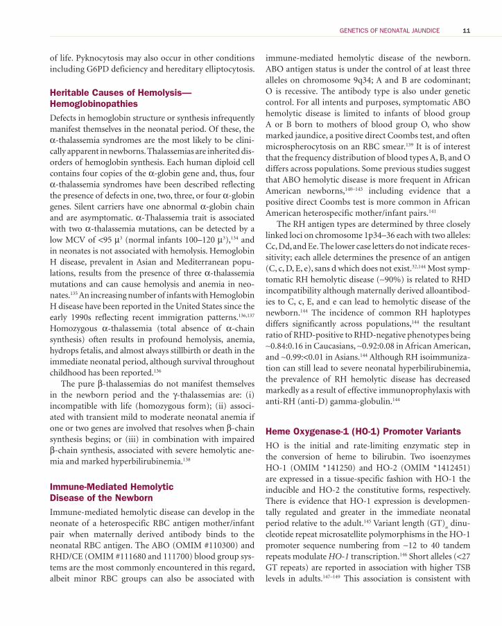
Heme Oxygenase-1 (HO-1) Promoter Variants HO is the initial and rate-limiting enzymatic step in the conversion of heme to bilirubin. Two isoenzymes HO-1 (OMIM *141250) and HO-2 (OMIM *1412451) are expressed in a tissue-specific fashion with HO-1 the inducible and HO-2 the constitutive forms, respectively. There is evidence that HO-1 expression is developmen-tally regulated and greater in the immediate neonatal period relative to the adult. 145 Variant length (GT)
n dinu-
cleotide repeat microsatellite polymorphisms in the HO-1 promoter sequence numbering from ∼12 to 40 tandem repeats modulate HO-1 transcription. 146 Short alleles (<27 GT repeats) are reported in association with higher TSB levels in adults. 147 – 149 This association is consistent with
Stev_Ch01_001-028.indd 11Stev_Ch01_001-028.indd 11 3/20/12 11:18:05 AM3/20/12 11:18:05 AM
12 Chap t e r 1
functional studies demonstrating greater basal HO-1 expression and HO-1 inducibility by oxidative stimuli in short (GT)
n repeat alleles as compared with their longer
counterparts. 150 To date, only two studies has explored the relationship between HO-1 (GT)
n repeats and TSB levels
in neonates and no effect of (GT) n number was observed
on peak hyperbilirubinemia risk, 151 , 152 albeit in one of these reports, short (<24 GT) alleles were associated with pro-longed breast milk jaundice. 152 A recent case report of a boy with hazardous hyperbilirubinemia, autoimmune hemo-lytic disease, and homozygosity for short (GT)
n repeats 153
suggests the potential modulatory role of HO-1 promoter polymorphisms on TSB in neonates merits further study, particularly when short HO-1 (GT) repeat alleles are coex-pressed with a genetic predisposition to hemolysis and increased heme production (e.g., G6PD deficiency, ABO hemolytic disease, hereditary spherocytosis). 153
Biliverdin Reductase Polymorphisms Biliverdin reductase A ( BLVRA ; OMIM *109750) efficiently reduces biliverdin to bilirubin. In theory, BLVRA polymor-phisms might affect hyperbilirubinemia risk in newborns. However, only one common nonsynonymous BLVRA gene variant (rs699512:A>G) has been reported in the dbSNP
database (allele frequency 0.23 Caucasians, 0.08 African Americans, 0.27 Chinese, and 0.40 Japanese) and this vari-ant is not associated with adult TSB levels across three Asian populations. 147 This BLVRA variant allele has not been studied in neonates. A recent case report of two unre-lated Inuit women with a homozygous nonsense BLVRA mutation indicates that complete absence of BLVRA activ-ity is a nonlethal condition, characterized phenotypically by green jaundice during episodes of cholestasis. 154
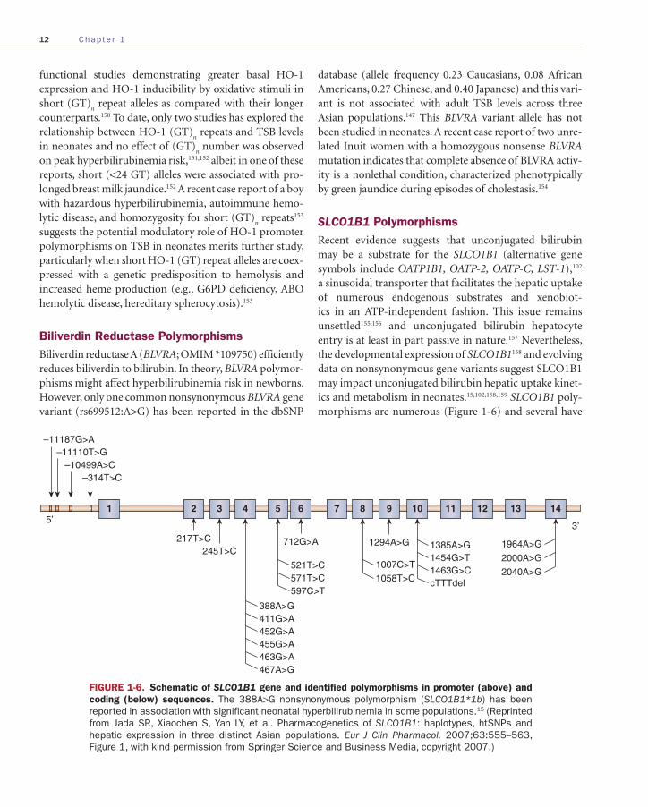
SLCO1B1 Polymorphisms Recent evidence suggests that unconjugated bilirubin may be a substrate for the SLCO1B1 (alternative gene symbols include OATP1B1, OATP-2, OATP-C, LST-1) , 102 a sinusoidal transporter that facilitates the hepatic uptake of numerous endogenous substrates and xenobiot-ics in an ATP-independent fashion. This issue remains unsettled 155 , 156 and unconjugated bilirubin hepatocyte entry is at least in part passive in nature. 157 Nevertheless, the developmental expression of SLCO1B1 158 and evolving data on nonsynonymous gene variants suggest SLCO1B1 may impact unconjugated bilirubin hepatic uptake kinet-ics and metabolism in neonates. 15 , 102 , 158 , 159 SLCO1B1 poly-morphisms are numerous ( Figure 1-6 ) and several have
388A>G
245T>C217T>C 712G>A
5’3’
411G>A452G>A455G>A463G>A467A>G
1 2 3 4 5 6 7 8 9 10 11 12 13 14
597C>T
521T>C 571T>C 1058T>C
1007C>T
1294A>G 1385A>G1454G>T1463G>CcTTTdel
1964A>G
2000A>G
2040A>G
–11187G>A–11110T>G
–10499A>C–314T>C
FIGURE 1-6. Schematic of SLCO1B1 gene and identified polymorphisms in promoter (above) and coding (below) sequences. The 388A>G nonsynonymous polymorphism ( SLCO1B1*1b ) has been reported in association with significant neonatal hyperbilirubinemia in some populations. 15 (Reprinted from Jada SR, Xiaochen S, Yan LY, et al. Pharmacogenetics of SLCO1B1: haplotypes, htSNPs and hepatic expression in three distinct Asian populations. Eur J Clin Pharmacol. 2007;63:555–563, Figure 1, with kind permission from Springer Science and Business Media, copyright 2007.)
Stev_Ch01_001-028.indd 12Stev_Ch01_001-028.indd 12 3/20/12 11:18:05 AM3/20/12 11:18:05 AM
GENETICS OF NEONATAL JAUNDICE 13
been studied in human neonates. 15 , 16 Their coexpression with other icterogenic genes is also common. 16 , 17 , 104 As detailed above, the SLCO1B1*1b variant allele is asso-ciated with increased risk for severe hyperbilirubine-mia in Taiwanese newborns 15 and coupling of UGT1A1 with SLCO1B1 variant alleles further enhances that risk. 15 Although homozygosity for SLCO1B1*1b was not observed at greater frequency in neonates with TSB >95th percentile in a United States–based cohort, 16 SLCO1B1*1b coexpression with G6PD A– was. 16 Some adult genome-wide association studies suggest that SLCO1B1 polymor-phisms are directly associated with higher TSB levels, albeit they account for only ∼1% of the TSB variance, as contrasted with UGT1A1 polymorphisms that account for ∼18% of TSB variance. 160
Glutathione S -transferase (Ligandin) Polymorphisms Human cytosolic GSTs are a superfamily of multifunc-tional proteins that in addition to their catalytic func-tion also demonstrate high-capacity ligand binding for a variety of nonsubstrate compounds. Although several different GST gene classes evidence a ligandin function, the class alpha (A) GSTs hGSTA1-1 and hGSTA2-2 appear to be the major ligand-binding and transporter proteins for unconjugated bilirubin in the hepatocyte. 161 Hepatic uptake of unconjugated bilirubin is enhanced by increas-ing concentrations of ligandin. 162 As such, the low hepatic ligandin concentration observed at birth 163 may con-tribute to the early hyperbilirubinemia risk in neonates. Moreover, a variant hGSTA1-1 allele (G-52A) within a polymorphic SP-1 binding site of the proximal promoter is associated with 4-fold lower mean hepatic expression than the referent allele 161 , 164 and presumably decreased hepatic unconjugated bilirubin binding, although the lat-ter has not been confirmed in functional assay. To date, only Muslu et al. have studied hGST polymorphisms in neonatal hyperbilirubinemia, specifically two non-α-GST isoenzymes hGSTT1 and hGSTM1 , and found no relationship between these allelic variants and neonatal hyperbilirubinemia risk. 165 However, proteins of the theta (T) and mu (M) classes bind bilirubin with a lower affinity than alpha-class GSTs so the aforementioned findings do not preclude an impact of hGSTA1-1 (or hGSTA2-2 ) vari-ant alleles on neonatal hyperbilirubinemia risk. It is clini-cally notable that induction of both hGSTA1 and hGSTA2 occurs in response to phenobarbital treatment. 166
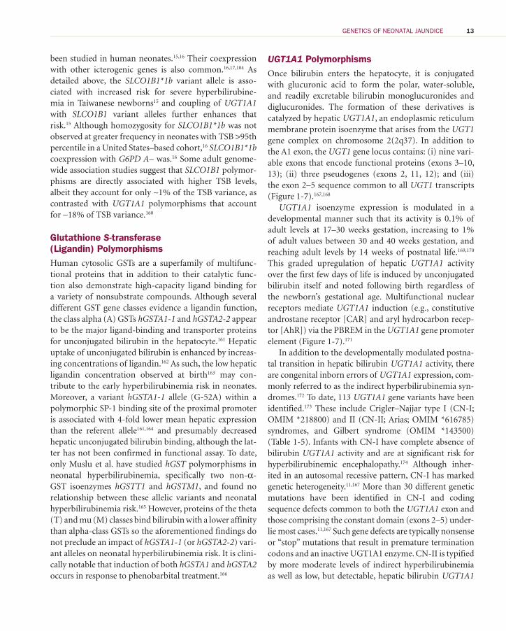
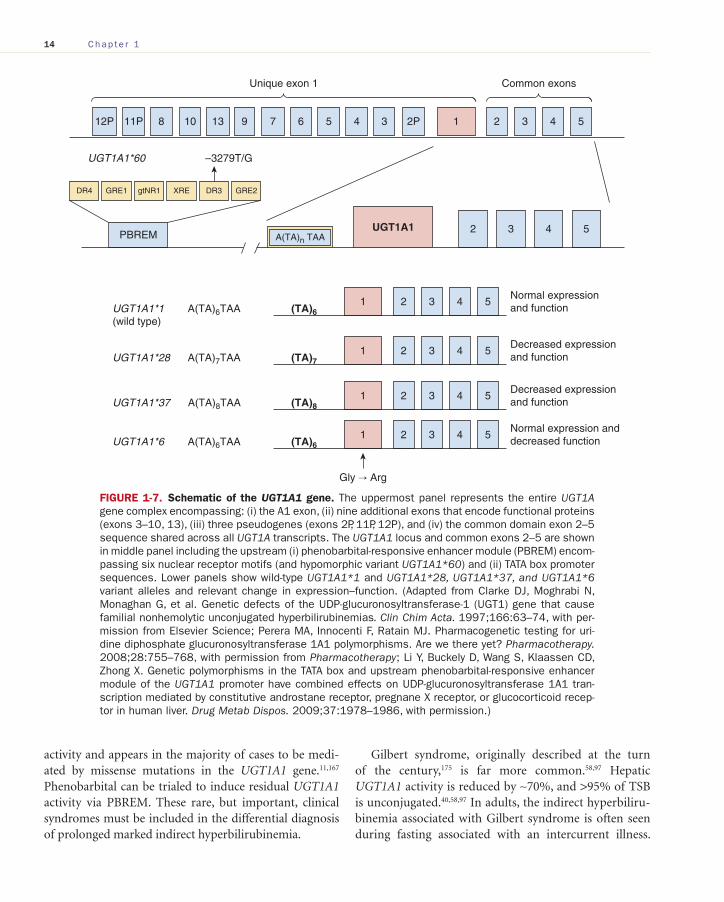
UGT1A1 Polymorphisms Once bilirubin enters the hepatocyte, it is conjugated with glucuronic acid to form the polar, water-soluble, and readily excretable bilirubin monoglucuronides and diglucuronides. The formation of these derivatives is catalyzed by hepatic UGT1A1 , an endoplasmic reticulum membrane protein isoenzyme that arises from the UGT1 gene complex on chromosome 2(2q37). In addition to the A1 exon, the UGT1 gene locus contains: (i) nine vari-able exons that encode functional proteins (exons 3–10, 13); (ii) three pseudogenes (exons 2, 11, 12); and (iii) the exon 2–5 sequence common to all UGT1 transcripts ( Figure 1-7 ). 167 , 168
UGT1A1 isoenzyme expression is modulated in a developmental manner such that its activity is 0.1% of adult levels at 17–30 weeks gestation, increasing to 1% of adult values between 30 and 40 weeks gestation, and reaching adult levels by 14 weeks of postnatal life. 169 , 170 This graded upregulation of hepatic UGT1A1 activity over the first few days of life is induced by unconjugated bilirubin itself and noted following birth regardless of the newborn’s gestational age. Multifunctional nuclear receptors mediate UGT1A1 induction (e.g., constitutive androstane receptor [CAR] and aryl hydrocarbon recep-tor [AhR]) via the PBREM in the UGT1A1 gene promoter element ( Figure 1-7 ). 171
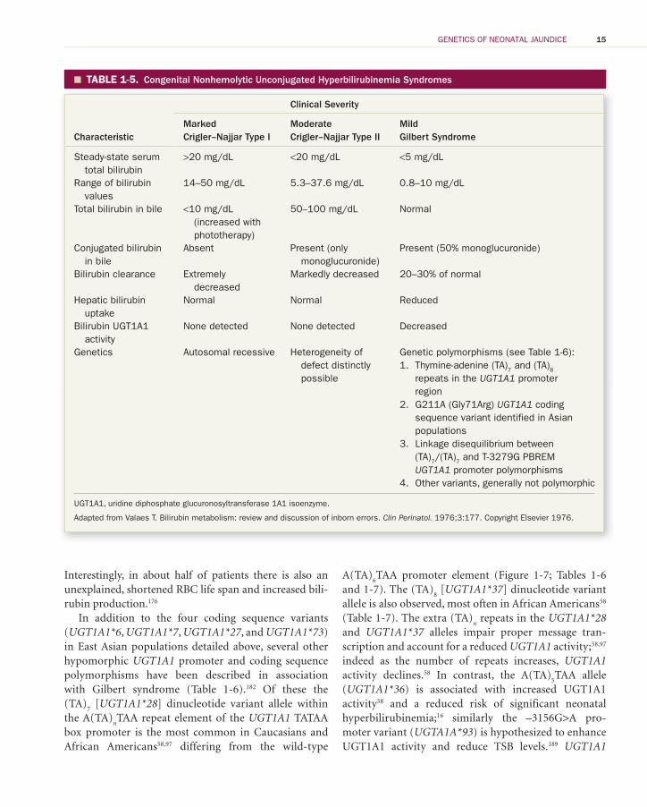
In addition to the developmentally modulated postna-tal transition in hepatic bilirubin UGT1A1 activity, there are congenital inborn errors of UGT1A1 expression, com-monly referred to as the indirect hyperbilirubinemia syn-dromes. 172 To date, 113 UGT1A1 gene variants have been identified. 173 These include Crigler–Najjar type I (CN-I; OMIM *218800) and II (CN-II; Arias; OMIM *616785) syndromes, and Gilbert syndrome (OMIM *143500) ( Table 1-5 ). Infants with CN-I have complete absence of bilirubin UGT1A1 activity and are at significant risk for hyperbilirubinemic encephalopathy. 174 Although inher-ited in an autosomal recessive pattern, CN-I has marked genetic heterogeneity. 11 , 167 More than 30 different genetic mutations have been identified in CN-I and coding sequence defects common to both the UGT1A1 exon and those comprising the constant domain (exons 2–5) under-lie most cases. 11 , 167 Such gene defects are typically nonsense or “stop” mutations that result in premature termination codons and an inactive UGT1A1 enzyme. CN-II is typified by more moderate levels of indirect hyperbilirubinemia as well as low, but detectable, hepatic bilirubin UGT1A1
Stev_Ch01_001-028.indd 13Stev_Ch01_001-028.indd 13 3/20/12 11:18:06 AM3/20/12 11:18:06 AM
14 Chap t e r 1
12P 11P
DR4
8 10 13 9 7 6 5 4 3 2P 1 2 3 4 5
1 2 3 4 5
2 3 4 5
Unique exon 1
UGT1A1*60 –3279T/G
Common exons
GRE1 gtNR1 XRE DR3 GRE2
PBREM A(TA)n TAAUGT1A1
(TA)6
1 2 3 4 5(TA)7
1 2 3 4 5(TA)8
1 2 3 4 5(TA)6
UGT1A1*1 A(TA)6TAA(wild type)
UGT1A1*28 A(TA)7TAA
Gly → Arg
UGT1A1*37 A(TA)8TAA
UGT1A1*6 A(TA)6TAA
Normal expression and function
Decreased expression and function
Decreased expression and function
Normal expression and decreased function
FIGURE 1-7. Schematic of the UGT1A1 gene. The uppermost panel represents the entire UGT1A gene complex encompassing: (i) the A1 exon, (ii) nine additional exons that encode functional proteins (exons 3–10, 13), (iii) three pseudogenes (exons 2P, 11P, 12P), and (iv) the common domain exon 2–5 sequence shared across all UGT1A transcripts. The UGT1A1 locus and common exons 2–5 are shown in middle panel including the upstream (i) phenobarbital-responsive enhancer module (PBREM) encom-passing six nuclear receptor motifs (and hypomorphic variant UGT1A1*60 ) and (ii) TATA box promoter sequences. Lower panels show wild-type UGT1A1*1 and UGT1A1*28, UGT1A1*37, and UGT1A1*6 variant alleles and relevant change in expression–function. (Adapted from Clarke DJ, Moghrabi N, Monaghan G, et al. Genetic defects of the UDP-glucuronosyltransferase-1 (UGT1) gene that cause familial nonhemolytic unconjugated hyperbilirubinemias. Clin Chim Acta. 1997;166:63–74, with per-mission from Elsevier Science; Perera MA, Innocenti F, Ratain MJ. Pharmacogenetic testing for uri-dine diphosphate glucuronosyltransferase 1A1 polymorphisms. Are we there yet? Pharmacotherapy. 2008;28:755–768, with permission from Pharmacotherapy ; Li Y, Buckely D, Wang S, Klaassen CD, Zhong X. Genetic polymorphisms in the TATA box and upstream phenobarbital-responsive enhancer module of the UGT1A1 promoter have combined effects on UDP-glucuronosyltransferase 1A1 tran-scription mediated by constitutive androstane receptor, pregnane X receptor, or glucocorticoid recep-tor in human liver. Drug Metab Dispos. 2009;37:1978–1986, with permission.)
activity and appears in the majority of cases to be medi-ated by missense mutations in the UGT1A1 gene. 11 , 167 Phenobarbital can be trialed to induce residual UGT1A1 activity via PBREM. These rare, but important, clinical syndromes must be included in the differential diagnosis of prolonged marked indirect hyperbilirubinemia.
Gilbert syndrome, originally described at the turn of the century, 175 is far more common. 58 , 97 Hepatic UGT1A1 activity is reduced by ∼70%, and >95% of TSB is unconjugated. 40 , 58 , 97 In adults, the indirect hyperbiliru-binemia associated with Gilbert syndrome is often seen during fasting associated with an intercurrent illness.
Stev_Ch01_001-028.indd 14Stev_Ch01_001-028.indd 14 3/20/12 11:18:06 AM3/20/12 11:18:06 AM
GENETICS OF NEONATAL JAUNDICE 15
Interestingly, in about half of patients there is also an unexplained, shortened RBC life span and increased bili-rubin production. 176
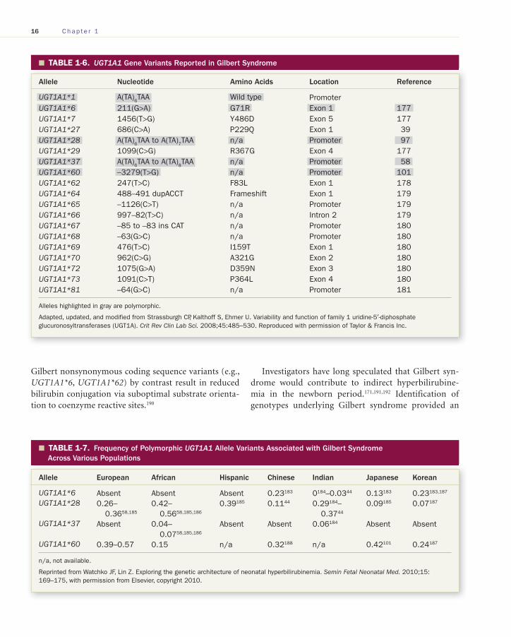
In addition to the four coding sequence variants ( UGT1A1*6 , UGT1A1*7 , UGT1A1*27 , and UGT1A1*73 ) in East Asian populations detailed above, several other hypomorphic UGT1A1 promoter and coding sequence polymorphisms have been described in association with Gilbert syndrome ( Table 1-6 ).182 Of these the (TA)
7 [ UGT1A1*28 ] dinucleotide variant allele within
the A(TA) n TAA repeat element of the UGT1A1 TATAA
box promoter is the most common in Caucasians and African Americans 58 , 97 differing from the wild-type
A(TA) 6 TAA promoter element ( Figure 1-7 ; Tables 1-6
and 1-7 ). The (TA) 8 [ UGT1A1*37 ] dinucleotide variant
allele is also observed, most often in African Americans 58
( Table 1-7 ). The extra (TA) n repeats in the UGT1A1*28
and UGT1A1*37 alleles impair proper message tran-scription and account for a reduced UGT1A1 activity; 58 , 97
indeed as the number of repeats increases, UGT1A1activity declines. 58 In contrast, the A(TA)
5 TAA allele
( UGT1A1*36 ) is associated with increased UGT1A1 activity 58 and a reduced risk of significant neonatal hyperbilirubinemia; 16 similarly the –3156G>A pro-moter variant ( UGTA1A*93 ) is hypothesized to enhance UGT1A1 activity and reduce TSB levels. 189 UGT1A1
TABLE 1-5. � Congenital Nonhemolytic Unconjugated Hyperbilirubinemia Syndromes
Characteristic
Clinical Severity
MarkedCrigler–Najjar Type I
ModerateCrigler–Najjar Type II
MildGilbert Syndrome
Steady-state serum total bilirubin
>20 mg/dL <20 mg/dL <5 mg/dL
Range of bilirubin values
14–50 mg/dL 5.3–37.6 mg/dL 0.8–10 mg/dL
Total bilirubin in bile <10 mg/dL (increased with phototherapy)
50–100 mg/dL Normal
Conjugated bilirubin in bile
Absent Present (only monoglucuronide)
Present (50% monoglucuronide)
Bilirubin clearance Extremely decreased
Markedly decreased 20–30% of normal
Hepatic bilirubin uptake
Normal Normal Reduced
Bilirubin UGT1A1 activity
None detected None detected Decreased
Genetics Autosomal recessive Heterogeneity of defect distinctly possible
Genetic polymorphisms (see Table 1-6 ): 1. Thymine-adenine (TA) 7 and (TA) 8
repeats in the UGT1A1 promoter region
2. G211A (Gly71Arg) UGT1A1 coding sequence variant identified in Asian populations
3. Linkage disequilibrium between (TA) 7 /(TA) 7 and T-3279G PBREM UGT1A1 promoter polymorphisms
4. Other variants, generally not polymorphic
UGT1A1, uridine diphosphate glucuronosyltransferase 1A1 isoenzyme.
Adapted from Valaes T. Bilirubin metabolism: review and discussion of inborn errors. Clin Perinatol. 1976;3:177. Copyright Elsevier 1976.
Stev_Ch01_001-028.indd 15Stev_Ch01_001-028.indd 15 3/20/12 11:18:08 AM3/20/12 11:18:08 AM
16 Chap t e r 1
Gilbert nonsynonymous coding sequence variants (e.g., UGT1A1*6 , UGT1A1*62 ) by contrast result in reduced bilirubin conjugation via suboptimal substrate orienta-tion to coenzyme reactive sites. 190
Investigators have long speculated that Gilbert syn-drome would contribute to indirect hyperbilirubine-mia in the newborn period. 171 , 191 , 192 Identification of genotypes underlying Gilbert syndrome provided an
TABLE 1-6. � UGT1A1 Gene Variants Reported in Gilbert Syndrome
Allele Nucleotide Amino Acids Location Reference
UGT1A1*1 A(TA) 6 TAA Wild type Promoter UGT1A1*6 211(G>A) G71R Exon 1 177 UGT1A1*7 1456(T>G) Y486D Exon 5 177 UGT1A1*27 686(C>A) P229Q Exon 1 39 UGT1A1*28 A(TA) 6 TAA to A(TA) 7 TAA n/a Promoter 97 UGT1A1*29 1099(C>G) R367G Exon 4 177 UGT1A1*37 A(TA) 6 TAA to A(TA) 8 TAA n/a Promoter 58 UGT1A1*60 –3279(T>G) n/a Promoter 101 UGT1A1*62 247(T>C) F83L Exon 1 178 UGT1A1*64 488–491 dupACCT Frameshift Exon 1 179 UGT1A1*65 –1126(C>T) n/a Promoter 179 UGT1A1*66 997–82(T>C) n/a Intron 2 179 UGT1A1*67 –85 to –83 ins CAT n/a Promoter 180 UGT1A1*68 –63(G>C) n/a Promoter 180 UGT1A1*69 476(T>C) I159T Exon 1 180 UGT1A1*70 962(C>G) A321G Exon 2 180 UGT1A1*72 1075(G>A) D359N Exon 3 180 UGT1A1*73 1091(C>T) P364L Exon 4 180 UGT1A1*81 –64(G>C) n/a Promoter 181
Alleles highlighted in gray are polymorphic.
Adapted, updated, and modified from Strassburgh CP, Kalthoff S, Ehmer U. Variability and function of family 1 uridine-5′-diphosphate glucuronosyltransferases (UGT1A). Crit Rev Clin Lab Sci. 2008;45:485–530. Reproduced with permission of Taylor & Francis Inc.
TABLE 1-7. � Frequency of Polymorphic UGT1A1 Allele Variants Associated with Gilbert Syndrome Across Various Populations
Allele European African Hispanic Chinese Indian Japanese Korean
UGT1A1*6 Absent Absent Absent 0.23 183 0 184 –0.03 44 0.13 183 0.23 183 , 187 UGT1A1*28 0.26–
0.36 58 , 185 0.42–
0.56 58 , 185 , 186 0.39 185 0.11 44 0.29 184 –
0.37 44 0.09 185 0.07 187
UGT1A1*37 Absent 0.04–0.07 58 , 185 , 186
Absent Absent 0.06 184 Absent Absent
UGT1A1*60 0.39–0.57 0.15 n/a 0.32 188 n/a 0.42 101 0.24 187
n/a, not available.
Reprinted from Watchko JF, Lin Z. Exploring the genetic architecture of neonatal hyperbilirubinemia. Semin Fetal Neonatal Med. 2010;15:169–175, with permission from Elsevier, copyright 2010.
Stev_Ch01_001-028.indd 16Stev_Ch01_001-028.indd 16 3/20/12 11:18:09 AM3/20/12 11:18:09 AM
GENETICS OF NEONATAL JAUNDICE 17
important tool to study the role of this condition in the pathogenesis of neonatal jaundice. Bancroft et al. were the first to explore this relationship and observed that newborn infants with the A(TA)
7 TAA UGT1A1
promoter polymorphism had accelerated jaundice and decreased fecal excretion of bilirubin monoglucuronides and diglucuronides. 98 Although some subsequent stud-ies demonstrated that UGT1A1*28 and/or UGT1A1*37 alleles are associated with modest 193 to more significant postnatal TSB elevation, 184 , 194 others have failed to dem-onstrate a clinically significant effect of UGT1A1*28 alone on neonatal hyperbilirubinemia risk 16 , 183 , 195 includ-ing a TSB >95th percentile on the Bhutani nomogram 16 or need for phototherapy. 184 The latter may reflect in part the incomplete penetrance of the UGT1A1*28 genotype. 97 Indeed in adults only about 50% of subjects homozygous for the UGT1A1*28 allele display a Gilbert phenotype; as stated by Bosma et al., the UGT1A1*28 variant allele is necessary, but not sufficient for com-plete phenotypic expression. 97 However, the coupling of UGT1A1*28 and/or UGT1A1*37 with other icterogenic conditions, for example, G6PD deficiency and heredi-tary spherocytosis, appears to markedly increase a new-born’s hyperbilirubinemia risk. 12 , 16 , 128 Several reports also convincingly demonstrate that UGT1A1*28 is prev-alent in breastfed infants who develop prolonged indi-rect hyperbilirubinemia. 45 , 99 , 193 In East Asian populations the UGT1A1*6 coding sequence variant described above appears to underlie a Gilbert phenotype and contribute to their widely recognized increased neonatal hyperbili-rubinemia risk 15 , 36 , 37 , 41 , 44 , 51,183 , 195 , 196 ( Table 1-7 ).
The PBREM is located ∼3 kb upstream to the TATA box on the UGT1A1 promoter ( Figure 1-7 ) and is a com-posite of six nuclear receptor motifs: DR4 (CAR), gtNR1 (CAR, pregnane X receptor [PXR]), DR3 (CAR, PXR), two glucocorticoid-receptor response elements (GRE1 and GRE2), and the receptor-type transcription factor AhR (xenobiotic response element [XRE]). 171 These nuclear receptor regulatory motifs modulate the expression of an overlapping set of target genes involved in the detoxifica-tion and transport of drugs and endogenous substances including bilirubin and impact neonatal hyperbilirubine-mia risk. 76 A single nucleotide polymorphism T-3279G ( UGT1A1*60 ) in the DR3 site of PBREM ( Figure 1-7 ) significantly reduces UGT1A1 transcription and is asso-ciated with an increased risk of hyperbilirubinemia. 101 , 171 It is of clinical interest that coexpression of UGT1A*60 with UGT1A1*28 is frequent and subjects with a Gilbert
genotype are often homozygous for both UGT1A1*28 and UGT1A1*60 . 16 , 104 , 197 Some investigators suggest such link-age is essential to the pathogenesis of Gilbert syndrome, 185 whereas others do not. 198 , 199 Recent reports also sug-gest that compound heterozygosity for UGT1A1*60 and UGT1A1*6 is associated with a Gilbert phenotype in Japanese patients. 101 Another promoter polymorphism UGT1A1*81 (–64[G>C]) 181 may also be associated with decreased UGT1A1 expression and in recent study, although expressed only in the heterozygous state, was found more frequently in neonates with a TSB >95th per-centile versus those with a TSB <40th percentile on the Bhutani nomogram. 16
Of physiologic note, the monoconjugated bilirubin fraction predominates over the diconjugated bilirubin fraction in Gilbert syndrome 200 and thereby enhances the enterohepatic circulation of bilirubin given that hydrolysis of monoglucoronides back to unconjugated bilirubin occurs at rates four to six times that of the dig-lucuronide. 201 These studies taken together demonstrate that Gilbert syndrome is a contributing factor to neonatal jaundice particularly when coexpressed with other ict-erogenic conditions. The role Gilbert syndrome may play in the genesis of extreme hyperbilirubinemia remains unclear, although a possible contribution is suggested by the low direct bilirubin fraction and evidence of poor feeding and prominent weight loss (i.e., a state resembling fasting) reported in several kernicterus cases. 3 , 202
Compound and Synergistic Heterozygosity Coexpression of variant alleles for genes involved in bili-rubin metabolism is common. 11 , 12 , 15 , 16 , 104 In one recent study of G6PD , UGT1A1 , and SLCO1B1 allele frequen-cies in 450 anonymous DNA samples of US residents with genetic ancestry from all the major regions of the world, more than three quarter of subjects demonstrated two or more variants. 104 This broad array of polymorphisms and high degree of variant coexpression underscore the potential for compound and/or synergistic heterozygosity to enhance hyperbilirubinemia risk, contributing to the etiologic heterogeneity and complex nature of neonatal hyperbilirubinemia. 16 , 17 , 104
Compound heterozygosity, that is, the expression of two different disease-causing alleles at a particular locus, has been reported in association with neona-tal hyperbilirubinemia risk and even kernicterus. 203 In particular, compound heterozygosity of a Gilbert-type
Stev_Ch01_001-028.indd 17Stev_Ch01_001-028.indd 17 3/20/12 11:18:09 AM3/20/12 11:18:09 AM
18 Chap t e r 1
promoter and coding region mutation of UGT1A1 has been reported in the genesis of CN-I and CN-II syndromes. 11 , 203 – 206 In addition, heterozygosities across different genes can also combine to produce subtle to more severe phenotypes, a process termed “synergistic heterozygosity.” 207 Two recent reports of kernicterus in females heterozygous for both G6PD mutations and UGT1A1*28 57 , 95 underscore the clinical potential of syn-ergistic heterozygosity to impact the genesis of hazard-ous hyperbilirubinemia. 17
Gene–Diet and Gene–Environment Interactions No discussion of the genetics of neonatal hyperbiliru-binemia would be complete without alluding to poten-tial gene–diet and gene–environment interactions, the most notable being exclusive breast milk feedings and environmental factors capable of triggering hemolysis in G6PD-deficient RBCs, respectively. We will consider exclusive breast milk feedings first. It is likely no coinci-dence that almost every reported case of kernicterus over the past three decades has been in breastfed infants. 3 As such, exclusive breast milk feeding, particularly if nursing is not going well and weight loss is excessive, is listed as a major hyperbilirubinemia risk factor in the 2004 AAP practice guideline. 34 What does the association between exclusive breast milk feeding and kernicterus imply with respect to the etiopathogenesis of marked neonatal jaun-dice? Numerous studies have reported an association between breastfeeding and an increased incidence and severity of hyperbilirubinemia, both during the first few days of life and in prolonged neonatal jaundice. 55 , 208 – 211 A pooled analysis of 12 studies comprising over 8000 neo-nates showed a 3-fold greater incidence in TSB of ≥12.0 mg/dL (205 μmol/L), and a 6-fold greater incidence in levels of ≥15 mg/dL (257 μmol/L) in breastfed infants as compared with their formula-fed counterparts. 210 Others, however, report that if adequate breastfeeding is established and sufficient lactation support is in place, breastfed infants should be at no greater risk for hyperbil-irubinemia than their formula-fed counterparts. 26,212 – 214 The later studies suggest that many breastfed infants who develop marked neonatal jaundice do so in the context of a delay in lactation or varying degrees of lactation failure. Indeed, an appreciable percentage of the breastfed infants who develop kernicterus have been noted to have inad-equate intake, and variable, but substantial, degrees of dehydration and weight loss. 202 , 215
Inadequate breast milk intake, in addition to con-tributing to dehydration, can further enhance hyperbili-rubinemia by increasing the enterohepatic circulation of bilirubin, and resultant hepatic bilirubin load. The enterohepatic circulation of bilirubin is already exagger-ated in the neonatal period, in part because the newborn gastrointestinal tract is not yet colonized with bacteria that convert conjugated bilirubin to urobilinogen and because intestinal β-glucuronidase activity is high. 216 , 217 Earlier studies in newborn humans and primates sug-gest that the enterohepatic circulation of bilirubin may account for up to 50% of the hepatic bilirubin load in neonates. 218 , 219 Fasting hyperbilirubinemia is largely due to intestinal reabsorption of unconjugated bilirubin, 220 , 221 a potential mechanism by which inadequate lactation and/or poor enteral intake may contribute to marked hyperbilirubinemia in some newborns. In the context of limited hepatic conjugation capacity in the immediate postnatal period, any further increase in hepatic biliru-bin load secondary to enhanced enterohepatic bilirubin recirculation will likely result in worsening hyperbiliru-binemia. Recent study confirms that early breastfeeding-associated jaundice is associated with a state of relative caloric deprivation 222 and resultant enhanced entero-hepatic recirculation of bilirubin. 20 , 222 Breastfeeding-associated jaundice, however, is not associated with increased bilirubin production. 223 , 224
Lactation failure, however, is not uniformly present in affected infants, suggesting that other mechanism(s) may be operative in breastfeeding-associated jaundice, a find-ing that merits further clinical study. Breast milk feeding may act as an environmental modifier for selected geno-types and thereby potentially predispose to the develop-ment of marked neonatal jaundice. 8 , 225 A recent report lends credence to this possibility demonstrating that the risk of developing a TSB ≥20 mg/dL (342 μmol/L) associ-ated with breast milk feeding was enhanced 22-fold when combined with expression of either a coding sequence gene polymorphism of the UGT1A1 ( UGT1A1*6 ) or SLCO1B1 ( SLCO1B1*1b ). 15 This hyperbilirubinemia risk increased to 88-fold when breast milk feedings were combined with both UGT1A1 and SLCO1B1 variants. 15 Others have previously reported an association between prolonged (>14 days) breast milk jaundice and expres-sion of the UGT1A1 gene promoter variant UGT1A1*28 41 and coding sequence variant UGT1A1*6 . 43 The mecha-nism driving this gene–environment augmentation of hyperbilirubinemia risk is not clear, but likely relates to
Stev_Ch01_001-028.indd 18Stev_Ch01_001-028.indd 18 3/20/12 11:18:09 AM3/20/12 11:18:09 AM
GENETICS OF NEONATAL JAUNDICE 19
enhanced enterohepatic recirculation as detailed above. While recognizing the relationship between breast milk feeding and jaundice, the benefits of breast milk feeds far outweigh the related risk of hyperbilirubinemia. Cases of severe neonatal hyperbilirubinemia with suboptimal breast milk feedings underscore the need for effective lac-tation support and timely follow-up exams.
The classic example of gene–environment interaction in the genesis of neonatal hyperbilirubinemia is oxidant-induced hemolysis of G6PD-deficient RBCs. Although acute hemolysis is not an absolute prerequisite for hazard-ous hyperbilirubinemia development in G6PD-deficient neonates, an oxidative stress exposure history is evident in many such cases including several with kernicterus. 3 ,
5 , 79 , 80 , 83 , 95 , 226 , 227 Agents reported to produce hemolysis in G6PD-deficient RBCs include: (i) antimalarials, (ii) sul-fonamides, (iii) fava beans (in utero exposure via mater-nal ingestion 228 or postnatal exposure via breast milk feedings 229 ), (iv) naphthalene (used in mothballs), (v) napthaquinones (used in mothballs), (vi) paradichlo-robenzenes (moth repellent, car freshener, bathroom deodorizer), (vii) henna (traditional cosmetic), and (viii) methylene blue among others. 79 , 83 , 109 , 229 These compounds differ in their chemical composition, but each is capable of inducing a chain of events including NADPH and glu-tathione oxidation, resulting in hemolysis of the G6PD-deficient RBC. 229 Another important hemolytic trigger in G6PD-deficient newborns is infection. 79 , 83 , 229 Regardless of the trigger, it is evident that environmental conditions can play a pivotal role in modulating neonatal hyperbili-rubinemia risk in the context of G6PD deficiency. Other potential gene–environment interactions including epi-genetic programming have not been studied in neonatal hyperbilirubinemia but merit investigation. 230 , 231
SUMMARY �
Adult studies suggest that up to ∼50% of TSB variance can be explained by genetic variables. 232 – 234 Although incomplete penetrance of allelic variants and develop-mental modulation of UGT1A1 and SLCO1B1 may par-tially mask genetic contributors in newborns, a growing literature shows the important modulatory role genetic variation across bilirubin metabolism genes can have on neonatal hyperbilirubinemia risk. Future study will fur-ther clarify the interactions among multiple bilirubin metabolism gene loci, other genes, and nongenetic factors to neonatal hyperbilirubinemia.
REFERENCES
1. Keren R, Tremont K, Luan X, Cnaan A. Visual assess-ment of jaundice in term and late preterm infants. Arch Dis Child Fetal Neonatal Ed. 2009;94:F317–F322.
2. Maisels MJ, Kring E. Length of stay, jaundice and hospi-tal readmission. Pediatrics. 1998;101:995–998.
3. Bhutani VK, Johnson LH, Maisels MJ, et al. Kernicterus: epidemiological strategies for its prevention through systems-based approaches. J Perinatol. 2004;24:650–662.
4. Newman TB, Escobar GJ, Gonzales VM, et al. Frequency of neonatal bilirubin testing and hyperbilirubinemia in a large health maintenance organization. Pediatrics. 1999;104:1198–1203.
5. Johnson L, Bhutani VK, Karp K, Sivieri EM, Shapiro SM. Clinical report from the Pilot USA Kernicterus Registry (1992 to 2004). J Perinatol. 2009;29:S25–S45.
6. Perlstein MA. The late clinical syndrome of pos-ticteric encephalopathy. Pediatr Clin North Am. 1960;7:665–687.
7. Watchko JF, Daood MJ, Biniwale M. Understanding neo-natal hyperbilirubinemia in the era of genomics. Semin Neonatol. 2002;7:143–152.
8. Watchko JF. Vigintiphobia revisited. Pediatrics. 2005;115:1747–1753.
9. Kaplan M, Hammerman C. Bilirubin and the genome: the hereditary basis of unconjugated neonatal hyperbili-rubinemia. Curr Pharmacogenomics. 2005;3:21–42.
10. Bosma PJ. Inherited disorders of bilirubin metabolism. J Hepatol. 2003;38:107–117.
11. Kadakol A, Ghosh SS, Sappal BS, et al. Genetic lesions of bilirubin uridine-diphosphoglucuronate glucuronosyl-transferase ( UGT1A1 ) causing Crigler–Najjar and Gil-bert syndromes: correlation of genotype to phenotype. Hum Mutat. 2000;16:297–306.
12. Kaplan M, Renbaum P, Levy-Lahad E, et al. Gilbert syn-drome and glucose-6-phosphate dehydrogenase defi-ciency: a dose-dependent genetic interaction crucial to neonatal hyperbilirubinemia. Proc Natl Acad Sci USA. 1997;94:12128–12132.
13. Beutler E. G6PD deficiency. Blood. 1994;84:3613–3636. 14. Huang CS, Huang MJ, Lin MS, et al. Genetic factors
related to unconjugated hyperbilirubinemia amongst adults. Pharmacogenet Genomics. 2005;15:43–50.
15. Huang MJ, Kua KE, Teng HC, et al. Risk factors for severe hyperbilirubinemia in neonates. Pediatr Res. 2004;56:682–689.
16. Watchko JF, Lin Z, Clark RH, et al. Complex multifac-torial nature of significant hyperbilirubinemia in neo-nates. Pediatrics. 2009;124:e868–e877.
17. Watchko JF, Lin Z. Exploring the genetic architecture of neonatal hyperbilirubinemia. Semin Fetal Neonatal Med. 2010;15:169–175.
Stev_Ch01_001-028.indd 19Stev_Ch01_001-028.indd 19 3/20/12 11:18:09 AM3/20/12 11:18:09 AM
20 Chap t e r 1
18. Bomprezzi R, Kovanen PE, Martin R. New approaches to investigating heterogeneity in complex traits. J Med Genet. 2003;40:553–559.
19. Kidd KK, Kidd JR. Human genetic variation of medical significance. In: Stearns SC, Koella JC, eds. Evolution in Health and Disease . New York, NY: Oxford University Press; 2008:51–62.
20. Maisels MJ. Epidemiology of neonatal jaundice. In: Maisels MJ, Watchko JF, eds. Neonatal Jaundice . Amster-dam, The Netherlands: Harwood Academic Publishers; 2000:37–49.
21. Dolan SM, Moore C. Linking family history in obstetric and pediatric care: assessing risk for genetic disease and birth defects. Pediatrics. 2007;120:S66–S70.
22. Keren R, Luan X, Friedman S, et al. A comparison of alternative risk-assessment strategies for predicting sig-nificant neonatal hyperbilirubinemia in term and near term infants. Pediatrics. 2008;121:e170–e179.
23. Gale R, Seidman DS, Dollberg S, Stevenson DK. Epi-demiology of neonatal jaundice in the Jerusalem population. J Pediatr Gastroenterol Nutr. 1990;10:82–86.
24. Newman TB, Xiong B, Gonzales VM, Escobar GJ. Pre-diction and prevention of extreme neonatal hyperbiliru-binemia in a mature health maintenance organization. Arch Pediatr Adolesc Med. 2000;154:1140–1147.
25. Khoury MJ, Calle EE, Joesoef RM. Recurrence risk of neonatal hyperbilirubinemia in siblings. Am J Dis Child. 1988;142:1065–1069.
26. Nielsen H, Hasse P, Blaabjerg J, Stryhn H, Hilden J. Risk factors and sib correlation in physiological neonatal jaundice. Acta Paediatr Scand. 1987;76:504–511.
27. Boomsma D, Busjahn A, Peltonen L. Classical twin stud-ies and beyond. Nat Rev Genet. 2002;3:872–882.
28. Ebbesen F, Mortensen BB. Difference in plasma bilirubin concentration between monozygotic and dizygotic new-born twins. Acta Paediatr. 2003;92:569–573.
29. Tan KL. Neonatal jaundice in twins. Aust Paediatr J. 1980;16:70–72.
30. Newman TB, Easterling MJ, Goldman ES, Stevenson DK. Laboratory evaluation of jaundice in newborns. Am J Dis Child. 1990;144:364–368 [erratum in: Am J Dis Child. 1992;146:1420–1421].
31. Setia S, Villaveces A, Dhillon P, Mueller BA. Neonatal jaundice in Asian, white and mixed-race infants. Arch Pediatr Adolesc Med. 2002;156:276–279.
32. Molnar S. Human Variation. Races, Types and Ethnic Groups . 6th ed. Upper Saddle River, NJ: Pearson Prentice Hall; 2006.
33. Risch N, Burchard E, Ziv E, Tang H. Categorization of humans in biomedical research: genes, race and disease. Genome Biol. 2002;3: comment 2007.
34. American Academy of Pediatrics, Subcommittee on Hyperbilirubinemia. Management of hyperbilirubine-mia in the newborn infant 35 or more weeks of gesta-tion. Pediatrics . 2004;114:297–316.
35. Ho NK. Neonatal jaundice in Asia. Baillieres Clin Hema-tol. 1992;5:131–142.
36. Huang CS, Luo GA, Huang MJ, Yu SC, Yang SS. Varia-tions of the bilirubin uridine-glucuronosyl transferase 1A1 gene in healthy Taiwanese. Pharmacogenetics. 2000;10:539–544.
37. Huang CS, Chang PF, Huang MJ, et al. Relationship between bilirubin UDP-glucuronosyl transferase 1A1 gene and neonatal hyperbilirubinemia. Pediatr Res. 2002;52:601–605.
38. Yamamoto K, Sato H, Fujiyama Y, et al. Contribution of two missense mutations (G71R and Y486D) of the biliru-bin UDP glycosyltransferase ( UGT1A1 ) gene to pheno-types of Gilbert’s syndrome and Crigler–Najjar syndrome type II. Biochim Biophys Acta. 1998;1406:267–273.
39. Koiwai O, Nishizawa M, Hasada K, et al. Gilbert’s syn-drome is caused by a heterozygous missense mutation in the gene for bilirubin UDP-glucuronosyltransferase. Hum Mol Genet. 1995;4:1183–1186.
40. Gourley GR. Disorders of bilirubin metabolism. In: Suchy FJ, ed. Liver Disease in Children . St. Louis: Mosby-Yearbook; 1994:401–413.
41. Maruo Y, Nishizawa K, Sato H, Doida Y, Shimada M. Association of neonatal hyperbilirubinemia with biliru-bin UDP-glucuronosyltransferase polymorphism. Pedi-atrics. 1999;103:1224–1227.
42. Huang CS, Chang PF, Huang MJ, Chen ES, Chen WC. Glucose-6-phosphate dehydrogenase deficiency, the UDP-glucuronosyl transferase 1A1 gene, and neona-tal hyperbilirubinemia. Gastroenterology. 2002;123:127–133.
43. Kang H, Lim JH, Kim JS, et al. The association of neo-natal hyperbilirubinemia with UGT1A1 and CYP1A2 gene polymorphism in Korean neonate. Korean J Pediatr. 2005;48:380–386.
44. Zhou YY, Lee LY, Ng SY, et al. UGT1A1 haplotype mutation among Asians in Singapore. Neonatology. 2009;96:150–155.
45. Maruo Y, Nishizawa K, Sato H, et al. Prolonged uncon-jugated hyperbilirubinemia associated with breast milk and mutations of the bilirubin uridine diphosphate-glucuronosyltransferase gene. Pediatrics . 2000;106:e59.
46. Sun G, Wu M, Cao J, Du L. Cord blood bilirubin level in relation to bilirubin UDP-glucuronosyltransferase gene missense allele in Chinese neonates. Acta Paediatr. 2007;96:1622–1625.
47. Gao ZY, Zhong DN, Liu Y, Liu YN, Wei LM. Roles of UGT1A1 gene mutation in the development of neonatal
Stev_Ch01_001-028.indd 20Stev_Ch01_001-028.indd 20 3/20/12 11:18:09 AM3/20/12 11:18:09 AM
GENETICS OF NEONATAL JAUNDICE 21
hyperbilirubinemia in Guangxi. Zhonghua Er Ke Za Zhi. 2010;48:646–649.
48. Ramirez J, Ratain MJ, Innocenti F. Uridine 5′- diphospho- glucuronosyltransferase genetic polymorphisms and response to cancer chemotherapy. Future Oncol. 2010;6:563–585.
49. Pasanen M, Neuvonen PJ, Niemi M. Global analysis of genetic variation in SLCO1B1. Pharmacogenomics. 2008;9:19–33.
50. Prachukthum S, Nunnarumit P, Pienvichit P, et al. Genetic polymorphisms in Thai neonates with hyper-bilirubinemia. Acta Paediatr. 2009;98:1106–1110.
51. Wong FL, Boo MY, Ainoon O, Wang MK. Variants of organic anion transporter polypeptide 2 gene are not risk factors associated with severe neonatal hyperbiliru-binemia. Malays J Pathol. 2009;31:99–104.
52. Chou SC, Palmer RH, Ezhuthachan S, et al. Management of hyperbilirubinemia in newborns: measuring per-formance by using a benchmarking model. Pediatrics. 2003;112:1264–1273.
53. Newman TB, Kuzniewicz, Liljestrand P, et al. Num-bers needed to treat with phototherapy according to American Academy of Pediatrics guidelines. Pediatrics. 2009;123:1352–1359.
54. Hardy JB, Drage JS, Jackson EE. The First Year of Life. The Collaborative Perinatal Project of the National Institute of Neurological and Communicative Disorders and Stroke. Baltimore: Johns Hopkins University Press; 1979:104.
55. Brown AK, Kim MH, Wu PYK, Bryla DA. Efficacy of phototherapy in prevention and management of neona-tal hyperbilirubinemia. Pediatrics. 1985;75:393–441.
56. Linn S, Schoenbaum SC, Monson RR, et al. Epidemiol-ogy of neonatal hyperbilirubinemia. Pediatrics. 1985;75:770–774.
57. Watchko JF. Hyperbilirubinemia in African American neonates: clinical issues and current challenges. Semin Fetal Neonatal Med. 2010;15:176–182.
58. Beutler E, Gelbert T, Demina A. Racial variability in the UDP-glucuronosyltransferase 1 ( UGT1A1 ) pro-moter: a balanced polymorphism for regulation of bili-rubin metabolism. Proc Natl Acad Sci U S A. 1998;95:8170–8174.
59. Li R, Darling N, Maurice E, Barker L, Grummer-Strawn LM. Breastfeeding rates in the United States by charac-teristics of the child, mother, or family: the 2002 National Immunization Survey. Pediatrics. 2005;115:e31–e37.
60. McDowell MM, Wang CY, Kennedy-Stephenson J. Breastfeeding in the United States: findings from the national health and nutrition examination surveys, 1999–2006. NCHS Data Brief. 2008;5:1–8.
61. Manning D, Todd P, Maxwell M, Platt MJ. Prospective surveillance study of severe hyperbilirubinaemia in the
newborn in the UK and Ireland. Arch Dis Child Fetal Neonatal Ed. 2007;92:342–346.
62. Nock ML, Johnson EM, Krugman RR, et al. Imple-mentation and analysis of a pilot in-hospital newborn screening program for glucose-6-phosphate dehy-drogenase deficiency in the United States. J Perinatol. 2011;31:112–117.
63. Maisels MJ, Gifford K, Antle CE, et al. Jaundice in the healthy newborn infant: a new approach to an old prob-lem. Pediatrics. 1988;81:505–511.
64. Fattah MA, Ghany EA, Adel A, Mosallam D. Glucose-6-phosphate dehydrogenase and red cell pyruvate kinase deficiency in neonatal jaundice cases in Egypt. Pediatr Hematol Oncol. 2010;27:262–271.
65. Adekunle-Ojo AO, Smitherman HF, Parker R, Ma L, Caviness AC. Managing well-appearing neonates with hyperbilirubinemia in the emergency department obser-vation unit. Pediatr Emerg Care. 2010;26:343–348.
66. Haymaker W, Margoles C, Pentschew A, et al. Pathol-ogy of kernicterus and posticteric encephalopathy. In: Kernicterus and its Importance in Cerebral Palsy . A con-ference presented by the American Academy for Cere-bral Palsy. Springfield, IL: Charles C Thomas; 1961:21–228.
67. Sieg A, Arab L, Schlierf G, et al. Prevalence of Gilbert’s syndrome in Germany. Dtsch Med Wochenschr . 1987;112:1206–1208.
68. Diamond LK, Vaughn VC, Allen FH Jr. Erythroblastosis fetalis. III. Prognosis in relation to clinical and serologic manifestations at birth. Pediatrics. 1950;6:630–637.
69. Armitage P, Mollison PL. Further analysis of controlled trials of treatment of haemolytic disease of the newborn. J Obstet Gynecol Br Emp. 1953;60:605–620.
70. Walker W, Mollison PL. Haemolytic disease of the new-born: deaths in England and Wales during 1953 and 1955. Lancet. 1957;1:1309–1314.
71. Crosse VM. The incidence of kernicterus (not due to haemolytic disease) among premature babies. In: Sass-Kortsak A, ed. Kernicterus . Toronto: University of Toronto Press; 1961:4–9.
72. Johnson L, Garcia ML, Figueroa E, et al. Kernicterus in rats lacking glucuronyl transferase. Am J Dis Child. 1961;101:322–349.
73. Cannon C, Daood MJ, Watchko JF. Sex specific regional brain bilirubin content in hyperbilirubinemic Gunn rat pups. Biol Neonate. 2006;90:40–45.
74. Becu-Villabos D, Gonzalez Iglesias A, Diaz-Torga G, et al. Brain sexual differentiation and gonadotropins secretion in the rat. Cell Mol Neurobiol. 1997;17:699–715.
75. Du L, Bayir H, Lai Y, et al. Innate gender-based proclivity in response to cytotoxicity and programmed cell death pathway. J Biol Chem. 2004;279:38563–38570.
Stev_Ch01_001-028.indd 21Stev_Ch01_001-028.indd 21 3/20/12 11:18:09 AM3/20/12 11:18:09 AM
22 Chap t e r 1
76. Huang W, Zhang J, Chua SS, et al. Induction of bilirubin clearance by the constitutive androstane receptor (CAR). Proc Natl Acad Sci U S A. 2003;100:4156–4161.
77. Pearson HA. Life-span of the fetal red blood cell. J Pedi-atr. 1967;70:166–171.
78. Vest MF, Grieder HR. Erythrocyte survival in the new-born infant, as measured by chromium 51 and its relation to the postnatal serum bilirubin level. J Pediatr. 1961;59:194–199.
79. Valaes T. Severe neonatal jaundice associated with glucose-6-phosphate dehydrogenase deficiency: patho-genesis and global epidemiology. Acta Paediatr Suppl. 1994;394:58–76.
80. Kaplan M, Hammerman C. Glucose-6-phosphate dehy-drogenase deficiency: a hidden risk for kernicterus. Semin Perinatol. 2004;28:356–364.
81. Ogunlesi TA, Ogunfowora OB. Predictors of acute bili-rubin encephalopathy among Nigerian term babies with moderate-to-severe hyperbilirubinemia. J Trop Pediatr. 2011;57:80–86 [Epub ahead of print June 15, 2010].
82. Beutler E. Glucose-6-phosphate dehydrogenase defi-ciency: a historical perspective. Blood. 2008;111:16–24.
83. Kaplan M, Hammerman C. Glucose-6-phosphate dehydrogenase deficiency and severe neonatal hyper-bilirubinemia: a complexity of interactions between genes and environment. Semin Fetal Neonatal Med. 2010;15:148–156.
84. Chinevere TD, Murray CK, Grant E, et al. Prevalence of glucose-6-phosphate dehydrogenase deficiency in U.S. Army personnel. Mil Med. 2006;171:905–907.
85. United Nations, Department of Economic and Social Affairs, Population Division. Trends in International Migrant Stock: The 2008 Revision. United Nations data-base, POP/DB/MIG/Stock/Rev.2008; 2009.
86. DeParle J. A world on the move. New York Times. June 27, 2010.
87. Lin Z, Fontaine JM, Freer DE, et al. Alternative DNA-based newborn screening for glucose-6-phosphate dehydrogenase deficiency. Mol Genet Metab. 2005;86:212–219.
88. Kaplan M, Herschel M, Hammerman C, Hoyer JD, Stevenson DK. Hyperbilirubinemia among African American, glucose-6-phosphate dehydrogenase- deficient neonates. Pediatrics. 2004;114:e213–e219.
89. Kaplan M, Herschel M, Hammerman C, et al. Neona-tal hyperbilirubinemia in African American males: the importance of glucose-6-phosphate dehydrogenase deficiency. J Pediatr. 2006;149:83–88.
90. Herschel M, Ryan M, Gelbart T, Kaplan M. Hemolysis and hyperbilirubinemia in an African American neonate heterozygous for glucose-6-phosphate dehydrogenase deficiency. J Perinatol. 2002;22:577–579.
91. O’Flynn ME, Hsia DY. Serum bilirubin levels and glucose-6-phosphate dehydrogenase deficiency in new-born American Negroes. J Pediatr. 1963;63:160–161.
92. Wolff JA, Grossman BH, Paya K. Neonatal serum biliru-bin and glucose-6-phosphate dehydrogenase. Am J Dis Child. 1967;113:251–254.
93. Zinkham WH. Peripheral blood and bilirubin values in normal full term primaquine-sensitive Negro infants. Effect of vitamin K. Pediatrics. 1963;31:983–995.
94. Slusher TM, Vreman HJ, McLaren DW, et al. Glucose-6-phosphate dehydrogenase deficiency and carboxyhemo-globin concentrations associated with bilirubin-related morbidity and death in Nigerian infants. J Pediatr. 1995;126:102–108.
95. Zangen S, Kidron D, Gelbart T, et al. Fatal kernicterus in a girl deficient in glucose-6-phophate dehydroge-nase: a paradigm of synergistic heterozygosity. J Pediatr. 2009;154:616–619.
96. Washington EC, Ector W, Abbound M, Ohning B, Holden K. Hemolytic jaundice due to G6PD deficiency causing kernicterus in a female newborn. South Med J. 1995;88:776–779.
97. Bosma PJ, Roy Chowdhury J, Bakker C, et al. The genetic basis of the reduced expression of bilirubin UDP-glucuronosyltransferase 1 in Gilbert’s syndrome. N Engl J Med. 1995;333:1171–1175.
98. Bancroft JD, Kreamer B, Gourley GR. Gilbert syndrome accelerates development of neonatal jaundice. J Pediatr. 1998;132:656–660.
99. Monaghan G, McLellan A, McGeehan A, et al. Gilbert’s syndrome is a contributory factor in prolonged uncon-jugated hyperbilirubinemia of the newborn. J Pediatr. 1999;134:441–446.
100. Bhutani V, Johnson L, Sivieri EM, et al. Predictive ability of a predischarge hour-specific serum bilirubin for sub-sequent significant hyperbilirubinemia in healthy term and near-term newborns. Pediatrics. 1999;103:6–14.
101. Sugatani J, Yamakawa K, Yoshinari K, et al. Identification of a defect in the UGT1A1 gene promoter and its asso-ciation with hyperbilirubinemia. Biochem Biophys Res Commun. 2002;292:492–497.
102. Cui Y, Konig J, Leier I, et al. Hepatic uptake of bilirubin and its conjugates by the human organic anion trans-porter SLC21A6. J Biol Chem. 2001;276:9626–9630.
103. Briz O, Serrano MA, MacIas RI, et al. Role of organic anion-transporting polypeptides, OATP-A, OATP-C and OATP-8, in the human placenta–maternal liver tan-dem excretory pathway for foetal bilirubin. Biochem J. 2000;371:897–905.
104. Lin Z, Fontaine J, Watchko, JF. Coexpression of gene polymorphisms involved in bilirubin production and metabolism. Pediatrics. 2008;122:e156–e162.
Stev_Ch01_001-028.indd 22Stev_Ch01_001-028.indd 22 3/20/12 11:18:09 AM3/20/12 11:18:09 AM
GENETICS OF NEONATAL JAUNDICE 23
105. Beutler E, Baluda MC. The separation of glucose-6-phosphate dehydrogenase deficient erythrocytes from the blood of heterozygotes for glucose-6-phosphate dehydrogenase deficiency. Lancet. 1964;1:189–192.
106. May J, Meyer CG, Grossterlinden L, et al. Red cell glu-cose-6-phosphate dehydrogenase status and pyruvate kinase activity in a Nigerian population. Trop Med Int Health. 2000;5:119–123.
107. Beutler E, Gelbart T. Estimating the prevalence of pyru-vate kinase deficiency from the gene frequency in the general white population. Blood. 2000;95:3585–3588.
108. Mentzer WC Jr. Pyruvate kinase deficiency and disorders of glycolysis. In: Nathan DG, Orkin SH, Ginsburgh D, Look AT, eds. Hematology of Infancy and Childhood. 6th ed. Philadelphia: WB Saunders; 2003:685–720.
109. Oski, FA. Disorders of red cell metabolism. In: Oski FA, Naiman JL, eds. Hematologic Problems in the Newborn . Philadelphia: WB Saunders; 1982:97–136.
110. Zanella A, Fermo E, Bianchi P, Valentini G. Red cell pyru-vate kinase deficiency: molecular and clinical aspects. Br J Haematol. 2005;130:11–25.
111. Bowman HS, McKusick VA, Dronamraju KR. Pyruvate kinase deficient hemolytic anemia in an Amish isolate. Am J Hum Genet. 1965;17:1–8.
112. Muir WA, Beutler E, Watson C. Erythrocyte pyruvate kinase deficiency in the Ohio Amish: origin and char-acterization of the mutant enzyme. Am J Hum Genet. 1984;36:634–639.
113. Christensen RD, Eggert LD, Baer VL, Smith KN. Pyru-vate kinase deficiency as a cause of extreme hyperbili-rubinemia in neonates from a polygamist community. J Perinatol. 2010;30:233–236.
114. Matthay KK, Mentzer WC. Erythrocyte enzymopathies in the newborn. Clin Hematol. 1981;10:31–55.
115. Oski FA, Nathan DG, Sidel VW, et al. Extreme hemo-lysis and red-cell distortion in erythrocyte pyruvate kinase deficiency. I. Morphology, erythrokinetics and family enzyme studies. New Engl J Med. 1964;270:1023–1030.
116. Oski FA. The erythrocyte and its disorders. In: Nathan DG, Oski FA, eds. Hematology of Infancy and Childhood . Philadelphia: WB Saunders; 1993:18–43.
117. Caprari P, Maiorana A, Marzetti G, et al. Severe neona-tal hemolytic jaundice associated with pyknocytosis and alterations of red cell skeletal proteins. Prenat Neonatal Med. 1997;2:140–145.
118. Tuffy P, Brown AK, Zuelzer WW. Infantile pyknocytosis: common erythrocyte abnormality of the first trimester. Am J Dis Child. 1959;98:227–241.
119. Stockman JA. Physical properties of the neona-tal red blood cell. In: Stockman JA, Pochedly C, eds.
Developmental and Neonatal Hematology . New York, NY: Raven Press; 1988:297–323.
120. Zipursky A, Brown E, Palko J. The erythrocyte differen-tial count in the newborn infant. Am J Pediatr Hematol Oncol. 1983;5:45–51.
121. Delaunay J. The molecular basis of hereditary red cell membrane disorders. Blood Rev. 2007;21:1–20.
122. Gallagher PG, Lux SE. Disorders of the erythrocyte membrane. In: Nathan, DG, Orkin SH, Ginsburgh D, Look AT, eds. Hematology of Infancy and Childhood. 6th ed. Philadelphia: WB Saunders; 2003:560–684.
123. Stamey CC, Diamond LK. Congenital hemolytic anemia in the newborn. Am J Dis Child. 1957;94:616–622.
124. Christensen RD, Henry E. Hereditary spherocyto-sis in neonates with hyperbilirubinemia. Pediatrics. 2010;125:120–125.
125. Sgro M, Campbell D, Shah V. Incidence and causes of severe neonatal hyperbilirubinemia in Canada. CMAJ. 2006;175:587–590.
126. Berardi A, Lugli L, Ferrari F, et al. Kernicterus asso-ciated with hereditary spherocytosis and UGT1A1 promoter polymorphism. Biol Neonate. 2006;90:243–246.
127. Burman D. Congenital spherocytosis in infancy. Arch Dis Child. 1958;33:335–338.
128. Iolascon A, Faienza MF, Moretti A, Perrotta S, Miraglia del Giudice E. UGT1 promoter polymorphism accounts for increased neonatal appearance of hereditary sphero-cytosis. Blood. 1998;91:1093.
129. Trucco JI, Brown AK. Neonatal manifestations of heredi-tary spherocytosis. Am J Dis Child. 1967;113:263–270.
130. Eyssette-Guerreau S, Bader-Meunier B, Garcon L, Guitton C, Cynober T. Infantile pyknocytosis: a cause of haemolytic anemia of the newborn. Br J Haematol. 2006;133:439–442.
131. Keimowitz R, Desforges JF. Infantile pyknocytosis. N Engl J Med. 1965;273:1152–1155.
132. Ackerman BD. Infantile pyknocytosis in Mexican-Amer-ican infants. Am J Dis Child. 1969;117:417–423.
133. Dahoui HA, Abboud MR, Saab R, et al. Familial infantile pyknocytosis in association with pulmonary hyperten-sion. Pediatr Blood Cancer. 2008;51:290–292.
134. Schmaier A, Maurer HM, Johnston CL, et al. Alpha thal-assemia screening in neonates by mean corpuscular vol-ume and mean corpuscular hemoglobin concentration. J Pediatr. 1973;83:794–797.
135. Pearson HA. Disorders of hemoglobin synthesis and metabolism. In: Oski FA, Naiman JL, eds. Hematologic Problems in the Newborn . Philadelphia: WB Saunders; 1982:245–282.
136. Vichinsky EP, MacKlin EA, Waye JS, Lorey F, Olivieri NF. Changes in the epidemiology of thalassemia in
Stev_Ch01_001-028.indd 23Stev_Ch01_001-028.indd 23 3/20/12 11:18:09 AM3/20/12 11:18:09 AM
24 Chap t e r 1
North America: a new minority disease. Pediatrics. 2005;116:e818–e825.
137. Benz EJ. Newborn screening for α-thalassemia—keeping up with globalization. N Engl J Med. 2011;364:770–771.
138. Oort M, Heerspink W, Roos D, et al. Haemolytic disease of the newborn and chronic anaemia induced by gam-ma-beta thalassemia in a Dutch family. Br J Haematol. 1981;48:251–262.
139. Watchko JF. Indirect hyperbilirubinemia in the neo-nate. In: Maisels MJ, Watchko JF, eds. Neonatal Jaundice. Amsterdam: Harwood Academic Publisher; 2000:51–66.
140. Bucher KA, Patterson AM, Elston RC, Jones CA, Kirk-man HN. Racial difference in incidence of ABO hemo-lytic disease. Am J Public Health. 1976;66:854–858.
141. Toy PTCY, Reid ME, Papenfus L, Yeap HH, Black D. Prevalence of ABO maternal–infant incompatibility in Asians, blacks, Hispanics, and Caucasians. Vox Sang. 1988;54:181–183.
142. Naiman JL. Erythroblastosis fetalis. In: Oski FA, Naiman JL, eds. Hematologic Problems in the Newborn . Philadel-phia: WB Saunders; 1982:326–332.
143. Kirkman HN. Further evidence for a racial differ-ence in frequency of ABO hemolytic disease. J Pediatr. 1977;90:717–721.
144. Liley HG. Immune hemolytic disease. In: Nathan DG, Orkin SH, Ginsburgh D, Look AT, eds. Hematology of Infancy and Childhood . 6th ed. Philadelphia: WB Saun-ders; 2003:56–85.
145. Zhao H, Wong R, Nguyen X, et al. Expression and regu-lation of heme oxygenase isozymes in the developing mouse cortex. Pediatr Res. 2006;60:518–523.
146. Exner M, Minar E, Wagner O, Schillinger M. The role of heme oxygenase-1 promoter polymorphisms in human disease. Free Radic Biol Med. 2004;37:1097–1104.
147. Lin R, Wang X, Wang Y, et al. Association of polymor-phisms in four bilirubin metabolism genes with serum bilirubin in three Asian populations. Hum Mutat. 2009;30:609–615.
148. Endler G, Exner M, Schillinger M, et al. A microsatellite polymorphism in the heme oxygenase-1 gene promoter is associated with increased bilirubin and HDL levels but not with coronary artery disease. Thromb Haemost. 2004;91:155–161.
149. D’Silva S, Borse V, Colah RB, Ghosh K, Mukherjee MB. Association of (GT) n repeats promoter polymorphism of heme oxygenase-1 gene and serum bilirubin levels in healthy Indian adults. Genet Test Mol Biomarkers. 2011;15:215–218 [Epub ahead of print].
150. Yamda N, Yamaya M, Okinaga S, et al. Microsatellite polymorphism in the heme oxygenase-1 gene promoter is associated with susceptibility to emphysema. Am J Hum Genet. 2000;66:187–195.
151. Kanai M, Akaba K, Sasaki A, et al. Neonatal hyperbili-rubinemia in Japanese neonates: analysis of the heme oxygenase-1 gene and fetal hemoglobin composition in cord blood. Pediatr Res. 2003;54:165–171.
152. Bozkaya OG, Kumral A, Yesilirmak DC, et al. Prolonged unconjugated hyperbilirubinemia associated with the haem oxygenase-1 gene promoter polymorphism. Acta Paediatr. 2010;99:679–683.
153. Immenschuh S, Shan Y, Kroll H, et al. Marked hyper-bilirubinemia associated with the heme oxygenase-1 gene promoter microsatellite polymorphism in a boy with autoimmune hemolytic anemia. Pediatrics. 2007;119:e764–e767.
154. Nytofte NS, Serrano MA, Monte MJ, et al. A homozy-gous nonsense mutation (c.214C → A) in the biliverdin reductase alpha gene (BLVRA) results in accumulation of biliverdin during episodes of cholestasis. J Med Genet. 2011;48:219–225 [Epub ahead of print].
155. Wang P, Kim RB, Chowdhury JR, Wolkoff AW. The human organic anion transport protein SLC21A6 is not sufficient for bilirubin transport. J Biol Chem. 2003;278:20695–20699.
156. McDonagh A. Controversies in bilirubin biochemistry and their clinical relevance. Semin Fetal Neonatal Med. 2010;15:141–147.
157. Zucker SD, Goessling W, Hoppin AG. Unconjugated bilirubin exhibits spontaneous diffusion through model lipid bilayers and native hepatocyte membranes. J Biol Chem. 1999;274:10852–10862.
158. Daood MJ, Watchko JF. Ontogeny of human and murine solute carrier organic anion transporter 1B1 (SLCO1B1) expression in liver. EPAS. 2006;59:5575.484.
159. van der Deure WM, Friesema EC, de Jong FJ, et al. Organic anion transporter 1B1: an important factor in hepatic thyroid and estrogen transport and metabolism. Endocrinology. 2008;149:4695–4701.
160. Johnson AD, Kavousi M, Smith AV, et al. Genome-wide association meta-analysis for total serum bilirubin lev-els. Hum Mol Genet. 2009;18:2700–2710.
161. Coles BF, Kadlubar FF. Human alpha class glutathi-one S -transferases: genetic polymorphism, expres-sion and susceptibility to disease. Methods Enzymol. 2005;401:9–42.
162. Wolkoff AW, Goresky CA, Sellin J, Gatmaitan Z, Aria I. Role of ligandin in transfer of bilirubin from plasma to liver. Am J Physiol. 1979;236:142–149.
163. Levi AJ, Gatmaitan Z, Aria IM. Deficiency of hepatic organic anion-binding protein, impaired organic anion uptake by liver and “physiologic” jaundice in newborn monkeys. N Engl J Med. 1970;283:1136–1139.
164. Morel F, Rauch C, Coles B, Le Ferrec E, Guillouzo A. The human glutathione transferase alpha locus:
Stev_Ch01_001-028.indd 24Stev_Ch01_001-028.indd 24 3/20/12 11:18:10 AM3/20/12 11:18:10 AM
GENETICS OF NEONATAL JAUNDICE 25
genomic organization of the gene cluster and func-tional characterization of the genetic polymorphism in the hGSTA1 promoter. Pharmacogenetics. 2002;12:277–286.
165. Muslu N, Dogruer ZN, Eskandari G, et al. Are glutathi-one S -transferase gene polymorphisms linked to neona-tal jaundice. Eur J Pediatr. 2008;167:57–61.
166. Morel F, Fardel O, Meyer DJ, et al. Preferential increase of glutathione S -transferase class α transcripts in cultured human hepatocytes by phenobarbital, 3- methylcholanthrene, and dithioethiones. Cancer Res. 1993;53:231–234.
167. Clarke DJ, Moghrabi N, Monaghan G, et al. Genetic defects of the UDP-glucoronosyltransferase-1 (UGT1) gene that cause familial non-haemolytic unconjugated hyperbilirubinemias. Clin Chim Acta. 1997;266:63–74.
168. Perera MA, Innocenti F, Ratain MJ. Pharmacogenetic testing for uridine diphosphate glucuronosyltransferase 1A1 polymorphisms. Are we there yet? Pharmacotherapy. 2008;28:755–768.
169. Kawade N, Onishi S. The prenatal and postnatal devel-opment of UDP glucuronyltransferase activity towards bilirubin and the effect of premature birth on this activ-ity in human liver. Biochem J. 1981;196:257–260.
170. Coughtrie MW, Burchell B, Leakey JE, Hume R. The inadequacy of perinatal glucuronidation: immunob-lot analysis of the developmental expression of indi-vidual UDP-glucuronosyltransferase isoenzymes in rat and human liver microsomes. Mol Pharmacol. 1988;34:729–735.
171. Sugatani J, Mizushima K, Osabe M, et al. Transcriptional regulation of human UGT1A1 gene expression through distal and proximal promoter motifs: implication of defects in the UGT1A1 promoter. Naunyn Schmiedebergs Arch Pharmacol. 2008;377:597–605.
172. Valaes T. Bilirubin metabolism: review and discussion of inborn errors. Clin Perinatol. 1976;3:177–209.
173. Bock KW, Burchell B, Guillemette C, et al. UGT Alleles Nomenclature Home Page . Available at: http://www.ugtalleles.ulaval.ca . Accessed April 11, 2010.
174. Crigler JF Jr, Najjar VA. Congenital familial nonhemolytic jaundice with kernicterus. Pediatrics. 1952;10:169–180.
175. Gilbert A, Lereboullet P. La cholemia simple familiale. Semaine Med. 1901;21:241–243.
176. Powell LW, Hemingway E, Billing BH, et al. Idiopathic unconjugated hyperbilirubinemia (Gilbert’s syn-drome): a study of 42 families. N Engl J Med. 1967;277:1108–1112.
177. Aono S, Yamada Y, Keino H, et al. Identification of defect in the genes for bilirubin UDP-glucuronosyl-transferase in a patient with Crigler–Najjar syndrome type II. Bio-chem Biophys Res Commun. 1993;197:1239–1244.
178. Sutomo R, Laosombat V, Sadewa AH, et al. Novel missense mutation of the UGT1A1 gene in Thai sib-lings with Gilbert’s syndrome. Pediatr Int. 2002;44:427–432.
179. Costa E, Vieira E, Martins M, et al. Analysis of the UDP-glucuronosyltransferase gene in Portuguese patients with a clinical diagnosis of Gilbert and Crigler–Najjar syndromes. Blood Cells Mol Dis. 2006;36:91–97.
180. Farheen S, Sengupta S, Santra A, et al. Gilbert’s syndrome: high frequency of the (TA)7 TAA allele in India and its interaction with a novel CAT insertion in promoter of the gene for bilirubin UDP-glucuronosyltransferase 1 gene. World J Gastroenterol. 2006;12:2269–2275.
181. Sai K, Saeki M, Saito Y, et al. UGT1A1 haplotypes associ-ated with reduced glucuronidation and increased serum bilirubin in irinotecan-administered Japanese patients with cancer. Clin Pharmacol Ther. 2004;75:501–515.
182. Strassburgh CP, Kalthoff S, Ehmer U. Variability and function of family 1 uridine-5′-diphosphate glucurono-syltransferases (UGT1A). Crit Rev Clin Lab Sci. 2008;45:485–530.
183. Akaba K, Kimura T, Sasaki A, et al. Neonatal hyper-bilirubinemia and mutation of the bilirubin uridine diphosphate-glucuronosyltransferase gene: a common missense mutation among Japanese, Koreans and Chi-nese. Biochem Mol Biol Int. 1998;46:21–26.
184. Agrawal SK, Kumar P, Rathi R, et al. UGT1A1 gene polymorphisms in North Indian neonates presenting with unconjugated hyperbilirubinemia. Pediatr Res. 2009;65:675–680.
185. Hall D, Ybazeta G, Destro-Bisol G, Petzl-Erler ML, Di Rienzo A. Variability at the uridine diphosphate glucuronosyltransferase 1A1 promoter in human populations and primates. Pharmacogenetics. 1999;9:591–599.
186. Haverfield EV, McKenzie CA, Forrester T, et al. UGT1A1 variation and gallstone formation in sickle cell disease. Blood. 2005;105:968–972.
187. Han JY, Lim HS, Shin ES, et al. Comprehensive analysis of UGT1A1 polymorphisms predictive for pharmacokinet-ics and treatment outcome in patients with non-small-cell lung cancer treated with irinotecan and cisplatin. J Clin Oncol. 2006;24:2237–2244.
188. Saito Y, Maekawa K, Ozawa S, Sawada J. Genetic poly-morphisms and haplotypes of major drug metaboliz-ing enzymes in East Asians and their comparison with other ethnic populations. Curr Pharmacogenomics. 2007;5:49–78.
189. Innocenti F, Undevia SD, Iyer L, et al. Genetic variants in the UDP-glucuronosyltransferase 1A1 gene predict the risk of sever neutropenia of irinotecan. J Clin Oncol. 2004;22:1382–1388.
Stev_Ch01_001-028.indd 25Stev_Ch01_001-028.indd 25 3/20/12 11:18:10 AM3/20/12 11:18:10 AM
26 Chap t e r 1
190. Takaoka Y, Ohta M, Takeuchi A, et al. Ligand orienta-tion governs conjugation capacity of UDP-glucurono-syltransferase 1A1. J Biochem. 2010;148:25–28.
191. Odell GB. Neonatal Hyperbilirubinemia . New York, NY: Grune and Stratton; 1980.
192. Oski FA. Unconjugated hyperbilirubinemia. In: Avery ME, Taeusch HW, eds. Diseases of the Newborn . Philadel-phia: WB Saunders; 1984:630–632.
193. Roy-Chowdury N, Deocharan B, Bejjanki HR, et al. Presence of the genetic marker for Gilbert syndrome is associated with increased level and duration of neonatal jaundice. Acta Paediatr. 2002;91:100–102.
194. Ergin H, Bican M, Atalay OE. A causal relationship between UDP-glucuronosyltransferase 1A1 promoter polymorphism and idiopathic hyperbilirubinemia in Turkish newborns. Turk J Pediatr. 2010;52:28–34.
195. Boo NY, Wong FL, Wang MK, Othman A. Homozygous variant of UGT1A1 gene mutation and severe neonatal hyperbilirubinemia. Pediatr Int. 2009;51:488–493.
196. Huang A, Tai BC, Wong LY, Lee J, Yong EL. Dif-ferential risk for early breastfeeding jaundice in a multi-ethnic Asian cohort. Ann Acad Med Singapore. 2009;38:217–224.
197. Maruo Y, Addario CD, Mori A, et al. Two linked poly-morphic mutations (A(TA)7TAA and T-3279G) of UGT1A1 as principal cause of Gilbert syndrome. Hum Genet. 2004;115:525–526.
198. Jirsa M, Petrasek J, Vitek L. Linkage between A(TA)7TAA and –3279T>G mutations in UGT1A1 is not essen-tial for pathogenesis of Gilbert syndrome. Liver Int. 2006;26:1302–1303.
199. Costa E, Vieira E, dos Santos R. The polymorphism c.–3279T>G in the phenobarbital responsive enhancer module of the bilirubin UDP-glucuronosyltransferase gene is associated with Gilbert syndrome. Clin Chem. 2005;51:2204–2206.
200. Muraca M, Fevery J, Blanckaert N. Relationships between serum bilirubins and production and conjugation of bilirubin. Studies in Gilbert’s syndrome, Crigler–Najjar disease, hemolytic disorders and rat models. Gastroen-terology. 1987;92:309–317.
201. Spivak W, DiVenuto D, Yuey W. Non-enzymatic hydro-lysis of bilirubin mono- and diglucuronide to unconju-gated bilirubin in model and native bilirubin systems. Biochem J. 1987;242:323–329.
202. Maisels MJ, Newman TB. Kernicterus in other-wise healthy, breast-fed term newborns. Pediatrics. 1995;96:730–733.
203. Kadakol A, Sappal BS, Ghosh SS, et al. Interaction of coding region mutations and the Gilbert-type pro-moter abnormality of the UGT1A1 gene causes mod-erate degrees of unconjugated hyperbilirubinemia
and may lead to neonatal kernicterus. J Med Genet. 2001;38:244–249.
204. Chalasani N, Roy-Chowdhury N, Roy-Chowdhury J, Boyer TD. Kernicterus in an adult who is heterozy-gous for Crigler–Najjar syndrome and homozygous for Gilbert-type genetic defect. Gastroenterology. 1997;112:2099–2103.
205. Ciotti M, Chen F. Rubatelli FF, Owens IS. Coding and a TATA box mutation at the bilirubin UDP-glucuronosyl transferase gene cause Crigler–Najjar syndrome type I disease. Biochim Biophys Acta. 1998;1407:40–50.
206. Yamamoto K, Soeda Y, Kamisako T, et al. Analysis of bilirubin uridine 5′-diphosphate (UDP)-glucurono-syltransferase gene mutations in seven patients with Crigler–Najjar syndrome type II. J Hum Genet. 1998;43:111–114.
207. Vockley J, Rinaldo P, Bennett MJ, Matern D, Vladutiu GD. Synergistic heterozygosity: disease resulting from multiple partial defects in one or more metabolic path-ways. Mol Genet Metab. 2000;71:10–18.
208. Kivlahan C, James EJP. The natural history of neonatal jaundice. Pediatrics. 1984;74:364–370.
209. Maisels MJ, Gifford K, Antle CE, et al. Normal serum bilirubin levels in the newborn and the effect of breast feeding. Pediatrics. 1986;78:837–843.
210. Schneider AP. Breast milk jaundice in the newborn. A real entity. JAMA. 1986;255:3270–3274.
211. Hansen TWR. Bilirubin production, breast-feeding and neonatal jaundice. Acta Paediatr. 2001;90:716–723.
212. Rubaltelli FF. Unconjugated and conjugated bilirubin pigments during perinatal development. IV: the influ-ence of breast-feeding on neonatal hyperbilirubinemia. Biol Neonate. 1993;64:104–109.
213. De Carvalho M, Klaus MH, Merkatz RB. Frequency of breastfeeding and serum bilirubin concentration. Am J Dis Child. 1982;136:737–738.
214. Yamauchi Y, Yamanouchi I. Breast-feeding frequency during the first 24 hours after birth in full term neonates. Pediatrics. 1990;86:171–175.
215. Johnson LH, Bhutani VK, Brown AK. System-based approach to management of neonatal jaundice and prevention of kernicterus. J Pediatr. 2002;140:396–403.
216. Takimoto M, Matsuda I. β-Glucuronidase activity in the stool of newborn infant. Biol Neonate. 1971;18:66–70.
217. Gourley GR. Perinatal bilirubin metabolism. In: Gluck-man PD, Heymann MA, eds. Perinatal and Pediatric Pathophysiology. A Clinical Perspective . Boston: Hodder and Stoughton; 1993:437–439.
218. Poland RD, Odell GB. Physiologic jaundice: the enterohepatic circulation of bilirubin. N Engl J Med. 1971;284:1–6.
Stev_Ch01_001-028.indd 26Stev_Ch01_001-028.indd 26 3/20/12 11:18:10 AM3/20/12 11:18:10 AM
GENETICS OF NEONATAL JAUNDICE 27
219. Gartner LM, Lee K-S, Vaisman S, et al. Development of bilirubin transport and metabolism in the newborn rhe-sus monkey. J Pediatr. 1977;90:513–531.
220. Gartner U, Goeser T, Wolkoff AW. Effect of fasting on the uptake of bilirubin and sulfobromopthalein by the isolated perfused rat liver. Gastroenterology. 1997;113:1707–1713.
221. Fevery J. Fasting hyperbilirubinemia. Unraveling the mech-anism involved. Gastroenterology. 1997;113:1798–1800.
222. Bertini G, Carlo C, Tronchin M, Rubaltelli FF. Is breast-feeding really favoring early neonatal jaundice. Pediat-rics. 2001;107:e41. Available at: http://www.pediatrics.org/cgi/content/full/107/3/e41 .
223. Stevenson DK, Bartoletti AL, Ostrander CR, et al. Pul-monary excretion of carbon monoxide in the human infant as an index of bilirubin production. IV: effects of breast-feeding and caloric intake in the first postnatal week. Pediatrics. 1980;65:1170–1172.
224. Hintz SR, Gaylord, TD, Oh W, et al. Serum bilirubin levels at 72 hours by selected characteristics in breastfed and formula-fed term infants delivered by cesarean sec-tion. Acta Paediatr. 2001;90:776–781.
225. Watchko JF. Genetics and the risk of neonatal hyperbili-rubinemia. Pediatr Res. 2004;56:677–678.
226. Kaplan M, Hammerman C, Vreman HJ, Wong RJ, Ste-venson DK. Severe hemolysis with normal blood count in a glucose-6-phosphage dehydrogenase deficient neo-nate. J Perinatol. 2008;28:306–309.
227. Valaes T. Neonatal jaundice in glucose-6-phosphate dehydrogenase deficiency. In: Maisels MJ, Watchko JF,
eds. Neonatal Jaundice . Amsterdam: Harwood Academic Publisher; 2000:67–72.
228. Corchia C, Balata A, Meloni GF, Meloni T. Favism in a female newborn infant whose mother ingested fava beans before delivery. J Pediatr. 1995;127:807–808.
229. Luzatto L. G6PD deficiency and hemolytic anemia. In: Nathan GD, Oski FA, eds. Hematology of Infancy and Childhood . Philadelphia: WB Saunders; 1993:674–695.
230. Guillemette C, Levesque E, Harvey M, Bellemare J, Menard V. UGT genomic diversity: beyond gene dupli-cation. Drug Metab Rev. 2010;42:24–44.
231. Gagnon JF, Bernard O, Villeneuve L, Tetu B, Guillemette C. Irinotecan inactivation is modulated by epigenetic silencing of UGT1A1 in colon cancer. Clin Cancer Res. 2006;12:1850–1858.
232. Lin JP, Cupples LA, Wilson PW, Heard-Costa N, O’Donnell CJ. Evidence for a gene influencing serum bilirubin on chromosome 2q telomere: a genomewide scan in the Framingham study. Am J Hum Genet. 2003;72:1029–1034.
233. Hunt SC, Wu LL, Hopkins PN, Williams RR. Evidence for a major gene elevating serum bilirubin concentra-tion in Utah pedigree. Arterioscler Thromb Vasc Biol. 1996;16:912–917.
234. Ki CS, Lee KA, Lee SY, et al. Haplotype structure of the UDP-glucuronosyltransferase A1A (UGT1A1) gene and its relationship to serum total bilirubin con-centration in a male Korean population. Clin Chem. 2003;49:2078–2081.
Stev_Ch01_001-028.indd 27Stev_Ch01_001-028.indd 27 3/20/12 11:18:10 AM3/20/12 11:18:10 AM

Stev_Ch01_001-028.indd 28Stev_Ch01_001-028.indd 28 3/20/12 11:18:10 AM3/20/12 11:18:10 AM