Genetic footprinting of the HIV co-receptor CCR5 ... · Genetic footprinting of the HIV co-receptor...
18
Genetic footprinting of the HIV co-receptor CCR5: delineation of surface expression and viral entry determinants Ricardo Quinonez, a Indu Sinha, a Ila R. Singh, b and Richard E. Sutton a, * a Department of Molecular Virology and Microbiology, Center for Cell and Gene Therapy, Baylor College of Medicine, Houston, TX 77030, USA b Department of Pathology, Columbia University College of Physicians & Surgeons, New York, NY 10032, USA Received 28 June 2002; returned to author for revision 26 September 2002; accepted 2 October 2002 Abstract Human immunodeficiency virus type 1 (HIV-1) utilizes CD4 as a primary receptor for viral entry and any of several 7-transmembrane chemokine receptors, including CCR5, as a co-receptor. Previous studies have demonstrated that multiple extracellular domains (ECDs) of CCR5 contribute to co-receptor function; here we applied genetic footprinting to CCR5 to confirm and extend those investigations. In genetic footprinting, a duplex oligonucleotide is inserted into the DNA sequence of interest by use of either a bacterial transposase or retroviral integrase. Here, CCR5 mutants were analyzed in bulk for their ability to be expressed on the recipient cell surface and to mediate viral entry of R5 HIV isolates. Most of the approximately 150 CCR5 mutants were not expressed on the cell surface. Of those remaining, 8 were specifically reduced or absent after macrophage (M)-tropic HIV infection, confirming a critical role of ECDs three (extracellular loop 2 or ECL2) and possibly four (ECL3) in viral entry. Mutational and functional analyses of ECD4 (ECL3) suggest it is under severe topological constraint for CCR5 surface expression and are consistent with it contributing to co-receptor function. © 2003 Elsevier Science (USA). All rights reserved. Introduction Human immunodeficiency virus type 1 (HIV-1), along with the other primate lentiviruses, utilizes a primary recep- tor (CD4) and any of a growing number of co-receptors to mediate viral entry into susceptible cells (for reviews, see Berson and Doms, 1998; Littman, 1998; and Berger et al., 1999). The HIV-1 co-receptors belong to the family of chemokine receptors, 7-pass transmembrane G-protein cou- pled proteins whose natural functions include stem cell homing, lymphocyte trafficking, and organ development (for reviews, see Rossi and Zlotnik, 2000; and Sallusto et al., 2000). The major co-receptors are considered to be CXCR4 and CCR5, which function for T cell (T)-tropic (X4) and macrophage (M)-tropic (R5) HIV isolates, respec- tively. Over the last several years, several mutagenesis studies of CCR5 have been performed, including functional analy- ses of human-mouse and chemokine receptor chimeric mol- ecules (Atchison et al., 1996; Rucker et al., 1996; Bieniasz et al., 1997; Doranz et al., 1997; Picard et al., 1997), ala- nine-scanning mutagenesis (Dragic et al., 1998; Farzan et al., 1998; Rabut et al., 1998), and targeted mutagenesis (Doranz et al., 1997; Ross et al., 1998), although the latter studies have not been particularly informative. The results of these investigations have highlighted the importance of the first extracellular domain (ECD1) of CCR5 (also known as the amino terminal domain or Nt) for binding to gp120 of both R5 and dual-tropic (R5X4) viruses (reviewed in Berger et al., 1999; and Dragic, 2001). For example, chemokine receptor chimeras utilizing Nt from CCR5 are functional as R5 co-receptor. In addition, negatively charged (Asp-2, Asp-11, and Glu-18) and tyrosine residues (Tyr-3, -10, -14, and -15) are critical for both binding to gp120 and viral entry. Multiple other residues in ECD1 have also been demonstrated to contribute to co- receptor function, includ- ing Ser-6, -7, Ile-9, Asn-13, Gln-21, and Lys-22. The other ECDs, especially the third (ECD3, also known as the second extracellular loop or ECL2), contribute to viral entry, in complex and envelope-specific manners. Gly- 163 (thought to be located in the fourth transmembrane * Corresponding author. Fax: 1-713-798-3586. E-mail address: [email protected] (R.E. Sutton). R Available online at www.sciencedirect.com Virology 307 (2003) 98 –115 www.elsevier.com/locate/yviro 0042-6822/03/$ – see front matter © 2003 Elsevier Science (USA). All rights reserved. doi:10.1016/S0042-6822(02)00032-6
Transcript of Genetic footprinting of the HIV co-receptor CCR5 ... · Genetic footprinting of the HIV co-receptor...

Genetic footprinting of the HIV co-receptor CCR5: delineation ofsurface expression and viral entry determinants
Ricardo Quinonez,a Indu Sinha,a Ila R. Singh,b and Richard E. Suttona,*a Department of Molecular Virology and Microbiology, Center for Cell and Gene Therapy, Baylor College of Medicine, Houston, TX 77030, USA
b Department of Pathology, Columbia University College of Physicians & Surgeons, New York, NY 10032, USA
Received 28 June 2002; returned to author for revision 26 September 2002; accepted 2 October 2002
Abstract
Human immunodeficiency virus type 1 (HIV-1) utilizes CD4 as a primary receptor for viral entry and any of several 7-transmembranechemokine receptors, including CCR5, as a co-receptor. Previous studies have demonstrated that multiple extracellular domains (ECDs) ofCCR5 contribute to co-receptor function; here we applied genetic footprinting to CCR5 to confirm and extend those investigations. Ingenetic footprinting, a duplex oligonucleotide is inserted into the DNA sequence of interest by use of either a bacterial transposase orretroviral integrase. Here, CCR5 mutants were analyzed in bulk for their ability to be expressed on the recipient cell surface and to mediateviral entry of R5 HIV isolates. Most of the approximately 150 CCR5 mutants were not expressed on the cell surface. Of those remaining,8 were specifically reduced or absent after macrophage (M)-tropic HIV infection, confirming a critical role of ECDs three (extracellular loop2 or ECL2) and possibly four (ECL3) in viral entry. Mutational and functional analyses of ECD4 (ECL3) suggest it is under severetopological constraint for CCR5 surface expression and are consistent with it contributing to co-receptor function.© 2003 Elsevier Science (USA). All rights reserved.
Introduction
Human immunodeficiency virus type 1 (HIV-1), alongwith the other primate lentiviruses, utilizes a primary recep-tor (CD4) and any of a growing number of co-receptors tomediate viral entry into susceptible cells (for reviews, seeBerson and Doms, 1998; Littman, 1998; and Berger et al.,1999). The HIV-1 co-receptors belong to the family ofchemokine receptors, 7-pass transmembrane G-protein cou-pled proteins whose natural functions include stem cellhoming, lymphocyte trafficking, and organ development(for reviews, see Rossi and Zlotnik, 2000; and Sallusto etal., 2000). The major co-receptors are considered to beCXCR4 and CCR5, which function for T cell (T)-tropic(X4) and macrophage (M)-tropic (R5) HIV isolates, respec-tively.
Over the last several years, several mutagenesis studiesof CCR5 have been performed, including functional analy-ses of human-mouse and chemokine receptor chimeric mol-
ecules (Atchison et al., 1996; Rucker et al., 1996; Bieniaszet al., 1997; Doranz et al., 1997; Picard et al., 1997), ala-nine-scanning mutagenesis (Dragic et al., 1998; Farzan etal., 1998; Rabut et al., 1998), and targeted mutagenesis(Doranz et al., 1997; Ross et al., 1998), although the latterstudies have not been particularly informative. The resultsof these investigations have highlighted the importance ofthe first extracellular domain (ECD1) of CCR5 (also knownas the amino terminal domain or Nt) for binding to gp120 ofboth R5 and dual-tropic (R5X4) viruses (reviewed in Bergeret al., 1999; and Dragic, 2001). For example, chemokinereceptor chimeras utilizing Nt from CCR5 are functional asR5 co-receptor. In addition, negatively charged (Asp-2,Asp-11, and Glu-18) and tyrosine residues (Tyr-3, -10, -14,and -15) are critical for both binding to gp120 and viralentry. Multiple other residues in ECD1 have also beendemonstrated to contribute to co- receptor function, includ-ing Ser-6, -7, Ile-9, Asn-13, Gln-21, and Lys-22.
The other ECDs, especially the third (ECD3, also knownas the second extracellular loop or ECL2), contribute toviral entry, in complex and envelope-specific manners. Gly-163 (thought to be located in the fourth transmembrane
* Corresponding author. Fax: �1-713-798-3586.E-mail address: [email protected] (R.E. Sutton).
R
Available online at www.sciencedirect.com
Virology 307 (2003) 98–115 www.elsevier.com/locate/yviro
0042-6822/03/$ – see front matter © 2003 Elsevier Science (USA). All rights reserved.doi:10.1016/S0042-6822(02)00032-6

domain), Tyr-184, Ser-185, Arg-197, all have been shownto affect co-receptor function by mutagenesis studies. Theimportance of ECD3 for viral entry (but not for gp120binding) has been buttressed by studies showing inhibitionof virus-cell fusion using monoclonal antibodies that rec-ognize epitopes in ECL2 (Wu et al., 1997; Lee et al., 1999;Olson et al., 1999). This suggests that ECD3 may play a roleafter gp120 binding to the Nt, perhaps during conforma-tional changes of CCR5 or co-receptor oligomerization.
Of note, the fourth ECD (ECL3) is absolutely conservedbetween man and mouse and has not been subjected to asextensive mutational analysis. However, limited evidencesuggests the importance of Asp-276 and Gln-280 (Doranz etal., 1997; Farzan et al., 1998). For the former, reducedco-receptor function was only observed in the context ofother mutations (namely Asp-11 and/or Arg-197) and onlyfor R5X4 and not R5 viral envelopes. In fact, in one studyD276A failed to express at detectable levels as measured byflow cytometry, and cell surface expression of Q280A wasconsistently lower than that of wild-type CCR5 (Farzan etal., 1998). Other point mutations in ECD4 appear to havelittle effect on co-receptor function (Doranz et al., 1997;Farzan et al., 1998). We sought to confirm and extend theresults of the mutagenesis studies by the use of geneticfootprinting.
Genetic footprinting is a saturation mutagenesis tech-nique in which either a bacterial transposase or retroviralintegrase is used in vitro to randomly insert a duplex oligo-nucleotide by a concerted integration event into a DNAsequence of interest (Singh et al., 1997). The entire collec-tion or library of mutant sequences is then subjected to afunctional selection and analyzed by PCR using a specificproperty of the inserted oligonucleotide. This method thusallows parallel analysis of hundreds if not thousands ofmutants without isolating a single one individually. Geneticfootprinting has been applied with success to the bacterialsuppressor tRNA SupF (Singh et al., 1997), cis acting nu-cleic acid sequences of the HIV-1 genome (Laurent et al.,2000), and the Maloney murine leukemia virus (MLV)envelope glycoprotein (Rothenberg et al., 2001). It has yetto be used to study the coding sequences of a cellulareukaryotic gene.
Here we report the application of genetic footprinting toCCR5. A library of oligonucleotide insertions was madeusing MuA transposase in a hemaglutinin (HA) epitope-tagged version of CCR5 already present in a MLV-basedvector. The library was used to transduce cells bearing CD4and several functional selections were performed. A major-ity of the mutant CCR5 proteins were not expressed on thecell surface, suggesting that membrane trafficking of CCR5is quite sensitive to its structure. Of the mutants that wereexpressed on the cell surface, 8 were still present aftertransduction of the cells with HIV pseudotyped with vesic-ular stomatitis virus (VSV) G protein but greatly reduced inamount after transduction of cells with HIV pseudotypedwith the envelopes of the R5 isolates ADA, BaL, and
SF162. The location of these insertions confirms the criticalrole of the third and possibly fourth ECDs (ECL2 andECL3, respectively) for CCR5 co-receptor function, whichwas corroborated by specific mutants of the fourth ECD.These results thus extend the general utility of geneticfootprinting to the functional analysis of protein-codingeukaryotic genes.
Results
Genetic footprinting strategy
Fig. 1 outlines the strategy for genetic footprinting ofCCR5. The gene of interest (CCR5) in an MLV-basedplasmid vector pBabeCCR5-HA (Liu et al., 1996) was sub-jected to a concerted integration reaction in vitro usingpurified MuA transposase (Fig. 1A, B). After oligonucleo-tide addition, the resulting DNA was cut with Mlu I, reli-gated, and transformed into E. coli. Approximately 4.5 �105 bacterial colonies were pooled and plasmid DNA pre-pared. Randomness of integration was confirmed by digest-ing the DNA with both Not I (cleaves the vector backboneonce) and Mlu I, which resulted in a smear of DNA frag-ments as judged by horizontal agarose gel electrophoresis.Greater than 95% of the plasmids had a single Mlu I site.Note that the coding sequence of CCR5 is only approxi-mately 1.1 kb in length, so roughly 17% of the oligonucle-otide inserts were inserted within CCR5 (the rest withinvector sequences).
Purified plasmid DNA along with pHIT60 (encodesMLV Gag-Pol) (Soneoka et al., 1995), and pMEVSV G(encodes VSV G) (Sutton et al., 1998) was used to tran-siently transfect 293T cells (Fig. 1B). The resultingpseudotyped viral supernatants were titered on HOS.T4cells by end- point dilution, and the titers were approxi-mately 6 � 104 IU/ml. Forty milliliters of viral supernatant(2.4 � 106 IU) were used to transduce 5 � 107 HOS.T4cells so that the MOI was �.05 and double transductantswould be avoided. This low MOI was important to reduceany type of trans-complementation in the targets. At thesame time, since this amount of IU represents only �1000MLV vector clones (see below), each is represented onaverage 2400-fold. Even if some are under-represented 50-fold, those clones would still be represented by �50 differ-ent transduction events, thus greatly reducing or eliminatingany clonal bias in transgene expression which is well-known to occur after retroviral transduction. After puromy-cin selection, cells were pooled, washed, expanded for sev-eral weeks, and frozen in aliquots of at least 107 cells toavoid bottle-necking and loss of representation. These cellswere then used for functional tests.
Fig. 1C demonstrates the principle behind genetic footprint-ing. The gene of interest has in each case no more than a singleduplex oligonucleotide inserted at a random position. Usingtwo PCR primers (one labeled with P-32 and the other one
99R. Quinonez et al. / Virology 307 (2003) 98–115

Fig. 1. Schematic of genetic footprinting. (A) shows MuA transposase-mediated concerted integration of the duplex oligonucleotide containing the M1u I site into the geneof interest contained on plasmid DNA. Note, however, that the integration event may occur anywhere in the plasmid DNA. (B) shows the MLV-based vector encodingHA-tagged CCR5. This was subjected to concerted integration and the plasmid library was co-transfected into 293T cells to produce VSV G-pseudotyped MLV particles(shown with a diploid RNA genome), which were then used to transduce HOS T4 cells. The puroR cells were then sorted and subjected to functional selections. (C) showsthe basic principle of the PCR technique to determine the point of oligonucleotide insertion. The oligo is represented by a closed box (region where function is not disrupted)or open box (function is disrupted). B � biotin, UN � unselected population, SEL � selected population. (D) shows some of the functional selections performed here. Attop are HOS.T4 cells, each with a single MLV integrant. Within the nucleus is the HA tagged CCR5 gene, with the inserted oligonucleotide as a black box. Note after sortingfor surface expression, CCR5 (grey circles) is on the cell surface. After infection with HIV- blasti (ADA), only one of the cell clones remains. At bottom is a schematic ofthe denaturing polyacrylamide gel, showing idealized results, with square brackets indicating regions of functional importance.


biotin), the appropriate PCR product is amplified, isolated, andin this case subjected to Mlu I restriction endonuclease cleav-age. Restriction digest products are separated on a denaturingpolyacrylamide sequencing gel and exposed by autoradiogra-phy. All insertions will be revealed in the unselected material.Once a functional selection has been applied, some productswill no longer be present, thus delineating the region of thegene required for function (bracket in Fig. 1C).
This strategy as applied to CCR5 is shown in Fig. 1D.The pooled puromycinR HOS.T4 cells described abovewere expanded and then first sorted by magnetic beadsusing the anti-HA epitope antibody 12CA5. Positivelysorted cells were expanded and divided. These cells werethen subjected to transduction to the following vector su-pernatants separately: HIV-blasti (VSV G), HIV-blasti(ADA), HIV-blasti (BaL), and HIV-blasti (SF162). In eachcase at least 106 IU were used, as titered on HOS.T4.CCR5cells. VSV G was used as a control since VSV uses phos-photidylserine and not CCR5 as its cellular receptor, and thelatter three are all M-tropic (R5) envelopes that utilizeCCR5. As a negative control, the titer of HIV-blasti (HXB2)was less than 10 IU/ml on HOS.T4.CCR5 cells.
After blasticidin selection, resultant colonies werepooled, expanded, and genomic DNA prepared. Anticipatedresults are shown at the bottom of Fig. 1D. As expected,there were no changes in the pattern of the radioactive PCRproducts when comparing the initial plasmid DNA pool tothat of the post-MLV transduction of the HOS.T4 cells (datanot shown). However, there was marked loss of productsafter sorting for surface expression of CCR5 (see below).This loss would thus define regions important for cell sur-face expression of CCR5. Unexpectedly, there was someadditional loss of bands after HIV-blasti (VSV G) transduc-tion (see below). However, there was additional productloss after transduction of the R5 envelope-bearing viruses(see below). This loss would define regions or amino acidresidues critical for infection with these R5 viruses.
Determination of the point of insertion of theoligonucleotide
Fig. 2A illustrates the different reading frames of the in-serted oligonucleotide. The Mlu I site is centrally locatedwithin the new 10 bp insertion and the 5 bp duplication. This
Fig. 2. Inserted oligonucleotide and PCR strategy. (A) shows for each of the three reading frames the resultant codons of the inserted 10 bp oligonucleotideand the 5 bp (N1–N5) flanking sequence duplication. The M1u I recognition site is underlined and the precise cleavage positions are indicated by verticalarrows. Since � 90% of the insertions have 5 bp duplications, the downstream reading frame is maintained (except, of course, when a stop codon isintroduced). (B) At top is a schematic of the CCR5 open reading frame. For the nested PCR, the initial set of primers was in vector flanking sequence. Fivepairs of PCR primers were used in separate, second PCR reactions. Note there is significant overlap between all except for the last two. Dotted lines indicatelength of the initial and final PCR products, and none of the latter was greater than 450 bp in size. Primers and products are approximately to scale.
102 R. Quinonez et al. / Virology 307 (2003) 98–115
5–5 bp duplication occurs greater than 90% of the time for theconcerted MuA transposase in vitro integration reaction (R.Crowley and P.O. Brown, unpublished results). Thus a total of15 bp are inserted so that the reading frame is maintained.Unfortunately, unavoidable DNA recognition sequence con-straints of the MuA transposase imposed a stop codon (TGA)if the oligonucleotide was inserted in frame. This would thenlead to protein truncation at the point of insertion. Note that forthe other two reading frames the predicted codons would leadto incorporation of a mix of hydrophobic, basic and acidicamino acid residues. In both cases there would also be dupli-cation of one residue. It is also important to realize that asshown in Fig. 2A the insertion point can either be consideredto be at position N5 or N1. Both interpretations are technicallycorrect, but since proteins are translated starting at the aminoterminus we have regarded N5 to be the insertion point and thesecond N1–N5 the duplicated sequence.
When determining the point of oligonucleotide insertion,the same radioactive DNA primer was used in both the PCRand the Sanger sequencing reaction and the homologous DNAsequence was electrophoresed on neighboring gel lanes. Notethat when the upper (5�) PCR primer was radioactive, afterMlu I digestion the radioactive product was 3 bp longer thanthe point of insertion, and in the case of the lower (3�) PCRprimer, the radioactive product was 8 bp longer than the truepoint of insertion (N5 in Figure 2A). From the original plasmidlibrary we isolated approximately 10 mutants and sequencedthe site of oligonucleotide insertion. In all of the examinedmutants, the expected 5 bp duplication was present at the pointof insertion. Although none of the mutants were informative(i.e., they were lost after bulk cell-surface sorting), the point ofinsertion agreed with that determined by the PCR and dena-turing sequencing gel method.
Fig. 2B illustrates the PCR strategy. Using the genomicDNA, an initial product of 1.4 kbp was amplified using theexternal primers. This 1.4 kbp product was used in nestedPCR reactions, with the approximate positions of the primerpairs and product sizes as shown. There is overlap betweenall PCR products (although somewhat minimal betweenproducts 4 and 5) and no product is longer than approxi-mately 450 bp. Thus, the point of insertion could be iden-tified in each PCR product by switching the position of theradioactive and biotinylated primers and performing two orthree loads on a 40 cm denaturing polyacrylamide sequenc-ing gel. In the regions of the PCR product where the pointof insertion could be read using either radioactive primer,the two results agreed perfectly, confirming the 5–5 dupli-cation rule as described above.
Multiple regions of CCR5 contribute to surface membranetrafficking
After transduction of the mutant CCR5 library intoHOS.T4 cells, cells were magnetic bead-sorted for surfaceexpression of CCR5. Sorted cells were expanded and trans-duced with the different vector supernatants as described
above. To confirm that the mutant CCR5 proteins wereactually being expressed on the cell surface, we performedflow cytometry on the different populations. As shown inFigure 3A, 30% of the unsorted cells were judged to bepositive, whereas after sorting and the different transduc-tions between 52% and 84% were positive. We are at a lossto explain why these numbers are low (i.e., not 100% afterR5 viral infection), but it appears to be due to insufficientincrease in mean cell fluorescence after antibody staining(see Figs. 3E and 3F).
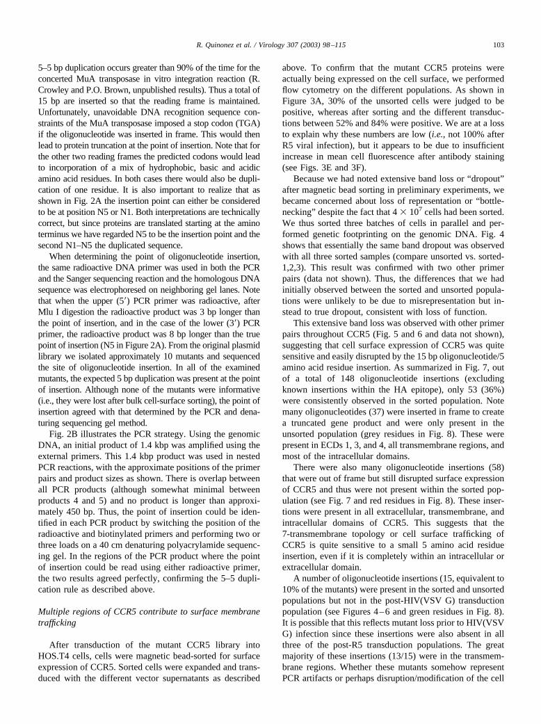
Because we had noted extensive band loss or “dropout”after magnetic bead sorting in preliminary experiments, webecame concerned about loss of representation or “bottle-necking” despite the fact that 4 � 107 cells had been sorted.We thus sorted three batches of cells in parallel and per-formed genetic footprinting on the genomic DNA. Fig. 4shows that essentially the same band dropout was observedwith all three sorted samples (compare unsorted vs. sorted-1,2,3). This result was confirmed with two other primerpairs (data not shown). Thus, the differences that we hadinitially observed between the sorted and unsorted popula-tions were unlikely to be due to misrepresentation but in-stead to true dropout, consistent with loss of function.
This extensive band loss was observed with other primerpairs throughout CCR5 (Fig. 5 and 6 and data not shown),suggesting that cell surface expression of CCR5 was quitesensitive and easily disrupted by the 15 bp oligonucleotide/5amino acid residue insertion. As summarized in Fig. 7, outof a total of 148 oligonucleotide insertions (excludingknown insertions within the HA epitope), only 53 (36%)were consistently observed in the sorted population. Notemany oligonucleotides (37) were inserted in frame to createa truncated gene product and were only present in theunsorted population (grey residues in Fig. 8). These werepresent in ECDs 1, 3, and 4, all transmembrane regions, andmost of the intracellular domains.
There were also many oligonucleotide insertions (58)that were out of frame but still disrupted surface expressionof CCR5 and thus were not present within the sorted pop-ulation (see Fig. 7 and red residues in Fig. 8). These inser-tions were present in all extracellular, transmembrane, andintracellular domains of CCR5. This suggests that the7-transmembrane topology or cell surface trafficking ofCCR5 is quite sensitive to a small 5 amino acid residueinsertion, even if it is completely within an intracellular orextracellular domain.
A number of oligonucleotide insertions (15, equivalent to10% of the mutants) were present in the sorted and unsortedpopulations but not in the post-HIV(VSV G) transductionpopulation (see Figures 4–6 and green residues in Fig. 8).It is possible that this reflects mutant loss prior to HIV(VSVG) infection since these insertions were also absent in allthree of the post-R5 transduction populations. The greatmajority of these insertions (13/15) were in the transmem-brane regions. Whether these mutants somehow representPCR artifacts or perhaps disruption/modification of the cell
103R. Quinonez et al. / Virology 307 (2003) 98–115

surface membrane such that not even VSV G pseudotypes areinfectious is unknown; since these mutants were not present inany of the post-infection populations they did not contribute toor interfere with the analysis of CCR5 function.
CCR5 is tolerant to oligonucleotide insertions
It was already known that short peptide epitopes could beplaced within the first ECD of CCR5 and still retain co--
receptor function. For example, the starting CCR5 constructfor these investigations has an eight amino acid HA epitopetag inserted at residue 14 (Liu et al., 1996). However, evenplacement of this epitope proved difficult since the impor-tance of the tyrosine residues in that first domain wereunappreciated at the time of vector construction (N. Landau,personal communication). This genetic footprinting proce-dure also identifies points of oligonucleotide insertion that
Fig. 3. Cell surface expression of CCR5. The six different HOS.T4 cell populations were analyzed by flow cytometry using an indirect method for surfaceexpression of CCR5. (A) Post-MLV transduction and unsorted, (B) post-magnetic bead sorted, (C) post-HIV (VSV G) transduction, (D) post-HIV (ADA)transduction, (E) post-HIV (BaL) transduction, and (E) post-HIV (SF162) transduction. Grey histograms: goat anti-mouse-FITC (GAM-FITC) alone added,black histograms, 12CA5 and GAM-FITC added. Bars and percentages indicate positive population (gated at �1% background).
104 R. Quinonez et al. / Virology 307 (2003) 98–115

Fig. 4. Genetic footprinting of CCR5, region 406–716. Genomic DNA was prepared from the cell populations shown in Figure 3 and nested PCR performed.In this case, the 627U primer was labelled with P-32. After M1u I digestion, products were size fractionated on a denaturing polyacrylamide gel along withthe homologous sequence and subjected to autoradiography. Sorted-1, Sorted-2, Sorted-3 are the three cell samples sorted in parallel for surface CCR5expression. * is the full-length, undigested PCR product (311 nt), closed arrowheads indicate products observed in the unsorted population only, and openarrowheads indicate products observed in the sorted population as well. Refer to Figure 7 for bp numbering.

Fig. 5. Genetic footprinting of CCR5, region 60–496. Here, the 693B primer was labeled with P-32. Please refer to Figure 4 for experimental details andlegend key.
106 R. Quinonez et al. / Virology 307 (2003) 98–115

are tolerated by CCR5 (depending upon the required func-tional tests, of course).
Multiple oligonucleotide insertions (26) were present inall the cell populations analyzed: unsorted, sorted, post-HIV(VSV G), and post-R5 viral transductions (see Figs.4–6 and blue residues in Fig. 8). Surprisingly, nearly half ofthese were in the transmembrane regions. There were 0–2insertions in most of the extracellular and intracellular do-mains, with the exception of the cytoplasmic tail, wherethere were 6 tolerated oligonucleotide insertions. This is notunexpected, since the cytoplasmic tail is not required forco-receptor function (Alkhatib et al., 1997) and C-terminalgreen fluorescent proteins are fully active (D. Littman, per-sonal communication). Whether all of these would be stillbe present if other functional tests were to be applied (e.g.,calcium/cellular signalling, chemokine binding, or cellularchemotaxis) is unknown.
Residues in the third and fourth ECD are critical forCCR5 co-receptor function
Of the 148 mutant CCR5 proteins, only 8 were present inthe unsorted, sorted, and post-HIV(VSV G) genomic DNAmaterial, but reduced or absent in the post-R5 envelopegenomic DNA. These oligonucleotide insertions were foundwithin the third ECD (ECL2), the fourth transmembraneregion, and the third intracellular loop (Figure 4 and yellowresidues in Figure 8). The first Cys in the third ECD (residue184 here) is thought to make a critical disulfide bond link-age with the Cys in the second ECD (residue 107 here, seeFigure 8). This bond is likely important for the properfolding and function of CCR5. Note one oligonucleotideinsertion at C184 led to protein truncation and was onlypresent in the unsorted population, whereas the other inser-tion was at bp 551 and was specifically absent from thepost-R5 envelope transduction populations (Figure 4). Thisinsertion results in the amino acid sequence . . . Tyr-Thr-Cys-Asp-Ala-Ser-Thr-Cys-Ser-Ser . . . , where the under-lined portion are the novel residues. Although it appears tobe expressed on the cell surface, this protein may be mis-folded because of the presence of a second Cys or alterna-tively critical determinants for CCR5 co-receptor functionmay be disrupted by this insertion.
The second oligonucleotide insertion specifically re-duced in the post-R5 envelope transduction populations waswithin the Tyr residue 193. This insertion results in theamino acid sequence. . . . Ser-Gln-Leu-Thr-Arg-His-Gln-Tyr-Gln . . . , where the novel residues are underlined. Al-though expressed on the cell surface, this mutant CCR5 wasreduced by �90% after HIV(ADA), HIV(BaL), andHIV(SF162) transductions. Presumably it too disrupts crit-ical co-receptor determinants.
The third and fourth oligonucleotide insertions specifi-cally reduced in the post-R5 envelope transduction popula-tions led to truncated proteins after the Trp residue 196 andthe Gln residue 200, respectively. Since both of these pro-
Fig. 6. Genetic footprinting of CCR5, region 686 to C-terminus. In thiscase, the 907U primer was labelled with P-32. Please refer to Figure 4 forexperimental details and legend key. Grey arrowheads indicate insertions3� of the stop codon at position 1075.
107R. Quinonez et al. / Virology 307 (2003) 98–115

teins were expressed on the cell surface, they likely havealtered conformations that will not support co-receptorfunction. Alternatively, there may be critical determinantsof co-receptor function that are C-terminal to these residues(see below).
The fifth and sixth oligonucleotide insertions variablypresent within the post-R5 envelope transduction popula-tions were at nucleotide positions 649 and 650 within theVal residue 217 (thought to lie within the fifth transmem-brane domain) (Figs. 4 and 8). These insertions gave rise toinserted peptides. . . . Val-Met-Val-Thr-Arg-His-Met-Val-Ile. . . . and. . . . Val-Met-Val-Asp-Ala-Ser-Met-Val-Ile. . . . , respectively, where the underlined portion is the
novel sequence. It is possible that the presence of the singlecharged residue (Arg or Asp) within the transmembranedomain disrupted conformation to such an extent these werereduced in the R5 envelope populations (despite the factthey are trafficked to the cell surface).
The seventh and eighth oligonucleotide insertions absentwithin the post-R5 envelope transduction populations wereat positions 688 and 691 (Fig. 4). These insertions led toprotein truncations after amino acid residues Arg 229 andCys 230, respectively (third intracellular domain, Fig. 8).These insertions were somewhat unusual in that they werepresent in the unsorted population, consistently reduced inthe post-sorted population, quite prominent in the post-
Fig. 7. Points of insertion of the oligonucleotide. Closed arrowheads indicate products only observed in the unsorted population, and open arrowheads indicateproducts observed in the sorted population as well. Bold type indicates the HA tag. Note the first residue of the epitope (Y) is also part of CCR5. In addition,two residues (Y15 and T16) of wild-type CCR5 were removed during the addition of the epitope, which did not appear to compromise co-receptor function.Underlined residues indicate putative transmembrane domains.
108 R. Quinonez et al. / Virology 307 (2003) 98–115

HIV(VSV G) transduction population, and virtually absentin all three of the post-R5 transduction populations (Fig. 4).It is unknown whether C230 forms a disulfide bond withany of the other intracellular Cys residues. These two oli-gonucleotide insertions both suggest that there are criticalCCR5 co-receptor determinants C-terminal to C230, quitepossibly within the fourth ECD.
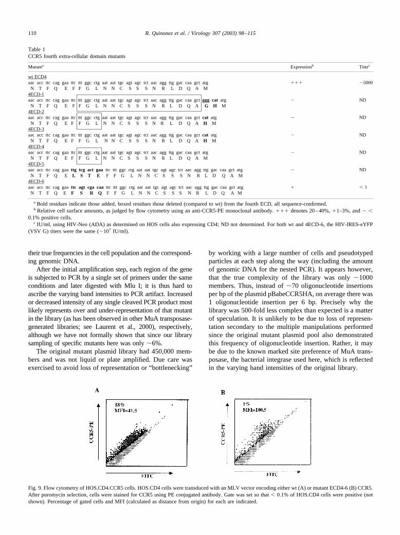
To address this question more directly, we made severalmutations in the highly conserved fourth ECD (Table 1).Some of these added one to a few residues, others deleted afew to most of the ECD. Of note, only one of the mutantswas expressed on the cell surface (4ECD-6), although thepercentage of positive cells, as judged by flow cytometry,was reduced 20-fold compared to wt control (Fig. 9). Thepositive mutant CCR5 cells, however, had the same, if notgreater, mean fluorescence intensity (MFI) as wt control,whereas the titer of HIV-Neo (ADA) on 4ECD-6 expressingcells was reduced more than 5,000-fold compared to wtcontrol (Table 1).
Discussion
Utility of genetic footprinting
We have described here the use of genetic footprinting toanalyze the function of a cellular eukaryotic gene. Previously,the use of this technique has been confined to the study of abacterial suppressor tRNA, the cis-acting sequences of the HIVgenome, and a retroviral envelope. The study of CCR5 co-receptor function adds a level of complexity since specificamino acid residues were introduced and multiple complexfunctional assays performed. Because we were analyzing in-sertions in mammalian genomic DNA, the use of PCR wasunavoidable and we cannot exclude representational bias afternested amplification. However, when comparing nested PCRperformed on the original plasmid DNA pool vs. the genomicDNA of the unsorted population, there was no clear represen-tational loss or gain, suggesting that the points of oligonucle-otide insertion and their relative intensities after PCR reflect
Fig. 8. Schematic of CCR5 structure. Plasma membrane is indicated by the curved cyan lines; ECDs are at top, intracellular loops at bottom (amino andcarboxy termini indicated). Black residues indicate the HA epitope tag, grey residues truncations at that position present only in the unsorted population, redresidues non-truncating insertions at that position present in the unsorted population only, blue residues insertions at that position present in all populations,green residues insertions at that position present in the unsorted and sorted populations only, and yellow residues insertions at that position present in theunsorted, sorted, and post- HIV(VSV G) but absent in the post-R5 envelope populations. Split circles indicate multiple insertions at that residue.
109R. Quinonez et al. / Virology 307 (2003) 98–115
their true frequencies in the cell population and the correspond-ing genomic DNA.
After the initial amplification step, each region of the geneis subjected to PCR by a single set of primers under the sameconditions and later digested with Mlu I; it is thus hard toascribe the varying band intensities to PCR artifact. Increasedor decreased intensity of any single cleaved PCR product mostlikely represents over and under-representation of that mutantin the library (as has been observed in other MuA transposase-generated libraries; see Laurent et al., 2000), respectively,although we have not formally shown that since our librarysampling of specific mutants here was only �6%.
The original mutant plasmid library had 450,000 mem-bers and was not liquid or plate amplified. Due care wasexercised to avoid loss of representation or “bottlenecking”
by working with a large number of cells and pseudotypedparticles at each step along the way (including the amountof genomic DNA for the nested PCR). It appears however,that the true complexity of the library was only �1000members. Thus, instead of �70 oligonucleotide insertionsper bp of the plasmid pBabeCCR5HA, on average there was1 oligonucleotide insertion per 6 bp. Precisely why thelibrary was 500-fold less complex than expected is a matterof speculation. It is unlikely to be due to loss of represen-tation secondary to the multiple manipulations performedsince the original mutant plasmid pool also demonstratedthis frequency of oligonucleotide insertion. Rather, it maybe due to the known marked site preference of MuA trans-posase, the bacterial integrase used here, which is reflectedin the varying band intensities of the original library.
Table 1CCR5 fourth extra-cellular domain mutants
Mutanta Expressionb Titerc
wt ECD4aac acc ttc cag gaa ttc ttt ggc ctg aat aat tgc agt agc tct aac agg ttg gac caa gct atg ��� �5000N T F Q E F F G L N N C S S S N R L D Q A M
4ECD-1aac acc ttc cag gaa ttc ttt ggc ctg aat aat tgc agt agc tct aac agg ttg gac caa gct ggg cat atg � ND
N T F Q E F F G L N N C S S S N R L D Q A G H M4ECD-2aac acc ttc cag gaa ttc ttt ggc ctg aat aat tgc agt agc tct aac agg ttg gac caa gct cat atg � ND
N T F Q E F F G L N N C S S S N R L D Q A H M4ECD-3aac acc ttc cag gaa ttc ttt ggc ctg aat aat tgc agt agc tct aac agg ttg gac caa gct cat atg � ND
N T F Q E F F G L N N C S S S N R L D Q A H M4ECD-4aac acc ttc cag gaa ttc ttt ggc ctg aat aat tgc agt agc tct aac agg ttg gac caa gct atg � ND
N T F Q E F F G L N N C S S S N R L D Q A M4ECD-5aac acc ttc cag gaa ttg tcg act gaa ttc ttt ggc ctg aat aat tgc agt agc tct aac agg ttg gac caa gct atg � ND
N T F Q E L S T E F F G L N N C S S S N R L D Q A M4ECD-6aac acc ttc cag gaa ttc agt cga caa ttc ttt ggc ctg aat aat tgc agt agc tct aac agg ttg gac caa gct atg � � 1
N T F Q E F S R Q F F G L N N C S S S N R L D Q A M
a Bold residues indicate those added, boxed residues those deleted (compared to wt) from the fourth ECD, all sequence-confirmed.b Relative cell surface amounts, as judged by flow cytometry using an anti-CCR5-PE monoclonal antibody. ��� denotes 20–40%, �1–3%, and � �
0.1% positive cells.c IU/ml, using HIV-Neo (ADA) as determined on HOS cells also expressing CD4; ND not determined. For both wt and 4ECD-6, the HIV-IRES-eYFP
(VSV G) titers were the same (�107 IU/ml).
Fig. 9. Flow cytometry of HOS.CD4.CCR5 cells. HOS.CD4 cells were transduced with an MLV vector encoding either wt (A) or mutant ECD4-6 (B) CCR5.After puromycin selection, cells were stained for CCR5 using PE conjugated antibody. Gate was set so that � 0.1% of HOS.CD4 cells were positive (notshown). Percentage of gated cells and MFI (calculated as distance from origin) for each are indicated.
110 R. Quinonez et al. / Virology 307 (2003) 98–115

This insertional bias was previously observed when an-alyzing the cis-acting elements of the HIV genome, wherethe original library had similar complexity to the one de-scribed here and yet the frequency of oligonucleotide inser-tion was approximately 1 every 7 bp (Laurent et al., 2000).Similar bias was observed in the study of ecotropic MLVenvelope (Rothenberg et al., 2001). The retroviral integrasesdo not appear to have such marked site preferences whennaked DNA is used as a template (Brown et al., 1987;Pryciak and Varmus, 1992; Singh et al., 1997). Unfortu-nately, they are also more difficult to purify in soluble activeform and have less intrinsic concerted integration enzymaticactivity in comparison to MuA transposase.
We also endeavored to simplify genetic footprinting bynot attempting to subclone the integration product but in-stead left it in the MLV-based vector. Previously, the inte-gration products were cloned into a small plasmid vector orinto the HIV genome (present within a plasmid vector).Thus, each member of the mutant plasmid library wouldhave at most a single oligonucleotide insertion only withinthe region of interest but construction of the library is morelaborious. Here, 15–20% of the members of the mutantplasmid library had a single oligonucleotide insertion withinthe coding sequence of CCR5 (the rest had none), but thelibrary was relatively easy to construct since the linearconcerted integration product was simply recircularized.Thus, to attain the same complexity as the subcloned li-brary, the direct library would have to be 5-fold greater insize in this specific case. But since the true complexity ofthese libraries was only a few thousand (see above), this isreadily accomplished. Achievement of equivalent complex-ity will obviously depend upon the size of both the plasmidand the region of interest. Direct libraries might be favoredwhen the region of interest is large and the plasmid is small,whereas subcloned libraries are preferred when the regionof interest is small and the plasmid large.
As in other studies using genetic footprinting, we did notattempt to reconstruct any of the specific CCR5 mutants inthe library. Even with a complexity of only 1000 members,direct isolation of the mutants of greatest interest would benon-trivial, and as mentioned above the specific mutantsisolated here were unfortunately all non-informative. Re-construction of select mutants would also be difficult (al-though not impossible). Indeed, the strength of this methodlies in the fact that the mutants are not analyzed individuallybut in bulk. When working well, the technique of geneticfootprinting should allow the parallel analysis of thousandof mutants in any of a number of functional selections,limited only by the parameters of the biological system (seebelow).
Genetic footprinting is non-trivial. Complexity of theanalyses grows substantially as the region of interest be-comes larger and as the number of functional analysesincreases. Cleaner results are obtained when the functional
selection is strong. Sorting for surface expression as de-scribed here is a moderately strong functional selection,although there is an unavoidable background (see Figure 3).Survival after lentiviral transduction is a strong functionalselection since the untransduced cells die and the transducedcells are expanded logarithmically (and the background isalmost non-existent). We did not attempt other functionalselections of CCR5 such as G-coupled calcium signaling,chemokine ligand binding, or cellular chemotaxis sincethose selections are likely to be considerably weaker and notabsolute.
Cell surface expression of CCR5
It is perhaps not surprising that approximately two-thirds(64%) of the oligonucleotide insertions resulted in a proteinthat did not get trafficked to the cell membrane, especiallysince many led to premature truncation. The well-known32 allele causes a frame-shift mutation and a prematurestop codon within the third ECD, resulting in a protein thatis expressed in the cell but not on the cell membrane (Liu etal., 1996; Samson, 1996). What is more surprising is thathalf of the insertions reported here were in either the puta-tive extracellular or intracellular domains, and half withinthe transmembrane regions (approximating the distributionof residues). This counter-intuitive result suggests that thetopological constraints of the 7-transmembrane proteinsmight be much greater than previously thought.
Of course, insertion of five amino acids any where inCCR5 might greatly disturb trafficking or function (reflect-ed here in the few mutants that survived the cell sorting).Efforts to develop “replacement” instead of “insertion” ge-netic footprinting libraries, in which a type II restrictionenzyme (e.g., Bsg I) is used to recircularize the plasmidafter the concerted integration reaction, are underway. Be-cause a few residues are simply replaced with an equivalentnumber, the mutagenesis should be less disruptive to overallprotein topology and structure.
It is instructive to examine the insertions that were ac-tually tolerated. Surprisingly, most (87%) of the onespresent in the sorted and unsorted populations were presentin the predicted transmembrane domains, again underscor-ing how little is known regarding the conformational re-quirements of CCR5. This is despite the fact that the inser-tion invariably leads to the addition of one or two chargedresidues (see Figure 2A). This was also true for the inser-tions present in all the cell populations, although there wasa slight bias towards insertion in the carboxy terminal cy-toplasmic domain (5 out of 50 compared to 21 out of 308).Note there were fully tolerated insertions in each of the fourECDs (Nt and ECL1–3), suggesting possible placement ofadditional epitope tags. This may be useful for structuralstudies using antibody reagents and fluorescence resonanceenergy transfer.
111R. Quinonez et al. / Virology 307 (2003) 98–115

Co-receptor function of CCR5
Of nearly 150 insertions in CCR5 analyzed here, only aminority was informative regarding co-receptor function.Previous investigations had identified critical residuesthroughout CCR5, especially in the first ECD (Dragic,1998; Farzan et al., 1998; Ross et al., 1998). Unfortunately,in that region (Nt) only a single insertion at K32 wasinformative here, and that was present in all populations.Thus, conclusions here are limited to examination of theinformative mutations in the other domains. Of interest isthe insertion in C184, which likely forms a covalent disul-fide bond with C107 (Berger et al., 1999). C184 is recreatedupon the oligonucleotide insertion, but the downstream res-idues are altered (see above). S186, which had been previ-ously identified by substituting mouse residues into humanCCR5 as being critical for co-receptor function (Ross et al.,1998), thus shifts position by five residues. In addition,other previously identified critical residues just C-terminalto S186, notably P189, Y190, and S191 (Kuhmann et al.,1997; Ross et al., 1998), would also be shifted by the sameamount. It may thus be difficult to separate direct fromindirect effects of the insertion at C184.
The same is true for the other three insertions (Y193,W196, and Q200) in ECL2 that specifically affected co-receptor function. The latter two resulted in protein trunca-tions, which may have disrupted the loop structure or elim-inated downstream determinants. For example, K203Aenhances the D11A loss-of-function mutation for R5X4envelopes (Doranz et al., 1997). No co-receptor functionhad previously been assigned to Y193. The effect observedat that residue may be direct or indirect, considering that itis a closed loop. There must be some flexibility in this loopstructure, however, because insertions at R174 and E178were well tolerated.
Can any conclusions be drawn from the truncating mu-tations at R229 and C230? The two major possibilities arethat these truncating mutations are either structurally desta-bilizing (despite the fact they are expressed on the cellsurface) or there are co-receptor determinants C-terminal, inthe fourth ECD (ECL3). With regard to the latter, the CCR5chimera studies have not been informative since the mousesequence is identical to that of the human. Alanine-scanningmutagenesis has demonstrated the importance of D282 (inthe context of D11A) for R5X4 strains (Doranz et al., 1997).This effect was not observed for the R5 strain JR-FL,although it is clear that the fourth ECD alone (in the contextof murine CCR5) is insufficient for co-receptor function(Bieniasz et al., 1997). Of the mutants in the fourth ECDthat we examined here, only one (4ECD-6) was informativein the sense that it was actually present on the cell surface.Minimal disruption of that loop (e.g., addition of one resi-due) led to undetectable surface expression. Based uponflow cytometry, 4ECD-6 was expressed on the cell surface,and although the percentage of positive cells was reduced20-fold compared to wild-type, the amount of expression on
a per cell basis (as quantitated by MCF) was the same, if nothigher. Thus, the fact that M-tropic viral titers were reducedapproximately four log10s is consistent with ECL3 playinga role in co-receptor function and probably deserves furtherexploration.
Previous investigations with mouse-human CCR5 chi-meras revealed differing requirements for the extracellularloops for strains ADA, SF162, and BaL, with BaL being themost sensitive to the presence of murine sequences (Bien-iasz et al., 1997; Picard et al., 1997). To a first approxima-tion, we did not observe substantial differences between thethree envelopes studied here, and whether that reflects thenature of the insertional mutagenesis versus the sensitivityof the PCR assay is unknown.
In summary, this report describes the application of ge-netic footprinting to the HIV co-receptor CCR5. Althoughthe density of the insertion oligonucleotides was lower thanexpected, the important conclusions are that surface expres-sion of CCR5 is relatively intolerant to 5 amino acid residueinsertions, and critical co-receptor determinants lie withinECL2 and possibly ECL3. This is consistent with previousinvestigations using envelopes of R5 HIV isolates. It isentirely conceivable that a denser insertion library or alter-natively a replacement genetic footprinting library may bemore illuminating, depending upon the topological con-straints of the gene under study.
Materials and methods
Plasmids
HIV-1 envelope expression plasmids pSM-ADA, pSM-BaL, and pSM-SF162 were gifts of Dan Littman (SkirballInstitute, NYU Medical Center). MLV Gag-Pol expressionconstruct pHIT60 (Soneoka et al., 1995) and pBa-beCCR5-HA (Liu et al., 1996) were obtained from AlanKingsman (Oxford) and Ned Landau (Salk Institute), re-spectively. HIV-blasti and HIV-Neo were constructed bysubstituting the coding region of alkaline phosphatase of thevector pHIV-APenvvifvpr (Sutton et al., 1998) withthat of either the blasticidin resistance gene from pcDNA6/V5-HisA (Invitrogen) or SV-Neo (from pBabeNeo), respec-tively. HIV-IRES-eYFP has been reported previously(Schroers et al., 2000), as has the VSV G expression plas-mid pME VSV G (Sutton et al., 1998).
Oligonucleotide insertion library of CCR5
Purified MuA transposase (gift of Harry Savilahti) wasused to create a library of insertional mutations in pBa-beCCR5-HA using modifications of described methods(Singh et al., 1997; Lauren et al., 2000). The donor oligo-nucleotide duplex used to create the insertions contained therecognition sequences for both the MuA transposase andMluI restriction endonuclease. This duplex was generated
112 R. Quinonez et al. / Virology 307 (2003) 98–115
by annealing together the oligonucleotides MuAMlu15A(5�-TGACGCGTCGCACGAAAAACGCGAAAGCGTTTC-ACGATAAATGCGAAAAC-3�) and MuAMlu15B (5�-GTT-TTCGCATTTATCGTGAAACGCTTTCGCGTTTTTCG-TGCGACGCGTCA-3�). The transposition reaction wasperformed by incubating 50 nM donor oligo duplex, 10 �gpBabeCCR5-HA, and 100nM MuA transposase in a buffercontaining 25 mM Tris–HCl pH 8.0, 144 mM NaCl, 10 mMMgCl2, 100 �g/ml bovine serum albumin, 15% (w/v) glycerol,15% (v/v) dimethyl sulfoxide and 0.1% (w/v) Triton X-100 for1 hour at 30°C. The reaction was extracted with phenol-chloroform and precipitated with ethanol. Plasmids that hadundergone a concerted integration of two oligonucleotides (andtherefore linear) were separated by horizontal agarose gel elec-trophoresis and purified. The five-base gap resulting from thetransposition was repaired by nick translation with Taq DNApolymerase. The resulting molecules were cleaved with MluI,recircularized using T4 DNA ligase, and transformed into E.coli strain DH10B by electroporation. The resulting librarycontained over 450,000 independent transformants, each withthe 15 bp insertion containing the MluI recognition sequencelocated at a random position in the pBabeCCR5-HA plasmid(see Figs. 1 and 2).
Cells and vector supernatants
293T cells were maintained in Dulbecco’s modified Ea-gle’s medium (DMEM) supplemented with 10% fetal calfserum (FCS, from Gibco-BRL) and penicillin-streptomycin.Hos.T4 cells which express human CD4 under the control ofan SV40 promoter were a gift of N. Landau and weremaintained in complete DMEM supplemented with 40�g/ml mycophenolic acid (MPA, Sigma) with xanthine andhypoxanthine. Hos.T4.CCR5 cells that express both CD4and wild-type CCR5 were a gift of D. Littman and weremaintained in 5 �g/ml puromycin (Sigma).
Pseudotyped HIV vector supernatants were produced bycalcium-phosphate co-transfection of 293T cells (Sutton etal., 1998) with pHIV-blasti and the relevant envelope ex-pression construct, titered on Hos.T4.CCR5 cells usingcomplete DMEM supplemented with 10 �g/ml blasticidin S(Invitrogen). HIV-IRES-eYFP (VSV G) was prepared sim-ilarly, 1 ml of supernatant was added to targets (HOS.T4cells expressing either wt or mutant CCR5) in 12-wellplates, and 72 h later cells were subjected to flow cytometry,gating on the eYFP � cells. HIV-Neo (ADA) was similarlyprepared, and 72 h after transduction, cells were positivelyselected by passaging at a ratio of 1:10 in completeDME supplemented with 1.0 mg/ml G418 (Invitrogen).Pseudotyped MLV vector supernatant was produced bycalcium-phosphate co-transfection of 293T cells usingpHIT60, pME VSV G, and the mutant pBabeCCR5-HAlibrary or appropriate MLV vector construct. It was titeredusing Hos.T4 cells as targets and also stored at �80°C.
Sorting for surface expression
Approximately 5 �107 HOS.T4 cells were transducedwith 2.4 � 106 IU of the pseudotyped MLV stock describedabove (m.o.i.�0.05), selected in DMEM containing bothMPA and puromycin, and resistant colonies were pooledand expanded. Approximately 4 � 107 cells were incubatedfor 60 min at 4°C with 50 �g of anti-HA mouse monoclonalantibody 12CA5 (Boehringer-Mannheim) in PBS contain-ing 2% FCS (PBS-FCS). Cells were washed three times inPBS-FCS and bound to goat anti-mouse-coated magneticbeads (Miltenyi-Biotec). Cells and beads were then appliedto a midi-MACS column (Miltenyi-Biotec). After washingextensively, bound cells were eluted according to the man-ufacturer’s instructions.
Recovered cells were expanded in complete DMEM sup-plemented with MPA and puromycin. Flow cytometry wasperformed by staining approximately 106 cells using 2 �g of12CA5 and goat anti-mouse-fluorescein isothiocyanate(FITC) conjugate as a secondary antibody and analyzingthem on a FACScan (Becton-Dickinson) using CellQuestsoftware. Gates were established at the �1% level usingcells stained with the secondary antibody alone.
Genetic footprinting
Positively sorted cells above were independently trans-duced with at least 106 IU of HIV-blasti(VSV G), HIV-blasti (ADA), HIV-blasti (BaL), and HIV-blasti (SF162).Transduced cells were selected in complete DMEM con-taining 10 �g/ml blasticidin S, and expanded. GenomicDNA from approximately 2.5 � 107 cells was prepared bySDS-proteinase K lysis and phenol:chloroform extraction.Nested PCR was performed starting with 0.8 �g of genomicDNA (approximately 100,000 cell equivalents) using theinitial primer pair 5�-GACCCTCCCTTTATCCAGCCCT-CACTCCTT-3� (Gag-U) and 5�-CCGTGGGCTTGTAC-TCGGTCATGGTAAGCT-3� (Puro-L). Conditions using 5U of Taqpluslong (Boehringer-Mannheim) were 40 cyclesof 94°C for 30 sec, 62°C for 30 sec, and 68° C for 90 sec toobtain a 1.4 kb product. Ten–20 ng of this product was usedfor the subsequent PCR reaction, using the following primerpairs (one of which was 5� end-labeled with biotin and theother 32-P): 5�-TTATACATCGGAGCCCTGCCAAA-3�(281U, 3� end at position 82) and 5�-AGCAAACACAG-CATGGACGACAG-3� (625L, 3� end at position 404), totallength 367 bp; 5�-CCCTCCCTTTATCCAGCCCTCAC-3�(BgagU) and 5�-TGGCCAGGTTGAGCAGGTAGATG-3�(437L, 3� end at position 216), approximate total length 280bp); 281U and 5�-ACGCAAACACAGCCACCACCC-AAGT-3� (693B, 3� end at position 472), total length 437bp; 5�-GTCGTCCATGCTGTGTTTGCTTT-3� (627U, 3�end at position 428) and 5�-GCCCTGTGCCTCTTCTTCT-CATT-3� (915L, 3� end at position 694), total length 311;and 5�-GGTGTCGAAATGAGAAGAAGAGG-3� (907U,3� end at position 708) and 5�-GGGACTTTCCACCCTA-
113R. Quinonez et al. / Virology 307 (2003) 98–115

ACTG-3� (1460L), approximate length 450 bp. Reactionconditions were 15 cycles of 94°C for 30 sec, 62°C for 30sec, and 72°C for 30 sec using 5 U Taq DNA polymerase.Full-length PCR products were gel-isolated after size sepa-ration by horizontal agarose gel electrophoresis and thenbound to streptavidin-agarose beads (SIGMA). After wash-ing unbound material, beads were incubated with 10 U MluI for 60 min at 37°C. Beads were pelleted by centrifugation,and released DNA in the supernatant was precipitated withethanol using glycogen as carrier. Mlu I-cleaved PCR prod-ucts were electrophoresed on 40 cm 7M urea-6% polyacryl-amide 0.4mm sequencing gels, along with the homologousdideoxy sequence. For quantitation of specific products,Cerenkov counts were obtained for the full length PCRproducts prior to binding to streptavidin-agarose beads (fornormalization purposes), and individual bands were quan-titated using a Betagen blot analyzer system.
Construction of mutations in the fourth ECD of CCR5and analysis of surface expression
4ECD-1 was constructed by PCR using pBabeCCR5-HAas a template with the following primers 5�-GGG-AATTCGGGCATATGCAGGTGACAGAGACTCTTGG-3�(Eco 4U) and 5�-GGGTCGACTCACAAGCCCACA-GATATTTCCTG-3� (Eco 4L). The �300 bp product was firstcloned into pCR 2.1-TOPO (Invitrogen), sequence confirmed,and then directionally ligated into pBabeCCR5-HA pre-cleaved with Eco RI and Sal I. 4ECD-2 was constructed bycleaving 4ECD-1 with Eco RI and Nde I and inserting a duplexoligonucleotide, with top strand sequence 5�-AATT-CAATAATTGCAGTAGCTCTAACAGGTTGGACCAAG-CTCA-3�. 4ECD-3 was constructed by cleaving 4ECD-1 withEco RI and Nde I and inserting a duplex oligonucleotide withtop s t rand sequence of 5 � -AATTCTTTGGCC-TGAATAATTGCAGTAGCTCTAACAGGTTGGACCAAG-CTCA-3�. 4ECD-4 was constructed by PCR using pBa-beCCR5-HA as a template and the following primers: 5�-GG-GAATTCAATAATTGCAGTAGCTCTAACAGGTTGG-3�and the above-mentioned Eco 4L primer. The �285 bp prod-uct was cloned into pBabeCCR5-HA as described for 4ECD-1.4ECD-5 and 4ECD-6 were constructed by cleaving pBa-beCCR5-HA with Eco RI and non-directionally ligating aduplex oligonucleotide with top strand sequence of 5�-AATTGTCGACTG- 3�. Both forward and reverse orientationclones were confirmed by DNA sequencing. The amino acidsequences of the mutant ECDs are shown in Table 1.
To examine cell surface expression of the 4ECD mutants,HOS.CD4 cells were transduced with either pBabeCCR5-HAor pBabeCCR5-HA-4ECD-6, both pseudotyped with VSV G.Stable transductants were selected using DME complete me-dium supplemented with 5 �g/ml puromycin. For surfacestaining, washed cells were incubated with PE-conjugated anti-human CCR5 mouse monoclonal antibody (FAB182P, R&DSystems) according to the manufacturer’s recommendationsand analyzed on a Becton-Dickinson FACScan. Gating was
established so that only 0.1% of the HOS.CD4 parental cellswere positive.
Acknowledgments
We would like to thank Dr. Patrick Brown (Stanford) forhis support of this project in its early stages, and Drs. NedLandau (Salk), Dan Littman (NYU), Sue and Alan Kings-man (Oxford), and Harry Savilahti (NIH) for generous re-agent gifts. This work was funded in part by the BaylorCenter for AIDS Research Core Support Grant numberAI36211 from NIAID, and R.Q. was supported by T32 AI07456. R.E.S. is an Edward Mallinckrodt, Jr. FoundationScholar.
References
Alkhatib, G., Locati, M., Kennedy, P.E., Murphy, P.M., Berger, E.A.,1997. HIV-1 coreceptor activity of CCR5 and its inhibition by chemo-kines: independence from G protein signaling and importance of co-receptor downmodulation. Virology 234, 340–348.
Atchison, R.E., Gosling, J., Monteclaro, F.S., Franci, C., Digilio, L., Charo,I.F., Goldsmith, M.A., 1996. Multiple extracellular elements of CCR5and HIV-1 entry: dissociation from response to chemokines. Science274, 1924–1926.
Berger, E.A., Murphy, P.M., Farber, J.M., 1999. Chemokine receptors asHIV-1 coreceptors: roles in viral entry, tropism, and disease. Annu.Rev. Immunol. 17, 657–700.
Berson, J.F., Doms, R.W., 1999. Structure-function studies of the HIV-1coreceptors. Semin. Immunol. 10, 237–248.
Bieniasz, P.D., Fridell, R.A., Aramori, I., Ferguson, S.S., Caron, M.G.,Cullen, B.R., 1997. HIV-1-induced cell fusion is mediated by multipleregions within both the viral envelope and the CCR-5 co-receptor.Embo. J. 16, 2599–2609.
Brown, P.O., Bowerman, B., Varmus, H.E., Bishop, J.M., 1987. Correctintegration of retroviral DNA in vitro. Cell 49, 347–356.
Doranz, B.J., Lu, Z.H., Rucker, J., Zhang, T.Y., Sharron, M., Cen, Y.H.,Wang, Z.X., Guo, H.H., Du, J.G., Accavitti, M.A., Doms, R.W., Peiper,S.C., 1997. Two distinct CCR5 domains can mediate coreceptor usageby human immunodeficiency virus type 1. J. Virol. 71, 6305–6314.
Dragic, T., 2001. An overview of the determinants of CCR5 and CXCR4co-receptor function. J. Gen. Virol. 82, 1807–1814.
Dragic, T., Trkola, A., Lin, S.W., Nagashima, K.A., Kajumo, F., Zhao, L.,Olson, W.C., Wu, L., Mackay, C.R., Allaway, G.P., Sakmar, T.P.,Moore, J.P., Maddon, P.J., 1998. Amino-terminal substitutions in theCCR5 coreceptor impair gp120 binding and human immunodeficiencyvirus type 1 entry. J. Virol. 72, 279–285.
Farzan, M., Choe, H., Vaca, L., Martin, K., Sun, Y., Desjardins, E.,Ruffing, N., Wu, L., Wyatt, R., Gerard, N., Gerard, C., Sodroski, J.,1998. A tyrosine-rich region in the N terminus of CCR5 is importantfor human immunodeficiency virus type 1 entry and mediates anassociation between gp120 and CCR5. J. Virol. 72, 1160–1164.
Kuhmann, S.E., Platt, E.J., Kozak, S.L., Kabat, D., 1997. Polymorphismsin the CCR5 genes of African green monkeys and mice implicatespecific amino acids in infections by simian and human immunodefi-ciency viruses. J. Virol. 71, 8642–8656.
Laurent, L.C., Olsen, M.N., Crowley, R.A., Savilahti, H., Brown, P.O.,2000. Functional characterization of the human immunodeficiency vi-rus type 1 genome by genetic footprinting. J. Virol. 74, 2760–2769.
Lee, B., Sharron, M., Blanpain, C., Doranz, B.J., Vakili, J., Setoh, P., Berg,E., Liu, G., Guy, H.R., Durell, S.R., Parmentier, M., Chang, C.N.,
114 R. Quinonez et al. / Virology 307 (2003) 98–115

Price, K., Tsang, M., Doms, R.W., 1999. Epitope mapping of CCR5reveals multiple conformational states and distinct but overlappingstructures involved in chemokine and coreceptor function. J. Biol.Chem. 274, 9617–9626.
Littman, D.R., 1998. Chemokine receptors: keys to AIDS pathogenesis?Cell 93, 677–680.
Liu, R., Paxton, W.A., Choe, S., Ceradini, D., Martin, S.R., Horuk, R.,MacDonald, M.E., Stuhlmann, H., Koup, R.A., Landau, N.R., 1996. Ho-mozygous defect in HIV-1 coreceptor accounts for resistance of somemultiply-exposed individuals to HIV-1 infection. Cell 86, 367–377.
Olson, W.C., Rabut, G.E., Nagashima, K.A., Tran, D.N., Anselma, D.J.,Monard, S.P., Segal, J.P., Thompson, D.A., Kajumo, F., Guo, Y.,Moore, J.P., Maddon, P.J., Dragic, T., 1999. Differential inhibition ofhuman immunodeficiency virus type 1 fusion, gp120 binding, andCC-chemokine activity by monoclonal antibodies to CCR5. J. Virol.73, 4145–4155.
Picard, L., Simmons, G., Power, C.A., Meyer, A., Weiss, R.A., Clapham,P.R., 1997. Multiple extracellular domains of CCR-5 contribute tohuman immunodeficiency virus type 1 entry and fusion. J. Virol. 71,5003–5011.
Pryciak, P.M., Varmus, H.E., 1992. Nucleosomes, DNA-binding proteins,and DNA sequence modulate retroviral integration target site selection.Cell 69, 769–780.
Rabut, G.E., Konner, J.A., Kajumo, F., Moore, J.P., Dragic, T., 1998.Alanine substitutions of polar and nonpolar residues in the amino-terminal domain of CCR5 differently impair entry of macrophage- anddualtropic isolates of human immunodeficiency virus type 1. J. Virol.72, 3464–3468.
Ross, T.M., Bieniasz, P.D., Cullen, B.R., 1998. Multiple residues contrib-ute to the inability of murine CCR-5 to function as a coreceptor formacrophage-tropic humanimmunodeficiency virus type 1 isolates.J. Virol. 72, 1918–1924.
Rossi, D., Zlotnik, A., 2000. The biology of chemokines and their recep-tors. Annu. Rev. Immunol. 18, 217–242.
Rothenberg, S.M., Olsen, M.N., Laurent, L.C., Crowley, R.A., Brown,P.O., 2001. Comprehensive mutational analysis of the Moloney murineleukemia virus envelope protein. J. Virol. 75, 11851–11862.
Rucker, J., Samson, M., Doranz, B.J., Libert, F., Berson, J.F., Yi, Y.,Smyth, R.J., Collman, R.G., Broder, C.C., Vassart, G., Doms, R.W.,Parmentier, M., 1996. Regions in beta-chemokine receptors CCR5and CCR2b that determine HIV-1 cofactor specificity. Cell 87,437– 446.
Sallusto, F., Mackay, C.R., Lanzavecchia, A., 2000. The role of chemokinereceptors in primary, effector, and memory immune responses. Annu.Rev. Immunol. 18, 593–620.
Samson, M., 1996. Resistance to HIV-1 infection in caucasian individualsbearing mutant alleles of the CCR-5 chemokine receptor gene. Nature382, 722–725.
Schroers, R., Sinha, I., Segall, H., Schmidt-Wolf, L.G.H., Rooney, C.M.,Brenner, M.K., Sutton, R.E., Chen, S.-Y., 2000. Transduction of humanPBMC-derived dendritic cells and macrophages by an HIV-1-basedlentiviral vector system. Molecular Therapy 1, 171–179.
Singh, I.R., Crowley, R.A., Brown, P.O., 1997. High-resolution functionalmapping of a cloned gene by genetic footprinting. Proc. Natl. Acad.Sci. USA 94, 1304–1309.
Soneoka, Y., Cannon, P.M., Ramsdale, E.E., Griffiths, J.C., Romano, G.,Kingsman, S.M., Kingsman, A.J., 1995. A transient three-plasmidexpression system for the production of high titer retroviral vectors.Nucleic Acids Res. 23, 628–633.
Sutton, R.E., Wu, H.T., Rigg, R., Bohnlein, E., Brown, P.O., 1998. Humanimmunodeficiency virus type 1 vectors efficiently transduce humanhematopoietic stem cells. J. Virol. 72, 5781–5788.
Wu, L., LaRosa, G., Kassam, N., Gordon, C.J., Heath, H., Ruffing, N.,Chen, H., Humblias, J., Samson, M., Parmentier, M., Moore, J.P.,Mackay, C.R., 1997. Interaction of chemokine receptor CCR5 with itsligands: multiple domains for HIV-1 gp120 binding and a single do-main for chemokine binding. J. Exp. Med. 186, 1373–1381.
115R. Quinonez et al. / Virology 307 (2003) 98–115