Genetic engineering of human NK cells to express CXCR2 ...
14
RESEARCH ARTICLE Open Access Genetic engineering of human NK cells to express CXCR2 improves migration to renal cell carcinoma Veronika Kremer 1 , Marteen A. Ligtenberg 2 , Rosa Zendehdel 1 , Christina Seitz 3 , Annet Duivenvoorden 1 , Erik Wennerberg 4 , Eugenia Colón 5,6 , Ann-Helén Scherman-Plogell 7 and Andreas Lundqvist 1,8* Abstract Background: Adoptive natural killer (NK) cell transfer is being increasingly used as cancer treatment. However, clinical responses have so far been limited to patients with hematological malignancies. A potential limiting factor in patients with solid tumors is defective homing of the infused NK cells to the tumor site. Chemokines regulate the migration of leukocytes expressing corresponding chemokine receptors. Various solid tumors, including renal cell carcinoma (RCC), readily secrete ligands for the chemokine receptor CXCR2. We hypothesize that infusion of NK cells expressing high levels of the CXCR2 chemokine receptor will result in increased influx of the transferred NK cells into tumors, and improved clinical outcome in patients with cancer. Methods: Blood and tumor biopsies from 14 primary RCC patients were assessed by flow cytometry and chemokine analysis. Primary NK cells were transduced with human CXCR2 using a retroviral system. CXCR2 receptor functionality was determined by Calcium flux and NK cell migration was evaluated in transwell assays. Results: We detected higher concentrations of CXCR2 ligands in tumors compared with plasma of RCC patients. In addition, CXCL5 levels correlated with the intratumoral infiltration of CXCR2-positive NK cells. However, tumor-infiltrating NK cells from RCC patients expressed lower CXCR2 compared with peripheral blood NK cells. Moreover, healthy donor NK cells rapidly lost their CXCR2 expression upon in vitro culture and expansion. Genetic modification of human primary NK cells to re-express CXCR2 improved their ability to specifically migrate along a chemokine gradient of recombinant CXCR2 ligands or RCC tumor supernatants compared with controls. The enhanced trafficking resulted in increased killing of target cells. In addition, while their functionality remained unchanged compared with control NK cells, CXCR2-transduced NK cells obtained increased adhesion properties and formed more conjugates with target cells. Conclusions: To increase the success of NK cell-based therapies of solid tumors, it is of great importance to promote their homing to the tumor site. In this study, we show that stable engineering of human primary NK cells to express a chemokine receptor thereby enhancing their migration is a promising strategy to improve anti-tumor responses following adoptive transfer of NK cells. Keywords: NK cells, Chemokines, CXCR2, Renal cell carcinoma, Adoptive cell therapy * Correspondence: [email protected] 1 Department of Oncology-Pathology, Karolinska Institutet, Stockholm, Sweden 8 Cell Therapy Institute, Nova Southeastern University, Fort Lauderdale, FL, USA Full list of author information is available at the end of the article © The Author(s). 2017 Open Access This article is distributed under the terms of the Creative Commons Attribution 4.0 International License (http://creativecommons.org/licenses/by/4.0/), which permits unrestricted use, distribution, and reproduction in any medium, provided you give appropriate credit to the original author(s) and the source, provide a link to the Creative Commons license, and indicate if changes were made. The Creative Commons Public Domain Dedication waiver (http://creativecommons.org/publicdomain/zero/1.0/) applies to the data made available in this article, unless otherwise stated. Kremer et al. Journal for ImmunoTherapy of Cancer (2017) 5:73 DOI 10.1186/s40425-017-0275-9 on November 21, 2021 by guest. Protected by copyright. http://jitc.bmj.com/ J Immunother Cancer: first published as 10.1186/s40425-017-0275-9 on 19 September 2017. Downloaded from on November 21, 2021 by guest. Protected by copyright. http://jitc.bmj.com/ J Immunother Cancer: first published as 10.1186/s40425-017-0275-9 on 19 September 2017. Downloaded from on November 21, 2021 by guest. Protected by copyright. http://jitc.bmj.com/ J Immunother Cancer: first published as 10.1186/s40425-017-0275-9 on 19 September 2017. Downloaded from
Transcript of Genetic engineering of human NK cells to express CXCR2 ...

RESEARCH ARTICLE Open Access
Genetic engineering of human NK cells toexpress CXCR2 improves migration to renalcell carcinomaVeronika Kremer1, Marteen A. Ligtenberg2, Rosa Zendehdel1, Christina Seitz3, Annet Duivenvoorden1,Erik Wennerberg4, Eugenia Colón5,6, Ann-Helén Scherman-Plogell7 and Andreas Lundqvist1,8*
Abstract
Background: Adoptive natural killer (NK) cell transfer is being increasingly used as cancer treatment. However,clinical responses have so far been limited to patients with hematological malignancies. A potential limiting factorin patients with solid tumors is defective homing of the infused NK cells to the tumor site. Chemokines regulatethe migration of leukocytes expressing corresponding chemokine receptors. Various solid tumors, including renalcell carcinoma (RCC), readily secrete ligands for the chemokine receptor CXCR2. We hypothesize that infusion of NKcells expressing high levels of the CXCR2 chemokine receptor will result in increased influx of the transferred NKcells into tumors, and improved clinical outcome in patients with cancer.
Methods: Blood and tumor biopsies from 14 primary RCC patients were assessed by flow cytometry and chemokineanalysis. Primary NK cells were transduced with human CXCR2 using a retroviral system. CXCR2 receptor functionalitywas determined by Calcium flux and NK cell migration was evaluated in transwell assays.
Results: We detected higher concentrations of CXCR2 ligands in tumors compared with plasma of RCC patients. Inaddition, CXCL5 levels correlated with the intratumoral infiltration of CXCR2-positive NK cells. However, tumor-infiltratingNK cells from RCC patients expressed lower CXCR2 compared with peripheral blood NK cells. Moreover, healthy donorNK cells rapidly lost their CXCR2 expression upon in vitro culture and expansion. Genetic modification of humanprimary NK cells to re-express CXCR2 improved their ability to specifically migrate along a chemokine gradient ofrecombinant CXCR2 ligands or RCC tumor supernatants compared with controls. The enhanced trafficking resulted inincreased killing of target cells. In addition, while their functionality remained unchanged compared with control NKcells, CXCR2-transduced NK cells obtained increased adhesion properties and formed more conjugates with target cells.
Conclusions: To increase the success of NK cell-based therapies of solid tumors, it is of great importance to promotetheir homing to the tumor site. In this study, we show that stable engineering of human primary NK cells to express achemokine receptor thereby enhancing their migration is a promising strategy to improve anti-tumor responsesfollowing adoptive transfer of NK cells.
Keywords: NK cells, Chemokines, CXCR2, Renal cell carcinoma, Adoptive cell therapy
* Correspondence: [email protected] of Oncology-Pathology, Karolinska Institutet, Stockholm,Sweden8Cell Therapy Institute, Nova Southeastern University, Fort Lauderdale, FL,USAFull list of author information is available at the end of the article
© The Author(s). 2017 Open Access This article is distributed under the terms of the Creative Commons Attribution 4.0International License (http://creativecommons.org/licenses/by/4.0/), which permits unrestricted use, distribution, andreproduction in any medium, provided you give appropriate credit to the original author(s) and the source, provide a link tothe Creative Commons license, and indicate if changes were made. The Creative Commons Public Domain Dedication waiver(http://creativecommons.org/publicdomain/zero/1.0/) applies to the data made available in this article, unless otherwise stated.
Kremer et al. Journal for ImmunoTherapy of Cancer (2017) 5:73 DOI 10.1186/s40425-017-0275-9
on Novem
ber 21, 2021 by guest. Protected by copyright.
http://jitc.bmj.com
/J Im
munother C
ancer: first published as 10.1186/s40425-017-0275-9 on 19 Septem
ber 2017. Dow
nloaded from
on Novem
ber 21, 2021 by guest. Protected by copyright.
http://jitc.bmj.com
/J Im
munother C
ancer: first published as 10.1186/s40425-017-0275-9 on 19 Septem
ber 2017. Dow
nloaded from
on Novem
ber 21, 2021 by guest. Protected by copyright.
http://jitc.bmj.com
/J Im
munother C
ancer: first published as 10.1186/s40425-017-0275-9 on 19 Septem
ber 2017. Dow
nloaded from

BackgroundNatural killer (NK) cells have received renewed attentionas an immunotherapeutic treatment against cancer inthe recent years. The advantage of NK cells is that theycan kill a wide spectrum of tumor cells without the re-quirement for specific antigen recognition. In differentanimal models, NK cells have been shown to eradicateengrafted tumors and, in particular, metastases. Adoptivetransfer of NK cells has successfully been used to treatpatients with leukemia and other hematological cancers[1–3]. However, clinical responses in patients with solidtumors treated with adoptively infused NK cells have sofar not been beneficial [4, 5]. A potential limiting factorfor the success of adoptive NK cell therapy is the ineffi-cient migration of NK cells to the tumor site [6–9],which may partially explain the poor clinical responsesin cancer patients. Several studies have correlated highnumbers of tumor-infiltrating NK cells with a goodprognosis in various cancers [10–12]. Renal cell carcin-oma (RCC) is particularly noteworthy as multipleindependent studies have associated intratumoral NKcell infiltration with increased survival of metastaticRCC patients [13–15], whereas CD8+ T cell infiltrationhas been indicative of a poor prognosis [16]. Therefore,improving NK cell infiltration into RCC tumors is anattractive therapeutic option to potentiate the successof NK cell-based therapies and eventually improveclinical responses.Chemokines are small secreted proteins that control
the recruitment of immune cells with correspondingchemokine receptors to sites of infection, tumors orother tissues. The presence of specific chemokines in thetumor microenvironment, including ligands for the che-mokine receptors CXCR3 and CX3CR1, has been shownto promote intratumoral infiltration of T and NK cellsand correlate with increased survival in a variety ofcancers [17–19]. We have recently shown a strategy toenhance the migration of CXCR3-positive human NKcells to melanoma tumors by stimulating local CXCL10secretion as CXCR3 ligands are not usually present atthe tumor site [20]. Ligands for the chemokine receptorCXCR2, on the other hand, are readily secreted by a var-iety of solid tumors, including RCC [21], to promoteangiogenesis, tumor growth and metastasis.The CXCR2 chemokine receptor is expressed on a
number of leukocytes, most prominently on neutrophilsand monocytes, facilitating their infiltration into solidtumors [22]. While CXCR2 is present on peripheralblood NK cells, we found that its expression is rapidlylost during their in vitro culture. We show here thatgenetic modification of human primary NK cells to re-express CXCR2 improves their ability to specificallymigrate along a tumor-derived chemokine gradientresulting in increased killing of target cells. In addition,
while their functionality remains unchanged, NK cellsincorporating the CXCR2 gene obtain increased adhe-sion properties. Thus, re-expression of CXCR2 throughgenetic engineering of ex vivo expanded NK cells repre-sents a novel strategy to improve anti-tumor responsesfollowing adoptive transfer of NK cells.
MethodsPatient samplesBlood and tumor biopsies were collected from 12 clearcell RCC patients, one papillary type 2 and one chromo-phobe RCC patient undergoing nephrectomy at the timeof surgery and 1 to 2 months after during the period ofApril 2014 till October 2015. The diagnosis was con-firmed histologically, and the cellular grading was deter-mined according to Fuhrman. All clear cell and papillaryRCC patients were Fuhrman grade II-IV. Plasma was ob-tained by centrifugation of the blood samples and storedat −20 °C until analysis. Whole blood was stained withappropriate antibody mixes and analyzed by flow cytom-etry. Tumor samples were snap-frozen in liquid nitrogenand stored at −80 °C until tissue lysates were preparedusing CelLytic MT extraction buffer (Sigma) followingthe manufacturer’s protocol.
Cell linesThe primary RCC cell lines TINCA1, TINCA3, TINCA7,and MAR were established from surgically resected tumorspecimens. The MAR cell line was kindly provided by Dr.Richard Childs (National Institutes of Health). The RCCcell lines ACHN, Caki-2, A498 and the myelogenousleukemia cell line K562 were obtained from the AmericanType Culture Collection (ATCC). Caki-1 and 786-O cellswere kindly provided by Prof. Barbara Seliger (MartinLuther University Halle-Wittenberg, Germany). All celllines were maintained in RPMI1640 or DMEM (ThermoFisher Scientific) supplemented with 10% FBS (ThermoFisher Scientific). The TINCA cell lines were cultured with20% FBS. Cell lines (ACHN, A498, Caki-2, and 786-O)were authenticated using the AmpFLSTR Identifiler PCRAmplification Kit (Thermo Fisher Scientific).
Expansion and retroviral transduction of NK cellsNK cells were isolated from PBMCs and expanded usinga GMP-compliant protocol with irradiated EBV-LCLfeeder cells as previously described [23]. Retroviral parti-cles containing the pMSGV1-CXCR2 vector were recov-ered from supernatant of confluent cultures of PG13packaging cells, kindly provided by Dr. Patrick Hwu(University of Texas M.D. Anderson Cancer Center,USA). Retrovirus containing the vector pMSGV1-NGFR-N which encodes the human nerve growth factorreceptor gene was used as control. NK cells expandedfor eight to 10 days and confirmed to be pure from
Kremer et al. Journal for ImmunoTherapy of Cancer (2017) 5:73 Page 2 of 13
on Novem
ber 21, 2021 by guest. Protected by copyright.
http://jitc.bmj.com
/J Im
munother C
ancer: first published as 10.1186/s40425-017-0275-9 on 19 Septem
ber 2017. Dow
nloaded from

feeder cells by flow cytometry were transduced usingRetroNectin reagent (Takara Bio) following the manufac-turer’s protocol. Briefly, viral supernatant was bound toRetroNectin-coated 6-well plates by 2 hour centrifuga-tion at 32 °C at 2000 x g. After virus removal, NK cellswere added to the wells at 0.5 × 10⁶/mL in X-Vivo 20medium containing 10% human AB serum and 1000 IU/mLIL-2 and centrifuged at 1000 x g for 10 min. Viral spino-culation was repeated on the following day to improvetransduction efficiency. The next day, NK cells werepooled and cultured at a concentration of 1 × 10⁶/mLsupplemented with 500 IU/mL IL-2 for two to 3 days. Forthe migration experiments, transgene-positive NK cellswere isolated by positive selection with anti-APC beads(Miltenyi Biotech) with >90% purities on average.
Chemokine analysisPrimary RCC cell line supernatants were collected from24-h cultures of 2 × 10⁵ cells/mL in 24-well plates. Su-pernatants from established RCC cell lines and MARwere generated by culturing 4 × 10⁵ cells/mL in serum-free RPMI1640 for 6 hours after they had attached over-night in their respective medium in 24-well plates. Thelevels of chemokines in RCC patient plasma, lysatesfrom tumor specimens and in the RCC cell line superna-tants were quantified using the Bio-Plex Pro HumanChemokine 40-plex Panel (Bio-Rad) according to themanufacturer’s instructions. The analysis was performedusing a Milliplex Magpix System with xPONENT 4.2control software (Merck Millipore) and Bio-Plex Man-ager 6.1 analysis software (Bio-Rad). The concentrationsof CXCL1 and CXCL8 in RCC tumor supernatants wereadditionally analyzed by ELISA (Bio-Techne) accordingto the manufacturer’s protocols.
Chromium release assayTo determine the cytotoxic activity of NK cells, K562,ACHN and Caki-2 cells were labeled with 51Cr and co-cultured with NK cells at different effector-to-target ra-tios for 5 or 20 h as previously described [20]. Specificlysis was calculated as the percentage of 51Cr releaseusing the following formula: % specific lysis = (samplerelease – spontaneous release)/(maximum release –spontaneous release) × 100%.
Flow cytometryStainings were performed using appropriate combina-tions of the following anti-human monoclonal anti-bodies: CD56-FITC, CD16-FITC, CD107a-FITC, CD3-PE, CXCR2 (CD182)-APC, NKp46-APC, CD11b-PE-Cy7(BD Biosciences), DNAM-1-FITC, TRAIL-PE, FasL(CD178)-PE, CD11a-PE, CXCR2 (CD182)-PE, CD56-PE-Cy7, CXCR2 (CD182)-APC, NGFR-N (CD271)-APC,NKp30-APC, IFN-γ-APC, CD56-Pacific Blue, CD19-
Brilliant Violet 570, CD3-Brilliant Violet 605 (Biolegend),CD2-APC (ImmunoTools), CD3-Pacific Orange (ThermoFisher Scientific), and active conformation LFA-1 (CD11/CD18, mAb24) (Hycult Biotech) labeled with ZenonPacific Orange Mouse IgG1 Labeling kit (Thermo FisherScientific). 7-AAD (BD Biosciences), LIVE/DEAD FixableNear-IR and LIVE/DEAD Fixable Aqua Dead Cell Stainkits (Thermo Fisher Scientific) were used to exclude deadcells. Data were acquired on a LSR II (BD Biosciences) ora NovoCyte flow cytometer (ACEA Biociences) and ana-lyzed using FlowJo software (TreeStar).NK cells (6 × 10⁵ cells/mL) were co-cultured with
K562 cells at a ratio of 1:1 and with RCC cells at a ratioof 2:1 to 1:1 for 6 h at 37 °C and stained for CD107a andIFN-γ to evaluate degranulation and IFN-γ productionby flow cytometry.For NK cell proliferation assays, NK cells were incubated
with 5 μM carboxyfluorescein succinimidyl ester (CellTraceCFSE cell proliferation kit, Thermo Fisher Scientific) in PBSfor 15 min at 37 °C, pelleted and incubated for additional30 min in medium. Labeled NK cells were plated in 96-wellplates, stimulated with 500 U/mL IL-2 and incubated at37 °C. IL-2 was replenished every 3 days. After 7 days, NKcells were stained with surface antibodies and proliferationwas evaluated by flow cytometry.
Conjugate formation assayNK cells and K562 cells were labeled with 0.36 μM CFSEor with 5 μM CellTracker Violet BMQC dye (ThermoFisher Scientific) following the manufacturer’s instruc-tions. In addition, NK cells were stained with APC-labeled anti-CXCR2 or anti-NGFR antibodies and, insome experiments, subsequently pre-incubated with10 mg/ml anti-CD11a for 20 min at 4 °C. Next, NK cells(1 × 10⁵) and K562 cells (1 × 10⁵) were mixed at aneffector-to-target ratio of 1:1 in a final volume of 200 μLof X-Vivo 20 medium with 10% human AB serum, cen-trifuged at 4 °C for 1 min at 20 x g, and incubated in a37 °C water bath for 10 min. Reactions were stopped byadding 0.5% paraformaldehyde. Conjugate formation wasanalyzed by flow cytometry, and the percentage of NKcells in conjugates was calculated as the ratio of doublepositive events to total effector cell events.
Calcium mobilization assayNK cells were loaded with 20 μM Fluo-3, AM(Thermo Fisher Scientific) in the presence of 0.1%(w/v) Pluronic F-127 (Thermo Fisher Scientific) in HBSSwith Ca2+/Mg2+ (Thermo Fisher Scientific) for 1 h at roomtemperature (RT). Cells were washed once and stainedwith APC-labeled anti-CXCR2 or anti-NGFR antibodies.After washing, 10 times diluted aliquots of cell suspensionswere kept at RT in the dark until analysis. Transient in-crease in Fluo-3 fluorescence upon intracellular calcium
Kremer et al. Journal for ImmunoTherapy of Cancer (2017) 5:73 Page 3 of 13
on Novem
ber 21, 2021 by guest. Protected by copyright.
http://jitc.bmj.com
/J Im
munother C
ancer: first published as 10.1186/s40425-017-0275-9 on 19 Septem
ber 2017. Dow
nloaded from

release in the presence of indicated stimuli was measuredat 530/30 nm using a LSR II flow cytometer for 300 s.Ionomycin (200 ng/mL) was used as a positive control fordye loading. Fluo-3 relative fluorescence units (RFU) werecalculated as mean fluorescence intensity (MFI) of Fluo-3normalized to the MFI mean of the baseline obtained inthe initial 30 s of the recording prior to the addition ofstimuli. The magnitude of the response after addition ofthe stimuli was calculated as the normalized area undercurve (AUC).
Chemotaxis assaysTo measure NK cell migration in real time, NK cells(5 × 10³) were placed in 60 μL medium containing 0.5%FBS into the upper chamber of an IncuCyte ClearViewchemotaxis plate (Essen Biosciences), which had beencoated with fibronectin (5 μg/mL in 0.1% BSA) for30 min at 37 °C and 30 min at RT. NK cells were thenallowed to settle for 1 h at RT. Next, the insert plate wastransferred onto the reservoir plate pre-filled with200 μL/well serum-free medium with or without recom-binant CXCL1, CXCL2, CXCL3 (PeproTech) at 50 ng/mLand CXCL8 (PeproTech) at 100 ng/mL. The assembledplate was then placed into the IncuCyte® ZOOM instru-ment inside a 37 °C incubator. The camera was set to takeimages of the top side of each well every 2 h. Migrationwas quantified by the IncuCyte® analysis software usingthe chemotaxis top mask as total area occupied by NKcells on the top well surface normalized to the occupiedarea at time point t = 0 h (n = 4 per condition).For transwell assays, 600 μL serum-free medium con-
taining recombinant CXCL1, CXCL2, CXCL3, CXCL5,CXCL6, CXCL7 and CXCL8 (Peprotech) at 50 ng/mL orconditioned medium from the indicated RCC cell lines(obtained as described above) was placed in the lowerchamber of a 24-well transwell plate (Corning). NK cells(2.5 × 10⁵) were added in 100 μL serum free-medium tothe upper chamber (5-μm pore size), and the plates wereincubated for 2 h at 37 °C. To block NK cell migration,NK cells were pre-incubated with 1 μM of the selectivenon-peptide CXCR2 inhibitor SB225002 (CaymanChemical) for 30 min at 37 °C. To distinguish betweenchemotaxis and chemokinesis, CXCR2 ligands wereadded to the upper and lower chambers in equal con-centrations. The number of NK cells that migrated tothe lower chamber was determined by automated count-ing of cells in a 200 μL aliquot using a NovoCyte flowcytometer. Data are presented as percentage of migra-tion based on total cell input.To evaluate NK cell-mediated cytotoxicity after migra-
tion, 51Cr-labeled K562 cells (7 × 10⁵) were placed in thelower transwell chamber in serum-free medium with orwithout CXCR2 ligands. After the NK cells were allowedto migrate for 2 h at 37 °C, the inserts were removed
and FBS was added to 10%. Supernatant was harvestedfrom each well in quadruplicates after an additional 4-hincubation (6 h in total).
StatisticsPaired Student’s t tests were performed for individualcomparisons of two paired groups after confirming nor-mal distribution of the data. Correlation analysis wasperformed using Pearson correlation for normally dis-tributed data. For multiple matched group comparisons,one-way or two-way repeated measures ANOVA wasapplied. For all statistical analyses the Prism softwareversion 6 and 7 (GraphPad Software) was used. Signifi-cance was defined by p-values less than 0.05 using atwo-tailed test. *, P < 0.05; **, P < 0.01; ****, P < 0.0001.
ResultsRCC tumors express CXCR2 ligands, whiletumor-infiltrating NK cells reduce CXCR2 expressionPrimary tumor tissues and plasma from 14 RCC patientsthat underwent nephrectomy were evaluated for thepresence of cognate ligands for the chemokine receptorCXCR2 by Bio-Plex chemokine array (Fig. 1a). ForCXCL1, CXCL2, CXCL6 and CXCL8, there was on aver-age a 10- to 24-fold concentration gradient (per mg pro-tein) between patient plasma and tumors. The greatestdifference in average concentration between tumor andplasma was found for CXCL5 (186-fold gradient) as thechemokine was largely not detectable in patient plasma,while in tumor lysates, its concentration was highest ofall analyzed CXCR2 ligands. However, CXCL5 was onlydetected in nine of the 14 tumor samples. The concen-tration of chemokines in the plasma one to two monthsafter surgery did not significantly change compared withthe concentration at surgery (data not shown). CXCR2ligands were also secreted by the low passage (≤3 pas-sages) RCC cell lines TINCA1, 3, and 7 established fromthree of the patient tumor samples (Fig. 1b). Further-more, the presence of CXCR2-positive NK cells in thetumors significantly increased with higher concentra-tions of CXCL5 (p = 0.039), while total NK cell frequen-cies were comparable in CXCL5 high and low tumors(Fig. 1c and data not shown). This correlation was notobserved for any of the other analyzed CXCR2 chemo-kines (data not shown). Overall, however, frequencies ofCXCR2-positive NK cells were significantly lower in thetumors compared with peripheral blood (p = 0.0003) aswere CXCR2 expression levels on those NK cells(p = 0.0016) (Figs. 1d-e). Moreover, we found that whilehuman circulating NK cells from healthy donorsexpressed CXCR2 at a resting state, they rapidly down-regulated CXCR2 expression upon ex vivo activationand expansion (Fig. 1f and Additional file 1: Figure S1).Hence, adoptively transferred ex vivo activated or
Kremer et al. Journal for ImmunoTherapy of Cancer (2017) 5:73 Page 4 of 13
on Novem
ber 21, 2021 by guest. Protected by copyright.
http://jitc.bmj.com
/J Im
munother C
ancer: first published as 10.1186/s40425-017-0275-9 on 19 Septem
ber 2017. Dow
nloaded from

expanded NK cells are unlikely to migrate to theCXCR2-ligand gradient present at the tumor site.
CXCR2 retroviral transduction does not alter the functionof human primary NK cellsIn order to promote the migration of adoptively trans-ferred ex vivo expanded NK cells to tumors that secreteCXCR2 chemokines, human primary NK cells weretransduced with human CXCR2 using a Murine StemCell Virus-derived retroviral expression system. NK celltransductions with the nerve growth factor receptor(NGFR) were carried out to control for effects resultingfrom the insertion of the virus into the genome. Trans-gene expression ranged from 26 to 93% (Fig. 2a) andwas stable over the course of the cell culture period oftwo to 3 weeks as well as after exposure to recombinantand RCC tumor-derived CXCR2 ligands (data not shown).Importantly, the transduction did not compromise theeffector functions of the transduced NK cells. Upon co-culture with K562 cells, NK cell cytotoxicity (ranging from
11 to 45%), degranulation (ranging from 20 to 55%) as wellas IFN-γ production (ranging from 5 to 11%) were similarin non-transduced, CXCR2- and NGFR-transduced NKcells (Fig. 2b, e-f and Additional file 2: Figure S2). Likewise,NK cell cytotoxicity against the RCC cell lines ACHN andCaki-2 (ranging from 13 to 79% and 19 to 45%, respect-ively) as well as degranulation (ranging from 2 to 15%)were not different in transduced compared with non-transduced NK cells (Fig. 2c-e). However, the RCC cellsdid not induce any detectable IFN-γ production (data notshown). Furthermore, the proliferation of transduced NKcells did not differ from non-transduced NK cells (Fig. 2g).
NK cells incorporating the CXCR2 transgene haveincreased adhesion propertiesAlthough the expression of activating receptors wassimilar between transduced and non-transduced NKcells (data not shown), the expression of CD2 byCXCR2-transduced NK cells significantly increased com-pared with non-transduced cells (1.7-fold change,
A
P B T u m o r0
2 0
4 0
6 0
8 0
1 0 0
%C
XC
R2
+N
Kc
ell
s
0.0003
P B T u m o r0
1 0 0
2 0 0
3 0 0
4 0 0
CX
CR
2M
FI
0.0016D
pg
/ml
CX
CL 1
CX
CL 2
CX
CL 5
CX
CL 6
CX
CL 8
1
1 0
1 0 0
1 0 0 0
1 0 0 0 0
T IN C A 7
T IN C A 3
T IN C A 1
% C X C R 2+
N K c e lls in tu m o r
CX
CL
5
[pg
/mg
pro
tein
]
0 2 0 4 0 6 0 8 0
0
2 0 0
4 0 0
6 0 0 r = 0 .6 9 ,
p = 0 .0 3 9
CXCR2
Isotype ctrl
Resting
Expanded
Blood
Tumor
CXCR2
Pla
sm
a
Tu
mo
r
Pla
sm
a
Tu
mo
r
Pla
sm
a
Tu
mo
r
Pla
sm
a
Tu
mo
r
Pla
sm
a
Tu
mo
r
0
2 0
4 0
6 0
2 0 0
4 0 0
6 0 0
pg
/mg
pro
tein
C X C L 1 C X C L 2 C X C L 5 C X C L 6 C X C L 8
B C
E F
Fig. 1 Expression of CXCR2 on NK cells and its ligands on RCC tumors. a Expression of CXCR2 ligands in the plasma and tumor lysate of patientswith primary RCC relative to mg total protein (n = 14). Samples were analyzed using Bio-Plex Pro Human Chemokine 40-plex panel. b Expressionof CXCR2 ligands by primary low-passage (P1 or P3) RCC cell lines. CXCL1 production by TINCA3 and TINCA7 as well as CXCL8 production byTINCA3 were above the quantification limits of 13,990 pg/mL and 31,093 pg/mL, respectively. c Pearson correlation of CXCL5 levels in tumorlysate in patients with primary RCC and frequency of intratumoral CXCR2-positive NK cells (n = 9). d Frequency and (e) levels of CXCR2 expressionby NK cells in peripheral blood (PB) and primary tumors of RCC patients (n = 13). A representative histogram from patient RCC007 is shown.f Flow cytometry analysis of CXCR2 expression by healthy donor peripheral blood non-activated NK cells and eight-day expanded NK cells. Resultsare representative of four experiments
Kremer et al. Journal for ImmunoTherapy of Cancer (2017) 5:73 Page 5 of 13
on Novem
ber 21, 2021 by guest. Protected by copyright.
http://jitc.bmj.com
/J Im
munother C
ancer: first published as 10.1186/s40425-017-0275-9 on 19 Septem
ber 2017. Dow
nloaded from

K 5 6 2 AC H N C a k i-20 .0
0 .5
1 .0
1 .5
2 .0
fold
-ch
an
ge
CD
10
7a
+N
Kc
ell
s
(re
lati
ve
toN
TN
Kc
ell
s) N G F R N K
C X C R 2 N K
N G F R C X C R 20
2 0
4 0
6 0
8 0
1 0 0
%tr
an
sg
en
ee
xp
r es
sio
n
5’LTR Ψ+ CXCR2 3’LTR
CXCR2
67%
CXCR2 NK NT NK
NGFR NK
NGFR
69%
5’LTR Ψ+ NGFR 3’LTR
A E
C
NG
F R
CX
CR
2
0 .0
0 .5
1 .0
1 .5
f old
-ch
an
ge
I FN
-g+
NK
ce
lls
(re
lati
ve
toN
TN
Kc
ell
s)
γ
NGFR NKCXCR2 NK
NT NKd7 d0
CFSE
B
D
F
G
0
2 0
4 0
6 0
1 :1 2 :1 4 :1
E :T ra t io
%s
pe
cif
icly
sis
N T N K
N G F R N K
C X C R 2 N K
ACHN
0 .5 :1 1 :1 2 :10
2 0
4 0
6 0N T N K
C X C R 2 N K
N G F R N K
E :T ra t io
%s
pe
cif
icly
sis
K562
0
2 0
4 0
6 0
1 :1 2 :1 4 :1
E :T ra t io
%s
pe
cif
icly
sis
N T N K
N G F R N K
C X C R 2 N K
Caki-2
Fig. 2 Retroviral transduction and functionality of NK cells. a Top and middle: Schematic representation of retroviral vectors containing humanCXCR2 and NGFR, respectively, and representative histograms of the transgene expression on non-transduced (NT), CXCR2- and NGFR-transducedprimary NK cells. Bottom: Flow cytometry analysis of CXCR2 and NGFR expression on NK cells after the transduction (n = 24). Horizontal barsrepresent the mean transduction efficiency. b NK cell-mediated cytotoxicity against K562 cells after 5 h. One of four representative experiments isshown. NK cell-mediated cytotoxicity against (c) ACHN and (d) Caki-2 cells after 20 h. One of five and three representative experiments is shown,respectively. e Flow cytometry analysis of NK cell degranulation after stimulation with K562 at a E:T ratio of 1:1 (n = 4) and with ACHN and Caki-2 at aE:T ratio of 2:1 to 1:1 (n = 3 for each cell line). f Flow cytometry analysis of NK cell IFN-γ production after stimulation with K562. Data in e and f aredepicted as mean ± SEM of fold-change compared with non-transduced NK cells and analyzed with repeated measures one-way ANOVA.g Proliferation of non-transduced (NT), NGFR-transduced, and CXCR2-transduced NK cells after 7 days as assessed by CFSE staining. Results arerepresentative of three experiments
Kremer et al. Journal for ImmunoTherapy of Cancer (2017) 5:73 Page 6 of 13
on Novem
ber 21, 2021 by guest. Protected by copyright.
http://jitc.bmj.com
/J Im
munother C
ancer: first published as 10.1186/s40425-017-0275-9 on 19 Septem
ber 2017. Dow
nloaded from

p = 0.0059) (Additional file 3: Figure S3). Overall, theexpression of the β2 integrins LFA-1 (CD11a/CD18 het-erodimer) and Mac-1 (CD11b/CD18 heterodimer) didnot change on transduced cells (data not shown).However, CD11a levels on NK cells incorporating theCXCR2 and NGFR transgene (CXCR2+ and NGFR+)were 1.4-fold (p = 0.0042) and 1.3-fold (p = 0.0138)higher, respectively, compared with NK cells that hadnot incorporated the transgene (CXCR2- and NGFR-)(Fig. 3a). Similarly, CD11b expression increased 1.6-foldon CXCR2+ NK cells compared with CXCR2- NK cells,but it did not differ among NGFR-transduced cells.CXCL8-induced CXCR2 signaling, however, did notchange CD11a expression or activate a high-affinityconformation of LFA-1 on CXCR2-transduced NK cells(data not shown). As a result of the increased expressionof adhesion molecules, CXCR2+ NK cells formed53 ± 21% more conjugates with K562 cells than CXCR2-NK cells (p = 0.0128) (Fig. 3b-c). However, these resultswere not observed for NGFR-transduced NK cells.Blocking CD11a with antibodies reduced conjugate forma-tion to background levels (Additional file 3: Figure S3).Although CXCR2+ NK cells formed conjugates at a higherrate compared with CXCR2- NK cells, no differencein their degranulation against K562 cells was observed(Fig. 3d).
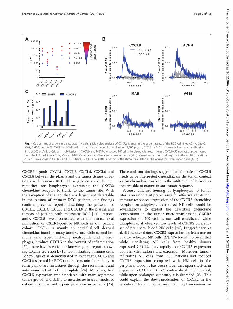
CXCR2-transduced NK cells show increased calciummobilization in the presence of CXCR2 ligandsTo determine if the ectopic CXCR2 receptor was func-tional and would respond to cognate ligands producedby RCC cell lines, the mobilization of calcium from theendoplasmatic reticulum into the cytosol was analyzed.Multiplex analysis revealed that ACHN cells producedthe highest levels of the analyzed CXCR2 ligands, whileA498 cells produced overall the least amount of CXCR2ligands (Fig. 4a). CXCL1, CXCL5 and CXCL8 were over-all the most abundant chemokines present in the RCCtumor supernatants. For the calcium flux assay, NK cellstransduced with CXCR2 or the control NGFR werestimulated with recombinant CXCL8 or conditionedRCC tumor supernatants. CXCR2-transduced, but notcontrol NK cells responded to CXCL8 with a rapid in-crease in intracellular calcium that dissolved over time(Fig. 4b). Supernatants from cell lines with high chemo-kine production (ACHN, 786-O) induced a stronger cal-cium flux in CXCR2-transduced NK cells thansupernatants with lower chemokine levels (Caki-2,MAR, A498) (Fig. 4b-c and Additional file 4: Figure S4).Importantly, NGFR-transduced NK cells did not respondto stimulation with CXCL8 or the RCC supernatants.NK cells transduced with both CXCR2 and NGFRshowed a similar increase in calcium when stimulatedwith ionomycin, indicating that their maximum capacity
to release calcium was comparable (Additional file 4:Figure S4).
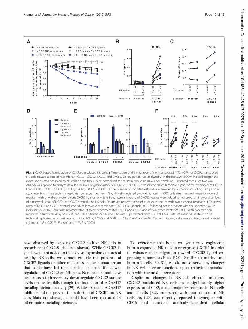
CXCR2-transduced NK cells have an increased ability tomigrate along a CXCR2 ligand gradientTo investigate if CXCR2-transduced NK cells had an in-creased ability to migrate toward RCC tumors that pro-duce CXCR2 ligands, two independent chemotaxisassays were performed. A microscopy based assay thatallows measurement of cell migration in real-time dem-onstrated that within 16 h, 71% of CXCR2-transducedNK cells migrated from the area on the top of a porousmembrane to the bottom chamber that containedpooled CXCR2 ligands, whereas less than 10% of non-transduced or control NGFR-transduced NK cells mi-grated (Fig 5a). There was 45% more migration ofCXCR2-transduced NK cells toward CXCR2 ligands wasthan to medium (p < 0.0001), while there was no signifi-cant ligand-induced migration for non-transduced orNGFR-transduced NK cells. These findings were corrob-orated in transwell assays showing that 3.4 times moreNK cells transduced with CXCR2 than those transducedwith NGFR migrated toward recombinant CXCR2ligands (p = 0.0083) (Fig. 5b). The enhanced migrationalong the CXCR2 gradient resulted in a significantlyhigher lysis of K562 target cells by CXCR2-transducedNK cells in contrast to NGFR-transduced NK cells (Fig. 5c).When CXCR2 ligands were present in both the upper andlower chambers, the migration of CXCR2-transduced NKcells was abrogated, confirming the enhanced migrationto be ligand-specific chemotaxis rather than increasedchemokinesis (Fig. 5d). To confirm that the migration wasspecifically dependent on CXCR2, NK cells were incu-bated with the selective CXCR2 inhibitor SB225502.Indeed, in the presence of SB225502, the migration ofCXCR2-transduced NK cells toward CXCL1, CXCL8 andCXCL5 was reduced to background levels, similar to thoseobserved for control NGFR-transduced cells (Fig. 5e). Fur-thermore, conditioned medium from the RCC cell linesACHN, 786-O and MAR that secrete high levels ofCXCR2 ligands enhanced the migration of CXCR2-transduced NK cells 2- to 2.5-fold compared with NGFR-transduced NK cells (Fig. 5f). By contrast, low-chemokinecontaining supernatant from Caki-2 and A498 cells didnot induce a significant increase in migration by CXCR2-transduced NK cells. For all assessed RCC tumorsupernatants, the CXCL1 but not CXCL8 concentrationcorrelated significantly (p = 0.014) with the migration ofCXCR2-transduced NK cells relative to NGFR-transducedNK cells (Additional file 5: Figure S5).
DiscussionIn this study, we show that genetic modification of hu-man NK cells to re-express the chemokine receptor
Kremer et al. Journal for ImmunoTherapy of Cancer (2017) 5:73 Page 7 of 13
on Novem
ber 21, 2021 by guest. Protected by copyright.
http://jitc.bmj.com
/J Im
munother C
ancer: first published as 10.1186/s40425-017-0275-9 on 19 Septem
ber 2017. Dow
nloaded from

CXCR2 conferred the ability to specifically migrate toRCC tumor-derived CXCR2 ligands resulting inincreased killing of target cells.
CXCR2 ligands promote several processes importantfor tumorigenesis, including angiogenesis, survival andmigration. We detected a concentration gradient for the
CD
11a
MF
I
CXCR2-
CXCR2+
NGFR-
NGFR+
CXCR2-
CXCR2+
CXCR2-
CXCR2+
NGFR-
NGFR+
CXCR2-
CXCR2+
NGFR-
NGFR+0
20000
40000
60000
80000
0
200
400
600
8000.0042 0.0138
CD
11 b
MF
I
0.0530 ns
0
20
40
60
80
100
% d
egra
nu
lati
ng
NK
cel
ls
ns
20673
1954
221
18020
17267
1763
3671
13580
0 min 10 min
NK cells (BMQC)
K56
2 ce
lls (
CF
SE
)%
NK
cel
ls in
co
nju
gat
es
0
10
20
30
40 0.0128 ns
A
DC
B
Fig. 3 Adhesion of NK cells incorporating the transgenes. a Flow cytometry analysis of CD11a (n = 6) and CD11b (n = 4) expression on NK cellsincorporating (CXCR2+ and NGFR+) and not incorporating the respective transgene (CXCR2- and NGFR-). b Representative dot plots depictingcounts of collected events within the double-positive gate after 0-min and 10-min co-cultures of CFSE-labeled K562 cells and BMQC-labeled NKcells. c NK cells incorporating and not incorporating the CXCR2 (n = 5) or NGFR (n = 6) transgenes in conjugates with K562 cells after 10 min ofco-culture. d Degranulation against K562 of CXCR2- and CXCR2+ NK cells, as assessed by flow cytometry (n = 4)
Kremer et al. Journal for ImmunoTherapy of Cancer (2017) 5:73 Page 8 of 13
on Novem
ber 21, 2021 by guest. Protected by copyright.
http://jitc.bmj.com
/J Im
munother C
ancer: first published as 10.1186/s40425-017-0275-9 on 19 Septem
ber 2017. Dow
nloaded from
CXCR2 ligands CXCL1, CXCL2, CXCL5, CXCL6 andCXCL8 between the plasma and the tumor tissues of pa-tients with primary RCC. These gradients are the pre-requisites for lymphocytes expressing the CXCR2chemokine receptor to traffic to the tumor site. Withthe exception of CXCL5 that was largely not detectablein the plasma of primary RCC patients, our findingsconfirm previous reports describing the presence ofCXCL1, CXCL3, CXCL5 and CXCL8 in the plasma andtumors of patients with metastatic RCC [21]. Import-antly, CXCL5 levels correlated with the intratumoralinfiltration of CXCR2-positive NK cells in our patientcohort. CXCL5 is mainly an epithelial-cell derivedchemokine found in many tumors, and while several im-mune cells types, including neutrophils and macro-phages, produce CXCL5 in the context of inflammation[22], there have been to our knowledge no reports show-ing CXCL5 secretion by tumor-infiltrating immune cells.López-Lago et al. demonstrated in mice that CXCL5 andCXCL8 secreted by RCC tumors constrain their ability toform pulmonary metastases through the recruitment andanti-tumor activity of neutrophils [24]. Moreover, lowCXCL5 expression was associated with more aggressivetumor growth and ability to metastasize in a rat model ofcolorectal cancer and a poor prognosis in patients [25].
These and our findings suggest that the role of CXCL5needs to be interpreted depending on the tumor contextas this chemokine can lead to the infiltration of leukocytesthat are able to mount an anti-tumor response.Because efficient homing of lymphocytes to tumor
sites is an important prerequisite for effective anti-tumorimmune responses, expression of the CXCR2 chemokinereceptor on adoptively transferred NK cells would beadvantageous to exploit the described chemokinecomposition in the tumor microenvironment. CXCR2expression on NK cells is not well established; whileCampbell et al. observed low levels of CXCR2 on a sub-set of peripheral blood NK cells [26], Inngjerdingen etal. did neither detect CXCR2 expression on fresh nor onin vitro activated NK cells [27]. We found, however, thatwhile circulating NK cells from healthy donorsexpressed CXCR2, they rapidly lost CXCR2 expressionupon in vitro culture and expansion. Moreover, tumor-infiltrating NK cells from RCC patients had reducedCXCR2 expression compared with NK cell in theperipheral blood. It has been shown that upon short-termexposure to CXCL8, CXCR2 is internalized to be recycled,while upon prolonged exposure, it is degraded [28]. Thiscould explain the down-modulation of CXCR2 in theligand-rich tumor microenvironment, a phenomenon we
pg
/ml
CX
CL 1
CX
CL 2
CX
CL 5
CX
CL 6
CX
CL 8
1
1 0
1 0 0
1 0 0 0
1 0 0 0 0
1 0 0 0 0 0
A 4 9 8
C a k i-2
M A R
7 8 6 -O
A C H N
Che
mok
ine
prod
uctio
n
CX
CL 8
AC
HN
7 8 6 -OM
AR
CA
KI-2
A4 9 8
0
2 0
4 0
6 0
8 0
Re
sp
on
se
(AU
C)
po
st
sti
mu
lus
(1
03
)
N G FR C X C R 2
A
C
B
S e c o n d s
Flu
o-3
RF
U
no
rma
l iz
ed
tob
as
eli
ne
1 0 0 2 0 0 3 0 0
1 .0
1 .5
2 .0
N G F R N K
C X C R 2 N K
CXCL8
S e c o n d s
Flu
o-3
RF
U
no
rma
l iz
ed
t ob
as
eli
ne
1 0 0 2 0 0 3 0 0
1 .0
1 .5
2 .0ACHN
S e c o n d s
Flu
o-3
RF
U
no
rma
l iz
ed
tob
as
eli
ne
1 0 0 2 0 0 3 0 0
1 .0
1 .5
2 .0MAR
S e c o n d s
Flu
o-3
RF
U
no
rma
liz
ed
tob
as
eli
ne
1 0 0 2 0 0 3 0 0
1 .0
1 .5
2 .0A498
�
Fig. 4 Calcium mobilization in transduced NK cells. a Multiplex analysis of CXCR2 ligands in the supernatants of the RCC cell lines ACHN, 786-O,MAR, CAKI-2, and A498. CXCL1 in ACHN cells was above the quantification limit of 13,990 pg/mL, CXCL5 in A498 cells was below the quantificationlimit of 603 pg/mL. b Calcium mobilization in CXCR2- and NGFR-transduced NK cells stimulated with recombinant CXCL8 (50 ng/mL) or supernatantfrom the RCC cell lines ACHN, MAR or A498. Values are Fluo-3 relative fluorescent units (RFU) normalized to the baseline prior to the addition of stimuli.c Calcium response in CXCR2- and NGFR-transduced NK cells after addition of the stimuli calculated as the normalized area under curve (AUC)
Kremer et al. Journal for ImmunoTherapy of Cancer (2017) 5:73 Page 9 of 13
on Novem
ber 21, 2021 by guest. Protected by copyright.
http://jitc.bmj.com
/J Im
munother C
ancer: first published as 10.1186/s40425-017-0275-9 on 19 Septem
ber 2017. Dow
nloaded from
have observed by exposing CXCR2-positive NK cells torecombinant CXCL8 (data not shown). While CXCR2 li-gands were not added to the ex vivo expansion cultures ofhealthy NK cells, we cannot exclude the presence ofCXCR2 ligands or other molecules in the human serumthat could have led to a specific or unspecific down-regulation of CXCR2 on NK cells. Nonligand stimuli havebeen shown to irreversibly down-regulate CXCR2 surfacelevels on neutrophils though the induction of ADAM17metalloproteinase activity [29]. While a specific ADAM17inhibitor did not prevent the reduction of CXCR2 on NKcells (data not shown), it could have been mediated byother matrix metalloproteinases.
To overcome this issue, we genetically engineeredhuman expanded NK cells to re-express CXCR2 in orderto enhance their migration toward CXCR2-ligand ex-pressing tumors such as RCC. Similar to murine andhuman T cells [30, 31], we did not observe any changesin NK cell effector functions upon retroviral transduc-tion with chemokine receptors.Despite no changes in NK cell effector functions,
CXCR2-transduced NK cells had a significantly higherexpression of CD2, a costimulatory receptor in NK cellsand T cells [32], compared with non-transduced NKcells. As CD2 was recently reported to synergize withCD16 and stimulate antibody-dependent cellular
%m
igra
tio
n
NG
FR
CX
CR
2
NG
FR
CX
CR
2
0
2 0
4 0
6 0
8 0
M e d iu m C X C R 2
lig a n d s
S tim u la n t:
N K c e lls :
0.0083
H o u rs
Are
ao
cc
up
ied
by
NK
ce
l ls
on
top
su
rfa
ce
no
rma
liz
ed
toa
rea
at
t=
0h
0 2 4 6 8 1 0 1 2 1 4 1 6
0 .0
0 .2
0 .4
0 .6
0 .8
1 .0
1 .2
N T N K v s m e d iu m
C X C R 2 N K v s m e d iu m
N G F R N K vs m e d iu m
N T N K v s C X C R 2 lig a n d s
C X C R 2 N K v s C X C R 2 lig a n d s
N G F R N K v s C X C R 2 lig a n d s
****
n s
ns
%m
igr a
tio
n
N G F R C X C R 20
1 0
2 0
3 0
4 0
5 0M e d iu m
L ig a n d s in
lo w e r w e ll
L ig a n d s in
to p a n d
lo w e r w e ll
%m
igra
tio
n
NG
FR
CX
CR
2
NG
FR
CX
CR
2
NG
FR
CX
CR
2
NG
FR
CX
CR
2
NG
FR
CX
CR
2
0
2 0
4 0
6 0
8 0
1 0 0
A C H N 7 8 6 -O M A R C a k i-2 A 498S tim u la n t:
N K c e lls :
****
2.4x 2.0x 1.4x2.5x 1.2x
%s
pe
cif
icly
sis
NG
FR
CX
CR
2
NG
FR
CX
CR
2
0
1 0
2 0
3 0
4 0
5 0
M e d iu m C X C R 2
lig a n d s
S tim u la n t:
N K c e lls :
0.0039
A
D
B C
E F
M e d iu m C X C L 1 C X C L 8
0
5
1 0
1 5
2 0C X C R 2 N G FR
S B 2 2 5 0 0 2 +- +- +- +-
%m
igra
tio
n
- -
M e d iu m C X C L 5
0
1 0
2 0
3 0
4 0
5 0
+- +-- -
Fig. 5 CXCR2-specific migration of CXCR2-transduced NK cells. a Time course of the migration of non-transduced (NT), NGFR- or CXCR2-transducedNK cells toward a pool of recombinant CXCL1, CXCL2, CXCL3, and CXCL8. Cell migration was analyzed with the IncuCyte ZOOM live cell imager andexpressed as area occupied by NK cells on the top surface normalized to the initial top value (n = 4 per condition). Repeated measures two-wayANOVA was applied to analyze data. b Transwell migration assay of NT, NGFR- or CXCR2-transduced NK cells toward a pool of the recombinant CXCR2ligands CXCL1, CXCL2, CXCL3, CXCL5, CXCL6, CXCL7, and CXCL8. The number of migrated cells was determined by automatic counting using a flowcytometer from three technical replicates per experiment (n = 7). c NK cell-mediated cytotoxicity against K562 cells after transwell migration towardmedium with or without recombinant CXCR2 ligands (n = 3). d Equal concentrations of CXCR2 ligands were added to the upper and lower chambersof a transwell assay of NGFR- and CXCR2-transduced NK cells. Results are representative of three experiments with two technical replicates. e Transwellassay of NGFR- and CXCR2-transduced NK cells toward recombinant CXCL1, CXCL8 and CXCL5 following pre-incubation with the selective CXCR2inhibitor SB225002. Results are representative of three experiments for CXCL1 and CXCL8 and of two experiments for CXCL5 with two technicalreplicates. f Transwell assay of NGFR- and CXCR2-transduced NK cells toward supernatants from RCC cell lines. Data are mean values from threetechnical replicates per experiment (n = 4 for ACHN, 786-O, and MAR; n = 3 for Caki-2 and A498). Percent migrated cells are calculated based on totalcell input. *, P < 0.05, **, P < 0.01 and ****, P < 0.0001
Kremer et al. Journal for ImmunoTherapy of Cancer (2017) 5:73 Page 10 of 13
on Novem
ber 21, 2021 by guest. Protected by copyright.
http://jitc.bmj.com
/J Im
munother C
ancer: first published as 10.1186/s40425-017-0275-9 on 19 Septem
ber 2017. Dow
nloaded from

cytotoxicity in NK cells [33], increased CD2 expressioncan be seen as an additional advantage of using CXCR2-transduced NK cells for adoptive cell therapy.LFA-1 is a key mediator of the firm adhesion and the
formation of the immunological synapse by cytotoxiclymphocytes [34]. We found that NK cells that had in-corporated the CXCR2 or NGFR transgene (CXCR2+ orNGFR+) had a significantly higher expression of CD11a,the α-subunit of LFA-1, compared with NK cells thathad not inserted the transgene (CXCR2- or NGFR-).The likely explanation for this observation is that NKcells with a high LFA-1 expression were more efficientlyinfected by the retrovirus as has been shown in T cellsinfected with the human immunodeficiency virus type 1bearing host-derived ICAM-1 [35]. Consequently,CXCR2+ NK cells formed more CD11a-dependent con-jugates with K562 cells than CXCR2- NK cells, a differ-ence not observed for NGFR-transduced NK cells.However, there were no differences in degranulationagainst K562 cells between CXCR2+ and CXCR2- NKcells. This is in agreement with Bryceson et al. who haveshown that in resting NK cells, cytotoxic granulepolarization is induced by LFA-1 signaling, whereas de-granulation is an LFA-1 independent event [36]. By con-trast, IL-2 activated NK cells were able to lyse ICAM-1coated insect target cells though LFA-1 signaling alone[37]. The latter would suggest that expanded CXCR2+ NKcells are more potent at killing target cells. However, tumorcells, such as K562, express a variety of ligands engaging awide range of receptors that can activate the redundantpathways for NK cell degranulation and cytotoxicity.We confirmed that the signaling machinery of the ectopic
CXCR2 receptor was functional as it could induce calciummobilization, one of the first steps in G-protein coupled re-ceptor signaling, in CXCR2-transduced NK cells uponstimulation with recombinant CXCL8 as well as with RCCtumor-derived supernatants containing CXCR2 ligands.While NGFR-transduced cells did not respond to CXCL8,they released calcium upon stimulation with tumor super-natants, albeit 2 to 6 times less than CXCR2-transduced NKcells. This release was likely due to other chemokines se-creted by RCC cells acting on NK cell chemokine receptors.For example, the tumor supernatants contained low levelsof CXCL9, CXCL10 and CXCL11 (data not shown) thatbind to CXCR3, a chemokine receptor known to be upregu-lated on NK cells upon activation and expansion [20].The transduction with CXCR2 conferred to the NK
cells a significantly (1.8- to 5.9-fold) increased migrationability to recombinant CXCR2 ligands compared withNGFR transduction. The enhanced trafficking along theCXCR2 gradient resulted in a significantly higher lysis ofK562 target cells by NK cells transduced with CXCR2compared with those transduced with NGFR. Inaddition, RCC supernatant from cell lines producing
high amounts of CXCR2 ligands induced 2- to 2.5-foldincreased migration of CXCR2-transduced NK cellscompared with NGFR-transduced NK cells. This in-crease in migration was dependent on the concentrationof CXCL1, but not CXCL8 in the tumor supernatants.While CXCL1 and CXCL8 have a similar potency of5 nM and 4 nM, respectively [38], CXCL1 was moreabundant in 80% of the assayed supernatants, whichcould explain its greater impact on NK cell migration.Other studies have recently addressed the issue of
lymphocyte homing as a key requirement for an effectiveanti-tumor immune response by modifying either thechemokine composition at the tumor site or the chemo-kine receptor repertoire of adoptively transferred lym-phocytes. While increased infiltration of T and NK cellshas been observed in tumors with a local production ofCCL5, CXCL10 or CX3CL1, these chemokines had to beartificially introduced into the tumor microenvironmentby intratumoral injections of chemokine-encoding DNAplasmids and adenoviral vectors or chemokine-stimulatingcytokines such as IFN-γ [20, 39, 40]. Taking for the firsttime advantage of the chemokines present at the targetedsite, Kershaw et al. transduced activated human T cellswith the chemokine receptor CXCR2 and observed im-proved migration toward melanoma supernatants in vitro[41]. Subsequent studies have shown increased infiltrationand anti-tumor responses of adoptively transferred mouseand human T cells engineered to express, for instance,CCR4, CXCR2 and CCR2 in transplantable mouse modelsof Hodgkin’s lymphoma, melanoma and mesothelioma,respectively [30, 31, 42]. With respect to NK cells, transi-ent expression of CCR7 in human NK cells acquired byeither mRNA electroporation or trogocytosis has beenshown to augment their migration to CCL19 in vitro andto lymph nodes in athymic nude mice, respectively[43, 44]. Moreover, transduction of the YTS NK cell line(containing an EGFR-specific chimeric antigen receptor)with CXCR4 has been shown to enhance infiltration inglioblastoma xenograft models overexpressing CXCL12,resulting in improved survival [45]. Our study furtheradvances the understanding how NK cell infiltration oftumors can be enhanced and shows, to our knowledge forthe first time, stable engineering of human primary NKcells to express a chemokine receptor, thereby improvingtheir migration. However, further research using in vivomodels is needed to corroborate our findings and addresshow infused NK cells overcome challenges such as migra-tion through the blood stream, extravasation into thetumor tissue and interplay with the tumor microenviron-ment to achieve an anti-tumor effect.
ConclusionsIn order to increase the success of NK cell-based therapiesof solid tumors, it is of great importance not only to
Kremer et al. Journal for ImmunoTherapy of Cancer (2017) 5:73 Page 11 of 13
on Novem
ber 21, 2021 by guest. Protected by copyright.
http://jitc.bmj.com
/J Im
munother C
ancer: first published as 10.1186/s40425-017-0275-9 on 19 Septem
ber 2017. Dow
nloaded from
maintain optimal in vivo proliferation and cytotoxic activ-ity, but also to promote homing of NK cells to the tumorsite. In this study, we report that increasing chemokine re-ceptor expression on NK cells is a promising approach toaugment the efficacy of adoptive cellular immunotherapies.
Additional files
Additional file 1: Figure S1. Time course of CXCR2 expression onhealthy donor NK cells in an expansion setup with EBV-LCL feeder cellsand IL-2, as assessed by flow cytometry. (PDF 16 kb)
Additional file 2: Figure S2. Gating strategy and representative flowcytometric assessment of NK cell degranulation and IFN-γ productionafter co-culture with K562 cells. (PDF 140 kb)
Additional file 3: Figure S3. Left: Flow cytometry analysis of CD2expression on non-transduced (NT), NGFR-and CXCR2-transduced NK cells.Middle: Flow cytometry analysis of CD2 expression of CXCR2-transduced NKcells incorporating (CXCR2+) and not incorporating the transgene (CXCR2-).Right: NGFR- and CXCR2-transduced NK cells in conjugates with K562 cellsco-cultured for 0 min and 10 min with and without CD11a-blockingantibodies (n = 3). (PDF 73 kb)
Additional file 4: Figure S4. Calcium mobilization in CXCR2- andNGFR-transduced NK cells stimulated with supernatant from the RCC celllines 786-O or Caki-2 or with ionomycin (200 ng/mL) as a positive control.Values are Fluo-3 relative fluorescent units (RFU) normalized to thebaseline prior to the addition of stimuli. (PDF 44 kb)
Additional file 5: Figure S5. Pearson correlation of CXCL1 and CXCL8levels in RCC tumor supernatants used in transwell assays and correspondingmigration of CXCR2-transduced NK cells relative to NGFR-transduced NK cells(n = 20). (PDF 19 kb)
AbbreviationsAUC: Area under curve; BMQC: 2,3,6,7-tetrahydro-9-bromomethyl-1H,5H–quinolizino(9,1-gh)coumarin; CCL: Chemokine (C-C motif) ligand; CCR: Chemokine(C-C motif) receptor; CFSE: Carboxyfluorescein succinimidyl ester; Cr: Chromium;CX3CL: Chemokine (C-X3-C motif) ligand; CX3CR: Chemokine (C-X3-C motif)receptor; CXCL: Chemokine (C-X-C motif) ligand; CXCR: Chemokine (C-X-C motif)receptor; DNAM-1: DNAX accessory molecule-1; E:T ratio: Effector-to-target ratio;EBV: Epstein-Barr virus; FBS: Fetal bovine serum; IFN: Inteferon; IL: Interleukin;LCL: Lymphoblastoid cell line; LFA-1: Leukocyte function-associated antigen 1;Mac-1: Macrophage-1 antigen; MFI: Mean fluorescence intensity; NGFR: Nervegrowth factor receptor; NK: Natural killer; NT: Non-transduced; PBMC: Peripheralblood mononuclear cell; PBS: Phosphate buffered saline; pMSGV: Murine stem cellvirus-based splice-gag vector plasmid; RCC: Renal cell carcinoma; RFU: Relativefluorescence unit; TRAIL: Tumor necrosis factor-related apoptosis-inducing ligand
AcknowledgementsThe authors would like to thank Dr. Ulrika Harmenberg, Karolinska UniversityHospital, for coordinating the collection of patient material and Dr. Vincentvan Hoef, Karolinska Institutet, SciLifeLab, for assistance with data analysis.
FundingThis work was supported by the Swedish Cancer Society (#CAN 2012/474 and#CAN 2015/421), the Swedish Childhood Cancer Foundation (#PR2014–0093),the Swedish Foundation for International Cooperation in Research andHigher Education (#IB2014–5690), and the Cancer Research Foundationsof Radiumhemmet (#141272 and 161192).
Availability of data and materialsThe datasets used and/or analyzed during the current study are availablefrom the corresponding author on reasonable request.
Authors’ contributionsVK designed and performed research, analyzed data, and wrote the paper.ML designed research, analyzed data, and wrote the paper. RZ designed andperformed research. CS performed research. AD performed research. EWdesigned and performed research. EC and ASP provided clinical material. AL
designed research, analyzed data, and wrote the paper. All authors read andapproved the manuscript.
Ethics approval and consent to participateThe study was approved by the Regional Ethical Review Board in Stockholm(Ethical approval # 2013–570-31). All patients provided written informed consent.
Consent for publicationNot applicable.
Competing interestsThe authors declare that they have no competing interests.
Publisher’s NoteSpringer Nature remains neutral with regard to jurisdictional claims inpublished maps and institutional affiliations.
Author details1Department of Oncology-Pathology, Karolinska Institutet, Stockholm,Sweden. 2Department of Molecular Oncology, The Netherlands CancerInstitute, Amsterdam, Netherlands. 3Department of Medicine Solna,Karolinska Institutet, Stockholm, Sweden. 4Department of RadiationOncology, Weill Cornell Medicine, New York, NY, USA. 5Department ofOncology-Pathology, Stockholm South General Hospital, Stockholm, Sweden.6Department of Woman and Child Health, Karolinska Institutet, Stockholm,Sweden. 7Department of Urology, Stockholm South General Hospital,Stockholm, Sweden. 8Cell Therapy Institute, Nova Southeastern University,Fort Lauderdale, FL, USA.
Received: 8 February 2017 Accepted: 14 August 2017
References1. Ruggeri L, Capanni M, Urbani E, Perruccio K, Shlomchik WD, Tosti A,
Posati S, Rogaia D, Frassoni F, Aversa F, et al. Effectiveness of donor naturalkiller cell alloreactivity in mismatched hematopoietic transplants. Science.2002;295:2097–100.
2. Benson DM Jr, Cohen AD, Jagannath S, Munshi NC, Spitzer G, Hofmeister CC,Efebera YA, Andre P, Zerbib R, Caligiuri MA. A phase I trial of the anti-KIRantibody IPH2101 and Lenalidomide in patients with relapsed/refractorymultiple myeloma. Clin Cancer Res. 2015;21:4055–61.
3. Miller JS, Soignier Y, Panoskaltsis-Mortari A, McNearney SA, Yun GH,Fautsch SK, McKenna D, Le C, Defor TE, Burns LJ, et al. Successful adoptivetransfer and in vivo expansion of human haploidentical NK cells in patientswith cancer. Blood. 2005;105:3051–7.
4. Geller MA, Cooley S, Judson PL, Ghebre R, Carson LF, Argenta PA, Jonson AL,Panoskaltsis-Mortari A, Curtsinger J, McKenna D, et al. A phase II study ofallogeneic natural killer cell therapy to treat patients with recurrent ovarianand breast cancer. Cytotherapy. 2011;13:98–107.
5. Parkhurst MR, Riley JP, Dudley ME, Rosenberg SA. Adoptive transfer ofautologous natural killer cells leads to high levels of circulating naturalkiller cells but does not mediate tumor regression. Clin Cancer Res.2011;17:6287–97.
6. Sconocchia G, Spagnoli GC, Del Principe D, Ferrone S, Anselmi M,Wongsena W, Cervelli V, Schultz-Thater E, Wyler S, Carafa V, et al. Defectiveinfiltration of natural killer cells in MICA/B-positive renal cell carcinomainvolves beta(2)-integrin-mediated interaction. Neoplasia. 2009;11:662–71.
7. Halama N, Braun M, Kahlert C, Spille A, Quack C, Rahbari N, Koch M, Weitz J,Kloor M, Zoernig I, et al. Natural killer cells are scarce in colorectalcarcinoma tissue despite high levels of chemokines and cytokines.Clin Cancer Res. 2011;17:678–89.
8. Sconocchia G, Arriga R, Tornillo L, Terracciano L, Ferrone S, Spagnoli GC.Melanoma cells inhibit NK cell functions. Cancer Res. 2012;72:5428–9.author reply 5430.
9. Erdag G, Schaefer JT, Smolkin ME, Deacon DH, Shea SM, Dengel LT,Patterson JW, Slingluff CL Jr. Immunotype and immunohistologiccharacteristics of tumor-infiltrating immune cells are associated with clinicaloutcome in metastatic melanoma. Cancer Res. 2012;72:1070–80.
10. Coca S, Perez-Piqueras J, Martinez D, Colmenarejo A, Saez MA, Vallejo C,Martos JA, Moreno M. The prognostic significance of intratumoral naturalkiller cells in patients with colorectal carcinoma. Cancer. 1997;79:2320–8.
Kremer et al. Journal for ImmunoTherapy of Cancer (2017) 5:73 Page 12 of 13
on Novem
ber 21, 2021 by guest. Protected by copyright.
http://jitc.bmj.com
/J Im
munother C
ancer: first published as 10.1186/s40425-017-0275-9 on 19 Septem
ber 2017. Dow
nloaded from
11. Ishigami S, Natsugoe S, Tokuda K, Nakajo A, Che X, Iwashige H, Aridome K,Hokita S, Aikou T. Prognostic value of intratumoral natural killer cells ingastric carcinoma. Cancer. 2000;88:577–83.
12. Villegas FR, Coca S, Villarrubia VG, Jimenez R, Chillon MJ, Jareno J, Zuil M,Callol L. Prognostic significance of tumor infiltrating natural killer cellssubset CD57 in patients with squamous cell lung cancer. Lung Cancer.2002;35:23–8.
13. Donskov F, von der Maase H. Impact of immune parameters on long-termsurvival in metastatic renal cell carcinoma. J Clin Oncol. 2006;24:1997–2005.
14. Eckl J, Buchner A, Prinz PU, Riesenberg R, Siegert SI, Kammerer R, Nelson PJ,Noessner E. Transcript signature predicts tissue NK cell content and definesrenal cell carcinoma subgroups independent of TNM staging. J Mol Med(Berl). 2012;90:55–66.
15. Geissler K, Fornara P, Lautenschlager C, Holzhausen HJ, Seliger B, Riemann D.Immune signature of tumor infiltrating immune cells in renal cancer.Oncoimmunology. 2015;4:e985082.
16. Igarashi T, Takahashi H, Tobe T, Suzuki H, Mizoguchi K, Nakatsu HO, Ito H.Effect of tumor-infiltrating lymphocyte subsets on prognosis andsusceptibility to interferon therapy in patients with renal cell carcinoma.Urol Int. 2002;69:51–6.
17. Mlecnik B, Tosolini M, Charoentong P, Kirilovsky A, Bindea G, Berger A,Camus M, Gillard M, Bruneval P, Fridman WH, et al. Biomolecular networkreconstruction identifies T-cell homing factors associated with survival incolorectal cancer. Gastroenterology. 2010;138:1429–40.
18. Park MH, Lee JS, Yoon JH. High expression of CX3CL1 by tumor cellscorrelates with a good prognosis and increased tumor-infiltrating CD8+ Tcells, natural killer cells, and dendritic cells in breast carcinoma. J SurgOncol. 2012;106:386–92.
19. Wendel M, Galani IE, Suri-Payer E, Cerwenka A. Natural killer cell accumulationin tumors is dependent on IFN-gamma and CXCR3 ligands. Cancer Res.2008;68:8437–45.
20. Wennerberg E, Kremer V, Childs R, Lundqvist A. CXCL10-induced migrationof adoptively transferred human natural killer cells toward solid tumorscauses regression of tumor growth in vivo. Cancer Immunol Immunother.2015;64:225–35.
21. Mestas J, Burdick MD, Reckamp K, Pantuck A, Figlin RA, Strieter RM. The roleof CXCR2/CXCR2 ligand biological axis in renal cell carcinoma. J Immunol.2005;175:5351–7.
22. Russo RC, Garcia CC, Teixeira MM, Amaral FA. The CXCL8/IL-8 chemokinefamily and its receptors in inflammatory diseases. Expert Rev Clin Immunol.2014;10:593–619.
23. Lundqvist A, Berg M, Smith A, Childs RW. Bortezomib treatment topotentiate the anti-tumor immunity of ex-vivo expanded adoptively infusedAutologous natural killer cells. J Cancer. 2011;2:383–5.
24. Lopez-Lago MA, Posner S, Thodima VJ, Molina AM, Motzer RJ, Chaganti RS.Neutrophil chemokines secreted by tumor cells mount a lung antimetastaticresponse during renal cell carcinoma progression. Oncogene. 2013;32:1752–60.
25. Speetjens FM, Kuppen PJ, Sandel MH, Menon AG, Burg D, van de Velde CJ,Tollenaar RA, de Bont HJ, Nagelkerke JF. Disrupted expression of CXCL5 incolorectal cancer is associated with rapid tumor formation in rats and poorprognosis in patients. Clin Cancer Res. 2008;14:2276–84.
26. Campbell JJ, Qin S, Unutmaz D, Soler D, Murphy KE, Hodge MR, Wu L,Butcher EC. Unique subpopulations of CD56+ NK and NK-T peripheralblood lymphocytes identified by chemokine receptor expression repertoire.J Immunol. 2001;166:6477–82.
27. Inngjerdingen M, Damaj B, Maghazachi AA. Expression and regulation ofchemokine receptors in human natural killer cells. Blood. 2001;97:367–75.
28. Fan GH, Lapierre LA, Goldenring JR, Richmond A. Differential regulation ofCXCR2 trafficking by Rab GTPases. Blood. 2003;101:2115–24.
29. Mishra HK, Long C, Bahaie NS, Walcheck B. Regulation of CXCR2 expressionand function by a disintegrin and metalloprotease-17 (ADAM17). J LeukocBiol. 2015;97:447–54.
30. Peng W, Ye Y, Rabinovich BA, Liu C, Lou Y, Zhang M, Whittington M, Yang Y,Overwijk WW, Lizee G, Hwu P. Transduction of tumor-specific T cells withCXCR2 chemokine receptor improves migration to tumor and antitumorimmune responses. Clin Cancer Res. 2010;16:5458–68.
31. Di Stasi A, De Angelis B, Rooney CM, Zhang L, Mahendravada A, Foster AE,Heslop HE, Brenner MK, Dotti G, Savoldo B. T lymphocytes coexpressingCCR4 and a chimeric antigen receptor targeting CD30 have improvedhoming and antitumor activity in a Hodgkin tumor model. Blood.2009;113:6392–402.
32. Siliciano RF, Pratt JC, Schmidt RE, Ritz J, Reinherz EL. Activation of cytolytic Tlymphocyte and natural killer cell function through the T11 sheeperythrocyte binding protein. Nature. 1985;317:428–30.
33. Liu LL, Landskron J, Ask EH, Enqvist M, Sohlberg E, Traherne JA, Hammer Q,Goodridge JP, Larsson S, Jayaraman J, et al. Critical role of CD2 co-stimulationin adaptive natural killer cell responses revealed in NKG2C-deficient humans.Cell Rep. 2016;15:1088–99.
34. Orange JS. Formation and function of the lytic NK-cell immunologicalsynapse. Nat Rev Immunol. 2008;8:713–25.
35. Fortin JF, Cantin R, Lamontagne G, Tremblay M. Host-derived ICAM-1glycoproteins incorporated on human immunodeficiency virus type 1 arebiologically active and enhance viral infectivity. J Virol. 1997;71:3588–96.
36. Bryceson YT, March ME, Barber DF, Ljunggren HG, Long EO. Cytolyticgranule polarization and degranulation controlled by different receptors inresting NK cells. J Exp Med. 2005;202:1001–12.
37. Barber DF, Faure M, Long EO. LFA-1 contributes an early signal for NK cellcytotoxicity. J Immunol. 2004;173:3653–9.
38. Ahuja SK, Murphy PM. The CXC chemokines growth-regulated oncogene(GRO) alpha, GRObeta, GROgamma, neutrophil-activating peptide-2, andepithelial cell-derived neutrophil-activating peptide-78 are potent agonistsfor the type B, but not the type a, human interleukin-8 receptor. J BiolChem. 1996;271:20545–50.
39. Lavergne E, Combadiere C, Iga M, Boissonnas A, Bonduelle O, Maho M, Debre P,Combadiere B. Intratumoral CC chemokine ligand 5 overexpression delays tumorgrowth and increases tumor cell infiltration. J Immunol. 2004;173:3755–62.
40. Xin H, Kikuchi T, Andarini S, Ohkouchi S, Suzuki T, Nukiwa T. Huqun,Hagiwara K, Honjo T, Saijo Y. Antitumor immune response by CX3CL1fractalkine gene transfer depends on both NK and T cells. Eur J Immunol.2005;35:1371–80.
41. Kershaw MH, Wang G, Westwood JA, Pachynski RK, Tiffany HL, Marincola FM,Wang E, Young HA, Murphy PM, Hwu P. Redirecting migration of T cells tochemokine secreted from tumors by genetic modification with CXCR2.Hum Gene Ther. 2002;13:1971–80.
42. Moon EK, Carpenito C, Sun J, Wang LC, Kapoor V, Predina J, Powell DJ Jr,Riley JL, June CH, Albelda SM. Expression of a functional CCR2 receptorenhances tumor localization and tumor eradication by retargeted human Tcells expressing a mesothelin-specific chimeric antibody receptor.Clin Cancer Res. 2011;17:4719–30.
43. Carlsten M, Levy E, Karambelkar A, Li L, Reger R, Berg M, Peshwa MV,Childs RW. Efficient mRNA-based genetic engineering of human NK cellswith high-affinity CD16 and CCR7 augments Rituximab-induced ADCCagainst lymphoma and targets NK cell migration toward the lymphnode-associated Chemokine CCL19. Front Immunol. 2016;7:105.
44. Somanchi SS, Somanchi A, Cooper LJ, Lee DA. Engineering lymph nodehoming of ex vivo-expanded human natural killer cells via trogocytosis ofthe chemokine receptor CCR7. Blood. 2012;119:5164–72.
45. Muller N, Michen S, Tietze S, Topfer K, Schulte A, Lamszus K, Schmitz M,Schackert G, Pastan I, Temme A. Engineering NK cells modified with anEGFRvIII-specific Chimeric antigen receptor to Overexpress CXCR4 improvesimmunotherapy of CXCL12/SDF-1alpha-secreting Glioblastoma. J Immunother.2015;38:197–210.
• We accept pre-submission inquiries
• Our selector tool helps you to find the most relevant journal
• We provide round the clock customer support
• Convenient online submission
• Thorough peer review
• Inclusion in PubMed and all major indexing services
• Maximum visibility for your research
Submit your manuscript atwww.biomedcentral.com/submit
Submit your next manuscript to BioMed Central and we will help you at every step:
Kremer et al. Journal for ImmunoTherapy of Cancer (2017) 5:73 Page 13 of 13
on Novem
ber 21, 2021 by guest. Protected by copyright.
http://jitc.bmj.com
/J Im
munother C
ancer: first published as 10.1186/s40425-017-0275-9 on 19 Septem
ber 2017. Dow
nloaded from

CORRECTION Open Access
Correction to: Genetic engineering ofhuman NK cells to express CXCR2 improvesmigration to renal cell carcinomaVeronika Kremer1, Maarten A. Ligtenberg2, Rosa Zendehdel1, Christina Seitz3, Annet Duivenvoorden1,Erik Wennerberg4, Eugenia Colón5,6, Ann-Helén Scherman-Plogell7 and Andreas Lundqvist1,8*
Unfortunately, after publication of this article [1], it wasnoticed that the name of Maarten A. Ligtenberg wasdisplayed incorrectly as Maarten Ligtenberg. The correctauthor list can be seen above and the original article hasbeen updated to reflect this.
Author details1Department of Oncology-Pathology, Karolinska Institutet, Stockholm,Sweden. 2Department of Molecular Oncology, The Netherlands CancerInstitute, Amsterdam, Netherlands. 3Department of Medicine Solna,Karolinska Institutet, Stockholm, Sweden. 4Department of RadiationOncology, Weill Cornell Medicine, New York, NY, USA. 5Department ofOncology-Pathology, Stockholm South General Hospital, Stockholm, Sweden.6Department of Woman and Child Health, Karolinska Institutet, Stockholm,Sweden. 7Department of Urology, Stockholm South General Hospital,Stockholm, Sweden. 8Cell Therapy Institute, Nova Southeastern University,Fort Lauderdale, FL, USA.
Received: 10 October 2017 Accepted: 10 October 2017
Reference1. Kremer V, Ligtenberg M, Zendehdel R, Seitz C, Duivenvoorden A,
Wennerberg E, Lundqvist A. Genetic engineering of human NK cells toexpress CXCR2 improves migration to renal cell carcinoma. Journal forImmunotherapy of Cancer. 2017;5:73. https://doi.org/10.1186/s40425-017-0275-9.
* Correspondence: [email protected] of Oncology-Pathology, Karolinska Institutet, Stockholm,Sweden8Cell Therapy Institute, Nova Southeastern University, Fort Lauderdale, FL,USA
© The Author(s). 2017 Open Access This article is distributed under the terms of the Creative Commons Attribution 4.0International License (http://creativecommons.org/licenses/by/4.0/), which permits unrestricted use, distribution, andreproduction in any medium, provided you give appropriate credit to the original author(s) and the source, provide a link tothe Creative Commons license, and indicate if changes were made. The Creative Commons Public Domain Dedication waiver(http://creativecommons.org/publicdomain/zero/1.0/) applies to the data made available in this article, unless otherwise stated.
Kremer et al. Journal for ImmunoTherapy of Cancer (2017) 5:88 DOI 10.1186/s40425-017-0292-8