Genetic Diseases and Antenatal Diagnosis - e-mjm.org · Med. J. Malaysia Vol. 40 No. 3 September...
12
Med. J. Malaysia Vol. 40 No. 3 September 1985 GENETIC DISEASES AND ANTENATAL DIAGNOSIS * WONG HOCK BOON SUMMARY Wong Hock Boon MBBS, FRCP (Edinl, FRCP (Glas), FRACP, DCH (Lond). PPA, PJG Head University Department of Paediatrics National University of Singapore Singapore General Hospital Director School of Postgraduate Medical Studies National University of Singapore College Road Singapore 0316 Head WHO Collaborating Centre for Research and Training in Human Genetics Singapore Address for correspondence: Professor Wong Hock Boon Head, Dept of Paediatrics National University of Singapore Singapore General Hospital Singapore 0316 The different methods of prenatal diagnosis are discussed with special reference to ultrasound scan, amniocentesis for cell culture with processing for chromosome study, biochemical analysis and DNA recombination analysis. Chorionic villi aspiration and fetoscopy are new methods which will enhance considerably the methods for pre- natal diagnosis. With regard to chromosome study of amniotic cells, experience with 623 cases is reviewed. 2. 7% demonstrated chromosome anomalies and of these Downs anomaly was the commonest. A large proportion of cases requesting for amniocentesis are Caucasians who represent only 2% of the population in Singapore, but 25% of the 440 requests were from Caucasions. The various problems associated with the different methods for prenatal deafness are discussed. INTRODUCTION All diseases have a certain genetic load, some more than others. 1 / 2 There are two groups of diseases with a high genetic load and they include Mendelian disorders and the chromosome diseases. The Mendelian disorders are due to mutations of single genes which normally have critical *The I.S. Puvan Memorial Lecture, delivered at the Inaugural Meeting - MASEAN Chapter of Obstetrics and Gynaecology, Kuala Trengganu, Malaysia on 15 April 1985. 153
Transcript of Genetic Diseases and Antenatal Diagnosis - e-mjm.org · Med. J. Malaysia Vol. 40 No. 3 September...

Med. J. Malaysia Vol. 40 No. 3 September 1985
GENETIC DISEASES ANDANTENATAL DIAGNOSIS *
WONG HOCK BOON
SUMMARY
Wong Hock BoonMBBS, FRCP (Edinl, FRCP (Glas),FRACP, DCH (Lond). PPA, PJG
Head
University Department of PaediatricsNational University of SingaporeSingapore General Hospital
DirectorSchool of Postgraduate Medical StudiesNational University of SingaporeCollege RoadSingapore 0316
HeadWHO Collaborating Centre for Research andTraining in Human GeneticsSingapore
Address for correspondence:
Professor Wong Hock BoonHead, Dept of PaediatricsNational University of SingaporeSingapore General HospitalSingapore 0316
The different methods of prenatal diagnosisare discussed with special reference to ultrasoundscan, amniocentesis for cell culture with processingfor chromosome study, biochemical analysis andDNA recombination analysis. Chorionic villiaspiration and fetoscopy are new methods whichwill enhance considerably the methods for prenatal diagnosis.
With regard to chromosome study of amnioticcells, experience with 623 cases is reviewed. 2.7%demonstrated chromosome anomalies and of theseDowns anomaly was the commonest. A largeproportion of cases requesting for amniocentesisare Caucasians who represent only 2% of thepopulation in Singapore, but 25% of the 440requests were from Caucasions. The variousproblems associated with the different methodsfor prenatal deafness are discussed.
INTRODUCTION
All diseases have a certain genetic load, somemore than others. 1
/2 There are two groups of
diseases with a high genetic load and they includeMendelian disorders and the chromosome diseases.
The Mendelian disorders are due to mutationsof single genes which normally have critical
*The I.S. Puvan Memorial Lecture, delivered at the Inaugural Meeting - MASEAN Chapter of Obstetrics and Gynaecology,Kuala Trengganu, Malaysia on 15 April 1985.
153
functions, and as a result such mutations result
in disease. If the mutation occurs in one of thetwo allelic genes, the disease is passed on in an
autosomal dominant manner, and if both allelesneed to be mutated before the disease state is
manifested, the disorder is termed autosomalrecessive; if the mutation occurs in a critical
gene resident in the X-sex chromosome, thedisease is termed Mendelian sex-linked. Because
of the very nature of these critical genes, therisk of offspring inheriting the disease from theirparents can be worked out clearly in a strict
mathematical manner.
The second group of diseases are the chromosome diseases ' where usually there is loss or gainof chromosome material, and because chromosomes carry many genes, in such states, largenumbers of genes are lost or gained. In thismanner, it is almost certain that some criticalgenes are definitely involved, and we are notsurprised that abnormalities result.
Although chromosomal diseases are strongly
genetic, yet only a few are inherited. For example,Down anomaly (D) or Mongolism is the result ofthe presence of an extra chromosome, No. 21,Most DAs are produced randomly by non-disjunction in meiosis of the parental gametes or nondisjunction during mitosis of the zygotes. Suchnon-disjunctional events are often environmentallytriggered e.g., old maternal age, drugs, infections,etc. The parents' chromosomes and genes arethemselves normal. However, a small percentageof DA are the result of abnormalities in chromosomes in one of the parents, e.g., DA can be
the result of abnormal translocated chromosomes,and in such instances, the risk of future offspringbeing DA reaches Mendelian orders of risk.
But both these diseases, i.e. Mendelian andchromosomal, only account for 5% of all humandiseases, at the most. The bulk of human diseasesdepend on both genes and the environment. Thegenes involved, each by each, is not 'critical',but because many of the genes are involved,they interact with each other, and produce amilleau whereby with the presence of unfavour-
154
able environmental factors, a disease state isproduced. Hence, such diseases are inherited ina multifactorial manner, and is the resultant ofpolygenes and the environment (Fig. 1). Therefore, 95% of all diseases are determined in thismanner, and nearly all congenital malformationsare found in this way together with commondiseases afflicting man, such as thyrotoxicosis,cardiac infarct, hypertension, bronchial asthma,
cancer, schizophrenia, etc.
In conclusion, whether the disease is Mendelian, chromosomal or multifactorial, as obstetricians, we should try and prevent the birth ofsuch foetuses, if this is possible in the particulardiseases. This is the essence of prenatal diagnosis.
The foetus had, for millions of years, beenshielded from the eyes of man and it was onlywhen he is born could man discover any abnormalities present. But doctors had always hopedthat they could diagnose these inherited diseasesbefore the foetus is born, in the hope of providing"treatment" it before birth, or abort it if unableto treat. We have arrived at the era when we cando the latter but are still very far from the former.It is the purpose to discuss the capability andaccuracy of modern methods in the diagnosis ofgenetic diseases in utero.
All the three types of genetic disease - Mendelian, chromosomal and multifactorial - areyielding to the probing methods of the medicalscientist, e.g., prenatal diagnosis of haemoglobinopathies, Down's anomaly and neural tube defectsrespectively, have been achieved in many instances.The methods available have been non-invasive aswell as invasive (Table I).
From Table I, it is seen that the privacy of thefoetus is now invaded and we can get an idea ofthe structure and functions of the foetus as neverbefore. But, of course, this brings with it manyproblems, some of which are far from being solved
and which will be discussed.
ULTRASOUNDAt the moment, high resolution ultrasound
machines can produce quite detailed foetal images.

FINAL PRODUCT
1ENVIRONMENT
INTERACT
1
A1A2
A3A4
w 8170:::0 822 «a.. I« DRW9 <.9 C1<.9
DR3 DR 3 z C2z «en I C3Cl)
A50 A50
CHROMOSOME 'A' CHROMOSOME '8'
C20 C20
CHROMOSOME 'C'
Fig. 1 Showing HLA haplotypes which increase susceptibility to lOOM among Singapore Chinese and Shanghai Chinese.B17, B22 and DR3 are significant in Singapore while DR3 and DRW9 are significant in Shanghai. 'Protective' haplotypes in Shanghai are All, CW4 and DR2.
155

TABLE I
METHODS OF PRENATAL DIAGNOSIS
of such experimental effects with comparable
'doses' necessary to produce these effects is inTable IV.
Non-Invasive:
1. Ultrasound
Invasive: TABLE 11
USES OF ULTRASOUND IN PRENATAL DIAGNOSIS
1.. Amniocentesis and examination of amniotic fluid and/or culture of amniotic cells for chromosome processing biochemical and other tests.
2. Chorionic villi aspiration and examination of the cellsin the same way as amniotic cells.
CNS:
Neural tube defects (spina bifida/anencephaly),hydrocephalus, microcephaly, porenchephaly.
3. Foetoscopy allows of direct visualisation of parts ofthe foetus as well as allowing of foetal blood aspiration and other tissue biopsies.
Alimentary Tract:
Obstructive Lesions, Omphalocele/Gastrochisis, diaphragmatic hern ia.
Renal System:Renal Aplasia, Dysplastic kidney, obstructive uro
pathy.
As a result, it has been used for conditionsmentioned in Table 11. Skeletal System:
Dwarfish and other malformations.
Possible Diagnosis only at certain gestational ages.
Role of intra-uterine treatment of detected anatomical defects.
Tissue Masses:Congenital tumours, cystic hygroma.
TABLE III
ULTRASOUND: PROBLEMS
Sister ChromatidPossible Foetal Tissue DamageExchange (SCE!.
Others:Foetal maturity, rnaruonc fluid volume, placental
site, hydrops foetalis, congenital heart, sex determination.
The problems which we have to resolve includethose in Table Ill.
The three main side-effects found, in vitro,and on animals in vivo, have been thermal effects,cavitation and other direct side-effects. A summary
There have been several series4,s of babies whohad been scanned by ultrasound as a foetus, with
follow-up not revealing any defects. However,these follow-up series are for short periods of timeonly, certainly only during childhood; longerperiods of follow-up are needed till adulthoodand ideally for a whole generation. This cautiousattitude is necessary because of the discovery ofcertain chromosome changes which may occurafter ultrasound in the young, viz. the presenceof sister chromatid exchanges 6
•7 discovered in
vitro. What the relevance is to the human foetusis not certain. It is wise to heed these findingsand a healthy attitude has developed amongworkers in that most have agreed that routineultrasound in foetuses should be deplored," itshould be carried out only in the presence ofcertain specific criteria or needs.
156

TABLE IV
EXPERIMENTAL EFFECTS OF ULTRASOUND
Tissue The lesion Doses Reference(X Normal)
Animal and human brain Necrotic lesions 10,000 X 9
Rat spinal cord Paraplegia 100 X 10
Cord made hypoxic Paraplegia 40 X 10
Mouse cells Unwinding of DNA Normal but 11long periods
Human lvmphocvtss Increased frequency of sister chromatid exchange Normal 12
Human cells in culture Decreased ability to attach to plastic Normal 13
So far no obvious damage has been observedafter 5-10 years observation of children who hadultrasound examination as foetuses. However, thepossible side-effects of any newly-introduceddiagnostic technologies take many more years ofobservation before side-effects can be seen. Forinstance, X-rays and their side-effects in producing thyroid cancer many years after they wereused in infancy and childhood for thymic enlargement, came to light only in the last twodecades. 8
, 9 I need not remind obstetricians aboutthe use of di-ethylstilboesterol and their effectson both female and male children only after theyhave reached puberty. Thus, recently, in UK, the
same advice, viz. foetal ultrasound should becarried out only with valid indications, was givenagain to all doctors there.
The second problem is that certain anomaliescan only be detected at a relatively late period ofgestation so that should therapeutic abortion bedecided on, it may be to late, e.g. ectrodactyly,syndactyly, polydactyly may be difficult todemonstrate till late pregnancy.
157
The third problem is what should be done withcertain abnormalities detected, usually unexpectedly, when scanning for other lesions, Forexample, when hydrocephalus is seen or whenrenal obstructive lesions are demonstrated, shouldfoetal surgery be carried out or should the foetusesbe left alone and only deal with the lesions postnatallv. when further damage would have accrued,or should therapeutic abortion be carried out.Some of these lesions have been found to betransient 10 with spontaneous intra-uterine
resolution.
In conclusion, with the advent of new medicaltechnologies, we must be prepared to deal withproblems arising from such advances. We havelearned that medical advances always bring in theirtrain certain problems as a cost we have to pay.
AMNIOCENTESIS
I shall deal mainly with chromosome analysisof amniotic cells for the diagnosis of foetuses withabnormal chromosome karvotvpes or for sexing

in dealing with sex-linked diseases. To date, ourHuman Genetics Division has dealt with 623requests for amniotic cell chromosome cultureand successful cultures were obtained in 97% ofcases. The chromosome failures were due tobacterial contamination, too few cells, heavyblood contamination, etc. Some of the date onthe first 440 cases are shown in Tables V-VIII.
It is noted that 64% of the requests came fromrelatively older mothers 35 years and beyond,and those 41 years and older comprised nearly10%. This confirms the fear that older mothers are
TABLE V
AMNIOCENTESIS AND CHROMOSOME CULTURE
Maternal Age (Years) No.
< 35 years 158
35 - 37 158
38 -40 83
41 and above 41
Total 440
TABLE VII
ETHNIC GROUP OF MOTHERS WITHAMNIOCENTESIS
Group No. %
Chinese 263 59.9
Caucasian 115 26.1
Indian 29 6.6
Malay 13 2.9
Others 20 44.5
Total 440 100.0
TABLE VIII
REASONS FOR AMNIOCENTESIS ANDCHROMOSOME CULTURE
To exclude Downs 210
Previous Downs 96
Previous child with ChromosomeAbnormality 8
Previous child with MD 4
Other reasons 122
TABLE VI
CHROMOSOME ANOMALIES DETECTED ONAMNIOCENTESIS
Total 440
Anomalies
Downs Anomaly
DA Translocation Carrier
Sex Chromosome Abnormality
E Trisomy
16/20 Translocation
C-Trisomy
No.
7
2
3
3
158
more prone to produce foetuses with abnormalchromosomes than younger mothers. This is seenin Fig. 2 from the European CollaborativeStudyll of 52,965 pregnancies dealing withmothers 35 years and over.
Among Caucasians, foetal trisomy-21 at maternal ages of 35 years and over is shown. The ratepeaks at a maternal age of 46 years and then declines significantly thereafter, showing very celarlythat foetuses of older mothers are more prone tochromosomal abnormalities. If all chromosomalabnormalities are considered, 1,200 foetal chrome-

-',.
% Rate
-8.0
6.0
-4.0
~ '---
2.0
035 36 37 38 3940 41 42 4344 45 46 47 48 49 50 51 52
Age at Amniocentesis
Maternal-age-specific rates (%) for Trisomy 21 at Amniocentesis
Totals
19 35 47 64 84 88 70 69 62 30 23 19 2 1 613
5409 6103 6956 7926 7682 7174 4763 3156 1912 1015 508 232 66 63 52965
Fig. 2 Showing incidence of Down Anomaly infants in 52,965 pregnancies in the European Collaborative Study. It is seenthat Down Anumaly is commoner among mothers with advanced maternal age at pregnancies.
some aberrations (with 613 trisomy 21 cases)were detected, i.e. an incidence of 2.3% of allpregnancies 35 years and over. The same increasein incidence with maternal age was seen with XXVKlinefelters and triplo-X females, trisomy 13(Patau's Syndrome) and trisomy 18 (Edward's
159
Syndrome). No significant increases with maternalage was seen in XYY and XO Turners Syndrome.It must be realised that these rates are not equivalent to the rates found at delivery for many ofthe most severely affected infants will be lost byspontaneous abortion or still birth between the

time of amniocentesis and delivery. It has beenestimated that for trisomy 21, the rate at birth islikely to be 30% less than at amniocentesis andthe reduction is even greater for the other autosomal trisomies and for 45 XO TurnersSyndrome. 1 2 On the other hand, few of thepregnancies with XXY, XXX and XYY Syndromewill be lost in this way, and amniocentesis ratesare likely to be similar to postnatal rates in theseconditions.
In our series of 440 and lately up to 623 casesstretching over the whole range of maternal ages,there were 17 chromosome abnormalities detectedas in Table VI.
This gives a 2.7% of positive rates for amniocentesis in the Department and considering thematernal ages requesting for amniocentesis (TableVj, the incidence of chromosomal aberrations ishigher than encountered in the European study.
The ethnic groups requesting amniocentesisare shown in Table VII.
It is thus seen that the Caucasians are significantly over-represented considering the ethnicrepresentation in Singapore. Among the threemain ethnic groups in Singapore, it is seen that:requests from Chinese are under-represented(p < 0.0001 ); requests from Malays are also underrepresented (p < 0.0001); requests from Indiansare appropriate (p < 0.05); requests from Caucasians are over-represented (p < 0.0001); requestsfrom other ethnic groups are over-represented(p< 0.05).
The reasons given (logical or not) for seekingchromosome culture were listed in Table v111. Thelargest fear. is Downs anomaly which is logical asit is the commonest serious chromosomal disorder. But what are the problems in chromosomalculture of amniotic cells? There are many besidesthe odd case where culture (Table IX) fails.
Maternal cell contamination is a real problemthough fortunately it is rare. In this instance, thematernal cells grow instead for the foetal cells.
160
TABLE IX
TECHNICAL PROBLEMS OF CHROMOSOMECULTURE
Maternal cell contamination
Laboratory errors
Mosaicism
This is more likely to occur when there is scantyfluid obtained, when there is heavy contaminationwith maternal blood, when there is more than oneattempt at amniocentesis and when the cellcultures' have taken longer than usual to grow. As aprecaution, it is wise to use a stillette and discardthe first few drops of amniotic fluid withdrawn.
Another precaution is to examine the amnioticcells for Barr bodies. If there are chromatinnegative cells and the subsequent culture shows afemale cell line, maternal contamination is likely.If there are chromatin positive cells, it may mean acorrect female fetus or maternal cells are beinggrown, and it is important to analyse cells fromat least two primary cultures especially if factorspredisposing to maternal cell contamination (asdescribed above) had been present before. Underthese circumstances, besides more than oneprimary culture, study of the parental karyotypesmay assist in clarification. In spite of all theseprecautions, once in a while, maternal cell growthdoes occur and it must be accepted that this isone of the hazards of amniotic chromosomeculture.
Laboratory errors are usually of two types inthe first type, administrative errors such as wronglabelling of specimens. This has happened oncewith us and now we do not accept more than onespecimen at the same time from the same obstetrician. Secondly, consider interpretation errors.This can happen with inexperience, poor preparations and poor growth, e.g. a female 47 trisomic21 Downs anomaly foetus may have poor preparations where 46 cells are counted and the presence

of five small acrocentrics, one of which is wrongly
interpreted as the Y chromosome will lead to a
wrong diagnosis of a normal male foetal.
More than one cell line may be grown and it is
not always indicative of a true mosaic, e.g., if there
is maternal cell contamination. When more than
one primary culture is carried out, these may show
its absence in' fhe other cultures and its presence
only in one. Occasionally such pseudomosaicims
may occur during cell culture itself. All so-called
mosaics should have a repetition of culture of the
cells of the baby after he is born. The other side
of the coin is also possible, viz. true mosaics fail
to demonstrate two cell lines because of failure
of growth of one of the cell lines.
In our series of 440 amniocentesis, there were
two errors in sex identification; in both cases they
were reported as females, and male babies were
born instead. Most probably, in both cases,
maternal cells were grown instead of foetal cells
and in both cases, the growth was scanty.
THALASSAEMIA PRENATALDIAGNOSIS
The next topic which is relevant in this part of
the world in prenatal diagnosis is the foetus
diagnosis of ~-thalassaemia major, a potentially
lethal genetic disease. There is no problem in the
technology which consists of Hb chain separation
and depending on the ~h chain ratio (whether
it is ~o or 'l-thalassaemia major), a firm diagnosis
can be made. The problem is the aspiration of
relatively pure fetal blood not contaminated by
maternal blood, a problem which is universal
when blind needle probe of the placenta is carried
out to obtain blood. Methods are available for
separating out the foetus cells from the maternal
cells but unless these methods are successful,
diagnosis may not be accurate.
Such haemoglobin chain separation carried out
on a foetal at 16 weeks gestation (Fig. 3), where
the foetal was not affected.
However, two alternative methods are available.
§occN
~.~
o~'E-o
1.0
0.5
Marker: 10 mg normal globinJ
cc
y r'1\
r .....
'1
/'!
1/, V '.I f'\ '... \.~
p I; \.h j
V -.. "~ "'" 1"-. -.r' -~ ' ... -- ..... _. ..f-J I'--. l!' -, -, ..... .~ f.-A
_.'-- 1--. ~
'- 1--i- ~ ./r-.. V <,
1000
800
600
400 1f:o
200
CM·23 Chromatography of normal foetal globin (16 weeks)
~ ~ 0.094 (normal range: 0.075· 0.161
Fig. 3 Showing haemoglobin chain separation in a normal foetus at 16 weeks.
161
Fetoscopy
Use of the fetoscope in competent hands has
allowed needle entry into foetal vessels in the
cord so that almost pure foetal blood can be
obtained. However, this expertise seems to reside
only in a few centres in the world but with practice,
it is hoped that more obstetricians may be com
petent enought with this procedure. Even in
competent hands there is a 5% incidence of foetal
mortality and a 2% perinatal mortality.
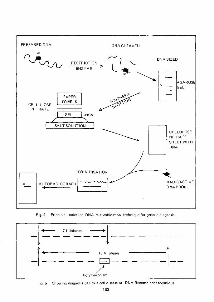
DNA Recombinant Technology
The difficu ltv in obtaining foetal blood has led
to an alternative, viz. culture of amniotic cells,
and examining the DNA sequences in these cells
to study more closely the DNA comprising the
{3-globin gene. In essence, the procedure consists
of utilising relevant restriction enzymes which
would cut the DNA chains in such a way that the
DNA of the {3-globin genes are separated. The DNA
fragments are sized by electrophoresis in agarose
gel (Fig. 4) and then transferred to nitrocellulose
by a method known as Southern blotting.
The actual DNA comprising the {3-gene and
the flanking DNA is 'brought out' by hybridisa
tion with a radioactivated normal {3-globin probe
which will 'seek out' a normal {3-gene while it will
fail to pair up if the {3-gene of the patient is
abnormal. The advantages with this method are: ituses the well tried relatively safe and simple pro
cedure of amniocentesis and cell culture; and cells
may also be obtained from chorionic villi which
allows of earlier sampling than amniocentesis orfoetal blood sampling, both of which can be
effectively done in the second trimester which
does not allow much time for analysis. Chorionic
villi aspiration can be carried out in the first
trimester and relatively large number of cells canbe obtained. However, the technique still hassome kinks which will have to be straightened
out.
However, the technology of DNA recombinantanalysis applied to the prenatal diagnosis of {3thalassaemia major suffers from one disadvantage
not inherent in the technology itself. It is inherent
162
in the genetic defect in {3-thalassaemia major wherein the majority base mutations within the {3
globin gene cannot be demonstrated but there
may be changes in the flanking genes or poly
morphic genes which are closely linked with the
{3-globin gene.
In, other words, these genes are markers for the
g-thalassaemia gene. These are termed restriction
fragment length polymorph isms (R F LP) and they
have to be studied in the parents as well as in the
patient. Another way whereby such examination
of neighbouring DNA is illustrated in the example
of sickle cell haernoqlobin.l " When the restriction
enzyme Hpal is used in normal individuals in the
region of the {3-globin gene, a 7.6 kilobase DNA
fragment can be elucidated while in 80% of cases
of sickle-cell {3-globin on different 13 kilobase
length fragment is obtained instead (Fig. 5).
The carrier parents are each heterozygous
for the disease allele and thus have one 7.6 kilo
base DNA fragment (associated with the normal
gene) and one 13 kilobase DNA fragment
(containing the sickle-globin gene). This poly
morphism is caused by a single-base change 5
kilobase to the 3' sides of the {3-globin gene which
eliminates the Hpal site.
CONCLUSION
To recapitulate, for the prenatal diagnosis of
genetic disorders, ultrasonography is used mainly
in delineating multifactorial genetic diseases, whilechromosome culture of foetal cells assist in the
diagnosis of chromosome diseases, and DNA
recombinant technology is solving problems in
the diagnosis of Mendelian genetic diseases, a
technology which can be applied to all tissuesobtained from the foetus.
One group of Mendelian diseases which has
not been alluded to include the biochemical
inborn errors of metabolism. Many of them can
be diagnosed by testing for the enzyme in amnio
tic cells after culture or, i,n some cases fromfoetal blood. However, one has to look at the
problem of Mendelian diseases as a whole; there
DNA SIZED
PREPARED DNA
RESTRICTION
ENZYME
DNA CLEAVED
0:AGAROSEGEL
CELLULOSENITRATE
PAPERTOWELS
GEL WICK
SALT SOLUTION
CELLULOSENITRATESHEET WITHDNA
0:
HYBRIDISATION
A';.TORAOIOGRAPHI0:
RADIOACTIVEDNA PROBE
Fig.4 Principle underline DNA re-combination technique for genetic diagnosis.
-1~ ~'O":: - - >1---------I~ ---- - 1~Obases - - - - -I--
/'Polymorphism
Fig. 5 Showing diagnosis of sickle cell disease of DNA Recombinant technique.
163

are approximately 1500 known diseases of which
only 200 lend themselves to be diagnosed at the
molecular level e.g. even cystic fibrosis which is
the commonest severe Mendelian gene among
Caucasians, we still do not know what the
abnormal enzyme or structural protein really is.
Thus in 1,300 such diseases due to single genemutations, no known exact diagnostic methodology is available.
Out of the 200 Mendelian diseases which arecapable of molecular diagnosis, only in 70 of
these has prenatal diagnosis been reliably achieved;
in other words only 5% of all Mendelian disorderslend themselves to exact reliable prenatal diagnosis.
We thus have to be aware of the present
limitations in prenatal diagnosis but basing in
previous experience in this field, many more
genetic diseases will be diagnosable in the nearfuture and this opens up a whole field for interest
ing and useful medical research.
REFERENCES
Wong HB. Genetic counselling. J Singapore PaediatSoc 1979; 21: 109-18.
2 Wong HB. Genes and the IQ. Singapore: PG PublishingPte Ltd, 1984.
3 Wong HB, Chua TS. The significance of human chromosone diseases in Singapore. J Singapore Paediat Soc1979; 21 :38-45.
164
4 Hellman LM, Duffus GM, Donald, Sunden B. Safety ofdiagnostic ultrasound in obstetrics. Lancet 1970;i:1133-5.
Stark CR, Orleans M, Haverkamp AD, Murphy J.Short and long-term risks after exposure to diagnosticultrasound in utero. Obstetrics & Gynaecology 1984;63: 194-200.
6 Watts PL, Hall AJ, Fleming JEE. Ultrasound and chromosome damage. B J Radial 1972; 45:335-339.
7 Becker R, Zimmer G, Schmidt CG, Sandbeg AA. Sisterchromatid exchange and proliferation pattern afterultrasound exposure in vivo. Am J Hum Genet 1983;35:932-7.
8 Bergman I. Questions concerning safety and use ofcranial ultrasonography in the neonate. J Pediat 1983:103:865-8.
9 Toyooka, Pifer, Hemplemann. Neoplasma in childrentreated with x-rays for thymic enlargement. J NatCancer Inst 1983; 31 :1379.
10 Refetoff S, Harrison J, Karanfilskin, et al. Continuingoccurrence of thyroid carcinoma after radiation to theneck in infancy and childhood. N Engl J Med 1975;292:171.
11 Ferguson-Smith. Prenatal chromosome analysis andits impact on the birth incidence of chromosome disorders. Brit Med Bullet 1983; 39:355-363.
12 Hook E B. Prevalence of chromosome abnormalitisduring human gestation and implications for studies ofenvironmental mutagens. Lancet 1981 :2: 169-172.
13 Kan, Dozy. Antenatal diagnosis of sickle-cell anaemiaby DNA analysis of amniotic fluid cells. Lancet 1978;2:901-911.