GENETIC ANOMALIES INVOLVED IN THE PROGNOSIS … · GENETIC ANOMALIES INVOLVED IN THE PROGNOSIS OF...
32
UNIVERSITY OF MEDICINE AND PHARMACY "GRIGORE T. POPA" IAŞI FACULTY OF MEDICINE GENETIC ANOMALIES INVOLVED IN THE PROGNOSIS OF MYELOPROLIFERATIVE NEOPLASMS ABSTRACT OF PhD THESIS Prof. univ. dr. DOINA AZOICĂI PhD STUDENT IVANOV (POPESCU) ROXANA -2015- PhD COORDINATOR
Transcript of GENETIC ANOMALIES INVOLVED IN THE PROGNOSIS … · GENETIC ANOMALIES INVOLVED IN THE PROGNOSIS OF...

UNIVERSITY OF MEDICINE AND PHARMACY
"GRIGORE T. POPA" IAŞI
FACULTY OF MEDICINE
GENETIC ANOMALIES INVOLVED
IN THE PROGNOSIS OF
MYELOPROLIFERATIVE
NEOPLASMS
ABSTRACT OF PhD THESIS
Prof. univ. dr. DOINA AZOICĂI
PhD STUDENT
IVANOV (POPESCU) ROXANA
-2015-
PhD COORDINATOR


ANOMALII GENETICE IMPLICATE ÎN PROGNOSTICUL NEOPLASMELOR mIELOPROLIFERATIVE
i
TABLE OF CONTENTS
INTRODUCTION ........................................................................................ 1
STATE OF KNOWLEDGE
1. MYELOPROLIFERATIVE NEOPLASMS BCR-ABL 1. POSITION -
CHRONIC MYELOID LEUKEMIA ..............................................................3
1.1. EPIDEMIOLOGICAL CONSIDERATIONS ...................................... 3 1.1.1. Descriptive epidemiology................................................................ 3 1.1.2. Aspects of analytical epidemiology ................................................. 4
1.2. CYTOGENETIC AND MOLECULAR BIOLOGY OF CHRONIC
MYELOID LEUKEMIA .............................................................................. 4 1.2.1. BCR-ABL translocation etiology ...................................................... 4 1.2.2. Genes and protein BCR-ABL, ABL and BCR .................................... 5 1.2.3. Role of BCR-ABL protein ................................................................ 8
1.3. CLINICAL FEATURES CHRONIC MYELOID LEUKEMIA .............. 14 1.3.1. The phases of the disease ............................................................... 14 1.3.2. Clinical aspects and hematology ..................................................... 14 1.3.3. Genetic evaluation ......................................................................... 15 1.3.4. Models risk stratification and prognostic variables for CML............. 16
1.4. THERAPEUTIC OPTIONS AND MONITORING RESPONSE TO
TREATMENT ........................................................................................... 19 1.4.1. Citototoxic agents and interferon alfa ............................................. 20 1.4.2. Tyrosine kinase inhibitors .............................................................. 20 1.4.3. Monitoring response to treatment ................................................... 23 1.4.4. New therapeutic targets ................................................................. 26 1.4.5. Imatinib resistance and drug resistance mechanisms ....................... 27 1.4.6. Treatment algorithms in chronic myeloid leukemia ......................... 33
2. BCR-ABL MYELOPROLIFERATIVE NEOPLASMS NEGATIVE .......... 36
2.1. PHENOTYPE FORMS BY GENETIC ANOMALIES INVOLVED ....... 36 2.2. GENETIC ETIOLOGY ....................................................................... 36 2.3. CLINICAL FEATURES OF BCR-ABL NEGATIVE
MYELOPROLIFERATIVE NEOPLASMS ............................................... 38 2.3.1. Polycythemia vera ......................................................................... 38 2.3.2. Essential thrombocythemia ............................................................ 39 2.3.3. Primary myelofibrosis .................................................................... 39

ANOMALII GENETICE IMPLICATE ÎN PROGNOSTICUL NEOPLASMELOR mIELOPROLIFERATIVE
ii
2.4. . TREATMENT OPTIONS ................................................................... 40
PERSONAL CONTRIBUTIONS
3. THE EVALUATION OF TREATMENT RESPONSE AND RATE OF
SURVIVAL FOR CML PATIENTS ............................................................. 42
3.1. INTRODUCTION ............................................................................... 42 3.2. PURPOSE AND OBJECTIVES OF THE STUDY ................................. 43 3.3. MATERIAL AND METHODS ............................................................. 43 3.4. RESULTS ............................................................................................ 48 3.5. DISCUSSIONS .................................................................................... 63 3.6. PARTIAL CONCLUSIONS ................................................................. 67
4. IDENTIFICATION OF THE MUTATIONS IN THE TYROSINE KINASE
DOMAIN OF THE BCR-ABL GENE ........................................................... 68
4.1. INTRODUCTION ............................................................................... 68 4.2. PURPOSE AND OBJECTIVES OF THE STUDY ................................. 68 4.3. MATERIAL AND METHODS ............................................................. 68 4.4. RESULTS ............................................................................................ 76 4.5. DISCUSSIONS .................................................................................... 80 4.6. PARTIAL CONCLUSIONS ................................................................. 85
5. EVALUATION OF GENETIC ANOMALIES RESPONSIBLE FOR THE
EVOLUTION TOWARDS BLASTIC CRISIS OF CML ................................ 86
5.1. INTRODUCTION ................................................................................... 86 5.2. PURPOSE AND OBJECTIVES OF THE STUDY .......................................... 88 5.3. MATERIAL AND METHODS ................................................................... 88 5.4. RESULTS .............................................................................................. 95 5.5. DISCUSSIONS ..................................................................................... 111 5.6. PARTIAL CONCLUSIONS .................................................................... 121
6. EVALUATION OF CLINICAL AND EVOLUTIVE IMPLICATIONS OF THE
CONCOMITANT PRESENCE OF THE JAK2V617F MUTATION AND THE BCR-ABL
FUSION GENE IN PATIENTS WITH MYELOPROLIFERATIVE NEOPLASMS .... 122
6.1. INTRODUCTION ................................................................................. 122 6.2. PURPOSE AND OBJECTIVES OF THE STUDY ........................................ 122 6.3. MATERIAL AND METHODS ................................................................. 123 6.4. RESULTS ............................................................................................ 125

ANOMALII GENETICE IMPLICATE ÎN PROGNOSTICUL NEOPLASMELOR mIELOPROLIFERATIVE
iii
6.5. DISCUSSIONS ..................................................................................... 127 6.6. PARTIAL CONCLUSIONS .................................................................... 131
7. FINAL CONCLUSIONS .......................................................................... 132
8. PERSPECTIVES OPENED BY THE DISSERTATION .................................. 134
REFERENCES. ......................................................................................... 135
PAPERS PUBLISHED DURING THE PHD STAGE ............................................... 1
The thesis is illustrated with 70 figures and 43 tables. The summary
includes a limited number of figures, retaining their numbering from
thesis.


GENETIC ANOMALIES INVOLVED IN THE PROGNOSIS OF MYELOPROLIFERATIVE NEOPLASMS
1
INTRODUCTION
The myeloproliferative neoplasms (MPNs) are diseases characterised
by clone proliferation, the distinction between various types of MPNs
being based on myelodysplasia (Vardiman et al. 2009). The management
of chronic myeloid leukemia (CML) in Romania is problematic because
of the defective alignment to the international standards for molecular
monitoring with consequences upon the modification of treatment at the
optimal time and the adequate use of the new tyrosine kinase inhibitors
(TKI). Although TKI induce remission in an important number of
patients, resistance and incomplete response to these agents occur
frequently leading to relapse and disease progression, this condition being
considered incurable. The choice of the doctoral research subject was
determined by the following arguments: the present research directions
are oriented both in the clinical field (a more efficient and standardized
monitoring of the disease evolution and the level of minimal residual
disease, the early relapse diagnosis) and in the fundamental one (the
identification of genetic and epigenetic alterations, the evaluation of their
involvement in the pathogeny of the disease, the clarification of cellular
communication inter-relations between stem leucemic cells and the
cellular micro-environment); the therapy efficiency depends on the
measure in which it identifies and controls the molecular targets, both the
optimal change of the therapy with new generations of TKI and the
personalization of treatment being compulsory.
The general objective aimed at consisted of the identification and
validation of a molecular markers panel (clone anomalies, mutations of
the kinase domain of the ABL gene, mutations of the JAK2 gene,
anomalies in the number of copies of other genes or epigenetic
anomalies) with a potential role in the progression or relapse of CML and
the evaluation of their involvement in the regulation of the expression of
the BCR-ABL fusion gene. The doctoral thesis follows the contemporary
advanced research directions by promoting useful tools that define
personalized medicine. Besides aspects related to the synthesis of existing
data, our personal research was targeted at studying the progressive
particularities of CML patients, identifying the genetic anomalies
involved in the evolution of the disease as well as their potential use as
markers in the identification of patients with a risk of CML progression.
The outcomes were possible due to the collaboration with the Oncology-Haematology Clinic and the Laboratory of Molecular Biology of the
Regional Institute of Oncology and the unconditioned support and

GENETIC ANOMALIES INVOLVED IN THE PROGNOSIS OF MYELOPROLIFERATIVE NEOPLASMS
2
expertise of the teams led by head of department Dr. Cătălin Dănăilă and
Professor Eugen Carasevici, PhD.
II. PERSONAL CONTRIBUTIONS
3. THE EVALUATION OF TREATMENT RESPONSE AND
RATE OF SURVIVAL FOR CML PATIENTS
3.1. Introduction CML patients are usually diagnosed in CF (chronic phase) (90%),
the clinical diagnosis being based on a characteristic complete blood
count where the differential blood count is left shifted (Melo et al. 1993,
Assouline, Lipton 2011). The introduction of the TKI treatment turned
this type of leukaemia from a reduced life expectancy disease
(approximately 4-6 years) into a chronic disease, with an increasing
global surviving rate (Deininger 2007). Classic prognosis indicators, such
as Sokal and Hasford scores, were used in order to estimate the relative
risk of evolution in CML-CP (Hochhaus et al. 2008). Due to the
prognosis value of the early response to treatment and the level of
response obtained, cytogenetic and molecular testing for the monitoring
of the therapeutical response and the minimal residual disease level have
become essential elements as far as decisions regarding CML patients are
concerned (Assouline, Lipton 2011).
3.2. Purpose and objectives of the study
The purpose of this study is to identify evolution particularities
concerning the therapeutic response, tolerance and survival rate in
patients treated with second-generation TKI in order to identify the most
useful markers for establishing the prognosis and optimizing treatment
changes.
3.3. Material and methods The study batch (batch A) was formed of 87 patients diagnosed with
CML, belonging to Romanian Institute of Oncology IRO, Iaşi. The
selection of the patients was performed by including all incidental cases
recorded in the period 2000-2012, their evolution being monitored in
successive stages up to the year 2014. The data analysis and interpretation
was performed for the cases that meet the conditions of molecular and
cytogenetic monitoring of minimum 18 months. For monitoring and
treatment response we performed the cytogenetic analysis and molecular
evaluation of BCR-ABL transcript and evaluated the type, frequency and
duration of treatment response.
Standard chromosomal analysis was performed on hematogenous
marrow culture, without stimulation (minimum 2 cultures). In order to
quantify the genetic expression of BCR-ABL we used the Translocation

GENETIC ANOMALIES INVOLVED IN THE PROGNOSIS OF MYELOPROLIFERATIVE NEOPLASMS
3
Kit t(9;22)/M-BCR-RQ (Experteam, Italy), based on the RealTimePCR
technique with hydrolysis probe (of TaqMan type). The reference gene
was ABL.
3.4. Results Characteristics of the patients batch. The statistic descriptive analysis
of the batch indicated a relatively equal distribution between sexes, the
average age was 41 years, people under 40 years of age being more
frequently affected by the disease (55%). The blood count is suggestive,
indicating hyperleukocytosis with a median of 143.2x10⁶/µL in 62.1% of
the cases over 100x106/µL. Thrombocytosis was signalled in 47.1% of
the total number of patients, with a severe form (>700x106/µL) in 13.6%
of the patients.
Evaluation of prognosis scores. The evaluation of the Sokal score
revealed that 36 patients belonged to the high risk category (41.9%),
while only 17 patients (19.5%) were included in the same prognosis
category when applying the European Hasford score. Statistical influence
was evaluated with the Kaplan Meyer survival curves, depending on the
prognosis scores. The survival rate in the batch of patient with Hasford
low risk was similar with that of the patients with intermediate and low
Sokal risk and of those with EUTOS low risk
Treatment. At diagnosis, in equal proportions of approximately 25%,
the patients of the studied batch received treatment with Glivec (Imatinib)
or Hidroxiuree (HUR) or HUR and cytarabine (C-ARA) while a very
small percentage received other treatments. The Imatinib dose was
increased in 66 patients, 17 patients displaying an improvement of the
therapeutic response. The treatment was interrupted in 19 patients
because of severe cytopenia, the evolution of the disease leading to
acceleration (N=14) and CB (N=4), lack of response or loss of response.
The Glivec treatment represented the second and respectively the third
therapeutic option in 66 patients (75.9%), the treatment being initiated in
less than 6 months from diagnosis in 54% of the cases (47 patients). At
the last evaluation, 11 patients were taken out of the batch, 51 patients
received the Glivec treatment while 18 (20.7%) received second-
generation TKI.
Survival rate according to treatment. For the studied batch the
survival rate for the patients under treatment Imatinib was, on average, of
56 months (3-126 months). The general survival rate was, on average, 76
months (18-180 months). The time until the initiation of treatment significantly influenced the survival rate, patients receiving Glivec in less
than 24 months from diagnosis responding much better than the ones who
received therapy later (p = 0,024).

GENETIC ANOMALIES INVOLVED IN THE PROGNOSIS OF MYELOPROLIFERATIVE NEOPLASMS
4
Treatment response monitoring was performed at a haematological,
cytogenetic and molecular level. All the patients received TKI treatment
throughout the evolution of the disease. The therapeutic response to the
Imatinib treatment varied, 59 patients (67.8%) obtaining CHR in 12
months and respectively 93.1% in 18 months from the beginning of the
treatment. The major cytogenetic response (partial and complete) was
obtained in only 64.5% of the patients in 12 months of treatment. At the
end of the study only 49 patients presented MCyR (56.3%) while 31%
had MCyR or ES. By the end of the study molecular examination was
performed in 62 patients (71.3%), while after the beginning of the Glivec
treatment an equal number of 22 patients CMR and MMR respectively.
The evaluation of the impact of prognosis scores upon the therapeutic
response to Imatinib revealed a significant statistic impact of the Sokal
score both on the cytogenetic response (p = 0,001) and the molecular
response (p = 0,025). The administration of Glivec as prime line therapy
greatly influenced both the cytogenetic (p = 0,005) and the molecular
response (p = 0,015), early administration (in less than 24 months)
determining a significantly better response (p = 0,026) as compared to the
late administration (third line of treatment).
3.5. Discussions Despite the fact that initial treatment was heterogeneous, throughout
the evolution of the disease all the patients received TKI treatment.In our
study, CHR rate was 93.1% comparable with the one obtained in the IRIS
study, of respectively 98% (Deininger M et al, 2009), as well as with the
response rate of 95% obtained in the phase II study (Kantarjian H et al,
2002).
The cytogenetic response rate for the patients included in our study is
lower than the one reported by the IRIS study, namely 64.5% for the
patients included in this comparative study as compared to 83%, yet it is
higher than the 13% rate reported in the phase I study – a study with
patients recruited after showing resistance at the αInterferon (Druker et al.
2001), being comparable with the results of the phase II study in which,
for a batch of 454 patients diagnosed with CML-CP, the CCyR rate was
of 60% (Kantarjian et al. 2002). This difference might be motivated by
the fact that our batch of patients was not homogeneous, since 54 patients
(62%) received the first line of treatment with Imatinib and only 38
patients began treatment within 12 months from the diagnosis. In our
study, only 74 patients were evaluated molecularly, MMR being obtained in 73% of the cases. At the end of the study only 50% of the patients
maintained MMR or CMR. Although it is difficult to compare these
outcomes with the ones described in literature because of the differences

GENETIC ANOMALIES INVOLVED IN THE PROGNOSIS OF MYELOPROLIFERATIVE NEOPLASMS
5
in expressing the level of the BCR-ABL transcript, the MMR rate
obtained in our study was similar with that reported by Lavallade et al.
In our study, the time until the initiation of treatment with Imatinib,
namely less than 12 months from diagnosis, had a positive impact on the
survival prognosis, as opposed to the data obtained Kantarjian et al. in a
study including 368 CML patients treated with Imatinib, after the failure
of the interferon treatment (Kantarjian et al. 2002).
The persistence of the prognosis Sokal score influence upon the
therapeutic response and the survival rate of the CML patients after the
apparition of Imatinib was discussed in several studies (Trask et al. 2012,
Hoffmann et al. 2013, Uz et al. 2013). Sokal maintained the impact and
the prognostic value upon the therapeutic response and survival rate even
in the Imatinib era. In our study the Sokal score at diagnosis had the
highest impact upon the cytogenetic molecular and survival responses.
The Hasford prognostic score had a less significant influence upon the
cytogenetic response, yet had a statistically more significant influence
upon the molecular response.
4. IDENTIFICATION OF THE MUTATIONS IN THE
TYROSINE KINASE DOMAIN OF THE BCR-ABL GENE
4.1. Introduction
The mutations in the tyrosine kinase situ (the kinase domain) of
BCR-ABL are the main mechanism associated with the resistance to the
TKI treatment in CML patients. More than 90 types of mutations were
reported, in patients with resistance to Imatinib (Jabbour et al. 2006,
O'Hare et al. 2007, Grant et al. 2010). The mutations in the kinase domain
are mainly grouped into nine amino-acids positions, including T315I,
Y253H/F, M351T, G250E, E255K/V, F359V and H396R, which
determine different levels of sensitivity to Imatinib. The in vitro
evaluation of their sensitivity to various generations of TKI offered the
adequate treatment options depending on the mutational statute of BCR-
ABL (eg. mutations Y253H, E255K/V or F359V/C/I – sensitivity to
Dasatinib and Ponatinib). Some mutations, such as T315I, are resistant to
all generations of TKI.
4.2. Purpose and objectives of the study The purpose of the study consists in the identification of mutations in
the kinase domain of the ABL gene in the BCR-ABL fusion gene which
induce resistance to the 1st generation TKI treatment, in order to achieve
controlled treatment alterations or transplant indication in patients who do
not respond to treatment.

GENETIC ANOMALIES INVOLVED IN THE PROGNOSIS OF MYELOPROLIFERATIVE NEOPLASMS
6
4.3. Material and methods
The study batch (batch B) selected for the analysis of mutations in
the kinase domain of BCR-ABL was formed of 15 patients diagnosed
with CML and treated with Imatinib, who did not obtain an adequate
molecular and cytogenetic response according to the ELN criteria
(without CHR, Ph +> 95% at 3 months; Ph+> 35%, or BCR-ABL> 10%
at 6 months and Ph+ ≥0% and/or BCR-ABL> 1% at 12 months from the
beginning of the treatment) or lost the response to treatment.
Sanger sequencing. The Sanger sequencing method with terminal
fluorescence was used in the study (Dye Terminator Sequencing). For the
sequencing of the kinase domain of the ABL gene involved in the
translocation we performed overlapped amplifications (nested PCR) for
BCR-ABL and for ABL. The primers sequences used in the
amplification of the kinase domain of the BCR-ABL (Branford et al.
2002) gene were: first amplification - the forward primer – 5’
TGACCAACTCGTGTGGTGTGAAACCTC 3’, the reverse primer– 5’
TCCACTTCGTCTGAGATACTGGATT 3’; second amplification
(nested) – Forward primer – 5’ CGCAACAAGCCCACTGTCT 3’,
reverse primer – 5’ TCCACTTCGTCTGAGATACTGGATT 3’.
Following the calculation of the PCR reaction parameters, a mix and a
start amplification programme were set, from which any other subsequent
optimizations derived. Sequencing is achieved with the GenomeLab™
Dye Terminator Cycle Sequencing Quick Start Kit (Beckman Coulter,
Fullerton, CA). Capilar electrophoretic migration was performed in the
Beckman Coulter CEQ8000 sequencer. The migration protocol was LFR-
a. The data obtained was initially interpreted with the CEQ8000 soft. The
data folders were afterwards exported in the .scf format, being interpreted
with the ChromasLite visualisation programe.
4.4. Results
4.4.1. The optimization process of the Sanger sequencing process
of the tyrosine kinase domain of the BCR-ABL gene 1. In the first stage it was decided to modify the PCR reaction
parameters in order to identify the optimal working conditions.
Concentration gradient of MgCl2. In order to verify the optimal
concentration of Mg, the following quantities were selected: 2; 3; 4; 5; 6
and 7μl MgCl2, corresponding to th final concentrations of 1.1; 1.6; 2.2;
2.7; 3.3; 3.8 mM MgCl2. The 2.7 mM MgCl2 concentration was
considered the accurate one.
Primers hybridization temperature gradient. Starting from the
hybridization temperature set at 64˚C, an amplification in temperature
gradient from 60 to70˚C was performed, as described below: 60; 61;

GENETIC ANOMALIES INVOLVED IN THE PROGNOSIS OF MYELOPROLIFERATIVE NEOPLASMS
7
Figura 40. The aspect of electrophoretic
migration in gel following the hybridization
temperatures gradient for nested primers
61.9; 62.9; 63.8; 64.6; 65.4; 66.3;
67.1; 68.1; 69; 70˚C (Figure 40).
The value of 66.5˚C was chosen as
the hybridization temperature.
Concentration gradient for
pimers. For concentrations below
0.5 μl (final concentration 0.2 μM),
no unspecific amplification signal
was obtained. A concentration of
0.2 μM was chosen.
Concentration gradient
for template. The template
concentration introduced in
the PCR reaction was
calculated theoretically (the technique is performed on cDNA). Following
the ARN extraction a dilution up to 500 ng/µl was performed, using in
the ReversTranscription reaction a quantity of 2µg. The final
concentration of cDNA will be equivalent of of 100 ng/µl of RNA. The
PCR amplifications were performed with 5µl undiluted cDNA and in
successive dilutions in scale 1/2. A dilution of 1/8 was selected.
2. In the second stage, in order to optimize the sequencing reaction
the reaction parameters were modified. The amplification programme was
optimized starting from the hybridization temperature of the primers used
in the PCR (66.5°C) amplification with an elongation of 72°C, and
eventually a two step programme was used with a hybridization
temperature and elongation of 60°C (the optimal temperature for the
amplification of the enzyme in the sequencing kit, respectively).
3. In the third stage we aimed at optimizing the electrophoretic
migration programme – the voltage and the migration time were modified
from 2.2 kV – 110 min to 3 kV – 180 min.
4.4.2. Identified mutations
A batch of 15 patients was investigated after concluding the
optimization process for the sequencing procedure. The level of the BCR-
ABL/ABL ratio was over 30% in the evaluated patients. Mutations were
identified only in two patients (13%).
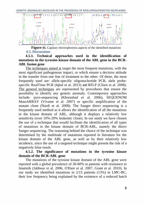
A patient displayed two mutations at two different moments of the
disease: the c.T107G mutation corresponding to the F359V phenotype
and the c.C951A mutation corresponding to the p.F317L phenotype
(Figure 45 A and B); one patient displayed the cG756T mutation,
corresponding to the Q252H phenotype (Figure 45 C).
GENETIC ANOMALIES INVOLVED IN THE PROGNOSIS OF MYELOPROLIFERATIVE NEOPLASMS
8
Figure 45. Capilary electrophoresis aspects of the identified mutations
4.5. Discussions
4.5.1. Technical approaches used in the identification of
mutations in the tyrosine-kinase domain of the ABL gene in the BCR-
ABL fusion gene
The techniques aimed at target the most frequent mutations, with the
most significant pathogenous impact, or which ensure a decisive attitude
in the transfer from one line of treatment to the other. Of these, the most
frequently used are: allele-specific oligonucleotide PCR, alele probe-
specific RealTime PCR (Iqbal et al. 2013) and RFLP (Chien et al. 2008).
The general techniques are represented by procedures that ensure the
possibility to identify any genetic anomaly. Contemporary approaches
include: pyro-sequencing (Khorashad et al. 2006), SEQUENOM
MassARRAY (Vivante et al. 2007) or specific amplification of the
mutant clone (Nardi et al. 2008). The Sanger direct sequencing is a
frequently used method as it allows the identification of all the mutations
in the kinase domain of ABL, although it displays a relatively low
sensitivity (over 10%-20% leukemic clone). In our study we have chosen
the use of a technique that would facilitate the identification of all types
of mutations in the kinase domain of BCR-ABL, namely the direct
Sanger sequencing. The reasoning behind the choice of the technique was
determined by the multitude of mutations reported in literature for the
kinase domain of the ABL gene, as well as by their relatively low
incidence, since the use of a targeted technique might present the risk of a
negatively false result.
4.5.2. The significance of mutations in the tyrosine kinase
domain of the BCR-ABL gene The mutations of the tyrosine kinase domain of the ABL gene were
reported with a global prevalence of 30-60% in patients with resistance to
Imatinib (Jabbour et al. 2006, O'Hare et al. 2007, Grant et al. 2010). In our study we identified mutations in 2/15 patients (13%) in LMC-BC,
their low frequency being explained by the existence of a reduced batch

GENETIC ANOMALIES INVOLVED IN THE PROGNOSIS OF MYELOPROLIFERATIVE NEOPLASMS
9
of patients and the possible existence of certain reduced mutant clones,
below the method detection limit.
One of the mutations identified in our study was Q252H, on a patient
displaying the loss of the molecular and haematological response, after 60
months of favourable evolution under treatment with Imatinib. The
Q252H mutation, together with other mutations of the coupling loop with
the ATP, were considered as having a great degree of resistance to
Imatinib and a very reserved prognosis (Branford et al. 2003). This aspect
was also noticed in the case of the patient included in our batch for which
no response was obtained during the treatment with Dasatinib, as the
patient deceased 4 months after the identification of the mutation.
Two mutations were identified in the ligation site of Imatinib, F359V
and F317L, respectively, in two successive samples (from different
moments in the evolution of the disease) at the same patient. The
c.1075T>G (F359V) mutation confers a reduction of response to
treatment with Imatinib and Nilotinib and partially retains the response to
the therapy with Dasatinib (Soverini et al. 2011). In the case of the
female patient included in our study this treatment was initiated, with a
favourable evolution (CCyR and MMR were obtained after three months,
the response being valid for 10 months). The c.951C>G (F317L)
mutation determines a reduced/moderate sensitivity to the treatment with
Imatinib (Shah et al. 2007) and Dasatinib (Laneuville et al. 2010) and
preserves high sensitivity to the therapy with Nilotinib (O'Hare et al.
2007, Soverini et al. 2011). In the case of the female patient included in
our study the identification of the mutation was achieved too late, as the
patient died before the result was obtained. The high incidence of the
F317L mutation following the treatment with Dasatinib (50% od all
detected mutations) support the in vitro studies that demonstrated the
potential occurrence of mutations (F317L included) under treatment with
Dasatinib (Bradeen et al. 2006). In a manner similar to the data reported
by Bradeen et al., in the case of the female patient included in our study,
the F317F mutation occurred during the treatment with Dasatinib.
5. EVALUATION OF GENETIC ANOMALIES
RESPONSIBLE FOR THE EVOLUTION TOWARDS BLASTIC
CRISIS OF CML
5.1. Introduction
Between 15% and 20% of the patients can progress towards CML-BC (Fabarius et al. 2011). The acquisition of secondary chromosomal
aberrations (CSA), such as + 8, the duplication of the Ph chromosome, i
17q, +19 or translocations and associate inversions LAM, was interpreted

GENETIC ANOMALIES INVOLVED IN THE PROGNOSIS OF MYELOPROLIFERATIVE NEOPLASMS
10
as a sign of evolution being associated with a high progression risk
towards the accelerated phase or the blastic crisis (Fabarius et al. 2011).
The evolution towards the BC limits therapeutic options, which seriously
reduces the survival rate (Cortes et al. 2008). Early recognition of the
patients with a risk of evolution towards BC is thus very important.
Deletions in IKZF1, PAX5, and/or CDKN2A were frequently reported in
lymphoid CML-BC (Mullighan et al. 2008, Alpar et al. 2012). Multiple
genes were identified as being methylate in the bone marrow in patients
with CML or AML, including CDH1, CDKN2A/CDKN2B, DAP kinase,
RUNX3, WT1 (Hess et al. 2008, Homig-Holzel, Savola 2012).
5.2. Purpose and objectives of the study
The purpose of our study is to identify genetic and epigenetic
anomalies in patients who either are resistant or lost their response to the
TKI treatment, in order to evaluate the overlapping events that could
explain the unfavourable evolution, as well as to identify new markers
that could discriminate between the respondent and non-respondent
patients as far as this therapy is concerned.
5.3. Material and methods
For this study, the batch (batch C) was formed of 92 patients
diagnosed with CML, for whom molecular and cytogenetic investigations
were performed. The selection of the patients was made by including the
cases recorded in the period 2005-2013, their evolution being monitored
until the year 2014 (cytogenetic or molecular monitoring of minimum 6
months). For all the patients we evaluated the frequency and types of
SCA identified through the standard karyotype. Chromosomal anomalies
were regarded as clonal evolution if at least two cells were discovered to
have the same chromosomal arrangement/suplimentary chromosome(s) or
at least three cells were identified to have the same monosomy.
From the study batch we selected the sub-batch D, made of 30
patients: 15 patients who showed no response to the treatment or relapsed
(the level of the BCR-ABL transcript went up to minimum 25%) and 15
control patients diagnosed with CML-CP, with a favourable evolution.
All the patients in sub-batch D were investigated using the MLPA method
and the P335 set (aimed at detecting anomalies in the number of copies of
the IKZF1, CDKN2A, PAX5, EBF1, ETV6, BTG1, RB1 genes and in the
PAR1 area: CRLF2, CSF2RA, IL3RA) and the ME002 set, which
evaluates the methylation stages of thye promontory regions of a set of
suppressing genes for tumours frequently involved in the carcinogenesis process. The number of DNA copies was estimated using the
Coffalyser.net programme.

GENETIC ANOMALIES INVOLVED IN THE PROGNOSIS OF MYELOPROLIFERATIVE NEOPLASMS
11
5.4. Results
5.4.1. The characterization of batch C as far as cytogenetic
anomalies and evolution are concerned
Conventional cytogenetic analysis revealed the presence of the Ph
chromosome in 92.4% (85/92) of the patients, as well as the presence of
other associated anomalies in 18% (17/92) of the patients. Fifteen of the
patients with SCA (15/17 – 88%) had an unfavourable evolution
(acceleration or accutization phases). The most frequent major SCA were
trisomy 8 (8 cases – 53%) and the duplication of the Ph chromosome (7
cases - 46%), 4 patients presenting the four anomalies simultaneously.
The identified chromosomal anomalies are presented in Table 38.
Nr Sex Age Months to the
SCA
Cytogenetic aberrations additional to t(9; 22)(q34;q11) or the
t(v,22) version, at diagnosis
1. F 46 60 47,XX,t(9,22)(q34;q11.2),+8[7]/46,XX,t(9,22)(q34;q11.2)[4]
2. F 55 96 46, XX[13]/47,XX,t(9,22)(q34;q11.2),+8[17]
3. F 41 6 46,XX,t(9,22)(q34;q11.2),inv(16)(p13;q22)[17]/46,XX,t(9,22)(q34;q11.2
)[2]/46,XX[1]
4. F 40 0 47,XXX,t(9;22)(q34;q11.2)[21]
5. F 55 6 46,XX [23]/46,XX,t(9,22)(q34;q11.2)[4]/46,XX,t(16;17)[3]
6. F 34 12 46,XX,t(9,22)(q34;q11.2)16]/47,XX,t(9,22)(q34;q11.2),+8[10]
7. F 56 12 46,XX[4]/47,XX,t(9;22)(q34;q11.2)X2[8]/48,XX,t(9;22)(q34;q11.2)X2,
+6[4]
8. F 63 6 46,XX[14]/46,XX,t(9,22)(q34;q11.2)[3]/48,XX,t(9,22)(q34;q11.2)x2,+8[
1
9. M 24 38 46,XY,t(9;22)(q34;q11.2)[15]/47,XY,t(9;22)(q34;q11.2)x2[5]/47,XY,t(9;
22)(q34;q11.2),+8[6]
10. M 30 12 46,XY,t(9;22)(q34;q11.2)[10]/47,XY,t(9;22)(q34;q11.2),+17[6]/47,XY,t(9;22)(q34;q11.2),+19[8]
11. F 24 0
36
46,XX,t(9;22)(q34;q11.2)[8]/47,XX, t(9;22)(q34; q11.2)x2[4]
47, XX,t(9;22)(q34;q11.2)x2[9]/48,XX,t(9;22)(q34; q11.2)x2,+8[5]
12. F 55 6 46,XX,t(9;22)(q34;q11.2)[16]/48,XX,t(9;22)(q34;q11.2)X2, +8[15]
13. M 20 0 46,XY,t(1;3)(p22;q29),t(9;22)(q34;q11.2)[20]
14. M 59 0 46, XY,i(17)(qter→q10::q10→qter)[10]
15. M 55 0 46,XY,der(2)?,der(2)?,t(9;22)(q34;q11.2)[20]
16. M 36 12 48,XY,t(9;22)(q34;q11.2)x2,+8[20]
17. M 28 0 46,XY,t(9;22;11)(q34;q11;q13)[30]
F - female, M - male; *age in years at the time of diagnosis; SCA- supplementary chromosomal
anomaly
5.4.2. Molecular and clinical characteriyation of a particular case
of CML which coexpresses the CBFβ-MYH11 and BCR-ABL fusion
genes
In the case of patient 3 we identified, at the moment of accutization,
the presence of a inv(16), while further on it was proved that it was
present even since the diagnosis. The evaluation of the CBFβ-MYH11

GENETIC ANOMALIES INVOLVED IN THE PROGNOSIS OF MYELOPROLIFERATIVE NEOPLASMS
12
Figura 60 Figura 61 Figura 62 Figure 1. Heterozygous deletion at the level of exon 1 of the IL3RA gene or polymorphism
mutation at this level (patient A1) ; Figure 61. Heterozygous deletion at the level of the entire
IKZF1 gene (patient A2); Figure 62. Heterozygous deletion of the exons 4-7 of the IKZF1
gene and of the CSF2RA, P2RY8 and IL3RA genes, in the PAR1 region (patient A3)
fusion gene revealed the E type transcript. Two mutations were identified
in the kinase domain of ABL F359V and F317L, respectively. The patient
deceased because of the progression of the disease and lack of response to
TKI and chemotherapy, 18 month after diagnostication.
5.4.3. Evaluation of the variations in the number of genic copies
through the MLPA technique, using the P335-B1 set In the sub-batch D there were identified 3 cases (10%) that
associated anomalies of the number of genic copies, in the group with
resistance to the TKI treatment (Figure 60, Figure 61, Figure 62 –
partial images).
5.4.4. The evaluation of the methylation status of a set of tumour-
suppressor genes through MS-MLPA using the ME002 kit With the help of the MS-MLPA technique there weas identified
abnormal methylation of the following tumour-suppressor genes: WT1,
CDH13, GATA5, CD44, ESR; they were methylated in 23% of the
patients (7/30). Out of the patients with abnormal methylation, 6 patients
were among the ones who did not respond to the TKI and evolved
towards BC (Table 40).
Table 40. Characteristics of patients who displayed abnormal
methylation through MS-MLPA
Age*/
Sex
BCR-
ABL*
Genes with abnormal
methylation Treatment* Evolution
A1 65 years, M 45% CDH13 Dasatinib LMC-CP
A2 58 years, F 35% ESR1, CDH13; (del CDK6, CFTR)
Imatinib 400 Myeloid BC - death
A3 58 years, M 79% CDH13; Dasatinib Lymphoid BC - death
A4 24 years, F 28% WT1 Dasatinib Myeloid BC–
A5 38 ani,M 68% CD44 Imatinib 400 LMC-CP
A6 58 years, M 100% WT1,CDH13, GATA5 - Lymphoid BC

GENETIC ANOMALIES INVOLVED IN THE PROGNOSIS OF MYELOPROLIFERATIVE NEOPLASMS
13
Figure 63. MS-MLPA aspect in patient A2. Superior panel: heterozygous deletion
at the level of genes CDK6 and CFTR. Inferior panel: abnormal methylation of genes
ESR and CDH13
A7 41 years, F 98% CD44 Imatinib 400 Myelomonocytic BC -
death
*- at the time of evaluation, Ph+ - presence of the Ph chromosome
In patient A2 (presented in the sub-chapter 5.4.3), the MS-MLPA
revealed a heterozygous deletion of genes CDK6 and CFTR, situated on
7q21-q22 and 7q31.2, respectively, as well as methylation anomalies at
the level of genes ESR and CDH13 (Figure 63). The frequency of
methylated genes was 13% (4/30) for the CDH13 gene, 6.6% (2/30) for
genes WT1 and CD44 and 3.3% (1/30) for genes ESR and GATA5.
5.5. Discussions
5.5.1. Secondary chromosomal anomalies
Types and frequency of secondary chromosomal anomalies
In our study 18% of the cases associated clonal SCA, their incidence
in the accelerated or blastic phase being of 75%. The type and frequency
of identified SCA are similar to those reported in previous studies (Farag
et al. 2004, Fabarius et al. 2011), the most frequent being major SCA
(11/17- 64%), respectively trisomy 8 (53%) and duplication of the Ph
chromosome (46%), 4 patients (23%) displaying a complex karyotype
with the association of both anomalies. In the study batch one patient
displayed t(v;22), with the further involvement of a single chromosome,
namely chromosome 11: 46,XY,t(9;22;11)(q34;q11;q13)
[30]. The patient’s evolution was favourable (CCyR, MMR), similar to
the one reported in previous studies (El-Zimaity et al. 2004, Marzocchi et
al. 2011). Rare SCA were identified (as far as we know, they have not
been reported) in three patients: t(16;17), t(1;3)(p22;q29) and respectively
two derivative chromosomes 2, possible by means of a mutual
translocation between homologous chromosomes.
Prognosis impact of secondary anomalies in CML

GENETIC ANOMALIES INVOLVED IN THE PROGNOSIS OF MYELOPROLIFERATIVE NEOPLASMS
14
Correspondingly to the data reported by Fabarius et al., who
evaluated 1151 patients with the same diagnosis and treatment, in our
study, major SCA had an unfavourable prognosis, with an average
survival rate of 41 months. Out of these, complex chromosomal
anomalies, with more than three abnormal cellular lines (two cases) were
associated with the patients death. Major chromosomal anomalies
developed during treatment were confirmed to be signals of
acceleration/accutization. Death occurred in two patients with
rarechromosomal anomalies usually detected in acute leukemias: inv(16)
and t(1;3). The co-existance of t(9; 22) and inv(16) in CML seems to
indicate an unfavourable prognosis, as well as resistance to both
chemotherapy and TKI (Merzianu et al. 2005, Roth et al. 2011).
5.5.2. Anomalies in the number of genic copies Anomalies in the number of copies were detected in only three cases
(3/30 -10%). The affected genes were similar to the ones reported in
literature (Mullighan et al. 2007, Mullighan et al. 2008, Kuiper et al.
2010, Calderon-Cabrera et al. 2013, van der Sligte et al. 2014). In the BC
cases there were identified deletions of the IKZF gene. Patient A3, with
lymphoid CML-BC limfoidă associated the most frequent type of
deletion, namely the deletion of exons 4-7 which was associated with the
deletion of genes in the pseudoautosomal region CSF2RA, P2RY8 and
IL3RA. In the case of the patient A2, we identified a rarely reported
deletion, namely the heterozygous deletion of the entire IKZF1 gene
(exons 1-8) which was phenotypically associated with myeloid CML-BC.
Thorough investigations through the use of MS-MLPA revealed
supplementary methylation anomalies and a heterozygous deletion of
genes CDK6 and CFTR, situated on 7q21-q22. The presence of the
deletion at the level of chromosome 7 on both arms can indicate the
absence of the entire chromosome 7 (monosomy 7). No cytogenetic
examination was performed for this case. Monosomy 7 was described in
approximately 5% of the supplementary chromosomal anomalies in
CML, being more frequently associated with lymphoid CML-BC
(Johansson et al. 2002). Deletions in the IKZF1 gene were associated
with a reserved prognosis, similar to the data reported in literature
(Mullighan et al. 2008, Kuiper et al. 2010). We have not been able to
establish a marker that could discriminate between patients with CML-CP
who are resistant to the therapy with Imatinib and those with an optimal
reponse to the treatment.
5.5.3. The methylation profile of tumour suppressor genes in
CML

GENETIC ANOMALIES INVOLVED IN THE PROGNOSIS OF MYELOPROLIFERATIVE NEOPLASMS
15
In our study, the genes identified with abnormal methylation and the
frequency of these alterations are similar to the results reported in
literature (Nguyen et al. 2000, Deutsch et al. 2003, Roman-Gomez et al.
2003, Janssen et al. 2010). In positive patients, methylation was
heterogenous, each individual having a different combination of affected
genes, an aspect which might be connected to an increased expression of
di-methyltransferases, as it was proven DNMT13A and 3B in CML-BC
(Mizuno et al. 2001). Abnormal methylation was significantly more
frequent in patients who did not respond to the TKI treatment (6 of 7
patients), 5 of them evolving towards BC. The increased number of
methylated genes in BC suggests that abnormal methylation is a non-
specific process which characterizes the acute stage of the disease, at least
for a sub-group of patients.
The CDH13 gene had the most frequent abnormal methylation, being
identified in both BC patients (3 cases) and in CP (1 case). These results
were similar to outcomes of previous studies (Roman-Gomez et al. 2003,
Janssen et al. 2010). The methylation of the CDH13 gene was not
correlated with a certain evolutive phenotype. The presence of abnormal
methylation in CP, as well in the progressive stages of the disease,
suggests that the aberrant methylation of the promoter takes place in an
early stage during the CML pathogenetics and it probably influences the
clinical behaviour of the disease. The abnormal methylation of the CD44
gene occurred in isolation in two patients, one with CML-CP who had a
favourable response to the Imatinib treatment and one with myeloid BC,
who displayed SCA, namely inv(16). In this context, the aberrant
methylation of the CD44 gene promoter takes place in an early stage in
the CML pathogenesis and probably influences the progression of the
disease by losing the control of the micro-environment upon the tumoral
cells. The hyper-methylation of the WT1 gene was identified in two
patients with progressive evolution. The alterations in the WT1
expression seem to be involved in the defective progenitor cells and the
progressive evolution of the disease (Rampal et al. 2014).
6. EVALUATION OF CLINICAL AND EVOLUTIVE
IMPLICATIONS OF THE CONCOMITANT PRESENCE OF THE
JAK2V617F MUTATION AND THE BCR-ABL FUSION GENE IN
PATIENTS WITH MYELOPROLIFERATIVE NEOPLASMS
6.1. Introduction The Ph chromosome is associated with CML and it occurs as the
effect of the balanced reciprocal translocation between chromosomes 9
and 22 (t(9;22)(q34;q11)), resulting in the production of the BCR-ABL

GENETIC ANOMALIES INVOLVED IN THE PROGNOSIS OF MYELOPROLIFERATIVE NEOPLASMS
16
fusion gene. In patients with negative MPN for BCR-ABL a punctiform
mutation was discovered that leads to the replacement of valine with
phenylalanine in codone 617 of the JAK2 gene. This mutation leads to the
constitutive activation of the JAK2 kinase and consequently to cellular
proliferation and resistance to apoptosis. It was initially believed that the
two anomalies are mutually exclusive (Jelinek et al. 2005), yet in the past
few years some rare cases were described about patients in which both
anomalies co-exist, the BCR-ABL fusion gene and the JAK2 V617F
mutation (Hussein et al. 2007, Inami et al. 2007), raising questions
regarding the phenotypical and prognosis relevance of the co-existance of
the two specific markers.
6.2. Purpose and objectives of the study This study aims at investigating the incidence of this double mutant
phenotype in patients with MPN at dignosis, in order to evaluate the
clinical characteristics of the differences in the responses to treatment for
patients testing positive for the JAK2V617F mutation and BCR-ABL, as
compared to patients who do not display the JAK2V617F mutation, but
are positive for BCR-ABL. A comparison with the outcomes described in
literature was also aimed at.
6.3. Material and methods This study was conducted in the period between January 2012 and
February 2014 and included 190 MPN cases selected from the Regional
Institute of Oncology Iasi, which formed batch E. for all the patients, we
evaluated both the JAK2V617F mutation, and the presence of the BCR-
ABL gene. The evaluation of the p210 BCR-ABL (b2a2, b3a2) transcript
was achieved by means of the Real Time PCR quantitative method, with
Taqman hydrolysis probe, using Translocation Kit t(9;22)/M-BCR-RQ
(Experteam, Italy). The evaluation of the JAK2V617F mutation was
performed with Real Time PCR Taqman hydrolysis probe.
6.4. Results Out of the 190 patients for which we evaluated both the BCR-ABL
fusion gene and the JAK2V617F mutation, 94 (84) patients (49,4%)
displayed the JAK2V617F mutation (79 (72) heterozygotes, 15 (12)
mutants) without the presence of the BCR-ABL gene, 68 (79) patients
(35,8 %) displayed JAK2 genotype wild type and tested negative for
BCR-ABL while 26 patients (13,7%) tested negative for the JAK2
mutation (wild type genotype) and positive for the BCR-ABL fusion
gene. Only two patients simultaneously displaed both the JAK2V617F mutation and the BCR-ABL transcript, namely 1.05%. The two patients’
phenotypes were essential thrombocythemia and polycythemia vera,
respectively.

GENETIC ANOMALIES INVOLVED IN THE PROGNOSIS OF MYELOPROLIFERATIVE NEOPLASMS
17
Case 1 is a 62 year old female patient in which the presence of the
b2a2 transcript (p210) was identified with a ratio of BCR-ABL/ABL of
99.54% at diagnosis. The patient also tested positive for the JAK2V617F
mutation in heterozygous state. In these circumstances, the diagnosis was
CML with associated JAK2V617F mutation. The therapeutic response
was favourable at Glivec 400mg/day, obtaining CHR and MMR after 3
months. The JAK2V617F mutation remains present in heterozygous state
in successive determinations. Case 2 is a 45 year old male patient in
which the JAK2V617F mjutation was identified in heterozygous state.
The analysis of the p210 transcript of the BCR-ABL fusion gene proved
the existence of the latter in reduced quantity at the time of diagnosis,
with a ratio BCR-ABL/ABL of 2.6%. the patient was diagnosed with
polycythemia vera, which associates the presence of the BCR-ABL major
transcript.
6.5. Discussions
The cases we presented contribute, along with the ones described in
literature in the last years, to a better understanding of the rare situation in
which the JAK2V617F mutation and the BCR-ABL fusion gene co-exist
in patients with MPN. In our study, the cases incidence that display the
BCR-ABL fusion gene at the same time with the JAK2V617F mutation
was of 1.05%, in a batch of 190 evaluated patients (2/190). A similar
result was reported by Cappetta et al. who analyzed 1320 cases with a
suspicion of MPN and identified 5 (0.37%), patients with non-typical
forms of MPN.
The two patients whose cases were detailed in this study associate
both anomalies concomitantly at diagnosis, having, however, different
phenotypes. Patient 1 displayed a non-typical phenotype for CML,
associating elements specific to essential thrombocythemia (ET). The
cases associating CML and ET phenotype are extremely rare (Veronese et
al. 2010, Lee et al. 2013, Pastore et al. 2013). Case 2 was diagnosed with
PV without phenotypical modifications specific to CML. Only two cases
described in literature display the BCR-ABL fusion gene (with reduced
levels of expression) without clinical signs of CML, throughout the
evolution of MPN BCR-ABL negative (Bornhauser et al. 2007, Park et al.
2013). As far as response to treatment is concerned, in most reported
patients the suppression of the BCR-ABL positive clone was obtained
under TKI treatment (CCyR or MMR/CMR), yet with a partial
haematological response, or the progression of marrow fibrosis. In the case of patient 1 described in this study, the response to the Imatinib 400
mg/day treatment was very good, leading to CHR and CMR in 3 months.
GENETIC ANOMALIES INVOLVED IN THE PROGNOSIS OF MYELOPROLIFERATIVE NEOPLASMS
18
Two hypotheses were proposed to explain the presence of the
JAK2V617F mutation and the BCR-ABL fusion gene at the same patient.
According to the first hypothesis negative MPN BCR-ABL and CML are
supposedly two distinctive diseases which develop in different clones of
the cellular progenitors, the phenotype being determined by the dominant
clone (Hussein et al. 2008, Bee et al. 2010, Veronese et al. 2010, Pastore
et al. 2013, Xu et al. 2014). The second hypothesis poposes the existence
of a single subclone of progenitors of the hematopoietic cells that acquire
the two anomalies in the same cell, at different moments (Bocchia et al.
2007, Bornhauser et al. 2007, Inami et al. 2007, Kramer et al. 2007,
Hussein et al. 2008, Jallades et al. 2008). The particularities of the cases
described in our study support the second hypothesis, namely the co-
existance of the two mutations within the same clone, with the occurance
of BCR-ABL in a sub-clone of positive JAK2V617F cells, the latter
gaining proliferative advantage. However, we cannot totally exclude the
possibility of two different mutant clones co-existing. The CML
evolution does not seem to be influenced by the presence of the
JAK2V617F mutation, yet it can constitute a rare cause explaining the
lack of haematological response to the TKI treatment and the
unfavourable prognosis for these patients.
7. FINAL CONCLUSIONS
1. Thorough monitoring of the cytogenetic and molecular response to
TKI allows for the accurate appreciation of the CML patients’ evolution,
the detection of patients with high risk of resistance and the identification
of candidates to the second-generation TKI treatment or transplant. Rates
of haematological, cytogenetical and molecular responses are similar to
the ones reported in literature.
2. Analysis at diagnosis of different parameters indicated that the
response to Imatinib therapy is influenced by the Sokal score and the
early initiation of treatment, in the first 12 month from diagnosis. The
Sokal score had the highest impact on the cytogenetic, molecular and
survival responses, the prognosis Hasford score having, in turn, a
statistically significant influence upon the molecular response. The time
to the initiation of the Imatinib treatment, less than 12 month from
diagnosis, had a positive impact upon the survival prognosis.
3. Molecular monitoring is essential for identifying patients with a high
risk of resistance and can optimally identify candidates for testing the mutations in the kinase domain of ABL. The sequencing technique has
the advantage of identifying all mutations in the tyrosine kinase domain
of BCR-ABL.

GENETIC ANOMALIES INVOLVED IN THE PROGNOSIS OF MYELOPROLIFERATIVE NEOPLASMS
19
4. The mutations identified in the study batch Q252H, F359V and
F317L are frequently reported in the cases of resistance to the Imatinib
treatment, being involved in the progression of the disease towards the
BC. Both the mutations in the P loop and those in the connection situ of
Imatinib were associated with an unfavourable prognosis, as none of the
patients survived.
5. Mutations sensitivity to the TKI treatment was similar to the data
reported in literature for the F359V mutation (induces sensitivity to the
treatment with Dasatinib) and F317L (generally reduced sensitivity to
Dasatinib), yet different for the Q252H mutation, that was resistant to the
Dasatinib treatment.
6. Systematic performance of the standard karyotype in CML Ph+
patients is necessary both for appreciating the response to treatment
(attaining and maintaining CCyR) and for identifying the clonal evolution
in patients losing response to the TKI treatment, whose disease
progresses. Classic cytogenesis is irreplaceable in the detection of
supplementary chromosomal anomalies, leading to additional molecular
testing.
7. Numerical chromosomal anomalies are the most frequent form of
clonal evolution, the most frequent being major SCA, trisomy 8 and the
duplication of the Ph chromosome. Patients displaying this anomaly
associate a significantly poorer prognosis as compared with the all other
patients, needing early monitoring and intensive therapeutical
intervention.
8. Identified minor SCA, associated with an unfavourable prognosis,
included derivative 2 chromosomes, t(1;3)(p22;q29), trisomy 19 and
mosaic trisomy 17 and inv(16). Inv(16) is a rare event in CML patients
being associated with the rapid progression of the disease. Particularly,
the case identified in our batch associated two successive mutations in the
kinase domain of BCR-ABL, the F359V mutation followed by the F317L
mutation, which worsened the clinical prognosis. Taking optimal
therapeutic decisions in such cases is very difficult, because of the limited
knowledge regarding the evolution of these patients.
9. The MLPA technique allowed the identification of clonal anomalies
in the number of copies of certain genes involved in the control and
regulation of the haemato-lymphoid system, which are rare in CML-CP,
yet are common to the progression stages of the disease. The deletion of
the IKZF1 gene was most commonly identified in patients with lymphoid BC.
10. MS-MLPA allowed the detection of abnormal methylation in the
samples taken from CML patients at the level of genes CDH13, CD44,

GENETIC ANOMALIES INVOLVED IN THE PROGNOSIS OF MYELOPROLIFERATIVE NEOPLASMS
20
WT1, GATA5 and ESR with an increased frequency in CML-BC, as
compared with the CP, in which methylation was almost absent. Our
results sustain the hypothesis according to which the progression of the
disease is frequently accompanied of methylation alterations. The
identification of certain clinically relevant methylation models in CML
can contribute to a better understanding of the disease progression
mechanisms. Generating a MS-MLPA set specific for CML by including
some specific targets could render this test more efficient in appreciating
the disease progression risks and optimal altering of the treatment in the
case of these patients.
11. The incidence of cases displaying the BCR-ABL fusion gene
concomitantly with the JAK2V617F mutation was low (1.05%, in the
batch of 190 evaluated patients), the identified cases, along with those
described in literature, supporting the possibility of the co-existance of
the two anomalies in the same patient, the most frequent phenotype being
the CML one.
12. The screening for the presence of the JAK2V617F mutation and the
BCR-ABL transcript is recommended at diagnosis in case of MPN
suspicion, as well in the case of patients diagnosed with CML who
develop myeloproliferations, despite achieving MMR
8. PERSPECTIVES OPENED BY THE DISSERTATION
In the context of molecular medicine development and molecularly
targeted medication, the aim of this paper was to broaden the perspective
by identifying genetic and epigenetic anomalies involved in the CML
progression, taking into account the evolutive particularities of patients in
North-Eastern Romania.
Applying the MLPA and MS-MLPA techniques in CML represents a
new approach for Romania; gathering information regarding the
genetic/epigenetic defects auxiliary in CML might lead to the generation,
in the near future, of a specific set for this condition.
Optimizing the sequencing technique for the mutations in the kinase
domain of BCR-ABL will allow its routine use, which will result in a
controlled change of treatment, thus saving critical time in the attempt to
conservate the state of disease remission. The sequencing technique has
the advantage of identifying new mutations, besides the ones already
described in literature, for which the gathering of data will allow a
stronger correlation with the response to various classes of inhibitors. Although it is a frequently studied disease, CML remains a high
interest field due to the constant need of identifying alternative variants
for both systemic and niche level treatments. In this respect, future

GENETIC ANOMALIES INVOLVED IN THE PROGNOSIS OF MYELOPROLIFERATIVE NEOPLASMS
21
research focus on the relationship between leukemic cells and marrow
stroma, especially the possibilities of artificial introduction of the immune
system cellular response in order to identify neoplastic entities for each
patient, creating thus the premises for the application of personalized
medicine. Another research direction is represented by following the
consecutive alterations or even those preceding the fusional alteration that
can generate the activation of genomic reconfiguration factors, that did
not undergo any cellular and/or micro-environment control system,
involving genic alterations recognition molecules genic alterations repair
molecules and control mechanisms for the repairs performed at the
genomic level. Another field of interest is represented by the study at the
marrow niche level of the communication between the leukemic cell and
the immune system cells responsible with the identification and
correlation of the defects, which eventually lead to the transformation of
the latter to the toleration of the leukemic cell (cancelling the anti-tumoral
response) or even offering a protection zone of the organism against the
tumour.
PUBLICATIONS DURING THE DOCTORAL PROGRAMME
Main author
1. Popescu R, Dăscălescu A, Dănăilă C, Ghiorghiu D, Zlei M, Ivanov A,
Sireteanu A, Gorduza EV, Azoicăi D. Co-expression of the CBFβ-MYH11
and BCR-ABL fusion genes in chronic myeloid leukaemia. Rev Romana
Med Lab. 2015;23(2):221-30. DOI:10.1515/rrlm-2015-0013
2. Popescu R, Dăscalescu A, Dănailă C, Gheorghiu D, Antohi I, Goriuc
A, Ivanov A., & Azoicai D. Myeloproliferative neoplasms with concurrent
BCR-ABL fusion gene and JAK2V617F mutation. Analele Stiintifice Ale
Universitatii "Alexandru Ioan Cuza" Din Iasi Sec. II A. Genetica Si
Biologie Moleculara, 2014;15(4), 25-34. Retrieved from
http://www.gbm.bio.uaic.ro/index.php/gbm/article/view/1160/1096 (B+)
Co-author
3. Grigore GE, Ivanov IC, Zlei M, Dăscălescu A, Popescu R, Petreuș T, et
al. Specific Associations Between Clinical Signs, Immune Cells, Disease
Genetic Background and Burden in a Group of Patients with B-Cell
Chronic Lymphocytic Leukemia. Rev Romana Med Lab. 2014;22(1):79-92.
DOI:10.2478/rrlm-2014-0004
4. Grigore G, Ivanov I, Popescu R, Zlei M, Dascalescu A, Jitaru D,
Petreus T, & Carasievici E. Genomic profiling by multiplex ligation -
dependent probe amplification in chronic lymphocytic leukemia
patients. Analele Stiintifice Ale Universitatii "Alexandru Ioan Cuza" Din

GENETIC ANOMALIES INVOLVED IN THE PROGNOSIS OF MYELOPROLIFERATIVE NEOPLASMS
22
Iasi Sec. II A. Genetica Si Biologie Moleculara, 2013; 14(3), 27-34.
Retrieved http://www.gbm.bio.uaic.ro/index.php/gbm/article/view/1072
SELECTIVE BIBLIOGRAPHY
Alpar D, de Jong D, Savola S, et al. MLPA is a powerful tool for detecting
lymphoblastic transformation in chronic myeloid leukemia and revealing
the clonal origin of relapse in pediatric acute lymphoblastic leukemia.
Cancer Genet. 2012; 205(9): 465-469.
Assouline S, Lipton JH. Monitoring response and resistance to treatment in
chronic myeloid leukemia. Curr Oncol. 2011; 18(2): e71-83.
Bee PC, Gan GG, Nadarajan VS, Latiff NA, Menaka N. A man with
concomitant polycythaemia vera and chronic myeloid leukemia: the
dynamics of the two disorders. Int J Hematol. 2010; 91(1): 136-139.
Bocchia M, Vannucchi AM, Gozzetti A, et al. Insights into JAK2-V617F
mutation in CML. Lancet Oncol. 2007; 8(10): 864-866.
Bornhauser M, Mohr B, Oelschlaegel U, et al. Concurrent JAK2(V617F)
mutation and BCR-ABL translocation within committed myeloid
progenitors in myelofibrosis. Leukemia. 2007; 21(8): 1824-1826.
Bradeen HA, Eide CA, O'Hare T, et al. Comparison of imatinib mesylate,
dasatinib (BMS-354825), and nilotinib (AMN107) in an N-ethyl-N-
nitrosourea (ENU)-based mutagenesis screen: high efficacy of drug
combinations. Blood. 2006; 108(7): 2332-2338.
Branford S, Rudzki Z, Walsh S, et al. High frequency of point mutations
clustered within the adenosine triphosphate-binding region of BCR/ABL in
patients with chronic myeloid leukemia or Ph-positive acute lymphoblastic
leukemia who develop imatinib (STI571) resistance. Blood. 2002; 99(9):
3472-3475.
Branford S, Rudzki Z, Walsh S, et al. Detection of BCR-ABL mutations in
patients with CML treated with imatinib is virtually always accompanied
by clinical resistance, and mutations in the ATP phosphate-binding loop (P-
loop) are associated with a poor prognosis. Blood. 2003; 102(1): 276-283.
Calderon-Cabrera C, Montero I, Morales RM, et al. Differential cytogenetic
profile in advanced chronic myeloid leukemia with sequential
lymphoblastic and myeloblastic blast crisis. Leuk Res Rep. 2013; 2(2): 79-
81.
Chien JH, Tang JL, Chen RL, Li CC, Lee CP. Detection of BCR-ABL gene
mutations in Philadelphia chromosome positive leukemia patients resistant
to STI-571 cancer therapy. Leuk Res. 2008; 32(11): 1724-1734.
Cortes J, Kim DW, Raffoux E, et al. Efficacy and safety of dasatinib in
imatinib-resistant or -intolerant patients with chronic myeloid leukemia in
blast phase. Leukemia. 2008; 22(12): 2176-2183.
Deininger MW. Optimizing therapy of chronic myeloid leukemia. Exp
Hematol. 2007; 35(4 Suppl 1): 144-154.

GENETIC ANOMALIES INVOLVED IN THE PROGNOSIS OF MYELOPROLIFERATIVE NEOPLASMS
23
Deutsch E, Jarrousse S, Buet D, et al. Down-regulation of BRCA1 in BCR-
ABL-expressing hematopoietic cells. Blood. 2003; 101(11): 4583-4588.
Druker BJ, Talpaz M, Resta DJ, et al. Efficacy and safety of a specific
inhibitor of the BCR-ABL tyrosine kinase in chronic myeloid leukemia. N
Engl J Med. 2001; 344(14): 1031-1037.
El-Zimaity MM, Kantarjian H, Talpaz M, et al. Results of imatinib mesylate
therapy in chronic myelogenous leukaemia with variant Philadelphia
chromosome. Br J Haematol. 2004; 125(2): 187-195.
Fabarius A, Leitner A, Hochhaus A, et al. Impact of additional cytogenetic
aberrations at diagnosis on prognosis of CML: long-term observation of
1151 patients from the randomized CML Study IV. Blood. 2011; 118(26):
6760-6768.
Farag SS, Ruppert AS, Mrozek K, et al. Prognostic significance of additional
cytogenetic abnormalities in newly diagnosed patients with Philadelphia
chromosome-positive chronic myelogenous leukemia treated with
interferon-alpha: a Cancer and Leukemia Group B study. Int J Oncol. 2004;
25(1): 143-151.
Grant H, Jiang X, Stebbing J, et al. Analysis of BCR-ABL1 tyrosine kinase
domain mutational spectra in primitive chronic myeloid leukemia cells
suggests a unique mutator phenotype. Leukemia. 2010; 24(10): 1817-1821.
Hess CJ, Errami A, Berkhof J, et al. Concurrent methylation of promoters
from tumor associated genes predicts outcome in acute myeloid leukemia.
Leuk Lymphoma. 2008; 49(6): 1132-1141.
Hochhaus A, Dreyling M, Group EGW. Chronic myelogenous leukemia:
ESMO clinical recommendations for the diagnosis, treatment and follow-
up. Ann Oncol. 2008; 19 Suppl 2: ii63-64.
Hoffmann VS, Baccarani M, Lindoerfer D, et al. The EUTOS prognostic
score: review and validation in 1288 patients with CML treated frontline
with imatinib. Leukemia. 2013; 27(10): 2016-2022.
Homig-Holzel C, Savola S. Multiplex ligation-dependent probe amplification
(MLPA) in tumor diagnostics and prognostics. Diagn Mol Pathol. 2012;
21(4): 189-206.
Hussein K, Bock O, Seegers A, et al. Myelofibrosis evolving during imatinib
treatment of a chronic myeloproliferative disease with coexisting BCR-
ABL translocation and JAK2V617F mutation. Blood. 2007; 109(9): 4106-
4107.
Hussein K, Bock O, Theophile K, et al. Chronic myeloproliferative diseases
with concurrent BCR-ABL junction and JAK2V617F mutation. Leukemia.
2008; 22(5): 1059-1062.
Inami M, Inokuchi K, Okabe M, et al. Polycythemia associated with the
JAK2V617F mutation emerged during treatment of chronic myelogenous
leukemia. Leukemia. 2007; 21(5): 1103-1104.

GENETIC ANOMALIES INVOLVED IN THE PROGNOSIS OF MYELOPROLIFERATIVE NEOPLASMS
24
Iqbal Z, Aleem A, Iqbal M, et al. Sensitive detection of pre-existing BCR-
ABL kinase domain mutations in CD34+ cells of newly diagnosed chronic-
phase chronic myeloid leukemia patients is associated with imatinib
resistance: implications in the post-imatinib era. PLoS One. 2013; 8(2):
e55717.
Jabbour E, Kantarjian H, Jones D, et al. Frequency and clinical significance
of BCR-ABL mutations in patients with chronic myeloid leukemia treated
with imatinib mesylate. Leukemia. 2006; 20(10): 1767-1773.
Jallades L, Hayette S, Tigaud I, et al. Emergence of therapy-unrelated CML
on a background of BCR-ABL-negative JAK2V617F-positive chronic
idiopathic myelofibrosis. Leuk Res. 2008; 32(10): 1608-1610.
Janssen JJ, Denkers F, Valk P, et al. Methylation patterns in CD34 positive
chronic myeloid leukemia blast crisis cells. Haematologica. 2010; 95(6):
1036-1037.
Jelinek J, Oki Y, Gharibyan V, et al. JAK2 mutation 1849G>T is rare in
acute leukemias but can be found in CMML, Philadelphia chromosome-
negative CML, and megakaryocytic leukemia. Blood. 2005; 106(10): 3370-
3373.
Johansson B, Fioretos T, Mitelman F. Cytogenetic and molecular genetic
evolution of chronic myeloid leukemia. Acta Haematol. 2002; 107(2): 76-
94.
Kantarjian H, Sawyers C, Hochhaus A, et al. Hematologic and cytogenetic
responses to imatinib mesylate in chronic myelogenous leukemia. N Engl J
Med. 2002; 346(9): 645-652.
Khorashad JS, Anand M, Marin D, et al. The presence of a BCR-ABL mutant
allele in CML does not always explain clinical resistance to imatinib.
Leukemia. 2006; 20(4): 658-663.
Kramer A, Reiter A, Kruth J, et al. JAK2-V617F mutation in a patient with
Philadelphia-chromosome-positive chronic myeloid leukaemia. Lancet
Oncol. 2007; 8(7): 658-660.
Kuiper RP, Waanders E, van der Velden VH, et al. IKZF1 deletions predict
relapse in uniformly treated pediatric precursor B-ALL. Leukemia. 2010;
24(7): 1258-1264.
Laneuville P, Dilea C, Yin OQ, et al. Comparative In vitro cellular data alone
are insufficient to predict clinical responses and guide the choice of BCR-
ABL inhibitor for treating imatinib-resistant chronic myeloid leukemia. J
Clin Oncol. 2010; 28(11): e169-171; author reply e172.
Lee YJ, Moon JH, Shin HC, et al. Two CML patients who subsequently
developed features of essential thrombocythemia with JAK2-V617F
mutation while in complete cytogenetic remission after treatment with
imatinib mesylate. Int J Hematol. 2013; 97(6): 804-807.
Marzocchi G, Castagnetti F, Luatti S, et al. Variant Philadelphia
translocations: molecular-cytogenetic characterization and prognostic

GENETIC ANOMALIES INVOLVED IN THE PROGNOSIS OF MYELOPROLIFERATIVE NEOPLASMS
25
influence on frontline imatinib therapy, a GIMEMA Working Party on
CML analysis. Blood. 2011; 117(25): 6793-6800.
Melo JV, Gordon DE, Cross NC, Goldman JM. The ABL-BCR fusion gene
is expressed in chronic myeloid leukemia. Blood. 1993; 81(1): 158-165.
Merzianu M, Medeiros LJ, Cortes J, et al. inv(16)(p13q22) in chronic
myelogenous leukemia in blast phase: a clinicopathologic, cytogenetic, and
molecular study of five cases. Am J Clin Pathol. 2005; 124(5): 807-814.
Mizuno S, Chijiwa T, Okamura T, et al. Expression of DNA
methyltransferases DNMT1, 3A, and 3B in normal hematopoiesis and in
acute and chronic myelogenous leukemia. Blood. 2001; 97(5): 1172-1179.
Mullighan CG, Goorha S, Radtke I, et al. Genome-wide analysis of genetic
alterations in acute lymphoblastic leukaemia. Nature. 2007; 446(7137):
758-764.
Mullighan CG, Miller CB, Radtke I, et al. BCR-ABL1 lymphoblastic
leukaemia is characterized by the deletion of Ikaros. Nature. 2008;
453(7191): 110-114.
Nardi V, Raz T, Cao X, et al. Quantitative monitoring by polymerase colony
assay of known mutations resistant to ABL kinase inhibitors. Oncogene.
2008; 27(6): 775-782.
Nguyen TT, Mohrbacher AF, Tsai YC, et al. Quantitative measure of c-abl
and p15 methylation in chronic myelogenous leukemia: biological
implications. Blood. 2000; 95(9): 2990-2992.
O'Hare T, Eide CA, Deininger MW. Bcr-Abl kinase domain mutations, drug
resistance, and the road to a cure for chronic myeloid leukemia. Blood.
2007; 110(7): 2242-2249.
Park SH, Chi HS, Cho YU, et al. Two cases of myeloproliferative neoplasm
with a concurrent JAK2 (V617F) mutation and BCR/ABL translocation
without chronic myelogenous leukemia phenotype acquisition during
hydroxyurea treatment. Ann Lab Med. 2013; 33(3): 229-232.
Pastore F, Schneider S, Christ O, Hiddemann W, Spiekermann K. Impressive
thrombocytosis evolving in a patient with a BCR-ABL positive CML in
major molecular response during dasatinib treatment unmasks an additional
JAK2V617F. Exp Hematol Oncol. 2013; 2(1): 24.
Rampal R, Alkalin A, Madzo J, et al. DNA hydroxymethylation profiling
reveals that WT1 mutations result in loss of TET2 function in acute
myeloid leukemia. Cell Rep. 2014; 9(5): 1841-1855.
Roman-Gomez J, Castillejo JA, Jimenez A, et al. Cadherin-13, a mediator of
calcium-dependent cell-cell adhesion, is silenced by methylation in chronic
myeloid leukemia and correlates with pretreatment risk profile and
cytogenetic response to interferon alfa. J Clin Oncol. 2003; 21(8): 1472-
1479.
Roth CG, Contis L, Gupta S, Agha M, Safyan E. De novo acute myeloid
leukemia with Philadelphia chromosome (BCR-ABL) and inversion 16

GENETIC ANOMALIES INVOLVED IN THE PROGNOSIS OF MYELOPROLIFERATIVE NEOPLASMS
26
(CBFB-MYH11): report of two cases and review of the literature. Leuk
Lymphoma. 2011; 52(3): 531-535.
Shah NP, Skaggs BJ, Branford S, et al. Sequential ABL kinase inhibitor
therapy selects for compound drug-resistant BCR-ABL mutations with
altered oncogenic potency. J Clin Invest. 2007; 117(9): 2562-2569.
Soverini S, Hochhaus A, Nicolini FE, et al. BCR-ABL kinase domain
mutation analysis in chronic myeloid leukemia patients treated with
tyrosine kinase inhibitors: recommendations from an expert panel on behalf
of European LeukemiaNet. Blood. 2011; 118(5): 1208-1215.
Trask PC, Mitra D, Iyer S, Candrilli SD, Kaye JA. Patterns and prognostic
indicators of response to CML treatment in a multi-country medical record
review study. Int J Hematol. 2012; 95(5): 535-544.
Uz B, Buyukasik Y, Atay H, et al. EUTOS CML prognostic scoring system
predicts ELN-based 'event-free survival' better than Euro/Hasford and
Sokal systems in CML patients receiving front-line imatinib mesylate.
Hematology. 2013; 18(5): 247-252.
van der Sligte NE, Krumbholz M, Pastorczak A, et al. DNA copy number
alterations mark disease progression in paediatric chronic myeloid
leukaemia. Br J Haematol. 2014; 166(2): 250-253.
Vardiman JW, Thiele J, Arber DA, et al. The 2008 revision of the World
Health Organization (WHO) classification of myeloid neoplasms and acute
leukemia: rationale and important changes. Blood. 2009; 114(5): 937-951.
Veronese L, Tchirkov A, Richard-Pebrel C, et al. A thrombocytosis occurring
in Philadelphia positive CML in molecular response to imatinib can reveal
an underlying JAK2(V617F) myeloproliferative neoplasm. Leuk Res. 2010;
34(4): e94-96.
Vivante A, Amariglio N, Koren-Michowitz M, et al. High-throughput,
sensitive and quantitative assay for the detection of BCR-ABL kinase
domain mutations. Leukemia. 2007; 21(6): 1318-1321.
Xu W, Chen B, Tong X. Chronic myeloid leukemia patient with co-
occurrence of BCR-ABL junction and JAK2 V617F mutation. Int J
Hematol. 2014; 99(1): 87-90.