Generation of an 870 kb deletion encompassing the Skt/Etl4 locus ...
17
RESEARCH ARTICLE Open Access Generation of an 870 kb deletion encompassing the Skt/Etl4 locus by combination of inter- and intra-chromosomal recombination Katrin Serth 1 , Anja Beckers 1 , Karin Schuster-Gossler 1 , Maria N. Pavlova 1,3 , Julia Müller 1,4 , Mariel C. Paul 1,5 , Richard Reinhardt 2 and Achim Gossler 1* Abstract Background: Etl4 lacZ (Enhancer trap locus 4) and Skt Gt (Sickle tail) are lacZ reporter gene integrations into the same locus on mouse chromosome 2 targeting a gene that is expressed in the notochord of early embryos and in multiple epithelia during later development. Both insertions caused recessive mutations that resulted exclusively in mild defects in the caudal vertebral column. Since notochord-derived signals are essential for formation of the vertebral column the phenotypes suggested that the lacZ insertions interfered with some notochord-dependent aspect of vertebral development. As both insertions occurred in introns it was unclear whether they represent hypomorphic alleles or abolish gene function. Here, we have generated a definitive null allele of the Skt/Etl4 gene and analysed homozygous mutants. Results: We have introduced loxP sites into three positions of the gene based on additional upstream exons that we identified, and deleted approximately 870 kb of the locus by a combination of inter- and intra-chromosomal Cre-mediated recombinations in the female germ line of mice. This deletion removes about 90 % of the coding region and results in the loss of the SKT/ETL4 protein. Similar to the Etl4 lacZ and Skt Gt alleles our deletion mutants are viable and fertile and show only mild defects in caudal vertebrae due to abnormal intervertebral disc development, although with higher penetrance. No other tissue with Skt/Etl4 expression that we analysed showed obvious defects. Conclusion: The complete loss of Skt/Etl4 function affects only development of caudal notochord derivatives and is compensated for in its other expression domains. Keywords: Targeted deletion, Tamere, etl4/skt, Notochord development Background In higher vertebrates the notochord is a transient rod-like structure in the midline of the embryo. In mouse embryos beginning at embryonic stage E8.5 the notochord arises as a distinct anatomical entity. Its anterior end is close to Radtke’ s pouch from which it extends posteriorly to the tip of the tail [1]. Early during development the notochord serves as an essential signalling centre for dorso-ventral patterning of the overlaying neural tube and the formation of the floorplate [2] by secretion of sonic hedgehog (Shh) that activates the hedgehog signalling pathway in adjacent tissues [3]. Likewise, signals from the notochord ventralise the somites [4], segmentally re- peated mesodermal units located at either side of the neural tube reviewed in [5], and induce the differenti- ation of sclerotome cells [6], the precursors of the vertebral bodies, intervertebral discs, neural arches and ribs reviewed in [5]. Consequently, notochord ab- lation and mutations that affect the formation or maintenance of the notochord lead to malformations * Correspondence: [email protected] 1 Institut für Molekularbiologie OE5250, Medizinische Hochschule Hannover, Carl-Neuberg-Str.1, 30625 Hannover, Germany Full list of author information is available at the end of the article © 2015 Serth et al. Open Access This article is distributed under the terms of the Creative Commons Attribution 4.0 International License (http://creativecommons.org/licenses/by/4.0/), which permits unrestricted use, distribution, and reproduction in any medium, provided you give appropriate credit to the original author(s) and the source, provide a link to the Creative Commons license, and indicate if changes were made. The Creative Commons Public Domain Dedication waiver (http://creativecommons.org/publicdomain/zero/1.0/) applies to the data made available in this article, unless otherwise stated. Serth et al. BMC Genetics (2015) 16:143 DOI 10.1186/s12863-015-0302-0
Transcript of Generation of an 870 kb deletion encompassing the Skt/Etl4 locus ...

RESEARCH ARTICLE Open Access
Generation of an 870 kb deletionencompassing the Skt/Etl4 locusby combination of inter- andintra-chromosomal recombinationKatrin Serth1, Anja Beckers1, Karin Schuster-Gossler1, Maria N. Pavlova1,3, Julia Müller1,4, Mariel C. Paul1,5,Richard Reinhardt2 and Achim Gossler1*
Abstract
Background: Etl4lacZ (Enhancer trap locus 4) and SktGt (Sickle tail) are lacZ reporter gene integrations into the samelocus on mouse chromosome 2 targeting a gene that is expressed in the notochord of early embryos and inmultiple epithelia during later development. Both insertions caused recessive mutations that resulted exclusively inmild defects in the caudal vertebral column. Since notochord-derived signals are essential for formation of thevertebral column the phenotypes suggested that the lacZ insertions interfered with some notochord-dependent aspectof vertebral development. As both insertions occurred in introns it was unclear whether they represent hypomorphicalleles or abolish gene function. Here, we have generated a definitive null allele of the Skt/Etl4 gene and analysedhomozygous mutants.
Results: We have introduced loxP sites into three positions of the gene based on additional upstream exons thatwe identified, and deleted approximately 870 kb of the locus by a combination of inter- and intra-chromosomalCre-mediated recombinations in the female germ line of mice. This deletion removes about 90 % of the codingregion and results in the loss of the SKT/ETL4 protein. Similar to the Etl4lacZ and SktGt alleles our deletion mutantsare viable and fertile and show only mild defects in caudal vertebrae due to abnormal intervertebral disc development,although with higher penetrance. No other tissue with Skt/Etl4 expression that we analysed showed obvious defects.
Conclusion: The complete loss of Skt/Etl4 function affects only development of caudal notochord derivatives and iscompensated for in its other expression domains.
Keywords: Targeted deletion, Tamere, etl4/skt, Notochord development
BackgroundIn higher vertebrates the notochord is a transientrod-like structure in the midline of the embryo. Inmouse embryos beginning at embryonic stage E8.5the notochord arises as a distinct anatomical entity.Its anterior end is close to Radtke’s pouch fromwhich it extends posteriorly to the tip of the tail [1].Early during development the notochord serves as anessential signalling centre for dorso-ventral patterning
of the overlaying neural tube and the formation ofthe floorplate [2] by secretion of sonic hedgehog(Shh) that activates the hedgehog signalling pathwayin adjacent tissues [3]. Likewise, signals from thenotochord ventralise the somites [4], segmentally re-peated mesodermal units located at either side of theneural tube reviewed in [5], and induce the differenti-ation of sclerotome cells [6], the precursors of thevertebral bodies, intervertebral discs, neural archesand ribs reviewed in [5]. Consequently, notochord ab-lation and mutations that affect the formation ormaintenance of the notochord lead to malformations
* Correspondence: [email protected] für Molekularbiologie OE5250, Medizinische Hochschule Hannover,Carl-Neuberg-Str.1, 30625 Hannover, GermanyFull list of author information is available at the end of the article
© 2015 Serth et al. Open Access This article is distributed under the terms of the Creative Commons Attribution 4.0International License (http://creativecommons.org/licenses/by/4.0/), which permits unrestricted use, distribution, andreproduction in any medium, provided you give appropriate credit to the original author(s) and the source, provide a link tothe Creative Commons license, and indicate if changes were made. The Creative Commons Public Domain Dedication waiver(http://creativecommons.org/publicdomain/zero/1.0/) applies to the data made available in this article, unless otherwise stated.
Serth et al. BMC Genetics (2015) 16:143 DOI 10.1186/s12863-015-0302-0
of the axial skeleton due to the lack or reduction ofsclerotome inducing signals [6].In addition to inducing sclerotome differentiation and
thereby indirectly regulating axial skeleton developmentnotochord cells directly contribute to the vertebral col-umn: at embryonic stage E12 sclerotome cells aroundthe notochord condense in a metameric pattern andmark the future vertebral body and the intervertebraldisc (IVD) regions. Concomitantly, cells of the noto-chord start to expand within the future IVDs and are ex-pelled from the vertebral body regions until theydisappear completely [7]. The molecular control of thiswithering process in the segmentation of the notochordis unknown, but is likely explained by biomechanicalforces that squeeze the notochord cells towards the IVDregions [8, 9]. IVDs consist of three main structures: thenucleus pulposus (NP), the annulus fibrosus (AF) andthe endplates (EP) reviewed in [10]. Lineage tracingstudies revealed that notochord cells persist in the adultspine solely within the NP [11, 12], a gelatinous tissue inthe centre of the IVD that produces collagen II and pro-teoglycans, and serves as a shock-absorbing structurebetween vertebrae.Genes that control notochord development in mice
have been identified by mutational analyses. The tran-scription factor FOXA2 as well as the upstream factorsTEAD1 and TEAD2 are essential for the initiation ofnotochord formation [13, 14]. High levels of the tran-scription factor Brachyury (T) are required for mainten-ance of the posterior notochord, as haploinsufficiencyresults in abnormal posterior notochord developmentand shortened tails [15]. Similarly, the homeodomaintranscription factor NOTO is required for normal caudalnotochord development and null mutants show inter-ruptions and malformations of the tail axial skeleton[16]. The highly related transcription factors SOX5 andSOX6 regulate extracellular matrix genes and are re-quired for formation of the peri-notochordal sheath, athick basement membrane surrounding the notochord,that is required for notochord cell survival, and NP de-velopment [17].Also mutations caused by lacZ enhancer and gene trap
insertions with reporter gene expression in the noto-chord led to abnormal vertebral column development.During a screen to identify developmentally regulatedgenes the enhancer trap line 4 (Etl4lacZ) mouse line wasestablished [18]. The integrated lacZ reporter gave riseto expression in the notochord and in the future IVDsas well as in branchial arches, limb buds and embryonickidney during embryogenesis [19]. The transgene inte-gration produced a recessive mutation characterised bymild tail kinks due to abnormally shaped vertebrae [19].Similarly, a gene trap insertion termed sickle tail (SktGt)showed primarily lacZ expression in the notochord, the
future IVDs, the mesonephros and in the nuclei pulposiof adult mice [20]. Homozygous SktGt mice had tailswith caudal kinks due to malformation and mislocationof the NP in the caudal region of the vertebral column[20]. Genomic mapping of the transgene integration sitesof Etl4lacZ and SktGt showed that both occurred onmouse chromosome 2 within the same genomic locusreferred to as the Etl4 or Skt gene [19, 20]. Severaltranscripts from the Skt/Etl4 locus were described,the longest coding for an approximately 150 kDa pro-tein containing a proline-rich region and a coiled-coildomain with a so far unknown function [20].Skt/Etl4 expression in the notochord and abnormal
development of vertebrae in Etl4lacZ and SktGt mutantssuggested that the Skt/Etl4 gene affects the function of agene important for notochord-dependent somite differ-entiation and subsequent vertebral development. How-ever, since both lacZ insertions occurred within intronicregions of the Skt/Etl4 gene (Etl4LacZ in intron 3, SktGt
in intron 14 according to Semba et al 2005 [20]) andcaused only a fairly mild axial skeleton phenotype, it wasunclear whether these insertions completely abolish genefunction or represent hypomorphic alleles that affectonly some aspect of Skt/Etl4 function in the notochord.Here, we generated a bona fide null allele of Skt/Etl4 toclarify its function during mouse development. Using acombination of gene targeting in ES cells and targetedmeiotic recombination [21] we deleted 868 kb of gen-omic DNA encompassing nearly the whole coding re-gion of Skt/Etl4. Surprisingly, mice homozygous for thisdeletion were viable and fertile. Despite expression ofSkt/Etl4 in multiple tissues during embryonic develop-ment the deletion caused only mild malformations ofthe axial skeleton virtually identical to the Etl4lacZ andSktGt alleles.
ResultsIdentification of transcripts derived from the Skt/Etl4 locusTo identify the gene detected by the Etl4lacZ insertionwe isolated and sequenced overlapping BAC and PACclones of the genomic Etl4 region on mouse chromo-some 2 (prior to publication of the mouse genome se-quence and identification of the Skt gene). Withinthis genomic region we identified two cDNA clones,mpm09263 and mbg07236, from the Kazusa MousecDNA Project [www.kazusa.or.jp/rouge [22], whichcontained exons flanking both, the Etl4lacZ and theSktGt insertions identified by Semba et al [20]. BothcDNA clones extensively overlap with other Skt/Etl4mRNA sequences (Fig. 1a), and contain two additional sofar unknown exons (red lines in Fig. 1a) located around360 kb and 168 kb upstream of the thus far known most5’exon in the genomic region. These cDNA clones con-tained 21 exons that are distributed over approximately
Serth et al. BMC Genetics (2015) 16:143 Page 2 of 17

871 kb of genomic sequence (Fig. 1a), and extend the gen-omic region of the Skt/Etl4 gene listed in GenBank entriesat NCBI (Fig. 1a). The longest open reading frame startsin exon 4 and ends in exon 21 (Fig. 1b). According to thisgene structure the Etl4lacZ and the SktGt insertions oc-curred in intron 4 and 15 of the gene, respectively.To verify that the newly identified exons 1 and 2 are
indeed part of the Skt/Etl4 gene we analysed poly (A+)RNA isolated from adult brain and stage E9.5 embryosby RT-PCR using primers pair located in exon 1 and 3(Fig. 1b). We obtained the expected PCR-Fragment (vali-dated by sequencing, data not shown) from adult brainmRNA (Fig. 1c lane 1), but not from E9.5 mRNA (Fig. 1clane 2). Furthermore a probe encompassing exon 1–3
hybridized to the same transcripts of around 7 and 6 kbas probes specific for exons 5–8, exons 9–13 and exons15–18 (Fig. 1d) in Northern blot hybridizations of poly(A+) RNA from adult testis, supporting that the newlyidentified exons contained in mbg07236 are part of theSkt/Etl4 gene. In addition to the 7 and 6 kb transcriptsdetected in testis we observed an approximately 1 kbtranscript in spleen with the exon 5–8 probe (Fig. 1d, Sp),an approx. 3 kb transcript in kidney with the exon 9–13probe (Fig. 1d, Ki) and an approx. 4 kb transcript in testiswith the exon 15–18 probe (Fig. 1d, Te), suggesting thegeneration of complex tissue-specific transcripts poten-tially derived from different promoters. In mRNAs fromvarious embryonic stages we detected two transcripts of
Fig. 1 Structure and transcripts of the Skt/Etl4 gene. a Scheme of mouse chromosome 2 with the integration sites of Enhancer trap locus 4(Etl4lacZ) and Sickle tail (SktGt). Above the scheme the distribution of Skt/Etl4 exon sequences obtained from GenBank/NCBI entries NM_178059.5(Etl4-a), NM_029895.4 (Etl4-b), NM_001081006.1 (Etl4-c), NM_001177630.2 (Etl4-d), NM_001177631.1 (Etl4-e), BC158051.1, BC050016.1, BC026657.1,AB125594.2 (Skt-a), AB125595.2 (Skt-b) is shown, below the exon distribution of Riken cDNA clones mpm09263 and mbg07236. The new exons 1and 2 located 360 kb and 168 kb upstream of the already published Skt/Etl4 mRNA are indicated in red. b Schematic representation of RikencDNA clones mpm09263 (4573 bp) and mbg07236 (5659 bp). Squares represent exons (drawn at scale) with continuous numbering above andsizes in bp below. Above exon 1, 2, 3 the location of the primer pair used in (c) is indicated. Grey squares: open reading frame with therespective N- and C-terminal amino acid sequences indicated below. Stippled box: proline-rich region. Black box: coiled-coil region. Redbox: new exons 1 and 2. c RT-PCR with primers located in exon 1 and 3 (depicted in (b)) with poly (A+) RNA isolated from adult brain and embryostage E9.5. Negative control (water) contains no cDNA. Red arrowhead marks PCR product used for sub-cloning and sequencing. d Northern blots withadult testis (Te), spleen (Sp) and kidney (Ki) poly (A+) RNA hybridized with Skt/Etl4 probes specific for exons indicated above. Detected transcripts ofaround 7, 6, 4, 3 and 1 kb in size are indicated by black arrowheads. e Northern blot with poly (A+) RNA from various embryo stages (indicated below)hybridized with exon 5-8 specific probe shows approx. 8 and 7 kb transcripts (arrowheads). f Whole mount in situ hybridization of wild type stage E9.5embryos (a, b) with Skt/Etl4 exon-specific probes indicated at the top
Serth et al. BMC Genetics (2015) 16:143 Page 3 of 17

around 8 kb and 6 kb (Fig. 1e) with the exon 5–8 (Fig. 1e)but not with the exon 1-3 probe (not shown), suggestingthat exon 1–3 are specific for testis (and brain as detectedby RT-PCR) expressed transcripts of Skt/Etl4. Consistentwith the Northern Blot results whole-mount in situhybridizations (WISH) on E9.5 embryos with the exon5-8 (n = 30) but not with the exon 1–3 (n = 10) probedetected expression in E9.5 embryos (Fig. 1f a, b).
Expression of Skt/Etl4 mRNATo get a comprehensive picture of the expression pat-tern and to detect expression domains that mighthave been missed in previous experiments using thelacZ reporter gene in Etl4lacZ or SktGt mice [19, 20]we performed in situ hybridizations with a Skt/Etl4specific probe. Consistent with the β-galactosidase stain-ing pattern of the lacZ alleles [19, 20] we found expression
in the notochord at similar levels along its entire length byWISH of E8.5 to E8.75 embryos (Fig. 2a, b). However, be-ginning at E8.75 up to approximately E11.5 Skt/Etl4 ex-pression in the notochord appeared graded with highexpression in the caudal region (Fig. 2c-g). Furthermorewe detected Skt/Etl4 expression within the otic placode(Fig. 2c), the branchial arches (Fig. 2c) and the AER of thefore- and hindlimbs (Fig. 2e, f ).In order to identify additional tissues expressing
Skt/Etl4, we performed section in situ hybridizations(SISH) with a Skt/Etl4 specific probe on transversesections of stage E15.5 wt embryos (n = 3) when mostof the vital organs are present or developing. In E15.5embryos when notochord cells are incorporated intothe intervertebral discs Skt/Etl4 expression persistedin the developing NP and AF throughout the wholevertebral column (Fig. 2i and j, [20]). In addition to
Fig. 2 Skt/Etl4 mRNA expression in early embryos. a-g Whole mount in situ hybridizations on wt E8.5 to E11.5 embryos with an exon 5-8 specificprobe showing graded notochord expression (E8.5–8.75:n = 11; E9.0: n = 5; E9.5: n = 30; E10: n = 3; E10.5: n = 3; E11.5: n = 5). Black line in d indicates theposition of a transverse HE stained section of the embryo shown in h. i, j Section in situ hybridization of wt E15.5 embryos (n = 3) showing expressionof Skt/Etl4 within the developing nucleus pulposus (NP, i) and annulus fibrosus (AF, j)
Serth et al. BMC Genetics (2015) 16:143 Page 4 of 17
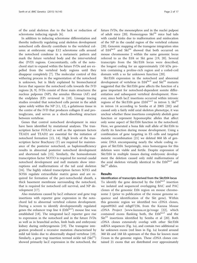
the expression in the emerging IVDs we verified theexpression in the kidney (Ki, Fig. 3a, b) with strongsignals within the cortical region presumably in devel-oping nephrons (Ne, Fig. 3a, b). Also the epitheliumof the ureter (Ur, Fig. 3a), the mesonephric duct(MeDu, Fig. 3b) and the bladder (Bl, Fig. 3c) were
positive for Skt/Etl4. In testis expression was stron-gest within the cortical region (Te, Fig. 3b) and lesswithin the outer region of the seminiferous tubules(SeTu, Fig. 3b). We additionally identified tissues wereSkt/Etl4 expression was not known hitherto: the epithe-lium of the lung (Lu, Fig. 3d, e) the bronchus (Br, Fig. 3e),
Fig. 3 Skt/Etl4 expression during organogenesis. a-l In situ hybridizations of sagittal sections of wt E15.5 embryos with an exon 5–8 specificprobe showing expression in developing nephrons (Ne, a) the cortical region of kidney (Ki, a, b), ureter epithelium (Ur, a), mesonephric duct(MeDu, b) bladder epithelium (Bl, c), cortical region of testis (Te, b) and seminiferous tubules (SeTu, b), lung epithelium (Lu, d, e), bronchus(Br, e), cochlea epithelium (Co, f), tympanic cavity epithelium (TyCa, f), nasopharynx epithelium (NaPh, g), lens (Le, h), optic nerve (OpNe, h),primordia of follicles of vibrissae (FoVi, j), ducts of submandibular gland (SuGl, i), thymus gland (ThymGl, k), thyroid gland (ThyrGl, k), exocrineacini of pancreatic primordium (Pa, l), gut epithelium and wall (Gu, l)
Serth et al. BMC Genetics (2015) 16:143 Page 5 of 17
as well as the epithelium of the cochlea (Co, Fig. 3f), thetympanic cavity (TyCa, Fig. 3f) and the nasopharynx(NaPh, Fig. 3g). In the developing eye we found that inaddition to the lens (Le, Fig. 3h, [20] and inner layer of theretina [20], Skt/Etl4 also is expressed within the opticnerve (OpNe, Fig. 3h). The primordia of follicles of vibris-sae associated with the upper lip (FoVi, Fig. 3j) as well asthe ducts of the submandibular gland (SuGl, Fig. 3i), thedeveloping thymus gland (ThymGl, Fig. 3k) and thyroidgland (ThyrGl, Fig. 3k) display Skt/Etl4 expression.Expression was also detected within the pancreatic prim-ordium most likely within the exocrine acini (Pa, Fig. 3l)and the wall and epithelium of the gut (Gu, Fig. 3l).
Targeted insertion of lacZ reporter genes into Skt/Etl4exons 1 and 5In both mouse lines, Etl4lacZ and SktGt, the insertion of alacZ transgene resulted in similar very mild phenotypicchanges [19, 20]. Both integrations occurred within in-trons (Fig. 1a) and in the SktGt allele a truncated proteinof 998 aa fused to β-galactosidase could potentially begenerated from the mutated locus [20], raising the pos-sibility that these mutations represent hypomorphic al-leles. To evaluate the phenotype of the complete loss ofthe gene we set out to generate a bona fide null alleleof Skt/Etl4.The presence of various transcripts presumably expressed
from different promoters [Fig. 1 d and e; and 20],and the large genomic distance between many codingexons (Fig. 1a) precluded a simple targeting strategyto ensure the complete elimination of Skt/Etl4 func-tion. Therefore, we decided to introduce lacZ reportergenes with strong transcriptional termination signals(triple poly (A) [23] and loxP sites into two differentregions of the Skt/Etl4 locus that are parts of differ-entially expressed transcripts. The termination signalshould prevent the expression of further downstreamsequences from a given promoter, the loxP sites shouldallow us to delete portions of the gene by site directed re-combination. Moreover the integration of the lacZ re-porter into different exons should allow us to readilyexamine the transcriptional activity of the gene from dif-ferent transcriptional start sites.We introduced an IRES driven lacZ reporter gene fused
to triple poly (A) into the most 5’exon (SktEx1IRESLacZ
allele, Fig. 4a b) to disrupt the transcript specificallydetected in brain and testis (Fig. 1c, d). Likewise weintroduced a lacZ triple poly (A) cassette into exon 5(SktEx5LacZ allele, Fig. 4a c), to disrupt transcriptsexpressed during embryogenesis. Correctly targetedES cells were used to establish mouse lines carryingthe SktEx1IRESLacZ and the SktEx5LacZ alleles (Fig. 4f,and data not shown). The presence of exon 1 con-taining transcripts was analysed by lacZ staining of
embryos and adult tissues of mice carrying the SktEx1-IRESLacZ allele. Consistent with RT-PCR and WISH re-sults (Fig. 1c and f ) we found no lacZ activity duringembryogenesis (data not shown). LacZ staining ofadult SktEx1IRESLacZ organs (brain, testis, epididymis,skeletal muscle, spleen, heart, salivary gland, small in-testine, stomach, kidney, liver, lung) revealed onlyspecific lacZ staining in the testis (n = 2) and the epi-didymis (n = 2; Additional file 1: Figure S1 A, anddata not shown). The staining results confirm theNorthern blot results that we obtained with the exon1-3 probe (Fig. 1d) but are at odds with the RT-PCRresults obtained with brain mRNA, where exon 1 po-tentially is expressed at low levels. Homozygous micecarrying the insertion of the lacZ-stop cassette intoexon1 were viable and fertile and had no obviousskeletal phenotypes (data not shown) indicating thatdisruption of Skt/Etl4 in its 5’most exon did notaffect the function during vertebrae development.With the SktEx5LacZ allele we observed specific lacZ re-
porter gene expression in stage E10.5 (n = 6) and E11.5(n = 10) embryos in the notochord (white triangles inAdditional file 1: Figure S1 B b, c, e and f ), the opticvesicle, the otic placode, the eye, the AER of the fore-and hindlimbs and on the surface of the branchial arches(Additional file 1: Figure S1 B b, c, e, f ). This expressionpattern was nearly identical to endogenous Skt/Etl4expression detected with the exon 5 to 8 probe inwhole mount in situ hybridizations (Fig. 2e-g) andsimilar to Etl4lacZ embryos of the same age [19]. Simi-lar to SktEx1IRESLacZ mice homozygous SktEx5lacZ micewere viable and fertile without obvious external phenotype(data not shown). To test if the termination signal 3’to thelacZ insertion in exon 5 prevents transcription ofdownstream exons we analysed the presence of exonsequences upstream and downstream of exon 5 inSkt/Etl4 mRNA by RT-PCR using RNA from wt, het-erozygous and homozygous SktEx5LacZ mutant E10.5embryos (for primer positions see Additional file 2:Figure S2A). As expected exons 4 and 5 upstream oflacZ were detected in wt and mutant embryos (PCR1,Additional file 2: Figure S2 B), as well as a Skt/Etl4-lacZ fusion transcript in embryos containing theSktEx5LacZ allele (PCR2, Additional file 2: Figure S2B). RT-PCR with primers binding to exon 4 and 7 de-tected a fragment in which exon 4 was fused to exon 6revealing an alternative splicing event, which removesthe targeted exon 5 (PCR3, Additional file 2: Figure S2B). In addition, further downstream sequences were stillexpressed in mutant embryos (PCR4, Additional file 2:Figure S2 B). Removal of exon 5 causes an in-frame de-letion of 96 of the total 1373 amino acids. Thus, mostlikely, this allele does not represent a null allele andtherefore was not analysed further.
Serth et al. BMC Genetics (2015) 16:143 Page 6 of 17

Fig. 4 (See legend on next page.)
Serth et al. BMC Genetics (2015) 16:143 Page 7 of 17

Deletion of the whole locus to eliminate Skt/Etl4 genefunctionSince the SktEx5LacZ allele did not prevent the generationof Skt/Etl4 transcripts that can give rise to likely func-tional protein (s) we set out to remove the vast majorityof the coding region using a two step deletion strategybased on the loxP sites present in the targeted allelesand an additional targeting event into exon 21 (outlinedin Fig. 4a, b).During the generation of the SktEx1IRESLacZ and
SktEx5LacZ alleles a floxed PGK-Neo cassette was in-troduced in both cases 3’to the lacZ reporter. Afterremoval of the neo gene by Cre-mediated recombin-ation a single loxP site (black triangle in Fig. 4a b, c)in the same orientation remained in the SktEx1IRESLacZ
and SktEx5LacZ alleles. We used these loxP sites incombination with Cre expression during oogenesis todelete 595 kb of genomic DNA between exon 1 and exon5 by targeted meiotic inter-chromosomal recombination[TAMERE 21]. Heteroallelic SktEx1IRESLacZ/Ex5LacZ femalescarrying a ZP3::Cre transgene were mated with wild typemales and offspring analysed for the presence of the inter-chromosomal recombination event (Fig. 4a d SktΔEx1-5 al-lele, and 4b). Among the first 20 offspring we identified afemale with the desired deletion by PCR analyses usingfour different primer pairs that amplified DNA fragmentsspecific for the various alleles (Fig. 4c, Primer pair positionin Fig. 4a). Successful inter-chromosomal recombinationwas indicated by PCR products obtained with PCR 1 and4 and lack of products with PCR 2 and 3, which differenti-ates between the deletion and the presence of the initialSktEx1IRESLacZ and SktEx5LacZ alleles (Fig. 4c). The correct
recombination event that removes the first 4 exons of thegene was validated by Southern blot analysis, whichshowed the expected polymorphisms between the threealleles (Fig. 4e). Mice homozygous for the 595 kb SktΔEx1-5
deletion were viable and fertile and did not show any obvi-ous phenotype (data not shown), indicating that the por-tions of Skt/Etl4 essential for vertebral development werestill functional.To delete the major part of the coding sequence con-
tained in further 273 kb of genomic DNA we generatedES cells from homozygous SktΔEx1-5 blastocysts, targetedexon 21 by introducing a floxed PGK-Neo cassette(SktΔEx1-5; Ex21GFP allele, Fig. 4a e), and generated micecarrying this allele. Intrachromosomal recombination(deletion of exons 5–20) was achieved in female miceharbouring the SktΔEx1-5; Ex21GFP allele together with theZP3::Cre transgene. Offspring carrying the deletion wereidentified by PCR analyses with 4 different primer paircombinations (Fig. 4d, Primer pair positions in Fig. 4a).Only mice with the second genomic deletion of 273 kbbetween exon 5 and 21 (Fig. 4b) generate PCR productswith PCR1, 5 and 6 in combination with lack of a prod-uct with PCR4, which distinguishes the second deletionand the initial SktΔEx1-5 and SktΔEx1-5; Ex21GFP alleles(SktΔEx1-5 allele, Fig. 4a f and d). Southern blot analysisusing a lacZ specific and a genomic probe locateddownstream of exon 21 (3’Exon21) and three differentrestriction digests of genomic mouse DNA (Fig. 4eand Additional file 3: Figure S3) confirmed successful de-letion of exons 1 to 20 of Skt/Etl4. Despite the deletion ofabout 88 % of the N-terminal portion of the proteinhomozygous SktΔEx1-20 mice were viable and fertile.
(See figure on previous page.)Fig. 4 Skt/Etl4 gene inactivation strategies. a Overview of Skt/Etl4 wt (a) and mutant alleles (b-f) that were generated by gene targeting. (b)Integration of an IRES-lacZ-triple poly a into exon 1. Floxed PGK-Neo-pA was removed with Cre recombinase leaving a single loxP site 3’to lacZ.(c) LacZ-triple poly a integration into exon 5 with 3’loxP site after Cre removal of the floxed PGK-Neo cassette. (d) Cre-induced TAMERE betweenloxP sites of SktEx1IRESlacZ (b) and SktEx5lacZ (c) alleles in the mouse resulting in 595 kb genomic deletion between exon 1 and 5 (depicted in B).(e) Integration of floxed PGK-Neo cassette together with a GFP-tagged exon 21into the SktΔEx1-5 allele. (f) Cre mediated excision of 273 kb genomicsequences between integrated loxP sites in the SktΔEx1-5, Ex21GFP allele (depicted in B). Numbered boxes represent Skt/Etl4 exons, coding exons aremarked in grey. lacZ-3pA: β-Galactosidase gene with triple poly a signal. Black triangle: lox P site. IRES: internal ribosome entry site. PGK-Neo-pA:Neomycin selection cassette with PGK promoter and poly a signal. GFP: green fluorescence protein tag with stop signal. PCR1 to PCR6 shows thepositions of Primer pairs that were used for mouse genotyping shown in (c) and (d). b Scheme of the Skt/Etl4 locus drawn to scale showing the twodeletion steps to eliminate Skt/Etl4 function. First TAMERE between the SktEx1IRESlacZ and SktEx5lacZ alleles generated a 595 kb deletion between exon 1and 5. Second around 273 kb genomic DNA between exon 5 and 21 were deleted by Cre-mediated intrachromosomal excision. c PCR Genotyping ofoffspring after the first deletion event between exon 1 and 5 (see B) with Primer pair combinations PCR 1 to 4 depicted in a from breedings of wtmales with SktEx1IRESlacZ/Ex5lacZ, ZP3::Cre females. Mice carrying the 595 kb deletion (SktΔEx1-5 allele, see A d) were positive for PCR 1 and 4 and negativefor PCR 2 and 3. d PCR Genotyping of offspring’s after the second deletion event between exon 5 and exon 21 (see B) with PCR Primerpair combinations PCR1, 4, 5 and 6 depicted in a from breeding’s of wt males with SktΔEx1-5; Ex21GFP, ZP3::Cre females. Only when 273 kbof genomic DNA was removed positive signals with PCR 1 and 5 were obtained. In addition PCR 6 specifically detects the whole 870 kbdeletion due to the flanking primer positions (red triangle). e Verification of chromosomal rearrangements by Southern blot analysis withgenomic mouse DNA digested with PstI (a) or NcoI (b) and hybridized with a lacZ specific probe for discrimination of the different Skt/Etl4 alleles thatwere generated. A polymorphism obtained with EcoRV-digested genomic DNA (c) hybridized with a probe downstream of exon 21 (3‘exon21) probewas used to demonstrate the presence of the wt, heterozygous or homozygous SktΔEx1-20 alleles in mice. The corresponding restriction maps with theexpected fragment sizes of Skt/Elt4 wt and mutant alleles are shown in Additional file 3: Figure S3. f Detection of endogenous SKT/ETL4 protein byWestern blot analysis using anti-NGS antibody and protein lysates isolated from adult brain of wt and homozygous SktΔEx1-20 animals. Red arrowheadspoint to bands missing in lysates of mutant tissue
Serth et al. BMC Genetics (2015) 16:143 Page 8 of 17

To test whether the truncated C-terminal portion ofthe protein comprising 167 AA encoded by exon 21 isgenerated we performed Western blot analysis with apolyclonal SKT/ETL4-specific antibody directed againsta C-terminal peptide (anti-NGS, see Materials andMethods), which detects SKT/ETL4 protein expressed inCHO cells with the expected size of approximately150 kDa (data not shown). In protein lysates of adultwild type brain we detected in addition to several othercross-reacting proteins a 200 kDa and a 150 kDa proteinspecies, which were not present in lysates of homozy-gous SktΔEx1-20 mice (Fig. 4f, red triangles in the leftpanel) and likely represent two variants of the SKT/ETL4 protein. We did not see any truncated version ofthe SKT/ETL4 protein at the predicted approximate sizeof 16,4 kDa in brain lysates of homozygous SktΔEx1-20
mice in a Western blot after separation of proteins bySDS-PAGE in higher percentage gels (Fig. 4f, rightpanel), indicating that no truncated SKT/ETL4 pro-tein is present in these mice. Thus, the establishedthe SktΔEx1-20 mouse line carrying a 868 kb deletionthat removes about 90 % of the N-terminal coding se-quence should represent a bona fide null allele of theSkt/Etl4 gene.
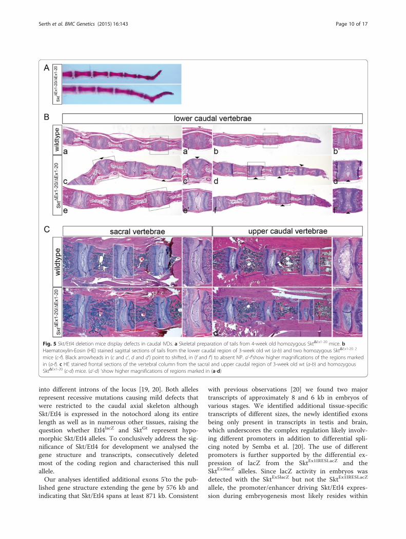
Sickle tail null mice display malformations of caudal IVDsExternal observation (at least n = 12) and skeletal prepa-rations (n = 3) revealed that most of the adult homozy-gous mutant animals displayed kinks within the tails(Fig. 5a) similar to the phenotype described for Etl4lacZ
[19] and SktGt [20] mice. For histological analysis paraf-fin sections of tails from 3-week old wt (n = 4) andhomozygous Skt/Etl4 mutant mice (n = 6) were stainedwith Haematoxylin-Eosin (HE, Fig. 5b). We found aber-rations in the morphology of the intervertebral discs(IVDs) that arose mostly in caudal vertebrae as shownin two examples of SktΔEx1-20 tails (Fig. 5b c-f ). Nor-mally in wt mice the NP of the IVDs is centrally located(Fig. 5b a and a’, b and b’), which we also observed inIVDs of SktΔEx1-20 mutants (Fig. 5b e and e’). However,in several SktΔEx1-20 mutant IVDs the NP was shifted tothe periphery (arrowheads Fig. 5b c, d and higher mag-nification in c’, d’) or in rare cases not present at all(arrowhead in Fig. 5 b f and higher magnification in f ’)in homozygous SktΔEx1-20 mutants. In addition the fibrouslayer of the AF surrounding the NP was reduced in size ornot present at all (Fig. 5b c’) resulting in some cases in thedirect contact of adjacent vertebral bodies. In more anter-ior IVDs (Fig. 5c, upper caudal vertebrae) we found only aslight lateral shift of the nucleus pulposus in one IVD ofone of four analysed mutant animals (Fig. 5c d and highermagnification in d’) but no other abnormalities comparedto wt (n = 2). In IVDs of the sacral region no structural al-terations of the NP or AF in homozygous mutant (n = 4)
mice were detected (Fig. 5c, sacral vertebrae). Together ourresults highly correspond with the phenotype described forthe Etl4lacZ and SktGt mutants, where only defects in sizeand position of the NP and AF, and corresponding verte-bral malformations were observed caudally, but not inother regions of the vertebral column [19, 20].
Skt/Etl4 null mice do not display major defects in thenotochordDuring mouse embryogenesis between E12 and E13notochordal cells in the region of the future disctransform and form the later NP and AF. The appear-ance of the previously described abnormalities in cau-dal IVDs of Skt/Etl4 deletion mutants may arise fromdefects in the formation or differentiation of thenotochord. To analyse if the Skt/Etl4 deletion leads toobvious notochord defects early during developmentwe hybridized stage E9.5 embryos with probes for thenotochordal markers Sonic hedgehog [Shh 3] andBrachyury [T 24]. We observed no differences in theexpression pattern of Shh in wt (n = 7) and in homo-zygous Skt/Etl4 (n = 5) deletion mutant embryos(Fig. 6a, b). Likewise expression of T at embryonicstage E9.5 was indistinguishable between wt (n = 7)and Skt/Etl4 (n = 6) mutants with the exception of anectopic expression domain at the forelimb level in one outof six analysed mutants (red arrowhead Fig. 6d) indicatingthat the loss of Skt/Etl4 expression does not have a majorimpact on early notochord development.
Histological analysis of adult Skt/Etl4 mutantsAs described earlier Skt/Etl4 is a gene expressed inmultiple tissues during embryogenesis. Therefore, weanalysed various tissues of juvenile Skt/Etl4 deletionmutants for histologically detectable abnormalities. InHE stained longitudinal kidney sections we found noobvious differences in the overall appearance (Fig. 7b)or structure of the cortex (Fig. 7 c, d), the medulla(Fig. 7 e, f ) and papilla (Fig. 7 g, h), or the numberof glomeruli (data not shown) between wild type (n = 2)and mutants (n = 2). Other organs that exhibited a thusfar not described expression of Skt/Etl4 were epithelia ofthe lung and the cochlea. We analysed lung tissue of2 week old wt (n = 3) and homozygous SktΔEx1-20 (n = 2)mice with HE staining and could not detect any obviousdifference in the tissue structure between both phenotypes(compare Fig. 7i, k, m with j, l, n). Likewise cochleae fromthe same stage did not exhibit any obvious variations be-tween wt (n = 2) and homozygous SktΔEx1-20 mice (n = 2;compare Fig. 7o, q, s with p, r, t).
DiscussionThe Skt/Etl4 gene was independently identified by twoinsertions of lacZ reporter constructs (Etl4lacZ and SktGt)
Serth et al. BMC Genetics (2015) 16:143 Page 9 of 17
into different introns of the locus [19, 20]. Both allelesrepresent recessive mutations causing mild defects thatwere restricted to the caudal axial skeleton althoughSkt/Etl4 is expressed in the notochord along its entirelength as well as in numerous other tissues, raising thequestion whether Etl4lacZ and SktGt represent hypo-morphic Skt/Etl4 alleles. To conclusively address the sig-nificance of Skt/Etl4 for development we analysed thegene structure and transcripts, consecutively deletedmost of the coding region and characterised this nullallele.Our analyses identified additional exons 5’to the pub-
lished gene structure extending the gene by 576 kb andindicating that Skt/Etl4 spans at least 871 kb. Consistent
with previous observations [20] we found two majortranscripts of approximately 8 and 6 kb in embryos ofvarious stages. We identified additional tissue-specifictranscripts of different sizes, the newly identified exonsbeing only present in transcripts in testis and brain,which underscores the complex regulation likely involv-ing different promoters in addition to differential spli-cing noted by Semba et al. [20]. The use of differentpromoters is further supported by the differential ex-pression of lacZ from the SktEx1IRESLacZ and theSktEx5lacZ alleles. Since lacZ activity in embryos wasdetected with the SktEx5lacZ but not the SktEx1IRESLacZ
allele, the promoter/enhancer driving Skt/Etl4 expres-sion during embryogenesis most likely resides within
Fig. 5 Skt/Etl4 deletion mice display defects in caudal IVDs. a Skeletal preparation of tails from 4-week old homozygous SktΔEx1-20 mice. bHaematoxylin-Eosin (HE) stained sagittal sections of tails from the lower caudal region of 3-week old wt (a-b) and two homozygous SktΔEx1-20 2
mice (c-f). Black arrowheads in (c and c‘, d and d‘) point to shifted, in (f and f‘) to absent NP. a‘-f‘show higher magnifications of the regions markedin (a-f). c HE stained frontal sections of the vertebral column from the sacral and upper caudal region of 3-week old wt (a-b) and homozygousSktΔEx1-20 (c-d) mice. (a‘-d) ‘show higher magnifications of regions marked in (a-d)
Serth et al. BMC Genetics (2015) 16:143 Page 10 of 17

the 595 kb genomic region between exon 1 and exon5. Similar to the existence of transcripts of differentsizes we detected in brain and testis lysates proteinsof different molecular weights. The faster migratingprotein species of approximately 150 kDa correlateswell with the SKT protein detected in lysates of inter-vertebral discs by Semba et al. [20]. The protein withhigher molecular weight might be encoded by someother transcript or generated by posttranslationalmodifications.Most of the published expression domains of Skt/Etl4
were determined indirectly using the lacZ reporter genein Etl4lacZ and SktGt mice [19, 20, 24] or with a SktCre
allele combined with a Rosa26lacZ reporter line [25]. Wefound additional tissues expressing Skt/Etl4 that werenot described so far, for instance the epithelium of thelung and the cochlea by detecting Skt/Etl4 mRNA by insitu hybridization. Given the complex regulation andsplicing patterns it appears possible that some of the en-dogenous expression domains were not detected due tothe intronic insertion sites of the reporter constructs.Analysis of the reporter gene activity in SktEx5lacZ em-bryos showed specific expression within the notochord
and other tissues consistent with lacZ activity in Etl4lacZ
and SktGt mice [19, 20]. However, a more thoroughcomparison of notochord staining of the Etl4lacZ andSktEx5lacZ alleles showed that lacZ expression along theentire length of the notochord in Etl4lacZ embryo wasnot recapitulated in SktEx5lacZ embryos: here the stainingshowed a gradient of expression with the strongest stain-ing in the tail area. Since this notochord expression pat-tern was also detected in WISH experiments with theexon 1-5 probe it presumably reflects the actual en-dogenous gene activity and might explain the regionallytail restricted IVD phenotype in Skt/Etl4 mutants.To study the function(s) of the brain/testis-specific
and the embryonic transcripts we introduced a lacZgene with a stop cassette (triple poly (A)), which waspreviously shown to effectively prevent transcriptionalread-through [23], into exon 1 and 5, respectively. Inser-tion into exon 1 did not cause any obvious phenotype,indicating that the transcript (s) required for Skt/Etl4function in the notochord was still functional, presum-ably due to a transcriptional start site downstream ofexon 1. Likewise the lacZ insertion into exon 5 did notcause any obvious phenotype, despite lacZ expression in
Fig. 6 Notochord marker expression in Skt/Etl4 deletion embryos. Whole mount in situ hybridization of E9.5 wt and Skt/Etl4 mutant embryoswith the notochord marker sonic hedgehog (Shh, a, b) and Brachyury (T, c, d). Red arrowhead point to a T expression abnormality seen in oneout of six embryos that were analysed
Serth et al. BMC Genetics (2015) 16:143 Page 11 of 17

Fig. 7 Histological analysis of kidneys, lungs and inner ears of SktΔEx1-20 mice. HE staining of organs isolated from 2 week old wt and SktΔEx1-20
mice. a-b overviews of longitudinal sections through the kidney and magnifications of the cortex (c), medulla (m) and papilla (p) regionsc-h indicated by lines in a. Arrowheads in c and d point to the cortex-specific Glomerular capsules. i-j Sections through lung tissue.Rectangles in i and j indicate the regions shown at higher magnification in k-n. Al: Alveolus, Bi: Bronchiole, PV: pulmonary vessel, Ac:Acini, AlSa: alveolar sac, AlDu: alveolar duct. o-r Midmodiolar cochlea sections. Rectangles in q and r indicate the regions shown at highermagnification in s and t. st: scala tympani, sv: scala vestibuli, sm: scala media, otc: otic capsule, ls: spiral limbus; rm: Reissner‘s membrane,stv: stria vascularis, sl: spiral ligament, co: organ of corti, sg: spiral ganglion, ohc: outer hair cells
Serth et al. BMC Genetics (2015) 16:143 Page 12 of 17
embryonic tissues similar to endogenous expression. Itturned out that the integration of the transcriptionalstop cassette did not eliminate Skt/Etl4 expression, butwas removed from a read-through primary transcript bysplicing around the lacZ cassette, indicating that thetriple poly (A) signal was not sufficient to terminatetranscription in this context.Since the lacZ insertions into exon 1 or 5 did not
phenocopy the SktGt or Etl4lacZ phenotype we deletedgenomic DNA containing exon sequences encodingnearly 90 % of the protein deduced from the longestknown open reading frame. To achieve this we deletedby site-specific inter- and intra-chromosomal recombin-ation events using the Cre/loxP recombinase system[26–28] a total of around 870 kb of genomic DNA. Theefficiency for creating genomic deletions in vivo with theTAMERE strategy decreases with size. It was successfulin males for trans-located loxP sites separated by 150 kb[21, 29], but failed for loxP sites with a trans distance of3,9 Mb [30]. We obtained a genomic deletion of 595 kbwith TAMERE using females that express Cre recombin-ase in oocytes [ZP3::Cre 32] in one offspring within thefirst two litters. However, due to the small numbers wecan not draw firm conclusions concerning the trans-chromosomal recombination efficiency in female mice.Excision of the residual 273 kb genomic DNA betweentwo cis-located loxP sites that occurred in all analysedoffspring (n = 26) demonstrating that an intrachromoso-mal deletion of this size can efficiently be obtained inthe female germ line.Our Skt/Etl4 deletion represents a bona fide null allele
whose phenotype is undistinguishable from Etl4lacZ andSktGt mice. All our mice that were homozygous for thedeletion and were histologically analysed (n = 9) showeddefects in the caudal vertebral column. In contrast, inEtl4lacZ and SktGt mutants only one third or half of themutants had obvious defects [19, 20]. Since these micewere analysed on a C57BL/6 background, whereas ourmice are on a mixed CD1/129Sv background, geneticbackground differences might contribute to the differentpenetrance of the phenotype. Thus, we cannot distin-guish at present whether the complete penetrance of thephenotype in our deletion mutant reflects that Etl4lacZ
and SktGt represent hypomorphic alleles or is due togenetic background differences.In SktGt as well in Etl4lacZ mutants no kidney abnor-
malities or impaired fertility were observed [19, 25]. Weanalysed selected organs of mutants highly expressingendogenous Skt/Etl4 histologically and found only ab-normalities within the IVDs solely in the caudal regionof the vertebral column. We cannot exclude that thereare subtle defects or that other tissues that we did notanalyse are affected in our deletion mutants. However, atpresent it appears that in almost all expression domains
the loss of Skt/Etl4 function can be compensated. Thespecific expression domain in the NP of the IVDs andstrong association of SKT polymorphisms with Lumbardisc herniation (LDH) or Disc degeneration (DD) in Fin-ish and Japanese populations [31, 32] makes SKT a goodcandidate for a LDH or DD susceptibility gene inhumans. These studies did not mention other healthproblems in these patients, supporting that Skt/Etl4function is only required for IVD formation or stability.However, it is still unclear how the LDH associated andintronic SNPs influence SKT/ETL4 function in thepatients [31].The region on chromosome 2 that we deleted contains
11 gene predictions including small nucleolar RNAs(snoRNAs), small nuclear RNA (snRNAs), pseudogenes,protein coding genes and unclassified non-coding RNAgenes (Gm25859, Gm13361, Gm13363, Gm13360,Gm13328, Gm34260, Gm23970, Gm17171, Gm13335,Gm27446, Gm13362) as well as two unclassified genesrepresented by cDNA clones (Gm16495, 8030447M02Rik)(http://www.informatics.jax.org). To the best of our know-ledge no functional data for these gene predictionsare available and is not clear whether these annota-tions represent true functional genes. However, as ourmice are largely normal none of these annotated se-quences can be essential for developmental processes,viability and fertility.
ConclusionInhibition of gene expression via integration of atranscriptional stop signal (triple poly (A)) can be in-efficient for gene loci with complex regulation. Forgenome modification inter- and intrachromosomal re-combination in the female germ line can be used todelete large genomic regions. The generation of abona fide null allele of the Skt/Etl4 gene with thistechnique demonstrates that this gene as well asother annotated sequences in this region are not es-sential for viability and fertility of mice, but Skt/Etl4is only required for caudal intervertebral disc develop-ment. These analyses also indicate that both Etl4lacZ
and SktGt abolish Skt/Etl4 function.
Materials and methodsEthics statementAll animal experiments were performed according tothe German rules and regulations (Tierschutzgesetz)and approved by the ethics committee of Lower Sax-ony for care and use of laboratory animals LAVES(Niedersächsisches Landesamt für Verbraucherschutzund Lebensmittelsicherheit; AZ 33-42502-02/543).Mice were housed in the central animal facility ofHannover Medical School (ZTL) and were maintainedas approved by the responsible Veterinary Officer of
Serth et al. BMC Genetics (2015) 16:143 Page 13 of 17
the City of Hannover. Animal welfare was supervisedand approved by the Institutional Animal WelfareOfficer (Tierschutzbeauftragter).
Mouse housing and husbandry conditionsMice were bred and maintained under routine hus-bandry procedures following standards published bythe Society for Laboratory Animal Science (SOLAS)and the EU directive 2010 63 EU at a temperature of21 °C, relative humidity of approximately 50 %, andartificial light from 06:00–18:00 h. Mice were kept inwire-topped type IIL Makrolon cages (Techniplast,Techniplast-Deutschland GmbH, Hohenpeienberg,Germany) on sterilised softwood granulate bedding(Lignocel, Altromin; Lage, Germany) and receivedautoclaved commercial pellet diet (Altromin1314)(protein 22, fat 5, raw fibre 4.5, ash 7 %, utilizing en-ergy 3.1 kcal/g) and water ad libitum. Microbiologicalstatus was measured at least every six months accord-ing to the recommendations for the health monitoringof rodent colonies in breeding and experimental units.
Shotgun cloning and sequencingShotgun libraries of BAC DNA with average insert sizesof 1.5 kb and 3.5 kb were generated and sequenced asdescribed [16].
Northern blot analysisCommercially available Northern blots (Mouse EmbryoMTN Blot (#7763-1, Clontech), Message Map NorthernBlot organs adult (#776900, Stratagene)) were hybridizedwith probes radioactively labelled with [α-32P] dCTPusing the Prime-it II Random Primer Labelling kit ac-cording to the manufacture’s instructions. Blots were hy-bridized in ULTRAhyb® hybridization solution (Ambion)for one hour without probe, then overnight with probeat 42 °C. Unspecific probe was removed by washing withNorthernMax® Low Stringency Wash buffer (Ambion)twice for five minutes and once with NorthernMax®High Stringency Wash buffer (Ambion) for 15 min at 42 °C.
Skt/Etl4 specific exon probesSkt/Etl4 specific fragments that were used as probes forwhole-mount in situ hybridization and Southern andNorthern blot experiments were amplified by PCR andsub-cloned into pGEM®-T Easy vector (Promega). Thefollowing primer pairs for amplification from cDNAclones were used: Exon 1–3 (AAGGTAGCGGAGGCTCAAG, CCCAGTATTTCCATCCCATAG), Exon 5–8(AGCCATCATGGGCCACC, GCATGTGAAGGATCCTTGTTG), Exon 9–13 (CCCTGATAGCCATTTGCCTACC, TTTGACAATCCGCTCTTCTTCG), Exon 15–18(GAACAGGGCAGTGTCCATTGAG, TCTTCTTCTTCCTCCTCCTCCTTG).
Whole-mount and section in situ hybridizationWhole-mount and section in situ hybridization of em-bryos was performed with digoxygenin-labelled anti-sense riboprobes as described [33, 34]. Documentationwas done with the Leica M420 microscope with Apo-zoom 1:6 and the Photograb-300Z version 2.0 software.In addition to Skt/Etl4 specific probes riboprobes spe-cific for T [35], and Shh [3] were used.
Skeletal preparations of newbornsNewborn mice were prepared, stained and documentedas described previously [36].
Cloning of targeting constructsCloning of the SktEx1IRESlacZ targeting construct: Thegenomic region including and surrounding Exon 1 wasamplified from a mouse genomic 129Svlmj PAC-clone(RZPD PAC-Library 711; clone 109.9.161) in three stepswith primer pairs CCAACTCAGGTTCTCGG, GGAGCACTTGTCCCCTTAG and ATGAGTATGAGCACATCGG, GAGATTGGGTGACTTACGG and GAGAGAGAGAGAGACTGGTCC, TCAAGGTCAGCCTGGTAG.The three fragments were linked by AccI and StuI sites,respectively. The IRESlacZ-reporter gene with a triplepoly (A) signal followed by a floxed PGK-Neo polyA-cassette was introduced into the SacII site of exon 1.Cloning of the Skt Ex5lacZ targeting construct: the
5’flank was amplified from genomic DNA with primerpair CTAATGGAGTGGTGGATGAGCG, TAACAAGAAAGGTCAGGAGCCG. The 3’flank region was ampli-fied in two steps with primer pairs GACCAGGTAGGAACACACTATCGG, CACAATCTATTTTTAGCCGCTTTAAT and AGTGTGTAGTCCTGGAGGGC, CAAAGTATGAATGGGGGCGG. The two fragments were com-bined using a BstZ17I site. A lacZ-reporter gene with atriple poly (A) signal was introduced between the ApaIsite of exon 5 and the StuI site of intron 5 into a PCRfragment obtained from genomic DNA with primersCCATCAAAACATACCCACG and TCAGATTTCAACTCAGGTCG, which leads to a 695 bp deletion of thesickle tail locus. The lacZ containing exon 5 togetherwith the 3’flank and the 5’flank were combined and afloxed PGK-Neo cassette was inserted into a SalI sitedownstream of the triple poly (A) signal. In addition aDT cassette was integrated upstream of the 5’flank intothe cloning vector. Cloning of the Skt Ex21GFP targetingconstruct: for amplification of the flanks from genomicDNA the following primer pairs were used: 5’flank pri-mer CTGTGGTTGATACTGACTTCG, GTTGTTTATGGAAGGCGAC; 3’flank primer ACATTCCTCTCCCAACTCG, TCACTCTTTCTCAGCGTCC. The C-terminalGFP tag was fused in frame with the Skt/Etl4 cDNAusing a PCR generated BglII site instead of the Skt/Etl4 stop codon. Combination of the 5’flank and
Serth et al. BMC Genetics (2015) 16:143 Page 14 of 17

3’flank containing exon 21 with GFP was performed usinga genomic SalI site. Into a genomic StuI site located in in-tron 20 a floxed PGK-Neo Cassette was introduced for EScell selection. Additionally a DT cassette was integratedinto the vector backbone using EcoRV and SacII sites.
Generation of ES cellsES cell lines homozygous for Skt ΔEx1-5 were obtainedfrom d 4.5 blastocysts collected from matings of homo-zygous Skt ΔEx1-5 mice on a mixed CD1/129Sv/ImJ gen-etic background as described [37].
Generation and genotyping of mutant micePositive ES cell clones that were electroporated withSktEx1IRESlacZ or SktEx5lacZ targeting constructs, wereverified by Southern blot analysis and used for chimeraproduction. For removal of the floxed PGKNeo cassettemice were crossed to ZP3::Cre mice [38]. Genomic DNAisolated from tails was used for genotyping using thefollowing PCR primer pairs (see also Fig. 4): PCR 1(GCTCCCAACTCTACCCAGAC, CCCTCACATTGCCAAAAGACG, 679 bp), PCR 2 (TCAGCCATACCACATTTGTAGAG, CTGGGGAGACGACTTTCAAG, 280 bp), PCR 3 (TCGTTAGCAACTGCCACAACC, TCGCCGCACATCTGAACTTC, 964 bp),PCR 4 (AACCTCCCACACCTCCCCTG, GCAAGACTGGTCCCCAAAATAAG, 624 bp), PCR 5 (TGGAAGTTCAAGCAAAGCC, TTGTGCCCCAGGATGTTG,492 bp), PCR 6 (AACCTCCCACACCTCCCCTG, ACAGCCCTTCTGAGCATCATTTAG, 404 bp). ForSouthern blot analysis a 1,9 kb ApaI/SacI fragment ofthe lacZ coding sequence was used as a probe. An889 bp Skt/Etl4 specific probe 3’to exon 21 (3’Ex21)was generated by PCR with primers TGAGTGGCATCATAATGGTGTGG and AAATACAGAGAGGAGGACAGGCGG.
RT-PCRRT-PCR was performed with poly (A+) RNA and theThermoscript RT-PCR system (Invitrogen) accordingto the manufacturer’s instructions with primer pairs:forward-primer exon 1 (AAGGTAGCGGAGGCTCAAG), backward primer exon 3 (CCCAGTATTTCCATCCCATAG) forward primer 4 (CCAGAAATGTGAGCCGAAC), backward-primer exon 5 (TCCATTAGAAAGGCGTTCCC), backward-primer lacZ (TCGCCGCACATCTGAACTTC), backward-primer exon 7 (TGTCTGTGCTTGTGACTTCATTCG), forward-primerexon 20 (CTTCCAAGAACAGACCCG), backward-primer exon 21 (CTTTCTTAGCACTTCCATTAGC).
β-galactosidase (lacZ) staining of embryosβ-galactosidase activity was detected as described [39].Briefly, embryos were fixed for 5 min in 0,1 M phosphate
buffer (pH 7,4) with 0,4 % glutardialdehyde, 2 mM MgCl2and 5 mM EGTA, and washed three times for 10 min in0,1 M phosphate buffer (pH 7,4) with 2 mM MgCl2, 0,1 %sodium deoxycholate and 0,02 % NP-40. Staining was per-formed at 37 °C in 0,1 M phosphate buffer (pH 7,4) with1 mg/ml X-Gal (5-bromo-4-chloro-3-indoyl-β-D-galacto-side), 5 mM potassium ferrocyanide and 5 mM potassiumferricyanide and stopped with 4 % paraformaldehyde.
Histological analysisKidney, lung, vertebral columns (sacral and caudal) andisolated inner ears of three week old mice were fixed in4 % PFA overnight, decalcified in 0,5 M EDTA/PBS for2 days (inner ears) or 2 weeks (vertebrae) respectively,dehydrated, paraffin wax embedded sectioned to 10 μmand stained with haematoxylin and eosin according tostandard procedures.
SKT/ETL4-specific antibodies and Western blot analysisSKT/ETL4 specific antibodies (anti-NGS) were gener-ated in rabbit (Biogenes GmbH, Berlin, Germany)against the C-terminal protein sequence NGSSSKATP-STAKETS and affinity purified as described [40]. Affin-ity purified anti-NGS antibodies were used for Westernblot analyses diluted (1:750) in PBS containing 5 %Milk powder and 0,5 % Tween followed by an incuba-tion with horseradish peroxidase-conjugated anti-rabbitantibody (GE Healthcare) and detection by ECL.
Availability of supporting dataAll supporting data are included in Supporting Informa-tion files.
Additional files
Additional file 1: Figure S1. lacZ reporter gene activity in SktEx1IRESlacZ
and SktEx5lacZ mice. (A) β-Galactosidase staining of wt (a, c) and homozy-gous SktEx1IRESlacZ (b, d) adult testes and epididymides. (B) β-Galactosidasestaining of wt (a, d), heterozygous (b, e) and homozygous (c, f) SktEx5lacZ
E10.5 and E11.5 embryos. White triangles in (b, c, e and f) point to lacZexpression in the caudal notochord. (TIF 8102 kb)
Additional file 2: Figure S2. Insertion of triple poly (A) into exon 5 ofthe Skt/Etl4 does not prevent transcription of downstream exons. (A)Schematic representation of the SktEx5lacZ allele and location of PCRprimer pairs used for RT-PCR. (B) RT-PCR results with PCR primer pairsdepicted in (A) with poly (A+) RNA isolated from E10.5 wt, heterozygousand homozygous SktEx5lacZ embryos. (TIF 186 kb)
Additional file 3: Figure S3. Restriction maps of Skt/Etl4 alleles.Schematic representation of the relevant regions of various Skt/Elt4 wtand mutant alleles with location of probes, restriction sites and expectedrestriction fragments detected by Southern blot hybridizations shown inFig. 4e. An EcoRV digest of wt DNA results in a 15 kb fragment detectedby the 3’Exon21 probe (A and Fig. 4e c), which shifts down to 11 kb inthe SktΔEx1-20 allele (B and Fig. 4e c). A lacZ probe detects in SktEx1IRESlacZ
DNA digested with PstI or NcoI a 5 kb or 7.1 kb fragment (C and Fig. 4ea and b), in SktΔEx1-5 DNA a 7.7 kb or 7.9 kb fragment (D and Fig. 4e aand b), and in SktEx5lacZ DNA a 12.6 kb or 12 kb fragment (E and Fig. 4e aand b). (TIF 413 kb)
Serth et al. BMC Genetics (2015) 16:143 Page 15 of 17
AbbreviationsAER: apical ectodermal ridge; AF: annulus fibrosus; BAC: bacterial artificialchromosome; DT: diphtheria toxin A; EDTA: ethylene demine tetraacetic acid;EP: endplate; ES cell: embryonic stem cell; Etl4: enhancer trap locus 4;Etl4lacZ: Etl4 mutagenesis line; FOXA2: forkhead box protein A2; GFP: greenfluorescent protein; HE: Haematoxylin-Eosin; IRES: internal ribosome entry site;IVD: intervertebral disc; lacZ: β-Galactosidase gene; loxP: LoxP sequence;NCBI: National Centre for Biotechnology Information; Neo: neomycin;Noto: notochord homeobox; NP: nucleus pulposus; PAC: P1 artificialchromosome; PBS: phosphate buffered saline; PGK: phosphoglyceratekinase 1 promoter; poly (A): polyadenylation signal; SDS-PAGE: sodiumdodecyl sulphate polyacrylamide gel electrophoresis; Shh: sonic hedgehog;SISH: section in situ hybridization; Skt: sickle tail locus; SktGT: Skt mutagenesisline; snoRNAs: small nucleolar RNAs; snRNAs: small nuclear RNA; Sox5: SRY-Box5; Sox6: SRY-box6; T: brachyury; TAMER: targeted meiotic recombination;TEAD1: TEA domain family member 1; TEAD2: TEA domain family member 2;WISH: whole mount in situ hybridization.
Competing interestsThe authors declare that they have no competing interests.
Authors’ contributionsKS performed Skt/Etl4 expression analysis, generated the Skt Ex5lacZ andSktΔEx1-5; Ex21GFP targeting constructs and analysed SktEx5lacZ and SktEx1IRESlacZ
mice, set up and analysed the genetic crosses, identified and analysed theSkt/Etl4 deletion mutants, drafted and wrote the manuscript. AB generatedthe SktEx1IRESlacZ targeting construct and ES cells. KSG generated SktΔEx1-5 EScells and germ line chimeras. MNP isolated and characterised BAC and PACclones. JM purified and characterised antibodies. MP analysed Skt/Etl4expression by in situ hybridization. RR sequenced BACs and PACs from theSkt/Etl4 region. AG planned the experiments and analysed data, wrote themanuscript. All authors read, reviewed and approved the final manuscript.
AcknowledgementsWe thank Dr. Osamu Ohara for generously providing the cDNA clones,Anatoli Heiser for mouse husbandry and maintenance, and PatriciaDelany-Heiken for technical support. This work was supported by a grantof the German Research Council to AG (GO 449/8). The funders had norole in study design, data collection and analysis, decision to publish, orpreparation of the manuscript.
Author details1Institut für Molekularbiologie OE5250, Medizinische Hochschule Hannover,Carl-Neuberg-Str.1, 30625 Hannover, Germany. 2Max Planck-Genome-CentreCologne, Carl-von-Linné-Weg 10, D-50829 Köln, Germany. 3Department ofAnaesthesiology and Pain Medicine, University of Washington, Seattle, WA98001, USA. 4Gasteiner Str. 31, 10717 Berlin, Germany. 5Department ofMedicine I, Institute of Cancer Research, Medical University of Vienna,Borschkegasse 8a, 1090 Vienna, Austria.
Received: 29 September 2015 Accepted: 8 December 2015
References1. Jurand A. Some aspects of the development of the notochord in mouse
embryos. J Embryol Exp Morphol. 1974;32:1–33.2. Yamada T, Placzek M, Tanaka H, Dodd J, Jessell TM. Control of cell pattern in
the developing nervous system: Polarizing activity of the floor plate andnotochord. Cell. 1990;64:635–47.
3. Echelard Y, Epstein DJ, St-Jacques B, Shen L, Mohler J, McMahon JA, et al.Sonic hedgehog, a member of a family of putative signalling molecules, isimplicated in the regulation of CNS polarity. Cell. 1993;75:1417–30.
4. Bumcrot DA, McMahon AP. Somite differentiation. Sonic signals somites.Curr Biol. 1995;5:612–4.
5. Gossler A, Hrabe De Angelis M. Somitogenesis. Curr Top Dev Biol. 1998;38:225–87.
6. Pourquie O, Coltey M, Teillet MA, Ordahl C, Le Douarin NM. Control ofdorsoventral patterning of somitic derivatives by notochord and floor plate.Proc Natl Acad Sci U S A. 1993;90:5242–6.
7. Smith LJ, Nerurkar NL, Choi KS, Harfe BD, Elliott DM. Degeneration andregeneration of the intervertebral disc: lessons from development. DisModel Mech. 2011;4:31–41.
8. Theiler K. Vertebral malformations. Adv Anat Embryol Cell Biol. 1988;112:1–99.9. Aszódi A, Chan D, Hunziker E, Bateman JF, Fässler R. Collagen II is essential
for the removal of the notochord and the formation of intervertebral discs.J Cell Biol. 1998;143:1399–412.
10. Chan WC, Au TY, Tam V, Cheah KS, Chan D. Coming together is abeginning: the making of an intervertebral disc. Birth Defects Res C EmbryoToday. 2014;102:83–100.
11. McCann MR, Tamplin OJ, Rossant J, Seguin CA. Tracing notochord-derivedcells using a Noto-cre mouse: implications for intervertebral discdevelopment. Dis Model Mech. 2012;5:73–82.
12. Choi KS, Cohn MJ, Harfe BD. Identification of nucleus pulposus precursorcells and notochordal remnants in the mouse: implications for diskdegeneration and chordoma formation. Dev Dyn. 2008;237:3953–8.
13. Ang SL, Rossant J. HNF-3 beta is essential for node and notochordformation in mouse development. Cell. 1994;78:561–74.
14. Sawada A, Kiyonari H, Ukita K, Nishioka N, Imuta Y, Sasaki H. Redundantroles of Tead1 and Tead2 in notochord development and the regulation ofcell proliferation and survival. Mol Cell Biol. 2008;28:3177–89.
15. Herrmann BG, Labeit S, Poustka A, King TR, Lehrach H. Cloning of the T generequired in mesoderm formation in the mouse. Nature. 1990;343:617–22.
16. Ben Abdelkhalek H, Beckers A, Schuster-Gossler K, Pavlova MN, Burkhardt H,Lickert H, et al. The mouse homeobox gene Not is required for caudalnotochord development and affected by the truncate mutation. Genes Dev.2004;18:1725–36.
17. Smits P, Lefebvre V. Sox5 and Sox6 are required for notochord extracellularmatrix sheath formation, notochord cell survival and development of thenucleus pulposus of intervertebral discs. Development. 2003;130:1135–48.
18. Korn R, Schoor M, Neuhaus H, Henseling U, Soininen R, Zachgo J, et al.Enhancer trap integrations in mouse embryonic stem cells give rise tostaining patterns in chimaeric embryos with a high frequency and detectendogenous genes. MOD. 1992;39:95–109.
19. Zachgo J, Korn R, Gossler A. Genetic interactions suggest that Danforth’sshort tail (Sd) is a gain-of-function mutation. Dev Genet. 1998;23:86–96.
20. Semba K. A novel murine gene, sickle tail, linked to the Danforth’s short taillocus, is required for normal development of the intervertebral disc.Genetics. 2005;172:445–56.
21. Hérault Y, Rassoulzadegan M, Cuzin F, Duboule D. Engineeringchromosomes in mice through targeted meiotic recombination (TAMERE).Nat Genet. 1998;20:381–4.
22. Ohara O, Temple G. Directional cDNA library construction assisted by the invitro recombination reaction. Nuc Acids Res. 2001;29:E22.
23. Soriano P. Generalized lacZ expression with the ROSA26 Cre reporter strain.Nat Genet. 1999;21:70–1.
24. Ando T, Semba K, Suda H, Sei A, Mizuta H, Araki M, Abe K, Imai K, NakagataN, Araki K, Yamamura K-I: The floor plate is sufficient for development of thesclerotome. MOD 2010:1–12
25. Abe K, Araki K, Tanigawa M, Semba K, Ando T, Sato M, et al. A Cre knock-inmouse line on the Sickle tail locus induces recombination in the notochordand intervertebral disks. Genesis. 2012;50:758–65.
26. Smith AJ, De Sousa MA, Kwabi-Addo B, Heppell-Parton A, Impey H, RabbittsP. A site-directed chromosomal translocation induced in embryonic stemcells by Cre-loxP recombination. Nat Genet. 1995;9:376–85.
27. Van Deursen J, Fornerod M, Van Rees B, Grosveld G. Cre-mediated site-specific translocation between nonhomologous mouse chromosomes. ProcNatl Acad Sci U S A. 1995;92:7376–80.
28. Ramirez-Solis R, Liu P, Bradley A. Chromosome engineering in mice. Nature.1995;378:720–4.
29. Kmita M, Fraudeau N, Hérault Y, Duboule D. Serial deletions andduplications suggest a mechanism for the collinearity of Hoxd genes inlimbs. Nature. 2002;420:145–50.
30. Olson LE, Tien J, South S, Reeves RH. Long-range chromosomal engineeringis more efficient in vitro than in vivo. Transgenic Res. 2005;14:325–32.
31. Karasugi T, Semba K, Hirose Y, Kelempisioti A, Nakajima M, Miyake A, et al.Association of the tag SNPs in the human SKT gene (KIAA1217) with lumbardisc herniation. J Bone Miner Res. 2009;24:1537–43.
32. Kelempisioti A, Eskola PJ, Okuloff A, Karjalainen U, Takatalo J, Daavittila I,et al. Genetic susceptibility of intervertebral disc degeneration amongyoung Finnish adults. BMC Med Genet. 2011;12:153.
Serth et al. BMC Genetics (2015) 16:143 Page 16 of 17

33. Serth K, Schuster-Gossler K, Cordes R, Gossler A. Transcriptional oscillation oflunatic fringe is essential for somitogenesis. Genes Dev. 2003;17:912–25.
34. Lescher B, Haenig B, Kispert A. sFRP-2 is a target of the Wnt-4 signallingpathway in the developing metanephric kidney. Dev Dyn. 1998;213:440–51.
35. Wilkinson DG, Bhatt S, Herrmann BG. Expression pattern of the mouse Tgene and its role in mesoderm formation. Nature. 1990;343:657–9.
36. Geffers I, Serth K, Chapman G, Jaekel R, Schuster-Gossler K, Cordes R, et al.Divergent functions and distinct localization of the Notch ligands DLL1 andDLL3 in vivo. J Cell Biol. 2007;178:465–76.
37. Maatman R, Zachgo J, Gossler A. The Danforth’s short tail mutation acts cellautonomously in notochord cells and ventral hindgut endoderm.Development. 1997;124:4019–28.
38. De Vries WN, Binns LT, Fancher KS, Dean J, Moore R, Kemler R, et al.Expression of Cre recombinase in mouse oocytes: a means to studymaternal effect genes. Genesis. 2000;26:110–2.
39. Beckers J, Caron A, Hrabe De Angelis M, Hans S, Campos-Ortega JA, GosslerA. Distinct regulatory elements direct delta1 expression in the nervoussystem and paraxial mesoderm of transgenic mice. MOD. 2000;95:23–34.
40. Braune E-B, Schuster-Gossler K, Lyszkiewicz M, Serth K, Preusse K, Madlung J,et al. S/T phosphorylation of DLL1 is required for full ligand activity in vitrobut dispensable for DLL1 function in vivo during embryonic patterning andmarginal zone B cell development. Mol Cell Biol. 2014;34:1221–33.
• We accept pre-submission inquiries
• Our selector tool helps you to find the most relevant journal
• We provide round the clock customer support
• Convenient online submission
• Thorough peer review
• Inclusion in PubMed and all major indexing services
• Maximum visibility for your research
Submit your manuscript atwww.biomedcentral.com/submit
Submit your next manuscript to BioMed Central and we will help you at every step:
Serth et al. BMC Genetics (2015) 16:143 Page 17 of 17