General Zoology Laborarory . Matt Nelson phylum - · PDF fileGeneral Zoology Laborarory ....
11
Lab exercise 7: Echinoderms and Chordates (introduction) General Zoology Laborarory . Matt Nelson Deuterostomia Both the Lophotrochozoa and the Ecdysozoa are examples of protostomous animals. Organisms that are protostomous possess a mouth that is derived from the blastopore of the embryo during development. Protostomes also share other characteristics, such as spiral, determinate cleavage. The Echinodermata, the Hemichordata, and the Chordata are members of the clade Deuterostomia. In the deuterostomes, the mouth arises later in development, and the anus is derived from the blastopore. The deuterostomes also possess radial, indeterminate cleavage. Echinoderms The phylum Echinodermata (Gr. [echinos, spiny, sea urchin] + [derma, skin]) are spiny skinned marine organisms such as sea stars and sea urchins. Because many of the echinoderms are near sessile, and the ancestors of modern echinoderms were totally sessile, the group has secondarily evolved radial symmetry. Most members of the Echinodermata possess pentaradial symmetry (five axes of symmetry). Along with the rest of the deuterostomes, echinoderms share a bilateral ancestor which is reflected by ontogeny. Echinoderms possess a bilaterally symmetrical larval form that undergoes a drastic rearrangement to develop into a pentaradial adult. Sea stars possess a bilaterally symmetrical swimming larva called a bipinnaria. The bipinnaria develops into a brachiolaria larva, which settles and develops into the adult form. Examine the preserved slides of sea star larva with a compound microscope. Classification (examples) Discussion of groups of organisms Class Ophiuroidea - Brittle stars are in this class. Tube feet with no suckers are located all around the arms. Mostly suspension feeders. Class Echinoidea - The sea urchins and irregular urchins. This group includes sand dollars and sea biscuits. Most feed by scraping algae or bacteria from substrates. Others feed on tiny particles moved toward the mouth by tube feet. Ossicles fused to form internal test. Class Crinoidea - Crinoids are near-sessile suspension feeders with feathery arms used for collecting tiny particles from the surrounding water. These echinoderms often attach to a substrate by a long stalk. phylum echinodermata 1 General Zoology Laboratory. Matthew K Nelson (2011) bilateria deuterostomia protostomia lophotrochozoa ecdysozoa hemichordata echinodermata chordata rotifera acanthocephala platyhelminthes mollusca annelida nemertea nemata nematomorpha tardigrada arthropoda onychophora acoela bipinnaria larva
Transcript of General Zoology Laborarory . Matt Nelson phylum - · PDF fileGeneral Zoology Laborarory ....
Lab exercise 7: Echinoderms and Chordates (introduction) General Zoology Laborarory . Matt Nelson
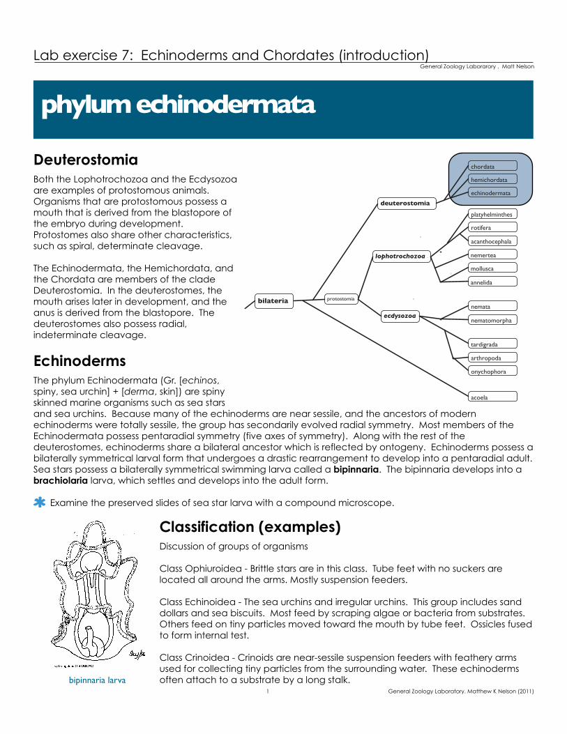
DeuterostomiaBoth the Lophotrochozoa and the Ecdysozoa are examples of protostomous animals. Organisms that are protostomous possess a mouth that is derived from the blastopore of the embryo during development. Protostomes also share other characteristics, such as spiral, determinate cleavage.
The Echinodermata, the Hemichordata, and the Chordata are members of the clade Deuterostomia. In the deuterostomes, the mouth arises later in development, and the anus is derived from the blastopore. The deuterostomes also possess radial, indeterminate cleavage.
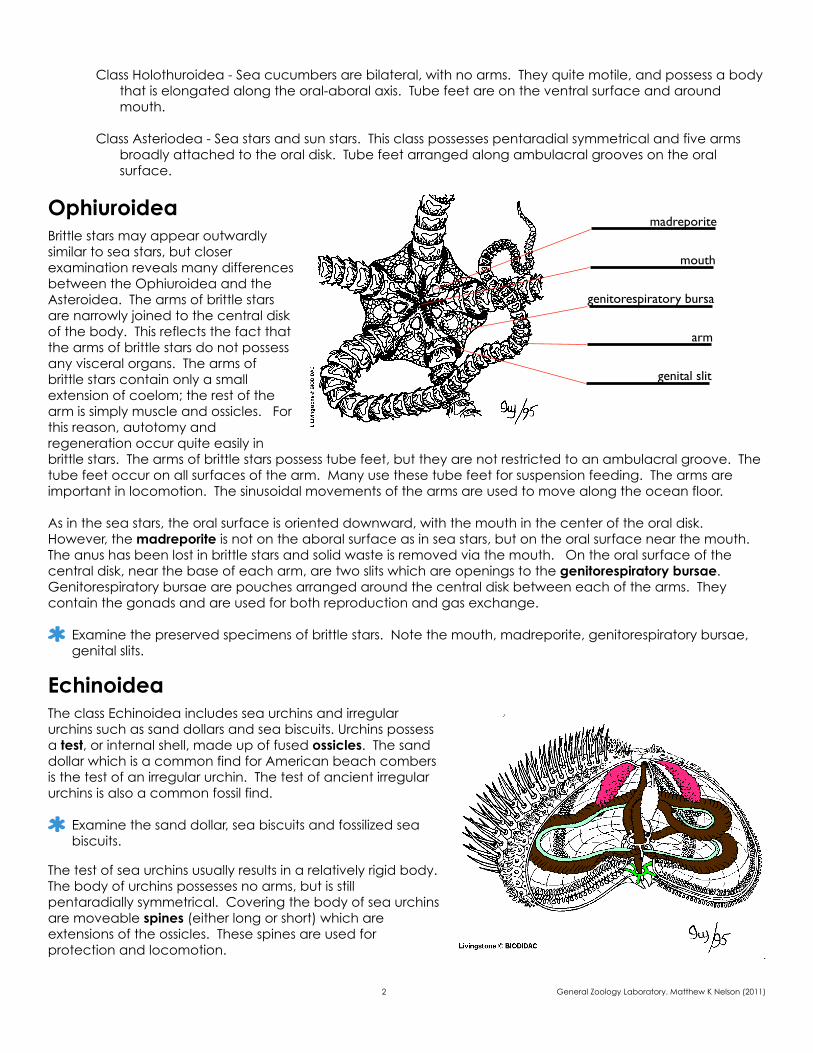
Echinoderms The phylum Echinodermata (Gr. [echinos, spiny, sea urchin] + [derma, skin]) are spiny skinned marine organisms such as sea stars and sea urchins. Because many of the echinoderms are near sessile, and the ancestors of modern echinoderms were totally sessile, the group has secondarily evolved radial symmetry. Most members of the Echinodermata possess pentaradial symmetry (five axes of symmetry). Along with the rest of the deuterostomes, echinoderms share a bilateral ancestor which is reflected by ontogeny. Echinoderms possess a bilaterally symmetrical larval form that undergoes a drastic rearrangement to develop into a pentaradial adult. Sea stars possess a bilaterally symmetrical swimming larva called a bipinnaria. The bipinnaria develops into a brachiolaria larva, which settles and develops into the adult form.
Examine the preserved slides of sea star larva with a compound microscope.
Classification (examples)Discussion of groups of organisms
Class Ophiuroidea - Brittle stars are in this class. Tube feet with no suckers are located all around the arms. Mostly suspension feeders.
Class Echinoidea - The sea urchins and irregular urchins. This group includes sand dollars and sea biscuits. Most feed by scraping algae or bacteria from substrates. Others feed on tiny particles moved toward the mouth by tube feet. Ossicles fused to form internal test.
Class Crinoidea - Crinoids are near-sessile suspension feeders with feathery arms used for collecting tiny particles from the surrounding water. These echinoderms often attach to a substrate by a long stalk.
phylum echinodermata
1 General Zoology Laboratory. Matthew K Nelson (2011)
bilateria
deuterostomia
protostomia
lophotrochozoa
ecdysozoa
hemichordata
echinodermata
chordata
rotifera
acanthocephala
platyhelminthes
mollusca
annelida
nemertea
nemata
nematomorpha
tardigrada
arthropoda
onychophora
acoela
bipinnaria larva
Class Holothuroidea - Sea cucumbers are bilateral, with no arms. They quite motile, and possess a body that is elongated along the oral-aboral axis. Tube feet are on the ventral surface and around mouth.
Class Asteriodea - Sea stars and sun stars. This class possesses pentaradial symmetrical and five arms broadly attached to the oral disk. Tube feet arranged along ambulacral grooves on the oral surface.
OphiuroideaBrittle stars may appear outwardly similar to sea stars, but closer examination reveals many differences between the Ophiuroidea and the Asteroidea. The arms of brittle stars are narrowly joined to the central disk of the body. This reflects the fact that the arms of brittle stars do not possess any visceral organs. The arms of brittle stars contain only a small extension of coelom; the rest of the arm is simply muscle and ossicles. For this reason, autotomy and regeneration occur quite easily in brittle stars. The arms of brittle stars possess tube feet, but they are not restricted to an ambulacral groove. The tube feet occur on all surfaces of the arm. Many use these tube feet for suspension feeding. The arms are important in locomotion. The sinusoidal movements of the arms are used to move along the ocean floor.
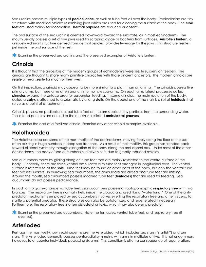
As in the sea stars, the oral surface is oriented downward, with the mouth in the center of the oral disk. However, the madreporite is not on the aboral surface as in sea stars, but on the oral surface near the mouth. The anus has been lost in brittle stars and solid waste is removed via the mouth. On the oral surface of the central disk, near the base of each arm, are two slits which are openings to the genitorespiratory bursae. Genitorespiratory bursae are pouches arranged around the central disk between each of the arms. They contain the gonads and are used for both reproduction and gas exchange.
Examine the preserved specimens of brittle stars. Note the mouth, madreporite, genitorespiratory bursae, genital slits.
EchinoideaThe class Echinoidea includes sea urchins and irregular urchins such as sand dollars and sea biscuits. Urchins possess a test, or internal shell, made up of fused ossicles. The sand dollar which is a common find for American beach combers is the test of an irregular urchin. The test of ancient irregular urchins is also a common fossil find.
Examine the sand dollar, sea biscuits and fossilized sea biscuits.
The test of sea urchins usually results in a relatively rigid body. The body of urchins possesses no arms, but is still pentaradially symmetrical. Covering the body of sea urchins are moveable spines (either long or short) which are extensions of the ossicles. These spines are used for protection and locomotion.
2 General Zoology Laboratory. Matthew K Nelson (2011)
madreporite
mouth
genitorespiratory bursa
arm
genital slit
Sea urchins possess multiple types of pedicellariae, as well as tube feet all over the body. Pedicellariae are tiny structures with modified ossicles resembling jaws which are used for cleaning the surface of the body. The tube feet are used mainly for locomotion. Dermal papulae are reduced or absent.
The oral surface of the sea urchin is oriented downward toward the substrate, as in most echinoderms. The mouth usually possess a set of five jaws used for scraping algae or bacteria from surfaces. Aristotle’s lantern, a uniquely echinoid structure derived from dermal ossicles, provides leverage for the jaws. This structure resides just inside the oral surface of the test.
Examine the preserved sea urchins and the preserved examples of Aristotle’s lantern.
CrinoidsIt is thought that the ancestors of the modern groups of echinoderms were sessile suspension feeders. The crinoids are thought to share many primitive characters with those ancient ancestors. The modern crinoids are sessile or near sessile for much of their lives.
On first inspection, a crinoid may appear to be more similar to a plant than an animal. The crinoids possess five primary arms, but these arms often branch into multiple sub-arms. On each arm, lateral processes called pinnules expand the surface area for suspension feeding. In some crinoids, the main radiation of the body, called a calyx is attached to a substrate by a long stalk. On the aboral end of the stalk is a set of holdfasts that serve as a point of attachment.
Crinoids possess no pedicellariae, but tube feet on the arms collect tiny particles from the surrounding water. These food particles are carried to the mouth via ciliated ambulacral grooves.
Examine the cast of a fossilized crinoid. Examine any other crinoid examples available.
HolothuroideaThe Holothuroidea are some of the most motile of the echinoderms, moving freely along the floor of the sea, often existing in huge numbers in deep sea trenches. As a result of their motility, this group has trended back toward bilateral symmetry through elongation of the body along the oral-aboral axis. Unlike most of the other echinoderms, the body of sea cucumbers is relatively soft, due to greatly reduced ossicles.
Sea cucumbers move by gliding along on tube feet that are mainly restricted to the ventral surface of the body. Generally, there are three ventral ambulacra with tube feet arranged in longitudinal rows. The ventral surface is referred to as the sole. Tube feet may be found on other parts of the body, but only the ventral tube feet possess suckers. In burrowing sea cucumbers, the ambulacra are closed and tube feet are missing. Around the mouth, sea cucumbers possess modified tube feet (tentacles) that are used for feeding. Sea cucumbers do not possess pedicellariae.
In addition to gas exchange via tube feet, sea cucumbers possess an autapomorphic respiratory tree with two brances. The respiratory tree is normally held inside the cloaca and used like a “water lung.” One of the anti-predator mechanisms employed by sea cucumbers involves everting the respiratory tree and other viscera, to startle a potential predator. These structures can also be autotomized and regenerated if necessary. Furthermore, the respiratory tree is often distasteful or toxic, which may also deter a predator.
Examine the preserved sea cucumbers. Note the tentacles, ventral tube feet, and respiratory tree (if everted).
AsteriodeaPerhaps the most well-known echinoderms are the Asteroidea, which includes sea stars (“starfish”) and sun stars. The Asteriodea generally possess pentaradial symmetry, with arms in multiples of five. It is not uncommon, however, to encounter individuals possessing six arms. This condition is often a consequence of regeneration.
3 General Zoology Laboratory. Matthew K Nelson (2011)
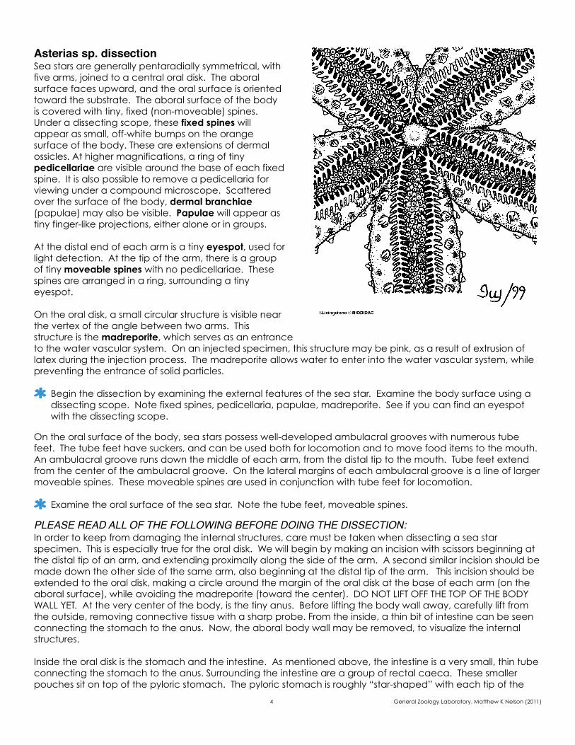
Asterias sp. dissectionSea stars are generally pentaradially symmetrical, with five arms, joined to a central oral disk. The aboral surface faces upward, and the oral surface is oriented toward the substrate. The aboral surface of the body is covered with tiny, fixed (non-moveable) spines. Under a dissecting scope, these fixed spines will appear as small, off-white bumps on the orange surface of the body. These are extensions of dermal ossicles. At higher magnifications, a ring of tiny pedicellariae are visible around the base of each fixed spine. It is also possible to remove a pedicellaria for viewing under a compound microscope. Scattered over the surface of the body, dermal branchiae (papulae) may also be visible. Papulae will appear as tiny finger-like projections, either alone or in groups.
At the distal end of each arm is a tiny eyespot, used for light detection. At the tip of the arm, there is a group of tiny moveable spines with no pedicellariae. These spines are arranged in a ring, surrounding a tiny eyespot.
On the oral disk, a small circular structure is visible near the vertex of the angle between two arms. This structure is the madreporite, which serves as an entrance to the water vascular system. On an injected specimen, this structure may be pink, as a result of extrusion of latex during the injection process. The madreporite allows water to enter into the water vascular system, while preventing the entrance of solid particles.
Begin the dissection by examining the external features of the sea star. Examine the body surface using a dissecting scope. Note fixed spines, pedicellaria, papulae, madreporite. See if you can find an eyespot with the dissecting scope.
On the oral surface of the body, sea stars possess well-developed ambulacral grooves with numerous tube feet. The tube feet have suckers, and can be used both for locomotion and to move food items to the mouth. An ambulacral groove runs down the middle of each arm, from the distal tip to the mouth. Tube feet extend from the center of the ambulacral groove. On the lateral margins of each ambulacral groove is a line of larger moveable spines. These moveable spines are used in conjunction with tube feet for locomotion.
Examine the oral surface of the sea star. Note the tube feet, moveable spines.
PLEASE READ ALL OF THE FOLLOWING BEFORE DOING THE DISSECTION: In order to keep from damaging the internal structures, care must be taken when dissecting a sea star specimen. This is especially true for the oral disk. We will begin by making an incision with scissors beginning at the distal tip of an arm, and extending proximally along the side of the arm. A second similar incision should be made down the other side of the same arm, also beginning at the distal tip of the arm. This incision should be extended to the oral disk, making a circle around the margin of the oral disk at the base of each arm (on the aboral surface), while avoiding the madreporite (toward the center). DO NOT LIFT OFF THE TOP OF THE BODY WALL YET. At the very center of the body, is the tiny anus. Before lifting the body wall away, carefully lift from the outside, removing connective tissue with a sharp probe. From the inside, a thin bit of intestine can be seen connecting the stomach to the anus. Now, the aboral body wall may be removed, to visualize the internal structures.
Inside the oral disk is the stomach and the intestine. As mentioned above, the intestine is a very small, thin tube connecting the stomach to the anus. Surrounding the intestine are a group of rectal caeca. These smaller pouches sit on top of the pyloric stomach. The pyloric stomach is roughly “star-shaped” with each tip of the
4 General Zoology Laboratory. Matthew K Nelson (2011)
star extending to form a duct passing into the arm. The largest organ in the oral disk is the cardiac stomach, which lies beneath the pyloric stomach. When feeding on a bivalve, the sea star can evert its cardiac stomach through the mouth, to pry open the shell of the bivalve. This process is referred to as gastric eversion.
If you follow the duct extending from the pyloric stomach into the arm that you opened, it leads to a complex, two-branched pyloric caecum that fills most of the arm. Lifting away the pyloric caecum reveals the gonads, which are usually much smaller and often shorter than the caecum. The gonads will look darker and grainier. Sea stars are dioecious, but the sexes can not be distinguished externally or internally without the help of a microscope.
Most of the canals which make up the water vascular system are located in the ventral portion of the main body cavity. In each arm, beneath the pyloric caecum and the gonads is the inside of the ambulacrum, running down the middle of the arm. Each of the tube feet possess an ampulla, a bulb at its proximal end. The ampulla is visible when looking down on the ambulacrum from the inside. When the ampulla contracts, it increases hydrostatic pressure inside the tube, causing the tube foot to extend. Each tube foot has a tiny lateral canal that connects it to a radial canal in the ambulacrum. The radial canal connects to a ring canal in the oral disk beneath the cardiac stomach. There is a tiny stone canal which extends from the ring canal to the madreporite. In order visualize the stone canal, you should gently examine the inside of the madreporite. If you did not damage the stone canal, it should still be in place.
Dissect the sea star as directed above. Draw your dissection, labeling the following organs: pyloric stomach, cardiac stomach, pyloric caecum, gonads, madreporite.
The deuterostomes include the Echinodermata, Hemichordata, and Chordata. The hemichordates are so named (Gr. [hemi, half] + L.[chorda, chord]) because they were previously thought to share the characteristic notochord of chordates. However, most molecular evidence places the hemichordates as sister to the echinoderms. The support structure found in hemichordates (the stomochord) is no longer thought to be homologous to the chordate notochord. Due to the highly derived nature of echinoderms, there are few morphological characteristics still shared between echinoderms and hemichordates. One of the similarities between echinoderms and hemichordates involves larval characteristics (these are plesiomorphic, of course.) The tornaria larva of many hemichordates is extremely similar to the bipinnaria larva of the Echinodermata.
Most of the hemichordates are worm-like. There are two classes of hemichordates: the Pterobranchia and the Enteropneusta. The pterobranchs are sessile and often colonial, constructing a tube similar to tubeworms. Complex tentacles are extended for suspension feeding. The Enteropneusta are the acorn worms. Acorn worms are usually burrowing deposit feeders. A large proboscis is used for burrowing. The mouth is located ventrally at the base of the proboscis. When feeding, water flows into the mouth and out through pharyngeal pores where food particles are collected.
Examine the preserved plastimount acorn worm. Identify the proboscis, pharyngeal pores, collar. It may be helpful to view the specimen under a dissecting scope.
The chordates possess several characteristics that can be seen at some point in the life cycle, if not in the adult. It should be noted that these characteristics are not necessarily chordate autapomorphies, but may be shared with other groups as well. Chordates possess a notochord, a support structure based on hydrostatic pressure. The notochord is not a cartilaginous structure, but consists of cells engorged with water, and
phylum hemichordata
phylum chordata
5 General Zoology Laboratory. Matthew K Nelson (2011)
surrounded by a sheath of connective tissue. In some groups of chordates, this structure is replaced in the adult by a cartilaginous or bony vertebral column. The nerve cord of chordates is dorsal and hollow, with the anterior end specialized as a brain. Chordates possess pharyngeal slits, which function in filter feeding and, in some groups, for gas exchange. The chordate body possesses metameric segmentation, as in the panarthropoda and the annelids. It is thought that segmentation has arisen independently in these three groups. The chordates have a post-anal tail, often involved in locomotion. Another characteristic of chordates is the presence of the endostyle. The endostyle is a ciliated, ventral portion of the pharynx, used for food capture. In the vertebrates, the thyroid gland is derived from the endostyle.
ClassificationThere are three subphyla in the phylum Chordata: Cephalochordata, Urochordata, and Craniata. The evolutionary relationships of these groups remain unclear.
Cephalochordata - Burrowing deposit feeders, also called lancelets. These possess all of the chordate characteristics as an adult.
Urochordata - Sessile suspension feeders as adults. Sea squirts are highly derived as adults, lacking most of the chordate characteristics. Sea squirts are also referred to as tunicates.
Craniata - This group includes the vertebrates. Members of this group possess a cartilaginous or bony cranium to support and protect the skull.
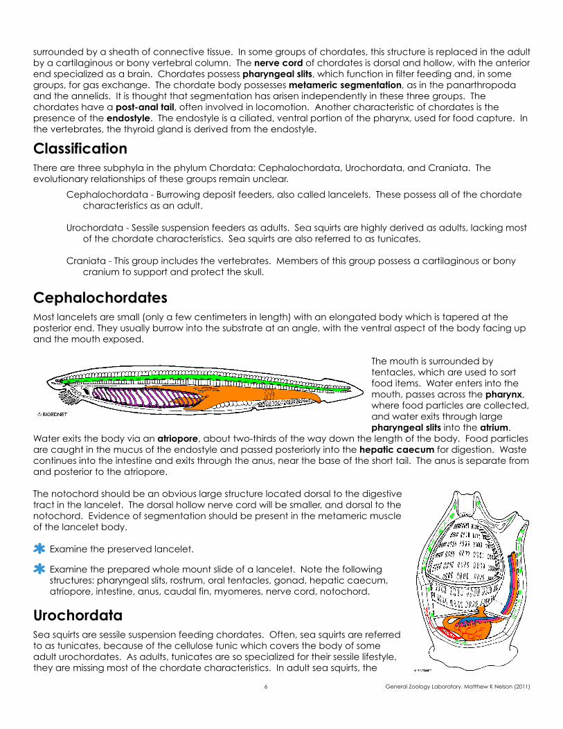
CephalochordatesMost lancelets are small (only a few centimeters in length) with an elongated body which is tapered at the posterior end. They usually burrow into the substrate at an angle, with the ventral aspect of the body facing up and the mouth exposed.
The mouth is surrounded by tentacles, which are used to sort food items. Water enters into the mouth, passes across the pharynx, where food particles are collected, and water exits through large pharyngeal slits into the atrium.
Water exits the body via an atriopore, about two-thirds of the way down the length of the body. Food particles are caught in the mucus of the endostyle and passed posteriorly into the hepatic caecum for digestion. Waste continues into the intestine and exits through the anus, near the base of the short tail. The anus is separate from and posterior to the atriopore.
The notochord should be an obvious large structure located dorsal to the digestive tract in the lancelet. The dorsal hollow nerve cord will be smaller, and dorsal to the notochord. Evidence of segmentation should be present in the metameric muscle of the lancelet body.
Examine the preserved lancelet.
Examine the prepared whole mount slide of a lancelet. Note the following structures: pharyngeal slits, rostrum, oral tentacles, gonad, hepatic caecum, atriopore, intestine, anus, caudal fin, myomeres, nerve cord, notochord.
UrochordataSea squirts are sessile suspension feeding chordates. Often, sea squirts are referred to as tunicates, because of the cellulose tunic which covers the body of some adult urochordates. As adults, tunicates are so specialized for their sessile lifestyle, they are missing most of the chordate characteristics. In adult sea squirts, the
6 General Zoology Laboratory. Matthew K Nelson (2011)
nerve cord is greatly reduced, and the notochord is lost. However, in the larva, sometimes referred to as a “tadpole,” all of the chordate characteristics are present.
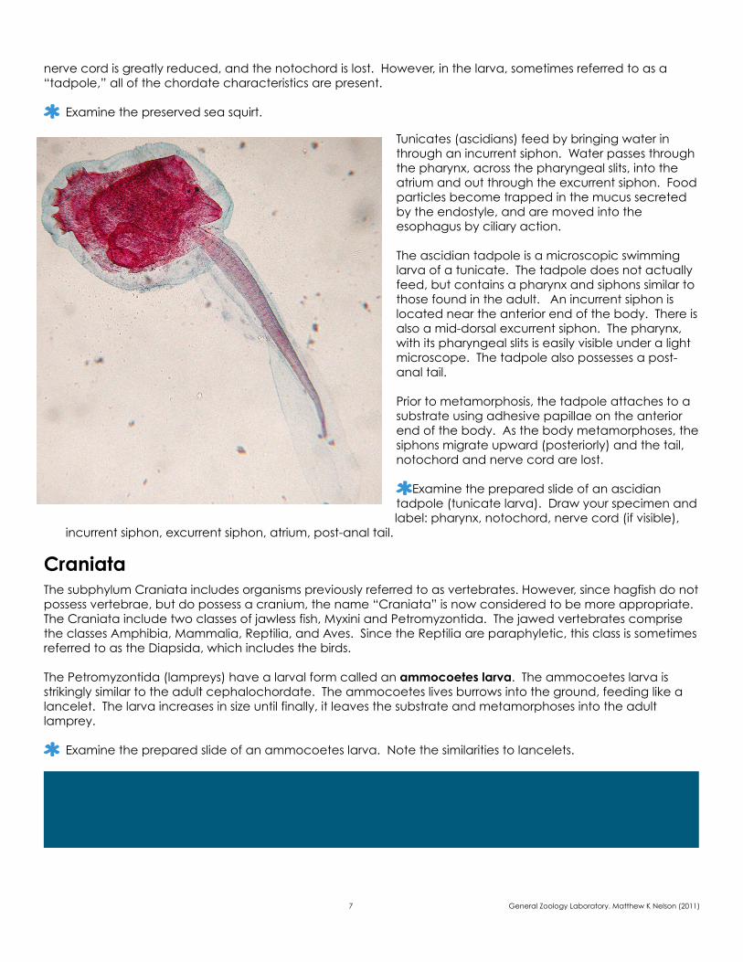
Examine the preserved sea squirt.
Tunicates (ascidians) feed by bringing water in through an incurrent siphon. Water passes through the pharynx, across the pharyngeal slits, into the atrium and out through the excurrent siphon. Food particles become trapped in the mucus secreted by the endostyle, and are moved into the esophagus by ciliary action.
The ascidian tadpole is a microscopic swimming larva of a tunicate. The tadpole does not actually feed, but contains a pharynx and siphons similar to those found in the adult. An incurrent siphon is located near the anterior end of the body. There is also a mid-dorsal excurrent siphon. The pharynx, with its pharyngeal slits is easily visible under a light microscope. The tadpole also possesses a post-anal tail.
Prior to metamorphosis, the tadpole attaches to a substrate using adhesive papillae on the anterior end of the body. As the body metamorphoses, the siphons migrate upward (posteriorly) and the tail, notochord and nerve cord are lost.
Examine the prepared slide of an ascidian tadpole (tunicate larva). Draw your specimen and label: pharynx, notochord, nerve cord (if visible),
incurrent siphon, excurrent siphon, atrium, post-anal tail.
CraniataThe subphylum Craniata includes organisms previously referred to as vertebrates. However, since hagfish do not possess vertebrae, but do possess a cranium, the name “Craniata” is now considered to be more appropriate. The Craniata include two classes of jawless fish, Myxini and Petromyzontida. The jawed vertebrates comprise the classes Amphibia, Mammalia, Reptilia, and Aves. Since the Reptilia are paraphyletic, this class is sometimes referred to as the Diapsida, which includes the birds.
The Petromyzontida (lampreys) have a larval form called an ammocoetes larva. The ammocoetes larva is strikingly similar to the adult cephalochordate. The ammocoetes lives burrows into the ground, feeding like a lancelet. The larva increases in size until finally, it leaves the substrate and metamorphoses into the adult lamprey.
Examine the prepared slide of an ammocoetes larva. Note the similarities to lancelets.
7 General Zoology Laboratory. Matthew K Nelson (2011)

8 General Zoology Laboratory. Matthew K Nelson (2011)
NAME: ________________________ SECTION:______________ LAB EXERCISE 7
Echinoderms1. Give three examples of modified ossicles in echinoderms:
2. Are there separate sexes in sea stars? If so, how can you tell them apart?
3. How is radial symmetry a selective advantage for echinoderms?
Chordates1. Draw a tree showing the currently accepted relationships among Chordates, Hemichordates, and
Echinoderms.
2. Describe the flow of water through a cephalochordate.
3. In what ways are Amphioxus and ammocoetes larvae similar?
questions
9 General Zoology Laboratory. Matthew K Nelson (2011)
Asterias sp. dissectionKingdom: _____________Phylum: ______________Class: ________________Order: ________________
Drawings
10 General Zoology Laboratory. Matthew K Nelson (2011)
Ascidian tadpole, WMKingdom: _____________Phylum: ______________Class: ________________Order: ________________magnification: __________
Drawings
11 General Zoology Laboratory. Matthew K Nelson (2011)